division of investigative sciences faculty of medicine ... · and the rest of the flu lab for...
TRANSCRIPT

Division of Investigative Sciences
Faculty of Medicine
Strategies for influenza vaccines
Submitted for the degree of
Doctor of Philosophy
Lorian Cedar Safir Hartgroves
Supervised by Wendy Barclay
August 2009

1
Declaration
I confirm that this is my own work and the use of all material from other
sources has been properly and fully acknowledged
Lorian Hartgroves

2
Abstract
The high mutation rate of influenza virus results in antigenic drift, meaning that
each of the three components in the trivalent influenza vaccine are updated
regularly so that the vaccine antigen closely matches the predominant or
emerging strain. The production of influenza vaccines from the chosen seed has
relied on embryonated chickens eggs for more than 40 years. Recent
technological advances have resulted in the evaluation of several cell lines as
alternative substrates for influenza vaccine production. Reverse genetics of
influenza viruses allows the creation of viruses at will from cDNA. However,
licensed cell lines have so far proved unpermissive for virus rescue and
permissive lines remain largely unlicensed. A reverse genetics system for the
production of influenza vaccines in PER.C6 cells has been developed and
optimised. Many recent clinical isolates do not grow in eggs and hence require
reassortment with a high growth, egg permissive backbone. Adventitious
agents aside, cell lines able to support growth of clinical isolates could be used
directly included in the vaccine, without the need for reassortment. However,
this could lead to year on year variation in the quality and characteristics of the
vaccine. Particularly pertinent, is the variation in the amount of IFN a clinical
isolate can induce, which could severely limit yields. Yields and effects of IFN
for a panel of high and low inducing viruses are investigated in a number of
potential vaccine cell lines. The mechanisms behind virus IFN induction have
been investigated. Using a panel of high and low inducing viruses the
differences in growth and viral protein expressions, and activation of PRR has
been analysed. The IFN response in PER.C6 cells has been characterised.
Through an improved understanding of the mechanisms of IFN induction and
inhibition, antagonists could be introduced into the vaccine cell line to improve
yields.

3
Acknowledgements
I would first like thank my supervisor, Wendy Barclay, for her help and
support, for believing in me enough to find the funding to give me another
opportunity to study towards a PhD and for her infectious enthusiasm for the
subject. I would also like to thank Sanofi Pasteur for funding my PhD and
Catherine Thompson and Wouter Koudstaal for their collaboration on the
PER.C6 work. Anna Hayman and Ben Johnson for the data that contributed to
the cytokine project and for their advice and training. Thanks to Ruth
Elderfield who has been through the highs and lows alongside me from the
start. Thanks to my parents for putting me up (or putting up with me?) while
writing up- to Lesley for always being there for a good chat and taking me out
the house every day to the gym and Steve for the donations of his genes to
what I hope he sees as a worthy cause, and doing the gracious thing by
conceding to his offspring the competition for the first PhD in the family. Many
thanks to Tei for helping me get to sleep, and entertaining me on the days I‟ve
had off. And the rest of the flu lab for helping along the way, reading my
drafts and making it fun to go into the work.

4
Abbreviations
β-gal β-galactosidase
4196 A/England/41/96
5‟3P 5‟ triphosphate
919/99 A/England/919/99
Ab antibody
ADCC Ab-dependent, cell-mediated cytotoxicity
AEM Adenovirus Expression Medium
Ag Antigen
Amp Ampicillin
ATP Adenosine triphosphate
BSA Bovine serum albumin
Ca Cold adapted
CARD Caspase recruitment domain
Cardif CARD adaptor inducing IFN β
CAT Chloramphenicol acetyltransferase
CBP Cap binding protein
CBP CREB-binding protein
CCLR Cell culture lysis reagent
cDC Conventional dendritic cells
cDNA Copy deoxyribonucleic acid
CEF Chick embryo fibroblasts
CEK Chick embryo kidney
CIP Calf intestinal phosphatase
CMV Cytomegalovirus
CP Coat protein
CPSF Cleavage and polyadenylation specificity factor
CRM Chromosomal region maintenance
CTL Cytotoxic lymphocyte
DABCO 1,4-diazabicyclo[2.2.2]octane

5
DAPI 4',6-diamidino-2-phenylindole
DI Defective interfering
DIG Digoxygenin
DMEM Dulbecco‟s modified eagles media
DMF Dimethylformamide
DNA Deoxyribonucleic acid
dNTP 2‟ deoxynucleoside 5´-triphosphates
dsRNA Double stranded RNA
DSS Dioctylsulfoccinate
DTT Dithiothreitol
ECL Enhanced chemiluminescence
EDTA Ethylenediaminetetraacetic acid
ELISA Enzyme Linked Immunosorbent assay
EMCV Encephalomyocarditis virus
EU European union
FAC Fluorescence-activated cell sorting
FCS Foetal calf serum
FIA Freund‟s incomplete adjuvant
FISH Fluorescent in situ histochemistry
GAPDH Glyceraldehyde-3-phosphate dehydrogenase
GSK Glaxo Smithkline
HA Haemagglutinin
HAE Human airway epithelial
HCV Hepatitis C virus
HDV Hepatitis delta virus
HEPES 4-(2-hydroxyethyl)-1-piperazineethanesulfonic acid
HIV Human immunodeficiency virus
HPAI High pathogenic avian influenza
HRP Horse radish peroxidase
Hrs Hours
ICH International Conference on Harmonisation of Technical
Requirements for Registration of Pharmaceuticals for Human Use

6
IF Immunofluorescence
IFN Interferon
IFNAR Interferon receptor
Ig Immunoglobulin
IKK Inducible κB kinase
IL Interleukin
ILL Influenza like illness
IPS-1 IFN-β promoter stimulator
IRAK Interleukin-1 receptor-associated kinase
IRAK Interleukin-1-receptor (IL-1R)-associated kinase-4
IRES Internal ribosome entry site
IRF Interferon responsive factor
ISCOM Immuno stimulating complex
ISG IFN stimulated gene
ISGF IFN stimulated gene factor
ISRE IFN stimulated response element
JAK Janus kinase
Kan Kanamycin
KLH Keyhole limpet haemocyanin
LAIV Live attenuated influenza vaccine
LB Luria broth
LMB Leptomycin B
LPAI Low pathogenic avian influenza
LGP Laboratory of genetics and physiology
LRR Leucine rich repeat
luc Luciferase
M Matrix protein
MAb Monoclonal Ab
MAVS Mitochondrial antiviral signalling molecule
MDA-5 Melanoma differentiation
MDCK Madin-Darby canine kidney
MDCK-N MDCKs expressing BVDV N protein

7
pro
MDCK-V MDCKs expressing paramyxovirus V protein
MEF Mouse embryo fibroblast
MEM Modified eagles medium
mins Minutes
M-MLV Moloney murine leukaemia virus
MOI Multiplicity of infection
MyD Myeloid differentiation primary response gene
NA Neuraminidase
NCR Non coding region
NDV Newcastle disease virus
NEMO NF-κB essential modifier
NEP Nuclear export protein
NES Nuclear export signal
NFκB Nuclear factor κB
NIBSC National Institute for Biological Standards and Control
NICE National institute for Clinical Excellence
NK Natural killer
NLS Nuclear localisation signal
NP Nucleoprotein
NS Non structural protein
OMPC Outer membrane protein complex
ONPG Ortho-Nitrophenyl-β-galactoside
p.i. Post infection
PA Polymerase subunit A
PABP Poly(A)-binding protein
PAMP Pattern associated molecular pattern
PapMV Papaya mosaic virus
PB1 Polymerase subunit B1
PB2 Polymerase subunit B2
PBS Phosphate buffered saline
PBST Phosphate buffered saline 0.1% tween

8
PCR Polymerase chain reaction
pDC Plasmacytoid dendritic cells
Pen/Strep Penicillin/streptomycin
PI3K Phosphatidylinositol-3 kinase
PIV5 Parainfluenza 5
PKR Protein kinase R
PLG Poly-lactide co-glycosides
poly IC,
pIC
Poly inositol choline
PR8 A/Puerto Rico/8/34
PRR Pattern recognition receptor
qRT PCR Quantitative real time PCR
RBC Red blood cells
rHA Recombinant HA
RIG-I Retinoic inducible gene I
RIP Receptor-interacting protein
RIPA Radio-immuno precipitation assay
RLR RIG-like receptor
RNA Ribonucleic acid
RNP Ribonucleoprotein
RPM Revolutions per minute
RSV Respiratory syncytial virus
RT Room temperature, reverse transcription, reverse transcriptase
SARS Severe acute respiratory syndrome
SCID Severe combined immunodeficiency
SDS Sodium dodecyl sulphate
PAGE Polyacrylamide gel electrophoresis
Secs Seconds
SeV Sendai virus
SFV Semliki forest virus
SIAT Sialtransferase
SOCS Suppressor of cytokine signalling
9
SOP Standard operating procedure
ssRNA Single stranded RNA
STAT Signal transducers and activators of transcription
STAT-1P Phosphorylated signal transducers and activators of transcription-1
STING Stimulator of interferon genes
SV Splice variant
SV5 Simian virus 5
Syd A/Sydney/5/97
TAB TAK1-binding proteins
TAE TRIS acetate EDTA
TAK Transforming growth factor β-activated kinase
TANK NK-κB activator
TB Tuberculosis
TBK-1 TANK-binding kinase
TCID50 Tissue culture infectious dose 50
Th T helper
TICAM TIR domain-containing adapter protein
TIR Toll/IL-1 receptor
TIV Trivalent inactivated vaccine
TLR Toll like receptor
TM Transmembrane
TNF- Tumour necrosis factor
TRAF TNF Receptor Associated Factor
TRIF TIR domain-containing adapter inducing IFN-β
TRIM25 Tripartite motif-containing 25
TRIS Tris(hydroxymethyl)aminomethane
TYK Tyrosine kinase
UTR Untranslated region
UV Ultraviolet
V Volts
Vic A/Victoria/3/75

10
VISA Virus-induced signalling adaptor
VLP Virus like particle
VP-SFM Virus production serum free medium
vRNA Viral ribonucleic acid
WHO World health organisation
WT Wild type

11
Amino acid abbreviations
Amino Acid 3 Letter 1 Letter
Alanine Ala A
Arginine Arg R
Asparagine Asn N
Aspartic acid Asp D
Cysteine Cys C
Glutamic acid Glu E
Glutamine Gln Q
Glycine Gly G
Histidine His H
Isoleucine Ile I
Leucine Leu L
Lysine Lys K
Methionine Met M
Phenylalanine Phe F
Proline Pro P
Serine Ser S
Threonine Thr T
Tryptophan Trp W
Tyrosine Tyr Y
Valine Val V

12
Table of contents
Declaration .................................................................................................... 1
Abstract ........................................................................................................ 2
Acknowledgements ....................................................................................... 3
Abbreviations ............................................................................................... 4
Amino acid abbreviations .............................................................................. 11
Table of contents ......................................................................................... 12
Table of figures ............................................................................................ 16
List of tables ................................................................................................ 18
Chapter 1 Introduction ........................................................................... 19
1.1 General Introduction ...................................................................... 19
1.2 Influenza Vaccines ......................................................................... 26
1.2.1 Antiviral drugs .............................................................................. 26
1.2.2 Current Vaccines ........................................................................... 27
1.2.3 The cell substrate .......................................................................... 34
1.2.4 Vaccine content ............................................................................ 40
1.2.5 Adjuvants ..................................................................................... 53
1.2.6 Conclusions .................................................................................. 58
1.3 The innate immune response to influenza viruses ........................... 60
1.3.1 JAK/STAT positive feedback .......................................................... 60
1.3.2 Pattern Recognition Receptors ........................................................ 61
1.3.3 TLR-3 signalling ............................................................................ 62
1.3.4 TLR-7 signalling ............................................................................ 64
1.3.5 RIG-Like Receptors ........................................................................ 65
1.3.6 Pathogen Associated Molecular Patterns ........................................ 69
1.3.7 NS1 as an antagonist of IFN-β ......................................................... 71
1.3.8 Conclusion .................................................................................... 73
Chapter 2 Materials and Methods ........................................................... 75
2.1 Materials ...................................................................................... 75
2.1.1 Plasmids ....................................................................................... 75

13
2.1.2 Primers ......................................................................................... 80
2.1.3 Virus Preparations ......................................................................... 82
2.1.4 Cell Lines ...................................................................................... 83
2.1.5 Antibodies .................................................................................... 84
2.1.6 Buffers, diluents and culture media ................................................ 85
2.1.7 Virus and cell culture reagents ....................................................... 85
2.1.8 Bacterial culture and cloning .......................................................... 86
2.1.9 Assays ........................................................................................... 87
2.2 Methods ....................................................................................... 90
2.2.1 Viral and cell culture techniques ..................................................... 90
2.2.2 Production of virus stocks .............................................................. 90
2.2.3 Infection of cell lines with influenza viruses ..................................... 91
2.2.4 Haemagglutinin assay ..................................................................... 91
2.2.5 Plaque assay................................................................................... 91
2.2.6 Transfections ................................................................................. 92
2.2.7 Virus rescue................................................................................... 93
2.2.8 Molecular biology techniques ........................................................ 94
2.2.9 Assays .......................................................................................... 100
Chapter 3 Rescue of recombinant influenza virus in PER.C6 cells ............ 105
3.1 Introduction ................................................................................ 105
3.2 Establishing a reverse genetics virus rescue system in PER.C6 .......... 108
3.2.1 PER.C6 cells support the rescue of recombinant influenza virus ...... 109
3.3 Optimising virus rescue conditions ................................................. 111
3.3.1 „Helper‟ plasmids derived from A/Victoria/3/75 virus drive replication
most efficiently ........................................................................................... 111
3.3.2 A single step rescue transfection procedure is most efficient for virus
rescue in the PER.C6 cell line ....................................................................... 114
3.3.3 The kinetics of virus rescue in the PER.C6 cells ............................... 117
3.3.4 PER.C6 cells support rescue of a range of backbones and subtypes .. 119
3.3.5 Virus rescue can be performed from a single PER.C6 suspension cell
line ......................................................................................................121
3.3.6 Comparison of PR8 backbones ..................................................... 124

14
3.4 Discussion .................................................................................... 126
Chapter 4 Rapid generation of a well-matched vaccine seed from a modern
influenza A virus primary isolate without recourse to eggs ........................... 129
4.1 Introduction ................................................................................ 129
4.2 PER.C6 cells support the faithful propagation of recent clinical isolates
......................................................................................................132
4.3 A/Eng/611/07 is typical of current circulating strains ....................... 135
4.4 Recombinant viruses with the HA of a recent clinical strain in a PR8
backbone can acquire the ability to grow in eggs ........................................ 136
4.5 G186V is less efficient adapting the growth of more in eggs ........... 139
4.6 PER.C6 cells faithfully support the growth of recombinant viruses
containing the HA of a recent clinical isolate A/England/611/07 ................... 143
4.7 Discussion .................................................................................... 145
Chapter 5 Characterisation of PER.C6 IFN-β response ............................ 149
5.1 Introduction ................................................................................ 149
5.2 PER.C6 cells make IFN-β in response to Newcastle disease virus .... 152
5.3 Influenza viruses poorly activate the IFN- β promoter in PER.C6 cells
.....................................................................................................154
5.4 PER.C6 cells respond to IFN-β ...................................................... 163
5.5 IFN-β promoter is activated in PER.C6 cells in response to poly IC but
not to in vitro transcribed viral-like dsRNA ................................................. 165
5.6 PER.C6 cells respond to IFN-β priming ......................................... 169
5.7 Expression of PRR differs in PER.C6 cells to IFN-β competent A549
cells .....................................................................................................171
5.8 Viruses do not upregulate PRR RIG-I and MDA-5 .......................... 173
5.9 Discussion .................................................................................... 175
Chapter 6 Characterising viral IFN induction, implications for vaccine
manufacture ..............................................................................................178
6.1 Introduction ................................................................................ 178
6.2 IFN-β induction does not map to HA, NA, NS ............................... 181
6.3 High IFN-β inducing viruses exhibit dysregulation of protein
expression ................................................................................................. 184

15
6.4 High IFN-β induction requires expression of RIG-I ......................... 188
6.5 RIG-I and MDA-5 mRNAs are upregulated during influenza infection
.....................................................................................................190
6.6 High IFN-β inducing viruses are prone to making more defective RNA
.....................................................................................................192
6.7 A/Sydney/97 virus drives replication early in infection ................... 196
6.8 Total RNA from infected cells induces similar levels of IFN-β
irrespective of strain .................................................................................. 200
6.9 Leptomycin B, inhibits the export of luciferase mRNA from the
nucleus and activates IRF-3 ....................................................................... 203
6.10 Cells infected with A/Sydney/97 do not have early nor greater
translocation of NP to the cytoplasm......................................................... 208
6.11 Discussion .................................................................................... 215
Chapter 7 General discussion ................................................................ 220
7.1 Emergence of a new pandemic- swine origin H1N1 influenza ........ 220
7.2 Swine-origin influenza flu vaccine ................................................ 222
7.3 Poor growth of vaccine seeds ...................................................... 222
7.4 Pathology of disease ................................................................... 224
Chapter 8 References ........................................................................... 229

16
Table of figures
Figure 1.1.1 Schematic of an influenza virion ................................................. 20
Figure 1.1.2 Schematic of influenza replication, depicting v, c and mRNA
synthesis, and 3‟ and 5‟ non coding regions. ................................................ 22
Figure 1.1.3 Schematic of influenza virus vRNP, depicting vRNA complexed
with NP and RdRp (PB2, PB1 and PA). ........................................................ 23
Figure 1.2.1 Schematic of reverse genetics. .................................................... 43
Figure 1.2.2 Schematic of multibasic cleavage. .............................................. 46
Figure 1.3.1 Schematic of TLR-3 signalling .................................................... 63
Figure 1.3.2 Schematic of RIG-I signalling ..................................................... 66
Figure 3.2.1 Schematic representing virus rescue procedure by either co-culture
or PER.C6 ................................................................................................... 110
Figure 3.3.1 “Helper” plasmids derived from A/Victoria/3/75 are the most
efficient at driving replication for influenza virus rescue ................................ 113
Figure 3.3.2 Schematic representing rescue procedures for one- or two- step
transfection protocol .................................................................................. 115
Figure 3.3.3 One-step virus rescue is the most efficient procedure ................. 116
Figure 3.3.4 Kinetics of virus rescue ............................................................. 118
Figure 3.3.5 Schematic representing virus rescue from a single cell line ......... 122
Figure 5.2.1 IFN-β promoter is activated in PER.C6 cells following Newcastle
disease virus infection ................................................................................ 153
Figure 5.3.1 A/Sydney/97 does not induce high levels of IFN-β in PER.C6 .... 155
Figure 5.3.2 A high MOI of virus does not activate the IFN-β promoter in
PER.C6 cells ............................................................................................... 156
Figure 5.3.3 Recent seasonal influenza isolates do not activate the IFN-β
promoter in PER.C6 cells ........................................................................... 157
Figure 5.3.4 The low IFN-β induction by influenza viruses in PER.C6 cells is not
a result of unproductive infection ............................................................... 159
Figure 5.3.5 Luciferase expression correlates with IFN-β expression and
secretion ..................................................................................................... 161

17
Figure 5.4.1 PER.C6 cells respond to IFN-β ................................................. 164
Figure 5.5.1 Schematic representation of in vitro transcribed dsRNA synthesis
................................................................................................................. 167
Figure 5.5.2 IFN-β promoter is activated in PER.C6 cells in response to poly IC
but not dsRNA .......................................................................................... 168
Figure 5.6.1 PER.C6 cells respond to IFN-β priming ..................................... 170
Figure 5.7.1 Expression of PRR differs in PER.C6 cells to IFN-β competent A549
cells ........................................................................................................... 172
Figure 5.8.1 Viruses do not upregulate PRR RIG-I and MDA-5 ..................... 174
Figure 6.2.1. IFN induction by wild type A/Sydney/97 virus does not map to
HA, NA, NS ............................................................................................... 183
Figure 6.3.1. A/Sydney/97 proteins accumulate early in infection ................. 186
Figure 6.3.2 A/Sydney/97 induces IFN-β early in infection .......................... 187
Figure 6.4.1. High IFN-β induction by influenza viruses is mediated by RIG-I 189
Figure 6.5.1 PRR are upregulated by IFN-β induction by influenza viruses ..... 191
Figure 6.6.1 Schematic representation of defective interfering RNA .............. 193
Figure 6.6.2 High IFN-β inducing viruses have a propensity to make short
RNAs ........................................................................................................ 195
Figure 6.7.1 Schematic of the “up-luc” assay ................................................ 198
Figure 6.7.2 A/Sydney/97 virus drives replication early in infection ............. 199
6.8.1 RNA from infected cells induces similar levels of IFN-β irrespective of
strain ........................................................................................................ 202
Figure 6.9.1 Inhibition of nuclear export with Leptomycin B ....................... 205
Figure 6.9.2 Leptomycin B immunofluorescence of NP and IRF-3 ............... 206
Figure 6.10.1 Cells infected with A/Sydney/97 do not have early nor greater
translocation of NP to the cytoplasm......................................................... 209

18
List of tables
Table 1.2.1 Attenuating mutation in cold adapted viruses. ............................ 32
Table 3.3.1 PER.C6 cells are able to support the rescue of range of virus
backbones and subtypes ............................................................................. 120
Table 3.3.2 Virus rescue can be performed in single PER.C6 cell line in
suspension ................................................................................................. 123
Table 3.3.3 Comparison of virus rescue backbones ..................................... 125
Table 4.2.1 PER.C6 cells support the faithful propagation of recent clinical
isolates ...................................................................................................... 134
Table 4.4.1 A Synthetic A/Eng/611 HA was rescued by co-culture of 293Ts with
MDCK-SIATs in a A/PR/8 backbone with NA derived from either A/PR/8 or
A/Sydney/97 ............................................................................................. 137
Table 4.4.2 Recombinant viruses with the HA of a recent clinical strain in a
PR8 backbone can acquire the ability to grow in eggs ................................. 138
Table 4.5.1 A/Eng/611 HA containing mutations thought to improve yields in
eggs, L194P or G186V, were rescued by co-culture in a A/PR/8 backbone .... 140
Table 4.5.2 G186V is less efficient adapting more modern viruses for growth in
eggs ............................................................................................................ 141
Table 4.6.1 PER.C6 cells faithfully support the growth of recombinant viruses
containing the HA of a recent clinical isolate A/England/611/07 ................... 144
Table 6.6.1 Defective RNAs accumulate during infection with A/Sydney/97
virus .......................................................................................................... 194

19
Chapter 1 Introduction
1.1 General Introduction
Influenza is a highly contagious, acute respiratory disease, which causes
significant mortality and morbidity worldwide. According to the World Health
Organisation (WHO) it causes between three and five million cases of severe
illness and 250,000 to 500,000 deaths every year around the world
(http://www.who.int/mediacentre/factsheets/fs211/en/). The elderly, young
children and those with chronic medical disorders such as asthma, diabetes and
the immune-compromised are most at risk. It has a range of symptoms
including rhinitis, cough, weakness, febrile illness and headache. Most infections
are self-limiting however, they can occasionally progress to pneumonia and
rare complications include encephalopathy, myocarditis, and myositis.
Influenza is caused by infection with a single stranded, negative sense, RNA
virus with a segmented genome (figure 1.1.1). The virus encodes 12 known
proteins, encoded on 8 segments. The viral envelope contains the
glycoproteins haemagglutinin (HA) and neuraminidase (NA) and matrix
protein (M2) in host derived lipids. This encloses a matrix of M1 protein,
around the virion core containing nuclear export protein (NEP) and each of
the 8 RNA segments. Each segment is coated with nucleoprotein (NP) and
complexed with the RNA-dependent RNA polymerase (RdRp), composed of
two polymerase basic and one polymerase acidic subunits (PB1, PB2, and PA),
and is known as an RNP. NS1 and PB1-F2 are non-structural proteins that help
the virus control the host cell immune responses and N40 is a recently
described N terminal product of the PB1 open reading frame which affects
replication (Wise, Foeglein et al. 2009).

20
Figure 1.1.1 Schematic of an influenza virion

21
HA mediates virus binding to the host cell receptor, sialic acid. The virus is
internalised by receptor mediated endocytosis (Lakadamyali, Rust et al. 2004).
Internalized viruses are trafficked along the endocytic pathway to late
endosomes. Exposure to the low pH results in a conformational change in the
HA which exposes the HA2 fusion peptide and triggers HA-mediated fusion
between the viral and endosomal membranes (Skehel and Wiley 2000). M2
forms an ion channel which results in the acidification of the virus interior,
triggering disassembly of the core and release the vRNPs from the matrix
proteins (reviewed in (Betakova 2007). vRNPs are imported into the nucleus
via nuclear localisation signals which direct cellular proteins to import the RNPs
and viral proteins into the host cell nucleus (Wu, Sun et al. 2007).
RdRp synthesizes viral mRNA and cRNA (copy RNA) from the same vRNA
(viral RNA) template (figure 1.1.2). The 3′ and 5′ non-coding regions are highly
conserved among each of the segments and partially complementary
(Desselberger, Racaniello et al. 1980). They base pair to form a helical hairpin
panhandle (figure 1.1.3), with which the RdRp associates (Gonzalez and Ortin
1999) The conserved non coding nucleotides function as the promoter (Luytjes
et al. 1990). The replication of vRNA is primer-independent and is terminated
by run-off transcription of the vRNA template to form a full-length cRNA, an
exact positive sense copy of the genome, which acts as a template for the
synthesis of subsequent vRNA. Initiation of viral mRNA synthesis requires
capped RNA fragments derived from host mRNA. PA has endonuclease activity
and “steals” or “cap snatches” 5′ capped primers from cellular RNA
polymerase II (Pol II) activity (Dias, Bouvier et al. 2009). PB2 subunit then
binds the 5' cap and is responsible for the initiation of transcription (Guilligay,
Tarendeau et al. 2008). PB1 subunit plays a central role in polymerase
elongation (Biswas and Nayak 1994). The non-coding regions also include the
mRNA polyadenylation signal and termination occurs following reiterative
copying of a sequence of uridine residues to generate a poly(A) tail (Luo,
Luytjes et al. 1991). 5′ capped, poly (A) mRNA can be exported and translated

22
Figure 1.1.2 Schematic of influenza replication, depicting v, c and mRNA
synthesis, and 3‟ and 5‟ non coding regions.

23
Figure 1.1.3 Schematic of influenza virus vRNP, depicting vRNA complexed
with NP and RdRp (PB2, PB1 and PA).

24
like host mRNA. Nuclear export of vRNA segments is mediated by the viral
proteins M1 and NEP via a CRM1-dependant pathway (Neumann, Hughes et
al. 2000; Elton, Simpson-Holley et al. 2001; Watanabe, Takizawa et al. 2001).
The mechanism controlling the synthesis of mRNA or cRNA from the same
template has been long debated. Most recently Brownlee and co-workers have
shown that both products are generated at the same time during infection but
that cRNA is unstable in the absence of associating NP and polymerases and
therefore only accumulates later in infection when intracellular viral protein
levels are high (Vreede and Brownlee 2007). This hypothesis is in line with a
much older theory that NP accumulation triggers the switch from transcription
to replication (Beaton and Krug 1984).
The envelope proteins HA, NA, and M2 accumulate at the cell membrane
(Nayak, Hui et al. 2004). M1 is thought to chaperone the RNPs to the cell
membrane for packaging, where it interacts with cytoplasmic tails of envelope
proteins HA, NA and M2, leading to assembly and budding of virions (Chen,
Leser et al. 2008; Nayak, Balogun et al. 2009). Virus budding may be initiated
by the accumulation of M1 matrix protein at the cytoplasmic side of the lipid
bilayer. The HA spikes continue to bind the virions to the sialic acid on the cell
surface until virus particles are released by the sialidase activity of the NA
protein (Barman, Adhikary et al. 2004).
The incorporation of the eight segments that comprise the genome into virus
particles has also long been a matter of debate. There is evidence for random
packaging whereby virions could contain more than eight segments and a
proportion would have the correct combination. However, recent elegant
work from Kawaoka has shown both genetically and by electron microscopy
that there are eight RNPs per virion because of the existence of segment
specific packaging signals that extend across both non-coding and coding
regions of each of the eight RNA segments (Fujii, Fujii et al. 2005; Noda,
Sagara et al. 2006). Interestingly this knowledge may lead to new strategies to
generate attenuated viruses suitable for vaccines (Gao, Brydon et al. 2008).

25
There are three serologically distinct types of the virus; A, B, and C- of which A
and B cause widespread outbreaks. Influenza A virus causes most severe
disease, whilst B causes significant epidemic outbreaks approximately every
three years. Influenza A viruses are further classified into subtypes based on the
surface glycoproteins; there are 16 haemagglutinin (HA) and 9 neuraminidase
(NA) subtypes. Three subtypes are known to infect and transmit within the
human population (H1N1, H2N2 and H3N2). Both, but particularly HA, are
major antigenic determinants and antibodies directed against HA and NA, are
crucial in current vaccine strategy.
New strains appear from gradual accumulation of point mutations in the
surface glycoproteins. This “antigenic drift” allows the virus to evade immune
recognition and forms the basis for the seasonal epidemics which occur each
year. Because influenza A is able to infect multiple organisms, occasionally a
“new” virus emerges, which is unrelated to the circulating strains and arises not
from mutation, but from reassortment between 2 different subtypes of the
virus, one of which was previously circulating only in an animal reservoirs. This
is called antigenic shift and can result in strains to which most adults do not
have pre-existing immunity. Antigenic shift can lead to influenza pandemics
such as the infamous 1918 “Spanish flu”- which killed 40 million, the Asian flu
1957, and Hong Kong flu 1968, and recently the swine-origin 2009 H1N1
pandemic. Despite the devastating effects of a pandemic, the cumulative deaths
during intervening years have been far greater than those associated with
pandemics. Influenza B virus infects only humans and therefore is only able to
undergo antigenic drift. Although most influenza virus infections are self-
limiting, few other diseases have such an effect on absenteeism, hospital
admission and economic loss.

26
1.2 Influenza Vaccines
Vaccination is the primary measure available to control influenza. The most
widely available vaccines are based on technology over 50 years old and give
inadequate protection to the most in need- the young and the elderly. Current
vaccine production could be considered somewhat archaic- involving
manufacture in chicken‟s eggs. With the threat of highly pathogenic avian
influenza (HPAI) gaining the ability to transmit between humans, the need to
develop new vaccine strategies is essential. Vaccine manufacturers need to be
able to respond quickly to new emerging strains, which will probably
necessitate an egg free system such as cell culture. Depending on the viruses
included in the vaccine and the method of vaccine generation, egg based
production may in any case be inappropriate for avian virus based vaccines
which could be pathogenic to eggs. New vaccines will need to be highly
immunogenic and provide protection to all age groups. This may demand the
addition of adjuvants, few of which are currently licensed. The first part of this
introduction will discuss at the current state of influenza vaccination and new
advances in the field.
1.2.1 Antiviral drugs
In the case of an influenza outbreak, there are two classes of antiviral drugs
available. Amantadine and rimantadine (only effective against influenza A)
block M2 ion channels, which regulate the internal pH of the virion and are
crucial during early virus replication. Amantadine has several adverse side
effects including insomnia, headaches and dizziness which can lead to falls
especially in the elderly. Although rimantadine has fewer side effects, it is not
widely available. Furthermore, viruses can rapidly acquire resistance to both
these drugs through single non-synonymous nucleotide changes in the
membrane spanning domain of M2.

27
The other antiviral agents available are NA inhibitors. These drugs are effective
against both influenza A and B. Zanamivir (Relenza) is administered by
inhalation (which is less desirable for elderly use but more convenient for
children), and may cause bronchospasm. Oseltamivir (Tamiflu) is administered
orally and can induce vomiting if not taken after food. These drugs can reduce
infection on average between 0.8-1.3 days. Resistance can occur but seems to
be strain and subtype specific (Meijer, Lackenby et al. 2009).
The National institute for Clinical Excellence (NICE) has issued guidance on the
use of antivirals; Amantadine is not recommended, whilst Zanamivir and
Oseltamivir are not recommended in adults or children unless they are at risk,
can start therapy within 48 hrs of the onset of symptoms, and it is known that
influenza is circulating. There is clearly scope for improvement to available
anti-influenza treatments, and effective vaccination should be both maintained
and improved in order to deal with the threat of an avian pandemic and to
alleviate morbidity in the interpandemic period.
1.2.2 Current Vaccines
1.2.2.1 Vaccine content
The emergence of new strains by antigenic drift demands the annual
reformulation of the vaccines. Retrospective analysis of variants from past
major epidemics shows that typically only 3 or 4 mutations in HA molecule are
all that is required for a new epidemic strain to emerge (Smith, Lapedes et al.
2004). New variants are identified from epidemiological surveillance studies
coordinated by WHO. A panel convenes in February in the northern
hemisphere, September in the south, to predict the circulating strains likely to
give most the most protection in the coming influenza season. Currently, two
influenza A virus strains, representing both subtypes that currently circulate in
humans (H1N1, H3N2) and one B strain are selected for inclusion in the

28
seasonal vaccine. Vaccine manufacturers then have approximately 6 months to
produce and register the vaccine. Approximately 50 countries have
government funded immunisation programmes (Nicholson, Wood et al. 2003)
and approximately 350 million doses are available to some 1.2 billion people
worldwide who are at risk
(http://www.who.int/vaccine_research/diseases/ari/en/index.html).
The industry is dominated by only a few manufacturing companies. In 2007,
Sanofi Pasteur supplied 180 million doses of seasonal influenza vaccine, an
estimated 40 percent of the world influenza vaccine market.
Vaccination results in a 30% reduction of mortality among elderly persons
during influenza epidemics (Groenwold, Hoes et al. 2009). Recommendations
for vaccination vary; and tend to be founded on rates of morbidity and
mortality. Therefore annual vaccination of at risk groups; the elderly, those
with chronic medical disorders, asthma, diabetes, immune-suppressed and
carers is generally recommended (Zimmerman and Middleton 2007).
1.2.2.2 Inactivated vaccines
The segmented nature of the influenza genome means a vaccine seed can be
generated from the reassortment of the WT epidemic strain and a high yield,
laboratory adapted strain such as A/Puerto Rico/8/34 (PR8). The reassortant
generally contains the six internal genes from PR8 and the surface antigen
genes, HA and NA, from the selected vaccine strains. PR8 has been used as the
recipient genetic backbone of the influenza A virus vaccine for more than 30
years (Kilbourne 1969).
No such lab adapted high growth master strains exists for influenza B viruses
and these viruses are simply strain selected each year. Often yields of influenza
B virus are lower than for influenza A as a result.

29
Trivalent inactivated vaccine (TIV) is the oldest licensed vaccine, its
effectiveness was first evaluated in 1938 (Francis 1953) and contains partially
purified HA and NA, from formaldehyde or β-propiolactone inactivated
virions, detergent treated to give a split product. It is administrated by
intramuscular injection. When there is a good match between the selected
vaccine strains and the circulating strains, the vaccine can confer up to 80%
immunity in healthy adults less than 65 years of age (Nicholson, Wood et al.
2003; Subbarao and Katz 2004) and is usually well tolerated with very few
adverse side effects. Protection is largely mediated by induction of neutralising
serum Ab against HA. Substantial but not complete protection is seen in the
elderly population. In young, naïve children, multiple injections are required
for high immunity. The split vaccines, delivered intramuscularly, are poor
inducers of local IgA in respiratory secretions and of cell mediated immunity,
important in protection against airborne pathogens and recovery respectively.
1.2.2.3 Live attenuated vaccines
Whole, live vaccine, administered intranasally is potentially a better alternative
to the split, intramuscular vaccine, as it should induce superior immunity more
analogous to a natural virus infection. To reduce virulence these vaccines are
designed to have limited replication in the upper respiratory tract. As with TIV,
a master backbone strain is generated. However, in the case of live attenuated
influenza vaccine (LAIV) the backbone confers an attenuated phenotype from
internal genes. Vaccines are made from 6:2 reassortants of attenuated
backbone and circulating HA and NA. There have been several strategies used
to attenuate the backbone strains (Reviewed in (Subbarao and Katz 2004)).
Some non-human viruses are naturally attenuated in humans, for example,
some avian viruses have restricted replication in primates and master backbone
strains have been developed from these (Snyder, Clements et al. 1986). This
overcomes the need for derivation or passage to attain attenuation.

30
A/Mallard/New York/6750/78 (H3N2) exhibited restricted replication in both
nonhuman primates and human adults and was shown to be safe, genetically
stable and immunogenic. However, in further clinical trials of vaccine strains
based on avian master backbones possessed some residual virulence in infants
and children, so use of host restricted attenuation has not been pursued
(Steinhoff, Halsey et al. 1991).
Another approach was to generate temperature sensitive (ts) viruses in
presence of the mutagen 5-fluorouracil, which showed restricted growth at
37°C (Mills, Chanock et al. 1971). They consequently have restricted replication
in the lower respiratory tract of animals (37-38°C) but are able to replicate in
the upper respiratory tract (32-33°C) and induce a humoral response that
protects against lethal virus challenge in mice. Two master strains were
developed but these proved genetically unstable and reverted to virulent
phenotypes in clinical trials (Tolpin, Massicot et al. 1981).
The last method involves adaptation of the virus for growth at sub-optimal
temperatures resulting in decreased virulence. This strategy has been used
independently by both American and Russian investigators, to generate highly
stable master strains, which have proved good candidates for LAIV.
In the US, A/Ann Arbor/6/60 (H2N2) and B/Ann Arbor/1/66 were used as
master strains (Maassab 1967). Viruses were isolated on chick kidney cells and
then adapted for growth at 25°C by 32 stepwise passages at successively lower
temperatures. They replicate efficiently between 25°C and 33°C but not 37°C,
thus they not only have a cold adapted (ca) but also a ts phenotype. In ferrets,
there is no replication in the lungs and attenuation in the upper respiratory
tract, with lower titres and decreased infection times compared to WT. A dose
of 107 TCID50 administered intranasally, is typically safe and immunogenic in all
age groups.

31
In Russia, A/Leningrad/134/57 (H2N2) was passaged 20 times in eggs followed
by a further 17 or 47 passages at reduced temperature 25-26°C (Ghendon,
Klimov et al. 1981). The mutants again, are ts as well as ca, being unable to
replicate in eggs at 37°C. A/Leningrad/134/17/57 is used in vaccines for adults,
whilst A/Leningrad/134/47/57 is used for children 3-14 yrs, in a trivalent
vaccine formation with B/USSR/60/69.
US and Russian strains have similar rates of infectivity in humans (Nicholson,
Tyrrell et al. 1987). The attenuating mutations (ca) are found mostly in NP and
the polymerase subunits, PB1 PB2 and PA (Jin, Lu et al. 2003; Hoffmann,
Mahmood et al. 2005; Chen, Aspelund et al. 2006) (see table 1.2.1).

32
Table 1.2.1 Attenuating mutation in cold adapted viruses.
Adapted from (Subbarao and Katz 2004). For B/Ann Arbor/1/66 only cold adapting mutations are shown
Gene Product A/Ann Arbor/6/60 A/Leningrad/137/57 B/ Ann Arbor/1/66
Amino acid Amino acid Amino acid
Residue WT Ca Residue WT Ca 17 Ca 47 Residue WT Ca
PB2 265 N S 478 V L L 630 S R
490 S - R
PB1 391 K E 265 L N N -
457 E D 317 M - I
581 E G 591 V I I
661 A T
PA 613 K E 28 L P P 431 V M
715 L P 341 V L L 497 Y H
NP 23 T N 341 I - I 114 V A
34 N G 410 P H
509 A T
M1 - 15 I V V 159 H Q
183 M V
M2 86 A S 86 A T T
BM2 - - -
NS1 153 A T -
NS2 - 100 M I I

33
LAIVs have been in use in Russia >50 years. MedImmune Vaccines Inc licensed
and marketed Flumist in US in 2002. There have been several evaluations of
the efficacy of LAIV (Palker, Kiseleva et al. 2004; Huber and McCullers 2006;
Belshe, Ambrose et al. 2008) and comparisons with inactivated vaccine. Serum
antibody titres are generally lower and mucosal IgA elevated, although in some
trials protection was conferred without detectable serum or nasal antibody
implying a CTL response. Generally, trials indicate that in adults LAIV is as
effective at eliciting a protective response as inactivated vaccine. In the elderly
it can be less immunogenic, possibly due to the limited replication of the
vaccine virus, which may be a result of higher levels of pre-existing antibodies
to influenza (Powers, Fries et al. 1991). Conversely, in naïve children a
substantial level of protection can be elicited after just one dose (Arvin and
Greenberg 2006) and children experienced more than 50% fewer cases of
influenza illness when vaccinated with LAIV compared to TIV (Ashkenazi,
Vertruyen et al. 2006; Belshe, Edwards et al. 2007; Rhorer, Ambrose et al.
2009). However in younger children 6–11 months, a small increase in the
number of hospitalizations (1% increase) and wheezing (2.1%) were noted
weeks to months after receiving the live vaccine. Although these are not
thought to be related specifically to influenza or temporally related to LAIV,
LAIV is not approved for use in children <2 years of age (Belshe, Ambrose et
al. 2008).
Since LAIV contains live virus there exists the possibility of reassortment with
native virus to produce new strains with unpredictable traits. Reassortants from
avirulent strains have previously been able to produce pathogenic strains
(Scholtissek, Vallbracht et al. 1979). Concerns have also been raised about the
potential for the virus to transmit from vaccinated to non-vaccinated
individuals and contact with immune-suppressed individuals is not advised. A
recent study investigated the duration and extent of virus shedding following
vaccination (Block, Yogev et al. 2008) and concluded that the amount of
shedding depended on the specific vaccine strains and the level of pre-existing
immunity in vaccine recipients. However, transmission was possible particularly

34
from younger children (<9 years old). The findings support the Advisory
Committee on Immunization Practices recommendation that LAIV recipients
should avoid contact with severely immunosuppressed. Despite this, ferrets
immune compromised by treatment with dexamethasone and cytarabine,
elicited improved responses (Huber and McCullers 2006) and a study in HIV
infected children showed equivalent responsiveness between groups with
different immunological status (Levin, Song et al. 2008). Also to be considered
are the benefits of herd immunity due to virus shedding and transmission,
which could increase the benefits of LAIV. Due to higher acquisition costs, LAIV
costs more per dose than TIV; however, as a result of increased protection and
reduced burden of disease, LAIV vaccination of children results in a net saving
(Luce, Nichol et al. 2008).
1.2.3 The cell substrate
Although the existing vaccines are comparatively successful in inducing immune
responses, there is evidently scope for improvement. Importantly, dependence
on eggs is a major limitation for current production procedure. It takes
between 3 and 6 months to develop the vaccines in eggs and efficiency is low,
requiring 1-2 eggs per dose of vaccine. Careful planning is needed each year to
ensure millions of fertile, embryonated eggs are available for vaccine
production, and this is often unable to take into account calls for increased
production. Furthermore, diseases in laying flocks can affect availability and
variation in the quality of the embryos can effect vaccine production. Some
highly pathogenic avian influenza (HPAI) strains such as H5N1 are lethal to
chicken embryos, and insufficient yields are obtained, making eggs an
inadequate method for production of vaccines against these viruses.
Furthermore, in the event of a pandemic many birds may be killed by the virus
or culled to limit the spread.
The SARS outbreak demonstrated that with increased air travel and
globalisation, a pandemic could spread in weeks or days and highlights the

35
need for a system which can respond rapidly, should HPAI gain the ability for
human-human transmission. Cell lines are the obvious alternative to egg based
vaccine production. Cell cultures can be used to manufacture vaccine at any
time of year, pertinent during the current H1N1 pandemic which emerged
outside of the typical winter influenza season in the northern hemisphere. Since
they are maintained in a strictly monitored and sterile environment, the risk of
contamination, and thus bio burden and end toxin content, is greatly reduced.
Consequently, the use of preservatives like thiomersal, which can cause
potential side effects, can be decreased. Cell lines are strictly monitored by
WHO, and licensed lines are free from contaminants. Moreover, cells can be
grown in serum free media, eliminating the potential for allergic reaction to
serum or chick embryo proteins. This is particularly relevant to whole, live
vaccine production, which does not involve the complex purification process
used for inactivated vaccine, and the virus is not purified away from cell
proteins. Egg derived vaccines are contraindicated for those with egg allergies.
Many recent clinical isolates, while becoming more adapted to the human host
exhibit altered receptor binding characteristics compared to strains that
circulated shortly after the emergence of the H3 or H1 subtype more than 40
years ago (Medeiros, Escriou et al. 2001; Mochalova, Gambaryan et al. 2003;
Thompson, Barclay et al. 2004). Since the receptor used for cell entry in eggs
differs from that in the human respiratory tract, this has the consequence that
fewer isolates are able to grow in eggs, and unless they acquire mutations in
HA (Schild, Oxford et al. 1983). These can, confer changes to glycosylation
patterns (Chen, Aspelund et al. 2008) and can give rise to mismatches in
antigenicity between the egg grown human influenza strains and the circulating
clinical virus (Mochalova, Gambaryan et al. 2003).
Cell cultures are easy to handle and scale-up and readily adjust to market
needs. However, licensed cell lines have largely proved unpermissive for
production of high yield influenza virus product, and many permissive lines
used in the laboratory are refractory to licensing.

36
The efficiency of a cell culture depends upon its ability to generate high yields,
and requires a well-optimised purification process. In the case of LAIV the
capacity of cell line to produce infectious particles is also very important.
The most common cell lines used in the laboratory for the amplification and
isolation of influenza viruses are Madin-Darby canine kidney (MDCK) or Vero
(African Green Monkey Kidney). These are adherent cell lines and can be
expanded for large scale manufacturing in a number of ways including fixed
bed reactors, fluidised bed reactors and microcarrier culture.
1.2.3.1 MDCK cells
Although in most published comparisons fluid from embryonated eggs yields
the highest virus titres (e.g. 3.9x10⁹ PFU/ml (Tree, Richardson et al. 2001),
yields from MDCKs remain respectable ranging from 3 to 10 fold less (Tree,
Richardson et al. 2001; Audsley and Tannock 2005). Often these comparisons
are carried out using the laboratory strain PR8 which has been specifically
adapted to be high yield in eggs. If a cell culture adapted backbone strain was
generated, equivalent yields could conceivably be achieved. Indeed, MDCKs
may well prove a better substrate for more recent clinical isolates which grow
less well in eggs. MDCK derived split vaccines have been tested in clinical trials
for efficacy. There were no statistical differences between the immunogenicity
(e.g. HAI titres) and post immunisation symptoms between subjects who were
immunised with either egg and cell derived vaccine (Halperin, Nestruck et al.
1998). As mentioned above recent clinical isolates have a decreased tropism for
egg propagation. In 2003/04 season the strain A/Fujian/411/02 was selected for
inclusion in the trivalent vaccine. However, following generation of the
vaccine seed the reassortant was unable to grow in eggs and an alternative had
to be sought, resulting in delays to vaccine issue. Recent research shows that
MDCK cells would have supported growth of the Fujian strain (Makizumi,

37
Kimachi et al. 2008). In terms of virus yield and vaccine efficacy, MDCKs pose
a truly viable alternative to eggs. However, there are concerns about the safety
of MDCKs due to the non-human origin, lack of documentation about their
transformation history and oncogenic potential. The line has consequently
been extensively characterised, shown to be free from detectable infectious
agents (viral or microbial) and highly unlikely to induce tumours in humans
(Kistner, Barrett et al. 1998; Genzel, Olmer et al. 2006). An assessment of the
risk of inclusion of adventitious agents in the vaccine product demonstrated
that MDCK cells are restricted in the number of mammalian viruses for which
they support growth (Gregersen 2008), and testing by infection with >20 virus
families and other pathogens, such as Mycoplasma and Chlamydia has shown
the manufacturing process capable of reducing adventitious agents to non
infectious level (Gregersen 2008). As such, Novartis, Solvay, GlaxoSmithKline
and MedImmune have all developed and characterised proprietary cell banks.
Moreover, Novartis and Solvay have extensively tested and characterized their
cell banks according to USA, EU and ICH requirements. These vaccines are not
inferior to those generated in eggs and some have been taken to clinical trials
and licensed. In 2001 Influvac-TC, Solvay Pharmaceuticals, was approved in the
Netherlands; Novartis have developed an MDCK based vaccine that is now on
the market (Gregersen 2008; Doroshenko and Halperin 2009). The high
permissivity of MDCKs makes the cell line an attractive prospect for vaccine
manufacture. However, MDCKs are difficult to transfect; as such, they are less
expedient for the generation of reverse genetics vaccines (see section 1.2.4.1).
They have also been evaluated for generation of reassortant vaccine seeds and
fell short of the yields attained in eggs. MDCK cells have also been shown to
support the growth of an efficacious LAIV (Ghendon, Markushin et al. 2005;
Liu, Hossain et al. 2008), an inactivated H5N1 vaccine (Hu, Weng et al. 2008)
and an equine vaccine (Genzel, Olmer et al. 2006).

38
1.2.3.2 Vero cells
Vero cells are an African green monkey kidney cell line, which are already
licensed for a range of other human vaccines including poliovirus, rabies and
even an inactivated influenza vaccine from Baxter healthcare corp. The
influenza vaccine generated in Vero cells is fully immunogenic (Youil, Su et al.
2004; Audsley and Tannock 2005; Ghendon, Markushin et al. 2005). A
whole, formalin and UV inactivated, H5N1 vaccine has been developed from
wt virus, which has proved immunogenic in both mice and guinea pigs
(Kistner, Howard et al. 2007; Howard, Kistner et al. 2008). In clinical trials
(against A/Vietnam/1203/2004), a relatively high dose (7.5-15 µg) was able to
induce neutralising and cross protecting antibodies to clades 1, 2 and 3 (Ehrlich,
Muller et al. 2008). The addition of alum adjuvant did not induce a dose
spearing effect. Generally, titres are at least a log lower than MDCKs, and
maximum yields are achieved in 4 rather than 2 days. A key advantage in the
use of Vero cells is their primate origin; since the cells are more human the
number of cell line specific mutations are reduced (Romanova, Katinger et al.
2003). Furthermore, glycosylation of HA has been implicated in the
antigenicity and immunogenicity of viral glycoproteins (Skehel, Stevens et al.
1984), and the infectivity of macrophages (Reading, Miller et al. 2000), this
could be particularly relevant for LAIV and could decrease the efficacy of a
vaccine.
The UK government has a sleeping contract with Baxter (now active) for 72
million doses of a whole, inactivated pandemic vaccine (Kistner, Howard et al.
2007) which will be produced in Vero cells
(http://www.parliament.uk/documents/upload/postpn331.pdf).
An LAIV was developed that is attenuated by inclusion of a truncated NS-1.
This vaccine is attenuated in most cell culture substrates via the induction of
IFN-β (Steel, Lowen et al. 2009). However, since Vero cells are IFN

39
incompetent IFN response they would represent a suitable cell line for vaccines
of this type.
1.2.3.3 PER.C6 cells
For commercial interest, a cell line to needs to be well characterised.
Developed by the Dutch company Crucell N.V., PER.C6 cells are a recently
derived cell line originating from the primary culture of foetal human
retinoblasts immortalised with an E1A minigene from type 5 adenovirus. They
can be grown in suspension, which eliminates the need for microcarriers,
making scale-up easier than for adherent MDCK and Vero cells (Fallaux, Bout
et al. 1998). PER.C6 cells support growth of a variety of influenza A and B
strains and produce high titres (1010 TCID50/ml) of virus in 4 days, in roller
bottles and bioreactors (Pau, Ophorst et al. 2001). Preliminary data, suggests
that passage of virus could result in higher yields of HA antigen. PER.C6 cells
are licensed for the use of a variety of vaccines and therapeutics, including a
West Nile virus vaccine (Samina, Havenga et al. 2007), a rabies vaccine
(Marissen, Kramer et al. 2005), recombinant adenovirus vectors for gene
delivery (Subramanian, Kim et al. 2007), production of recombinant proteins
such as an HIV-1 gag protein vaccine (Ledwith, Lanning et al. 2006) and
neutralizing monoclonal antibodies, e.g. SARS-CoV (SARS-AB) (Havenga,
Holterman et al. 2008). Sanofi Pasteur purchased an exclusive license from
Crucell N.V. in 2003 to research, develop, manufacture and market cell-based
human influenza vaccines using PER.C6 cell line. Crucell retains the commercial
rights for human vaccines in Japan and pandemic vaccine.
1.2.3.4 PBS-1
A recently developed cell line derived from 11 day old immortalized chick
embryo cell line, termed PBS-1, is especially novel because it can support the

40
replication of influenza in the absence of exogenous trypsin. PBS-1 cells support
the growth of recent isolates of human influenza A and B viruses as well as
avian viruses to high titres (up to 107.5
PFU/ml). The authors claim that PBS-1
cells outperformed other licensed cell lines: CEK cells, MDCK cells and Vero
(Smith, Colvin et al. 2008). PBS-1 cells are free of exogenous agents, are non-
tumorigenic, and are readily adaptable to a variety of culture conditions,
including growth on microcarrier beads.
1.2.4 Vaccine content
1.2.4.1 The use of reverse genetics to generate the vaccine seed
The strains of virus used in the vaccine are updated every year. High growth
6:2 reassortants with the backbone strain need to be generated and selected.
The process can be time consuming and unpredictable. In 1990 a technique to
generate designer influenza viruses was developed. At this time, the „reverse
genetic‟ technique used helper viruses and exchanged synthetic segments
derived from cDNAs one at a time into the influenza genome. RNA was
transcribed in vitro from cDNA and contained RNP proteins purified from virus
particles. The reconstituted RNPs were transfected into cells simultaneously
infected with „helper‟ virus. Reassortants containing the novel segment were
then selected from the progeny which also contained abundant titres of helper
virus progeny. Even with the use of tight selection markers such as antigenic
differences or ts/ca mutations, the technique was laborious. Still, some
candidate vaccines were generated using this technique (Li et al 1997).
Two laboratories independently came up with a procedure to “rescue” whole
virus genomes entirely from viral cDNAs (Fodor, Devenish et al. 1999;
Neumann, Watanabe et al. 1999). Cells are co-transfected with 8 plasmids
encoding each of the viral genomic RNAs and 4 helper plasmids directing
expression of viral mRNAs (fig 1.2.1). The 8 viral RNAs are under the control

41
of an RNA polymerase I (pol I) promoter; a hepatitis delta virus (HDV)
genomic ribozyme or a polymerase terminator, downstream of the vRNA-
coding region ensures that RNA is processed correctly from 3' end of the
vRNA. The helper plasmids typically express proteins from a viral promoter
such as that of CMV, which directs transcription by the host pol II apparatus, a
poly A tail is added via a 3‟ polyadenylation site. The helper plasmids encode
the three subunits of the viral RNA-dependent RNA polymerase complex (PB1,
PB2, and PA) and the nucleoprotein (NP), and compromise the minimal set of
viral proteins required for encapsidation, transcription and replication of the
viral genome (Parvin, Palese et al. 1989). More recently, an 8 plasmid
expression system was developed. The cDNA of each of the 8 influenza virus
segments was inserted between the pol I promoter and the pol I terminator.
The pol I transcription unit is flanked by the pol II promoter of the human
cytomegalovirus and the polyadenylation signal of the gene encoding bovine
growth hormone. After transfection of the eight expression plasmids, two types
of molecules are synthesized. From the human pol I promoter, negative-sense
vRNA is synthesized by cellular pol I. The synthesized vRNA contains the
noncoding regions (NCR) at the 5' and 3' ends. Transcription by pol II yields
mRNAs with 5' cap structures and 3' poly (A) tails; these mRNAs are translated
into viral proteins. The ATG of the viral cDNA is the first ATG downstream of
the pol II transcription start site. The orientation of the two transcription units
allows the synthesis of both negative-sense viral RNA and positive-sense mRNA
from one viral cDNA template (Hoffmann, Krauss et al. 2002). More recently
still all the viral cDNAs have been combined onto a single plasmid for ease of
transfection (Neumann, Fujii et al. 2005). The efficiency of recovery of virus
may be enhanced by reducing the number of plasmids required for the
transfection.
Due to the species specificity of the pol I promoter the earliest reverse genetics
systems were ineffective in MDCK cells. Two labs cloned the canine RNA
polymerase I (pol I) promoter from MDCK cells and exchanged the promoter
regions of the respective reverse genetics systems; a human pol I promoter in a

42
Figure 1.2.1 Schematic of reverse genetics systems.
There are several reverse genetics systems available. Plasmids from each system
are transfected into cells and virus recovered.
In the 12 plasmid system 8 plasmids encode each of the viral genomic RNAs
and 4 helper plasmids directing expression of viral mRNAs of the minimal set
of virus proteins required to drive virus replication (PB2, PB1, PA and NP). The
viral RNAs are under the control of an RNA polymerase I (pol I) promoter; a
hepatitis delta virus (HDV) genomic ribozyme or a polymerase terminator,
downstream of the vRNA-coding region ensures that RNA is processed
correctly from 3' end of the vRNA. The helper plasmids typically express
proteins from a viral promoter such as that of CMV, which directs transcription
by the host pol II apparatus. A poly A tail is added via a 3‟ polyadenylation
site. This system has been adapted for use in both human and canine cells
(MDCK) by using a species specific pol I promoters.
The 10 plasmid system (described in chapter 3) encodes the helper plasmids on
just two plasmids, one carrying PB1 and PB2 separated by an internal
ribosomal entry site (IRES) sequence of EMCV (Clontech) and another carrying
NP and PA also separated by an IRES sequence. These double constructs are
cloned between a CMV promoter and BGH polyadenylation site. The vRNA
plasmids are of a similar design to the 12 plasmid rescue system.
The T7 system directs vRNA transcription from a T7 promoter. The T7 RNA
polymerase is co-transfected with 8 vRNA plasmids.
The 8 plasmid system is bidirectional, the human pol I promoter, synthesizes
vRNA. Proteins are expressed from a pol II promoter.

43
Figure 1.2.1 Schematic of reverse genetics.

44
cold adapted backbone (Wang and Duke 2007) or the chicken pol I promoter
in a PR8 backbone (Murakami, Horimoto et al. 2008). Thus, providing MDCK
cells can be transfected efficiently, they can be used to generate the vaccine
seed, and represent a single cell suitable for all stages of vaccine manufacture.
Both these labs report rescue titres in the range of 106 PFU/ml, equivalent to
titres achieved by co-culture with 293T cells.
An alternative reverse genetics system based on the T7 promoter rather than
pol I has also been described (de Wit, Spronken et al. 2007). The vector
contains the T7 RNA polymerase promoter, hepatitis delta virus ribozyme
sequence and T7 RNA polymerase terminator sequence. The system is not
species specific since the T7 RNA polymerase is provided in the form of
plasmid DNA that is co-transfected with the vRNA and has been used in cell
lines from a variety of species including 293T, MDCK and QT6 cells. vRNA is
transcribed from the T7 promoter in a negative-sense orientation and efficiency
is improved when a T7 RNA polymerase with the inclusion of a nuclear-
localization signal was used.
1.2.4.2 Novel reverse genetics strategies
Reverse genetics not only permits the generation of 6:2 reassortants but also
has the potential for easy genetic modification of viruses. Individual segments
can be manipulated to investigate the role of viral proteins on replication and
dissemination, to generate higher growth, higher yielding viruses or perhaps
viruses more immunogenic to humans, all of which have scope for improving
vaccine production and efficacy. It also makes possible the generation of safer
vaccine strains from HPAI strains by removing known pathogenicity
determinants by genetic engineering. For example, the replication cycle of the
virus requires cleavage of HA by host proteases to activate fusion and infect
cells. Natural cleavage of the HA protein is performed by the trypsin-like
enzyme Clara tryptase which is secreted from the Clara cells of the respiratory

45
tract. This restricts virus replication to sites in the host where such enzymes are
found. HPAI viruses possess multiple, basic amino acids (arginine and lysine) at
their cleavage sites and these are cleavable by a ubiquitous protease, furin (fig
1.2.2) These viruses are thus able to replicate throughout the host, damaging
vital organs and tissues causing more severe disease and death. These highly
pathogenic HA are unsuitable for inclusion in a vaccine strain since they may
confer pathogenicity to the backbone strain and pose the risk of recombination
with other circulating human strains. For H5 and H7 vaccines, designed to
protect against HPAI, viruses bearing HAs with the multibasic site deleted have
been constructed (Stieneke-Grober, Vey et al. 1992; Li, Liu et al. 1999;
Subbarao, Chen et al. 2003; Subbarao, Chen et al. 2003; Horimoto, Takada et
al. 2006; Whiteley, Major et al. 2007).
A whole, live HA attenuated vaccine has been designed in which the HA
cleavage site has been modified to be dependent on elastase for cleavage
(Stech, Garn et al. 2005). The virus is a conditional mutant, attenuated in vivo
where elastase like enzymes are not present naturally, but able to grow as well
as wild type in vitro, in the presence of exogenous elastase. This means an
epidemic strain can be converted to a live attenuated vaccine. The chief
difference between this live attenuated vaccine strategy and the cold adapted
strains is the self-limiting replication. Ca virus can cause shedding up to 11 days
pi which increases the probability of reversion and recombination.
Initial elastase dependant mutants were made in a lab-adapted virus,
A/WSN/33 (WSN). Replication was restricted to very few cycles and there
were no revertants after five passages. Inactivation of elastase dependant
mutants (WSN-E) with formalin abrogated the ability of the vaccine to protect
against a wt challenge, so minimal replication of the WSN-E mutant is essential
for protection. One intranasal dose was sufficient to generate HA inhibiting
titres, serum IgG and mucosal IgA. However a high dose of WSN-E (106 PFU) is
required to protect against challenge.

46
Figure 1.2.2 Schematic of multibasic cleavage.
High pathogenic avian influenza (HPAI) viruses possess multiple, basic amino
acids (arginine and lysine) at their cleavage sites and these are cleavable by a
ubiquitous protease, furin.

47
Reverse genetics has also been used to develop an attenuated virus which
could serve as a LAIV against classical swine influenza virus (SIV) (Masic, Babiuk
et al. 2009). The HA cleavage site was mutated from arginine to valine at
amino acid 345, which like the WSN-E mutant removes sensitivity to trypsin
cleavage but retains elastase sensitivity. The resultant virus was attenuated in
both mice and swine.
Reverse genetics has been used to assess growth determinants of the PR8
backbone in eggs compared to MDCK cell culture (Murakami, Horimoto et al.
2008). There are several different strains of PR8 maintained in laboratories
across the world. Certain strains contain amino acids which confer higher
growth characteristics in MDCKs which could be important if this cell line were
used for the manufacture of vaccines. Two amino acid positions were found to
be important for the MDCK high yield phenotype, S360 on PB2 and E55 on
NS1. Only the PR8 (Cambridge) strain possesses S360 in PB2, all the others
strains possess Y360. E55 NS1 is expressed in several strains and seems to confer
improved IFN antagonism.
1.2.4.3 Baculovirus expression systems
Cell culture has been used for the production of whole virus, with similar
inactivation and downstream processing steps, effectively replacing eggs
directly with cell culture; however, this still requires generation of high yield re-
assorted virus. Novel approaches to vaccine manufacture, presentation and
delivery are also being developed. Recombinant protein technology, the
cloning of virus subunits into DNA vectors, enables the production of pure
viral antigens such as HA and NA, in cell culture, as the active ingredient in
vaccine (Wang, Holtz et al. 2006). Recombinant DNA vaccines have been
shown to be highly effective inducers of both humoral and cellular immunity,
most notably in the development of HIV vaccines (Barouch, Santra et al. 2000;
Barouch, Santra et al. 2001). Several have been tested in clinical trials, and

48
indications are that they are safe to use, immunogenic and may confer more
cross-reactivity than inactivated viruses.
Indeed a plethora of biotech companies have been investing in developing
different cell lines, vectors, influenza strains and fusion proteins (Crawford,
Wilkinson et al. 1999; Treanor, Wilkinson et al. 2001; de Wit, Munster et al.
2005; Wang, Holtz et al. 2006; Huber and McCullers 2008; Huber, Thomas et
al. 2009). The protein-based approach represents a safe alternative to egg-
based technology for pandemic vaccines; since it uses HA proteins as antigens,
it does not require the large-scale production of potentially dangerous live
virus. Purification is rapid- fermentation to 95% purity can be achieved in 6
hrs, with a yield of 57% (Wang, Holtz et al. 2006). Following the H5
influenza outbreak in Hong Kong in 1997, a vaccine based on a recombinant
baculovirus, was prepared 4 months after isolation of etiological virus and was
ready for trial before any other vaccine candidates (Treanor, Wilkinson et al.
2001).
Baculovirus DNA vectors have been applied to the development of influenza
vaccines production and have been particularly employed in development of
HPAI H5 vaccines. They can also be fused with other proteins with adjuvant or
immune stimulatory effects.
A trivalent epidemic vaccine of full length, recombinant HA (rHA) H1N1,
H3N2 and a flu B, marketed as FluBlØk has been developed by Protein
Sciences Corp and UMN Pharma Inc (Huber and McCullers 2008). It has been
developed in a high-yield SF9-derived insect cell line, expresSF+, in serum-free
conditions (McPherson 2008). FluBlØk has recently been through phase III
clinical trials which are awaiting publication. Phase II clinical trials showed that
although high concentrations of protein were required (45 µg) to induce serum
antibody response, even higher doses were well tolerated and were more
immunogenic than the H3 component of an inactivated TIV, 15 µg Fluzone
(Sanofi Pasteur) (Treanor, Schiff et al. 2006).

49
Chickens were effectively protected against H5N1 (Crawford, Wilkinson et al.
1999) and mice against H7N7 (de Wit, Munster et al. 2005) following
inoculation with rHA vaccines, and these to some extent also conferred
heterosubtypic immunity. The main drawback to rHA is the high doses
required to induce neutralising antibody (two 90 µg doses) and indeed thus far
have proved less immunogenic than conventional virus based vaccines
(Treanor, Wilkinson et al. 2001).
This has partly been explained by the conformation of the recombinant HA
(rHA). HAs derived from insect cells are monomeric, whereas they naturally
exists as trimers. Development of a trimeric and oligomeric HA has improved
the immunogenicity (Wei, Xu et al. 2008).
A vaccine strategy combining rHA with LAIV, in a prime-boost approach shows
promise for wide cross protection (Huber, Thomas et al. 2009). Priming with
rHA followed by an LAIV boost, strengthened and broadened the antibody
response, such that it elicited immunity against multiple strains within the H3
subtype. Although not a universal vaccine, since protection is limited to
circulating viruses within a two-decade period, full protection within a subtype
may be possible with the inclusion of multiple HAs from current and predicted
future influenza strains.
rHA can be stored in PBS without the need for preservatives such as thiomersal
(a mercury derivative which is required in egg-based vaccines), or antibiotics.
The vaccine contains no live virus, and so does not require any
biocontainment, nor chemical inactivating treatments, and could be the reason
for decrease in observed in side effects compared with licensed vaccines (Wang,
Holtz et al. 2006). If the problems with immunogenicity can be resolved,
perhaps by delivery with adjuvants, this vaccine strategy could be a reliable,
affordable and effective alternative.

50
1.2.4.4 Recombinant viruses
A number of other recombinant viruses have been used to direct expression of
HA and generate live attenuated vaccines for influenza.
Parainfluenza 5 (PIV5) (formerly SV5) infects humans but is not associated with
any known human illness. The HA gene from A/Udorn/72 (H3N2) was
inserted into the PIV5 genome, as an extra gene between the haemagglutinin-
neuraminidase (HN) gene and the large (L) polymerase gene. Infection with
this recombinant was able to protect mice against infection with WSN-H3, a
7:1 reassortant virus containing all of the genome segments from A/WSN/33
virus except the HA from A/Udorn/72 (Tompkins, Lin et al. 2007). Vesicular
stomatitis virus (VSV) expressing H5 has been used as an experimental vaccine
vectors (Schwartz, Buonocore et al. 2007). A recombinant fowlpox virus co-
expressing HA and NA (H5N1) genes was evaluated for its ability to protect
chickens against intramuscular challenge with a lethal dose of HPAI (Qiao, Yu
et al. 2003).
An adenovirus vector with E1/E3 deletion, expressing the HA gene from
A/Vietnam/1203/2004, protected mice and chickens from wt H5N1 with broad
and potent HA specific humoral and cellular immune response (Gao, Soloff et
al. 2006), and pigs were protected from swine influenza virus by
immunisation with an adenovirus H3N2 (Wesley, Tang et al. 2004). Studies in
pigs and mice reveal adenovirus vaccines can afford cross protection in the
absence of neutralising humoral immunity. Phase 1 clinical trials indicate
Adenovirus vaccines to be safe (Van Kampen, Shi et al. 2005).
An important consideration for the use of vector based vaccines is vector-
specific immunity, which could reduce the overall efficiency of a vaccine and
poses a major drawback for the use of vectors in vaccination. However, in
spite of the presence of pre-existing anti-adenovirus antibodies, these vaccines

51
have still proved efficient, suggesting vector directed immunity can be
overcome. Should vector immunity become an issue, a wide range of human
and simian adenovirus serotypes are being developed as alternative vectors
(Bangari and Mittal 2006).
1.2.4.5 Influenza M2 protein
Since the origin of the next human pandemic is impossible to predict, the holy
grail of the vaccine industry would be to produce a universal vaccine which
cross-protects against all strains of influenza (Kaiser 2006; Hellemans 2008).
The vaccines discussed above, still require sufficient homology between
circulating and selected strains to give adequate protection. In 2003-4 season
the vaccine strain A/Panama/2007/99 did not match circulating strain
A/Fujian/411/2002, and vaccine protection was decreased by 50% (De Filette,
Min Jou et al. 2005).
HA and NA are highly immunogenic, but antigen drift means new epidemic
strains arise every 1-2 years and any HA based vaccine will need to be updated
in response. An ideal vaccine candidate is one which gives full heterosubtypic
protection between strains of influenza. The membrane protein, M2, is an
integral membrane protein like HA and NA but is much smaller- only 97 amino
acids. The M2 extracellular domain (M2e) is extremely highly conserved
among human viruses, although there is more variation among the avian
strains sequenced (Buckler-White, Naeve et al. 1986; Obenauer, Denson et al.
2006). Amino acid changes have been recorded in only two human influenza
viruses sequenced since the first virus sequence in 1933 (A/WSN/33- H1N1)- 1
amino acid in PR8, 2 amino acids in A/Fort Monmouth/1/47 (Lamb and Lai
1981). Segment 7 encodes for both M1 and M2. Since M2 shares its first 9
amino acids directly with M1, and the next 15 in a different reading frame, this
could constrain possible mutations and maintain conservation. M2 tetramers
are present in low abundance on the virus particle and since it has a small

52
external domain may be shielded from interaction with antibodies and effector
cells (Black, Rota et al. 1993).
The most prominent M2 vaccine is a recombinant vaccine derived from M2e
fusion to hepatitis B core protein (M2eHBc) designed by Walter Fiers at Ghent
University, Belgium (Neirynck, Deroo et al. 1999) which has recently been
tested in phase I clinical trials as ACAM-FLU-ATM, in collaboration with
Acambis, Cambridge, although the results are yet to be published. The authors
claim it is highly immunogenic and provides protection against a potentially
lethal virus challenge in mice (Neirynck, Deroo et al. 1999), especially in the
presence of adjuvants and confers full protection when administered
parenterally or intranasally (De Filette, Min Jou et al. 2005). The mechanism
of protection from M2e vaccines is still unclear, but since protective immunity
is transferred in serum it suggests that the humoral response confers protection
(Neirynck, Deroo et al. 1999).
However, despite the apparent success of M2e vaccines in mice there has been
limited evidence of testing in other animal models. An M2eHBc vaccine was
tested in pigs. Surprisingly no protection was conferred when challenged with
classical swine influenza virus (Heinen, de Boer-Luijtze et al. 2001; Heinen,
Rijsewijk et al. 2002). Clinical signs and virus excretion were not reduced in
vaccinated group compared to the control and vaccinated pigs showed more
severe clinical signs, suggesting that M2e antibodies may exacerbate disease.
Side by side testing of two M2 vaccine in mice provided equivalent protection
(Fan, Liang et al. 2004), however differences in the level of protection were
seen in rhesus monkeys (Fu, Grimm et al. 2009). Pigs are a natural reservoir for
influenza and the pathology of disease in pigs and monkeys is more similar to
humans than the pathology in mice. This highlights the need for stringent
analysis and the use of adequate controls in assessing vaccine efficacy.
The M2e is currently well conserved in human influenza however this could be
partly due to a poor M2e-specific Ab response and thus the absence of pressure

53
for change. Evolutionary pressure from vaccination against could induce
mutations, rendering the vaccine ineffective. SCID mice were infected in the
presence or absence of passive M2e-specific monoclonal antibodies (mAbs)
(Zharikova, Mozdzanowska et al. 2005). Escape mutants emerged following
weekly treatment with M2e mAbs, suggesting that M2e may not prove to be
the universal vaccine its creator‟s desire.
1.2.5 Adjuvants
Intramuscular injection of inactivated vaccine does not elicit a strong immune
response and nasopharyngeal administration of soluble antigen to mucosal
surfaces is inefficient. Vaccination often result in a poor response in those with
poor susceptibility, particularly the elderly who have had been repeatedly
infected or vaccinated with influenza. The decline of the immune system
related to impaired immunity in elderly is a growing problem with the global
rise in the ageing population. Early H5N1 vaccine trials required 2 high doses
(95 µg compared to 15 µg of annual trivalent vaccines to elicit an adequate
serum antibody response (Treanor, Campbell et al. 2006). This could in part
be due to immunological naivety of humans to avian subtypes of influenza.
The newer generation of vaccines such as recombinant proteins and synthetic
peptides, yield purer products which although considered safer, often prove
less immunogenic. Unsurprisingly, attention has focused on ways to improve
the immunogenicity and enhance the immune response to these vaccines. In
the event of a pandemic, large quantities of vaccine need to be made available,
and dose-sparing measures will greatly facilitate vaccine distribution
(Radosevic, Rodriguez et al. 2008). Recent developments involving the use of
adjuvants has generated increased confidence in the ability to vaccinate against
pandemic influenza.
Adjuvants have an important role to play in the efficacy of vaccines. Many
substances have adjuvant properties, although their mechanisms of action are
often still unknown and most are unlicensed due to their potential toxicity and

54
side effects. The type of adjuvant employed depends upon a number of factors
ranging from the target species, type of antigen presented, route of
inoculation, and type and duration of immune response required. Hence, there
is currently no “universal” adjuvant, able to fulfil all requirements.
1.2.5.1 Aluminium hydroxide
Aluminium hydroxide generically called alum, was first described as an
adjuvant 1926 by Glenny et al (as cited in (Singh, Ugozzoli et al. 2006).
Aluminium hydroxide and aluminium phosphate have since been used as the
preferred adjuvant in a number of human vaccines, although this tendency is
perhaps more a result of its long-standing history, than its suitability. Alum is a
good adjuvant for bacterial toxins e.g. diphtheria and tetanus, but has had
limited success with newer vaccine antigens. It stimulates IgE, a Th2 dependant
response and is a poor inducer of Th1, MHC class 1 restricted, cellular immune
responses, in influenza vaccination this corresponds to high IgG1 levels and a
low number of IFN-γ producing T cells. Th1 responses are favourable for most
infectious diseases (e.g. TB, HIV, HCV and influenza) which benefit from IL-2
induction and antiviral IFN activity. Mice vaccinated with inactivated vaccine
plus alum adjuvant had higher antibody titres than unadjuvanted vaccinated
mice. However, they suffered more severe weight loss and had significantly
higher virus loads in their lung tissue than mice receiving TIV alone (Bungener,
Geeraedts et al. 2008). The major difference between these groups of mice was
the type of immune response induced, Th2 instead of Th1, indicating that a Th1
response plays a major role in viral clearance. Alum adjuvants are therefore not
well suited to the new generation of influenza vaccines, and yet they continue
to be used (e.g. (Ehrlich, Muller et al. 2008; Nolan, Richmond et al. 2008;
Cox, Madhun et al. 2009).

55
1.2.5.2 MF59
Emulsions have proved highly effective adjuvants however many are not well
tolerated. A proprietary squalene based oil in water emulsion adjuvant MF59
(4.3% squalene, 0.5% tween, 0.5% span 85), has been developed by Novartis
(Podda 2001). MF59 has been licensed for use in an influenza vaccine, FLUAD
(Novartis), in 20 countries. In clinical trials it has been shown to be safe and
tolerable, and stimulates both humoral and cellular immune responses (Gupta
and Siber 1995). The systemic reaction between adjuvanted and non
adjuvanted vaccines were similar, there were reports of pain, erythema and
induration, at site of injection but these were short lived and self limiting and
in most cases described as mild.
Trials comparing adjuvanted and non-adjuvanted licensed vaccines found both
vaccines to be effective. In one trial (Frey, Poland et al. 2003) the adjuvanted
vaccine gave rise to a fourfold increase in haemagglutination titres against the
influenza B antigen component of the vaccine after 28 days and a fourfold
increase in the H3N2 component titres 28 days after the second dose, although
this effect was minimal after 180 days. Importantly the adjuvanted vaccine was
more immunogenic in elderly recipients (≤75 years of age (Squarcione, Sgricia
et al. 2003) (<64 years of age (Puig-Barbera, Diez-Domingo et al. 2004).
As mentioned previously avian viruses are poor immunogens in humans
(Nicholson, Colegate et al. 2001) and have even proved poor in vaccination of
poultry (Stephenson, Nicholson et al. 2003). Antibody responses in a number
of cases to vaccines containing avian derived HA have been low. As an
example, responses to A/Duck/Singapore (H5N3) were poor, even with two
30 µg HA doses; but when adjuvanted with MF59, two 7.5 µg doses were
sufficient to give levels of antibody associated with protection. In further trials
in humans (Stephenson, Bugarini et al. 2005), adjuvanted A/Duck/Singapore

56
(H5N3) induced antibodies against H5N1 viruses, having significant
implications for potential avian influenza outbreaks.
Addition of MF59 to a subunit H5N1 vaccine A/Vietnam/1203/2004(H5N1)
was able to significantly reduce the dose required, 15 µg of adjuvanted vaccine
elicited a better response than 45 µg vaccine alone (Bernstein, Edwards et al.
2008). Two 7.5 µg doses of a subunit H5N1 vaccine (A/Vietnam/1194/2004)
(clade 1) elicited high antibody titres and induced heterologous Abs to a clade
2 virus, H5N1/turkey/Turkey/05 (Banzhoff, Gasparini et al. 2009). This
response is sustained, priming subjects with MF59-adjuvanted H5 vaccine
induces cross reactive B-cell memory responses that can be rapidly mobilized
many years later by a single booster dose (Galli, Hancock et al. 2009). The
data is encouraging for the priming strategy currently being considered by the
WHO, which have previously been dismissed due to the lack of cross-reactivity
of antibodies.
Use of MF59 is not limited to H5N1; it has also been shown to improve
antigenicity of an H9N2 vaccine (Radosevic, Rodriguez et al. 2008). Much
publicity surrounds the pandemic potential of H5N1, yet other influenza
viruses circulate in bird or swine populations have equal potential for zoonosis
as recently seen with pandemic H1N1 2009.
Senescence of the immune system means the ability to mount novel immune
responses may be compromised in the elderly who often don‟t respond well to
vaccination (Puig-Barbera, Diez-Domingo et al. 2004). The ability of MF59 to
improve the immune responses of the elderly has also been widely tested (de
Bruijn, Meyer et al. 2007). The addition of MF59 increased the
immunogenicity of the influenza vaccines in the elderly but was particularly
pronounced in those with low pre-vaccination titres, who are at greatest risk of
developing severe influenza disease and vaccine failure (De Donato, Granoff et
al. 1999; Banzhoff, Nacci et al. 2003). Systemic reactions were more common

57
in those receiving the adjuvanted vaccine, however, these were predominantly
mild and transient, and none were serious.
MF59 adjuvanted vaccine (FLUAD, Novartis) also exhibited better
immunogenicity in HIV patients than non adjuvanted vaccine (Agrippal,
Novartis) in an analysis that compared the geometric mean antibody titres
(Durando, Fenoglio et al. 2008).
1.2.5.3 AS03
GSK has also produced a proprietary oil in water emulsion adjuvant (5% DL-α-
tocopherol and squalene, 2% Tween 80 in the aqueous phase) as an antigen-
sparing strategy (Leroux-Roels, Borkowski et al. 2007). Two doses of an
inactivated split NIBRG-14 (A/Vietnam/1194/2004) vaccine were well tolerated
and no serious adverse events were reported during the trial. There were no
more participants reporting unsolicited symptoms in adjuvanted or non-
adjuvanted vaccine groups. Adjuvantation substantially improved both
seroconversion and seroprotection rates; after the second dose 86% of
participants administered 3.8 µg HA adjuvanted vaccine had neutralising
antibody responses compared to only 65% of participants vaccinated with the
highest dose of unadjuvanted vaccine (30 µg) (Leroux-Roels, Borkowski et al.
2007; Sambhara and Poland 2007). The vaccine also induced heterosubtypic
protection; the neutralising seroconversion rate was 77% after the second dose
of adjuvanted vaccine, whereas in the non-adjuvanted groups, rates were just
9%. Interestingly, the two dose prime-boost regime was found to be more
effective when the second dose was administered 6 months after the first rather
than the typical 21 days (Schwarz, Horacek et al.).
Vaccine adjuvanted with AS03 was approved for use in Europe in May 2008 as
Prepandrix and Pandemrix, to be administered as pre-pandemic and pandemic
vaccines respectively (Jones 2009). Furthermore, 60 million doses of 2009

58
pandemic swine origin influenza vaccine being manufactured as a result of a
sleeping contract between the government and GSK
(http://www.parliament.uk/documents/upload/postpn331.pdf) is adjuvanted
with AS03.
1.2.6 Conclusions
In the current swine-origin H1N1 2009 influenza pandemic, vaccines are being
manufactured in both eggs and cell culture. Vaccine seeds have been produced
by classical reassortment and reverse genetics
(http://www.who.int/immunization/sage/4.Zhang_SAGE_vaccine_virus_reagen
ts_-_Final.pdf). In the UK, the government has activated two sleeping contacts
with Baxter and GSK
(http://www.parliament.uk/documents/upload/postpn331.pdf). These are for
72 million doses from Baxter of a whole, inactivated Vero derived vaccine,
and 60 million doses from GSK of egg grown, split vaccine.
There are many new adjuvants being developed, those mentioned here are a
small cross-section. An adjuvant of some kind is certainly required to enhance
the immunogenicity of split pandemic and recombinant vaccines, to stretch the
number of doses available, and to enhance the immunogenicity.
The vaccine industry has the tools and techniques available to update the
antiquated way in which influenza vaccines are made. Cell culture is a
convenient alternative to eggs, and easy scale-up means the number of doses
can be controlled as required. Reverse genetics means vaccines can be designed
as and when new strains arise. The current pandemic has seen the fast tracking
of new vaccines
(http://www.emea.europa.eu/pdfs/human/press/pr/46856809en.pdf) and the
use of reverse genetics, which is not covered by IP in the event of a pandemic,
to generate vaccine seeds

59
(http://whqlibdoc.who.int/hq/2009/WHO_IVB_09.05_eng.pdf). Hopefully,
the experience will leave us well prepared in the event of a more pathogenic
pandemic in the future.

60
1.3 The innate immune response to influenza viruses
Multiple cytokines and chemokines are produced in response to a viral
infection. The principal cytokines involved in the antiviral response are the
interferons (IFN), a family of secreted cytokines which are grouped according
to amino acid sequence into three classes; I, II and III. Type I IFNs comprising
IFN-α and IFN-β were discovered in 1957 by Isaacs and Lindemann (Isaacs and
Lindenmann 1957) in classic experiments performed at Mill Hill in London
using influenza virus infected embryonated chicken eggs. There are multiple
isoforms of IFN-α and between one and three forms of IFN-β plus other
members such as -ε, -κ, -ω; type I IFNs are ubiquitously expressed by all
nucleated cells in response to infection. IFN-γ is the only type II IFN isoform
and is secreted by mitogenically activated T cells and natural killer (NK) cells.
The type III IFN group (IFN-λ) have been described quite recently and were
previously known as IL-29, IL-28A and IL-28B (Sommereyns, Paul et al. 2008).
IFN-λ expression is prominent in the stomach, intestine and lungs but less in
other organs, suggesting that IFN-λ may contribute to the prevention of viral
invasion through skin and mucosal surfaces.
IFN-α and -β activate a ubiquitously expressed heterodimeric type 1 IFN
receptor, IFNAR, to initiate a signalling cascade which results in the induction
of transcription of an assortment of IFN stimulated (IS) genes which set up an
antiviral state in the cell.
1.3.1 JAK/STAT positive feedback
Activation of IFNAR induces the expression of antiviral response gene signals
(Samuel 2001). STAT (signal transducer and activator of transcription) proteins
are transcription factors that reside in the cytoplasm and become
phosphorylated by JAK-1 (Janus kinase) and TYK-2 (tyrosine kinase) after
IFNAR activation. Phosphorylated STAT-1 and -2 recruit IRF-9 (IFN response

61
factor) and translocate to the nucleus in a trimeric complex known as ISGF-3
(IFN stimulated gene factor). This binds to the cis–acting DNA element
designated the IFN stimulated response element (ISRE promoter) which up-
regulates the expression of many genes e.g. PKR (protein kinase R), Mx.
Cells also express SOCS (suppressor of cytokine signalling) proteins which are
potent endogenous inhibitors of JAK/STAT signalling. SOCS-1 and -3 levels
were elevated in influenza virus infection and SOCS deficient cells had elevated
expression of ISRE genes and reduced viral titres (Pauli, Schmolke et al. 2008;
Pothlichet, Chignard et al. 2008). The mechanism of SOCS induction by
influenza virus is still unclear since two groups have described different
pathways. (Pauli, Schmolke et al. 2008) show that SOCS-3 but not SOCS-1
mRNA induction is dependent upon NF-κB and is independent of IFN.
(Pothlichet, Chignard et al. 2008) demonstrated that SOCS-1 and SOCS-3 are
up-regulated during influenza infection in a manner independent of both the
TLR-3, and of the RIG-I/MAVS pathways. They also show that SOCS-1 and
SOCS-3 differentially modulate inflammatory signalling pathways.
1.3.2 Pattern Recognition Receptors
To mediate an antiviral response, a cell must first detect the virus or viral
antigen in the cell. Cellular pattern-recognition receptors (PRRs) recognise
specific molecular structures on viral particles or products of viral replication
known as pathogen associated molecular patterns (PAMPs) (Mogensen and
Paludan 2005).
Toll receptors were first identified in Drosophila melanogaster as the major
PRR involved in the innate immune response and this subject is
comprehensively reviewed in (Koyama, Ishii et al. 2008; Randall and
Goodbourn 2008). The mammalian homologue Toll-like receptors (TLRs) are
membrane-bound PRRs and are extensively expressed on antigen presenting
cells such as macrophages and dendritic cells. There are at least 11 TLRs which

62
are characterised by a Toll/IL-1 receptor (TIR) domain in the cytoplasmic tail
and leucine rich repeats (LRR) in the extracellular domain which are believed
to interact directly with their cognate ligands. TLRs are widely distributed on
and in the immune cells and recognize and respond to a diverse range of
molecules such as lipids, proteins, and nucleic acids derived from pathogens
and damaged host cells (Ishii, Coban et al. 2005). Many TLRs are expressed
on the cell surface and so are not involved in virus recognition. TLR-3, -7, -8
and -9 are expressed in endosomes. TLR-3 is able to detect dsRNA, TLR-7
recognises ssRNA and TLR-9 recognizes unmethylated DNA with a CpG motif
(CpG-DNA).
1.3.3 TLR-3 signalling
Tyrosine phosphorylation and dimerisation of TLR-3 following exposure to
dsRNA initiates two distinct pathways (Sarkar, Peters et al. 2004) (fig 1.3.1).
Upon TLR-3 activation the TRIF (TIR (Toll-interleukin 1 receptor) domain-
containing adapter inducing IFN-β) (also known as TIR domain-containing
adapter protein TICAM-1) is recruited to the receptor and associates with
TRAF-3, TRAF-6 (tumour necrosis factor (TNF) receptor-associated factors -3
and -6) and RIP-1 and -3 (receptor interacting protein) (Yamamoto, Sato et al.
2002; Meylan, Burns et al. 2004). Oligomerisation of TRAF-6 initiates its
ubiquitin E3 ligase activity which polyubiquitinates itself and RIP-1. The
polyubiquitin chains are recognized by TAB-2 and TAB-3 (TAK1-binding
proteins -2 and -3) (Kanayama, Seth et al. 2004), which recruit TAK-1
(transforming growth factor β-activated kinase 1) to the complex (Wang, Deng
et al. 2001). Polyubiquitinated RIP-1 is also recognised by a subunit of IKK,
NEMO (NF-κB essential modifier) (Ea, Deng et al. 2006; Li, Kobayashi et al.
2006) which recruits IKK (Wu, Conze et al. 2006). TAK-1 is then able to
phosphorylate the IKKβ subunit of the IKK complex (Wang, Deng et al. 2001),
leading to the downstream phosphorylation of IκB. Following subsequent
ubiquitination and degradation, NF-κB is translocated to the nucleus.

63
Figure 1.3.1 Schematic of TLR-3 signalling

64
The alternative signalling cascade involves the interaction of TRAF-3 with
TANK (TRAF family member-associated NK-κB activator) which associates with
TBK-1 (TANK-binding kinase 1) and inducible IκB kinase (IKK-i) (Sato, Sugiyama
et al. 2003), which are able to phosphorylate IRF-3. Phosphorylated IRF-3
homodimerises, and translocates to the nucleus. This recruits the transcriptional
co-activators p300 and CBP (CREB-binding protein) and the synthesis of IFN-β
is induced. IFN-β triggers the expression of IRF-7, which up-regulates the
synthesis of IFN-α.
Phosphorylation of TLR-3 also recruits and activates PI3K
(phosphatidylinositol-3 kinase) (Sarkar, Peters et al. 2004), activating the
downstream kinase Akt leads to full phosphorylation and dimerisation of IRF-
3.
TLR-3 is more widely expressed than the other TLRs, being found in
conventional dendritic cells (cDCs), macrophages and non-immune cells such as
fibroblasts and epithelial cells. It recognises dsRNA, a predicted replication
intermediate for ssRNA viruses, although it has yet to be detected in influenza
virus infected cells (Weber, Wagner et al. 2006).
1.3.4 TLR-7 signalling
TLR-7 recognises ssRNA and signals through a TIR domain-containing adapter,
MyD88 (myeloid differentiation factor 88). Upon exposure to its ligand,
MyD88 forms a complex with IRAK-4 (interleukin-1-receptor (IL-1R)-associated
kinase-4), IRAK-1, TRAF-3, TRAF-6, Ikkα and IRF-7 (Honda, Yanai et al. 2004;
Kawai, Sato et al. 2004; Uematsu, Sato et al. 2005). TRAF-6 can then initiate
the NF-κB signalling pathway described for TLR-3. Alternatively IRF-7 can be
bound and ubiquitinated by TRAF-6 and then interacts with RIP-I (Huye, Ning
et al. 2007). Recruited IRF-7 is phosphorylated by IRAK-1 and also possibly
IKKα (Hoshino, Sugiyama et al. 2006), and is then able to translocate into the
nucleus (still in association with MyD88, TRAF-6 and IRAK-1), where it binds to

65
the ISRE. Expression of TLR-7 is however, restricted to plasmacytoid dendritic
cells (pDCs), thus their involvement in influenza antigen recognition is limited.
1.3.5 RIG-Like Receptors
Respiratory viruses, such as influenza, target airway epithelial cells which have
limited expression of TLRs. These cells instead encode other PRR, such as the
intracellular RNA helicases RIG-I (retinoic inducible gene I (Yoneyama, Kikuchi
et al. 2004) and MDA-5 (melanoma differentiation-associated gene-5
(Andrejeva, Childs et al. 2004) and the negative regulator, LGP2 (laboratory of
genetics and physiology 2 (Murali, Li et al. 2008).
LGP2 has homology to RIG-I and MDA-5 but lacks a caspase recruitment
domain (CARD), and binds dsRNA but not ssRNA with a higher affinity
(Murali, Li et al. 2008). LGP2 inhibits virus induction of ISRE genes and NF-κB-
dependent pathways, and as such acts as a dominant negative regulator of
RIG-I/ MDA-5 signalling through competitive sequestering of dsRNA,
suggesting a role in negative feedback (Rothenfusser, Goutagny et al. 2005;
Yoneyama, Kikuchi et al. 2005; Komuro and Horvath 2006)
The signalling pathways of RIG-I (figure 1.3.2) and MDA-5 are less well
characterised than those of TLRs. Both RIG-I and MDA-5 have two N-terminal
CARDs and a C-terminal DExD/H box RNA helicase, also known as the
regulatory domain (RD).
The helicase activity is essential for MDA-5 and RIG-I but not LGP-2 indicating
a differential mode of action (Bamming and Horvath 2009). A single-point
mutation (K270A) in the ATPase site rendered RIG-I inactive in antiviral
signalling, despite retention of RNA binding. Helicases unwind duplex nucleic
acids by translocating along one of the strands of nucleic acid by ATP

66
Figure 1.3.2 Schematic of RIG-I signalling

67
hydrolysis (Myong, Cui et al. 2009). This suggests that the ATP-dependant
dsRNA translocation activity is linked to sensing of the PAMPs.
Recently RIG-I has been the focus of intense research and the mechanisms of
downstream activation are now more clearly defined than for MDA-5. RIG-I
has been shown to associate with the actin cytoskeleton via the CARDs
although the significance has yet to be determined (Mukherjee, Morosky et al.
2009). Engagement of the PAMP is thought to induce a conformational change
(Yoneyama, Kikuchi et al. 2004), which exposes the CARDs and promotes the
formation of homodimers (Childs, Andrejeva et al. 2009). Yeast 2-hybrid
experiments showed no self-interaction suggesting that dimer formation does
not occur via direct interaction and requires the binding of RNA to regulatory
domain.
Following activation, RIG-I is ubiquitinated by two proteins; ubiquitination is
not required for MDA-5 activation (Gack, Shin et al. 2007). RIG-I CARDs
interact with tripartite motif protein 25 (TRIM25), an E3 ubiquitin ligase which
contains a RING finger domain. The first and second card domains have
distinct roles in RIG-I signalling. TRIM25 binds the first CARD (Gack, Kirchhofer
et al. 2008), the C-terminal SPRY domain of TRIM25 then delivers K63-linked
ubiquitin moieties to the second CARD (Gack, Shin et al. 2007). Mutation of
T55I in the first CARD abolishes TRIM25 interaction, whereas mutation of
K172R in the second CARD eliminates polyubiquitin attachment. TRIM25
expression did not result in ubiquitination of MDA-5 CARD. Polyubiquitination
may facilitate the interaction of RIG-I with MAVS in a similar fashion to RIP
and NEMO in TLR-3 signalling.
Another E3 ubiquitin ligase, Riplet (RING finger protein leading to RIG-I
activation), shares 60% sequence homology to TRIM25, but ubiquitinates the
C-terminal RD of RIG-I via a PRY domain (Oshiumi, Matsumoto et al. 2009).
Since RIG-I C-terminal RD region is a negative regulator of CARD activity, it is
suggested that ubiquitination by Riplet stabilizes RIG-I by inhibiting conversion

68
from the active to inactive conformation and providing sufficient structure for
RIG-I to maintain the accessibility to TRIM25.
Upon ubiquitination of the CARD, RIG-I binds to MAVS (mitochondrial
antiviral signalling molecule (Seth, Sun et al. 2005) (also known as IPS-1 (IFN-β
promoter stimulator (Kawai, Takahashi et al. 2005)), VISA (virus-induced
signalling adaptor (Xu, Wang et al. 2005) and Cardiff (CARD adaptor inducing
IFN β (Meylan, Curran et al. 2005). MAVS localises to the mitochondrial
membrane via its C-terminal, transmembrane domain. This localisation is
essential for its function. The transmembrane domain mediates the dimerisation
of MAVS which mediates association and subsequent activation of the E3
ligase, TRAF-3 (Tang and Wang 2009). MAVS N-terminal CARD is required for
the interaction with RIG-I, CARD-CARD interactions may allow the
stabilisation and accumulation of active MAVS homodimers which interact with
TRAF-3.
Expression of a RIG-I splice variant (SV) is up-regulated upon viral infection
and IFN-β pre-treatment. It carries a short deletion (amino acids 36–80) within
the first CARD which renders it unable to bind TRIM25; CARD ubiquitination
and downstream signalling ability is ablated. The RIG-I SV is detectable in a
number of cell lines upon IFN-β treatment (Gack, Kirchhofer et al. 2008), and
maintains the ability to bind MAVS, so competitively inhibits downstream RIG-
I signalling.
Two novel proteins have recently been shown to interact with MAVS. NLRX1
interacts with the CARD domain of MAVS, and may compete with RIG-I as a
negative regulator of IFN induction (Moore, Bergstralh et al. 2008). STING
(Bowzard, Ranjan et al. 2009) interacts with both MAVS and RIG-I and is
essential for IFN induction. It is proposed that STING recruits TBK-1 to the
mitochondria, where it phosphorylates IRF-3. IRF-3 can also be
phosphorylated by another IκB kinase- IKKε (Fitzgerald, McWhirter et al.
2003). IKKε and TBK1 differ functionally in DNA virus-mediated IFN responses;

69
however, they are redundant in RNA virus-mediated IFN responses (Miyahira,
Shahangian et al. 2009).
Alternative splicing of TBK-1 pre-mRNA which removes the kinase domain is
induced by Sendai virus infection or priming with IFN-β (Deng, Shi et al. 2008)
and results in negative regulation of IFN-β induction. Since binding to IRF-3 is
maintained, an interaction with RIG-I that disrupts the subsequent interaction
with MAVS is thought to occur, as opposed to prevention of IRF-3 activation.
Ultimately RIG-I signalling results in the activation of IRF-3 by phosphorylation
and homodimerisation. The dimers translocate to the nucleus and initiate the
synthesis of IFN-β as described for TLR-3 signalling by recruitment of the
transcriptional co-activators p300 and CBP.
1.3.6 Pathogen Associated Molecular Patterns
As cytoplasmic RLRs, RIG-I and MDA-5 seem to maintain both unique and
redundant roles of RIG-I and MDA-5 in signalling (Loo, Fornek et al. 2007).
Their signalling pathways converge on MAVS activation which is essential for
signalling via IRF-3 activation by both RLRs.
RIG-I is essential for IFN induction by negative sense single stranded RNA
viruses such as influenza virus A and B, human respiratory syncytial virus (RSV),
and Ebola virus. Whilst MDA-5 is required for the detection of PAMPS made
by positive sense single stranded Picornaviridae such as Encephalomyocarditis
(EMCV) (Gitlin, Barchet et al. 2006) and Paramyxoviruses (PIV5) (Childs, Stock
et al. 2007). However, since both MDA-5 and RIG-I can transduce IFN
triggered by dsRNA Reoviruses and the positive sense ssRNA Flavivirus,
Dengue virus; it is not simply the species of RNA. Indeed the nature of the
PAMPs still remains somewhat elusive.
70
The PAMP in an influenza infection was historically considered to be dsRNA, a
predicted intermediate of viral replication, however detectable levels of dsRNA
have yet to be observed in influenza infected cells by immunofluorescence or
FACs (Pichlmair, Schulz et al. 2006; Weber, Wagner et al. 2006). One group
(Kato, Takeuchi et al. 2006) suggested MDA-5 and RIG-I are differentially
activated by different species of dsRNA; MDA-5 recognizing the dsRNA
analogue, poly IC whilst RIG-I detects in vitro transcribed dsRNA. MDA-5 has
been shown to be more readily activated by lower concentrations of poly IC
than RIG-I in in vitro assays (Childs, Andrejeva et al. 2009), suggesting MDA-5
may be more sensitive to poly IC ligand.
RIG-I is activated by 5‟ triphosphate (5‟3P) ssRNA in influenza infection
(Pichlmair, Schulz et al. 2006). ssRNA lacking a 5'3P can bind RIG-I in vitro but
is not able to activate signal transduction (Ranjith-Kumar, Murali et al. 2009)
and treatment with calf intestinal phosphatase (CIP) to remove the 5‟3P
completely abrogated ssRNA IFN induction (Pichlmair, Schulz et al. 2006).
More recent research suggests that short doubled stranded secondary structures
may form within 5‟3P ssRNA, which are important for RIG-I detection of the
PAMP (Schlee, Roth et al. 2009; Schmidt, Schwerd et al. 2009). RIG-I -/- MEFs
(Kato, Takeuchi et al. 2006; Loo, Fornek et al. 2007), dominant negative RIG-I
(Matikainen, Siren et al. 2006; Siren, Imaizumi et al. 2006) and small
interfering RNA (siRNA) knockdown of RIG-I (Guo, Chen et al. 2007;
Mibayashi, Martinez-Sobrido et al. 2007) abrogate the response to influenza
viruses suggesting that IFN induction by influenza PAMP is RIG-I dependant.
Ligand specificity may also be a result of length, since poly IC can be converted
to a RIG-I ligand following shortening (Kato, Takeuchi et al. 2008). Thus, RIG-I
and MDA-5 may be differentially activated depending on the PAMP length,
and RIG-I and MDA-5 may selectively recognize short and long dsRNAs,
respectively.

71
RIG-I is also the RLR involved in the detection of dsRNA lacking 5'3P in
infection with Vesicular stomatitis virus (Kato, Takeuchi et al. 2008) and SeV
(Hausmann, Marq et al. 2008). It seems that RIG-I interacts with poly IC and
dsRNA in different ways, indeed poly IC has been shown to activate RIG-I
variants which lack the C-terminal regulatory domain (Hausmann, Marq et al.
2008).
At the moment there exists a certain amount of discrepancy in the field
detailing the precise nature of the PAMPs and the redundancy and differential
roles of each RLR.
1.3.7 NS1 as an antagonist of IFN-β
Many viruses encode proteins which can negatively regulate the IFN response.
Influenza encodes the non-structural protein NS1, on the smallest influenza
segment (segment 8) whose mRNA upon splicing also encodes nuclear export
protein (NEP, previously known as NS2). The numerous functions of NS1 are
extensively reviewed in (Hale, Randall et al. 2008). An influenza virus with the
NS1 protein knocked out induces very high levels of IFN, the growth of these
viruses is attenuated in most cell lines but restored in the IFN incompetent cell
line Vero, although still reduced compared to WT virus. The ability of NS1 to
antagonise IFN has been shown to be strain specific (Hayman, Comely et al.
2006; Kochs, Garcia-Sastre et al. 2007).
NS1 is a small protein of ~26 KD, which varies in length from 230 to 237
amino acids, although some strains may encode further C-terminal deletions.
There are two functional domains, an N-terminal RNA-binding domain
(residues 1–73) and a C-terminal effector domain (residues 74–230) which
mediates the interactions with host-cell proteins, and also stabilizes the RNA-
binding domain. NS1 has been shown to form homodimers (Xia, Monzingo et
al. 2009).

72
NS1 contains one or two nuclear localisation signals (NLS) depending on strain,
and is found predominantly in the nucleus although again this is strain
dependant and can depend upon the time post infection. The N terminal NLS 1
(residues 35-41) (Melen, Kinnunen et al. 2007) also contains the RNA binding
domain (Arg-35, Arg-38 and Lys-41). Not all strains of influenza contain the
second NLS in the C terminus (Lys-219, Arg-220, Arg-231 and Arg-232). There
is also a third potential nucleolar localisation signal (Arg-224 and Arg-229).
Pre transcriptional regulation, suppressing the activation of IRF-3 dependant
signalling is dependent on the RNA binding domain of NS1 (Arg-35, Arg-38
and Lys-41) and NS1 has been shown to interact with the dsRNA (Donelan,
Basler et al. 2003) (Hatada and Fukuda 1992) (Wang, Riedel et al. 1999),
Initially it was proposed that NS1 interacted with dsRNA sequestering it from
PRR, but dsRNA has yet to be detected in infected cells (Weber, Wagner et al.
2006). NS1 co-precipitates with the PRR, RIG-I (Pichlmair, Schulz et al. 2006),
and the stability of the interaction is enhanced by the inclusion of RNA
(Mibayashi, Martinez-Sobrido et al. 2006). Residues E96/E97 of NS1 mediate
an interaction with the coiled-coil domain (CCD) of TRIM25, and so block
TRIM25 multimerisation and subsequent ubiquitination of the RIG-I CARD
(Gack, Albrecht et al. 2009) which binds to the SPRY domain of TRIM25
(Gack, Shin et al. 2007). Thus NS1 and RIG-I do not compete for binding to
TRIM25, instead the data suggest that a triple complex is formed in influenza
virus-infected cells. The RNA binding residues R38 and K41 are important for
the interaction with TRIM25, since the mutations R38A/K41A lack TRIM25
binding perhaps as a result of changes to NS1 conformation that affects its
dimerisation or stability.
NS1 also prevents nuclear post-transcriptional processing and inhibits the
nuclear export of mRNAs. The CPSF30 (C-terminal effector domain binds to
cleavage and polyadenylation specificity factor) (Nemeroff, Barabino et al.
1998) mediated by residues P103, M106, and interacts with PABPII (poly(A)-
binding protein II) via residues 223–237aa (Chen, Li et al. 1999). These

73
interactions interfere with CPSF30 binding of cellular pre-mRNAs; and thus
inhibit the cleavage and polyadenylation of the 3'-end of host-cell mRNAs
(Nemeroff, Barabino et al. 1998). Since the polyadenylation of influenza A
virus mRNAs is independent of cellular 3'-end processing factors, viral mRNAs
are not affected by CPSF30 inhibition. NS1 also forms an inhibitory complex
with the host mRNA nuclear export machinery, NXF1/TAP, p15/NXT,
Rae1/mrnp41, and E1B-AP5, which direct mRNAs through the nuclear pore
complex (Satterly, Tsai et al. 2007) abrogating the export of cellular mRNAs,
including those that encode anti-viral genes.
NS1 is also involved in splicing of vRNAs and the temporal regulation of vRNA
synthesis and translation. NS1 protein can enhance transcription or translation
of viral RNAs although this may be strain specific. NS1 also interacts with the
IFN induced protein, PKR (protein kinase R) (Wang, Riedel et al. 1999;
Salvatore, Basler et al. 2002).
1.3.8 Conclusion
Incongruent induction of IFN may play an important role in pathogenesis.
Infection with HPAI viruses (such as H5N1 and 1918) can elicit
hypercytokaemia or “cytokine storm” (Cheung, Poon et al. 2002; Heui Seo,
Hoffmann et al. 2002; Zambon and Barclay 2002; R Deng, M Lu et al. 2008).
It has been suggested that this contributes to the highly pathogenic phenotype
of these viruses. Unrestricted viral replication in the face of cytokine induction
leads to exponential cytokine secretion and corresponds with acute respiratory
distress, lymphopaenia, haemophagocytosis and eventually multiple organ
failure. However, recent publications suggest that lack of hypercytokaemia in
transgenic mice did not alter the pathogenicity of these viruses (Droebner,
Reiling et al. 2008). The contribution of individual inflammatory cytokines to
the morbidity and mortality was not apparent, and they may either not be
involved in the destructive cytokine response or are redundant to other factors
elicited during such a response (Salomon, Hoffmann et al. 2007).

74
Understanding the balance between cytokine induction and viral antagonism,
and the mechanism of induction, will provide insight into their effects on both
virus and host, including their role in pathogenicity and severity of infection.
Importantly, a better understanding of IFN induction by influenza viruses may
reveal further targets for drug use and vaccines.

75
Chapter 2 Materials and Methods
2.1 Materials
2.1.1 Plasmids
Construct Description Source
pPol I pPol I vector for reverse genetics
containing human pol I promoter
and hepatitis delta virus ribozyme
Dr T. Zurcher
GlaxoSmithKline
pPol I PR8 PB2 pPol I vector containing PR8
Segment 1 cDNA FluPAN backbone
(Whiteley, Major et
al. 2007)
pPol I PR8 PB1 pPol I vector containing PR8
Segment 2 cDNA FluPAN
backbone
(Whiteley, Major et
al. 2007)
pPol I PR8 PA pPol I vector containing PR8
Segment 3 cDNA FluPAN
backbone
(Whiteley, Major et
al. 2007)
pPol I PR8 HA pPol I vector containing PR8
Segment 4 cDNA FluPAN
backbone
(Whiteley, Major et
al. 2007)
pPol I PR8 NP pPol I vector containing PR8
Segment 5 cDNA FluPAN
backbone
(Whiteley, Major et
al. 2007)
pPol I PR8 NA pPol I vector containing PR8
Segment 6 cDNA FluPAN
backbone
(Whiteley, Major et
al. 2007)
pPol I PR8 M pPol I vector containing PR8
Segment 7 cDNA FluPAN
backbone
(Whiteley, Major et
al. 2007)
pPol I PR8 NS pPol I vector containing PR8
Segment 8 cDNA FluPAN
backbone
(Whiteley, Major et
al. 2007)
pPol I PR8 PB2
Crucell
pPol I vector containing PR8
Segment 1 cDNA Crucell backbone
(Koudstaal,
Hartgroves et al.
2009)
pPol I PR8 PB1
Crucell
pPol I vector containing PR8
Segment 2 cDNA Crucell backbone
(Koudstaal,
Hartgroves et al.
2009)
pPol I PR8 PA
Crucell
pPol I vector containing PR8
Segment 3 cDNA Crucell backbone
(Koudstaal,
Hartgroves et al.
2009)

76
Construct Description Source
pPol I PR8 HA
Crucell
pPol I vector containing PR8
Segment 4 cDNA Crucell backbone
(Koudstaal,
Hartgroves et al.
2009)
pPol I PR8 NP
Crucell
pPol I vector containing PR8
Segment 5 cDNA Crucell backbone
(Koudstaal,
Hartgroves et al.
2009)
pPol I PR8 NA
Crucell
pPol I vector containing PR8
Segment 6 cDNA Crucell backbone
(Koudstaal,
Hartgroves et al.
2009)
pPol I PR8 M
Crucell
pPol I vector containing PR8
Segment 7 cDNA Crucell backbone
(Koudstaal,
Hartgroves et al.
2009)
pPol I PR8 NS
Crucell
pPol I vector containing PR8
Segment 8 cDNA Crucell backbone
(Koudstaal,
Hartgroves et al.
2009)
pPol I/pPol II PR8
PB2
pPol I/pPol II vector containing
PR8 Segment 2 cDNA
(Whiteley, Major et
al. 2007)
pPol I/pPol II PR8
PB1
pPol I/pPol II vector containing
PR8 Segment 3 cDNA
(Whiteley, Major et
al. 2007)
pPol I/pPol II PR8
PA
pPol I/pPol II vector containing
PR8 Segment 4 cDNA
(Whiteley, Major et
al. 2007)
pPol I/pPol II PR8
NP
pPol I/pPol II vector containing
PR8 Segment 5 cDNA
(Whiteley, Major et
al. 2007)
pPol I Vic PB2 pPol I vector containing
A/Victoria/3/75 segment 1 cDNA
Dr T Zurcher
GlaxoSmithKline
pPol I Vic PB1 pPol I vector containing
A/Victoria/3/75 segment 2 cDNA
Dr T Zurcher
GlaxoSmithKline
pPol I Vic PA pPol I vector containing
A/Victoria/3/75 segment 3 cDNA
Dr T Zurcher
GlaxoSmithKline
pPol I Vic HA pPolI vector containing
A/Victoria/3/75 segment 4 cDNA
Dr T Zurcher
GlaxoSmithKline
pPol I Vic NP pPol I vector containing
A/Victoria/3/75 segment 5 cDNA
Dr T Zurcher
GlaxoSmithKline
pPol I Vic NA pPol I vector containing
A/Victoria/3/75 segment 6 cDNA
Dr T Zurcher
GlaxoSmithKline
pPol I Vic M pPol I vector containing
A/Victoria/3/75 segment 7 cDNA
Dr T Zurcher
GlaxoSmithKline
pPol I Vic NS pPol I vector containing
A/Victoria/3/75 segment 8 cDNA
Dr T Zurcher
GlaxoSmithKline

77
Construct Description Source
pPol II Vic PB2 pPol II vector containing
A/Victoria/3/75 segment 1 cDNA
Dr T Zurcher
GlaxoSmithKline
pPol II Vic PB1 pPol II vector containing
A/Victoria/3/75 segment 2 cDNA
Dr T Zurcher
GlaxoSmithKline
pPol II Vic PA pPol II vector containing
A/Victoria/3/75 segment 3 cDNA
Dr T Zurcher
GlaxoSmithKline
pPol II Vic NP pPol II vector containing
A/Victoria/3/75 segment 5 cDNA
Dr T Zurcher
GlaxoSmithKline
pPol II WSN PB2 pPol II vector containing WSN
segment 1 cDNA
Ervin Fodor,
Oxford University
pPol II WSN PB1 pPol II vector containing WSN
segment 2 cDNA
Ervin Fodor,
Oxford University
pPol II WSN PA pPol II vector containing WSN
segment 3 cDNA
Ervin Fodor,
Oxford University
pPol II WSN NP pPol II vector containing WSN
segment 5 cDNA
Ervin Fodor,
Oxford University
pPol I/pPol II PR8
PB2
pPol I/pPol II vector containing
PR8 Segment 2 cDNA
(Koudstaal,
Hartgroves et al.
2009)
pPol I/pPol II PR8
PB1
pPol I/pPol II vector containing
PR8 Segment 3 cDNA
(Koudstaal,
Hartgroves et al.
2009)
pPol I/pPol II PR8
PA
pPol I/pPol II vector containing
PR8 Segment 4 cDNA
(Koudstaal,
Hartgroves et al.
2009)
pPol I/pPol II PR8
NP
pPol I/pPol II vector containing
PR8 Segment 5 cDNA
(Koudstaal,
Hartgroves et al.
2009)
pPol I B PB2 pPol I vector containing
B/Beijing/87 segment 1 cDNA
(Jackson, Cadman
et al. 2002)
pPol I B PB1 pPol I vector containing
B/Beijing/87 segment 2 cDNA
(Jackson, Cadman
et al. 2002)
pPol I B PA pPol I vector containing
B/Beijing/87 segment 3 cDNA
(Jackson, Cadman
et al. 2002)
pPol I B HA PPol I vector containing
B/Beijing/87 segment 4 cDNA
(Jackson, Cadman
et al. 2002)
pPol I B NP pPol I vector containing
B/Beijing/87 segment 5 cDNA
(Jackson, Cadman
et al. 2002)
pPol I B NA pPol I vector containing
B/Beijing/87 segment 6 cDNA
(Jackson, Cadman
et al. 2002)

78
Construct Description Source
pPol I B M pPol I vector containing
B/Beijing/87 segment 7 cDNA
(Jackson, Cadman
et al. 2002)
pPol I B NS pPol I vector containing
B/Beijing/87 segment 8 cDNA
(Jackson, Cadman
et al. 2002)
pPol II B PB2 pPol II vector containing
B/Beijing/87 segment 1 cDNA
(Jackson, Cadman
et al. 2002)
pPol II B PB1 pPol II vector containing
B/Beijing/87 segment 2 cDNA
(Jackson, Cadman
et al. 2002)
pPol II B PA pPol II vector containing
B/Beijing/87 segment 3 cDNA
(Jackson, Cadman
et al. 2002)
pPol II B NP pPol II vector containing
B/Beijing/87 segment 5 cDNA
(Jackson, Cadman
et al. 2002)
pPol I A/Sydney/97
HA
pPol I vector containing
A/Sydney/97/5 segment 4 cDNA
(Hayman, Comely
et al. 2006)
pPol I A/Sydney/97
NA
pPol I vector containing
A/Sydney/97/5 segment 6 cDNA
(Hayman, Comely
et al. 2006)
pPol I A/Sydney/97
NS
pPol I vector containing
A/Sydney/97/5 segment 8 cDNA
(Hayman, Comely
et al. 2006)
pPol I Panama HA pPol I vector containing
A/Panama/ segment 4 cDNA
Dr Alison Whiteley,
Reading University
pPol I Panama NA pPol I vector containing
A/Panama/ segment 6 cDNA
Dr Alison Whiteley,
Reading University
pPol I New
Caledonia HA
pPol I vector containing A/New
Caledonia/20/99 segment 4 cDNA
Isabelle Legastalois,
Sanofi Pasteur
pPol I New
Caledonia NA
pPol I vector containing A/New
Caledonia/20/99 segment 6 cDNA
Isabelle Legastalois,
Sanofi Pasteur
pPol I RD3 HA pPol I vector containing
A/Chicken/Italy/1347/99 segment 4
cDNA
(Whiteley, Major et
al. 2007)
pPol I RD3 NA pPol I vector containing
A/Chicken/Italy/1347/99 segment 6
cDNA
(Whiteley, Major et
al. 2007)
pPol I HK/97 HA pPol I vector containing A/Hong
Kong/156/97segment 4 cDNA
(Koudstaal,
Hartgroves et al.
2009)
pPol I HK/97 NA pPol I vector containing A/Hong
Kong/156/97segment 6 cDNA
(Koudstaal,
Hartgroves et al.
2009)

79
Construct Description Source
pPol I DI 220 pPol I vector containing 225 ntd
of 3' and 220 ntd of 5' from PB2
A/equine/Newmarket/7339/79
(Duhaut and
Dimmock 2002)
pPol I DI 317 pPol I vector containing 268 ntd
of 3' and 317 ntd of PB2 from
A/chicken/Dobson/27
(Duhaut and
Dimmock 2002)
pCAGG
A/Victoria/75 NS
pCAGG vector containing
A/Victoria/3/75 segment 8 cDNA
(Hayman, Comely
et al. 2006)
pCAGG
A/England/24/00
NS
pCAGG vector containing
A/England/24/00 segment 8
cDNA
Dr. Anna Hayman,
Reading University
pCAGG
A/England/492/95
NS
pCAGG vector containing
A/England/492/95 segment 8
cDNA
Dr. Anna Hayman,
Reading University
pCAGG
A/Equine/Sussex/89
NS
pCAGG vector containing
A/Equine/Sussex/89 segment 8
cDNA
Sarah Linton,
Reading University
IFN-β luc Fire fly luciferase reporter gene
under the control of IFN- β
promoter
Steve Goodbourn,
St. Georges,
London
ISRE LUC Fire fly luciferase reporter gene
under the control of ISRE promoter
Steve Goodbourn,
St. Georges,
London
β-gal β-galactosidase reporter with a rat
actin promoter and a 3‟ SV40 T Ag
splice /polyadenylation site
Steve Goodbourn,
St. Georges,
London
Up-luc A/Victoria/75/3 segment 7 with
fire fly luciferase reporter gene
under the control of 358 up
promoter
Dan Brookes, ICL
pEGFP Green fluorescent protein Clontech

80
2.1.2 Primers
Primer Name Sequence Description
PB2-BsmBI F 5‟ TAT TGG CGT CTC ACC CAG CGA
AAG CAG GTC 3‟
cloning
segment 1
PB2-BsmBI R 5‟ ATA TCG CGT CTC TTA TTA GTA
GAA ACA AGG TCG TTT 3‟
cloning
segment 1
PB1-BsmBI F 5‟ TAT TGG CGT CTC ACC CAG CGA
AAG CAG GCA 3‟
cloning
segment 2
PB1-BsmBI R 5‟ ATA TGC CGT CTC TTA TTA GTA
GAA ACA AGG CAT TT 3‟
cloning
segment 2
PA-BsmBI F 5‟ TAT TGG CGT CTC ACC CAG CGA
AAG CAG GTA C 3‟
cloning
segment 3
PA-BsmBI R 5‟ ATA TGC CGT CTC TTA TTA GTA
GAA ACA AGG TAC TT 3‟
cloning
segment 3
HA-BsmBI F 5‟ TAT TGG CGT CTC ACC CAG CGA
AAG CAG GGG 3‟
cloning
segment 4
HA-BsmBI R 5‟ ATA TGC CGT CTC TTA TTA GTA
GAA ACA AGG GTG TTT T 3‟
cloning
segment 4
NP-BsmBI F 5‟ TAT TGG CGT CTC ACC CAG CGA
AAG CAG GGT A 3‟
cloning
segment 5
NP-BsmBI R 5‟ ATA TGC CGT CTC TTA TTA GTA
GAA ACA AGG GTA TTT TT 3‟
cloning
segment 5
NA-BsmBI F 5‟ TAT TGG CGT CTC ACC CAG CGA
AAG CAG GAG T 3‟
cloning
segment 6
NA-BsmBI R 5‟ ATA TGC CGT CTC TTA TTA GTA
GAA ACA AGG AGT TTT TT 3‟
cloning
segment 6
M-BsmBI F 5‟ TAT TGG CGT CTC ACC CAG CGA
AAG CAG GTA G 3‟
cloning
segment 7
M-BsmBI R 5‟ ATA TGC CGT CTC TTA TTA GTA
GAA ACA AGG TAG TTT TT 3‟
cloning
segment 7
NS-BsmBI F 5‟ TAT TGG CGT CTC ACC CAG CGA
AAG CAG GGT G 3‟
cloning
segment 8
NS-BsmBI R 5‟ ATA TGC CGT CTC TTA TTA GTA
GAA ACA AGG GTG TTT T 3‟
cloning
segment 8
Universal
primer 4a
5‟ TAT TGG CGT CTC ACC CAG CGA
AAG 3‟
cDNA RT
PCR
Universal
primer 4g
5‟ TAT TGG CGT CTC ACC CAG CGA
AAA 3‟
cDNA RT
PCR

81
Primer Name Sequence Description
T7 Flu G F 5‟ GCA TCC TAA GAC TCA CTA TGG
AGC GAA GAC AGG 3‟ dsRNA
T7 Flu R 5‟ GGA TCC TAA TAC GAC TCA CTA
TGG AGT AGA AAC AAG 3‟ dsRNA
RIG-I F 5‟ TGT TTC CAG GGA TCC CAG CAA
TGA 3‟ RT PCR
RIG-I R 5‟ ACT TCA CAT GGA TCC CCC AGT
CAT GGC 3‟ RT PCR
MDA-5 F 5‟ GCT CAC AGT GGA TCC GGA GTT
ATC AA 3‟ RT PCR
MDA-5 R 5‟ CAC CAT CAT GGA TCC CCA AGC
CTG GCC 3‟ RT PCR
SOCS-3 F 5‟ TCA CCC ACA GCA AGT TTC CCG C
3‟ RT PCR
SOCS-3 R 5‟ GTT GAC GGT CTT CCG ACA GAG
ATG C 3‟ RT PCR
Primers were synthesised by eurofins MWG operon.

82
2.1.3 Virus Preparations
Virus Comments Source
A/PR8/34 Wild type virus H1N1 Dr Alison Whiteley,
University of Reading
A/Victoria/3/7
5
Wild type virus H3N2 Dr Wendy Barclay,
Reading University
A/Sydney/5/9
7
Wild type virus H3N2- high
inducer
Dr. Maria Zambon,
HPA CfI
A/England/41/
96
Wild type virus H3N2- high
inducer
Dr. Maria Zambon,
HPA CfI
A/England/91
9/99
Wild type virus H3N2- low
inducer
Dr. Maria Zambon,
HPA CfI
A/England/37
0/02
Wild type virus H3N2- high
inducer
Dr. Maria Zambon,
HPA CfI
Vic +Syd NS 7:1 Recombinant virus
A/Victoria/3/75 + A/Sydney/5/97
NS
(Hayman, Comely et
al. 2006)
Vic +Syd HA 7:1 Recombinant virus
A/Victoria/3/75 + A/Sydney/5/97
HA
(Hayman, Comely et
al. 2006)
Vic +Syd NA 7:1 Recombinant virus
A/Victoria/3/75 + A/Sydney/5/97
NA
(Hayman, Comely et
al. 2006)
Vic +Syd HA
+NS
6:2 Recombinant virus
A/Victoria/3/75 + A/Sydney/5/97
HA +NS
(Hayman, Comely et
al. 2006)
PR8P 6:2 Recombinant virus PR8
A/Panama HA NA
(Hayman, Comely et
al. 2006)
PR8 +Syd HA
NA
6:2 Recombinant virus PR8 +
A/Sydney/5/97 HA NA
(Hayman, Comely et
al. 2006)
PR8 +Syd NS 7:1 Recombinant virus PR8 +
A/Sydney/5/97 NS
(Hayman, Comely et
al. 2006)
PR8P +Syd
HA +NS
5:2:1 Recombinant virus PR8 +
A/Panama NA + A/Sydney/5/97
HA +NS
(Hayman, Comely et
al. 2006)
PR8P +Syd
HA
5:2:1 Recombinant virus PR8 +
A/Panama NA + A/Sydney/5/97
HA
(Hayman, Comely et
al. 2006)
PR8P +Syd
NA
5:2:1 Recombinant virus PR8P +
A/Panama HA + A/Sydney/5/97
HA NA
(Hayman, Comely et
al. 2006)

83
Virus Comments Source
PR8P +Syd
NS
5:2:1 Recombinant virus PR8P +
A/Sydney/5/97 NS
(Hayman, Comely et
al. 2006)
RD3 6:2 A/PR/8/34 +
A/Chicken/Italy/1347/99 HA &
NA H7N1
(Koudstaal, Hartgroves
et al. 2009)
RG14 6:2 A/PR/8/34 + A/Hong
Kong/156/97 HA & NA H5N1
(Koudstaal, Hartgroves
et al. 2009)
2.1.4 Cell Lines
Cell line Description Source
PER.C6 Human retina cell line immortalised
with adenovirus
Crucell N.V.
Vero African Green Monkey Kidney cell line ATCC
MDCK Madin Darby canine kidney cell Glaxo
Smithkline
MDCK-SIAT Madin Darby canine kidney cell over-
expressing 2-6 sialidase
HPA,
(Matrosovich,
Matrosovich
et al. 2003)
293 T Human embryonic kidney cell line Glaxo
Smithkline
Huh-7 Human hepatoma cell line Dr. Michael
McGarvey, IC
Huh-7.5 Human hepatoma cell line containing a
point mutation in RIG-I TRIM25
binding domain
Dr. Michael
McGarvey, IC
A549 Human lung cell line ATCC
A549 IFN-luc Human lung cell line stably expressing
Fire fly luciferase reporter gene under
the control of IFN-β promoter
(Hayman,
Comely et al.
2006)

84
2.1.5 Antibodies
Antibody Description Dilution for
Western
blot
Dilution
for IF
Source
-NP Mouse monoclonal
antibody raised
against Influenza A
H7 NP clone 2F6.C9
1/300 1/300 Dr. Maria
Zambon,
HPA CfI
-NS1 Rabbit polyclonal
raised against A/PR/8
NS1 clone V29
1/300 NA Dr. Paul
Digard,
Cambridge
University
-M1 Mouse monoclonal
antibody raised
against Influenza A
matrix protein
1/1000 NA Oxford
Biotechnolo
gicals
-IRF3 Rabbit polyclonal
raised against IRF-3
NA 1/300 Santa Cruz
-Stat-1 P Goat polyclonal
antibody raised
against Stat-1 Tyr 701
1/500 NA Santa Cruz
-vinculin Goat polyclonal
antibody raised
against vinculin
1/1000 NA Santa Cruz
Goat -
rabbit
FITC
FITC conjugated
antibody used in
confocal microscopy
and flow cytometry
NA 1/500 Immunologi
cals Direct
Goat -
mouse
Texas Red
Texas Red conjugated
antibody used in
confocal microscopy
NA 1/500 Oxford
Biotechnolo
gicals
Sheep -
rabbit
HRPO
Horse radish
peroxidase
conjugated antibody
1/1000 NA AbD Serotec
Goat -
mouse
HRPO
Horse radish
peroxidase
conjugated antibody
1/1000 NA AbD Serotec
donkey -
sheep/goat
HRPO
Horse radish
peroxidase
conjugated antibody
1/1000 NA AbD Serotec
Goat -
mouse -
gal
-galactosidase
conjugated antibody,
used in blue cell assay
NA 1/400 AbD Serotec

85
2.1.6 Buffers, diluents and culture media
Unless otherwise stated, all reagents were obtained from Sigma
2.1.7 Virus and cell culture reagents
2.1.7.1 Virus diluent
3% FCS (Bio Sera International)
0.1% Pen/Strep (Gibco)
PBS
2.1.7.2 Plaque assay overlay
0.7x EMEM (Gibco)
0.303 % BSA (fraction V) (Gibco)
0.7xL-Glutamine (Gibco)
0.2% NaHC02 (Gibco)
0.7M HEPES (Gibco)
0.007% dextran DEAE (Sigma)
0.7x Pen/Strep (Gibco)
2.1.7.3 Agarose for plaque assay
2% purified agar in ddH2O, Oxoid Ltd
2.1.7.4 RIPA buffer
50 mM Tris HCl pH 8
150 mM NaCl
1% NP-40
0.5% sodium deoxycholate

86
0.1% SDS
10% sodium deoxycholate stock solution (protected from light)
5 mM Iodoacetamide
2.1.8 Bacterial culture and cloning
2.1.8.1 LB
1% Oxoid tryptone
0.5% Oxoid yeast extract
0.5% NaCl
0.1% glucose
2.1.8.2 SOCS
2% Oxoid tryptone
0.5% Oxoid yeast extract
10 mM NaCl
2.5 mM KCl
10 mM MgCl2
10 mM MgSO4
20 mM glucose
2.1.8.3 TAE Buffer
4 mM Tris acetate
1 mM EDTA
2.1.8.4 6 x DNA loading buffer
0.25% bromophenol blue
30% glycerol

87
2.1.9 Assays
2.1.9.1 6 x SDS sample buffer
300 mM Tris HCl
600 mM DTT
2% SDS
0.1% bromophenol blue
50% glycerol
2.1.9.2 Tris-glycine SDS-Polyacrylamide Gel running buffer (10X)
25 mM Tris base
250 mM Glycine
0.1% SDS
pH 8.3
2.1.9.3 Transfer buffer
0.037 % (w/v) SDS
48 mM Tris
39 mM Glycine
20% (v/v) Ethanol added just before use
2.1.9.4 X-gal
10% in DMF

88
2.1.9.5 10X CN buffer
3 mM Potassium Ferricyanide
3 mM Potassium Ferrocyanide
1 mM MgCl2
PBS
2.1.9.6 X-gal substrate
8.75 ml PBS
1 ml CN buffer
0.25 ml 10% X-gal
2.1.9.7 Molviol
6g glycerol
2.4g Molviol 4-8 Hoechst
6 ml ddH2O
12 ml 0.2M TRIS pH 8
DAPI 1000x stock
DABCO final concentration 2.5%
Glycerol, Molviol and ddH2O were dissolved O/N rotating. Tris was added
and allowed to dissolve for 2-3 hours alternating between 50°C and rotation.
Following centrifugation 2500 RPM x 5 minutes, the supernatant was
transferred, DAPI and DABCO were added as required, aliquotted and stored -
20°C up to 6 months.

89
2.1.9.8 Lac Z buffer
60 mM Na2HPO4.7H2O
40 mM NaH2PO4. H2O
10 mM KCl
1 mM MgSO4 7.H2O

90
2.2 Methods
2.2.1 Viral and cell culture techniques
2.2.1.1 Standard cell culture
Cells were passaged in Dulbecco‟s modified Eagle‟s medium (DMEM) (Gibco
BRL) supplemented with 10% FCS (v/v) and 1% penicillin streptomycin and
were maintained at 37°C, 5% CO2.
Media for A549 luc cells was supplemented with 2 mg/ml Genetcin (G418).
Media for MDCK-SIAT cells was supplemented with 0.4 mg/ml Genetcin
(G418).
2.2.1.2 PER.C6 cell culture
PER.C6 cells were passaged in DMEM +10% FCS, 1% penicillin streptomycin
and 10 mg/ml MgCl2 and were maintained at 37°C, 10% CO2.
2.2.2 Production of virus stocks
Viruses were grown in MDCK or MDCK-SIATs cells. All viruses were
propagated at the 33°C, except A/Victoria/3/75 and A/Sydney/5/97, at 37°C.
Cells were infected at an MOI of 0.01 in serum free DMEM (SF DMEM) for 1
hour. Media was replaced with SF DMEM containing 2.5g/ml of porcine
trypsin (Sigma). Viruses were harvested 2-3 days post infection by collecting
supernatants. These were clarified by centrifugation at 2,500 RPM for 10
minutes, aliquotted and stored at –80°C.

91
2.2.3 Infection of cell lines with influenza viruses
PER.C6, Huh-7, Huh-7.5 and A549 cell lines were infected with influenza virus
at the relevant MOI in SF DMEM. After 1 hour incubation at 34°C or 37°C the
media was removed and cells washed with PBS, the media was replaced with
2% DMEM.
2.2.4 Haemagglutinin assay
Haemagglutination assays were performed by mixing two-fold dilutions of
virus in PBS with equal volumes of 0.5% chicken, turkey or guinea pig red
blood cell suspensions in PBS in 96 well V-bottom plates. These were incubated
for 1 hour, at RT. The HA titre of the virus was determined as the well prior to
the first well displaying a red blood cell pellet.
2.2.5 Plaque assay
Plaque assays were carried out in 12 well plates. Serial 10-fold dilutions of
samples were created. 200 l of diluted virus was incubated on MDCK
monolayers for 1 hour at 34°C or 37°C depending on the virus. Media was
then removed and 1 ml semisolid agar overlay media (17.5 ml plaque assay
overlay, 2.1.7.2 plus 7.5 ml Oxoid agar, 2.1.7.3) containing 0.4 g/ml trypsin
(Worthington Biochemical Corporation) was added to each well. These were
incubated at 34°C or 37°C for 3 to 4 days and then visualised by staining with
crystal violet stain. Plaques were counted and used to determine viral titres in
terms of PFU/ml.

92
2.2.6 Transfections
2.2.6.1 PER.C6 cell transfection
Adherent PER.C6 cells in a 6 well plate in 2% DMEM were transfected with at
a 2:1 ratio reagent (µl): µg DNA, poly IC or RNA. Generally 0.5 µg plasmid
(e.g. IFN luc) and 0.25 µg b-gal was used. Lipofectamine was added to 250 µl
Optimem and the plasmids added to 250 µl Optimem. These were incubated 5
minutes RT, combined and incubated a further 20 mins, then added drop wise
to cells, and incubated for 24-48 hours. Volumes and concentrations were
scaled appropriately for smaller wells.
2.2.6.2 A549 cell transfection
A549 cells in a 6 well plate in 1.5 ml/well of 2% DMEM media were
transfected with plasmids. Generally 0.5 µg LUC reporter and 0.25 µg b-gal
reporter. These were combined in a total volume of 100 µl/reaction of serum
free DMEM, in a 1.5 ml Eppendorf. 12 µl of Polyfect (QIAGEN) was added,
vortexed for 15 seconds, and incubated for 10 minutes at RT. 600 µl of 10%
DMEM added to the Eppendorf and pipetted gently twice. The transfection
mix was added directly to each well and incubated at 37°C for 24-48 hours.
Volumes and concentrations were scaled appropriately for smaller wells.
2.2.6.3 Huh-7 cell transfection
Huh-7 cells were transfected following the same protocol used for PER.C6
cells.

93
2.2.7 Virus rescue
2.2.7.1 Co-culture virus rescue
293T cells in DMEM+ 3% FCS in 12 well plates, 70-80% confluent were
transfected with 12 plasmids for virus rescue and a ratio of 3:1 Fugene reagent:
DNA. 20 µl Fugene was added to 200 µl SF DMEM. Incubated 5 minutes, RT.
Plasmids required for virus rescue as below were added and incubated a further
15 mins, then added drop wise to cells. These were incubated overnight at
37°C. Confluent MDCK cells were detached using trypsin and cells resuspended
in 20 ml DMEM +10% FCS + pyruvate/75 cm2 flask. 4 ml MDCK cell
suspension was transferred to a 25 cm2 flask. Transfected 293T cells were
washed in PBS and detached by adding 5 drops of PBS, EDTA, trypsin. 1 ml
MDCK cell suspension was added to the detached 293T cells, mixed and
transferred to 25 cm2 flask. These were allowed to adhere for 6 hours at 37°C.
Cells were washed in serum-free DMEM and then incubated 5 ml serum-free
media + pyruvate with 2.5 µg/ml trypsin, 37°C (A/Victoria/75 backbone
viruses) or 33°C (A/PR/8 backbone viruses and influenza B) until cytopathic
effect was observed. Samples were clarified by centrifugation, 3000 RPM for 3
minutes, and stored -80°C.
The amounts of each plasmid used for the rescue are as follows:
vRNA Plasmids:
pPol I PB1 0.5 µg
pPol I PB2 0.5 µg
pPol I PA 0.5 µg
pPol I HA 0.5 µg
pPol I NP 0.5 µg
pPol I NA 0.5 µg

94
pPol I M 0.5 µg
pPol I NS 0.5 µg
Helper Plasmids:
pCMV PB1 0.5 µg
pCMV PB2 0.5 µg
pCMV PA 0.05µg
pCMV NP 1 µg
2.2.7.2 PER.C6 virus rescue
For rescue in PER.C6, adherent cells in DMEM +2% FCS in 6 well plates were
transfected with 12 plasmids for virus rescue. Lipofectamine 2000 2:1 ratio
reagent: DNA. 26 µl Lipofectamine was added to 250 µl Optimem and double
the amount of the plasmids described above added to 250 µl Optimem. These
were incubated 5 mins RT, combined and incubated a further 20 minutes, then
added drop wise to cells. The transfection reagent was removed after 3 hrs and
cells resuspended in 2 ml virus growth media (2/3 adenovirus expression
media-AEM, 1/3 VPSF) +3 µg/ml trypsin. Cells were then shaken 80 RPM
(Shaker flask table IKA KS 260). Samples were clarified by centrifugation, 3000
RPM for 3 minutes, and stored -80°C.
2.2.8 Molecular biology techniques
2.2.8.1 vRNA extraction
vRNA was extracted using the QIAamp viral RNA mini kit (QIAGEN) or TRIzol
LS (Invitrogen) according to manufacturer‟s instructions. For the QIAamp kit
280 µl of virus stock was lysed using the AVL buffer containing Raisin

95
(Promega). This was bound to silica membrane, washed and eluted in 30 µl of
AVE elution buffer. vRNA was stored at -20°C. For TRIzol LS protocol 250 µl
virus preparation was combined with 750 µl TRIzol LS. 200 µl of chloroform
was added, shaken 15 seconds, incubated RT for 2-3 minutes and centrifuged
at 12,000g, 4°C, for 15 minutes. The supernatant was removed using RNase
free tips, and transferred to a clean Eppendorf. An equal amount of
isopropanol was added and incubated RT for 10 minutes, then centrifuged
12,000 RPM, 4°C, for 15 minutes. The supernatant was removed and the pellet
washed in pre-chilled -20°C 75% ethanol, and centrifuged 7,500g, 4°C, for 5
mins. The pellet was air dried for 10 minutes and dissolved in 50-100 l
nuclease free water.
RNA was quantified by spectrophotometry by measurement of absorbance at
260 nm. Concentration (µg/ml) = A260 X where for RNA = 44.19
2.2.8.2 RNA extraction
Total cellular RNA was extracted using TRIzol (Invitrogen) according to
manufacturer‟s instructions. Cells were washed with PBS, lysed in 1 ml TRIzol,
transferred to an RNase free Eppendorf tube. Following addition of 200 µl of
chloroform, the protocol is the same as for TRIzol LS RNA extraction.
2.2.8.3 Reverse Transcription
Generation of cDNA by reverse transcription of RNA was performed using
Promega reagents. A reaction mix consisting of 2 g of RNA, 0.5g of oligo dT
primers (Promega), nuclease free water to 15l. The mix was incubated 70°C x
5 minutes, then 5 µl 5x RT buffer, 1.25 l of 10 mM dNTP mix (Promega), 200
units M-MLV RT 200 U/l (Promega), 25 units RNasin (Promega) was added

96
and mixed by flicking. This was incubated 42°C for 1 hour, then stored -20°C
until required for PCR.
2.2.8.4 PCR amplification
PCR was performed with KOD Taq (Novagen) or GoTaq green master mix
(Promega). For cDNA amplification, 2 l was used, for plasmid amplification
200 ng was used.
For KOD, a 25 µl PCR mix was made consisting of 2.5 µl 10X Buffer for KOD
DNA Polymerase, 0.4 µM forward and reverse primers, 2.5 l dNTPs 2 mM,
0.2l KOD polymerase (2.5 U/µl), nuclease free water to 25 l.
30 cycles of: 95° C for 15 seconds, optimised annealing temperature for 5
seconds/ 1 Kb DNA, 72° C for 30 seconds.
For GoTaq, 12.5 µl 2X GoTaq® Green Master Mix, forward and reverse
primers, 0.25 µM, nuclease free water to 25 µl. 25 cycles was sued for semi-
quantitative RT-PCR
Cycling conditions were: 95° C for 2 minutes, 30 cycles of 95° C for 30
seconds, optimised annealing temperature for 1 minute/ Kb DNA, 72°C for 30
seconds, followed by a final extension of 72° C 5 minutes
2.2.8.5 In vitro transcribed dsRNA synthesis
In vitro transcribed dsRNA was synthesised using T7 RiboMAX Express RNAi
System (Promega). Two complementary RNA strands were synthesized from
DNA templates, in this instance a plasmid encoding an influenza DI (kindly
provided by Nigel Dimmock). T7 RNA polymerase promoters were added at
the 5´ ends of both DNA strands with amplification by PCR with primers
containing the T7 promoter sequence at the 5´ end. Generating the two DNA

97
templates requires two separate PCR amplifications, each containing one gene
specific primer and the converse T7 primers. PCR was performed with KOD as
section 2.2.8.4; the first 10 cycles were performed at an annealing temperature
5°C above the melting temperature of the gene-specific sequences, followed by
25 cycles of annealing approximately 5°C above the melting temperature of
the entire primer, including the T7 promoter. A 20 µl reaction was set up at RT
included: Express T7 2X Buffer 10, linear DNA template (~1 µg total; 1-8 µl,
nuclease free water 0-7 µl, T7 Express Enzyme Mix, 2 µl to a final Volume of
20 µl. the reaction was mixed gently and incubated at 42°C, since the RNA
contains a high level of secondary structure, for 30 minutes. The RNA strands
were annealed by mixing equal volumes of complementary RNA reactions
together and incubated at 70°C for 10 minutes, then slowly cooled to room
temperature (~20 minutes). The DNA template was removed by digestion
with RQ1 RNase-Free DNase. The RNase Solution was diluted 1:200 in
nuclease free water. 1 µl freshly diluted RNase Solution was added to 1 µl RQ1
RNase-Free DNase per 20 µl reaction volume, and incubated for 30 minutes at
37°C. 0.1 volume of 3M Sodium Acetate (pH 5.2) and 1 volume of
isopropanol was added and incubated on ice for 5 minutes, then centrifuged
for 10 minutes, 13,000 RPM. The supernatant was aspirated and the pellet
washed with 2 volumes of cold 70% ethanol. The pellet was air dried for
approximately 15 minutes RT, and resuspended in nuclease free and stored -
20°C.
2.2.8.6 Site directed mutagenesis
Site directed mutagenesis was performed using the Quikchange XL site directed
mutagenesis kit (Stratagene), following the manufacturers protocol. The PCR
reaction mixtures comprised of 10 ng of DNA template, 125 ng of each of
forward and reverse primers, 1 µl 10 M dNTPs, 1x reaction buffer, 3 µl
Quikchange solution, 2.5 units of Pfu turbo DNA polymerase and made up to
50 µl with sterile water. The PCR was carried out using an initial denaturation

98
stage of 1 minute at 95°C, followed by 18 cycles of denaturation at 95°C for 50
seconds, annealing at 60°C for 50 seconds and elongation at 68°C for 8
minutes. The final elongation step occurred at 68°C for 7 minutes. The PCR
products were digested with 10 U of DpnI restriction enzyme to remove the
methylated parental (non-mutated) DNA prior to transformation. Plasmids
were transformed into XL10-Gold ultracompetent cells (Stratagene) following
manufacturers‟ instructions, and plated out onto L Agar + 100 µg/ml ampicillin
plates.
2.2.8.7 Restriction digest
Restriction enzymes were from New England Biosciences (NEB) unless
otherwise stated. They were used according to manufacturers‟ instructions in
the buffers suggested. pPol I constructs were digested with EcoRI and HindIII.
2.2.8.8 Transformation
Plasmids were transformed into One Shot TOP 10 chemically competent cells
(Invitrogen) according to manufacturers‟ instructions. 100 µl of cells were
plated onto LB plates containing 100 µg/ml ampicillin and incubated inverted
overnight at 37°C.
2.2.8.9 Small scale plasmid purification
A single bacterial colony was picked from selection plates and grown up
overnight in LB + 100 µg/ml ampicillin at 37°C, shaking Bacteria were pelleted
from 1.5 ml of the bacterial suspension in a 1.5 ml Eppendorf using a
microcentrifuge at 13,000 RPM for 3 minutes. The supernatant was discarded,
1.5 ml fresh bacterial suspension added and the process repeated. Plasmids
were recovered using the QIAprep spin miniprep kit (QIAGEN) according to

99
manufacturer‟s instructions. This involves alkaline lysis of bacterial cells
followed by neutralisation and binding of plasmid DNA to a silica membrane.
The membrane is then washed and plasmids eluted in 30 µl of sterile water
and stored at -20°C. 300 µl of the original suspension were mixed with 300 µl
of sterile glycerol and stored at -80°C as a glycerol stock.
2.2.8.10 Large scale plasmid purification
A single bacterial colony was picked from selection plates and grown in LB plus
100 g/ml ampicillin at 37°C for 8 hours, shaking. 100 l of this was inoculated
into 100 ml of LB plus 100 g/ml ampicillin. This was grown overnight at 37°C,
shaking. Plasmids were recovered using the high-speed plasmid purification
maxi kit (QIAGEN) according to the manufacturer‟s instructions. Briefly,
bacterial cells were pelleted by centrifugation at 6000 RPM for 15 minutes at
4°C using a Sorval GSA rotor. The supernatant was discarded and cells
resuspended in 10 ml of resuspension buffer containing RNaseA. Alkaline lysis
buffer containing SDS was added to lyse the cells and denature the
chromosomal DNA. The suspension was neutralised, precipitating
contaminants, and passed through a resin containing column that binds plasmid
DNA. The columns were washed and plasmid DNA eluted. Isopropanol was
added to desalt and precipitate plasmid DNA, which was then ethanol washed
and eluted in 500 µl of nuclease free water. Plasmid preparations were stored
at -20°C.
2.2.8.11 Agarose gel electrophoresis
DNA fragments were separated on 1% agarose gels diluted with TAE and
supplemented with 1x gel red (Cambridge Bioscience). Gels were made and run
in TAE buffer. Samples were run alongside commercially available DNA size
markers at 80-100V until the bands had sufficiently separated. DNA was

100
visualised using a UV transilluminator. Where required bands were purified
using a QIAquick gel extraction kit (QIAGEN), following manufacturer‟s
instructions. PCR products were eluted in 30 µl nuclease free water and stored
at -20°C.
2.2.8.12 DNA Sequencing
Constructs were sequenced using the in-house sequencing service.
Reactions were sequenced using the ABI Prism 3100 (ABI), a fluorescence based
automated capillary electrophoresis system.
2.2.9 Assays
2.2.9.1 Blue cell assay
Cells were infected with virus at serial dilutions incubated at 37°C for 1 hour.
The infection media was replaced with DMEM plus 2% FCS for 8 hours 37°C.
Cells were fixed and permeabilised using ice-cold methanol: acetone (50:50
v/v) for 10 minutes, RT. NP was detected by incubation with mouse -NP
antibody (1/300) for 1 hour at room temperature, washed 3 times with PBS,
followed by goat -mouse antibody conjugated to -galactosidase (1/400) for
1 hour RT. Infected cells were visualised by incubation with CN buffer (X-Gal
substrate) which changes from yellow to blue in the presence of -
galactosidase, for 2 hours- overnight at 37°C depending on cell type.
2.2.9.2 SDS-PAGE
A549 or Huh-7 cells were infected with virus at a relevant MOI or left
uninfected for 8 hours. Cells were washed in PBS, then lysed in an appropriate

101
volume of RIPA buffer for 10 minutes, centrifuged at 1000 RPM, for 1 minute,
and stored -80°C. Lysates were mixed with 6x loading buffer containing 2% -
mercapto-ethanol. Samples were heated at 55°C for 5 minutes. Proteins were
separated by denaturing SDS-PAGE on polyacrylamide gels at 100V for 15 mins,
followed by 180V.
2.2.9.3 Western blotting
Proteins were transferred onto ethanol activated PVDF at 200 mA for 1 hour.
Membranes were blocked in blocking solution: 5% Marvel milk powder in PBS
+ 0.1% Tween20 (PBST) except NS1 antibody 5% Marvel milk powder, 10%
horse serum, in PBS, for 1 hour, RT rocking. Membranes were incubated
primary antibodies at appropriate dilution (see 2.1.5) in blocking solution for 1
hour at RT. Membranes were washed 3 times 5 minutes in PBST, then
incubated with the appropriate secondary antibody conjugated to horseradish
peroxidase for 1 hour at RT. Membranes were washed 3 times as before then
incubated with enhanced chemiluminescence (ECL) reagents (Amersham
Biosciences) according to manufacturer‟s instructions for 1 minute and
developed using autorad film (Kodak).
2.2.9.4 Immunofluorescence
A549 cell monolayers grown on poly-L-lysine coated coverslips, were infected
with influenza virus at the relevant MOI in serum free DMEM for 1 hour, then
replaced with 2% DMEM for the desired time. Coverslips were washed with
PBS and fixed with freshly made 4% paraformaldehyde for 20 minutes at RT.
After fixing, cells were blocked with blocking buffer (3% BSA in PBS, 0.02%
azide) for 30 minutes. cells were permeabilised in 0.1% Triton, PBS for 30
mins. Antibodies were diluted in blocking buffer and incubated for 1 hour, RT,
rocking, followed by 3 washes with PBS. Coverslips were washed in water and

102
mounted with Molviol. Cells were visualised using a Pascal confocal
microscope and analysed using Leica software.
2.2.9.5 Flow cytometry
Cells were transfected with eGFP for 8 hrs, washed once with PBS, suspended
in PBS containing 2% FCS and 0.02% Azide and analysed by flow cytometry
using a Becton Dickinson flow cytometer.
2.2.9.6 Mini replicon assay
Cells were transfected with 0.5g of PB2 pol II, PB1 and PA, 1 g of NP and s
reporter construct: CAT (chloramphenicol acetyltransferase) reporter construct,
or M-luc (segment 7 UTRs flanking a luciferase reporter). After 48 hours, cells
were washed with PBS. For CAT, production was quantified using a CAT ELISA
kit (Roche) following manufacturers‟ instructions. Briefly, cells were lysed and
diluted appropriately in sample buffer provided. Samples and standards were
loaded onto a 96 well plate pre-coated with -CAT antibodies and incubated
at 37°C for 1 hour. Wells were washed and incubated with a DIG labelled -
CAT antibody for 1 hour at 37°C. Following washing, wells were incubated
with an -DIG antibody conjugated to peroxidase, followed by washing and
visualisation using the peroxidase substrate ABTS. The absorbance at both 405
and 490 nm was measured and the value for 405 nm subtracted from the
value at 490 nm. The levels of CAT present in the samples were determined by
reading off a standard curve. For Luciferase reporter, cells were washed in PBS,
lysed in CCLR. Luciferase was measured using a luminometer.

103
2.2.9.7 IFN Luciferase assays
Cells transiently or stably expressing IFN luc reporter construct were infected
with influenza virus at MOI 2 (unless otherwise stated) in SF DMEM. After 1
hour incubation at 34°C or 37°C the media was removed, cells were washed
with PBS, and the media replaced with 2% DMEM. After 8 hours or
appropriate incubation cells were lysed in CCLR for 5 minutes, centrifuged
1,000 RPM for 1 minute, supernatants were transferred to clean Eppendorf and
stored -20°C. Luciferase was measured using a luminometer.
2.2.9.8 ISRE Luciferase assays
Cells transiently expressing ISRE luc reporter construct were treated with 1U/ml
IFN for 16 hours at 37°C media. Cells were lysed in CCLR for 5 minutes,
centrifuged 1000 RPM for 1 minute transferred to clean Eppendorf. Luciferase
was measured using a luminometer.
2.2.9.9 Up luc assay
Cells transiently expressing the up luc reporter construct were infected with
influenza virus at the MOI 2 (unless otherwise stated) in SF DMEM. After 1
hour incubation at 34°C or 37°C media was removed and cells washed with
PBS, replaced with 2% DMEM. After 8 hours or appropriate incubation cells
were lysed in CCLR for 5 minutes, centrifuged 1000 RPM for 1 minute
transferred to clean Eppendorf. Luciferase was measured using luminometer.

104
2.2.9.10 β-galactosidase assay
In a 96 well plate CCLR lysates (20 µl PER.C6, 2 µl 293T, 20 µl A549, 20 µl
Huh-7), plus 800 µl lacZ buffer and 200 µl ONPG (4 mg/ml) were incubated
37°C until lemon yellow. Absorbance measured at 420 nm. If reactions are
very fast (2 hours or less), stopped by addition of 500 µl of 1M Na2CO3.
105
Chapter 3 Rescue of recombinant influenza virus
in PER.C6 cells
3.1 Introduction
Influenza vaccines of the future will be manufactured in cell cultures rather than
eggs. Future influenza vaccines may be generated from recombinant proteins
purified from insect cells infected with engineered baculoviruses (Cox, Patriarca
et al. 2008; He, Madhan et al. 2009), or from mammalian cells infected with
other viral vectors (adenovirus (Hoelscher, Singh et al. 2008; Holman, Wang
et al. 2008), modified vaccinia virus Ankara (MVA) (Kreijtz, Suezer et al. 2009;
Rimmelzwaan and Sutter 2009)). However, it is likely that the bulk of doses
manufactured during the next few decades will be derived from whole
influenza viruses, as they are today. Thus, whether further processed to
become split, subunit or whole virion inactivated product, or administered as a
live attenuated vaccine, most influenza vaccines will continue to be made from
a live influenza virus. Many cell culture systems that might be used to generate
influenza vaccines have low permissivity to influenza infection and this is a
serious problem for production and yield. The most permissive cell culture
system for amplification of influenza viruses in the laboratory are Madin Darby
Canine Kidney (MDCK) cells. Solvay has successfully licensed a vaccine grown
in MDCK cells. The product is known as Influvac©TC (Medema, Wijnands et
al. 2004). Licensing required rigorous testing to prove that the final purified
product would not contain any oncogenic or other adventitious agents.
Since African Green Monkey Vero cells have been used for many years for the
production of vaccines for other viruses such as polio, their use for influenza
vaccines requires less validation to be licensed. However, Vero cells show
relatively low permissivity for influenza viruses, although Baxter describe Vero
cell clones that can yield higher titres of virus under suspension growth

106
conditions (Kistner, Barrett et al. 1998; Audsley and Tannock 2004; Youil, Su et
al. 2004) and they are the substrate for one of the vaccine for the current
swine H1N1 pandemic.
An important requirement of the cell line in which influenza vaccine is
produced is that it supports faithful virus replication, without inducing adapting
mutations. After all, the vaccine administered should be a close match to strains
that circulate in humans. The propagation of influenza viruses in eggs is
associated with changes in the HA protein which can alter antigenicity.
Therefore human virus HA sequences were analysed following propagation in
the Vero cell line (Romanova, Katinger et al. 2003), all were identical to that
of the original clinical isolates. Meanwhile, viruses passaged in MDCK cells
acquired several different mutations within the HA. Two of the viruses, despite
having the same sequence as the clinical isolates, displayed differential abilities
to agglutinate chicken red blood cells suggesting that cell type specific post-
translational modifications can also occur. In this instance, all of the viruses
passaged in eggs acquired mutations located within the receptor binding site.
The MDCK-SIAT cell line which over-expresses α2-6 sialic acid, although not
currently used for vaccine manufacture, are routinely used for the isolation of
clinical viruses and have been shown to faithfully replicate influenza viruses
(Oh, Barr et al. 2008).
Sanofi Pasteur, the vaccine arm of Sanofi Aventis, is the largest manufacturer of
influenza vaccine in the world. Currently, their principal product is a beta-
propiolactone inactivated, split virion influenza vaccine; marketed in the UK as
Vaxigrip. Sanofi Pasteur manufactured 170 million doses of Vaxigrip in 2008 by
propagating influenza virus in eggs. Like any large biopharma company, Sanofi
Pasteur is investing in research and development that will allow them to keep
abreast of new developments in their relevant market place. This includes the
investigation of the use of cell lines for influenza vaccine production.
Crucell N.V. a Dutch company, have developed a human cell line, PER.C6,
originating from the primary culture of foetal human retinoblasts immortalised

107
with the oncogenic Adenovirus 5 early region 1 (AdE1) gene. These cells have
been rigorously documented since their conception and comply with all
regulatory requirements; therefore, they are suitable for the production of
human biopharmaceuticals. PER.C6 cells can be grown in suspension, which
eliminates the need for microcarriers and makes scale-up easier (Fallaux, Bout
et al. 1998). These human cells support growth of a variety of influenza A and
B strains and can produce high titres (1010 TCID50/ml) of virus in 4 days
following infection in roller bottles or bioreactors. Preliminary data suggests
that passage of virus to adapt it to this cell line could result in even higher
yields of HA antigen (Pau, Ophorst et al. 2001).
Sanofi Pasteur has licensed the PER.C6 cell line from Crucell for manufacture of
human influenza vaccine in Europe and the Americas. The aim of work
described in the first part of this thesis is to investigate the suitability of PER.C6
cells for production of influenza vaccines. Since it is likely that future influenza
vaccines will be generated by the reverse genetics procedure, the first part of
this work describes the evaluation of PER.C6 cell line to support recombinant
influenza virus rescue from cDNA.
Although it is not absolutely necessary that the vaccine seed strain be produced
in the same cell line as the vaccine product, using a single cell line would
simplify the procedures and regulatory requirements for vaccine production.
Recognizing this, several groups have already described the application of the
other two cell lines, MDCK and Vero for recombinant influenza virus rescue
(Ozaki, Govorkova et al. 2004; Nicolson, Major et al. 2005; Murakami,
Horimoto et al. 2008).
Thus, a reverse genetics system for use in canine cells, has been developed
(Murakami, Horimoto et al. 2007). This adaptation from the standard reverse
genetics systems used widely in research laboratories has required the
delineation and cloning of the species-specific canine polymerase I promoter to
drive in situ synthesis of influenza viral RNA in the MDCK cell nuclei. Although

108
MDCK cells are notoriously difficult to transfect, reported titres of rescued
influenza viruses are typically 103-4
PFU/ml up to a maximum of 106 PFU/ml.
The use of Vero cells for influenza virus rescue has been widely described but
because of the generally low permissivity of Vero cells for influenza
amplification, the success of this approach relies on passage of transfection
products through eggs (Nicolson, Major et al. 2005), or chick embryo
fibroblasts (CEFs) (Whiteley, Major et al. 2007). Further adaptation of the
plasmids used for influenza virus rescue has allowed this procedure to be
performed in chicken cells such as CEFs (Massin, Rodrigues et al. 2005).
However, virus rescue in CEFs is difficult, inconsistent and unreliable, and
therefore is unlikely to be utilized routinely for generation of vaccine seeds.
3.2 Establishing a reverse genetics virus rescue system in PER.C6
The development of a reverse genetics system in which influenza viruses can be
rescued from cloned cDNAs was first described by Palese and co-workers in
1989 (Luytjes, Krystal et al. 1989). However, in 1999, the Palese group (Fodor
et al. 1999) refined their methodology and a second group (Neumann, Fujii et
al. 2005) described a similar approach that enhanced our ability to generate
recombinant influenza viruses at will. This more recent method is likely to be
the route for vaccine seed generation in the future. In this procedure (figure
1.1.1), cells are co-transfected with 8 plasmids encoding each of the viral
genomic RNAs under the control of an RNA polymerase I (pol I) promoter and
4 plasmids directing expression of viral mRNAs from a host cell pol II
promoter. The pol II plasmids encode the three subunits of the viral RNA-
dependent RNA polymerase complex (PB1, PB2, and PA) and the
nucleoprotein (NP), and are referred to later in this chapter as „helper
plasmids‟. They comprise the minimal set of viral proteins required for
encapsidation, transcription and replication of the viral genome (Parvin, Palese
et al. 1989). In the nucleus of transfected cells, the viral promoters of the

109
negative sense vRNAs produced in situ are recognized by the polymerase
complex, and then amplified and expressed. Viral gene products and newly
synthesized genome segments then assemble to form de novo infectious
influenza virus whose genome sequence has been predetermined by the cDNAs
that were originally transfected. In order to be a suitable cell line for influenza
vaccine seed generation, PER.C6 cells must be able to support the procedure to
generate infectious influenza virus from cDNA.
3.2.1 PER.C6 cells support the rescue of recombinant influenza virus
The first step in assessing the suitability of PER.C6 cells for the generation of
influenza viruses by reverse genetics was to determine how efficiently they
could be transfected. Cells were transfected with an expression plasmid
encoding the reporter gene, eGFP, under a variety of conditions and reagents
according to the manufacturers‟ instructions. Transfection efficiency was
measured by monitoring the number of fluorescent cells using fluorescence
microscopy and the percentage positive cells using FACs (data not shown).
Adherent PER.C6 cells are readily transfectable. 13.7% PER.C6 cells were
positive for GFP expression compared to 12.5% achieved with the optimized
protocol used in the laboratory to transfect the 293T cell line that is routinely
used for recombinant influenza virus rescue. In contrast, suspension PER.C6
cells were found not to be transfectable by lipofection.
The optimum transfection conditions established for transfection of a single
plasmid into adherent PER.C6 cells were achieved using Lipofectamine 2000
(Invitrogen) at a ratio of 2 µl of reagent: 1 µg DNA. Cells were most
transfectable when ~95% confluent. This correlates to 2x10⁶ adherent cells in a
well of a 6 well plate. To achieve this, 1x10⁶ cells were plated the day prior to
transfection. A 95% confluent monolayer of adherent PER.C6 cells from a T80
cm2 flask split at a 1:2.5 ratio will realize this.

110
Figure 3.2.1 Schematic representing virus rescue procedure by either co-culture
or PER.C6
For co-culture: 293T cells in 12 well plates are transfected with the 12 plasmids
for virus rescue using Fugene (Roche). Following an overnight incubation, cells
are mixed with an MDCK cell suspension in T25 cm2 flask. These are allowed
to adhere for 6 hours at 37°C in 10% DMEM. Media is then replaced with
serum-free DMEM containing 2.5 µg/ml trypsin, and incubated until cytopathic
effect was observed (4-6 days).
For rescue in PER.C6 cells: adherent PER.C6 cells in 6 well plates are
transfected with the 12 plasmids for virus rescue using Lipofectamine 2000
(Invitrogen). The transfection reagent was removed after 3 hrs and cells
resuspended in 2 ml virus growth media (2/3 adenovirus expression media-
AEM, 1/3 VPSF) +3 µg/ml trypsin. Cells were then shaken 80 RPM (Shaker flask
table IKA KS 260). Virus was harvested 6-7 days post transfection.

111
In the laboratory, the established procedure for rescue of recombinant
influenza virus for research purposes involves the transfection of 293T cells
with the 12 plasmids for virus rescue, followed by co-culture with MDCK cells
the following day. Since PER.C6 cells in suspension are more permissive to
virus growth but are resistant to transfection, a two-step procedure was
developed. The ratios determined above were applied to the 12 plasmid virus
rescue in PER.C6 cells. The transfection of 12 plasmids compared to a single
plasmid means higher concentrations of DNA and accordingly larger volumes
of reagents will be applied to the transfected cells. Higher quantities of
transfection reagents are associated with increased cytotoxicity; however,
PER.C6 cells tolerated these higher concentrations.
Adherent cells were transfected with the 12 plasmids and incubated for 3 hours,
37°C, 10% CO2. This was deemed sufficient time to allow transfection to
occur. The media was then removed and cells resuspended in virus growth
media (2/3 AEM, 1/3 VPSF, Pen/Strep), 1x10⁶ cells/ml (i.e. 2 ml). Using this
procedure, recombinant influenza virus of PR8 vaccine strain was rescued from
cDNA and titres of 1x10⁶ PFU/ml were achieved 6-7 days after transfection (fig
3.2.1). This protocol was written as an SOP for use by national and
international reference laboratories for the generation of vaccine seeds in the
PER.C6 cell line.
3.3 Optimising virus rescue conditions
3.3.1 ‘Helper’ plasmids derived from A/Victoria/3/75 virus drive replication
most efficiently
Having established that the PER.C6 cell line can support virus rescue, it was
next investigated whether rescue efficiency could be improved by adapting
rescue conditions. Virus rescue requires transfection of 4 „helper‟ plasmids (PB1,

112
PB2, PA and NP) which express the minimal set of viral proteins required for
virus RNA replication, (Parvin, Palese et al. 1989). The viral origin of the helper
plasmids may influence the efficiency of virus rescue because of differences in
the processivity of the viral polymerases they encode. For example, the
polymerase of the H3N2 virus A/Victoria/3/75 has been used in the lab to
efficiently drive minireplicon expression and recombinant virus rescue (Elleman
and Barclay 2004). Others have described sets of helper plasmids derived from
influenza PR8 vaccine strain (Schickli, Flandorfer et al. 2001; Whiteley, Major
et al. 2007; Koudstaal, Hartgroves et al. 2009) or the laboratory adapted
A/WSN/33 virus (Pleschka, Jaskunas et al. 1996; Fodor, Devenish et al. 1999).
It is important to emphasise that, since the helper plasmids only encode viral
proteins, and not vRNA, at no point will the sequences encoding the helper
polymerases form the genome of the rescued virus. To compare efficiency of
rescue using different helper polymerases, helper plasmid sets were transfected
into cells and their ability to drive the replication of an influenza-like
minireplicon RNA, encoding the non-coding regions of segment 8 flanking a
luciferase reporter was assessed. Some influenza A virus strains encode a PA
protein which can be toxic to cells, so a range of concentrations of PA were
tested. Five helper plasmids sets were compared in their ability to drive
influenza minireplicon expression (figure 3.3.1), including the polymerases
cloned from A/Victoria/75, and polymerases cloned from a number of
different sources of the vaccine strain of influenza A virus, PR8. PR8
polymerases cloned from a virus supplied by NIBSC were expressed from
developed bi-directional pol I/II plasmids (Whiteley, Major et al. 2007); these
direct production in transfected cells of both proteins and vRNA and are used
in transfection systems described by (Hoffmann, Krauss et al. 2002). A set of
plasmids encoding PR8 polymerase proteins were also independently cloned
by Wouter Koudstaal of Crucell N.V. and supplied for testing in this
experiment and either as 4 separate plasmids or just 2 plasmids expressing
proteins separated by and IRES (fig 1.1.1 (Koudstaal, Hartgroves et al. 2009).

113
Figure 3.3.1 “Helper” plasmids derived from A/Victoria/3/75 are the most
efficient at driving replication for influenza virus rescue
The efficiency of different helper plasmid sets was measured by their ability to
drive the replication of an influenza-like minireplicon RNA, encoding the non-
coding region of segment 8 flanking a luciferase reporter. PER.C6 cells were
transfected with helper plasmid constellations cloned from A/Victoria/75 and
A/WSN/33, bi-directional pol I/ pol II A/PR/8/34 and IRES/BGH A/PR/8/34,
and the luciferase reporter. Since PA from certain strains of influenza can be
toxic, two concentrations were tested for A/Victoria/75 and A/PR/8 plasmid
sets. 42 hrs post transfection cells were washed with PBS, lysed in CCLR and
luciferase activity measured. Means and standard error were calculated for
duplicate wells. This assay is representative of three repeat experiments.
mock
victo
ria (0.5
ug PA)
victo
ria (0.0
5ug P
A)
PR8 p
ol i/ii (0.5
ug P
A)
PR8 p
ol i/ii (0.0
5ug PA)
cru
cell PR8 (0.5
ug PA)
cru
cell PR8 (0.0
5ug P
A)
W
SN (0.5
ug P
A)
cru
cell PR8 p
ol i/ii
0
1.0×10⁷
2.0×10⁷
3.0×10⁷
Relative Luciferase units

114
Finally, polymerase expression plasmids for the virus strain A/WSN/33 that are
routinely used at NIBSC to generate vaccine seeds in Vero cells were provided
by Ervin Fodor, (SWDSOP, Oxford). The data (figure 3.3.1) shows that the
most efficient expression of the minireplicon luciferase reporter was driven
following transfection of helper plasmid encoding the polymerase complex
cloned from A/Victoria/3/75 virus and 0.05 µg of PA. These are the conditions
previously optimised for rescue in co-culture and therefore fortuitously the
conditions used to establish the virus rescue procedure described for PER.C6
cells above; thus no alterations to this part of the procedure were proscribed.
3.3.2 A single step rescue transfection procedure is most efficient for virus
rescue in the PER.C6 cell line
Rather than transfecting all 12 plasmids at once, a variation to the procedure
was made whereby helper plasmids were added on day 1, to allow strong
expression of viral polymerase proteins before transfection of plasmids
encoding viral RNAs (figure 3.3.2). Transfection of all plasmids at once resulted
in >3 logs more virus than a two step procedure (figure 3.3.3). A single step
transfection is the most efficient and also the most straightforward protocol for
virus rescue in PER.C6 cells.

115
Figure 3.3.2 Schematic representing rescue procedures for one- or two- step
transfection protocol
In the one-step procedure cells were transfected with all 4 Victoria helper
plasmids and 8 RD3 (6:2 PR8 +HA and NA A/Chicken/Italy/1347/99 H7N7)
rescue plasmids. 3 hours later the transfection reagent was removed and the
cells resuspended in virus growth media (2/3 AEM, 1/3 VPSF with pen/strep).
In the two-step procedure cells were transfected with 4 Victoria helper
plasmids. 16 hours post transfection the media was removed and 8 RD3 rescue
plasmids were transfected, 3 hours later the transfection reagent was removed
and the cells resuspended in virus growth media (2/3 AEM, 1/3 VPSF with
pen/strep). Virus was harvested and clarified 7 days following the first
transfection.

116
Figure 3.3.3 One-step virus rescue is the most efficient procedure
The efficiency of one- and two- step rescue procedures of the vaccine strain
RD3 (6:2 A/PR/8 internal gene segments plus HA and NA from
A/Chicken/Italy/1347/99) were compared in PER.C6 cells. In the one-step
procedure cells were transfected with all 4 Victoria helper plasmids and 8 RD3
rescue plasmids and incubated 37°C for 3 hours. The transfection reagent was
removed and the cells resuspended in virus growth media (2/3 AEM, 1/3 VPSF
with pen/strep). In the two-step procedure cells were transfected with 4
Victoria helper plasmids incubated 37°C. 16 hours post transfection the media
was removed and 8 RD3 rescue plasmids were transfected, incubated 37°C x16
hours. The transfection reagent was removed and the cells resuspended in virus
growth media (2/3 AEM, 1/3 VPSF with pen/strep). Virus was harvested and
clarified 7 days following the first transfection. Virus titres were assayed by
plaque assay. This assay is representative of two repeat experiments.
One step
Two step
100
101
102
103
104
105
106
107
108
Rescue Method
PFU
/m
l

117
3.3.3 The kinetics of virus rescue in the PER.C6 cells
In order to establish the optimum day post-transfection to yield the highest
titre of infectious influenza virus in PER.C6 cells supernatants were taken over
the course of the rescue. Following the SOP, the 12 plasmids that direct the
rescue of A/PR/8/34 were transfected on day 0. Cell supernatants were taken
daily from day 1, clarified by centrifugation (3000 RPM for 3 minutes) and
stored at -80°C until all samples were collected and could be assayed
simultaneously for the presence of virus by HA assay using CRBC and plaque
assay on MDCK cells. The procedure was performed in parallel in PER.C6 cells
or in 293T/MDCK cell co-culture. Virus was detected on day 2 post
transfection using the co-culture method or day 3 using PER.C6 cells (figure
3.3.4). Peak recovery of virus was obtained 6 days after transfection of PER.C6
cells. Peak titres determined by plaque assay, were similar for each method
(approximately 1x10⁶ PFU/ml). In PER.C6 an increase in HA units consistently
occurred towards the end of infection despite a decrease in infectious plaque
forming units. The reasons for this have not been determined.

118
Figure 3.3.4 Kinetics of virus rescue
Following the SOP for PER.C6 or standard lab procedure for co-culture, cells
were transfected on day 0 with the 12 plasmids that direct the rescue of
A/PR/8/34 were. Cell supernatants were taken daily from day 1, clarified by
centrifugation (3000 RPM x3 min) and stored at -80°C. Samples were assayed
for the presence of virus by HA assay using CRBC and plaque assay on MDCK
cells. This data is representative of 2 repeat experiments.
1.E+00
1.E+01
1.E+02
1.E+03
1.E+04
1.E+05
1.E+06
1.E+07
1 2 3 4 5 6 7day
PF
U/m
l
0
10
20
30
40
50
60
70
HA
Units
Per.C6 PFU/ml CC PFU/mlPer.C6 HA CC HA

119
3.3.4 PER.C6 cells support rescue of a range of backbones and subtypes
Next, the ability of PER.C6 cells to support the rescue a range of influenza
viruses and subtypes was assessed. Two complete reverse genetics systems with
plasmids for the rescue of influenza A/PR/8/34 (H1N1) or A/Victoria/3/75
(H3N2) were tested. PR8 has been used as the recipient genetic backbone of
the influenza A virus vaccine for more than 30 years (Kilbourne 1969), and was
developed particularly for high yields in eggs. A/Victoria/3/75 (H3N2) is more
representative of a typical human influenza strain. PER.C6 cells were able to
support the rescue of both PR8 and A/Victoria/3/75 viruses. Moreover, PR8
based 6+2 reassortants with a wide variety of HA and NA subtypes,
representing typical vaccine seed strains were reliably rescued in these cells
(table 3.3.1), yields can be low but are recovered following subsequent
passage. In general, yields were higher and more reliable in PER.C6 than in the
licensed alternatives- rescue in Vero cells co-cultured with chick embryo
fibroblasts. Interestingly rescue of the A PR8-HK97 virus was rather inefficient
in PER.C6 cells, even though the rescued virus was amplified during one
additional passage in PER.C6 cells and eventually a model vaccine was
prepared from this strain (Koudstaal, Hartgroves et al. 2009).

120
Table 3.3.1 PER.C6 cells are able to support the rescue of range of virus
backbones and subtypes
PER.C6 cells were transfected according to the SOP and assayed 6 days post
transfection by plaque assay.
Virus Subtype
Titre
(PFU/ml)
A/PR/8/34 H1N1 5x10⁶-
2x10⁶
A/Victoria/3/75 H3N2 8x10³
B/Beijing/87 B 1x10⁴
6:2 A/PR/8/34 + A/New Caledonia/20/99 HA & NA H1N1 1x10²
6:2 A/PR/8/34 + A/Victoria/3/75 HA & NA H3N2 2x10⁵
6:2 A/PR/8/34 + A/Panama/2007/99 HA & NA H3N2 19
6:2 A/PR/8/34 + A/Wisconsin/67/2005 HA & NA H3N2 5x10²
6:2 A/PR/8/34 + A/Chicken/Italy/1347/99 HA & NAa H7N1 5x10
7
6:2 A/PR/8/34 + A/Viet Nam/1194/2004 HA & NAa H5N1 9x10
6
6:2 A/PR/8/34 + A/Hong Kong/156/97 HA & NAa H5N1 <10
a Sequence of the HA cleavage site modified to remove pathogenic traits

121
3.3.5 Virus rescue can be performed from a single PER.C6 suspension cell line
Initially the procedure for rescue of influenza virus was established in adherent
PER.C6 cells because suspension cells were reported to show very low
transfection efficiency that may preclude virus rescue. On the other hand, virus
propagation is most efficient in PER.C6 suspension cells (Pau et al. 2005), For
vaccine generation and production this would require maintenance of both
adherent and suspension cell lines. It would be preferable to maintain a single
cell line which could produce recombinant vaccine seed and high titre vaccine
manufacture. The addition of serum in the growth media rapidly drives PER.C6
suspension cells to adherence, thus one solution to this would be the
generation of recombinant virus by transfection in adherent cells that had only
recently been derived from a suspension culture (fig 3.3.5). The health agencies
responsible for seed generation (e.g. NIBSC or CDC) would then be required to
maintain only PER.C6 cells in suspension. Once the rescued virus had been
generated, it could then be propagated in the same suspension cells from which
the adherent cells were derived.
We were provided with a line of PER.C6 suspension cells that have been
adapted by Sanofi Pasteur for influenza propagation and represent the cell line
they use for vaccine manufacture. In parallel, suspension cells were derived
from the adherent cell stock originally supplied from Crucell, which has been
used throughout for the development of virus rescue. To generate the
suspension cells, the adherent cells were passaged a minimum of six times in
suspension cell media (AEM +L-glutamine), until stable growth characteristics
were observed. One day before transfection, the cells were returned to
adherent state by transfer to serum containing media as before, at a seeding
density of 2x10⁶ cells. Following the standard SOP, the 12 plasmids that direct
rescue of PR8 virus were transfected into duplicate dishes of either the
suspension cells of Sanofi Pasteur or the derived cells of Crucell. Many cells

122
Figure 3.3.5 Schematic representing virus rescue from a single cell line
Adherent PER.C6 cells were reported to show very low transfection efficiency
that may preclude virus rescue, whilst virus propagation is most efficient in
PER.C6 suspension cells. It would be preferable to maintain a single cell line
which could produce both recombinant vaccine seed and high titre vaccine.
The addition of serum to the growth media rapidly drives PER.C6 suspension
cells to adherence, so virus can be rescued from adherent cells that have only
recently been derived from a suspension culture. Rescued virus is then
propagated in the same suspension cell stock from which the adherent cells
were derived.

123
Table 3.3.2 Virus rescue can be performed in single PER.C6 cell line in
suspension
PER.C6 suspension cells were driven to adherence by the addition of serum to
the growth media. PER.C6 suspension cells adapted by Sanofi Pasteur for
influenza propagation were compared to suspension cells derived from the
original adherent PER.C6 cell stock supplied from Crucell, by passage in
suspension cell media (AEM +L-glutamine) with adherent PER.C6 cells as a
rescue control. Cells were transfected with 12 plasmids that drive rescue of PR8
virus, following the SOP. Virus titres 7 days post transfection were determined
by TCID50/ml in MDCK cells.
Cell origin # TCID50 HA
Adherent: Crucell
1 3.2x10⁴ 64
2 3.2x101 0
Suspension: Crucell
1 3.2x10³ 0
2 5.6x10⁴ 32
Suspension: Sanofi
1 1.3x10⁴ 16
2 4.2x10⁵ 32
124
looked granulated and were not well adhered to the well, nonetheless, when
titres of recombinant viruses at day 7 were compared by TCID50 assay in
MDCK cells and HA of chicken RBC (table 3.3.2) they were comparable to
those achieved from rescue in cells fully adapted to adherent growth.
By increasing the total number of cells undergoing transfection, yields might be
further increased; therefore, cells were also allowed to adhere over night then
trypsinised and reseeded on day 2 (P1). This did not affect rescue titres (data
not shown).
3.3.6 Comparison of PR8 backbones
Although the PR8 strain was rescued and propagated efficiently in PER.C6 cells,
this vaccine backbone has been adapted for high growth in eggs. It may be that
PR8 is not well adapted for growth in the human cell line and that by further
passage of a PR8 stock, a derivative could be generated that would more
efficiently be rescued. The stock of PR8 virus originally obtained from NIBSC
was passaged 11 times in suspension PER.C6 by Wouter Koudstaal at Crucell.
The P11 virus did indeed show enhanced growth in the PER.C6 line, and whole
genome sequencing revealed changes in three viral gene segments, HA, NA and
NP (L37F and I349F in HA, S349N in NA, I33T and F346S in NP, Wouter
Koudstaal personal communication). The entire eight segments of P11 PR8 virus
were cloned into polymerase I rescue plasmids for generation of recombinant
virus by Wouter Koudstaal. The ability of this set of P11 plasmids to direct virus
rescue was compared with those generated in the Barclay laboratory as part of
the FLUPAN EU funded project (Whiteley, Major et al. 2007). The rescue
efficiency was compared in either PER.C6 or 293T/MDCK co-culture rescue.
The rescue of three different vaccine strain viruses were assessed; these
contained plasmids encoding the 6 internal gene segments from either P11 or
FLUPAN PR8 virus, together with plasmids encoding the surface antigens, HA
and NA, from either wt PR8 or avian influenza viruses A/HK/97 (H5N1) or
A/Chicken/Italy/1347/99 (RD3/ H7N1).

125
Table 3.3.3 Comparison of virus rescue backbones
A backbone was generated in house in the Barclay laboratory as part of the
FLUPAN EU funded project. Another backbone was derived from a stock of
PR8 virus originally obtained from NIBSC passaged 11 times in suspension
PER.C6 by Wouter Koudstaal at Crucell. Rescue by co-culture and PER.C6 were
compared. Cells were transfected with either backbone according to the
standard SOP, virus titres were assayed by plaque assay in MDCK cells.
backbone transfection method PR8 H5N1 H7N1
FluPAN Co-culture 3.0x10⁷ 1.9x10
7 1.5x10
5
Crucell Co-culture 3.1x10⁷ 2.5x10
6 1.5x10
5
FluPAN PER.C6 3.0x105 ND* 5.0x10
7
Crucell PER.C6 1.2x10⁵ ND* ND*
*ND- not detected

126
Wt A/PR8/34 virus of both P11 and FLUPAN derived backbone was rescued
using either set of cells (table 3.3.3). Interestingly, RD3 H7N1 6:2 recombinant
virus was only rescued in conjunction with the FLUPAN plasmids. This might
imply that the sequence changes in the P11 NP gene are incompatible with the
H7 or N1 genes. The 6:2 PR8 -HK97 H5N1 recombinant virus was not rescued
under any conditions in PER.C6, although it was rescued using the
293T/MDCK co-culture protocol. Further work has shown that upon passage
of transfected cell supernatants through PER.C6 cells the PR8 -HK97 virus can
be propagated and indeed a vaccine batch has been prepared and tested in
mice for this strain (Koudstaal, Hartgroves et al. 2009). The data suggests the
co-culture protocol is more efficient for virus rescue, and that certain
combination of internal and surface antigens may be more compromised than
others for rescue and propagation in PER.C6 cells.
3.4 Discussion
Adherent PER.C6 cells are easily transfectable. Furthermore, they support the
rescue of recombinant viruses by reverse genetics. A rescue procedure has been
developed and optimised and an SOP has been supplied to NIBSC for
generation of seed virus for vaccine production in PER.C6.
The procedure has been optimised using the available reverse genetics
backbone and the current vaccine donor strain PR8, which was originally
adapted for high growth in eggs. Yields in PER.C6 cells could be increased by
adapting or improving the backbone of the virus. H5N1 viruses have proved
difficult to grow to sufficiently high titres. A PR8 based backbone containing a
chimeric NA was able to increase the yields for a prototype H5N1 vaccine
(Adamo, Liu et al. 2009).
Following the generation of reassortants for the 2006/7 vaccine season, a
reassortant of the H3N2 strain, A/Wisconsin/67/2005, was selected (X116)

127
which did not grow well in eggs. An alternative reassortant was then selected
(X116B) which replicated well in eggs. The passage of these viruses in PER.C6
cells resulted in the opposite growth kinetics to eggs; X116 grew to high titres
whilst X116B grew to poor titres. Whilst a 6:2 reassortant is ideally required,
only the incorporation of surface antigens (HA and NA) and segment 7 into
the vaccine seed are routinely screened. Subsequent sequencing of both
reassortants revealed that X116 contained only segment 7 from PR8 and all
other 7 segments from Wisconsin, whilst X116B was a true 6:2 reassortant. This
suggests that an H3N2 backbone may be better suited for higher yields of virus
in PER.C6 cells. However, titres of rescued virus were not higher when the
A/Victoria/75 (H3N2) backbone was used. A/Victoria/75 is a lab adapted older
strain of virus; a more modern H3N2 might represent a more suitable donor
strain for rescue of viruses PER.C6.
PR8 virus has been serially passaged in PER.C6 cells by both Sanofi and Crucell
in order to adapt the virus to the cell line. Eleven serial passages by Crucell
resulted in amino acid changes in HA and NA and NP; the mutations in the
surface antigens are likely to improve the efficiency by which the virus is able
to enter cells but would confer no additional benefits to PR8 as a vaccine
donor strain, since only the internal genes are donated. The mutation to NP
did not seem to increase the rescue efficiency (section 3.3.6). Alternatively, ten
serial passages by Sanofi resulted in a log increase in virus with yields (Sanofi
personal communication). Sequencing of the virus revealed two mutations in
segment 7 (M). Segment 7 encodes two matrix proteins- M1 and M2. M1 is
involved in assembly and release of the virus. Whilst alternative splicing
encodes M2 a small ion channel protein, which alters pH of vesicles allowing
conformational change of HA (Elleman and Barclay 2004) and has also been
associated with the formation of infectious viral particles (McCown and Pekosz
2006). One mutation is located in M1, H175D, and the second in M2, V92A.
However, although inclusion of PER.C6 adapted segment 7 resulted in the
detection of virus a day earlier than rescue with wt PR8 segment 7 (day 2
compared to day 3), there was no increase in the final titres of rescued virus

128
(data not shown). Although the addition of PER.C6 adapted segment 7 confers
no increase in rescued virus titres its inclusion may benefit difficult to rescue
viruses, and may confer higher titres when propagating the vaccine seed.
PR8 is the approved donor strain currently used in vaccine manufacture.
Although increases in yields could potentially be obtained with adapted viruses
and other backbones these will require validation and safety approval. The
continued use of PR8 as a backbone means seed viruses can be generated by
reverse genetics in PER.C6 and used for vaccine manufacture in both eggs and
PER.C6 cell culture.

129
Chapter 4 Rapid generation of a well-matched
vaccine seed from a modern influenza A virus
primary isolate without recourse to eggs
4.1 Introduction
The production of influenza vaccines from seed reassortants has relied on
embryonated chickens eggs for large-scale viral growth for more than 40 years.
However, the receptor used for cell entry in eggs differs from that in the
human respiratory tract. As a consequence of host adaptation combined with
antigenic drift, many recent isolates of influenza A virus have developed
altered receptor binding characteristics compared to strains that circulated
shortly after the emergence of the H3 or H1 subtype as pathogens in man more
than 40 years ago (Medeiros, Escriou et al. 2001; Mochalova, Gambaryan et
al. 2003; Thompson, Barclay et al. 2004) and now show decreased potential
to propagate in eggs.
It has long been known that for many human influenza isolates, propagation in
eggs selects for mutations (Schild, Oxford et al. 1983) some of which give rise
to a mismatch in antigenicity between the egg-grown virus and the naturally
circulating virus (Mochalova, Gambaryan et al. 2003). For influenza B viruses,
this often manifests as changes in the glycosylation patterns of HA during egg
adaptation that may additionally affect the antigenicity (Schild, Oxford et al.
1983; Chen, Aspelund et al. 2008). Indeed the positions of egg-adapting
mutations are key to whether the antigenicity of the vaccine would be
affected. Egg passage of the H3N2 virus A/Memphis/7/90 resulted in two
different egg-adapted mutants, containing HA mutations at either G156K or
S186I. Although the S186I virus induced a protective antibody profile similar to
that of the wt virus, antibodies induced by the virus with G156K mutation

130
were cross-reactive but did not protect against challenge with a lethal dose of
unadapted A/Memphis/7/90 virus (Widjaja, Ilyushina et al. 2006). Thus,
strains chosen for inclusion in the vaccine should be monitored for the
occurrence of antigenic mismatches. Moreover, for some viruses, propagation
in eggs is impossible altogether and in this case, reliance on eggs for vaccine
manufacture may result in the exclusion of the most suitable strain from
incorporation into the vaccine or a delay in vaccine manufacture. A recent
example is the H3N2 influenza A/Fujian/411/02 virus that was predicted to be
the dominant strain in the 2003/04 season. Since this virus did not grow in
eggs and an alternative seed had to be sought (A/Wyoming/3/2003) resulting
in delays to the delivery of the vaccine seed. Subsequent molecular analysis
revealed that mutations at G186V and V226I were sufficient to confer egg
adaptation to Fujian and that this had little or no effect on the antigenicity of
the strain (Lu, Zhou et al. 2005). The G186V mutation alone was also able to
confer egg adaption to other H3 viruses unable to grow in eggs (Lu, Zhou et al.
2006).
Several cell lines are currently being explored as alternative substrates for
influenza vaccine production. These include the MDCK, Vero and PER.C6 cell
lines (Halperin, Nestruck et al. 1998; Kistner, Barrett et al. 1998; Brands, Visser
et al. 1999; Pau, Ophorst et al. 2001; Audsley and Tannock 2005). Using cell
culture systems would add substantial flexibility to the influenza vaccine
production process, for example in times requiring rapid scale up, such as onset
of a pandemic. Furthermore, the use of mammalian cell substrates for
propagation of vaccine strains reduces the likelihood of variants with altered
antigenicity occurring during propagation since HA mutations are not
necessarily selected for during replication in cell culture (Minor, Engelhardt et
al. 2009).
The generation of vaccine seed strains on cell culture has been inhibited by
concerns about the increased potential for carryover of adventitious agents
from clinical specimens. Egg propagation has been previously been thought of

131
as a filter for such agents. However, reverse genetics technology (Fodor,
Devenish et al. 1999; Neumann, Watanabe et al. 1999), allows for the
generation of recombinant influenza viruses entirely from cDNA, and thus
eliminates this risk. Moreover, by removing the random aspect of the
reassortment process, reverse genetics makes it possible to create vaccine seed
strains at unprecedented speed and even to incorporate desirable safety and
growth characteristics (Stieneke-Grober, Vey et al. 1992; Li, Liu et al. 1999;
Subbarao, Chen et al. 2003; Stech, Garn et al. 2005; Horimoto, Takada et al.
2006).
Technological advances in DNA synthesis mean that long sequences can be
synthesised de novo, introduced directly into the appropriate plasmids for use
in reverse genetics, and rescued in a suitable backbone strain. If rescue and
subsequent propagation does not introduce mutations, this approach should
provide the best possible antigenic match between vaccine and circulating
strains.
Previously we showed that PER.C6 cells are suitable for the recovery of
recombinant influenza viruses by the reverse genetic technique and subsequent
production of vaccine (chapter 3, (Koudstaal, Hartgroves et al. 2009). Here we
assess whether the PER.C6 cell line faithfully supports replication of recently
isolated human influenza strains and facilitates production of appropriate
vaccine seeds based on their sequence.
However, current manufacturing capacity for the production of influenza
vaccines on cell culture is insufficient to meet vaccine demand, and some
proportion of the vaccine quota will require propagation in eggs. Therefore, at
least for the immediate future, an ideal seed virus should replicate in both cell
culture and eggs.
Here we investigated whether it was possible to generate a universal seed
appropriate for both egg and cell culture derived vaccine manufacture for a

132
wild type strain that did not replicate in eggs. As a representative strain to
demonstrate proof of principle we used A/Eng/611/07 (E611) that is
antigenically similar to the vaccine strain A/Brisbane/10/07 (the H3 component
of the 2008/09 vaccine) but whose suitability as a vaccine seed would have
been precluded by its inability to grow in eggs. Since the G186V mutation has
been shown to confer egg growth to a number of strains from the first part of
this decade (2000-2005), we engineered G186V into E611 HA. Additionally,
we found a novel mutation in the E611 HA gene, L194P, was selected during
egg passage of the recombinant virus. Therefore, we also directly engineered
this mutation into the wild type E611 HA gene and compared its ability to
adapt a virus with E611 HA for growth in eggs with the G186V mutation. We
found that combination of the wild type HA gene into the PR8 vaccine
backbone allowed for the generation in PER.C6 cells of a vaccine seed that
would be capable of efficient propagation in both cell substrates and eggs.
4.2 PER.C6 cells support the faithful propagation of recent clinical
isolates
Contemporary circulating strains of human influenza A virus are frequently
isolated in surveillance laboratories on the MDCK-SIAT cell line. These cells
have been genetically manipulated to over-express the α2-6 sialyltransferase
enzyme and consequently display a higher level of α2-6 linked SA at the cell
surface than do unmanipulated MDCK cells (Matrosovich, Matrosovich et al.
2003). Recent strains of human influenza A virus that tend to display low SA
receptor binding affinity are isolated in these cells more efficiently than in
standard MDCK cells (Oh, Barr et al. 2008). In contrast, inoculation of eggs
with recent clinical isolates results in sporadic recovery of infectious virus. To
assess whether recent human influenza viruses were faithfully propagated in
PER.C6 cells, a small panel of isolates of human influenza A (H3N2 and H1N1)
and B viruses isolated from clinical cases of influenza like illness in the winters

133
of 2003/04 and 2004/05 were obtained from the Health Protection Agency
Centre for Infections and had been isolated in the MDCK-SIAT cell line.
The viruses were passaged again once in either MDCK-SIAT cells, in PER.C6
cells or inoculated into eggs. After 3 days incubation, cell supernatants and
allantoic fluids were harvested and virus was detected by haemagglutination
assay using either chicken or turkey red blood cells. Since the 1999/2000
influenza season, turkey red blood cells have been more sensitive for the
detection of clinical strains of human influenza viruses than the traditional
chicken red blood cells. This is most likely because the density of SA on turkey
RBC is higher than on chicken RBC, and viruses whose HA proteins have low
affinity for SA maintain the ability to agglutinate them (Kumari, Gulati et al.
2007). An inability to agglutinate either type of red blood cell was taken as
absence of virus growth and this was confirmed by plaque assay of undiluted
samples on both MDCK cells and MDCK-SIAT cells. Table 4.2.1 shows that the
only viruses to be propagated efficiently in eggs were two strains of influenza B
virus, B/E/216/05 and B/E/429/05. These isolates had a strong HA titre
measured with chicken RBC even before passage through eggs suggesting they
retained a receptor binding phenotype typical of older influenza virus isolates.
For all the other H3N2, H1N1 and influenza B viruses analysed, no virus
growth in eggs was detected. Moreover, following passage through either
PER.C6 cells or SIAT-MDCK cells, this group of viruses did not acquire the
ability to agglutinate chicken RBCs suggesting that receptor binding profile of
the original isolate was faithfully maintained. The entire HA gene of a
representative H3N2 virus (A/Eng/26/05) passaged in PER.C6 and MDCK-SIAT
cells was sequenced and found to be identical from both cell substrates.

134
Table 4.2.1 PER.C6 cells support the faithful propagation of recent clinical
isolates
Clinical isolates from the 2004/2005 influenza season were previously
passaged once on MDCK-SIATs. PER.C6 cells in suspension, MDCK-SIAT cells,
or 10 day old embryonated chicken eggs were inoculated with virus innoculum
at equivalent HA units. Supernatants were harvested after 4 days for cell lines
and 3 days for eggs. Receptor binding characteristics of clarified supernatants or
allantoic fluid were determined by HA assay with chicken and turkey RBC to
analyse.
Strain name Virus type
SIAT PER.C6 Egg
TRBC CRBC TRBC CRBC TRBC CRBC
B/E/216/05
B
4 >8 4 >8 32 >32
B/E/429/05 4 >8 4 >8 16 >32
B/E/319/05 8 >8 4 2 < >32
B/E/354/05 4 >8 4 2 < <
B/E/426/05 4 >8 4 2 < <
A/E/67/04
H1N1
8 < 4 < < <
A/E/82/05 8 < 4 < < <
A/E/48/04 4 < 4 < < <
A/E/204/05 4 < 4 < < <
A/E/208/05 4 < 4 < < <
A/E/280/05 4 < 4 < < <
A/E/180/05
H3N2
8 < 4 < < <
A/E/81/05 8 < 4 < < <
A/E/26/05 4 < 4 < < <
A/E/13/05 4 < 4 < < <
A/E/29/05 4 < 4 < < <

135
4.3 A/Eng/611/07 is typical of current circulating strains
The current vaccine strain selection procedure requires growth of viruses in
eggs before they are put forward for consideration for inclusion in the new
season‟s vaccine. Since many circulating strains do not grow in eggs, the best
vaccine candidates may potentially be excluded. In addition, unpredictable
delays occur while a strain that does grow in eggs is found. Therefore, we
wanted to assess the feasibility of generating a vaccine seed with the HA
antigen of a typical circulating influenza A virus that did not grow in eggs. This
approach is facilitated by two novel technologies: firstly, long sequences of
DNA can now be generated de-novo and secondly, influenza viruses can be
generated directly from cDNA. Using this strategy, we reasoned that
recombinant viruses displaying the surface antigen of circulating viruses could
be generated directly from sequenced viruses without the need for virus
propagation in eggs or in cell culture.
From the clinical isolates available through the surveillance programme at the
HPA, we obtained a sample of a recent H3N2 influenza A virus and deduced
the complete nucleotide sequence of the HA gene. HAI tests with ferret
antisera showed that the specific isolate, A/Eng/611/07 (E611), is antigenically
like A/Brisbane/10/07 (Brisbane), the virus included in the trivalent seasonal
vaccine for 2008/9 (data not shown). Sequence analysis revealed that the HA
of E611 differs from that of Brisbane by only two amino acids, one in HA1 at
residue 194 where E611 has leucine and Brisbane has proline, and the other in
HA2 at residue 418 where E611 has valine and Brisbane has isoleucine. E611 was
isolated and propagated on MDCK-SIAT cells and like other very recent
influenza A H3N2 strains, E611 agglutinated guinea pig RBC but did not
agglutinate turkey or chicken RBC. Unlike Brisbane virus however, it was not
possible to attain a primary egg isolate of E611, as this virus did not acquire the
ability to grow in eggs, even following two blind passages. Absence of virus
growth in eggs was confirmed by the inability of allantoic fluid to agglutinate

136
guinea pig RBC, and the absence of cytopathic effect or expression of viral
antigens in MDCK-SIAT cells after inoculation with undiluted allantoic fluid.
Taken together these data show that E611 is a typical contemporary human
influenza virus that has highly-adapted human receptor binding characteristics
and may also be restricted for egg growth by incompatibilities within the
internal viral genes. Despite the fact that E611 is an antigenic representative of
the majority of H3N2 clinical isolates of the 2007/08 influenza season, it
would not have been put forward as a vaccine candidate because the wild type
E611 virus did not grow in eggs.
4.4 Recombinant viruses with the HA of a recent clinical strain in a
PR8 backbone can acquire the ability to grow in eggs
The cDNA representing a full length HA gene from E611 flanked at the 3‟UTR
by a HDV ribozyme and at the 5‟UTR by a human polymerase I promoter was
synthesised de novo (GENEART) and directly cloned into the pUC19 vector.
Using our previously described protocols (Koudstaal, Hartgroves et al. 2009),
recombinant influenza viruses bearing the E611 HA gene in combination with
the A/Puerto Rico/8/34 (PR8) genetic backbone were rescued in either 293T
cells with MDCK co culture, or directly in PER.C6 cells. Two different
recombinant viruses were rescued, combining the E611 HA gene with either a
modern N2 NA cloned from the H3N2 vaccine strain, A/Sydney/97, or with
the NA gene of the PR8 vaccine strain. Both viruses were readily rescued in
both cell systems. The virus containing PR8 NA grew to a high enough titre to
agglutinate guinea pig RBCs but retained the wt E611 phenotype, being unable
to agglutinate chicken RBCs (Table 4.4.1). Even passage of rescued viruses in
MDCK-SIATs, increasing titres above the limit of detection did not affect the
wild type E611 phenotype.

137
Table 4.4.1 A Synthetic A/Eng/611 HA was rescued by co-culture of 293Ts with
MDCK-SIATs in a A/PR/8 backbone with NA derived from either A/PR/8 or
A/Sydney/97
A Synthetic A/Eng/611 HA was rescued by co-culture of 293Ts with MDCK-
SIATs in a A/PR/8 backbone with NA derived from either A/PR/8 or
A/Sydney/97. Viruses were passaged once in MDCK-SIATs. Virus titres of
recombinant influenza viruses were determined by plaque assay in MDCK-SIAT
cells. Receptor binding characteristics of the recombinant viruses were inferred
by HA assay with either chicken or guinea pig RBC.
Passage status
Rescue Passage 1- SIATs
HA NA CRBC GPRBC PFU/ml CRBC GPRBC PFU/ml
611
PR8 < 16 3.5 x10⁵ < 256 5.8 x10
7
Syd < < 2.5x10⁴ < 256 7.5x10
7
< Not detectable

138
Table 4.4.2 Recombinant viruses with the HA of a recent clinical strain in a
PR8 backbone can acquire the ability to grow in eggs
Rescued recombinant influenza viruses with the HA of A/Eng/611, the NA of
either PR8 or A/Sydney/97 (Syd) in a A/PR/8 backbone were inoculated into
the allantoic cavity of three 10 day old embryonated chicken eggs with
equivalent HA units and incubated for 3 days at 37°C. Allantoic fluid was
clarified by centrifugation at 2500 RPM for 10 mins, 4°C and stored at -80°C.
Growth and adaptation in eggs were determined by ability to agglutinate
chicken and turkey RBC. Virus titres of recombinant influenza viruses were
determined by immuno-staining of plaques in MDCK-SIAT cells
HA NA TRBC CRBC PFU/ml
E611 PR8
16 4 8.8x10⁴
4 4 8.8x10⁴
8 < 5x10³
E611 Syd
< < 9x10³
< < 5.5x10⁴
32 32 2x10⁵
< Not detectable

139
Therefore, the rescued viruses retain the receptor binding characteristics of wt
E611. The rescued E611 HA virus containing A/Sydney/97 NA grew to a titre
below the threshold of detection by HA assay but was detected by immuno-
staining of plaques in MDCK cells. The ability to agglutinate guinea pig RBCs
was recovered following passage in MDCK-SIATs.
These two recombinant viruses were subsequently inoculated in triplicate in 10-
day-old embryonated chicken eggs. The E611 HA: PR8 NA virus grew to high
titre in eggs. However, egg grown E611 HA virus displayed an altered receptor
binding phenotype inferred by the agglutination of chicken and turkey RBCs
(Table 4.4.2) and attributable to mutation in the HA at L194P. Interestingly,
proline is the amino acid present at residue 194 in the HA gene of
Brisbane/10/07 virus, which also grows in eggs. Virus propagated in one of the
three eggs inoculated with the E611 HA: A/Sydney/97 NA virus also acquired
the L194P mutation, concurrent with the ability to agglutinate chicken RBCs.
Although we cannot exclude the possibility that the viruses which do not
agglutinate chicken RBCs are below the limit of detection since they have
lower titres.
4.5 G186V is less efficient adapting the growth of more in eggs
We had demonstrated that the introduction of the E611 HA gene into a virus
background that facilitates replication in eggs had increased the propensity for
adaptive mutations in the HA gene to be selected. In this genetic background,
all of the mutations that conferred egg growth were the same, L194P.
However, it has previously been shown that another mutation, G186V, can
confer egg adaptation to a range of recent influenza H3N2 viruses so we were
surprised that this mutation had not arisen in at least some of the viruses with
egg-adapted E611 HA. We introduced either the G186V or the L194P mutations
into the synthetic E611 HA cDNA. Viruses containing each of the HA variants

140
Table 4.5.1 A/Eng/611 HA containing mutations thought to improve yields in
eggs, L194P or G186V, were rescued by co-culture in a A/PR/8 backbone
Amino acid mutations L194P or G186V were introduced into synthetic
A/Eng/611 HA by QuikChange XL site directed mutagenesis kit (Stratagene).
Recombinant viruses were rescued by co-culture in a PR8 backbone. Receptor
binding characteristics of the recombinant viruses were inferred by HA assay
with either chicken or guinea pig RBC.
HA CRBC GPRBC
E611 wt < 512
E611 G186V < 512
E611 L194P 64 512
< Not detectable

141
Table 4.5.2 G186V is less efficient adapting more modern viruses for growth in
eggs
Recombinant influenza viruses containing either the wild type HA of
A/Eng/611, or G186V or L194P mutation, in a PR8 backbone (including PR8
NA) were inoculated into the allantoic cavity of three 10 day old embryonated
chicken eggs with equivalent HA units and incubated for 3 days at 37°C.
Allantoic fluid was clarified by centrifugation at 2500 RPM for 10 minutes, 4°C
and stored at -80°C. Adaptation in eggs was determined by ability to
agglutinate chicken RBC. Virus titres of recombinant influenza viruses were
determined by plaque assay in MDCK-SIAT cells.
HA CRBC GPRBC PFU/ml
E611 wt
1,024 64 3x10⁵
1,024 64 2x10⁵
E611 G186V
< 16 1.4x10⁵
< < 8x10⁴
E611 L194P
1,024 256 3x10⁷
256 128 6.3x10⁶
< Not detectable

142
were rescued in a PR8 backbone in conjunction with the PR8 NA gene (7:1
recombinants) (Table 4.5.1). All viruses were rescued regardless of whether the
293T MDCK co-culture system or direct rescue in PER.C6 cells was employed.
Viruses grew in cell culture to equivalent HA titres measured by HA assay with
guinea pig RBCs. Only the L194P mutation conferred the ability to agglutinate
chicken RBCs on viruses with E611 HA. Contrary to the implications of previous
studies, G186V did not confer the ability to agglutinate chicken RBCs.
Sequencing of the HA genes of each virus confirmed the presence of the
engineered mutations, and that no other mutations were present.
Eggs were inoculated in duplicate with each of the recombinant viruses (Table
4.5.2). As before, eggs inoculated with a PR8 based recombinant virus with wt
E611 HA supported virus replication and the propagated virus gained the
ability to agglutinate chicken RBCs. HA gene sequencing revealed the
acquisition of the L194P mutation but not the G186V change. Viruses with the
G186V mutation engineered into the E611 HA gene grew to slightly lower titres
in eggs than those with the L194P mutation and did not agglutinate chicken
RBCs, although we cannot differentiate whether this was due to insufficient
virus titre or to inability of the HA to bind SA on chicken RBCs. Although the
recombinant virus with wt E611 HA gained the ability to agglutinate chicken
RBCs, and the L194P mutation, it grew to lower titres than the virus engineered
with the same mutation. However, this could be expected, since the virus had
to acquire this mutation over multi-cycle growth. Of the two eggs inoculated
with the L194P, the HA titres measured with chicken RBCs differed appreciably
(1024 vs. 256). Interestingly, the HA gene of the virus with the higher chicken
RBC HA titre contained an additional mutation G188D that may have
increased the HA affinity for chicken RBCs. To ensure that these virus are still
antigenically similar to the clinical isolate, and the mutations do not alter the
antigenicity and therefore the protection provided if they were to be used as
vaccines, in the future haemagglutinin inhibition assays (HAI) could be carried
out with sera raised against the clinical isolate. However, as Brisbane, which

143
has the L194 mutation, has been included in the seasonal trivalent vaccine, this
mutation is not expected to change the antigenicity.
4.6 PER.C6 cells faithfully support the growth of recombinant viruses
containing the HA of a recent clinical isolate A/England/611/07
A universal vaccine seed must be able to grow in both eggs and cell culture.
PER.C6 cells have proven suitable for the rescue a number of subtypes of
viruses (Koudstaal, Hartgroves et al. 2009) and we have shown here that they
faithfully support the growth of recent clinical isolates. To ensure that PER.C6
cells could support the rescue and propagation of proposed universal vaccine
seeds, E611 wt, G186V and L194P HAs were rescued in the PR8 backbone, and
passaged through PER.C6 cells. The viruses grew to high titres as measured by
TCID50 in MDCK cells (Table 4.6.1). The resultant viruses were sequenced, each
virus was replicated accurately and no sequence changes were observed
suggesting that each virus was replicated accurately.

144
Table 4.6.1 PER.C6 cells faithfully support the growth of recombinant viruses
containing the HA of a recent clinical isolate A/England/611/07
Recombinant influenza viruses were rescued in PER.C6 cells, containing either
the wild type HA of A/Eng/611, or G186V or L194P mutation, in a PR8
backbone (including PR8 NA). Virus was harvested 7 days post transfection
and passaged once on PER.C6 cells. Titres of recombinant influenza viruses
were determined by TCID50 in MDCK cells.
HA TCID50/ml
E611 wt 3.9x10⁸
E611 G186V 4.4x10⁷
E611 L194P 1.9x10⁷

145
4.7 Discussion
The application of cell culture for the generation of influenza vaccine is widely
being investigated as a supplement or replacement for egg grown vaccine
(Minor, Engelhardt et al. 2009). Of particular consequence will be the ease of
scale-up to produce more vaccine should the need arise (e.g. during an
epidemic or pandemic). Furthermore, over the last decade, human influenza
viruses have evolved a decreased capacity for growth in eggs to the extent that
many recent clinical isolates are unable to grow. Here we show that the
PER.C6 cell line faithfully supports the growth of recent influenza virus isolates,
many of which were unable to grow in eggs. Receptor binding changes in the
HA gene were not incurred during amplification of clinical isolates in PER.C6
cells. Sequencing corroborated the absence of mutations in PER.C6 passaged
material. Thus, PER.C6 cells represent a suitable media for the propagation of
vaccine viruses bearing authentic HA derived from clinical isolates, including
those which are unable to grow in eggs.
However, since the use of cell culture for influenza vaccine is still in
development, there is not yet the capacity to comprehensively supply the
vaccine market with cell grown influenza vaccine. Therefore, a lag period is
foreseeable, in which both cell culture and egg vaccine are in production.
Rather than considering the generation of two disparate vaccine seeds, in the
current study we investigated whether it was possible to generate a seed virus
appropriate for both egg and cell culture derived vaccine manufacture for a
typical influenza A virus H3N2 strain that originally did not replicate in eggs.
We approached this using two different strategies: Firstly, we generated the
recombinant vaccine virus and passaged it through eggs to assess whether egg-
adapting mutations would be generated. In the second approach we pre-
empted the „random‟ generation of egg-adapting mutations by engineering
mutations into the HA gene before rescue as recombinant virus. For this
purpose, we tested two different mutations close to the receptor-binding site of

146
the H3 HA protein to see if they would confer egg adaptation to a virus with
the H3 HA from a 2007/8 season clinical isolate. The first mutation, G186V,
was proposed by others as a universal mutation to confer growth in eggs (Lu,
Zhou et al. 2005) and was certainly able to do so when engineered into the
HA gene of viruses from the early half of this decade (Lu, Zhou et al. 2006).
However, the G186V mutation did not fully confer egg adaptation to the HA
gene of our very recent (2007) clinical isolate, A/England/611/07 (E611). The
second mutation we engineered was at position 194 in HA1. This change was
present in the closely related vaccine strain A/Brisbane/10/07 and was also
selected during passage of the 7:1 PR8/E611 HA recombinant virus in eggs. As
expected, the L194P mutation was able to confer egg adaptation to the E611
HA protein. Thus, it would appear that the very recent human influenza
isolates of the H3 HA subtype have an altered receptor-binding site in
comparison with viruses isolated even just 5 years previously, a finding borne
out by their inability to agglutinate turkey red blood cells in comparison to
viruses of the Fujian/02 era. It may be difficult to find a universal egg adapting
mutation for the H3N2 human influenza viruses.
It is noteworthy that the wild type E611 clinical isolate was not propagated in
eggs even on serial passage. This suggests that its growth in eggs might be
limited by incompatibilities within several viral genes. On the other hand,
when the HA gene in isolation was conferred to the egg-adapted PR8 genetic
backbone, mutants of the 7:1 recombinant virus were readily selected upon
single passage in eggs. Thus, many clinical isolates whose HAs might be very
suitable as components of the annual vaccine would not be considered due to
limitations in the egg growth conferred by internal genes that are irrelevant to
the vaccine process.
The combination of the HA gene of E611 with the NA gene from the PR8
vaccine strain may also account for the ability of the recombinant virus to
grow sufficiently in eggs for adapting mutations to be selected. Influenza virus
adaption to new hosts can be mediated by mutations in NA (Mitnaul,

147
Matrosovich et al. 2000; Wagner, Matrosovich et al. 2002). The NA enzyme
activity and/or specificity of recent human influenza viruses may be insufficient
to accommodate growth in eggs. Indeed, Fujian/02 and later strains acquired
growth in eggs either with the G186V mutation in HA mentioned above (Lu,
Zhou et al. 2006) but also through mutations in NA that affect the enzyme
activity and consequently the balance between HA:SA affinity and receptor
destroying activity. The most significant of the NA mutations for egg-adaption
of Fujian-like strains was E119Q, but Q136K and H374Y were also noted (Lu,
Zhou et al. 2005). Neither E611 nor Brisbane NA genes possess any of those
key egg-adapting NA mutations. It is possible that the Brisbane NA gene that
supports growth in eggs of viruses with Brisbane HA contains its own unique
egg-adapting motifs. There are seven amino acid differences between the NA
proteins of A/Eng/611/07 virus and the vaccine strain A/Brisbane/10/07 at
positions 26, 45, 126, 147, 212, 267 and 312. Whether any of these accounts
for the high growth properties of viruses with Brisbane HA in eggs has not
been assessed. As for HA, it may be difficult to elucidate universal mutations to
be introduced to wild type NA genes for egg-adaptation.
Incorporation of the E611 HA gene into the PR8 backbone and combination
with PR8 NA was sufficient to allow a virus to acquire egg adaptation, and
therefore supersedes the need for prior knowledge of the identity of the
favoured mutation of any virus lineage. This suggests that PR8 NA, which
shows only 42% identity with NAs of currently circulating viruses such as
Brisbane, is well adapted to growth in eggs, and that matching HA and NA
activity for high yield in eggs is not easily predicted.
This approach would mean that vaccines would not be updated with respect
to the NA component. However, vaccine efficacy is measured by
haemagglutination inhibition assay (HAI), which only measures the
haemagglutinin specific antibodies and, vaccine content is largely described in
quantities of HA. Moreover, recombinant baculovirus vaccines are being
developed which express HA alone (Treanor, Wilkinson et al. 2001; Wang,

148
Holtz et al. 2006; Huber and McCullers 2008); these have proved to be
immunogenic and protective. Furthermore, a 7:1 vaccine strategy has been
described for the generation of H5N1 vaccine and H5 vaccine viruses with PR8
NA showed improved growth in eggs (Horimoto, Murakami et al. 2007). Thus
matching with PR8 NA removes potential delays and facilitates timely and
efficient vaccine production. To determine the contribution of PR8 NA, virus
could be rescued in the future as a true 6:2 recombinant PR8: E611.
The quantity of HA gene sequences available has grown exponentially over
recent years. Synthesis of HA genes using the data from sequences derived from
clinical isolates allows the rapid generation of recombinant vaccine seeds from
circulating viruses which otherwise might have been excluded from
consideration as potential vaccine strains due to their inability to grow in eggs,
and simultaneously eliminates the potential for transfer of adventitious agents
to the vaccine seed. A 7:1 recombinant virus in a PR8 backbone generates a
single vaccine seed which is suitable for influenza vaccine culture in both cell
lines and eggs.
PER.C6 cells are able to support the rescue of vaccine seeds derived from any
strategy. Furthermore, virus propagation in PER.C6 cells induces no adapting
mutations, so they provide a highly suitable substrate to produce both vaccine
seeds and large-scale vaccine which is an exact antigenic match for the chosen
clinical isolates.

149
Chapter 5 Characterisation of PER.C6 IFN-β
response
5.1 Introduction
The type I IFN response is a major element of the innate immune system‟s
antiviral defence (reviewed by (Haller, Kochs et al. 2006), (Randall and
Goodbourn 2008), and chapter 1). Virus infected cells synthesise and secrete
type I IFN (IFN-α and -β), these cytokines alert the immune system to the
invading pathogens and cause cells to express antiviral proteins which inhibit
viral replication and dissemination. Upon detection of virus by pattern
recognition receptors (PRR), such as intracellular RNA helicases retinoic
inducible gene I (RIG-I (Yoneyama, Kikuchi et al. 2004) and melanoma
differentiation-associated gene-5 (MDA-5 (Andrejeva, Childs et al. 2004), IRF-3
and NFκB are activated and this leads to the induction of IFN-β (Sato, Tanaka
et al. 1998; Sato, Suemori et al. 2000).
The induction of IFN-β by influenza viruses could have an adverse effect on the
ultimate yield of influenza vaccine propagation under multicycle growth
conditions. African green monkey kidney (Vero) cells, which are licensed for a
range of human vaccines including poliovirus, rabies and an inactivated
influenza vaccine from Baxter healthcare corp. vaccines (Kistner, Howard et al.
2007), are able to respond to IFN-β but lack a functional IFN-β gene and
therefore do not secrete it. This may contribute to their success as a vaccine
substrate. The IFN-β response of PER.C6 has yet to be characterised and it is
unknown whether it is complete and functional.
PER.C6 cells have been shown to support the growth of wild type clinical
strains of influenza viruses (chapter 3, (Pau, Ophorst et al. 2001). Since growth
in cell culture does not require the high growth backbone that is needed for

150
vaccine manufacture in eggs, pharmaceutical companies may consider the
generation of vaccines directly from wild type clinical isolates. In this event,
with no backbone donor, the internal genes of vaccine strains will vary.
Influenza viruses have been shown to induce strain dependant levels of IFN-β
in IFN competent cells lines (e.g. A549 cells (Hayman, Comely et al. 2006), pig
macrophages (Seo, Hoffmann et al. 2004), and certain strains such as
A/Sydney/97, a chosen vaccine strain, induce particularly high levels.
Accordingly, the use of wt viruses as vaccine seeds will be significantly less
predictable than recombinant or reassortant viruses and the IFN-β induction
could limit virus propagation.
Also pertinent is the novel strategy for the generation of LAIV from
recombinant viruses attenuated through the truncation or deletion of the
influenza encoded IFN antagonist, NS1. Deletion of NS1 results in high IFN-β
induction by the recombinant virus (Egorov, Brandt et al. 1998; Garcia-Sastre,
Egorov et al. 1998). LAIV has been developed with either truncated or deleted
NS1 proteins, that are highly attenuated in both immunologically mature,
embryonated eggs and mice (Talon, Salvatore et al. 2000; Romanova, Krenn
et al. 2009). These viruses have been rescued in Vero cells which as stated
above, lack the ability to secrete IFN-β. They are likely to be attenuated in IFN
competent cell lines. The strategy of NS1 mutation has also been used to make
H5N1 vaccine. These were highly attenuated in mice, and reduced growth
correlated with high levels IFN-β. These viruses protected against challenge
with HPAI H5N1 viruses in poultry, ferrets and macaques and hold the
potential for use in mammalian hosts (Romanova, Krenn et al. 2009; Steel,
Lowen et al. 2009). Vaccines of this design will require propagation in IFN
incompetent cell lines.
It has already been demonstrated that cell lines which lack an IFN response
give rise to higher yields for other virus vaccines (Young, Andrejeva et al.
2003) and this general strategy could be applied to the manufacture of

151
influenza vaccines. Hepatitis C virus (HCV) has been notoriously difficult to
research due to the lack of permissive cell lines. It was established that a
hepatoma cell line, Huh-7, could support the replication of HCV replicons
(Lohmann, ouml et al. 1999; Blight, McKeating et al. 2002). A subline of Huh-
7, Huh-7.5, showed much increased susceptibility and this has been attributed
to a deficiency in the induction of IFN signalling as a result of a point mutation
in RIG-I (Sumpter, Loo et al. 2005) in the TRIM25 binding domain (Gack,
Albrecht et al. 2009). Cells lines have been developed which have artificially
abrogated IFN responses by transduction with viral antagonists. Many viruses
encode proteins which antagonise IFN induction. Paramyxovirus 5 encodes V
protein, which interacts with MDA-5 and targets STAT-1 for degradation, thus
blocking IFN-α, -β and -γ signalling within infected cells (Didcock, Young et al.
1999; Andrejeva, Childs et al. 2004). Bovine Viral Diarrhoea virus (BVDV)
encodes N-pro, which blocks signalling of IRF-3 by preventing translocation to
the nucleus and targeting it for degradation (Hilton, Moganeradj et al. 2006).
MDCK and A549 cell lines have been developed by selection of stable
transfection (Young, Andrejeva et al. 2003; Hayman, Comely et al. 2006;
Hayman, Comely et al. 2007)) and more recently infection with lentivirus
encoding these viral proteins (Hale personal communication). The
development of several negative-strand RNA virus recombinant vaccines that
lack IFN antagonists, such as respiratory syncytial virus (RSV) and Bunyamwera
virus, has been compromised by the inability of these viruses to grow to high
titres in susceptible cell types which are IFN competent. MRC5 and HEp2 cell
lines were transduced with the V protein of PIV5 (Young, Andrejeva et al.
2003). The most significant results were seen in the HEp2/SV5-V cells. In this
cell line there was a 4,000-fold increase in the yield of Bunyamwera ∆NS virus
compared to wt HEp2 cells. Similarly, there was a ~200-fold increase in the
yield of RSV ∆NS1 and RSV ∆NS2 in HEp2/SV5-V cells compared to the wt cell
line. Only a 35-fold increase was seen in Vero cells for the RSV ∆NS viruses.
Evidently, IFN competent cells have the potential to severely limit viral yields.
If PER.C6 cells have a vigorous IFN response, then antagonism of the response
by strategies that ablate the activity of genes involved in IFN activation may

152
help to further improve influenza vaccine yields that may naturally vary in the
extent to which they trigger an IFN-β response.
The aim of this chapter is therefore to characterise the IFN response in PER.C6
cells to establish whether antagonism of the IFN response is appropriate and
whether PER.C6 are a suitable cell line in which to prepare vaccines from
clinical isolates.
5.2 PER.C6 cells make IFN-β in response to Newcastle disease virus
To ascertain whether PER.C6 cells produce IFN-β in response to virus infection,
cells were transiently transfected with a reporter construct in which luciferase
gene expression is driven by IFN-β promoter (IFN-luc). Newcastle disease virus
(NDV) is a negative sense RNA virus which replicates in the cytoplasm and is a
potent activator of IFN in mammalian cells (figure 5.2.1). In IFN-β luc
transiently transfected PER.C6 cells, NDV infection resulted in approximately a
40-fold activation of the IFN-β promoter. This level was similar to that seen in
other IFN competent cell lines such as A549s (human lung epithelial cells) and
293T cells (human epithelial kidney cells). PER.C6 cells must therefore possess
the upstream components of the IFN signalling pathway to activate the IFN-β
promoter.

153
Figure 5.2.1 IFN-β promoter is activated in PER.C6 cells following Newcastle
disease virus infection
Infection with Newcastle disease virus (NDV) induces IFN-β in PER.C6 cells.
PER.C6 and 293T cells were transiently transfected with IFN-β luc reporter
plasmid. An A549 cell line stably expressing IFN-β luc was previously generated
(Hayman, Comely et al. 2006). Cells were infected with NDV (48 hrs post
transfection for PER.C6 and 293T) for 1 hour, washed in PBS, then incubated
2% DMEM, for 7 hours at 37°C. Cells were washed PBS, lysed in CCLR,
clarified 1000 RPM for 1 min and luciferase measured. Data is represented as
ratio of induction corrected to unchallenged cells. Means and standard error
were calculated for duplicate wells. This assay is representative of three repeat
experiments.
A549 293T PER.C6
0
10
20
30
40
50
60
70
80
90
100
110
120
130
+NDV
-NDV
cell line
Relative Luciferase A
ctivity

154
5.3 Influenza viruses poorly activate the IFN- β promoter in PER.C6
cells
NDV replicates in the cytoplasm, whilst influenza replicates in the nucleus; the
IFN response may therefore differ for these viruses. Most influenza viruses are
able to suppress the IFN response; however, Hayman et al (Hayman, Comely
et al. 2006) described significant variation in the IFN response induced by
different strains of influenza in A549 cells. The strain A/Sydney/97 (Syd)
typified an influenza virus with relatively high IFN-β inducing properties.
A/Victoria/3/75 (Vic) is an older H3N2 strain which induces low levels of IFN-
β in A549 cells.
Tested on several occasions with different preparations of virus, A/Sydney/97
did not activate the IFN-β promoter in PER.C6 cells. In figure 5.3.1, two
separate preparations of A/Sydney/97 were compared to A/Victoria/75. As
seen previously in A549 IFN-β luc cells (figure 5.3.1A), A/Sydney/97 activated
the IFN-β promoter by 20-40 fold. In PER.C6 cells (figure 5.3.1 B),
A/Sydney/97 activated the IFN-β promoter to insignificant levels similar to the
negligible activation by A/Victoria/3/75. Even at a higher MOI A/Sydney/97
did not activate the IFN-β promoter to high levels in PER.C6 (figure 5.3.2). A
panel of recent clinical isolates from 04/05 season was also tested in this assay,
these included H1N1 viruses (A/Eng/48/04, (A/Eng/208/05, A/Eng/280/05,
A/Eng/204/05), H3N2 viruses (A/Eng/26/05, A/Eng/29/05, A/Eng/81/05,
A/Eng/180/05), influenza B viruses (B/Eng/216/05, B/Eng/319/05). None of
these viruses activated the IFN-β promoter to significant levels in PER.C6
(figure 5.3.3).
It was possible that the low IFN-β induction in PER.C6 cells reflected a low
infectivity of influenza viruses in these cells. This was deemed unlikely since it
has been shown that influenza viruses grow to high titres in PER.C6 cells
(chapters 3&4). Moreover, immuno-staining of cells infected with virus (figure

155
Figure 5.3.1 A/Sydney/97 does not induce high levels of IFN-β in PER.C6
A549 cells stably transfected with IFN-β luc (A) were compared to PER.C6 cells
transiently transfected IFN-β luc (B). 48 hours post transfection, cells were
infected with two different A/Sydney/97 preparations, or A/Victoria/3/75 MOI
2, for 1 hour, washed in PBS, then incubated 2% DMEM, for 7 hrs at 37°C.
Cells were washed PBS, lysed in CCLR, clarified by centrifugation, 13,000 RPM
for 1 minute and luciferase measured. Data are represented as ratio of
induction corrected to unchallenged cells. Means and standard error were
calculated for duplicate wells. Error bars represent standard error.
A)
Syd 1 Syd 2 Vic NDV mock
0
10
20
30
40
50
600
700
800
Virus
Relative Luciferase A
ctivity
B)
Syd 1 Syd 2 Vic NDV mock
0
1
2
3
4
5
6
Virus
Relative Luciferase A
ctivity

156
Figure 5.3.2 A high MOI of virus does not activate the IFN-β promoter in
PER.C6 cells
PER.C6 cells were transiently transfected IFN-β luc and compared to A549 cells
stably transfected with IFN-β luc. 48 hours post transfection cells were infected
with virus MOI 5 A/Sydney/97 (Syd) or A/Victoria/3/75 (Vic), for 1 hour,
washed in PBS, then incubated 2% DMEM, for 7 hrs at 37°C. Cells were
washed PBS, lysed in CCLR, clarified by centrifugation, 13,000 RPM for 1
minute and luciferase measured. Data are represented as ratio of induction
corrected to unchallenged cells. Means and standard error were calculated for
duplicate wells. This assay is representative of three repeat experiments. Error
bars represent standard error.
A549 PER.C6
0.0
2.5
5.0
7.5
A/Vic/75
A/Syd/97
25
30
35
40
45
50
55
Cell line
Relative Luciferase A
ctivity

157
Figure 5.3.3 Recent seasonal influenza isolates do not activate the IFN-β
promoter in PER.C6 cells
PER.C6 cells were transiently transfected IFN-β luc. 48 hrs after transfection
cells were infected with virus MOI 2, for 1 hour, washed in PBS, then incubated
2% DMEM, for 7 hours at 37°C. Cells were washed PBS, lysed in CCLR,
clarified by centrifugation, 13,000 RPM for 1 minute and luciferase measured.
Data are represented as ratio of induction corrected to unchallenged cells.
Means and standard error were calculated for duplicate wells. This assay is
representative of two repeat experiments. Error bars represent standard error.

158
5.3.4A) in parallel with the luciferase assay (figure 5.3.2) demonstrate that the
low IFN-β induction in PER.C6 was not a result of an unproductive infection,
since most cells express NP. Despite coating the plates with poly-L-lysine, the
PER.C6 cells do not withstand the repeated wash steps and become easily
detached, resulting in gaps in the monolayer. The cells also form clumps which
do not stain well; the clumps are possibly inaccessible to either the virus or the
antibody. These effects may arise because PER.C6 cells are essentially a
suspension cell line which can be driven to adherence by the addition of serum
to the media. To use an alternative method for assessing infection of PER.C6
cells by influenza, the generation of vRNA was measured using qRT PCR to
determine the number of copies of segment 7 (M) vRNA/ml. This data also
showed that viral replication was occurring to at least equivalent levels in
PER.C6 cells and A549 cells (figure 5.3.4B). In accordance with data to be
presented in chapter 6, following infection with A/Sydney/97 in either cell line
more vRNA is generated than infection with A/Victoria/75, suggesting faster
replication. Furthermore, both viruses generate more vRNA in PER.C6 cells
suggesting that PER.C6 cells are more permissive for RNA replication than
A549 cells. Thus, the low IFN-β induction in PER.C6 cells is not a result of
either reduced viral infection or replication.
The luciferase assay does not measure secreted IFN-β, rather the activation of
the IFN-β promoter. A complete and functioning set of upstream signalling
proteins will activate the IFN-β promoter but may not correlate with synthesis
of IFN-β. However, the induction of the IFN-β promoter measured by the
luciferase reporter assay correlated with IFN-β RNA measured by qRT PCR
(figure 5.3.5). In A549 cells A/Sydney/97 induced very high levels IFN-β
mRNA which correlate to ~65 fold induction over A/Victoria/75, in line with
results seen using the reporter assay. In PER.C6 cells neither A/Victoria/75 nor
A/Sydney/97 virus infection results in induction of IFN-β mRNA. An IFN-β
ELISA (R&D systems) was used to determine whether luciferase expression
corresponded to secreted IFN-β.

159
Figure 5.3.4 The low IFN-β induction by influenza viruses in PER.C6 cells is not
a result of unproductive infection
A) A549 cells and PER.C6 cells were infected with A/Victoria/75 or
A/Sydney/97 virus at an MOI 5 for 1 hour, washed in PBS, then incubated 2%
DMEM, for 7 hours at 37°C. Cells were fixed and permeabilised using ice-cold
methanol: acetone (50:50 v/v) for 10 minutes. NP was detected by incubation
with mouse -NP antibody (1/300) for 1 hour at RT, washed 3x PBS, followed
by goat -mouse antibody conjugated to -galactosidase (1/400) for 1 hour at
RT. Infected cells were visualised by incubation overnight at 37C with CN
buffer (X-Gal substrate) which changes from yellow to blue in the presence of
-galactosidase.

160
B) A549 cells and PER.C6 cells were infected with A/Victoria/75 or
A/Sydney/97 virus at an MOI 2 for 1 hour, washed in PBS, then incubated 2%
DMEM, for 7 hours at 37°C. RNA was extracted with TRIzol, and cDNA
generated with QuantiTect RT (Qiagen) using random hexamer primers. cDNA
was amplified by qRT PCR, using primers specific for H3N2 influenza matrix
protein. Copies/ml were normalised to GAPDH
A549 PER.C6
100
101
102
103
104
105
106
107
108
109
A/Vic/3/75
A/Syd/5/97
Cell Line
vR
NA
co
pie
s/m
l
no
rm
alised
to
G
APD
H

161
Figure 5.3.5 Luciferase expression correlates with IFN-β expression and
secretion
A) A549 cells and PER.C6 cells were infected with A/Victoria/75 or
A/Sydney/97 virus at an MOI 2 for 1 hour, washed in PBS, then incubated 2%
DMEM, for 7 hours at 37°C. RNA was extracted with TRIzol, and cDNA
generated with QuantiTect RT kit, Qiagen. cDNA was amplified by qRT PCR,
using primers specific for human IFN-β. Copies/ml were normalised to 18s
ribosomal RNA. Overleaf, A549 cells (B) and PER.C6 cells (C) were infected
with influenza virus A/England/41/96 (4196) at an MOI 2, and PER.C6 cells
with NDV as a positive control, for 16 hours. Supernatants were removed,
clarified by centrifugation 13,000 RPM for 1 min and then applied to an ELISA
which employs streptavidin-conjugated horseradish peroxidase (HRP) and
Tetramethyl-benzidine (TMB) as the substrate, to measure IFN-β secretion. This
assay is representative of two repeat experiments.
A)
A549 PERC.6
0
500
1000
1500
2000 A/Vic/75
A/Syd/97
Cell line
IFN
m
RN
A co
pie
s/m
l
no
rm
alised to
18s R
NA
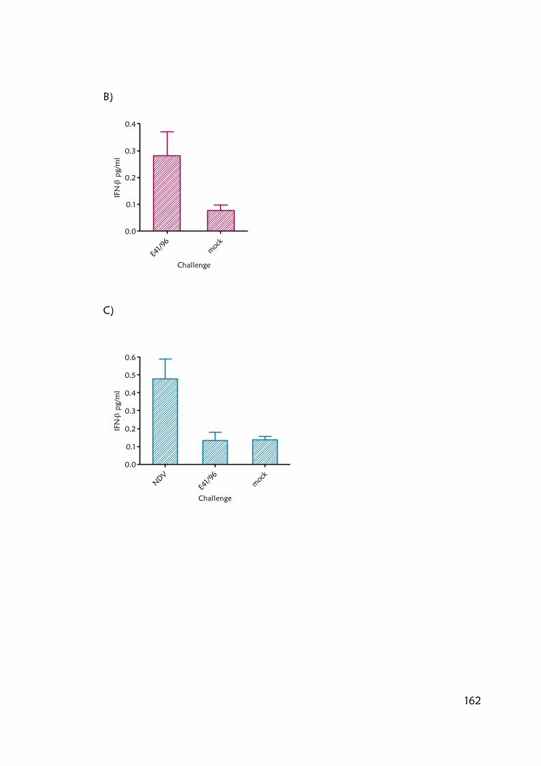
162
B)
E41/96
mock
0.0
0.1
0.2
0.3
0.4
Challenge
IFN
- pg/m
l
C)
NDV
E41/96
mock
0.0
0.1
0.2
0.3
0.4
0.5
0.6
Challenge
IFN
- pg/m
l

163
Cells were infected with A/England/41/96 (4196) an alternative high IFN-β
inducing strain of influenza (Hayman, Comely et al. 2006), which resulted in
IFN-β secretion in A549 cells (figure 5.3.5B). In contrast, infection of PER.C6
cells with A/England/41/96 virus did not result in secretion of IFN-β. This
correlated with both the activation of the IFN-β promoter measured by the
luciferase assay and the number of RNA copies measured by qRT PCR (figure
5.3.4C). However, infection with NDV does result in the secretion of IFN-β in
PER.C6 cells, albeit to a low level. Taken together the data suggests that the
mechanisms involved in IFN-β induction by influenza viruses may be defective
in PER.C6 cells.
5.4 PER.C6 cells respond to IFN-β
Paracrine IFN signalling results in the formation of signalling complexes which
translocate to the nucleus and bind to the IFN stimulated response element
(ISRE promoter) leading to the expression of many antiviral genes. Infection of
Vero cells, which lack a functional IFNAR, suggest that positive feedback
enhances IFN-β induction by influenza viruses (Johnson et al manuscript in
preparation). The ISRE promoter has been cloned upstream of a luciferase gene
by Steve Goodbourn (St. Georges, University of London) to provide a simple
quantification of ISRE promoter activation. To assess whether PER.C6 cells
were able to respond to IFN-β they were transiently transfected with an IFN
stimulating response element upstream of a luciferase reporter gene (ISRE-luc)
and stimulated with 1U/ml of IFN-β for 16 hours (figure 5.4.1). Priming of
PER.C6 ISRE-luc cells resulted in a 10-fold activation of the ISRE promoter.
Although the response was not as marked as in 293T ISRE-luc cells transfected
with the same reporter, this could be due to a less efficient IFN-β induction in
PER.C6 or less transfected cells since β-gal assay was not performed for this
assay. However, data shown in chapter 3 indicates little difference in the
transfection efficiency between PER.C6 and 293T cell lines (section 3.2.1)

164
Figure 5.4.1 PER.C6 cells respond to IFN-β
293T cells and PER.C6 cells were transiently transfected with ISRE-luc. 24 hours
post transfection cells stimulated with 1U/ml IFN-β for 16 hours. Cells were
washed PBS, lysed in CCLR, clarified by centrifugation, 13,000 RPM for 1
minute and luciferase measured. Data are represented as ratio of induction
corrected to unchallenged cells. Means and standard error were calculated for
duplicate wells. This assay is representative of three repeat experiments. Error
bars represent standard error.
293T PER.C6
0
5
10
15
20
25
+IFN
-IFN
Cell line
Relative Luciferase A
ctivity

165
In summary, PER.C6 cells respond to IFN-β and secrete it following NDV
infection, but respond only poorly to influenza viruses.
5.5 IFN-β promoter is activated in PER.C6 cells in response to poly IC
but not to in vitro transcribed viral-like dsRNA
It is intriguing that PER.C6 cells do not secrete IFN-β in response to infection
by influenza virus. It is possible that they lack the ability to detect the influenza
RNA PAMP. In order to identify the possible PAMPs that PER.C6 cells can
recognise and thus further characterise the IFN response in PER.C6, cells were
stimulated with three different RNA based PAMPs. Poly IC is a synthetic dsRNA
analogue thought to activate the MDA-5 pathway (Kato, Takeuchi et al.
2006). Poly IC was obtained from a commercial source (GE healthcare,
formerly Pharmacia). In vitro transcribed dsRNA with 5‟ triphosphate is
thought to activate the RIG-I pathway. A particularly relevant dsRNA would
contain influenza sequences. Influenza-like dsRNA was synthesised from
plasmids encoding short defective viral RNAs. These short RNAs result from
spontaneously occurring deletions in full-length viral RNAs during RNA
synthesis. The short RNAs are incorporated into virions, often called defective
interfering particles (DIs) which are able to infect cells, but are unable to
replicate without the assistance of an intact helper virus. dsRNA representing
influenza DI RNA was generated by in vitro transcription from appropriate
cDNA that contained a T7 promoter upstream of influenza sequences (figure
5.5.1). RNPs are the packaged state of influenza viral genomes and consist of
influenza viral RNA complexed with nucleoproteins. RNPs represent a form of
viral RNA which is known to exist in cells during viral replication, although it
has yet to be determined if RNPs induce IFN-β. RNPs were obtained by
purifying the NP rich fraction of egg derived virus particles through CsCl
gradient ultracentrifugation purification (Parvin, Palese et al. 1989)

166
Fig 5.5.1 Schematic representation of in vitro transcribed dsRNA synthesis
Influenza-like dsRNA was synthesised from plasmids encoding defective
interfering RNAs. T7 RNA polymerase promoters are added at the 5´ ends of
both DNA strands with amplification by PCR with primers containing the T7
promoter sequence at the 5´ end. Generation of the two DNA templates
requires two separate PCR amplifications, each containing one gene specific
primers, and the converse T7 primers. Two complementary RNA strands are
then synthesized from the DNA templates using T7 RiboMAX Express RNAi
System (Promega). The resulting RNA strands are annealed to form dsRNA.

167
Figure 5.5.1 Schematic representation of in vitro transcribed dsRNA synthesis

168
Figure 5.5.2 IFN-β promoter is activated in PER.C6 cells in response to poly IC
but not dsRNA
A549-luc cells (A) or PER.C6 cells transiently transfected with IFN-β luc (B)
were stimulated with a panel of stimuli. Poly IC (pI:C), in vitro transcribed
dsRNA and RNPs were transfected into cells 48 hours post IFN-β luc
transfection. 6 hours post transfection cells were washed PBS, lysed in CCLR,
clarified by centrifugation, 13,000 RPM for 1 minute and luciferase measured.
Data are represented as ratio of induction corrected to unchallenged cells.
Means and standard error were calculated for duplicate wells. This assay is
representative of two repeat experiments. Error bars represent standard error.
A)
pI:C 10ug/ul
pI:C 5
ug/ul
pI:C 1ug/ul
dsR
NA 10ug/ul
dsR
NA 5
ug/u
l
dsR
NA 1ug/ul
RNP 10ul
RNP 5
ul
RNP 1ul
mock
0
500
1000
1500
2000
2500
3000
3500
4000
4500
Challenge
Relative Luciferase U
nits
B)
pI:C 10ug/ul
pI:C 5
ug/ul
pI:C 1ug/ul
dsR
NA 10ug/ul
dsR
NA 5
ug/u
l
dsR
NA 1ug/ul
RNP 10ul
RNP 5
ul
RNP 1ul
mock
0
5
10
15
20
25
30
35
40
45
50
55
Challenge
Re
lative
Lu
cife
rase
Un
its

169
A549s stably expressing the IFN-luc reporter or PER.C6 cells transiently
transfected with IFN-luc reporter plasmid, were stimulated with poly IC, in
vitro transcribed dsRNA, RNPs or NDV (figure 5.5.2). All three RNA based
PAMPs activated the IFN-β promoter in A549 cells (figure 5.5.2 A). However,
only poly IC was able to activate the IFN-β promoter in PER.C6 cells (figure
5.5.2 B). This implies that PER.C6 cells have an intact MDA-5 pathway but are
impaired in RIG-I signalling. Alternatively poly IC may be detected by TLR-3
and both MDA-5 and RIG-I could be defective in PER.C6 cells.
5.6 PER.C6 cells respond to IFN-β priming
RIG-I and MDA-5 are IFN stimulated genes (ISGs), their expression is
upregulated by IFN-β (Yoneyama, Kikuchi et al. 2004; Siren, Imaizumi et al.
2006). In order to test whether IFN-β primed PER.C6 cells would respond
more robustly to challenge with influenza viruses or viral like dsRNA, after up-
regulation of PRRs, cells were transiently transfected with IFN-β luc plasmid
and pre-treated with exogenous IFN-β overnight. Cells were stimulated as
before with influenza virus, poly IC or dsRNA (figure 5.6.1). Following
stimulation with virus and dsRNA a two-fold greater activation of IFN-β
promoter is seen in IFN-β primed cells than in naïve cells. This could be the
result of up-regulation of RIG-I following IFNAR signalling. IFN-β promoter
activation by poly IC transfection was unaffected by IFN-β pre-treatment. Poly
IC IFN-β activation was much greater than for other stimuli. The amount of
poly IC added could be in excess and may have already saturated the response.
Alternatively, the high levels of IFN-β could be a result of very high or even
constitutive expression of the poly IC PRR.

170
Figure 5.6.1 PER.C6 cells respond to IFN-β priming
PER.C6 cells were transiently transfected with IFN-β luc plasmid. 48 hours post
transfection cells were primed with 0.5 U/ml IFN-β, for 16 hours. Cells were
subsequently infected with virus MOI 2 A/Sydney/97 (Syd) and
A/Victoria/3/75 (Vic), or transfected with 10 µg/ml poly IC or dsRNA. 7 hours
post transfection/infection cells were washed PBS, lysed in CCLR, clarified by
centrifugation, 13,000 RPM for 1 minute and luciferase measured. Data are
represented as ratio of induction corrected to unchallenged cells. Means and
standard error were calculated for duplicate wells. This assay is representative
of two repeat experiments. Error bars represent standard error.
Syd Vic poly IC dsRNA mock
0
250
500
750
- IFN
+ IFN
Agonist
Relative Luciferase A
ctivity

171
5.7 Expression of PRR differs in PER.C6 cells to IFN-β competent
A549 cells
Semi-quantitative RT-PCR was used to determine the levels of expression of
MDA-5 and RIG-I mRNAs, pre and post-IFN-β treatment. Cells were primed
with 0.5U/ml IFN overnight and lysed in TRIzol according to manufacturer‟s
instructions. Total cellular RNA was quantified by spectrophotometer.
Equivalent amounts of RNA were used to generate cDNA and then equal
volumes of cDNA were used in the PCR reaction to render the PCR
quantitative. The PCR was limited to 25 cycles to maintain the proportional
relationship between amplification and input RNA. Expression of RIG-I, MDA-
5, SOCS3 and GAPDH mRNA as a loading control, were evaluated (figure
5.7.1). As expected in A549 cells RIG-I and MDA-5 mRNAs are present in low
amounts in untreated cells, and were up-regulated following priming with IFN-
β (Siren, Imaizumi et al. 2006). SOCS3 is moderately upregulated. Although
total signals for SOCS3 are low, SOCS3 expression has been shown to peak 3
hours p.i. in A549 cells (Pauli, Schmolke et al. 2008), therefore it is possible
that expression of SOCS3 had already peaked after overnight IFN-β treatment.
In a human bronchial epithelial cell line, BEAS-2B SOCS3 expression peaks after
14 hours (Pothlichet, Chignard et al. 2008). In PER.C6 cells both RIG-I and
MDA-5 were upregulated following IFN-β pre-treatment. Interestingly, MDA-5
was constitutively expressed prior to IFN-β treatment; this may contribute to
the high reactivity to poly IC. Since no standards were used, it is not possible to
compare the levels of expression from gene to gene; the levels of MDA-5
might not be higher, rather its primers may be more efficient than those of
SOCS-3 for example. If either PRR are functionally competent in un-primed
cells, their residual expression could certainly have an adverse effect vaccine
yields for viruses that are IFN-β sensitive.

172
Figure 5.7.1 Expression of PRR differs in PER.C6 cells to IFN-β competent A549
cells
Cells were primed with IFN-β for 16 hours. Cells were lysed in TRIzol
(Invitrogen) and RNA extracted according to instructions. 2 µg RNA was used
in an RT reaction to synthesize cDNA. 2 µl of this reaction was used in a 25 µl
PCR reaction using primers against RIG-I, MDA-5, SOCS-3, GAPDH was used as
loading control.

173
5.8 Viruses do not upregulate PRR RIG-I and MDA-5
IFN-β priming moderately enhanced the sensitivity of PER.C6 cells to PAMPs,
but this would only be relevant to vaccine production if it were to occur
during multicycle virus growth in response to virus infection. Therefore,
expression levels of PRRs were also analysed following infection with a panel
of high inducing viruses A/Sydney/97 (Syd) and A/England/41/96 (4196), or
low inducing viruses (A/Victoria/3/75 (Vic) and A/England/919/99 (919) (figure
5.8.1). Cells were infected with the viruses and treated as previously described
(section 5.7). As before, MDA-5 expression is constitutive in PER.C6 cells and
priming with IFN-β or infection with NDV both up-regulated expression of the
PRR mRNAs in both A549s cells (data not shown) and PER.C6 cells. However,
infection with influenza virus did not elicit induction of PRR expression.

174
Figure 5.8.1 Viruses do not upregulate PRR RIG-I and MDA-5
PER.C6 cells were infected with influenza virus for 8 hours MOI 2
A/Victoria/3/75 (Vic), A/Sydney/97 (Syd) A/England/919/99 (919) and
A/England/41/96 (4196), or infected with NDV for 8 hours, or were primed
with IFN-β for 16 hours. Cells were lysed in TRIzol (Invitrogen) and RNA
extracted according to instructions. 2 µg RNA was used in an RT reaction to
synthesize cDNA. 2 µl of this reaction was used in a 25 µl PCR reaction.
Screened with primers against MDA-5, RIG-I and GAPDH as a loading control.

175
5.9 Discussion
Vaccine manufacturers are investigating the potential of cell culture for the next
generation of influenza vaccines. Most laboratory based cell lines available are
unpermissive, unlicensable or low yielding, hence the need to test new lines
such as PER.C6. PER.C6 cells are also able to support the growth of both
clinical isolates and vaccine strains (chapter 3 and chapter 4). There exists great
variation in the levels of IFN-β induction from clinical isolates from year to
year and even within a single season, which could lead to inconsistency in
yields (Hayman, Comely et al. 2006).
PER.C6 cells appear to be IFN-β competent; the IFN-β promoter is activated
and IFN-β protein is secreted in response to poly IC or NDV. They also
respond to exogenous IFN-β by activation of the IFN-β responsive promoter,
ISRE. However, infection of PER.C6 cells by influenza virus results only in low
activation of IFN-β promoter, suggesting a defect in either the detection of the
influenza PAMP or the downstream signalling pathway. As such, PER.C6 cells
represent a good cell line for the propagation of clinical influenza isolates
which may vary in their ability to induce IFN-β.
The key PRRs involved in detection of RNA PAMPs are MDA-5 and RIG-I.
Previously MDA-5 has been associated with detection of poly IC and positive
sense RNA viruses whilst RIG-I has been associated with influenza virus (Kato,
Takeuchi et al. 2006) and in particular the presence of 5‟ triphosphate RNA
(Pichlmair, Schulz et al. 2006). However, recent research suggests that rather
than being related to the nature of the nucleic acids, the recognition by each
PRR is associated to its length; RIG-I and MDA-5 selectively recognize short
(<1kb) and long (>1kb) dsRNAs, respectively (Kato, Takeuchi et al. 2008). The
DI dsRNAs used here (DI 316) was 585 nucleotides long (Duhaut and Dimmock
2002) and the poly IC (GE healthcare (formerly Pharmacia) ranges between

176
4,000 and 6,000 nucleotides. These therefore represent distinct activators of
RIG-I and MDA-5 respectively.
The constitutive expression of the PRR MDA-5 may explain why PER.C6 cells
respond more robustly to poly IC. Alternatively, the response to poly IC in
PER.C6 may also be transduced by TLR-3. More accurate and sensitive
techniques, such as quantitative RT PCR, would allow comparisons between
the levels of gene expression in different cell lines and give further indication of
any irregularities in genes associated with IFN induction. Following activation
of RIG-I, expression of an inactive splice variant (SV) is up-regulated (Gack,
Kirchhofer et al. 2008). Analysis of MDA-5 has yet to be performed but
inactive splice variants may also be generated following IFN induction. Full
length RT PCR of PRRs could determine potential defects in PRR splice variant
expression in PER.C6. Furthermore, application of a micro-array would detect
difference in expression of the full range of proteins involved in IFN signalling
and clarify precisely if the induction or expression of any proteins is defective.
Sequencing of both MDA-5 and RIG-I, and proteins flagged in the micro array
might indicate where the defects lie. However, since PER.C6 cells are able to
support the propagation of influenza virus, further characterisation of these
genes may not be necessary.
On the other hand, antagonism of the residual IFN response albeit low may
increase yields further. Cell lines expressing virally encoded antagonists have
improved yields for viruses which induce high levels of IFN e.g. MDCK cells
that express PIV5 V protein (Young, Andrejeva et al. 2003). No significant
increase in yield was noted for viruses which do not induce significant levels of
IFN-β (unpublished observation Anna Hayman). However, since very little
IFN-β has been detected following infection of PER.C6 with influenza with
either vaccine strains made by reverse genetics, clinical isolates or reassortant
viruses, no significant benefit is likely to be gained by growth in an IFN
antagonised cell line. Furthermore, an altered cell line would also require re-

177
validation to ensure the antagonism has no ensuing adverse side effects on the
safety, composition and efficacy of the vaccine.
The constitutive expression of MDA-5 may be a concern for the propagation
and vaccine development for positive sense RNA viruses such as members of
the Picornaviridae family, e.g. Encephalomyocarditis (EMCV) (Gitlin, Barchet et
al. 2006) or the negative sense paramyxoviruses (PIV5) (Childs, Stock et al.
2007) which are known to induce IFN-β expression in an MDA-5 dependant
manner. In this instance, antagonism of the IFN response by transduction of
the cells with viral antagonist proteins such as PIV5-V may be appropriate for
the propagation of these viruses.
Further characterisation is required to describe the irregularities in IFN response
in PER.C6 cells. However, since the secreted IFN-β following infection with
influenza is lower than for other IFN competent cell lines and the cell line is
able to support the growth of clinical isolates, it may not generate any further
constructive data. As such, PER.C6 cells represent a highly suitable cell line for
the generation of influenza vaccine.

178
Chapter 6 Characterising viral IFN induction,
implications for vaccine manufacture
6.1 Introduction
Comparisons in the induction of IFN-β by a panel of human H1N1 and H3N2
clinical isolates found considerable variation in their ability to induce IFN-β in
A549 lung epithelial cells. A number of strains induced high levels of IFN-β,
exemplified by the H3N2 strain A/Sydney/5/97. Although the PER.C6 cell line
elicits only a very limited IFN-β response to influenza virus infection (chapter
5), other cell lines used in vaccine manufacture may still be IFN competent.
Should vaccines be made directly from clinical isolates in IFN competent cell
lines, IFN induction may be of consequence. Although high IFN-β induction in
immortalised cell culture has yet to be correlated with high cytokine induction
in vivo, the variation in IFN-β induction by different strains of influenza A
viruses is likely to contribute to the biological consequences of infection
including the pathology, pathogenicity and outcome of disease.
The concept that the cytokine response, including IFN, might contribute to the
disease process following influenza infection is most recognised following
infection with H5N1 virus. Since 2003, the H5N1 virus has caused devastating
outbreaks in poultry in Asia, Africa and Europe, and resulted in 438 human
infections, with 262 fatalities thus an overall case fatality rate of 60%,
(according to the WHO 5th Feb. 2009
(http://www.who.int/csr/disease/avian_influenza/country/cases_table_2009_0
8_11/en/index.html). Thankfully, human-to-human transmission so far remains
restricted. Post mortem analysis showed elevated levels of the inflammatory
cytokines interleukin-6 (IL-6), TNF- α and IFN-γ (To, Chan et al. 2001). This
correlates with H5N1-infected ferret lungs, where IFN stimulated genes were

179
more strongly expressed than in lungs from ferrets infected with the less
pathogenic H3N2 subtypes (Cameron, Cameron et al. 2008).
The 1918 “Spanish flu” pandemic was unusually severe, resulting in
approximately 50 million deaths. Analysis of the 1918 pandemic virus was not
possible until RT-PCR of viral RNA isolated from preserved tissue obtained
from formalin-fixed pathology specimens and a cadaver buried in permafrost
at the time of the pandemic, allowed the sequencing and subsequent
reconstruction of the virus (Taubenberger, Reid et al. 1997; Basler, Reid et al.
2001; Tumpey, Basler et al. 2005; Tumpey, Garcia-Sastre et al. 2005). Infection
of macaques with 1918 virus caused an ultimately lethal, highly pathogenic
respiratory disease (Kobasa, Jones et al. 2007). Infected animals mounted an
immune response, characterized by dysregulation of the antiviral response that
was insufficient for protection. Several key cytokines (including IL-6, IL-8), were
expressed at a high level in 1918-virus-infected animals but not in low
pathogenic H1N1 infected animals. The dysregulation of IFN induction by
H5N1 and 1918 have been reviewed by (Basler and Aguilar 2008). The IFN
response clearly plays an important role in pathogenesis and understanding the
mechanisms will enable more co-ordinated treatment of viral infection.
Some high IFN-β inducing strains of influenza virus, in some cell lines seem to
be resistant to the IFN they induce. For example the replication of the H5N1
strain, A/HK/97, in pig epithelial cells was not affected by pre-treatment of
cells with a number of cytokines including, IFN-α, IFN-γ or TNF-α. In this
instance, resistance was shown to map to glutamic acid at position 92 on the
non-structural protein, NS1, replacing the usual aspartic acid (Seo, Hoffmann et
al. 2002; Seo, Hoffmann et al. 2004). The seasonal strain A/Sydney/97 which
induces a high level of IFN-β, was less affected by the presence of exogenous
IFN-β in A549 cells; whilst viruses which induced lower levels of IFN-β, e.g.
A/Victoria/75, were more susceptible (Hayman, Comely et al. 2006). Since
A/Sydney/97 does not possess the D92E mutation, there must also exist

180
alternative mechanisms of IFN-β resistance. Furthermore, the mechanisms
behind high IFN-β induction are also yet to be established.
Many viruses encode immunomodulatory proteins that interfere with the
induction, response to, or action of host cytokines. The influenza NS1 protein
antagonises the innate immune response in several ways (Egorov, Brandt et al.
1998; Garcia-Sastre, Egorov et al. 1998; Hale, Randall et al. 2008). NS1 has
two domains involved in suppression of the IFN response. The mechanisms are
strain-dependent since not all strains retain all functions, and are likely to be
determined by the amino acid sequence of NS1. NS1 has been shown to
interact with both the PAMP (currently believed to double stranded structures
within 5‟ triphosphate ssRNA (Pichlmair, Schulz et al. 2006) mediated via an
RNA binding domain within residues 38 and 41 (Hatada and Fukuda 1992;
Wang, Riedel et al. 1999; Donelan, Basler et al. 2003), and PRR such as RIG-I
(Garcia-Sastre, Egorov et al. 1998; Talon, Horvath et al. 2000; Wang, Li et al.
2000; Pichlmair, Schulz et al. 2006). It also interacts with many other proteins
involved in IFN induction such as protein kinase R (PKR) (Wang, Riedel et al.
1999), and the p85β regulatory subunit of host-cell phosphoinositide 3-kinase
(PI3K) which activates signalling (Hale, Batty et al. 2008). The NS1 from some
strains of influenza A virus have been shown to bind to the 30 kDa subunit of
the cellular processivity and specificity factor (CPSF-30) and poly (A)-binding
protein II (PABII), thus abrogating the export of cellular mRNA, including those
that encode anti-viral genes and IFN-β (Nemeroff, Barabino et al. 1998; Chen,
Li et al. 1999; Wang, Riedel et al. 1999; Noah, Twu et al. 2003).
As part of the investigation into the role IFN may play in the ultimate yield of
vaccine strains and the extent to which antagonism can be beneficial, the
mechanism by which viruses induce IFN-β, and the reasons for the disparity in
levels of IFN-β induction between virus strains, needs to be understood. This
supplements research done by two previous lab members: initially Anna
Hayman and subsequently Ben Johnson.

181
6.2 IFN-β induction does not map to HA, NA, NS
Since the NS1 protein has been shown to antagonise IFN-β induction by
multiple mechanisms it is the obvious candidate to account for the variation in
IFN-β induction. Using reverse genetics, a panel of recombinant influenza
viruses that differed only in the origin of segment 8 RNAs, was generated
(Hayman, Comely et al. 2006). Initially this was done in the A/Victoria/75
background (Anna Hayman, figure 6.2.1). To exclude the possibility that an
additional gene(s) of A/Victoria/75 was acting to suppress IFN-β induction and
thus mask a defect in NS1 function, a selection of viruses were also rescued in
PR8P background (a 6:2 recombinant virus with six internal genes of
A/PR/8/34 and surface antigens HA and NA of A/Panama/2007/99,
representative of a typical vaccine seed). Using the A549 IFN-β reporter cell
line, recombinant viruses with NS1s derived from human viruses with both low
and high IFN-β inducing phenotypes, all only activated the IFN-β promoter to
low amounts, similar to that induced by the parental strains. Moreover,
endogenous expression of high inducing virus NS1 proteins in the context of
virus infection, were able to control IFN-β induction by Sendai and influenza
virus (Johnson et al manuscript in preparation). Thus, the high induction of
IFN-β in human cells by wild-type influenza viruses was not accounted for by a
deficiency in their NS genes.
Besides NS1, the surface glycoproteins HA and NA of influenza have also been
implicated in the high cytokine induction of the 1918 “Spanish” pandemic virus
and high TNF-α induction by Sydney in porcine macrophages in vitro (Seo,
Webby et al. 2004). For vaccine manufacture, the properties of the surface
antigens are the most significant since the vaccine is a 6:2 recombinant.
Recombinant viruses in both A/Victoria/75 and PR8P backgrounds, containing
constellations of HA, NA and NS derived from A/Sydney/97, all induced low
amounts of IFN-β, similar to that induced by the parental strains in the A549
IFN-β reporter cell line (figure 6.2.1), suggesting that high IFN-β induction in

182
A549 cells is not conferred by either the influenza surface glycoproteins or NS1.
If vaccine seeds are 6:2 reassortants derived from a low inducing donor strains
then IFN-β induction should not be an issue for vaccine manufacture.

183
Figure 6.2.1. IFN induction by wild type A/Sydney/97 virus does not map to
HA, NA, NS
Figure 6.2 The A549 IFN-β luc reporter cell line was infected with recombinant
viruses containing constellations of HA, NA and NS genes from A/Sydney/97 in
stable human genetic backgrounds (A/Victoria/3/75 or A/PR8/) at an MOI of 3
PFU/cell. 8 hours post infections cells were washed PBS, lysed in CCLR, clarified
by centrifugation, 13,000 RPM for 1 minute and luciferase measured. Data are
represented as ratio of induction corrected to unchallenged cells. Means and
standard error were calculated for duplicate wells. This assay is representative
of two repeat experiments. Error bars represent standard error.

184
6.3 High IFN-β inducing viruses exhibit dysregulation of protein
expression
Although it had been shown that A/Sydney/97 virus NS1 protein was
functional and able to antagonise IFN-β induction, it was possible that its
expression could be dysregulated in the context of wild type virus infection
such that when PAMP is generated insufficient NS1 protein has accumulated to
antagonise IFN-β induction. To assess this possibility protein levels over the
course of infection were measured by Western blot. Expression of A/Sydney/97
viral proteins were compared to a low IFN-β inducing virus A/Victoria/75. A
recombinant virus containing A/Sydney/97 NS in the A/Victoria/75 background
was used as a second, low IFN-β inducing virus for comparison.
A dose of virus was established that infected an equal number of cells. The
expression of viral proteins, NS1, M1 and NP, were assessed by Western blot
(figure 6.3.1). JAK/STAT signalling following IFNAR stimulation by IFN-β
results in the phosphorylation of STAT-1, so phosphorylated STAT-1 was used
as a marker for the induction of IFN-β.
Infection with A/Sydney/97 was found to be synchronized differently than for
A/Victoria/75 based viruses. Viral proteins in A/Sydney/97 infection
accumulated much earlier in infection than did proteins expressed by the two
low inducing viruses. Viral proteins (NP, M and NS1) were detected as early as
2 hours post infection with A/Sydney/97 and this correlated with earlier STAT-1
phosphorylation. Furthermore, the expression of A/Sydney/97 NS1 protein
appeared to lag behind the other viral proteins. Although differences in the
affinity of the antibodies to each of the viral proteins cannot be excluded, the
data suggests that viral replication resulting in the synthesis of certain viral
proteins, and the accumulation of PAMP, occurred earlier during infection with
A/Sydney/97 without a corresponding increase in the levels of NS1 to
antagonise the IFN-β induction

185
Viral proteins also accumulated, to a lesser extent, earlier following infection
with the recombinant virus containing A/Sydney/97 NS1 in the A/Victoria/75
background, than during infection with wild type A/Victoria/75. However, this
was associated with much higher relative levels of NS1. The increased
expression of NS1 can antagonise the IFN-β induced by the increased levels of
PAMP and thus results in much reduced levels of phosphorylated STAT-1;
which do not accumulate until later in infection. The NS1 protein has been
shown to mediate the temporal regulation of viral RNA synthesis via
interactions with the viral polymerase (Min, Li et al. 2007) and also enhance
the rate of translation of certain viral proteins (Enami, Sato et al. 1994; de la
Luna, Fortes et al. 1995) and this could in part explain the differences in viral
protein expression of the recombinant virus.
Viral proteins associated with infection with A/Victoria/75 accumulated slowly
and did not even reach equivalent levels to those expressed by the other two
viruses. However, this does not correlate with outcome of infection;
A/Victoria/75 is an extremely “fit” virus and can achieve titres of 108 PFU/ml
following propagation in cell culture whilst A/Sydney/97 usually attains a log
less.
Furthermore, the earlier phosphorylation of STAT-1 correlates with early IFN-β
induction by A/Sydney/97 virus (figure 6.3.2). A549 cells harbouring the IFN-β
luciferase reporter were infected with the same panel of viruses. IFN-β
induction was detected as early as 2 hours post infection by A/Sydney/97.
Infection with the recombinant virus in the A/Victoria/75 background with
A/Sydney/97 NS resulted in a slight induction of IFN-β which correlates to the
lower levels of phosphorylated STAT-1 noted by Western blot.

186
Figure 6.3.1. A/Sydney/97 proteins accumulate early in infection
A/Sydney/97 proteins are synthesised earlier in infection than those of
A/Victoria/75. A549 cells were infected with viruses (A/Victoria/75- V,
A/Sydney/97- S, and a recombinant containing NS from A/Sydney/97 in the
A/Victoria/75 background- NS) at a dose twice that required for every cell to
express antigen, cells were lysed in RIPA buffer containing iDOC at 2, 4, 6, and
8 hours post infection. Cell lysates were run on 10% or 15% SDS PAGE gels
and Western blotted on to PVDF membrane. Membranes were probed with
antibodies and detected with ECL reagent. Vinculin was used as a loading
control. (Antibody concentrations were Vinculin 1/1000, Stat-1 1/500, NP
1/400, M 1/1000, NS 1/300, secondary antibodies 1/2000).

187
Figure 6.3.2 A/Sydney/97 induces IFN-β early in infection
The A549 IFN-β luc reporter cell line was infected with A/Sydney/97,
A/Victoria/75 and a recombinant containing NS from A/Sydney/97 in the
A/Victoria/75 background at a dose twice that required for every cell to
express antigen. At 2, 4, 6, 8, 12 and 24 hours post infection cells were washed
PBS, lysed in CCLR, clarified by centrifugation, 13,000 RPM for 1 minute and
luciferase measured. Data are represented as ratio of induction corrected to
unchallenged cells. Means and standard error were calculated for duplicate
wells. This assay is representative of three repeat experiments. Error bars
represent standard error.
2 4 6 8 12 24 2 4 6 8 12 24 2 4 6 8 12 24
0
10
20
30
A/Vic/75
A/Syd/97
AVic/75 +A/Syd/97 NS
hours pi
Rela
tiv
e Luciferase A
ctiv
ity

188
To normalise infections, the infectivity of viruses in the A549 cell line was
measured by “blue cell” assay, where expression of the viral protein, NP was
immuno-detected. Interestingly, it was observed that not all viruses expressed
the same levels of NP in infected cells. Infection with some strains resulted in
cells which stained very weakly whilst for others staining was more intense.
The "blue cell" assay standardises the volume of virus required to infect an
equal number of cells regardless of intensity. In some instances, Western blots
of virus proteins have been used to standardise assays and ensure equivalent
levels of infectivity. The results of both the Western blot and the blue cell assay
highlight the need to be aware when comparing some virus strains that the
levels of virus protein from different strains are not absolute.
6.4 High IFN-β induction requires expression of RIG-I
Work in section 6.3 suggested that the higher levels of IFN-β induced by
A/Sydney/97 may be due to a dysregulation in the levels of PAMP relative to
the levels of NS1 protein. Alternatively, A/Sydney/97 may also generate a
different kind of PAMP which NS1 is unable to control. The PRR, RIG-I, has
been shown to mediate the IFN-β response to influenza viruses (Pichlmair,
Schulz et al. 2006; Le Goffic, Pothlichet et al. 2007; Loo, Fornek et al. 2007;
Pothlichet, Chignard et al. 2008), however, these experiments have largely
been performed with lab strains of viruses which tend to have a low IFN-β
inducing phenotype.
Huh-7.5 cells have a point mutation the PRR, RIG-I (Sumpter, Loo et al. 2005)
in the TRIM25 binding domain (Gack, Albrecht et al. 2009) and are believed
to be unable to respond to RIG-I activating PAMPs. Huh-7.5s were used to
determine whether the high IFN-β induction by A/Sydney/97 virus was
mediated via the RIG-I dependant PRR pathway.
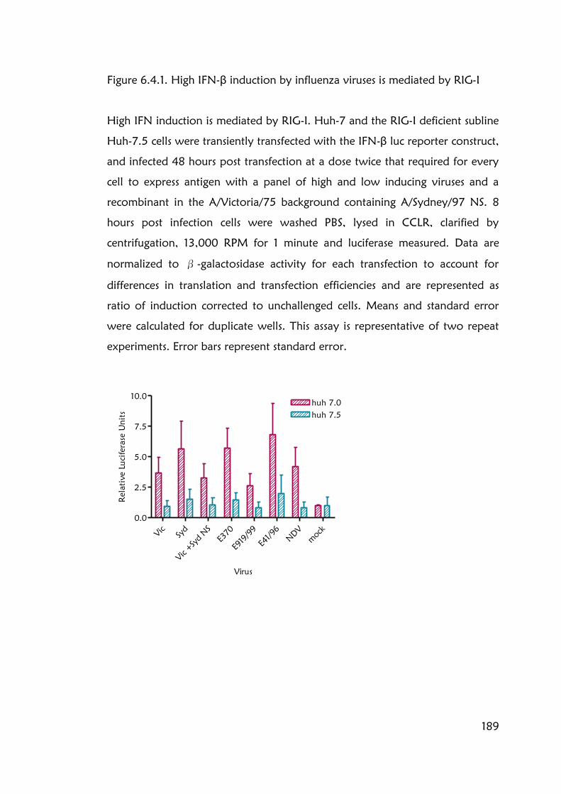
189
Figure 6.4.1. High IFN-β induction by influenza viruses is mediated by RIG-I
High IFN induction is mediated by RIG-I. Huh-7 and the RIG-I deficient subline
Huh-7.5 cells were transiently transfected with the IFN-β luc reporter construct,
and infected 48 hours post transfection at a dose twice that required for every
cell to express antigen with a panel of high and low inducing viruses and a
recombinant in the A/Victoria/75 background containing A/Sydney/97 NS. 8
hours post infection cells were washed PBS, lysed in CCLR, clarified by
centrifugation, 13,000 RPM for 1 minute and luciferase measured. Data are
normalized to β-galactosidase activity for each transfection to account for
differences in translation and transfection efficiencies and are represented as
ratio of induction corrected to unchallenged cells. Means and standard error
were calculated for duplicate wells. This assay is representative of two repeat
experiments. Error bars represent standard error.
Vic
Syd
Vic +
Syd N
S
E370
E919/99
E41/96
NDV
mock
0.0
2.5
5.0
7.5
10.0
huh 7.5
huh 7.0
Virus
Relative Luciferase U
nits

190
Huh-7 and Huh-7.5 cells were transiently transfected with the IFN-β luc
reporter construct, and infected with a panel of high and low inducing WT
viruses and the recombinant virus containing A/Sydney/97 NS in the
A/Victoria/75 background (figure 6.4.1). Huh-7 cells with intact RIG-I pathway
displayed a similar pattern of IFN-β induction upon infection had been seen
following as infection of A549 cells. In contrast, only low amounts of IFN-β
were induced following infection of Huh-7.5 cells irrespective of the virus
strain. This suggests that the high induction of IFN-β by wild-type influenza
viruses, such as A/Sydney/97, is mediated via RIG-I.
6.5 RIG-I and MDA-5 mRNAs are upregulated during influenza
infection
PRR such as MDA-5 and RIG-I, are IFN stimulated genes (ISGs) which are
upregulated by IFN-β induction. To determine whether the high IFN-β
induction by some strains of influenza viruses elicited a typical upregulation of
ISG, semi-quantitative RT-PCR was used to determine the levels of expression
of MDA-5 and RIG-I, in response to virus infection of A549 cells. Total RNA
was extracted from infected cells at various times post infection. Equal amounts
of RNA, quantified by spectrophotometer, were used to generate cDNA, then
equivalent volumes used to generate cDNA. Only 25 cycles of PCR were
performed to ensure the reaction was terminated in the exponential phase.
mRNA expression of RIG-I, MDA-5 and GAPDH as a loading control, were
analysed (fig 6.5.1). In A/Sydney/97 infected A549 cells RIG-I and MDA-5 were
up-regulated early in infection; RIG-I mRNA signal increased as early as 2 hours
post infection. Both PRR are upregulated by positive feedback in a typical
autocrine manner as a consequence of IFN-β induction during infection with
A/Sydney/97.

191
Figure 6.5.1 PRR are upregulated by IFN-β induction by influenza viruses
MDA-5 and RIG-I are upregulated earlier in infection in cells infected with
A/Sydney/97. A549 cells were infected with virus at a dose twice that required
for every cell to express antigen and lysed in TRIzol (Invitrogen) according to
manufacturer‟s instructions at the given time points. Purified RNA was
quantified by spectrophotometer. Equivalent amounts of RNA were used to
generate cDNA. Equivalent volumes of cDNA were used in the PCR reaction,
for 25 cycles to ensure PCR was in the quantitative phase. Expression of RIG-I,
MDA-5, and GAPDH as a loading control, were evaluated.

192
6.6 High IFN-β inducing viruses are prone to making more defective
RNA
UV treatment denatures nucleic acids, but viral proteins are undamaged and
the virion particles remain intact. UV treatment abrogated IFN-β induction by
influenza viruses including A/Sydney/97 (Johnson et al manuscript in
preparation). Therefore, IFN-β induction is not activated by virus cell binding
or entry but rather must be reliant on RNA replication. The most systematic
way to dissect the mechanisms for high IFN-β induction would be to test the
contribution of each RNA segment in the phenotype by generating
recombinant viruses containing constellations of segments from high and low
inducing viruses; this approach had eliminated HA, NA and NS genes (figure
6.2.1). However, we do not possess a full reverse genetics system for a high
inducing virus. While attempting to clone the remaining genes of A/Sydney/97,
using primer pairs complementary to the 5‟ and 3‟ termini of each viral RNA, a
number of shorter products were consistently generated and for some viral
RNA segments the full-length band was not even detectable. This occurred
frequently during attempts to clone segments from viruses which induced high
levels of IFN-β, and although these short products could be PCR artefacts, they
were rarely seen in low inducing viruses. A number of these bands were
sequenced and had the properties of defective interfering RNAs (figure 6.6.1
and table 6.6.1). Defective RNAs result from spontaneously occurring deletions
within the full-length viral RNAs during RNA synthesis. These shorter RNAs
retain the 3‟ and 5‟ non coding regions which encode the packaging signals.
This means they can be incorporated into virions to generate defective
interfering particles (DIs) which are able to infect cells but since they lack a
complete genome are unable to replicate without the assistance of an intact
helper virus. In this event, as a result of the small size of the defective RNAs the
shorter RNAs are replicated to much higher levels than full-length segments, so

193
Figure 6.6.1 Schematic representation of defective interfering RNA
Defective RNAs result from spontaneously occurring deletions within the full-length viral RNAs during RNA synthesis. These
RNAs are incorporated into virions to generate defective interfering particles which are able to infect cells but due to the lack
of a complete genome are unable to replicate without the assistance of an intact helper virus. As a result of the small size of
defective interfering RNA it is replicated to much higher levels than full-length segments, so the cell produces defective virions
in place of infectious progeny (Duhaut and Dimmock 2000; Duhaut and Dimmock 2002).

194
Table 6.6.1 Defective RNAs accumulate during infection with A/Sydney/97
virus
Sequence analysis of shorter PCR products generated from amplification of
A/Sydney/97 virus show signatures of defective RNAs. Alignment against the
full length sequence allows the junctions of the DI to be determined.
Segment
of origin
Approx. PCR
product size
Full Length
of segment
5'
junction
3'
junction
Deduced
length of
RNA
PB2 600 2280 259 2010 529
PB1 900 2274 275 1943 606
PB1 600 2274 204 2025 453
PA 900 2183 115 1586 712

195
Figure 6.6.2 High IFN-β inducing viruses have a propensity to make short
RNAs
32P labelled oligos complimentary to PB2 gene segment 3‟UTR were used in an
extension assay with total RNA from infected cells as a template. Cells were
infected at an MOI 0.01 and harvested 40 hours post infection. RNA was
extracted with TRIzol according to manufacturers‟ instructions. Equivalent
amounts of RNA were used in each reaction. Only the lower part of the gel is
shown. Data provided by Matt Smith.

196
the cell produces defective virions in place of infectious progeny (Duhaut and
Dimmock 2000; Duhaut and Dimmock 2002). DIs usually occur late in
infection or when a high MOI is used (Odagiri and Tobita 1990), preparations
that contain them are known to induce high levels of IFN-β (Johnston 1981).
The amplification of short products suggests that A/Sydney/97, and other high
IFN-β inducing viruses, have a propensity to make defective RNAs. Primer
extension analysis was used to quantify the RNA species in virus infected cells
(fig 6.6.2). 32P labelled oligos complimentary to viral gene segments were
used in an extension assay with total RNA as a template. In this case, primers
specific to the 3' UTR of vRNA were used. Shorter RNAs were produced during
infection with A/Sydney/97 or A/England/370/02 again suggesting that viruses
that induce high IFN-β have a propensity to make defective RNAs. Although
these short RNAs could be the result of mispriming, this is unlikely because they
did not occur when RNA from cells infected with A/Victoria/75. Analysis
showed that the primer sequence is unique and specific for the 3‟UTR of the
PB2 segment 1. Plaque picking removes DIs which accumulate during an
infection, because virus isolated from a plaque will be derived only from a
single infectious event. Bearing in mind Ben Johnson showed the high IFN-β
inducing phenotype was carried over in plaqued picked viruses and was also
genetically determined (Johnson et al, manuscript in preparation) this suggests
that the DIs are generated spontaneously during infection with viruses which
induce high levels IFN-β.
6.7 A/Sydney/97 virus drives replication early in infection
Taken together the data suggested that the high IFN-β inducing phenotype
maybe inherent to the polymerase complex. To assess this the rate of viral
replication was measured using a luciferase reporter assay. The principal
involves transfection of cells with a plasmid encoding the non-coding regions
of segment 8 flanking a luciferase reporter gene. The amount of luciferase
expression driven by viral polymerase allows replication to be quantified. This

197
Figure 6.7.1 Schematic of up-luc assay (overleaf)
In a minireplicon assay it is sufficient to transfect cells with the polymerase
subunits, PB2, PB1, PA and NP together with a minireplicon reporter construct
of viral like RNA containing a luciferase gene flanked by the 5‟ and 3‟ UTR of
segment 8. However, in virus infection the viral polymerases are unable to
drive the replication of such a reporter, mutation of nucleotides 3, 5 and 8 in
3‟ UTR, to generate a so-called “up-promoter, are able to restore this capacity.
Cells are transfected with the up-luc reporter construct, then infected with
virus. Viral polymerases drive the amplification and expression of the viral like
RNA. Quantification of the luciferase signal allows the rate of virus replication
to be determined.

198
Figure 6.7.1 Schematic of the “up-luc” assay

199
Figure 6.7.2 A/Sydney/97 virus drives replication early in infection
Cells were transfected with the up-luc reporter construct. After 48 hours, cells
were infected with virus. 8hours post infection cells were washed PBS, lysed in
CCLR, clarified by centrifugation, 13,000 RPM for 1 minute and luciferase
measured. Data are represented as ratio of induction corrected to unchallenged
cells. Means and standard error were calculated for duplicate wells. This assay
is representative of three repeat experiments. Error bars represent standard
error.
2 4 6 8 12 2 4 6 8 12 2 4 6 8 12 2 4 6 8 12
0
2
4
6
8
10
12
14
A/Vic/75
A/Syd/97
A/Vic/75 +A/Syd/97 NS
NDV
hours PI
Rela
tiv
e Luciferase U
nits

200
is apt for minireplicon assays, however, the polymerases, in the context of virus
infection, seem unable to recognise and replicate „naked‟ vRNA present inside
a cell to detectable levels (Lutz et al., 2005) unless three nucleotide changes at
positions 3, 5 and 8 in the 3‟ non-coding region are present. This mutation
appears to increase viral polymerase transcription of a synthetic viral-like RNA
(Neumann & Hobom, 1995). Therefore these three point mutations were
engineered into a minireplicon containing the luciferase reporter gene (Dan
Brookes), and function as an „up-promoter‟ enhancing recognition of the
reporter gene by virus polymerases (here on called the “up-luc” reporter)
(figure 6.7.1).
The extent to which the virus polymerase of A/Sydney/97, A/Victoria/75 or
recombinant, in the A/Victoria/75 background containing A/Sydney/97 NS
drove RNA replication and expression were compared. Huh-7 cells were
transiently transfected with the up-luc reporter construct and infected with
twice the volume of virus required to infect every cell. Luciferase production
was measured at various times post-infection (figure 6.7.2). A/Sydney/97 virus
drove expression of the up-luc reporter very early in infection. Luciferase was
measured at the earliest time point; just 2 hours post infection. The low
inducing viruses A/Victoria/75 or the A/Victoria/75 +A/Sydney/97 NS
recombinant eventually attained similar or higher levels of luc expression, but
not until much later in infection at 12 hours. These data imply that the
polymerases of A/Sydney/97 virus are particularly active and initiated
replication very early in infection.
6.8 Total RNA from infected cells induces similar levels of IFN-β
irrespective of strain
The increased rate of replication of A/Sydney/97 appears to correlate
kinetically with its high IFN-β induction. However, the nature of the PAMP
which drives the high IFN-β induction is as yet undetermined. 5‟ triphosphate

201
RNA has been shown to mediate IFN-β induction for laboratory strains of
influenza virus but this has yet to be associated with the high IFN-β phenotype.
The earlier replication observed in A/Sydney/97 virus infection may result in
the generation of vRNAs, possessing 5‟3P, which accumulate in the infected
cells in the absence of sufficient NS1 protein to antagonise IFN-β induction. To
investigate the ability of RNAs generated in infected cells to trigger IFN-β
induction, total RNA was extracted from cells infected over the course of
infection with RNA extracted from uninfected cells serving as a control. The
RNA was then applied to the same cell type expressing the IFN-β luc reporter,
and its ability to induce activation of the IFN-β promoter was assessed (figure
6.8.1).
RNA from uninfected cells did not activate the IFN-β promoter. RNA from
infected cells induced no IFN-β until 4 hours post infection after which up to
20 fold induction of IFN-β was observed, irrespective of strain. This was higher
than the 5 fold induction measured by infection with A/Sydney/97 virus and
transpired at a later time during infection. The data suggest that virus infection
causes accumulation of a PAMP from 4 hours post infection which is not strain
dependant, perhaps via a general modification to RNAs present in the cells
which renders them immuno-stimulatory.
It is surprising that IFN-β induction by RNA extracted from A/Sydney/97
infected cells was not detected earlier since A/Sydney/97 polymerases were
active as early 2 hours post infection (section 6.3 and 6.7) and IFN-β induction
was also seen at that time (figure 6.3.2). The assay may not be sufficiently
sensitive to respond to the levels of PAMP present in the total RNA extraction
at the earlier time points. 500 ng of RNA extracted from virions was able to
induce IFN-β promoter induction, yet 100 ng of this RNA could not (data not
shown). In the present assay, 250 µg of total RNA from infected cells was
applied to IFN-β luc reporter cells.

202
6.8.1 RNA from infected cells induces similar levels of IFN-β irrespective of
strain
Total cellular RNA from infected cells induces similar levels of IFN-β,
irrespective of IFN inducing phenotype of virus. Cells were infected at a dose
twice that required for every cell to express antigen. Total cellular RNA was
extracted with TRIzol (Invitrogen) according to manufacturer‟s instructions,
from infected Huh-7 cells at given times (1, 2, 3, 4, 6, 7 hours) post infection.
250 µg RNA was then transfected onto Huh-7 cells transiently transfected with
IFN- luc and β-galactosidase, 48 hours post transfection. After 8 hours post
transfection cells were washed PBS, lysed in CCLR and clarified by
centrifugation, 13,000 RPM for 1 minute. Luciferase activity was measured and
normalized to β-galactosidase activity for each transfection to account for
differences in transfection efficiencies. Data are represented as ratio of
induction corrected to unchallenged cells. Means and standard error were
calculated for duplicate wells. This assay is representative of two repeat
experiments. Error bars represent standard error.

203
Should the proportion of viral RNA contained within the total RNA fall below
the threshold of detection (between 500 ng and 100 ng vRNA), IFN-β
induction would not be measured. Increasing the amount of total RNA applied
to cells and thereby increasing the proportion of PAMP or increasing the
amount of time the RNA is incubated on the cells before taking measurements
may improve the sensitivity.
6.9 Leptomycin B, inhibits the export of luciferase mRNA from the
nucleus and activates IRF-3
It seemed likely that A/Sydney/97 induced IFN when replicated RNA reached
the cytoplasm, where it would be detected by RIG-I. If this were the case it was
reasoned that inhibition of the export of viral RNAs from the nucleus would
prevent IFN-β induction. Leptomycin B is a specific nuclear export inhibitor,
which alkylates and thus inhibits CRM-1 (chromosomal region maintenance)
mediated nuclear export of proteins containing nuclear export sequences
(NES). Addition of LMB has been shown to prevent the export of viral RNPs
(Elton, Simpson-Holley et al. 2001). A549 IFN-β luc reporter cells infected with
virus were incubated in the presence of 10 nM LMB. Luciferase activity,
reflecting activation of the IFN-β promoter, was measured after 8 hours (figure
6.9.1). IFN-β promoter induction by A/Sydney/97 was greatly reduced
although not completely prevented. However, LMB also reduced IFN-β
induction by NDV infection. Since NDV replicates in the cytoplasm it is not
dependant on nuclear export of its PAMP to mediate an IFN-β response. This
result suggests that LMB treatment retains luciferase mRNA in the nucleus and
that the luciferase mRNA is also exported from the nucleus in a CRM-1
dependant manner. The abrogation of IFN-β induction by A/Sydney/97 virus
was not complete, and some induction of the IFN-β promoter was measured.
LMB was shown to be effective at retaining NP in the nucleus when added 3
hours post infection at a concentration of 11 nM (Elton, Simpson-Holley et al.
2001). Other groups have used a similar range of concentrations (2-40 nM)

204
and addition as late as 6 hours post infection to inhibit export of vRNA and
RNPs from the nucleus (Ma, Roy et al. 2001; Watanabe, Takizawa et al. 2001;
Wu and Pante 2009).Typical virus infection procedures allows 1 hour for the
virus to enter the cells, after which the media is replaced with serum free
DMEM. This minimises the carryover of artefacts in the virus preps which are
able to induce IFN-β. The LMB was therefore added to the replacement serum
free media. Since it has been demonstrated that virus replication and IFN-β
induction occurs early during A/Sydney/97 virus infection, the IFN-β luc
measured could have been induced during the hour prior to the addition of
LMB and whilst the later addition of LMB is appropriate for other virus strains,
addition from the onset of infection is required for viruses such as A/Sydney/97
to completely block export from the nucleus.
Because LMB inhibits the export of luciferase messenger RNA from the nucleus,
the luciferase reporter assay is not an appropriate assay to determine whether
nuclear export is required for influenza mediated IFN-β induction.
Immunofluorescence was used as an alternative assay; the activation of the
IFN-β promoter can be inferred by the translocation of IRF-3 from the
cytoplasm to the nucleus. Cells were infected as above but fixed at 7 hours
post infection and immunostained for viral antigen (NP) and IRF-3.
Surprisingly, however, even in uninfected cells treated with LMB, IRF-3 was
predominantly nuclear (figure 6.9.2 top right panel). Thus, the addition of
LMB seems to activate IRF-3 and this precluded an assessment of the
relationship between RNP localisation during influenza infection and RIG-I
activation. Interestingly, in A/Sydney/97 untreated infected cells treated with
LMB, IRF-3 translocation to the nucleus was still more substantial than in
infected cells. This again could be due to IRF-3 activation during the first hour
of infection when LMB is not present. Alternatively, IFN-β induction by
A/Sydney/97 virus could indeed occur in the absence of nuclear export. NP
was also more nuclear in LMB treated cells confirming NP required CRM-1 for
export from the nucleus, and that inhibition of CRM-1 by LMB had occurred.
In conclusion, it is not possible to equivocally establish the importance of vRNP

205
Figure 6.9.1 Inhibition of nuclear export with Leptomycin B
The A549 IFN-β Luc reporter cell line was infected with a high IFN inducing
virus A/Sydney/97, a low inducing viruses A/Eng/99/00 at a dose twice that
required for every cell to express antigen and NDV. 10 nM Leptomycin B was
added 1 hours p.i. 8 hours post infection cells were washed PBS, lysed in CCLR,
clarified by centrifugation, 13,000 RPM for 1 minute and luciferase measured.
Data are represented as ratio of induction corrected to unchallenged cells.
Means and standard error were calculated for duplicate wells. This assay is
representative of three repeat experiments. Error bars represent standard error.
A/Syd/97
E/919/00
NDV
mock
0
1
2
3
4
5
6
7
8
9
-LMB
+LMB
Virus
Relative Luciferase U
nits

206
Figure 6.9.2 Leptomycin B immunofluorescence of NP and IRF-3
A549 cells were infected with A/Sydney/97 at a dose twice that required for
every cell to express antigen. 10 nM Leptomycin B was added 1 hours post
infection. 7 hours post infection cells were washed PBS, then fixed 4% PFA/PBS
for 10 minutes, RT. Coverslips were blocked 3% BSA for 30 minutes RT.
Proteins were immunodetected (top left panel- goat IRF-3 1/300, anti goat
FITC 1/500, bottom left panel- mouse NP 1/300, anti mouse Texas red 1/500,
top right panel- phase contrast, bottom right panel- overlay).

207

208
nuclear export in the A/Sydney/97 high IFN-β inducing phenotype because
the drug used to inhibit CRM-1 had unexpected albeit interesting effects.
6.10 Cells infected with A/Sydney/97 do not have early nor greater
translocation of NP to the cytoplasm
Viral proteins, particularly NEP (Neumann, Hughes et al. 2000; Robb, Smith et
al. 2009) and M1 (Liu and Ye 2002) are involved in the export of vRNA and
RNPs from the nucleus. The accumulation of viral proteins earlier in
A/Sydney/97 (figure 6.3.1) infection may have promoted the export of vRNAs
and RNPs from the nucleus before sufficient NS accumulated to antagonise the
IFN-β response. It has been noted previously that following infection with
A/Sydney/97 virus, a portion of virus infected cells have IRF-3 in the nucleus.
Typically such cells showed increased levels of NP (and by inference, RNPs) in
the cytoplasm (Hayman, Comely et al. 2006). In this instance, the cells were
fixed 8 hours post infection, however, data presented in this chapter (sections
6.3.1 and 6.5.2) indicates that IFN-β promoter activation occurs already at
earlier time points. Therefore, the location of NP and IRF-3 in virus infected
A549 cells were determined by IF at 4, 5, 6 and 7 hours p.i. (figure 6.6.1).
Since IFN-β was induced as early as 2 hours post infection (figure 6.3.2) and
STAT-1 was phosphorylated 4 hours post infection (figure 6.3.1), IRF-3
translocation to the nucleus ought to occur early in infection and be detectable
by 4 hours post infection. The number of A/Sydney/97 infected cells with
nuclear IRF-3 was expected to be higher than in cells infected with either
A/Victoria/75 or the recombinant. As previously observed, infection with
A/Sydney/97 virus resulted in an increase in the number of cells in which IRF-3
had translocated to the nucleus (figure 6.10.1), corresponding to induction of
IFN-β. Activated IRF-3 had translocated to the nucleus even at the earliest time
point, 4 hours. At 7 hours post infection, the latest time point examined, 40%
of A/Sydney/97 infected cells had IRF-3 in the nucleus, compared to just 14%
for the recombinant with A/Victoria/75 with A/Sydney/97 NS and none for
WT A/Victoria/75.

209
Figure 6.10.1 Cells infected with A/Sydney/97 do not have early nor greater
translocation of NP to the cytoplasm
A549 cells were infected with A/Victoria/75, A/Sydney/97 or a recombinant
A/Victoria/75 with A/Sydney/97 NS at a dose twice that required for every cell
to express antigen. A) At the times indicated post infection cells were washed
PBS, then fixed 4% PFA/PBS 10 minutes, RT. Coverslips were blocked with 3%
BSA for 30 minutes, RT. Proteins were immunodetected (top left panel- rabbit
IRF-3 1/300, anti rabbit FITC 1/500, bottom left panel- mouse NP 1/300, anti
mouse Texas red 1/500, top right panel- phase contrast, bottom right panel-
overlay).
A) On this page uninfected cells, on the following pages virus infected cells at
4, 5, 6 and 7 hours post infection.

210

211

212

213

214
B) Three fields were stained for each virus and each time point. Cells were
scored on expression of NP exclusively in the nucleus or expression in both the
nucleus and the cytoplasm, and cells with nuclear IRF-3 and those with both
nuclear IRF-3 and cytoplasmic NP. Three fields stained for NP were counted,
the percentage of cells with nuclear NP are shown by blue bars, the percentage
of cells with nuclear and cytoplasmic NP are shown by the pink hashed bars.
Three fields stained for IRF-3 were counted, the percentage of cells with
nuclear IRF-3 are shown by green hashed bars, for three fields the number of
cells with cytoplasmic NP as a percentage of cells with nuclear IRF-3 are shown
in grey bars.
Vic 4
hrs
Vic 5
hrs
Vic 6
hrs
Vic 7
hrs
Syd 4
hrs
Syd 5
hrs
Syd 6
hrs
Syd 7
hrs
V+S N
S 4
hrs
V+S N
S 5
hrs
V+S N
S 6
hrs
V+S N
S 7
hrs
0
20
40
60
80
100
120
140
Nuclear NP
Cytoplasmic+Nuclear NP
Nuclear IRF-3
Nuclear IRF-3+Cytoplasmic NP
Virus/ Time (hrs p.i.)
%

215
However, at the same time point 65% cells infected with either of the low
inducing viruses expressed NP in the cytoplasm; compared to only 40% cells
infected with A/Sydney/97. Thus, there was no evidence for an early or greater
translocation of NP to the cytoplasm in cells infected with A/Sydney/97.
Moreover, more of the infected cells in which IRF-3 was nuclear had
cytoplasmic NP after infection with the recombinant virus than after
A/Sydney/97 infection, 36% compared to 26%). An increase in the expression
of NP, based on the intensity of the antibody staining, in A/Sydney/97 infected
cells was not observed. The experiment was carried out in the same cell line,
with the same virus preparations and equivalent innoculum as the Western
blot. Since the Western blots have been repeated a number of times, this
suggests that immunofluorescence is a less quantitative technique. At optimised
concentrations digitonin can selectively lyse the plasma membrane leaving the
nuclear membrane intact allowing the cytoplasm to be separated from the
nuclear fraction. Western blots of cell fractions could be used in the future to
quantify the amount of NP export from the nucleus. However, it should be
noted that IRF-3 translocation is transient and the dimer shuttles in and out of
the nucleus (Reich 2002).
6.11 Discussion
Ongoing work in the lab has attempted to establish the mechanisms
responsible for the variation in IFN-β induction between influenza strains. This
body of work has not been fully resolved the issue but has alluded to the viral
genes involved. In human strains of influenza, high IFN-β induction does not
map to segments 4, 5 and 8; those of the surface antigens HA and NA nor
surprisingly the IFN-β antagonist, NS.
These experiments have shown that the IFN-β induction profile is set very early
in induction. Polymerases from high IFN-β inducing viruses initiate RNA
synthesis and viral replication within 2 hours of infection. This has a knock on

216
effect of increased protein expression early in infection. Thus, viral proteins are
synthesised early and ultimately accumulate to greater abundance over the
course of infection. Surprisingly, this does not correlate with increased viral
yields or indeed increased virus fitness in immortalised cell lines. It could
however, result in the dysregulation of viral RNA or protein targeting within
the infected cells. The experiments designed thus far were not able to
demonstrate this. Since the high IFN-β induction requires replication, the early
start by A/Sydney/97 could be due to very quick entry and uncoating. The
viral inhibitor, Amantadine, inhibits formation of M2 mediated pores in the
virion whilst within endosomes and thus prevents virus uncoating. Its use
would provide further evidence that input virus does not induce IFN-β. Its
addition at staggered times post infection might demonstrate that A/Sydney/97
infects and uncoats earlier in infection than viruses such as A/Victoria/75.
Whilst the use of RNA dependant RNA polymerase specific inhibitor such as
Ribavirin would corroborate that viral replication is required.
The ideal way to assay the molecular basis for high IFN-β induction would be
to employ a reverse genetics system. Recombinant viruses could be used to
determine the contribution of each gene segment and subsequently which
motifs within these genes are required to elicit the high IFN-β induction.
However, difficulties have been encountered trying to obtain full length PCR
products. Two laboratories have published the use of a A/Sydney/97 virus
reverse genetics system (Parks, Latham et al. 2007). They were approached for
access, but unfortunately, did not respond.
The attempt to establish a reverse genetics system revealed further irregularities
in the activity of A/Sydney/97 polymerase. RNA analysis and the generation of
smaller PCR products amplified while attempting to clone each segment,
suggest these viruses have a propensity to make short defective RNAs which
are known to induce high levels of IFN-β in virus preparations. Thus far,
primer extensions have only been performed using the PB2 gene as a template.
Although DIs most frequently occur within the longer segments, using the mid-

217
range or smaller segments of the influenza genome may infer whether the
phenomenon is the result of an unstable polymerase complex for example or a
result of genetic encoding within the RNA sequence. A mutation in PA, R638A
of A/WSN/33 virus has been shown to drive the synthesis of DIs (Fodor,
Mingay et al. 2003). The mutation in PA was proposed to destabilize
polymerase-RNA template interactions during elongation which results in the
release of the RNA template, the polymerase then reinitiates downstream on
the template and completes elongation. These viruses were severely attenuated
in MDBK cells, but were rescued by a compensating mutation in PA, C453R
(Fodor, Mingay et al. 2003). It is interesting to speculate that the attenuation is
due to the IFN-β induction, although this was not investigated. A mutation in
NEP, I32T of A/Aichi/2/68 virus was also shown to drive the aberrant synthesis
of RNAs derived from the PA vRNA segment (Odagiri and Tobita 1990;
Odagiri, Tominaga et al. 1994); thus there seem to be multiple mechanisms
which can lead to the generation of DIs. A/Sydney/97 virus does not encode
either of the mutations described above (neither R638 in its PA, nor T32 NEP),
but it and other high IFN-β inducing virus could encode other mutations which
are compensated through mutations which drive resistance to IFN-β induction
caused by DIs, rather than mutations which repair the destabilisation of the
polymerase.
Pichlmair et al, (Pichlmair, Schulz et al. 2006) showed 5‟3P RNA to be
involved in influenza virus driven IFN-β induction since CIP treatment of vRNA
abrogated the response. This work was all performed using the laboratory
strains A/PR/8, and high inducing viruses may make another PAMP which has
yet to be determined. RIG-I thus far seems to be the most important transducer
of IFN-β induction in influenza infection, and although MDA-5 was found to
be upregulated in influenza virus infected cells in an autocrine manner, it is not
involved in viral PAMP recognition. Short doubled stranded secondary
structures within single stranded RNA, such as the proposed pan handle of
influenza vRNA (Brownlee and Sharps 2002), have also been shown important
for RIG-I detection of PAMP (Schlee, Roth et al. 2009; Schmidt, Schwerd et al.

218
2009). In order to understand the nature and localisation of the influenza
PAMP, an antagonist of CRM-1 dependant nuclear export, LMB was used. It
was hoped this would demonstrate whether the PAMPs presumably produced
during viral RNA replication in the nucleus, required transport into the
cytoplasm. Unfortunately, LMB also blocked export of the reporter luciferase
mRNA and so IFN-β induction could not be measured. Intriguingly, LMB also
seemed to activate IRF-3 and this masked the ability to detect influenza
activated IRF-3 translocation by immunofluorescence. The situation may be
resolved in the future by isolating RNA from nuclear and cytoplasmic extracts
from infected cells, to help determine the location of the PAMP.
Increased expression of viral proteins particularly NEP (Neumann, Hughes et
al. 2000; Robb, Smith et al. 2009) and M (Liu and Ye 2002) which are
involved in nuclear export, could result in dysregulation in protein and RNP
targeting. FISH can be used to determine the localisation of influenza RNA
species (Hutchinson, Curran et al. 2008), but has yet to used to investigate
differences in RNA localisation between high and low IFN-β inducing viruses. It
would be interesting to determine whether high inducing viruses yield more
RNAs in the cytoplasm, whether this correlates to all segments or is segment
specific and NP localisation. An increase in the export of RNAs or another
PAMP, either by increased expression or more efficient protein function, could
account for A/Sydney/97 high IFN-β induction phenotype.
The mechanisms and consequences of high IFN induction have yet to be
determined. Experiments to investigate the consequences of high IFN induction
are to be carried out in the ferret model and in redifferentiated human airway
epithelial (HAE) cells. Preliminary experiments in the ferret model, suggest that
animals infected with A/Sydney/97 showed more severe symptoms, they had
audible respiratory infection and wheezing, they were less interactive and more
lethargic (personal communication Kim Roberts). To determine whether this
correlates with high IFN-β induction by A/Sydney/97 virus, cytokine levels,
measured by qRT-PCR are in progress on both ferret tissue samples and human

219
airway epithelial cells cultures. Infection of HAE cells resulted in high cytokine
induction (personal communication Holly Shelton) and also resulted in more
cell death- a phenotype not seen in immortalised cell lines.
Through understanding the mechanisms of high IFN-β induction by
A/Sydney/97, improvements and modifications can be made to virus
antagonism, prophylaxis and vaccine design. Furthermore, this could have
implications for other viruses which induce high levels IFN but have more
severe pathology, such as HPAI H5N1.

220
Chapter 7 General discussion
7.1 Emergence of a new pandemic- swine origin H1N1 influenza
March 2009 saw the emergence of the first pandemic of the 21st century. The
work in this thesis has concerned two aspects of influenza research that are
pertinent to the emergence of this pandemic. Firstly, the acknowledgment that
novel vaccine substrates are required in order to meet the global needs for
pandemic vaccination policy. The PER.C6 cell line is one of a number of cell
lines that might be part of this new wave of vaccine substrates and it has been
important to evaluate their usefulness for the propagation of a range of
influenza strains, also as a vehicle in which recombinant influenza vaccine seeds
might be generated quickly and efficiently. Secondly, we need to understand
how severe the consequence of the new pandemic will be. The viral factors
that determine pathogenicity of influenza strains are not completely elucidated
but the induction of host cytokine response is one of the foremost mechanisms
by which some viruses appear to display increased virulence. The second part
of this thesis aimed towards understanding the molecular basis of high cytokine
induction by naturally occurring variants of influenza virus and may indeed be
directly relevant for predicting the biological outcome of the H1N1 swine
influenza viruses.
The new H1N1 2009 pandemic virus originated not from the much anticipated
highly pathogenic H5N1 avian influenza, but from a classical swine influenza
virus. Thanks to extensive prepandemic planning, the UK has a large stockpile
of the antiviral drug oseltamivir (Tamiflu) and despite several isolated cases the
virus largely remains sensitive to this drug (http://www.thelancet.com/H1N1-
flu/egmn/0c03a65d). However, prophylaxis is clearly preferable to treatment.
In contrast to previous pandemics, we possess the knowledge and technology
to respond quickly and an H1N1 vaccine will hopefully be available before the

221
winter season (in the northern hemisphere) when the potential for more
sustained transmission will increase (Lipsitch and Viboud 2009). The threat of
an HPAI pandemic has lead to the foundation of sleeping contracts with
vaccine manufacturers by many governments worldwide. However, these
agreements are complex and may only be activated once WHO pandemic alert
level 6 is reached. Furthermore, the ability to deliver the vaccine to a country
will be dependent on the number of other countries which activate their
contracts and the relative scale of each. This, and the desire to assuage public
anxiety, may explain why WHO was reticent to raise the pandemic alert level
to 6 despite widespread transmission in several countries on different
continents in late May and early June. The UK has two, now active, contracts
for 72 million doses from Baxter of a whole, inactivated Vero cell culture
derived vaccine, and 60 million doses from GSK of egg grown, split vaccine
(http://www.thelancet.com/H1N1-flu/egmn/0c03a65d). Both vaccines will be
given as a 2 dose regime of a prime followed by boost, since the main target
groups (under 60 years of age) are expected to be immunologically naive to
the H1 antigen. In addition, the immunogenicity of the vaccines will be
enhanced by their formulation. Whole virion preparations have an
immunostimulatory effect compared to split vaccine (Geeraedts, Bungener et
al. 2008). Otherwise they will be given in conjunction with novel adjuvants
such as MF59 (Radosevic, Rodriguez et al. 2008; Banzhoff, Gasparini et al.
2009) or AS03 (Leroux-Roels, Borkowski et al. 2007; Sambhara and Poland
2007). In our own work with an H5 vaccine prepared in the PER.C6 cell line,
the immune response was greatly enhanced by a proprietary adjuvant (a
double oil emulsion of 2% Montanox-80) (Koudstaal, Hartgroves et al. 2009).
In human trials with an H7N1 prepandemic vaccine prepared in the PER.C6
cell line, the use of alum adjuvant was essential to enhance the otherwise
rather low response in naive volunteers (Cox, Madhun et al. 2009). Influenza
vaccines generated in the PER.C6 cell line are still at the clinical trial stage, so
the cell line was not certified for vaccine production during the current
pandemic. The ability of PER.C6 to generate vaccine seed in the same substrate

222
as vaccine production and the high vaccine yields, shown in this thesis, would
have made them an ideal substrate.
7.2 Swine-origin influenza flu vaccine
Once the severity of the current outbreak became clear, reference labs across
the globe began generating vaccine seeds. Five seeds have, as of August 09,
been approved by WHO. These have been generated by both classical
reassortment (IVR-153, NYMC X-179a) and by reverse genetics, (IDCDC-RG15,
NIBRG-121, CBER-RG2)
(http://www.who.int/immunization/sage/4.Zhang_SAGE_vaccine_virus_reagen
ts_-_Final.pdf). NIBSC, the UK institute responsible for generating vaccine seeds
for Europe, along with the other labs attempting to make vaccine candidates
encountered some technical difficulties with the gene cloning step
(http://www.hpa.org.uk/web/HPAwebFile/HPAweb_C/1245138053330). The
use of synthetically derived sequences (as discussed in chapter 4) could
potentially have overcome the problems encountered with cloning. In the
present outbreak, our lab encountered delays in the delivery of ordered
sequences due to the sudden high demand for the service, however reference
laboratories might in the future hold sleeping contracts with gene synthesis
companies, whereby the required sequences can be prioritised, and synthesised
within days of submission.
7.3 Poor growth of vaccine seeds
Some of the reassortant and recombinant swine influenza strains vaccine seeds
generated (x179a, CBER-RG2, NIBRG-121) were reported to grow poorly in
either eggs or cell culture; yields were approximately 50% less than expected
for H1N1 component of the seasonal vaccine

223
(http://www.who.int/immunization/sage/4.Zhang_SAGE_vaccine_virus_reagen
ts_-_Final.pdf). A serial passage derivative of NIBRG-121, NIBRG-121xp, has
shown a 4 fold improvement in yields. Since we have shown that vaccine seeds
can be generated and propagated in PER.C6 this adaption may not have been
required, meaning high growth recombinants might have been available for
production pipelines earlier.
The most commonly used donor strain for inactivated influenza vaccine is
A/PR/8, which was selected for its high growth characteristics in eggs. Recent
clinical trial data (Kistner, Howard et al. 2007) and unpublished data for a
wild type swine origin influenza vaccine (A/California/7/09)
(http://www.who.int/immunization/sage/4.Zhang_SAGE_vaccine_virus_reagen
ts_-_Final.pdf) suggest that yields with wild type strains can be higher than 6:2
recombinants in Vero cells. The use of wild type strains for seasonal vaccine
manufacture is being investigated. However, these are subject to seasonal
variability in growth characteristics, and will also require more rigorous
containment. An alternative would be to develop a high growth strain adapted
for high growth in cell culture, based either on a cell passaged derivative of
A/PR/8 or an entirely different strain. Indeed, experience with PER.C6 cells
suggests that an H3N2 virus may be a more suitable backbone (as discussed in
chapter 3). During the 2006/7 vaccine season, the reassortant of the H3N2
strain, A/Wisconsin/67/2005 which grew best in PER.C6 cell culture (X116)
actually contained 7 genes from the seasonal strain and only one gene, (M),
from the A/PR/8 donor. Reverse genetics could be used in the future to map
which „wild type‟ genes confer the increased growth advantage to X116 in
PER.C6 and perhaps stably utilize their genotypes as a novel vaccine backbone.
The chief obstacle in developing a cell culture adapted donor strain is the wide
selection of cell culture substrates that are being developed. It is unlikely that
the reference labs would be willing to generate more than one reference strain,
nor the pharma companies willing to incur the costs of having their preferred
donor incorporated. Furthermore, regulatory bodies may also be reticent to
approve multiple reference strains. However, improvements in vaccine yield

224
will not only be more cost effective for the manufacturer but also allow more
doses of vaccine to be generated. This is still more important during a
pandemic, when vaccine is in high demand. The choice of the „universal
backbone‟ for influenza vaccine seeds in the future will require careful
negotiations and open mindedness from manufacturers and regulators alike.
7.4 Pathology of disease
There are now more than 150,000 confirmed cases and more than 1,000
deaths, in 168 countries resulting from infection with swine origin H1N1 virus,
and all continents are affected (WHO). The infection is associated with mild or
moderate clinical signs but an increase in morbidity has been noted compared
to seasonal influenza
(http://www.ecdc.europa.eu/en/files/pdf/Health_topics/090731_Influenza_A(H
1N1)_Analysis_of_individual_data_EU_EEA-EFTA.pdf). Virus shedding in ferrets
was equal (Maines, Jayaraman et al. 2009) or slightly higher (1.5 fold)
(Munster, de Wit et al. 2009) than seasonal influenza depending on the study.
However, in all cases the virus replicated efficiently in both the upper and
lower respiratory tract of ferrets. The disease was associated with lesions in the
lower respiratory tract and virus was detectable in the lungs (Munster, de Wit
et al. 2009) which correlates with a cough as a prevalent symptom in human
infection (Novel Swine-Origin Influenza A Virus Investigation Team 2009).
Although the virus is currently not as virulent, one of the possible contributions
to the severity of the 1918 virus was the tropism to both the upper and lower
respiratory tract. Infection of the upper respiratory tract facilitated
transmission; it has been suggested lack of replication in the upper respiratory
tract is a factor contributing to the restriction of H5N1 virus transmission
(Shinya, Ebina et al. 2006; van Riel, Munster et al. 2006). However,
replication in the lower respiratory tract has been correlated with higher
pathogenicity (Watanabe, Watanabe et al. 2009).

225
In Britain, the number of cases has increased such that testing of all influenza
like illness (ILI) is impossible and deemed unnecessary, and has now been
superseded by estimates based upon the numbers of people consulting their
GPs. There is no information relating the proportion of pandemic (H1N1) 2009
cases who consult their GP. A telephone helpline The National Pandemic Flu
Service (NPFS) will have reduced the proportion of pandemic (H1N1) 2009
cases who consulted their GP. However, it is thought, that this proportion is
likely to lie within the range 20% to 50% based on seasonal influenza
(http://www.hpa.org.uk/web/HPAwebFile/HPAweb_C/1248940851283), and
a website set up to monitor the activity of ILI in the population through the
internet (www.flusurvey.co.uk) also generates estimates within this range.
Interestingly reporting levels are still lower than the peak during the winter
08/09.
Bearing this in mind 110,000 new cases per week were estimated in England at
the beginning of August 2009. Children and young adults still seem to be the
most affected and this is a profile that has been noted for previous pandemics‟
of the 20th century including the notorious 1918 (Lipsitch and Viboud 2009).
To date in the UK, there have been more than 30 deaths, ~800 hospitalised
patients. It is too early for information on human pathology, particularly
cytokine profiles to be published but initial accounts suggest that swine origin
pandemic H1N1 virus is not particularly virulent and does not elicit extreme
responses such as the cytokine storm induced by H5N1 and 1918 type viruses.
Cytokine arrays are to be performed on hospitalised patients with severe
disease; this may provide information on the pathology of the virus, but may
also be skewed by variability in patients‟ immune response. Hospitalisation can
be caused by two factors, patients may be either genetically predisposed or
have pre-existing conditions such as asthma or pregnancy, which can result in
severe illness; alternatively they may become infected with a more virulent
strain of the virus. Sequencing is being performed on all cases admitted to St.
Mary‟s hospital, London. Aside from mutations universally occurring in severe
cases, virally encoded virulence markers will be anticipated. Of the sequences

226
available to date, the virus does not contain these markers which could infer
the emergence of a more dangerous strain. Using the knowledge acquired from
virulent strains, particularly 1918 and HPAI (H5N1, H7N1), as benchmarks key
virulence factors have been identified. These viruses often displayed both
increased viral titres in vitro and in vivo (Hatta, Gao et al. 2001; Tumpey,
Basler et al. 2005; Watanabe, Watanabe et al. 2009) that have been
associated with the polymerase complex (Rolling, Koerner et al. 2009). In
H5N1, this has been attributed to the presence of a lysine at position 627 in
place of glutamic acid on PB2. Another virulence factor associated with 1918
influenza, is the presence of a full length protein from an alternative reading
frame of PB1, PB1-F2, encoding the mutation in N66S (Conenello, Zamarin et
al. 2007). 1918 PB1-F2 has also been associated with increased inflammation
which rendered the patient more susceptible to secondary bacterial infections
which were particularly prevalent in the 1918 pandemic (McAuley, Hornung et
al. 2007). Secondary bacterial infection has been proposed as a factor for the
increased severity seen in Mexico (Perez-Padilla, de la Rosa-Zamboni et al.
2009). The increased replication of 1918 virus in the lungs, which appears to
have been a contributing factor to the pathogenicity of the virus has also been
linked to the polymerases (Watanabe, Watanabe et al. 2009). Fortunately,
sequencing gazing suggests that the circulating 2009 pandemic H1N1 swine
influenza has a very truncated F2, and requires the reversion of 2 stop codons
for full length protein expression to be regained, thus the likelihood of it
acquiring a more virulent form through mutation alone is low. However, this
virus could equally reassort with other circulating human seasonal viruses, and
pick up more virulent polymerase genes.
HPAI and 1918 viruses display high virulence associated with hypercytokinemia,
(Tumpey, Garcia-Sastre et al. 2005; de Jong, Simmons et al. 2006; Kobasa,
Jones et al. 2007; Basler and Aguilar 2008). Although no information relating
to cytokine storms and their role in the severity of disease associated with the
current pandemic has been published, infection of pigs with swine origin H1N1
influenza suggest that the IFN response may play a role in the pathology

227
(Barbé, Saelens et al. 2009). Work presented in this thesis suggests that there is
a correlation between increased polymerase activity and cytokine induction,
and this has also been associated with severe pathology by H5N1 in the avian
model (Suzuki, Okada et al. 2009). Although the mechanism have yet to be
fully clarified, the data suggests that destabilisation of the polymerase genes
may result in the generation of defective RNAs which induce cytokines to
which the viruses have evolved resistance.
The mutation in swine origin H1N1 that could have the most impact on our
public health policies is the acquisition of resistance to Tamiflu. Previously the
resistance acquired through mutation of amino acid 275 from histidine to
tryptophan, has been associated with attenuation of the virus (Meijer,
Lackenby et al. 2009). However, recent seasonal H1N1 viruses have acquired
subsequent mutations which compensate this and they replicate as well as
Tamiflu susceptible viruses. Worryingly, although the majority of the swine flu
isolates do not have the Tamiflu resistance mutation they do have the
associated mutations which suggest that the virus will not be attenuated if they
are acquired (Rameix-Welti, Enouf et al. 2008).
These virulence determinants can be picked up by pyrosequencing. However,
other unknown mutations may also arise which result in a similar phenotype.
The techniques described in this thesis provide a useful panel with which to
analyse viruses isolated from severe and fatal cases and determine quickly and
easily whether any viruses display a different phenotype. Moreover, the
techniques could be adapted to could include analysis of other important
cytokines such as TNF-α, IL-6 and those associated with apoptosis such as
CCLR5 (RANTES) (To, Chan et al. 2001; Tyner, Uchida et al. 2005; Zhou, Law
et al. 2006) and caspase-1 (Allen, Scull et al. 2009). Monitoring of these
features may help to reveal whether the virus is evolving and whether this
correlates with a shift from the current mild symptoms to more severe
pathology.

228
Use of reverse genetic will allow the contribution of each gene segment to the
severity of disease to be assessed, and clarify why this virus is different to
either the TRIG (triple reassortant) swine influenza which has circulated in pigs
for the last decade (Shinde, Bridges et al. 2009) or other virus which
transmitted from pigs to humans (Yassine, Khatri et al. ; Vincent, Swenson et al.
2009) but did not results in sustained human to human transmission.
The pathology of disease can be considered a balance between transmissibility
and pathogenicity, a virus that kills its hosts or incapacitates them may not be
optimally transmissible (Morens, Taubenberger et al. 2009). Some viruses such
as A/Sydney/97 virus are somewhat resistant to the IFN-β they induce
(Hayman, Comely et al. 2006), whilst other strains rely on antagonism. These
differences could have important implications for the pathology of disease.
The consequences of globalisation; increased air travel and free movement
between countries and continents mean viruses are able to spread with
increased ease throughout the world. Nevertheless, we are also better
equipped than ever before to deal with the effects of a pandemic. It is too
early to say whether sufficient vaccine doses will be produced in time or
indeed whether the pandemic will reach the scale that has been forecast
(Fraser, Donnelly et al. 2009). However, a new era of influenza vaccine
production in cell culture is coming to the fore which means together with
reverse genetic derived seeds, in the future vaccines will be available more
readily and the terrible consequences of previous pandemics may be averted.

229
Chapter 8 References
Adamo, J. E., T. Liu, et al. (2009). "Optimizing Viral Protein Yield of Influenza
Strain A/Vietnam/1203/2004 by Modification of the Neuraminidase
Gene." J Virol.
Allen, I. C., M. A. Scull, et al. (2009). "The NLRP3 Inflammasome Mediates In
Vivo Innate Immunity to Influenza A Virus through Recognition of Viral
RNA." Immunity 30(4): 556-565.
Andrejeva, J., K. S. Childs, et al. (2004). "The V proteins of paramyxoviruses
bind the IFN-inducible RNA helicase, mda-5, and inhibit its activation of
the IFN-{beta} promoter." PNAS 101(49): 17264-17269.
Arvin, A. M. and H. B. Greenberg (2006). "New viral vaccines." Virology
344(1): 240.
Ashkenazi, S., A. Vertruyen, et al. (2006). "Superior relative efficacy of live
attenuated influenza vaccine compared with inactivated influenza
vaccine in young children with recurrent respiratory tract infections."
Pediatr Infect Dis J 25(10): 870-879.
Audsley, J. M. and G. A. Tannock (2004). "The role of cell culture vaccines in
the control of the next influenza pandemic." Expert Opin Biol Ther 4(5):
709-717.
Audsley, J. M. and G. A. Tannock (2005). "The growth of attenuated influenza
vaccine donor strains in continuous cell lines." Journal of Virological
Methods 123(2): 187.
Bamming, D. and C. M. Horvath (2009). "Regulation of signal transduction by
enzymatically inactive antiviral RNA helicase proteins MDA-5, RIG-I and
LGP2." J. Biol. Chem.: M807365200.
Bangari, D. S. and S. K. Mittal (2006). "Current strategies and future directions
for eluding adenoviral vector immunity." Curr Gene Ther 6(2): 215-226.
Banzhoff, A., R. Gasparini, et al. (2009). "MF59-adjuvanted H5N1 vaccine
induces immunologic memory and heterotypic antibody responses in
non-elderly and elderly adults." PLoS ONE 4(2): e4384.
Banzhoff, A., P. Nacci, et al. (2003). "A New MF59-Adjuvanted Influenza
Vaccine Enhances the Immune Response in the Elderly with Chronic
Diseases: Results from an Immunogenicity Meta-Analysis." Gerontology
49: 177-184
Barbé, F., X. Saelens, et al. (2009). "Role of IFN-[alpha] during the acute stage
of a swine influenza virus infection." Research in Veterinary Science In
Press, Corrected Proof.
Barman, S., L. Adhikary, et al. (2004). "Role of Transmembrane Domain and
Cytoplasmic Tail Amino Acid Sequences of Influenza A Virus
Neuraminidase in Raft Association and Virus Budding." J. Virol. 78(10):
5258-5269.
Barouch, D. H., S. Santra, et al. (2001). "Reduction of Simian-Human
Immunodeficiency Virus 89.6P Viremia in Rhesus Monkeys by

230
Recombinant Modified Vaccinia Virus Ankara Vaccination." J. Virol.
75(11): 5151-5158.
Barouch, D. H., S. Santra, et al. (2000). "Control of Viremia and Prevention of
Clinical AIDS in Rhesus Monkeys by Cytokine-Augmented DNA
Vaccination." Science 290(5491): 486-492.
Basler, C. F. and P. V. Aguilar (2008). "Progress in identifying virulence
determinants of the 1918 H1N1 and the Southeast Asian H5N1 influenza
A viruses." Antiviral Research 79(3): 166-178.
Basler, C. F., A. H. Reid, et al. (2001). "Sequence of the 1918 pandemic
influenza virus nonstructural gene (NS) segment and characterization of
recombinant viruses bearing the 1918 NS genes." Proc Natl Acad Sci U S
A 98(5): 2746-2751.
Beaton, A. R. and R. M. Krug (1984). "Synthesis of the templates for influenza
virion RNA replication in vitro." Proc Natl Acad Sci U S A 81(15): 4682-
4686.
Belshe, R. B., C. S. Ambrose, et al. (2008). "Safety and efficacy of live
attenuated influenza vaccine in children 2-7 years of age." Vaccine 26
Suppl 4: D10-16.
Belshe, R. B., K. M. Edwards, et al. (2007). "Live Attenuated versus Inactivated
Influenza Vaccine in Infants and Young Children." N Engl J Med 356(7):
685-696.
Bernstein, D. I., K. M. Edwards, et al. (2008). "Effects of adjuvants on the
safety and immunogenicity of an avian influenza H5N1 vaccine in
adults." J Infect Dis 197(5): 667-675.
Betakova, T. (2007). "M2 protein-a proton channel of influenza A virus." Curr
Pharm Des 13(31): 3231-3235.
Biswas, S. K. and D. P. Nayak (1994). "Mutational analysis of the conserved
motifs of influenza A virus polymerase basic protein 1." J. Virol. 68(3):
1819-1826.
Black, R. A., P. A. Rota, et al. (1993). "Antibody response to the M2 protein of
influenza A virus expressed in insect cells." J Gen Virol 74(Pt 1): 143-146.
Blight, K. J., J. A. McKeating, et al. (2002). "Highly Permissive Cell Lines for
Subgenomic and Genomic Hepatitis C Virus RNA Replication." J. Virol.
76(24): 13001-13014.
Block, S. L., R. Yogev, et al. (2008). "Shedding and immunogenicity of live
attenuated influenza vaccine virus in subjects 5-49 years of age." Vaccine
26(38): 4940-4946.
Bowzard, J. B., P. Ranjan, et al. (2009). "Antiviral defense: RIG-Ing the
immune system to STING." Cytokine Growth Factor Rev.
Brands, R., J. Visser, et al. (1999). "Influvac: a safe Madin Darby Canine Kidney
(MDCK) cell culture-based influenza vaccine." Dev Biol Stand 98: 93-
100; discussion 111.
Brownlee, G. G. and J. L. Sharps (2002). "The RNA Polymerase of Influenza A
Virus Is Stabilized by Interaction with Its Viral RNA Promoter." J. Virol.
76(14): 7103-7113.

231
Buckler-White, A. J., C. W. Naeve, et al. (1986). "Characterization of a gene
coding for M proteins which is involved in host range restriction of an
avian influenza A virus in monkeys." J Virol 57(2): 697-700.
Bungener, L., F. Geeraedts, et al. (2008). "Alum boosts TH2-type antibody
responses to whole-inactivated virus influenza vaccine in mice but does
not confer superior protection." Vaccine 26(19): 2350-2359.
Cameron, C. M., M. J. Cameron, et al. (2008). "Gene expression analysis of
host innate immune responses during Lethal H5N1 infection in ferrets." J
Virol 82(22): 11308-11317.
Chen, B. J., G. P. Leser, et al. (2008). "The Influenza Virus M2 Protein
Cytoplasmic Tail Interacts with the M1 Protein and Influences Virus
Assembly at the Site of Virus Budding." J. Virol. 82(20): 10059-10070.
Chen, Z., A. Aspelund, et al. (2008). "Stabilizing the glycosylation pattern of
influenza B hemagglutinin following adaptation to growth in eggs."
Vaccine 26(3): 361-371.
Chen, Z., A. Aspelund, et al. (2006). "Genetic mapping of the cold-adapted
phenotype of B/Ann Arbor/1/66, the master donor virus for live
attenuated influenza vaccines (FluMist)
Multiple amino acid residues confer temperature sensitivity to human influenza
virus vaccine strains (FluMist) derived from cold-adapted A/Ann
Arbor/6/60." Virology 345(2): 416-423. Epub 2005 Nov 2010.
Chen, Z., Y. Li, et al. (1999). "Influenza A virus NS1 protein targets poly(A)-
binding protein II of the cellular 3'-end processing machinery." Embo J
18(8): 2273-2283.
Cheung, C. Y., L. L. M. Poon, et al. (2002). "Induction of proinflammatory
cytokines in human macrophages by influenza A (H5N1) viruses: a
mechanism for the unusual severity of human disease?" The Lancet
360(9348): 1831-1837.
Childs, K., N. Stock, et al. (2007). "mda-5, but not RIG-I, is a common target
for paramyxovirus V proteins." Virology 359(1): 190.
Childs, K. S., J. Andrejeva, et al. (2009). "Mechanism of mda-5 Inhibition by
Paramyxovirus V Proteins." J. Virol. 83(3): 1465-1473.
Conenello, G. M., D. Zamarin, et al. (2007). "A Single Mutation in the PB1-F2
of H5N1 (HK/97) and 1918 Influenza A Viruses Contributes to Increased
Virulence." PLoS Pathog 3(10): e141.
Cox, M. M., P. A. Patriarca, et al. (2008). "FluBlok, a recombinant
hemagglutinin influenza vaccine." Influenza Other Respi Viruses 2(6):
211-219.
Cox, R. J., A. S. Madhun, et al. (2009). "A phase I clinical trial of a PER.C6®
cell grown influenza H7 virus vaccine." Vaccine 27(13): 1889-1897.
Crawford, J., B. Wilkinson, et al. (1999). "Baculovirus-derived hemagglutinin
vaccines protect against lethal influenza infections by avian H5 and H7
subtypes." Vaccine 17(18): 2265.
de Bruijn, I., I. Meyer, et al. (2007). "Antibody induction by virosomal, MF59-
adjuvanted, or conventional influenza vaccines in the elderly." Vaccine
26(1): 119-127.

232
De Donato, S., D. Granoff, et al. (1999). "Safety and immunogenicity of MF59-
adjuvanted influenza vaccine in the elderly." Vaccine 17(23-24): 3094-
3101.
De Filette, M., W. Min Jou, et al. (2005). "Universal influenza A vaccine:
Optimization of M2-based constructs." Virology 337(1): 149.
de Jong, M. D., C. P. Simmons, et al. (2006). "Fatal outcome of human
influenza A (H5N1) is associated with high viral load and
hypercytokinemia." Nat Med 12(10): 1203-1207. Epub 2006 Sep 1210.
de la Luna, S., P. Fortes, et al. (1995). "Influenza virus NS1 protein enhances the
rate of translation initiation of viral mRNAs." J. Virol. 69(4): 2427-
2433.
de Wit, E., V. J. Munster, et al. (2005). "Protection of Mice against Lethal
Infection with Highly Pathogenic H7N7 Influenza A Virus by Using a
Recombinant Low-Pathogenicity Vaccine Strain." J. Virol. 79(19): 12401-
12407.
de Wit, E., M. I. Spronken, et al. (2007). "A reverse-genetics system for
Influenza A virus using T7 RNA polymerase." J Gen Virol 88(Pt 4): 1281-
1287.
Deng, W., M. Shi, et al. (2008). "Negative Regulation of Virus-triggered IFN-
{beta} Signaling Pathway by Alternative Splicing of TBK1." J. Biol. Chem.
283(51): 35590-35597.
Desselberger, U., V. R. Racaniello, et al. (1980). "The 3' and 5'-terminal
sequences of influenza A, B and C virus RNA segments are highly
conserved and show partial inverted complementarity." Gene 8(3): 315-
328.
Dias, A., D. Bouvier, et al. (2009). "The cap-snatching endonuclease of
influenza virus polymerase resides in the PA subunit." Nature
458(7240): 914-918.
Didcock, L., D. F. Young, et al. (1999). "The V Protein of Simian Virus 5 Inhibits
Interferon Signalling by Targeting STAT1 for Proteasome-Mediated
Degradation." J. Virol. 73(12): 9928-9933.
Donelan, N. R., C. F. Basler, et al. (2003). "A recombinant influenza A virus
expressing an RNA-binding-defective NS1 protein induces high levels of
beta interferon and is attenuated in mice." J Virol 77(24): 13257-13266.
Doroshenko, A. and S. A. Halperin (2009). "Trivalent MDCK cell culture-
derived influenza vaccine Optaflu (Novartis Vaccines)." Expert Rev
Vaccines 8(6): 679-688.
Droebner, K., S. J. Reiling, et al. (2008). "Role of Hypercytokinemia in NF-
{kappa}B p50-Deficient Mice after H5N1 Influenza A Virus Infection." J.
Virol. 82(22): 11461-11466.
Duhaut, S. and N. J. Dimmock (2000). "Approximately 150 Nucleotides from
the 5' End of an Influenza A Segment 1 Defective Virion RNA Are
Needed for Genome Stability during Passage of Defective Virus in
Infected Cells." Virology 275(2): 278.
Duhaut, S. D. and N. J. Dimmock (2002). "Defective segment 1 RNAs that
interfere with production of infectious influenza A virus require at least

233
150 nucleotides of 5' sequence: evidence from a plasmid-driven system."
J Gen Virol 83(2): 403-411.
Durando, P., D. Fenoglio, et al. (2008). "Safety and immunogenicity of two
influenza virus subunit vaccines, with or without MF59 adjuvant,
administered to human immunodeficiency virus type 1-seropositive and
-seronegative adults." Clin Vaccine Immunol 15(2): 253-259.
Ea, C.-K., L. Deng, et al. (2006). "Activation of IKK by TNF[alpha] Requires
Site-Specific Ubiquitination of RIP1 and Polyubiquitin Binding by
NEMO." Molecular Cell 22(2): 245-257.
Egorov, A., S. Brandt, et al. (1998). "Transfectant influenza A viruses with long
deletions in the NS1 protein grow efficiently in Vero cells." J Virol 72(8):
6437-6441.
Ehrlich, H. J., M. Muller, et al. (2008). "A clinical trial of a whole-virus H5N1
vaccine derived from cell culture." N Engl J Med 358(24): 2573-2584.
Elleman, C. J. and W. S. Barclay (2004). "The M1 matrix protein controls the
filamentous phenotype of influenza A virus." Virology 321(1): 144.
Elton, D., M. Simpson-Holley, et al. (2001). "Interaction of the Influenza Virus
Nucleoprotein with the Cellular CRM1-Mediated Nuclear Export
Pathway." J. Virol. 75(1): 408-419.
Elton, D., M. Simpson-Holley, et al. (2001). "Interaction of the influenza virus
nucleoprotein with the cellular CRM1-mediated nuclear export
pathway." J Virol 75(1): 408-419.
Enami, K., T. A. Sato, et al. (1994). "Influenza virus NS1 protein stimulates
translation of the M1 protein." J. Virol. 68(3): 1432-1437.
Fallaux, F. J., A. Bout, et al. (1998). "New helper cells and matched early
region 1-deleted adenovirus vectors prevent generation of replication-
competent adenoviruses." Hum Gene Ther 9(13): 1909-1917.
Fan, J., X. Liang, et al. (2004). "Preclinical study of influenza virus A M2
peptide conjugate vaccines in mice, ferrets, and rhesus monkeys."
Vaccine 22(23-24): 2993.
Fitzgerald, K. A., S. M. McWhirter, et al. (2003). "IKKepsilon and TBK1 are
essential components of the IRF3 signaling pathway." Nat Immunol
4(5): 491-496.
Fodor, E., L. Devenish, et al. (1999). "Rescue of Influenza A Virus from
Recombinant DNA." J. Virol. 73(11): 9679-9682.
Fodor, E., L. J. Mingay, et al. (2003). "A Single Amino Acid Mutation in the PA
Subunit of the Influenza Virus RNA Polymerase Promotes the
Generation of Defective Interfering RNAs." J. Virol. 77(8): 5017-5020.
Francis, T., Jr. (1953). "Vaccination against influenza." Bull World Health Organ
8(5-6): 725-741.
Fraser, C., C. A. Donnelly, et al. (2009). "Pandemic Potential of a Strain of
Influenza A (H1N1): Early Findings." Science 324(5934): 1557-1561.
Frey, S., G. Poland, et al. (2003). "Comparison of the safety, tolerability, and
immunogenicity of a MF59-adjuvanted influenza vaccine and a non-
adjuvanted influenza vaccine in non-elderly adults." Vaccine 21(27-30):
4234.

234
Fu, T. M., K. M. Grimm, et al. (2009). "Comparative immunogenicity
evaluations of influenza A virus M2 peptide as recombinant virus like
particle or conjugate vaccines in mice and monkeys." Vaccine 27(9):
1440-1447.
Fujii, K., Y. Fujii, et al. (2005). "Importance of both the Coding and the
Segment-Specific Noncoding Regions of the Influenza A Virus NS
Segment for Its Efficient Incorporation into Virions." J. Virol. 79(6):
3766-3774.
Gack, M. U., R. A. Albrecht, et al. (2009). "Influenza A Virus NS1 Targets the
Ubiquitin Ligase TRIM25 to Evade Recognition by the Host Viral RNA
Sensor RIG-I." Cell Host & Microbe 5(5): 439-449.
Gack, M. U., A. Kirchhofer, et al. (2008). "Roles of RIG-I N-terminal tandem
CARD and splice variant in TRIM25-mediated antiviral signal
transduction." Proceedings of the National Academy of Sciences
105(43): 16743-16748.
Gack, M. U., Y. C. Shin, et al. (2007). "TRIM25 RING-finger E3 ubiquitin ligase
is essential for RIG-I-mediated antiviral activity." Nature 446(7138): 916-
920.
Galli, G., K. Hancock, et al. (2009). "Fast rise of broadly cross-reactive
antibodies after boosting long-lived human memory B cells primed by
an MF59 adjuvanted prepandemic vaccine." Proceedings of the National
Academy of Sciences 106(19): 7962-7967.
Gao, Q., E. W. A. Brydon, et al. (2008). "A Seven-Segmented Influenza A Virus
Expressing the Influenza C Virus Glycoprotein HEF." J. Virol. 82(13):
6419-6426.
Gao, W., A. C. Soloff, et al. (2006). "Protection of Mice and Poultry from
Lethal H5N1 Avian Influenza Virus through Adenovirus-Based
Immunization." J. Virol. 80(4): 1959-1964.
Garcia-Sastre, A., A. Egorov, et al. (1998). "Influenza A virus lacking the NS1
gene replicates in interferon-deficient systems." Virology 252(2): 324-
330.
Geeraedts, F., L. Bungener, et al. (2008). "Whole inactivated virus influenza
vaccine is superior to subunit vaccine in inducing immune responses and
secretion of proinflammatory cytokines by DCs." Influenza Other Respi
Viruses 2(2): 41-51.
Genzel, Y., R. M. Olmer, et al. (2006). "Wave microcarrier cultivation of
MDCK cells for influenza virus production in serum containing and
serum-free media." Vaccine 24(35-36): 6074.
Ghendon, Y. Z., A. I. Klimov, et al. (1981). "Analysis of genome composition
and reactogenicity of recombinants of cold-adapted and virulent virus
strains." J Gen Virol 53(Pt 2): 215-224.
Ghendon, Y. Z., S. G. Markushin, et al. (2005). "Development of cell culture
(MDCK) live cold-adapted (CA) attenuated influenza vaccine." Vaccine
23(38): 4678-4684.
Ghendon, Y. Z., S. G. Markushin, et al. (2005). "Development of cell culture
(MDCK) live cold-adapted (CA) attenuated influenza vaccine." Vaccine
23(38): 4678.

235
Gitlin, L., W. Barchet, et al. (2006). "Essential role of mda-5 in type I IFN
responses to polyriboinosinic:polyribocytidylic acid and
encephalomyocarditis picornavirus." Proceedings of the National
Academy of Sciences 103(22): 8459-8464.
Gonzalez, S. and J. Ortin (1999). "Distinct regions of influenza virus PB1
polymerase subunit recognize vRNA and cRNA templates." Embo J
18(13): 3767-3775.
Gregersen, J. P. (2008). "A quantitative risk assessment of exposure to
adventitious agents in a cell culture-derived subunit influenza vaccine."
Vaccine 26(26): 3332-3340.
Gregersen, J. P. (2008). "A risk-assessment model to rate the occurrence and
relevance of adventitious agents in the production of influenza
vaccines." Vaccine 26(26): 3297-3304.
Groenwold, R. H. H., A. W. Hoes, et al. (2009). "Impact of influenza
vaccination on mortality risk among elderly." Eur Respir J:
09031936.00190008.
Guilligay, D., F. Tarendeau, et al. (2008). "The structural basis for cap binding
by influenza virus polymerase subunit PB2." Nat Struct Mol Biol 15(5):
500-506.
Guo, Z., L. M. Chen, et al. (2007). "NS1 protein of influenza A virus inhibits the
function of intracytoplasmic pathogen sensor, RIG-I." Am J Respir Cell
Mol Biol 36(3): 263-269. Epub 2006 Oct 2019.
Gupta, R. K. and G. R. Siber (1995). "Adjuvants for human vaccines--current
status, problems and future prospects." Vaccine 13(14): 1263.
Hale, B. G., I. H. Batty, et al. (2008). "Binding of influenza A virus NS1 protein
to the inter-SH2 domain of p85 suggests a novel mechanism for
phosphoinositide 3-kinase activation." J Biol Chem 283(3): 1372-1380.
Hale, B. G., R. E. Randall, et al. (2008). "The multifunctional NS1 protein of
influenza A viruses." J Gen Virol 89(Pt 10): 2359-2376.
Haller, O., G. Kochs, et al. (2006). "The interferon response circuit: Induction
and suppression by pathogenic viruses." Virology 344(1): 119.
Halperin, S. A., A. C. Nestruck, et al. (1998). "Safety and immunogenicity of a
new influenza vaccine grown in mammalian cell culture." Vaccine
16(13): 1331.
Hatada, E. and R. Fukuda (1992). "Binding of influenza A virus NS1 protein to
dsRNA in vitro." J Gen Virol 73(Pt 12): 3325-3329.
Hatta, M., P. Gao, et al. (2001). "Molecular Basis for High Virulence of Hong
Kong H5N1 Influenza A Viruses." Science 293(5536): 1840-1842.
Hausmann, S., J. B. Marq, et al. (2008). "RIG-I and dsRNA-Induced IFNbeta
activation." PLoS ONE 3(12): e3965.
Havenga, M. J., L. Holterman, et al. (2008). "Serum-free transient protein
production system based on adenoviral vector and PER.C6 technology:
high yield and preserved bioactivity." Biotechnol Bioeng 100(2): 273-
283.
Hayman, A., S. Comely, et al. (2007). "NS1 proteins of avian influenza A
viruses can act as antagonists of the human alpha/beta interferon
response." J Virol 81(5): 2318-2327. Epub 2006 Dec 2320.

236
Hayman, A., S. Comely, et al. (2006). "Variation in the ability of human
influenza A viruses to induce and inhibit the IFN-beta pathway."
Virology 347(1): 52-64. Epub 2005 Dec 2027.
He, F., S. Madhan, et al. (2009). "Baculovirus vector as a delivery vehicle for
influenza vaccines." Expert Rev Vaccines 8(4): 455-467.
Heinen, P. P., E. A. de Boer-Luijtze, et al. (2001). "Respiratory and systemic
humoral and cellular immune responses of pigs to a heterosubtypic
influenza A virus infection." J Gen Virol 82(11): 2697-2707.
Heinen, P. P., F. A. Rijsewijk, et al. (2002). "Vaccination of pigs with a DNA
construct expressing an influenza virus M2-nucleoprotein fusion protein
exacerbates disease after challenge with influenza A virus." J Gen Virol
83(8): 1851-1859.
Hellemans, A. (2008). "Beating the flu in a single shot." Sci Am 298(6): 104,
106-107.
Heui Seo, S., E. Hoffmann, et al. (2002). "Lethal H5N1 influenza viruses escape
host anti-viral cytokine responses." Nat Med 8(9): 950-954.
Hilton, L., K. Moganeradj, et al. (2006). "The NPro product of bovine viral
diarrhea virus inhibits DNA binding by interferon regulatory factor 3
and targets it for proteasomal degradation." J Virol 80(23): 11723-
11732. Epub 12006 Sep 11713.
Hoelscher, M. A., N. Singh, et al. (2008). "A broadly protective vaccine against
globally dispersed clade 1 and clade 2 H5N1 influenza viruses." J Infect
Dis 197(8): 1185-1188.
Hoffmann, E., S. Krauss, et al. (2002). "Eight-plasmid system for rapid
generation of influenza virus vaccines." Vaccine 20(25-26): 3165.
Hoffmann, E., K. Mahmood, et al. (2005). "Multiple gene segments control the
temperature sensitivity and attenuation phenotypes of ca B/Ann
Arbor/1/66." J Virol 79(17): 11014-11021.
Holman, D. H., D. Wang, et al. (2008). "Multi-antigen vaccines based on
complex adenovirus vectors induce protective immune responses against
H5N1 avian influenza viruses." Vaccine 26(21): 2627-2639.
Honda, K., H. Yanai, et al. (2004). "Role of a transductional-transcriptional
processor complex involving MyD88 and IRF-7 in Toll-like receptor
signaling." Proceedings of the National Academy of Sciences of the
United States of America 101(43): 15416-15421.
Horimoto, T., S. Murakami, et al. (2007). "Enhanced growth of seed viruses
for H5N1 influenza vaccines." Virology 366(1): 23-27.
Horimoto, T., A. Takada, et al. (2006). "The development and characterization
of H5 influenza virus vaccines derived from a 2003 human isolate."
Vaccine 24(17): 3669-3676. Epub 2005 Jul 3621.
Hoshino, K., T. Sugiyama, et al. (2006). "I[kappa]B kinase-[alpha] is critical for
interferon-[alpha] production induced by Toll-like receptors 7 and 9."
Nature 440(7086): 949-953.
Howard, M. K., O. Kistner, et al. (2008). "Pre-clinical development of cell
culture (Vero)-derived H5N1 pandemic vaccines." Biol Chem 389(5):
569-577.

237
Hu, A. Y., T. C. Weng, et al. (2008). "Microcarrier-based MDCK cell culture
system for the production of influenza H5N1 vaccines." Vaccine 26(45):
5736-5740.
Huber, V. C. and J. A. McCullers (2006). "Live Attenuated Influenza Vaccine Is
Safe and Immunogenic in Immunocompromised Ferrets." J Infect Dis
193(5): 677-684.
Huber, V. C. and J. A. McCullers (2008). "FluBlok, a recombinant influenza
vaccine." Curr Opin Mol Ther 10(1): 75-85.
Huber, V. C., P. G. Thomas, et al. (2009). "A multi-valent vaccine approach
that elicits broad immunity within an influenza subtype." Vaccine 27(8):
1192-1200.
Hutchinson, E. C., M. D. Curran, et al. (2008). "Mutational analysis of cis-
acting RNA signals in segment 7 of influenza A virus." J Virol 82(23):
11869-11879.
Huye, L. E., S. Ning, et al. (2007). "Interferon Regulatory Factor 7 Is Activated
by a Viral Oncoprotein through RIP-Dependent Ubiquitination." Mol.
Cell. Biol. 27(8): 2910-2918.
Isaacs, A. and J. Lindenmann (1957). "Virus interference. I. The interferon."
Proc R Soc Lond B Biol Sci 147(927): 258-267.
Ishii, K. J., C. Coban, et al. (2005). "Manifold mechanisms of Toll-like receptor-
ligand recognition." J Clin Immunol 25(6): 511-521.
Jackson, D., A. Cadman, et al. (2002). "A Reverse Genetics Approach for
Recovery of Recombinant Influenza B Viruses Entirely from cDNA." J.
Virol. 76(22): 11744-11747.
Jin, H., B. Lu, et al. (2003). "Multiple amino acid residues confer temperature
sensitivity to human influenza virus vaccine strains (FluMist) derived
from cold-adapted A/Ann Arbor/6/60." Virology 306(1): 18-24.
Johnston, M. D. (1981). "The Characteristics Required for a Sendai Virus
Preparation to Induce High Levels of Interferon in Human
Lymphoblastoid Cells." J Gen Virol 56(1): 175-184.
Jones, T. (2009). "GSK's novel split-virus adjuvanted vaccines for the
prevention of the H5N1 strain of avian influenza infection." Curr Opin
Mol Ther 11(3): 337-345.
Kaiser, J. (2006). "A one-size-fits-all flu vaccine?" Science 312(5772): 380-382.
Kanayama, A., R. B. Seth, et al. (2004). "TAB2 and TAB3 Activate the NF-
[kappa]B Pathway through Binding to Polyubiquitin Chains." Molecular
Cell 15(4): 535-548.
Kato, H., O. Takeuchi, et al. (2008). "Length-dependent recognition of double-
stranded ribonucleic acids by retinoic acid-inducible gene-I and
melanoma differentiation-associated gene 5." J. Exp. Med. 205(7):
1601-1610.
Kato, H., O. Takeuchi, et al. (2006). "Differential roles of MDA5 and RIG-I
helicases in the recognition of RNA viruses." Nature 441(7089): 101.
Kawai, T., S. Sato, et al. (2004). "Interferon-[alpha] induction through Toll-like
receptors involves a direct interaction of IRF7 with MyD88 and TRAF6."
Nat Immunol 5(10): 1061-1068.

238
Kawai, T., K. Takahashi, et al. (2005). "IPS-1, an adaptor triggering RIG-I- and
Mda5-mediated type I interferon induction." Nat Immunol 6(10): 981-
988. Epub 2005 Aug 2028.
Kilbourne, E. D. (1969). "Future influenza vaccines and the use of genetic
recombinants." Bull World Health Organ 41(3): 643-645.
Kistner, O., P. N. Barrett, et al. (1998). "Development of a mammalian cell
(Vero) derived candidate influenza virus vaccine." Vaccine 16(9-10):
960-968.
Kistner, O., M. K. Howard, et al. (2007). "Cell culture (Vero) derived whole
virus (H5N1) vaccine based on wild-type virus strain induces cross-
protective immune responses." Vaccine 25(32): 6028-6036.
Kobasa, D., S. M. Jones, et al. (2007). "Aberrant innate immune response in
lethal infection of macaques with the 1918 influenza virus." Nature
445(7125): 319-323.
Kochs, G., A. Garcia-Sastre, et al. (2007). "Multiple anti-interferon actions of
the influenza A virus NS1 protein." J Virol 81(13): 7011-7021. Epub 2007
Apr 7018.
Komuro, A. and C. M. Horvath (2006). "RNA- and Virus-Independent
Inhibition of Antiviral Signaling by RNA Helicase LGP2." J. Virol.
80(24): 12332-12342.
Koudstaal, W., L. Hartgroves, et al. (2009). "Suitability of PER.C6® cells to
generate epidemic and pandemic influenza vaccine strains by reverse
genetics." Vaccine 27(19): 2588-2593.
Koyama, S., K. J. Ishii, et al. (2008). "Innate immune response to viral
infection." Cytokine 43(3): 336-341.
Kreijtz, J. H., Y. Suezer, et al. (2009). "Recombinant modified vaccinia virus
Ankara expressing the hemagglutinin gene confers protection against
homologous and heterologous H5N1 influenza virus infections in
macaques." J Infect Dis 199(3): 405-413.
Kumari, K., S. Gulati, et al. (2007). "Receptor binding specificity of recent
human H3N2 influenza viruses." Virology Journal 4(1): 42.
Lakadamyali, M., M. J. Rust, et al. (2004). "Endocytosis of influenza viruses."
Microbes and Infection 6(10): 929-936.
Lamb, R. A. and C. J. Lai (1981). "Conservation of the influenza virus
membrane protein (M1) amino acid sequence and an open reading
frame of RNA segment 7 encoding a second protein (M2) in H1N1 and
H3N2 strains." Virology 112(2): 746-751.
Le Goffic, R., J. Pothlichet, et al. (2007). "Cutting Edge: Influenza A virus
activates TLR3-dependent inflammatory and RIG-I-dependent antiviral
responses in human lung epithelial cells." J Immunol 178(6): 3368-3372.
Ledwith, B. J., C. L. Lanning, et al. (2006). "Tumorigenicity assessments of
Per.C6 cells and of an Ad5-vectored HIV-1 vaccine produced on this
continuous cell line." Dev Biol (Basel) 123: 251-263; discussion 265-256.
Leroux-Roels, I., A. Borkowski, et al. (2007). "Antigen sparing and cross-
reactive immunity with an adjuvanted rH5N1 prototype pandemic
influenza vaccine: a randomised controlled trial." The Lancet
370(9587): 580-589.

239
Levin, M. J., L. Y. Song, et al. (2008). "Shedding of live vaccine virus,
comparative safety, and influenza-specific antibody responses after
administration of live attenuated and inactivated trivalent influenza
vaccines to HIV-infected children." Vaccine 26(33): 4210-4217.
Li, H., M. Kobayashi, et al. (2006). "Ubiquitination of RIP Is Required for
Tumor Necrosis Factor {alpha}-induced NF-{kappa}B Activation." J. Biol.
Chem. 281(19): 13636-13643.
Li, S., C. Liu, et al. (1999). "Recombinant influenza A virus vaccines for the
pathogenic human A/Hong Kong/97 (H5N1) viruses." J Infect Dis
179(5): 1132-1138.
Lipsitch, M. and C. c. Viboud (2009). "Influenza seasonality: Lifting the fog."
Proceedings of the National Academy of Sciences 106(10): 3645-3646.
Liu, B., M. J. Hossain, et al. (2008). "Evaluation of a virus derived from MDCK
cells infected persistently with influenza A virus as a potential live-
attenuated vaccine candidate in the mouse model." J Med Virol 80(5):
888-894.
Liu, T. and Z. Ye (2002). "Restriction of Viral Replication by Mutation of the
Influenza Virus Matrix Protein." J. Virol. 76(24): 13055-13061.
Lohmann, V., ouml, et al. (1999). "Replication of Subgenomic Hepatitis C Virus
RNAs in a Hepatoma Cell Line." Science 285(5424): 110-113.
Loo, Y.-M., J. Fornek, et al. (2007). "DISTINCT RIG-I AND MDA5 SIGNALING
BY RNA VIRUSES IN INNATE IMMUNITY." J. Virol.: JVI.01080-01007.
Lu, B., H. Zhou, et al. (2006). "Single amino acid substitutions in the
hemagglutinin of influenza A/Singapore/21/04 (H3N2) increase virus
growth in embryonated chicken eggs." Vaccine 24(44-46): 6691.
Lu, B., H. Zhou, et al. (2005). "Improvement of Influenza A/Fujian/411/02
(H3N2) Virus Growth in Embryonated Chicken Eggs by Balancing the
Hemagglutinin and Neuraminidase Activities, Using Reverse Genetics." J.
Virol. 79(11): 6763-6771.
Luce, B. R., K. L. Nichol, et al. (2008). "Cost-effectiveness of live attenuated
influenza vaccine versus inactivated influenza vaccine among children
aged 24-59 months in the United States." Vaccine 26(23): 2841-2848.
Luo, G. X., W. Luytjes, et al. (1991). "The polyadenylation signal of influenza
virus RNA involves a stretch of uridines followed by the RNA duplex of
the panhandle structure." J. Virol. 65(6): 2861-2867.
Luytjes, W., M. Krystal, et al. (1989). "Amplification, expression, and packaging
of a foreign gene by influenza virus." Cell 59(6): 1107-1113.
Ma, K., A.-M. M. Roy, et al. (2001). "Nuclear Export of Influenza Virus
Ribonucleoproteins: Identification of an Export Intermediate at the
Nuclear Periphery." Virology 282(2): 215-220.
Maassab, H. F. (1967). "Adaptation and Growth Characteristics of Influenza
Virus at 25[deg] C." Nature 213(5076): 612.
Maines, T. R., A. Jayaraman, et al. (2009). "Transmission and Pathogenesis of
Swine-Origin 2009 A(H1N1) Influenza Viruses in Ferrets and Mice."
Science 325(5939): 484-487.
Makizumi, K., K. Kimachi, et al. (2008). "Timely production of A/Fujian-like
influenza vaccine matching the 2003-2004 epidemic strain may have

240
been possible using Madin-Darby canine kidney cells." Vaccine 26(52):
6852-6858.
Marissen, W. E., R. A. Kramer, et al. (2005). "Novel rabies virus-neutralizing
epitope recognized by human monoclonal antibody: fine mapping and
escape mutant analysis." J Virol 79(8): 4672-4678.
Masic, A., L. A. Babiuk, et al. (2009). "Reverse genetics-generated elastase-
dependent swine influenza viruses are attenuated in pigs." J Gen Virol
90(Pt 2): 375-385.
Massin, P., P. Rodrigues, et al. (2005). "Cloning of the Chicken RNA
Polymerase I Promoter and Use for Reverse Genetics of Influenza A
Viruses in Avian Cells." J. Virol. 79(21): 13811-13816.
Matikainen, S., J. Siren, et al. (2006). "Tumor Necrosis Factor Alpha Enhances
Influenza A Virus-Induced Expression of Antiviral Cytokines by
Activating RIG-I Gene Expression." J. Virol. 80(7): 3515-3522.
Matrosovich, M., T. Matrosovich, et al. (2003). "Overexpression of the
{alpha}-2,6-Sialyltransferase in MDCK Cells Increases Influenza Virus
Sensitivity to Neuraminidase Inhibitors." J. Virol. 77(15): 8418-8425.
McAuley, J. L., F. Hornung, et al. (2007). "Expression of the 1918 Influenza A
Virus PB1-F2 Enhances the Pathogenesis of Viral and Secondary Bacterial
Pneumonia." Cell Host & Microbe 2(4): 240-249.
McCown, M. F. and A. Pekosz (2006). "Distinct Domains of the Influenza A
Virus M2 Protein Cytoplasmic Tail Mediate Binding to the M1 Protein
and Facilitate Infectious Virus Production." J. Virol. 80(16): 8178-8189.
McPherson, C. E. (2008). "Development of a novel recombinant influenza
vaccine in insect cells." Biologicals 36(6): 350-353.
Medeiros, R., N. Escriou, et al. (2001). "Hemagglutinin Residues of Recent
Human A(H3N2) Influenza Viruses That Contribute to the Inability to
Agglutinate Chicken Erythrocytes." Virology 289(1): 74.
Medema, J. K., D. T. Wijnands, et al. (2004). "The role of MDCK-based
influenza vaccine Influvac®TC in (inter)pandemics." International
Congress Series 1263: 822-825.
Meijer, A., A. Lackenby, et al. (2009). "Oseltamivir-resistant influenza virus A
(H1N1), Europe, 2007-08 season." Emerg Infect Dis 15(4): 552-560.
Melen, K., L. Kinnunen, et al. (2007). "Nuclear and Nucleolar Targeting of
Influenza A Virus NS1 Protein: Striking Differences between Different
Virus Subtypes." J. Virol. 81(11): 5995-6006.
Meylan, E., K. Burns, et al. (2004). "RIP1 is an essential mediator of Toll-like
receptor 3-induced NF-[kappa]B activation." Nat Immunol 5(5): 503-
507.
Meylan, E., J. Curran, et al. (2005). "Cardif is an adaptor protein in the RIG-I
antiviral pathway and is targeted by hepatitis C virus." Nature
437(7062): 1167.
Mibayashi, M., L. Martinez-Sobrido, et al. (2006). "Inhibition of Retinoic Acid-
Inducible Gene-I-Mediated Induction of Interferon-{beta} by the NS1
Protein of Influenza A Virus." J. Virol.: JVI.01265-01206.

241
Mibayashi, M., L. Martinez-Sobrido, et al. (2007). "Inhibition of Retinoic Acid-
Inducible Gene I-Mediated Induction of Beta Interferon by the NS1
Protein of Influenza A Virus." J. Virol. 81(2): 514-524.
Mills, J., V. Chanock, et al. (1971). "Temperature-sensitive mutants of influenza
virus. I. Behavior in tissue culture and in experimental animals." J Infect
Dis 123(2): 145-157.
Min, J.-Y., S. Li, et al. (2007). "A site on the influenza A virus NS1 protein
mediates both inhibition of PKR activation and temporal regulation of
viral RNA synthesis." Virology 363(1): 236-243.
Minor, P. D., O. G. Engelhardt, et al. (2009). "Current challenges in
implementing cell-derived influenza vaccines: Implications for
production and regulation, July 2007, NIBSC, Potters Bar, UK." Vaccine
In Press, Corrected Proof.
Mitnaul, L. J., M. N. Matrosovich, et al. (2000). "Balanced Hemagglutinin and
Neuraminidase Activities Are Critical for Efficient Replication of
Influenza A Virus." J. Virol. 74(13): 6015-6020.
Miyahira, A. K., A. Shahangian, et al. (2009). "TANK-Binding Kinase-1 Plays an
Important Role during In Vitro and In Vivo Type I IFN Responses to
DNA Virus Infections." J Immunol 182(4): 2248-2257.
Mochalova, L., A. Gambaryan, et al. (2003). "Receptor-binding properties of
modern human influenza viruses primarily isolated in Vero and MDCK
cells and chicken embryonated eggs." Virology 313(2): 473.
Mogensen, T. H. and S. R. Paludan (2005). "Reading the viral signature by
Toll-like receptors and other pattern recognition receptors." J Mol Med
83(3): 180-192. Epub 2005 Jan 2006.
Moore, C. B., D. T. Bergstralh, et al. (2008). "NLRX1 is a regulator of
mitochondrial antiviral immunity." Nature 451(7178): 573-577.
Morens, D. M., J. K. Taubenberger, et al. (2009). "The Persistent Legacy of the
1918 Influenza Virus." N Engl J Med 361(3): 225-229.
Mukherjee, A., S. A. Morosky, et al. (2009). "Retinoic acid induced gene-1
(RIG-I) associates with the actin cytoskeleton via CARD-dependent
interactions." J. Biol. Chem.: M807547200.
Munster, V. J., E. de Wit, et al. (2009). "Pathogenesis and Transmission of
Swine-Origin 2009 A(H1N1) Influenza Virus in Ferrets." Science
325(5939): 481-483.
Murakami, S., T. Horimoto, et al. (2008). "Growth determinants for H5N1
influenza vaccine seed viruses in MDCK cells." J Virol 82(21): 10502-
10509.
Murakami, S., T. Horimoto, et al. (2007). "Establishment of Canine RNA
Polymerase I-Driven Reverse Genetics for Influenza A Virus: its
Application for H5N1 Vaccine Production." J. Virol.: JVI.01876-01807.
Murakami, S., T. Horimoto, et al. (2008). "Establishment of Canine RNA
Polymerase I-Driven Reverse Genetics for Influenza A Virus: Its
Application for H5N1 Vaccine Production." J. Virol. 82(3): 1605-1609.
Murali, A., X. Li, et al. (2008). "Structure and Function of LGP2, a DEX(D/H)
Helicase That Regulates the Innate Immunity Response." J. Biol. Chem.
283(23): 15825-15833.

242
Myong, S., S. Cui, et al. (2009). "Cytosolic Viral Sensor RIG-I Is a 5'-
Triphosphate-Dependent Translocase on Double-Stranded RNA." Science
323(5917): 1070-1074.
Nayak, D. P., R. A. Balogun, et al. (2009). "Influenza virus morphogenesis and
budding." Virus Research 143(2): 147-161.
Nayak, D. P., E. K.-W. Hui, et al. (2004). "Assembly and budding of influenza
virus." Virus Research 106(2): 147-165.
Neirynck, S., T. Deroo, et al. (1999). "A universal influenza A vaccine based on
the extracellular domain of the M2 protein." Nature Medicine 5(10):
1157.
Nemeroff, M. E., S. M. Barabino, et al. (1998). "Influenza virus NS1 protein
interacts with the cellular 30 kDa subunit of CPSF and inhibits 3'end
formation of cellular pre-mRNAs." Mol Cell 1(7): 991-1000.
Nemeroff, M. E., S. M. L. Barabino, et al. (1998). "Influenza Virus NS1 Protein
Interacts with the Cellular 30 kDa Subunit of CPSF and Inhibits 3' End
Formation of Cellular Pre-mRNAs." Molecular Cell 1(7): 991-1000.
Neumann, G., K. Fujii, et al. (2005). "An improved reverse genetics system for
influenza A virus generation and its implications for vaccine production."
PNAS 102(46): 16825-16829.
Neumann, G., M. T. Hughes, et al. (2000). "Influenza A virus NS2 protein
mediates vRNP nuclear export through NES-independent interaction
with hCRM1." EMBO J 19(24): 6751-6758.
Neumann, G., M. T. Hughes, et al. (2000). "Influenza A virus NS2 protein
mediates vRNP nuclear export through NES-independent interaction
with hCRM1." Embo J 19(24): 6751-6758.
Neumann, G., T. Watanabe, et al. (1999). "Generation of influenza A viruses
entirely from cloned cDNAs." PNAS 96(16): 9345-9350.
Nicholson, K. G., A. E. Colegate, et al. (2001). "Safety and antigenicity of non-
adjuvanted and MF59-adjuvanted influenza A/Duck/Singapore/97
(H5N3) vaccine: a randomised trial of two potential vaccines against
H5N1 influenza." The Lancet 357(9272): 1937.
Nicholson, K. G., D. A. Tyrrell, et al. (1987). "Infectivity and reactogenicity of
reassortant cold-adapted influenza A/Korea/1/82 vaccines obtained from
the USA and USSR." Bull World Health Organ 65(3): 295-301.
Nicholson, K. G., J. M. Wood, et al. (2003). "Influenza." The Lancet
362(9397): 1733.
Nicolson, C., D. Major, et al. (2005). "Generation of influenza vaccine viruses
on Vero cells by reverse genetics: an H5N1 candidate vaccine strain
produced under a quality system." Vaccine 23(22): 2943.
Noah, D. L., K. Y. Twu, et al. (2003). "Cellular antiviral responses against
influenza A virus are countered at the posttranscriptional level by the
viral NS1A protein via its binding to a cellular protein required for the 3'
end processing of cellular pre-mRNAS." Virology 307(2): 386-395.
Noda, T., H. Sagara, et al. (2006). "Architecture of ribonucleoprotein
complexes in influenza A virus particles." Nature 439(7075): 490-492.
Nolan, T. M., P. C. Richmond, et al. (2008). "Phase I and II randomised trials
of the safety and immunogenicity of a prototype adjuvanted inactivated

243
split-virus influenza A (H5N1) vaccine in healthy adults." Vaccine
26(33): 4160-4167.
Novel Swine-Origin Influenza A Virus Investigation Team (2009). "Emergence
of a Novel Swine-Origin Influenza A (H1N1) Virus in Humans." N Engl J
Med 360(25): 2605-2615.
Obenauer, J. C., J. Denson, et al. (2006). "Large-scale sequence analysis of
avian influenza isolates." Science 311(5767): 1576-1580. Epub 2006 Jan
1526.
Odagiri, T. and K. Tobita (1990). "Mutation in NS2, a nonstructural protein of
influenza A virus, extragenically causes aberrant replication and
expression of the PA gene and leads to generation of defective
interfering particles." Proc Natl Acad Sci U S A 87(15): 5988-5992.
Odagiri, T., K. Tominaga, et al. (1994). "An amino acid change in the non-
structural NS2 protein of an influenza A virus mutant is responsible for
the generation of defective interfering (DI) particles by amplifying DI
RNAs and suppressing complementary RNA synthesis." J Gen Virol
75(1): 43-53.
Oh, D. Y., I. G. Barr, et al. (2008). "MDCK-SIAT1 Cells Show Improved
Isolation Rates for Recent Human Influenza Viruses Compared to
Conventional MDCK Cells." J. Clin. Microbiol. 46(7): 2189-2194.
Oshiumi, H., M. Matsumoto, et al. (2009). "Riplet/RNF135, a RING Finger
Protein, Ubiquitinates RIG-I to Promote Interferon-{beta} Induction
during the Early Phase of Viral Infection." J. Biol. Chem. 284(2): 807-
817.
Ozaki, H., E. A. Govorkova, et al. (2004). "Generation of High-Yielding
Influenza A Viruses in African Green Monkey Kidney (Vero) Cells by
Reverse Genetics." J. Virol. 78(4): 1851-1857.
Palker, T., I. Kiseleva, et al. (2004). "Protective efficacy of intranasal cold-
adapted influenza A/New Caledonia/20/99 (H1N1) vaccines comprised
of egg- or cell culture-derived reassortants." Virus Res 105(2): 183-194.
Parks, C. L., T. Latham, et al. (2007). "Phenotypic properties resulting from
directed gene segment reassortment between wild-type A/Sydney/5/97
influenza virus and the live attenuated vaccine strain." Virology 367(2):
275-287.
Parvin, J. D., P. Palese, et al. (1989). "Promoter analysis of influenza virus RNA
polymerase." J Virol 63(12): 5142-5152.
Pau, M. G., C. Ophorst, et al. (2001). "The human cell line PER.C6 provides a
new manufacturing system for the production of influenza vaccines."
Vaccine 19(17-19): 2716-2721.
Pauli, E.-K., M. Schmolke, et al. (2008). "Influenza A Virus Inhibits Type I IFN
Signaling via NF-ΰB-Dependent Induction of SOCS-3 Expression." PLoS
Pathogens 4(11): e1000196.
Perez-Padilla, R., D. de la Rosa-Zamboni, et al. (2009). "Pneumonia and
Respiratory Failure from Swine-Origin Influenza A (H1N1) in Mexico." N
Engl J Med: NEJMoa0904252.

244
Pichlmair, A., O. Schulz, et al. (2006). "RIG-I-Mediated Antiviral Responses to
Single-Stranded RNA Bearing 5'-Phosphates." Science 314(5801): 997-
1001.
Pleschka, S., R. Jaskunas, et al. (1996). "A plasmid-based reverse genetics system
for influenza A virus." J. Virol. 70(6): 4188-4192.
Podda, A. (2001). "The adjuvanted influenza vaccines with novel adjuvants:
experience with the MF59-adjuvanted vaccine." Vaccine 19(17-19):
2673-2680.
Pothlichet, J., M. Chignard, et al. (2008). "Cutting Edge: Innate Immune
Response Triggered by Influenza A Virus Is Negatively Regulated by
SOCS1 and SOCS3 through a RIG-I/IFNAR1-Dependent Pathway." J
Immunol 180(4): 2034-2038.
Powers, D. C., L. F. Fries, et al. (1991). "In elderly persons live attenuated
influenza A virus vaccines do not offer an advantage over inactivated
virus vaccine in inducing serum or secretory antibodies or local
immunologic memory." J Clin Microbiol 29(3): 498-505.
Puig-Barbera, J., J. Diez-Domingo, et al. (2004). "Effectiveness of the MF59-
adjuvanted influenza vaccine in preventing emergency admissions for
pneumonia in the elderly over 64 years of age." Vaccine 23(3): 283.
Qiao, C. L., K. Z. Yu, et al. (2003). "Protection of chickens against highly lethal
H5N1 and H7N1 avian influenza viruses with a recombinant fowlpox
virus co-expressing H5 haemagglutinin and N1 neuraminidase genes."
Avian Pathol 32(1): 25-32.
R Deng, M Lu, et al. (2008). "Distinctly different expression of cytokines and
chemokines in the lungs of two H5N1 avian influenza patients." The
Journal of Pathology 216(3): 328-336.
Radosevic, K., A. Rodriguez, et al. (2008). "Antibody and T-cell responses to a
virosomal adjuvanted H9N2 avian influenza vaccine: impact of distinct
additional adjuvants." Vaccine 26(29-30): 3640-3646.
Rameix-Welti, M.-A., V. Enouf, et al. (2008). "Enzymatic Properties of the
Neuraminidase of Seasonal H1N1 Influenza Viruses Provide Insights for
the Emergence of Natural Resistance to Oseltamivir." PLoS Pathog 4(7):
e1000103.
Randall, R. E. and S. Goodbourn (2008). "Interferons and viruses: an interplay
between induction, signalling, antiviral responses and virus
countermeasures." J Gen Virol 89(Pt 1): 1-47.
Ranjith-Kumar, C. T., A. Murali, et al. (2009). "Agonist and Antagonist
Recognition by RIG-I, a Cytoplasmic Innate Immunity Receptor." J. Biol.
Chem. 284(2): 1155-1165.
Reading, P. C., J. L. Miller, et al. (2000). "Involvement of the Mannose
Receptor in Infection of Macrophages by Influenza Virus." J. Virol.
74(11): 5190-5197.
Reich, N. C. (2002). "Nuclear/cytoplasmic localization of IRFs in response to
viral infection or interferon stimulation." J Interferon Cytokine Res
22(1): 103-109.

245
Rhorer, J., C. S. Ambrose, et al. (2009). "Efficacy of live attenuated influenza
vaccine in children: A meta-analysis of nine randomized clinical trials."
Vaccine 27(7): 1101-1110.
Rimmelzwaan, G. F. and G. Sutter (2009). "Candidate influenza vaccines based
on recombinant modified vaccinia virus Ankara." Expert Rev Vaccines
8(4): 447-454.
Robb, N. C., M. Smith, et al. (2009). "NS2/NEP protein regulates transcription
and replication of the influenza virus RNA genome." J Gen Virol 90(6):
1398-1407.
Rolling, T., I. Koerner, et al. (2009). "Adaptive Mutations Resulting in
Enhanced Polymerase Activity Contribute to High Virulence of Influenza
A Virus in Mice." J. Virol. 83(13): 6673-6680.
Romanova, J., D. Katinger, et al. (2003). "Distinct host range of influenza h3n2
virus isolates in vero and mdck cells is determined by cell specific
glycosylation pattern." Virology 307(1): 90-97.
Romanova, J., B. M. Krenn, et al. (2009). "Preclinical Evaluation of a
Replication-Deficient Intranasal ΔNS1 H5N1 Influenza Vaccine." PLoS
ONE 4(6): e5984.
Rothenfusser, S., N. Goutagny, et al. (2005). "The RNA Helicase Lgp2 Inhibits
TLR-Independent Sensing of Viral Replication by Retinoic Acid-Inducible
Gene-I." J Immunol 175(8): 5260-5268.
Salomon, R., E. Hoffmann, et al. (2007). "Inhibition of the cytokine response
does not protect against lethal H5N1 influenza infection." Proceedings of
the National Academy of Sciences 104(30): 12479-12481.
Salvatore, M., C. F. Basler, et al. (2002). "Effects of Influenza A Virus NS1
Protein on Protein Expression: the NS1 Protein Enhances Translation and
Is Not Required for Shutoff of Host Protein Synthesis." J. Virol. 76(3):
1206-1212.
Sambhara, S. and G. A. Poland (2007). "Breaking the immunogenicity barrier
of bird flu vaccines." The Lancet 370(9587): 544-545.
Samina, I., M. Havenga, et al. (2007). "Safety and efficacy in geese of a
PER.C6-based inactivated West Nile virus vaccine." Vaccine 25(49):
8338-8345.
Samuel, C. E. (2001). "Antiviral Actions of Interferons." Clin. Microbiol. Rev.
14(4): 778-809.
Sarkar, S. N., K. L. Peters, et al. (2004). "Novel roles of TLR3 tyrosine
phosphorylation and PI3 kinase in double-stranded RNA signaling." Nat
Struct Mol Biol 11(11): 1060-1067.
Sato, M., H. Suemori, et al. (2000). "Distinct and essential roles of transcription
factors IRF-3 and IRF-7 in response to viruses for IFN-alpha/beta gene
induction." Immunity 13(4): 539-548.
Sato, M., N. Tanaka, et al. (1998). "Involvement of the IRF family transcription
factor IRF-3 in virus-induced activation of the IFN-beta gene." FEBS Lett
425(1): 112-116.
Sato, S., M. Sugiyama, et al. (2003). "Toll/IL-1 Receptor Domain-Containing
Adaptor Inducing IFN-{beta} (TRIF) Associates with TNF Receptor-
Associated Factor 6 and TANK-Binding Kinase 1, and Activates Two

246
Distinct Transcription Factors, NF-{kappa}B and IFN-Regulatory Factor-
3, in the Toll-Like Receptor Signaling." J Immunol 171(8): 4304-4310.
Satterly, N., P.-L. Tsai, et al. (2007). "Influenza virus targets the mRNA export
machinery and the nuclear pore complex." Proceedings of the National
Academy of Sciences 104(6): 1853-1858.
Schickli, J. H., A. Flandorfer, et al. (2001). "Plasmid-only rescue of influenza A
virus vaccine candidates." Philos Trans R Soc Lond B Biol Sci 356(1416):
1965-1973.
Schild, G. C., J. S. Oxford, et al. (1983). "Evidence for host-cell selection of
influenza virus antigenic variants." Nature 303(5919): 706-709.
Schlee, M., A. Roth, et al. (2009). "Recognition of 5' Triphosphate by RIG-I
Helicase Requires Short Blunt Double-Stranded RNA as Contained in
Panhandle of Negative-Strand Virus." Immunity 31(1): 25-34.
Schmidt, A., T. Schwerd, et al. (2009). "5′-triphosphate RNA requires base-
paired structures to activate antiviral signaling via RIG-I." Proceedings of
the National Academy of Sciences 106(29): 12067-12072.
Scholtissek, C., A. Vallbracht, et al. (1979). "Correlation of pathogenicity and
gene constellation of influenza A viruses. II. Highly neurovirulent
recombinants derived from non-neurovirulent or weakly neurovirulent
parent virus strains." Virology 95(2): 492-500.
Schwartz, J. A., L. Buonocore, et al. (2007). "Vesicular stomatitis virus vectors
expressing avian influenza H5 HA induce cross-neutralizing antibodies
and long-term protection." Virology 366(1): 166-173.
Schwarz, T. F., T. Horacek, et al. "Single dose vaccination with AS03-
adjuvanted H5N1 vaccines in a randomized trial induces strong and
broad immune responsiveness to booster vaccination in adults." Vaccine
In Press, Corrected Proof.
Seo, S. H., E. Hoffmann, et al. (2002). "Lethal H5N1 influenza viruses escape
host anti-viral cytokine responses." Nat Med 8(9): 950-954.
Seo, S. H., E. Hoffmann, et al. (2004). "The NS1 gene of H5N1 influenza
viruses circumvents the host anti-viral cytokine responses." Virus
Research 103(1-2): 107-113.
Seo, S. H., R. Webby, et al. (2004). "No apoptotic deaths and different levels
of inductions of inflammatory cytokines in alveolar macrophages
infected with influenza viruses." Virology 329(2): 270-279.
Seth, R. B., L. Sun, et al. (2005). "Identification and Characterization of MAVS,
a Mitochondrial Antiviral Signaling Protein that Activates NF-[kappa]B
and IRF3." Cell 122(5): 669.
Shinde, V., C. B. Bridges, et al. (2009). "Triple-Reassortant Swine Influenza A
(H1) in Humans in the United States, 2005-2009." N Engl J Med
360(25): 2616-2625.
Shinya, K., M. Ebina, et al. (2006). "Avian flu: Influenza virus receptors in the
human airway." Nature 440(7083): 435-436.
Singh, M., M. Ugozzoli, et al. (2006). "A preliminary evaluation of alternative
adjuvants to alum using a range of established and new generation
vaccine antigens." Vaccine 24(10): 1680.

247
Siren, J., T. Imaizumi, et al. (2006). "Retinoic acid inducible gene-I and mda-5
are involved in influenza A virus-induced expression of antiviral
cytokines." Microbes and Infection 8(8): 2013.
Skehel, J. J., D. J. Stevens, et al. (1984). "A carbohydrate side chain on
hemagglutinins of Hong Kong influenza viruses inhibits recognition by a
monoclonal antibody." Proceedings of the National Academy of
Sciences of the United States of America 81(6): 1779-1783.
Skehel, J. J. and D. C. Wiley (2000). "RECEPTOR BINDING AND MEMBRANE
FUSION IN VIRUS ENTRY: The Influenza Hemagglutinin." Annual
Review of Biochemistry 69(1): 531-569.
Smith, D. J., A. S. Lapedes, et al. (2004). "Mapping the Antigenic and Genetic
Evolution of Influenza Virus." Science 305(5682): 371-376.
Smith, K. A., C. J. Colvin, et al. (2008). "High titer growth of human and avian
influenza viruses in an immortalized chick embryo cell line without the
need for exogenous proteases." Vaccine 26(29-30): 3778-3782.
Snyder, M. H., M. L. Clements, et al. (1986). "Evaluation of live avian-human
reassortant influenza A H3N2 and H1N1 virus vaccines in seronegative
adult volunteers." J Clin Microbiol 23(5): 852-857.
Sommereyns, C., S. Paul, et al. (2008). "IFN-Lambda (IFN-λ) Is Expressed in a
Tissue-Dependent Fashion and Primarily Acts on Epithelial Cells In
Vivo." PLoS Pathog 4(3): e1000017.
Squarcione, S., S. Sgricia, et al. (2003). "Comparison of the reactogenicity and
immunogenicity of a split and a subunit-adjuvanted influenza vaccine in
elderly subjects." Vaccine 21(11-12): 1268.
Stech, J., H. Garn, et al. (2005). "A new approach to an influenza live vaccine:
modification of the cleavage site of hemagglutinin." Nat Med 11(6):
683-689. Epub 2005 May 2029.
Steel, J., A. C. Lowen, et al. (2009). "Live Attenuated Influenza Viruses
Containing NS1 Truncations as Vaccine Candidates against H5N1 Highly
Pathogenic Avian Influenza." J. Virol. 83(4): 1742-1753.
Steinhoff, M. C., N. A. Halsey, et al. (1991). "The A/Mallard/6750/78 avian-
human, but not the A/Ann Arbor/6/60 cold-adapted, influenza
A/Kawasaki/86 (H1N1) reassortant virus vaccine retains partial virulence
for infants and children." J Infect Dis 163(5): 1023-1028.
Stephenson, I., R. Bugarini, et al. (2005). "Cross-reactivity to highly pathogenic
avian influenza H5N1 viruses after vaccination with nonadjuvanted and
MF59-adjuvanted influenza A/Duck/Singapore/97 (H5N3) vaccine: a
potential priming strategy." J Infect Dis 191(8): 1210-1215. Epub 2005
Mar 1214.
Stephenson, I., K. G. Nicholson, et al. (2003). "Boosting immunity to influenza
H5N1 with MF59-adjuvanted H5N3 A/Duck/Singapore/97 vaccine in a
primed human population." Vaccine 21(15): 1687.
Stieneke-Grober, A., M. Vey, et al. (1992). "Influenza virus hemagglutinin with
multibasic cleavage site is activated by furin, a subtilisin-like
endoprotease." EMBO J 11(7): 2407-2414.

248
Subbarao, K., H. Chen, et al. (2003). "Evaluation of a Genetically Modified
Reassortant H5N1 Influenza A Virus Vaccine Candidate Generated by
Plasmid-Based Reverse Genetics." Virology 305(1): 192.
Subbarao, K., H. Chen, et al. (2003). "Evaluation of a genetically modified
reassortant H5N1 influenza A virus vaccine candidate generated by
plasmid-based reverse genetics." Virology 305(1): 192-200.
Subbarao, K. and J. M. Katz (2004). "Influenza vaccines generated by reverse
genetics." Curr Top Microbiol Immunol 283: 313-342.
Subramanian, S., J. J. Kim, et al. (2007). "Scaleable production of adenoviral
vectors by transfection of adherent PER.C6 cells." Biotechnol Prog
23(5): 1210-1217.
Sumpter, R., Jr., Y.-M. Loo, et al. (2005). "Regulating Intracellular Antiviral
Defense and Permissiveness to Hepatitis C Virus RNA Replication
through a Cellular RNA Helicase, RIG-I." J. Virol. 79(5): 2689-2699.
Suzuki, K., H. Okada, et al. (2009). "Association of Increased Pathogenicity of
Asian H5N1 Highly Pathogenic Avian Influenza Viruses in Chickens with
Highly Efficient Viral Replication Accompanied by Early Destruction of
Innate Immune Responses." J. Virol. 83(15): 7475-7486.
Talon, J., C. M. Horvath, et al. (2000). "Activation of Interferon Regulatory
Factor 3 Is Inhibited by the Influenza A Virus NS1 Protein." J. Virol.
74(17): 7989-7996.
Talon, J., M. Salvatore, et al. (2000). "Influenza A and B viruses expressing
altered NS1 proteins: A vaccine approach." Proceedings of the National
Academy of Sciences of the United States of America 97(8): 4309-4314.
Tang, E. D. and C.-Y. Wang (2009). "MAVS SELF-ASSOCIATION MEDIATES
ANTIVIRAL INNATE IMMUNE SIGNALING." J. Virol.: JVI.02623-
02608.
Taubenberger, J. K., A. H. Reid, et al. (1997). "Initial genetic characterization of
the 1918 "Spanish" influenza virus." Science 275(5307): 1793-1796.
Thompson, C. I., W. S. Barclay, et al. (2004). "Changes in in vitro susceptibility
of influenza A H3N2 viruses to a neuraminidase inhibitor drug during
evolution in the human host." J. Antimicrob. Chemother. 53(5): 759-
765.
To, K. F., P. K. Chan, et al. (2001). "Pathology of fatal human infection
associated with avian influenza A H5N1 virus." J Med Virol 63(3): 242-
246.
Tolpin, M. D., J. G. Massicot, et al. (1981). "Genetic factors associated with loss
of the temperature-sensitive phenotype of the influenza A/Alaska/77-ts-
1A2 recombinant during growth in vivo." Virology 112(2): 505-517.
Tompkins, S. M., Y. Lin, et al. (2007). "Recombinant parainfluenza virus 5
(PIV5) expressing the influenza A virus hemagglutinin provides
immunity in mice to influenza A virus challenge." Virology 362(1): 139-
150.
Treanor, J. J., J. D. Campbell, et al. (2006). "Safety and Immunogenicity of an
Inactivated Subvirion Influenza A (H5N1) Vaccine." N Engl J Med
354(13): 1343-1351.

249
Treanor, J. J., G. M. Schiff, et al. (2006). "Dose-related safety and
immunogenicity of a trivalent baculovirus-expressed influenza-virus
hemagglutinin vaccine in elderly adults." J Infect Dis 193(9): 1223-1228.
Treanor, J. J., B. E. Wilkinson, et al. (2001). "Safety and immunogenicity of a
recombinant hemagglutinin vaccine for H5 influenza in humans."
Vaccine 19(13-14): 1732.
Tree, J. A., C. Richardson, et al. (2001). "Comparison of large-scale mammalian
cell culture systems with egg culture for the production of influenza virus
A vaccine strains." Vaccine 19(25-26): 3444.
Tumpey, T. M., C. F. Basler, et al. (2005). "Characterization of the
reconstructed 1918 Spanish influenza pandemic virus." Science
310(5745): 77-80.
Tumpey, T. M., A. Garcia-Sastre, et al. (2005). "Pathogenicity of Influenza
Viruses with Genes from the 1918 Pandemic Virus: Functional Roles of
Alveolar Macrophages and Neutrophils in Limiting Virus Replication and
Mortality in Mice." J. Virol. 79(23): 14933-14944.
Tyner, J. W., O. Uchida, et al. (2005). "CCL5-CCR5 interaction provides
antiapoptotic signals for macrophage survival during viral infection."
Nat Med 11(11): 1180-1187.
Uematsu, S., S. Sato, et al. (2005). "Interleukin-1 receptor-associated kinase-1
plays an essential role for Toll-like receptor (TLR)7- and TLR9-mediated
interferon-{alpha} induction." J. Exp. Med. 201(6): 915-923.
Van Kampen, K. R., Z. Shi, et al. (2005). "Safety and immunogenicity of
adenovirus-vectored nasal and epicutaneous influenza vaccines in
humans." Vaccine 23(8): 1029-1036.
van Riel, D., V. J. Munster, et al. (2006). "H5N1 Virus Attachment to Lower
Respiratory Tract." Science 312(5772): 399-.
Vincent, A. L., S. L. Swenson, et al. (2009). "Characterization of an influenza A
virus isolated from pigs during an outbreak of respiratory disease in
swine and people during a county fair in the United States." Veterinary
Microbiology 137(1-2): 51-59.
Vreede, F. T. and G. G. Brownlee (2007). "Influenza Virion-Derived Viral
Ribonucleoproteins Synthesize both mRNA and cRNA In Vitro." J. Virol.
81(5): 2196-2204.
Wagner, R., M. Matrosovich, et al. (2002). "Functional balance between
haemagglutinin and neuraminidase in influenza virus infections." Rev
Med Virol 12(3): 159-166.
Wang, C., L. Deng, et al. (2001). "TAK1 is a ubiquitin-dependent kinase of MKK
and IKK." Nature 412(6844): 346-351.
Wang, K., K. M. Holtz, et al. (2006). "Expression and purification of an
influenza hemagglutinin--one step closer to a recombinant protein-based
influenza vaccine." Vaccine 24(12): 2176.
Wang, W., K. Riedel, et al. (1999). "RNA binding by the novel helical domain
of the influenza virus NS1 protein requires its dimer structure and a small
number of specific basic amino acids." RNA 5(2): 195-205.

250
Wang, X., M. Li, et al. (2000). "Influenza A virus NS1 protein prevents
activation of NF-kappaB and induction of alpha/beta interferon." J Virol
74(24): 11566-11573.
Watanabe, K., N. Takizawa, et al. (2001). "Inhibition of nuclear export of
ribonucleoprotein complexes of influenza virus by leptomycin B." Virus
Research 77(1): 31-42.
Watanabe, T., S. Watanabe, et al. (2009). "Viral RNA polymerase complex
promotes optimal growth of 1918 virus in the lower respiratory tract of
ferrets." Proceedings of the National Academy of Sciences 106(2): 588-
592.
Weber, F., V. Wagner, et al. (2006). "Double-Stranded RNA Is Produced by
Positive-Strand RNA Viruses and DNA Viruses but Not in Detectable
Amounts by Negative-Strand RNA Viruses." J. Virol. 80(10): 5059-5064.
Wei, C.-J., L. Xu, et al. (2008). "Comparative Efficacy of Neutralizing
Antibodies Elicited by Recombinant Hemagglutinin Proteins from Avian
H5N1 Influenza Virus." J. Virol. 82(13): 6200-6208.
Wesley, R. D., M. Tang, et al. (2004). "Protection of weaned pigs by
vaccination with human adenovirus 5 recombinant viruses expressing
the hemagglutinin and the nucleoprotein of H3N2 swine influenza
virus." Vaccine 22(25-26): 3427.
Whiteley, A., D. Major, et al. (2007). "Generation of candidate human
influenza vaccine strains in cell culture – rehearsing the
European response to an H7N1 pandemic threat." Influenza and Other
Respiratory Viruses 1(4): 157-166.
Widjaja, L., N. Ilyushina, et al. (2006). "Molecular changes associated with
adaptation of human influenza A virus in embryonated chicken eggs."
Virology 350(1): 137.
Wise, H. M., A. Foeglein, et al. (2009). "A Complicated Message: Identification
of a Novel PB1-Related Protein Translated from Influenza A Virus
Segment 2 mRNA." J. Virol. 83(16): 8021-8031.
Wu, C.-J., D. B. Conze, et al. (2006). "Sensing of Lys 63-linked
polyubiquitination by NEMO is a key event in NF-[kappa]B activation."
Nat Cell Biol 8(4): 398-406.
Wu, W. and N. Pante (2009). "The directionality of the nuclear transport of
the influenza A genome is driven by selective exposure of nuclear
localization sequences on nucleoprotein." Virology Journal 6(1): 68.
Wu, W., Y.-H. Sun, et al. (2007). "Nuclear import of influenza A viral
ribonucleoprotein complexes is mediated by two nuclear localization
sequences on viral nucleoprotein." Virology Journal 4(1): 49.
Xia, S., A. F. Monzingo, et al. (2009). "Structure of NS1A effector domain from
the influenza A/Udorn/72 virus." Acta Crystallogr D Biol Crystallogr
65(Pt 1): 11-17.
Xu, L.-G., Y.-Y. Wang, et al. (2005). "VISA Is an Adapter Protein Required for
Virus-Triggered IFN-[beta] Signaling." Molecular Cell 19(6): 727.
Yamamoto, M., S. Sato, et al. (2002). "Cutting Edge: A Novel Toll/IL-1
Receptor Domain-Containing Adapter That Preferentially Activates the

251
IFN-{beta} Promoter in the Toll-Like Receptor Signaling." J Immunol
169(12): 6668-6672.
Yassine, H. M., M. Khatri, et al. "Characterization of triple reassortant H1N1
influenza A viruses from swine in Ohio." Veterinary Microbiology In
Press, Corrected Proof.
Yoneyama, M., M. Kikuchi, et al. (2005). "Shared and Unique Functions of the
DExD/H-Box Helicases RIG-I, MDA5, and LGP2 in Antiviral Innate
Immunity." J Immunol 175(5): 2851-2858.
Yoneyama, M., M. Kikuchi, et al. (2004). "The RNA helicase RIG-I has an
essential function in double-stranded RNA-induced innate antiviral
responses." Nat Immunol 5(7): 730-737. Epub 2004 Jun 2020.
Youil, R., Q. Su, et al. (2004). "Comparative study of influenza virus replication
in Vero and MDCK cell lines." J Virol Methods 120(1): 23-31.
Young, D. F., L. Andrejeva, et al. (2003). "Virus Replication in Engineered
Human Cells That Do Not Respond to Interferons." J. Virol. 77(3):
2174-2181.
Zambon, M. and W. Barclay (2002). "Unravelling the mysteries of influenza."
The Lancet 360(9348): 1801-1802.
Zharikova, D., K. Mozdzanowska, et al. (2005). "Influenza type A virus escape
mutants emerge in vivo in the presence of antibodies to the ectodomain
of matrix protein 2." J Virol 79(11): 6644-6654.
Zhou, J., Helen K.  W. Law, et al. (2006). "Differential Expression of
Chemokines and Their Receptors in Adult and Neonatal Macrophages
Infected with Human or Avian Influenza Viruses." The Journal of
Infectious Diseases 194(1): 61-70.
Zimmerman, R. K. and D. B. Middleton (2007). "Vaccines for persons at high
risk, 2007." J Fam Pract 56(2 Suppl Vaccines): S38-46, C34-35.