dna-binding specificities of the gata transcription
TRANSCRIPT

MOLECULAR AND CELLULAR BIOLOGY, July 1993, p. 4011-4022 Vol. 13, No. 70270-7306/93/074011-12$02.00/0Copyright © 1993, American Society for Microbiology
DNA-Binding Specificities of the GATA TranscriptionFactor Family
LINDA J. KO AND JAMES DOUGLAS ENGEL*Department ofBiochemistry, Molecular Biology and Cell Biology, Northwestern University,
Evanston, Illinois 60208-3500
Received 9 March 1993/Returned for modification 8 April 1993/Accepted 21 April 1993
Members of the GATA family of transcription factors, which are related by a high degree of amino acidsequence identity within their zinc finger DNA-binding domains, each show distinct but overlapping patternsof tissue-restricted expression. Although GATA-1, -2, and -3 have been shown to recognize a consensussequence derived from regulatory elements in erythroid cell-specific genes, WGATAR (in which W indicatesA/T and R indicates A/G), the potential for more subtle differences in the binding preferences of each factorhas not been previously addressed. By employing a binding selection and polymerase chain reactionamplification scheme with randomized oligonucleotides, we have determined the binding-site specificities ofbacterially expressed chicken GATA-1, -2, and -3 transcription factors. Whereas all three GATA factors bindan AGATAA erythroid consensus motif with high affinity, a second, alternative consensus DNA sequence,AGATCTTA, is also recognized well by GATA-2 and GATA-3 but only poorly by GATA-1. These studiessuggest that all three GATA factors are capable of mediating transcriptional effects via a common erythroidconsensus DNA-binding motif. Furthermore, GATA-2 and GATA-3, because of their distinct expressionpatterns and broader DNA recognition properties, may be involved in additional regulatory processes beyondthose of GATA-1. The definition of an alternative GATA-2-GATA-3 consensus sequence may facilitate theidentification of new target genes in the further elucidation of the roles that these transcription factors playduring development.
Many transcription factors have been found to be mem-bers of highly related multifactor families, and thus theirspecificity of action must be addressed in order to ascertaintheir respective functions. Consequently, it is critical todetermine which factor, from an array of factors with closelyrelated DNA-binding motifs, acts upon a particular recogni-tion site from a variety of sites with similar sequences.Equally important is the elucidation of the means by whichthis discrimination is achieved; a variety of mechanisms bywhich related transcription factors are targeted to distinctregulatory elements have been discovered. Differences inDNA-binding properties direct the zinc finger estrogen andprogesterone receptors to their appropriate targets (7, 44), asis the case with the Antennapedia-related homeodomainproteins Ultrabithorax and Deforned (8, 9). The relatedretinoic acid, thyroid hormone, and vitamin D receptorsexemplify a unique solution to the problem of differentialtarget recognition by discriminating between the spacing andorientation of closely related binding sites of these obligatedimers (27, 45). For the dimeric basic helix-loop-helix pro-teins MyoD and E2A, the choice of dimerization partner hasbeen shown to dictate binding-site preference (2). Further-more, some factor families have indistinguishable bindingspecificities, and specific protein-protein interactions medi-ated through regions outside of the DNA-binding domain areproposed to result in promoter targeting, as exemplified bythe POU proteins Oct-1 and Oct-2 (40) and the basic helix-loop-helix proteins myogenin and MRF4 (5).GATA-1 was originally identified as an erythroid cell-
specific DNA-binding protein that bound to a WGATARconsensus sequence (in which W indicates A/T and R
* Corresponding author.
indicates A/G) found in the regulatory regions of manyglobin and nonglobin erythroid-specific genes (4, 13, 20, 24,41, 46). After GATA-1 was cloned (11, 42), it was later foundto be but one member of a transcription factor family relatedby their high degree of amino acid identity throughout thetwo-zinc-finger DNA-binding domain (48). The expressionpattern of each of the GATA family members appears to behighly evolutionarily conserved among vertebrates: GATA-1is expressed only in cells of the myeloid lineage (erythroidcells, mast cells, and megakaryocytes [25, 34, 42, 48]), withthe notable exception of an abundant testis-specific formtranscribed from an alternate promoter (18). GATA-2 isexpressed in a wide variety of tissues, and GATA-3 is mostabundantly expressed in T lymphocytes and in the develop-ing central nervous system (21a, 48). Each of the chickenGATA factors, cGATA-1, -2, and -3, has been shown to bindto the GATA site found in the mouse a-globin promoter(TGATAA), and all three factors are expressed during avian,murine, and human erythropoiesis (22, 48). Therefore, eachfactor has the potential to bind to the same sites withindownstream target gene regulatory elements. Determinationof which specific GATA factor is bound to any givenpromoter or enhancer region is thus critical to understandingthe role that each of these factors plays during developmentand differentiation. We therefore postulated that each factormight have a different, distinguishable binding specificitystill encompassed within the WGATAR consensus thatcould allow each member of the GATA family to fulfilldistinct functions.When considering the further observation that the expres-
sion patterns of the various GATA family members aresometimes overlapping but decidedly distinct, the issue ofbinding specificity takes on further significance, since thecell types of most abundant expression differ for the variousGATA proteins. Although all three GATA factors have been
4011
on April 13, 2018 by guest
http://mcb.asm
.org/D
ownloaded from

4012 KO AND ENGEL
shown to bind a consensus sequence based on regulatoryelements from within erythroid cell-specific genes (theWGATAR erythroid consensus), GATA-3, for example,may recognize a different site in T lymphocytes, the cell typeof its highest expression (17, 19, 21, 30, 31). For this reason,we felt that the recognition element(s) of the GATA factorsmediating transcriptional regulation in nonerythroid cellsshould be examined in detail.
Utilizing a binding-site selection procedure, we show herethat the bacterially expressed chicken GATA proteins havevirtually identical binding specificity for the originally de-fined erythroid GATA consensus site. However, cGATA-2and -3 (but not cGATA-1) can also recognize a new alterna-tive binding site with high affinity, consistent with the
This PCR product was digested with BamHI and insertedinto the pGEX vector containing the 3' coding sequences ofcGATA-3. The resulting clone was confirmed by DNAsequencing.The cGATA-1, -2, and -3 expression constructs were each
transformed by electroporation into BL21 cells (39). Expres-sion of each of the fusion proteins was carried out accordingto published procedures (38). Partially purified cGATA-1was prepared from mature adult chicken erythrocytes (10,15) by DNA cellulose chromatography followed by wheatgerm agglutinin-agarose affinity chromatography (50).
Binding site selection procedure. (i) Generation of probesfor gel mobility shift assay. The following synthetic oligonu-cleotides were used for the binding site selections:
A TCCGAATTCCTACAGNGAT TCCGAATTCCTACAGGACNNNGATNNNNACTTGTTCACATGTAGACTGCAATGGTACCOGTCTB ACGTTACCATGGCAGA
possibility that cGATA-2 and cGATA-3 regulate a broaderconstellation of target genes than cGATA-1.
MATERIALS AND METHODSBacterial expression of the cGATA-1, -2, and -3 proteins.
cGATA-1, -2, and -3 proteins were prepared by introducingthe relevant portions of the cGATA cDNAs (48) into thebacterial expression vector pGEX-2T (Pharmacia). ThecGATA-1 cDNA clone was digested to completion withEcoRI (which cleaves at the 3' end of the cDNA) and thenpartially digested with NcoI (which cleaves in two positions,25 and 562, in the cGATA-1 cDNA sequence [48]). TheCCATGG NcoI recognition sequence at the 5' end of thisfragment contains the initiation methionine for the cGATA-1protein. The resulting 1.0-kb fragment corresponding to theentire coding region plus the 3' untranslated region wasisolated, and the protruding ends were filled with the Klenowfragment of DNA polymerase I (Promega), then ligated tothe pGEX-2T vector digested with SmaI, and treated withcalf intestinal alkaline phosphatase (Boehringer Mannheim).The resulting construct was confirmed by DNA sequencing.The cGATA-2 cDNA was digested with Ncol at positions408 to 1800, thereby excising almost the complete codingregion (410 to 1810). The CCATGG at the 5' end of thefragment corresponds to the initiation methionine of thecGATA-2 protein. The ends were filled with the Kienowfragment of DNA polymerase I and ligated into SmaI-digested pGEX-2T vector (described above). The cGATA-3bacterial expression construct was made in two steps: first,the 3' end of the cGATA-3 cDNA from the BamHI site atnucleotide 675 to the SmaI site in the downstream polylinkerof the original vector (pGEM 7; Promega) was cloned intothe corresponding sites of pGEX-2T. Second, a 522-bpregion at the 5' end of the cDNA was amplified by thepolymerase chain reaction (PCR) using a downstream primercorresponding to nucleotides 692 to 672 (48) and an upstreamprimer corresponding to nucleotides 171 to 123 that intro-duced a BamHI site (underlined) and an NcoI site (boldface)not present in the original cDNA:
GOCAGCGAGCGaQATfSATGGAG primerGOCAGCGAGCOGCGGAAGATGGAG cDNA
Direct sequencing of the NGAT oligonucleotide with prim-ers A and B confirmed that the starting material contained anequal representation of all four bases at each of the random-ized positions; equal-intensity bands were detected in se-quencing lanes for each of the four bases (data not shown).Results from the first binding-site selection experiment indi-cated that the original pool of NGAT was a mixture of63-mers due to a deletion in the N region and full-length64-mers. The latter were gel isolated and used for the secondexperiment.
(ii) Individual selection reactions. For each round of selec-tion, the starting oligonucleotides were amplified for 30cycles and then radiolabeled in a subsequent step. Becauseof the sensitivity of PCR when many cycles are performed,special attention was given to ensure the absence of contam-ination in these reactions. For each set of amplifications, aparallel control with no added template was included andduplicate reactions were performed. To one reaction 10 ,uCiof 32P-labeled dCTP was added, and 10 ,ul of labeled productwas electrophoresed on a 10% denaturing gel to verify thatthe major species recovered was the predicted 64-bp oligo-nucleotide and that it was detected only in a template-dependent fashion. After confirmation of a lack of contami-nation, the unlabeled amplification products werefractionated on a Sephadex G-25 column. Of 100 ,ul ofselection reaction mixture, 10 ,ul was then used as a templatefor extending 500 ng (each) of primers A and B in thepresence of 10 ,uCi of a-32P-labeled dCTP. The products ofthese reactions were again passed over Sephadex G-25columns and then used directly as probes in gel mobility shiftassays.As a mobility standard for the first round of selection, the
30-bp double-stranded MaP GATA site from the mousea-globin promoter MaP30 (29) was cloned into the BamHIsite of pGEM4 and a 63-bp EcoRI-SalI fragment was ex-cised, end labeled with [y- 2P]ATP by using T4 polynucle-otide kinase, and used as a probe, MaP63, to determine themobility of the 64-bp NGAT complexed with each of theGATA factors.
(iii) Gel mobility shift assays. Probes prepared as describedabove were used in 20-,ul binding reaction mixtures contain-
MOL. CELL. BIOL.
on April 13, 2018 by guest
http://mcb.asm
.org/D
ownloaded from

GATA FACTOR FAMILY BINDING SITE SPECIFICITY 4013
ing 25 mM HEPES (N-2-hydroxyethylpiperazine-N'-2-ethanesulfonic acid) (pH 7.9), 80 mM NaCl, 5 mM MgCI2,0.1 mg of bovine serum albumin per ml 10 mM dithiothreitol,and 2.5% Ficoll. Each reaction mixture was incubated for 30min at 0°C before electrophoresis as described previously(48). Bands corresponding to the GATA protein-DNA com-plex (the mobility of which was determined by comparisonwith the binding reactions with the MctP63 probe; see above)were excised from the dried gel and placed in 200 ,ul of 10mM Tris, pH 7.5. The DNA was allowed to elute from thegel slice during a 3-h incubation at 37°C. Ten microliters ofthe eluted DNA was then used directly in a PCR to amplifythe selected sites.The same process was repeated for a total of four rounds
of selection. While the initial rounds of selection wereperformed at lower salt concentrations, in the subsequentrounds salt concentrations were raised. The first selectionexperiment with cGATA-1 was performed by one round ofselection at 80 mM NaCl followed by three rounds at 200mM NaCl. All other experiments were performed using tworounds of selection at 80 mM NaCl followed by two roundsat 200 mM NaCl.
(iv) Cloning and sequencing of selected sites. After fourrounds, the pools of oligonucleotides selected by eachGATA factor were PCR amplified a final time before beingcleaved by KjpnI and EcoRI (recognition sites in oligonucle-otides A and B, above) and cloned into a KpnI-EcoRI-cleaved, phosphatase-treated pBSIIKS vector (Stratagene).Denatured double-stranded templates containing individualsites were sequenced from the T7 promoter by dideoxy chaintermination (6, 35).
Gel mobility shift assays with individual binding sites.Oligonucleotides generated from the cloned, selected siteswere PCR amplified for 12 cycles starting with 200 ng ofplasmid containing the single sites to generate probes for gelmobility shift assays. The amplification products were sep-arated from the larger plasmid templates by electrophoresisof the samples on a 4% low-melting-point agarose gel(NuSieve GTG agarose; FMC) and isolation of the 64-bpPCR products. The fragments were resuspended in 10 mMTris (pH 7.5)-i mM EDTA, and 4% of the reaction mixturewas labeled with Klenow fragment plus [a-32P]dCTP asdescribed above.As a standard for gel shifts with selected cloned sites, the
30-bp MaP site (42) was kinase labeled and used to deter-mine the amount of each protein required to give a shiftedband of comparable intensity. This same quantity of proteinwas then used in binding reactions with individual sites. Gelmobility shifts used to determine equilibrium binding char-acteristics were performed in buffer containing 200 mMNaCl.
Dissociation rate analysis. Thirty-nucleotide double-strandedDNAs corresponding to the selected sequences B20 andCC21 (see Fig. 2A and 3B) were synthesized, and thedissociation rates for the three GATA factors from these tworepresentative binding sites were determined as follows. Foreach off rate, a master binding reaction equivalent to fourreactions as described above was used, with the followingexceptions: the NaCl concentration was lowered to 40 mMin order to retard the off-rate kinetics, and the probe usedwas 1 ng of kinase-labeled, double-stranded B20 or CC21.Reactions were allowed to come to equilibrium for 30 min at0°C before the addition of a 100-fold excess of synthetic,unlabeled double-stranded MaP competitor oligonucleotideat time zero. Aliquots (10 ,ul) were removed at the timesindicated in Fig. 6 and loaded onto a gel running at 50 V. A
separate, single reaction including a 100-fold excess ofcompetitor was allowed to come to equilibrium to determinethe end point of dissociation. After loading of all of the timepoints, the gel was run at 175 V for the remainder of the run.Complex formation at each time point was determined bydensitometric scanning on a Molecular Dynamics Phosphor-Imager. For quantitative evaluation of the kinetic and equi-librium binding experiments, the amount of complexed DNAand protein from the coelectrophoresed final equilibriumvalue sample was subtracted from intermediate time pointvalues. The Kd was determined by plotting the equationln(fraction bound) = -Kdt by utilizing the Cricket Graphsoftware program to determine the best fit.
Relative equilibrium affinities. Synthetic oligonucleotidescorresponding to the GATA sites within selected sites B20and CC21 (see Results) were quantitated with a spectropho-tometer, diluted to appropriate concentrations, and used fortitration as competitors in binding reactions with the MaP30site probe. Quantitation of relative affinities was determinedby densitometry using a Molecular Dynamics Phosphor-Imager.
RESULTS
Bacterial expression of the cGATA factors. In order toobtain pure and abundant quantities of the GATA proteins tostudy their relative binding characteristics, each of thechicken GATA factors was expressed in bacteria as aglutathione S-transferase (GST) fusion protein. PurifiedGST-cGATA fusion proteins were shown to specifically bindto a high-affinity site from the mouse a-globin promoter(MaP30; see Materials and Methods and Fig. 3), while thefull-length GST protein alone did not (data not shown). Thefusion proteins were used in all of the following studiesunless noted otherwise.
Binding-site selection. We initially postulated that theGATA factors might exert differential effects dictated bysimilar yet unique binding specificities. A number of GATAfactor-regulated genes have been identified by the presenceof a consensus recognition sequence (4, 13, 20, 24, 41, 46),but which GATA factor regulates any particular sequence invivo is not yet resolved. Therefore, we initiated theseexperiments by fixing the most critical sequence determinantfor GATA factor recognition and then determined the bind-ing specificities of the three factors at adjacent nucleotideresidues in an attempt to correlate the site recognitionproperties of each of the factors with the known cis regula-tory elements.The initial erythroid consensus binding site contained a
GATA core by one analysis (13) and a GAT by another (46),each flanked by other nucleotides with some degree ofdegeneracy. Confirmation of the necessity of the first threenucleotides was demonstrated by methylation interferenceexperiments with human GATA-1, suggesting that the mostcritical contact point identified in the recognition sequencewas the G (46); subsequent studies with cGATA-1 confirmedthis conclusion (36). Other experiments showed that muta-tion of the first A or T resulted in an 80- or 60-fold-loweraffinity of factor binding, respectively, while mutation of thesecond A led to only a 20-fold decrease in affinity (29),consistent with the GAT consensus derivation. On the basisof these observations, we chose to undertake the binding-site selection experiment utilizing an oligonucleotide with acentral GAT core DNA sequence bordered by randomnucleotides (NGAT; see Materials and Methods). Random-ized positions are designated -3 to -1 (5' of the GAT core),
VOL. 13, 1993
on April 13, 2018 by guest
http://mcb.asm
.org/D
ownloaded from

KO AND ENGEL MOL. CELL. BIOL.
1 GGAC A3 GGAC GAC5 GGAC ACA9 GGAC TCT10 GGAC GTT.3 GGAC GAA14 GGAC CA16 GGAC CGT17 GGAC ACCLs GGAC GAT19 GGAT AAA22 GGAC GCA24 GGAC GCA
- 3 - 2 - 1
GATGATGATGATGATGATGATGATGATGATGATGATGAM
AAAC ACTTAAGG ACTTAAC ACTTAACA TCTTAAGT ACTTAATC ACTTAAC ACTTAAAC ACTTAACA ACTTAACA ACTTAACA ACTTAACA ACTTATCA ACTT
1 2 3 4
BAA1AA3AA4AA7AA8AA9AA10AAl1AA12AA13AA14AAl5AA1 6AA1 7AA18AA19AA20AA2 1AA2 2AA25AA2 7AA29AA30AA32AA3 4AA3 5AA36AA3 7AA38AA3 9AA41AA42AA43AA44
D
N N N GAT N N N N
GGAC CCAGGAC CTAGGAC GCAGGAC CTAGGAC GAAGGAC GCTGGAC CGAGGAC CTTGCAC GCAGGAC CCTGGAC ACAGGAC CTGGAC AAAGGAC CGAGGAC ACAGGAC ACCGGAC GATGGAC CCAGGAC AAAGGAC CTAGGAC CTTGGAC ATAGGAC CTTGGAC AGCGGAT ATAGGAC GAGGGAC GAGGAC ATAGGAC ACAGGAC CTAGGAC GCTGGAC CATGGAC TAAGGAC GAA
,GAT,GAT,GAT,GATIGATGATIGATGATIGATGAT,GATGATIGATIGATIGATGATGATIGATIGATIGATGATIGATGATGAT
IGATGATIGATIGATIGATIGATGATGAT
IGAT, GAT
AAAAG ACTTCTTA TCTTATC ACTTAATG ACTTAATC ACTTAACA TCTTAAGA TCTTAACA ACTTAAGA TCTTGTTA TCTTCTTA TCTTAAGA TCTTTTTA TCTTAAAA TCTTCTTA TCTTAAGA ACTTTATC ACTTAAGA TCTTAACG ACTTCAAG ACTTCTTA TCTTAAGA ACTTGTTA TCTTAAGA ACTTAAGG ACTTTATC ACTTAACA ACTTAAGC ACTTAAGA ACTTCTTA TCTTCTAA TCTTTAGA TCTTAAGA ACTTAATC ACTT
-3-2-1 1 2 3 4
N N N GAT N N N N
- - 2 1 G 1 3 - 7 5
A 3 2 4 10 10 2 6 A 7 2 11 14 15 3 9
T I 1 4
C 2 6 2
- - - 1 T 1 6 2
- - 6 2 C 6 4 2
1010 10 10 10 10 10
N N q GAT A A N N
1 - 4 1
B152B324-1B4-2B4-3B5B6B759Bll-lB11-2B11-3B14B15B16B20B21B22B23-1B23-2B23-3B24B25-1B25-2B25-3529B31B33-1B33-2B33-3B35B36537B38B39540-1B40-2B40-3B41-1B41-2B4 1-3B42-1B42-2B42-3B47
C
15 15 15 15 15 15 15
N N A GAT A A N N
FIG. 1. Binding-site selection of cGATA-1. (A and B) Sequencesof cloned sites after four rounds of selection for cGATA-1 binding inthe first (A) and second (B) experiments. The sites identified inboldface are the single sites used for the analysis shown in panels Cand D. (See Results for a description of the basis for exclusion fromsingle-site analysis). The central GAT core encoded in NGAT (seeMaterials and Methods) is indicated by the boxes. Deletions are
represented by gaps in the sequence. (C and D) Frequency ofrecovery of each nucleotide at each selected position and thederived consensus recognition sequence in the first (C) and second(D) experiments. To be considered part of the consensus, thethreshold level of representation of a nucleotide at a given positionwas arbitrarily set at >60%. In the case of deletions, the adjacentfixed nucleotides next in sequence were considered the selectednucleotide. -, no single sites were recovered with that nucleotide atthat position.
and +1 to +4 (3' of the GAT core). This initial assumptionfor the necessity of the GAT motif for the binding of theGATA factors was further borne out by the results (detailedbelow) in which selected oligonucleotides displaying muta-tions in the GAT core were found to contain a GAT motifelsewhere in the randomized portions of the oligonucleotide.To determine the binding-site specificity of the various
GATA factors, a selection and amplification procedure wasemployed (1, 2). Radiolabeled, double-stranded NGAT wassynthesized and used as a probe in gel mobility shift assayswith GST fusion proteins of each cGATA-1, -2, and -3.GATA factor-binding sites were selected by isolating thelower-mobility band of the protein-DNA complex separatedby gel electrophoresis, followed by amplification by PCRusing primers A and B corresponding to the ends of NGAT(Materials and Methods). The process was repeated for four
GGAC CCAGGAC TTAGGAC CATGGAC GCTGGAC AGAGGAC CACGGAC AGAGGAT GACGGAC AACGGAC GAAGGAC ACTGGAC GTGGGAC AGTGGAC CGCGGAC TAAGGAC GTAGGAC CAAGGAC GCAGGAC TTAGGAC AAAGGAC AGAGGAC GCTGGAC ACAGGAC ACAGGAC CCAGGAC AGCGGAC TGTGGAC ATAGGAC ATAGGAC CTTGGAC AGTGGAC CATGGAC ACCGGAC GCCGGAC AACGGAC AGAGGAC ACAGGAC ACCGGAC AGCGGAC CGGAC GGTGGAC ATTGGAC CCAGGAC AGCGGAC GAGGGAC ACA
GATGATGATGATGATCATGAMGATGATGATGATGATGAMGATGATGATGATGATGATGATGATGATGATGATGATGATGATGATGATGATGATGATCATGATGATGATGATCATGATGATGATGATGATGATGATTAM
ATCT ACTTCAAA ACTTAAGT ACTTAAGA ACTTAAGT ACTTTATC GCTTAACT ACTTAACA ACTTTATC ACTTATTT ACTTAATC ACTTATCA CCTTAAAG ACTTTATC ACTTAACA ACTTAACA ACTTAAGA ACTTATC ACTTAAGA ACTTAAC ACTTAACA ACTTAAGA ACTTAAGG ACTTAAGC GCTTATCG ACTTAAGA ACTTAACG ACTTAAGA ACTTATAC ACTTAAGA ACTTAACG ACTTAAGA ACTTTATC ACTTAAGG ACTTTATC ACTTAAAG ACTTAAGC ACTTTATC ACTTAAGA ACTTATC ACTAAGG ACTTATCA ACTTAACA ACTTAAGA ACTTTATC ACTTAATA ACTT
- 3 - 2 - 1 1 2 3 4
N N N GAT N N N N
G 7 11 -
BBB2BB3BB4BB5BB6BB8BB9BB10BB11BB12BB13BB14BB15BB16BB17BB18BB19BB2 0BB21BB23BB24BB25BB2 6BB28B52 9BB3 05531BB33BB34BB35BB36BB37BB38BB39BB40BB411BB4 12BB413BB42BB43
D
GGAC GCAGGAC CCAGGAC GATGGAC AGCGGAT ATAGGAC TGAGGAC AATGGAC TAAGGAC ACCGCAC GATGGAC GAAGGAC GCAGGAC TGTGGAC ACTGGAC AACGGAC AGAGGAC AGAGGAC GCAGGAC GAGGAC AGCGGAC GCCGGAC GAAGGAC CAGGAC GCAGGAC ACTGGAC AGCGGAC AGAGGAC ACAGGAT AAAGGAC AGAGGAC CCAGGAC AAAGGAC GCAGGAC CATGGAC ATAGGCC GATGGAC GCAGGAC ACTGGAC CGAGGAC GCA
GATGATGATGATGATGATGATGATGATGATGATGATGATGATGATGATGATGATGATGATGATGATGATGATGATGATGATGATGATGATGATGATGATGATGATGATGATGATGATGAT
ATC ACTTAAGA ACTTTATC ACTTAACA TCTTGTTA ACTTAAGA ACTTCTTA TCTTCTTA ACTTATCA TCTTATCT ACTTAAGA TCTTATC ACTTAAGA ACTTAGTA ACTTTATC ACTTATCA TCTTCTTA ACTTATC ACTTAACA ACTTAACA TCTTAAGA ACTTAAGA ACTTAAAA ACTTATC ACTTAAGA ACTTCTTA TCTTAGCA TCTTCTAA TCTTCTTA ACTTAAGA ACTTATC TCTTCTTA ACTTATC ACTTAATC ACTTCTTA TCTTATCT ACTTATC ACTTCTTA TCTTAAGA ACTTATC ACTT
- 3 - 2 - 1 1 2 3 4
N N N GAT N N N N
- - 177 G 2 9 - - 2 8 -
A 15 7 17 30 29 4 17 A 9 3 12 14 12 2 18
T 4 6 9 - 2 1 4 T 3 - 3 - 4 4 -
C 5 7 5 1 - 9 3 C 4 6 3 4 - 4 -
31 31 31 31 331 31
N N 4 GAT A A N N
18 18 18 18 18 18 18
N N A GAT A A N A
FIG. 2. Binding-site selection of cGATA-2. (A and B) Sequencesof cloned sites after selection for cGATA-2 binding in the first (A)and second (B) experiments. The sites identified in boldface are thesingle sites used for the analysis shown in panels C and D. Sitesidentified as, e.g., B4-3 indicate that in clone B4, there was aninsertion of three independent 64-bp oligonucleotides as a result ofthe cloning procedure (see Materials and Methods). (C and D)Frequency of recovery of each nucleotide at each selected positionand the derived consensus recognition sequence in the first (C) andsecond (D) experiments. See the legend to Fig. 1 for details of theanalysis.
consecutive rounds of selection, the pools of selected oligo-nucleotides were then cloned, and individual sites weresequenced.The sites selected by GST-cGATA-1, -2, and -3, in two
independent experiments, are shown in Fig. 1A and B, 2Aand B, and 3A and B, respectively. Because of the cloningstrategy employed (incorporating different restriction sites ateither end of the selected sites in the A and B primers; seeMaterials and Methods), trimer or pentamer ligations in thecloning step were recovered in some cases; each sequencethus identified was analyzed as an independent site (Fig. 2Band 3B).
Several of the sites selected by either of the GATAproteins contained the sequence AGATCTTAIC1 (e.g.,
4014
A:A'A!A!A:
A:A:AlAlAlA:A2
A
C
G 4 1 -
on April 13, 2018 by guest
http://mcb.asm
.org/D
ownloaded from

GATA FACTOR FAMILY BINDING SITE SPECIFICITY 4015
GATGATGATGATGATGATGATGATGATGATGATGATGATGATAATGATGATGATGATGATAATGATGATGATGATGATGATCATGATGATGATGATGATTATGATGATGATGATGGTGATGATGATGATGATGATGATCATGATGATGATGATGATCATGATGATGATGATGATAATGATGATGATGATGATGATGATGATGATGATGATAATGATGATGATGTTGATGATGATAATGATGATGATGATGAT
AAGA ACTTTATC ACTATCA ACTTAATA ACTTAATC ACTTAAGA ACTTAATA ACTTAAAA ACTTAACT ACTTTATC ACTTAAT ACTTAACA ACTTTATC ACTTAAGG ACTTAATC ACTTTAAT ACTTAAGT ACTTAATC ACTTATCC ACTTAAGA ACTTATC ACTAAGA ACTTTCT ACTTAAGA ACTTGATC ACTTCAAG ACTTAACA ACTTAATC ACTTATCA ACTTATCG ACTTCTTA ACTTATC ACTTATC ACTTTATC ACTTACCA ACTTAATC ACTTTATC ACTTAAGA ACTTGGTC ACTTAAC ACTTAAGA ACTTTAGG ACTTAATC ACTTAAAT ACTTTAA AGTTAAGA ACTTATC ACTAAGG ACTTATT ACTTATCA ACTTAAGA ACTTATCC ACTTATCC ACTTAACA ACTTATCT ACTTCTTA ACTTTATC ATCCCTTT ACTTTATC ACTTAACG ACTTAACC ACTTAATC ACTTATCA TCTTAATC ACTTAAT ACTTAAAG ACTTAAGA ACTTATCG ACTTAAGA ACTTAATC ACTTAAC ACTTAAG ACTTAATA ACTTTATC ACTTCATC ACTTAATC ACTTATC ACTTATTT ACTTATC ACTTATC ACTCTTA ACTTCTCT ACTTTATC ACTAATC ACTT
Bcci
CC2CC3-1CC3-2CC3-3CC4
cc6CC7CC13CC14Cci 5CC1 6Cci1CC2 0CC21CC23CC2 4CC2 6
CC27CC28CC30CC31CC33CC35CC3 6
CC4O
CC41CC42CC44CC45CC46
CC47CC48
C
D
GGAC ACTGGAC ACAGGAC CGAGGAC GTTGGAC ATAGGAC ACAGGAC TAAGGAC ATAGGAG ATAGGAC GAAGGAC GAAGGAC CATGGAC AACGGAC ATTGCAC CAAGGAC TAAGGAC GCAGGAC AGAGGAC AGCGGAC AAAGGAC CTGGAC AAAGGAC TGAGGAC AAAGGAC AGAGGAC AAAGGAG ATTGGAT GATGGAC AGAGGAC GATGGAC AAAGGAC ATAGGAC CAG
- 3 - 2 - 1
N N N
G 823 2
A 20 10 26
T 6 3 7
C
a
A
T
C
GATGATGATGATGATGATGATGATGATGATGATGATGATGATGATGATGATGATGATGATGATGATGATGATGATGATGATGATGATGATGATGATGAT
AAAC ACCITAAA ACTICTTA TCTIAAGA TCT1CTTA TCTICTAA ACTIAAAA TCTIAATA ACT1CTTA ACTIGTTA TCTIAAGA TCTICGAT ACTICTTA ACTICTTA ACTICTTA ACT1CTTA ACT1CTTA ACT1TATA ACT1CTTA ACT1CTAG ACT1AAGA ACT1TAAA ACT1CTTA ACT1CTTT ACT1CTTA ACT1CTAC ACT1CTGT ACT1AATC ACT7TTTA ACT7ATTA TCT1CAA ACTIAAGA ACT7TATC ACT7
GAT N N N N
7 5 6 5 2 8 2
414141 4141 4141
N N A GAT A A N A
-3-2-1 1 2 3 4
N N N GAT N N N N
4 5 - - - 5 1
17 11 18 8 11 8 21
3 5 6 4 15 13 2
2 5 2 14 - - 2
26 26 26 26 26 26 26
A N A GAT C T N AACA
FIG. 3. Binding-site selection of cGATA-3. (A and B) Sequencesof cloned sites after selection for cGATA-3 binding in the first (A)and second (B) experiments. The sites identified in boldface arethose utilized in panels C and D. Sites identified as, e.g., C3-3indicate that in clone C3, there was an insertion of three independentoligomers as a result of the cloning procedure. (C and D) Frequencyof recovery of each nucleotide at each selected position and thederived consensus recognition sequence in the first (C) and second(D) experiments. See the legend to Fig. 1 for details of the analysis.
AA14, BB33, and CC3-3), in which a second GATA site waspresent on the bottom strand (TATC). Several other selectedsites were found to have similar patterns of a GATA se-quence in the inverse orientation on the bottom strand:
TABLE 1. Recovery frequency of erythroid consensus and otherbinding sites from GATA factor-selected oligonucleotides
% of sites with sequence (no.Factor Sequence with sequence/no. of sites) in:
Expt 1 Expt 2
cGATA-1 GAT AA 100 (10/10) 93 (14/15)cGATA-2 GAT AA 94 (29/31) 67 (12/18)
GAT CT 0 22 (4/18)cGATA-3 GAT AA 71 (29/41) 27 (7/26)
GAT CT 10 (4/41) 50 (13/26)GAT TA 7 (2/41) 12 (3/26)
T GATAIC (e.g., AA4, BB2, and C26-2) or GATTAIC (e.g.,T AA20, B14, and C8-1). Because of the possibility that eachT GAT sequence (one on the top and one on the bottom strand)T serves as a separate recognition site, these particular prod-T ucts might be considered double sites with inverted orienta-T tion and are considered separately below. Other oligonucle-
otides (recovered less frequently) contained two GATAsequences in a direct repeat orientation (e.g., AA34). Therecovery rate of double sites was very frequent and is notsurprising given that double sites would be theoreticallypredicted to be of twofold-higher affinity than single sites.However, to address the specificity of each factor for asingle binding site, data from recovered (potential) multiplesites were excluded from the analyses described immedi-ately below.
Sites with mutations in the GAT core were also excludedfrom the analysis. During several rounds of amplification,the intrinsic incorporation error frequency of Taq poly-merase occasionally mutated the GAT core, yet these oligo-nucleotides were still selected by GATA factor binding (e.g.,B4-3 and C9-3). Of a total of 15 oligonucleotides in thiscategory, 11 were found to contain a GAT sequence else-where in the oligonucleotide (generally on the bottom strand,within the +1 to +4 randomized positions). This observationfurther supports the initial assumption that the GAT coresequence would be critical for GATA factor binding.To identify the relative contributions of specific nucleotides
at defined positions in determining the ability of a site to berecognized, the single binding-site sequences recovered werecompiled and analyzed for the frequency of encountering anynucleotide at each randomized nucleotide position, and a
consensus was derived for each of the factors based on themost highly favored nucleotide at each position and thefrequency at which it was recovered (Fig. 1C and D, 2C andD, and 3C and D). The nucleotides immediately 3' to the GATcore were the most highly selected, with each of the factorsselecting GATAA at a very high frequency (Fig. 1C and D, 2Cand D, and 3C and D), consistent with the canonical WGA-TAR erythroid consensus. In two independent experiments,GATA-1 very specifically selected GATAA, while GATA-2,in comparison, had a lower specificity for those same nucle-otide positions, and GATA-3 selected this same site with thelowest frequency. In the first experiment, 100, 94, and 71% ofthe single sites selected by GATA-1, -2, and -3, respectively,contained a GATAA motif. A similar trend was apparent withthe single sites in the second experiment, in which 93, 67, and27% of the sites selected by GATA-1, -2, and -3, respectively,displayed the identical sequence (Table 1). Thus, all threecGATA factors selected an erythroid consensus GATAA siteat a relatively high frequency.
Inspection of the recovered sites also revealed that other,
VOL. 13, 1993
AC3-1C3-2C3-3cs-iC5-2C5-3cl-iC7-2C7-3C8-1C8-2C8-3C9-1C9-2C9-3dlo-iC1O-3
C11-2C11-3C11-4Cll-SC12-1C12-2C12-3C12-4C14-1C14-2C14-3C15-1C15-2C15-3C17C18-1CIS-2C18-3C19-1C19-2C19-3C21C22C23-1C23-2C23-3C24C26-1C2 6-2C26-3C27-1C27-2C27-3C28-1C28-2C28-3C29-1C29-2C2 9-3C30C31-1C31-2C32C34-1C34-2C34-3C35-1C35-2C35-3C37-1C37-2C37-3C39-1C39-2C39-3C41C43C44C45C46C47-1C47-2C47-3C48-1C48-2C48-3
GGAC CGCGGAC AGCGGAC CGTGGAC AAAGGAC ATGGGAC AAAGGAC CGAGGAC AACGGAC CGGGGAC AATGGAC GTGGGAC ACAGGAC CGCGGAC AGAGGAC GTCGGAC GGCGGAC ACTGGAC AGCGGAC AGAGGAC GCTGGAC ATAGGAC TGAGGAC AACGGAC GTGGAC AGTGGAC AGAGGAC GAAGGAC CCCGGAC ACGGGAC GCAGGAC TTAGGAC ATGGATC GCGGAC CCTGGAC TAAGGAC GCCGGAC ACAGGAC AGCGGAC ATTGCAC AGAGGAC GTAGGAC CAAGGAC TGAGGAC ACAGGAT GAGGAT ACGGAC ACAGGAC GATGGAT AGAGGAC GGGGGAC AGAGGAC AAAGGAC AACGGAC AGAGGAG ATAGGAC TGAGGAC CAGGGAC GAAGGAC AGCGGAC AGAGGAC TGTGGAC AGGGGAC CCCGGAC TAAGGAC AGAGGAC CGAGGAC AGAGGAT ACTGGAC AGAGGAC GATGGAC GCAGGAC CGAGGAC GCAGGAG ATAGGAC GTTGGAC CATGGAC CGAGGAC AGTGGAC ACTGGAC CGCGGAC AACGGAT AAAGGAC GGGGGAC AGC
TT'TTTTTTTTTTTTTTTTTTTTT
on April 13, 2018 by guest
http://mcb.asm
.org/D
ownloaded from

4016 KO AND ENGEL
non-WGATAR consensus sites were also selected at asignificant frequency by the GATA-2 and -3 factors. Sitescontaining a GATCT motif accounted for 22% of the singlesites selected by GATA-2 in the second experiment and 10and 50% of GATA-3-selected sites in the first and secondexperiments, respectively (Table 1). Another site, GATTA,was recovered at a much lower but reproducible frequencyby GATA-3. These three types of sites, GATAA, GATCT,and GATTA, accounted for the majority of single sitesrecovered in the selection assay (Table 1) and further showthat the nucleotide identity at a given position is not inde-pendent of one selected at a neighboring position. By chi-square analysis, the dependence of the identity of the +2position upon the +1 identity exceeds the 0.005 level ofsignificance for the GATAA sites selected by all three factorsin the first experiment and for the GATCT sites selected byGATA-3 in the second experiment. Although the total num-ber of sites selected with the GATTA motif is too low toeffectly employ statistical analysis, inspection of the se-quences recovered suggests a dependence between the +1and +2 identities for this motif as well.The fact that there exist numerical differences in the
representation of GATAA and GATCT sites in the first andsecond experiments may indicate that early selection eventslimited the population of oligonucleotides carried throughsubsequent rounds of amplification and selection. However,the general conclusions derived from the two independentexperiments are internally consistent: all three GATA fac-tors select canonical GATAA sites, but in addition, GATA-2and GATA-3 have a broader specificity, also selecting novel,alternative consensus sites.The previously derived GATA consensus, WGATAR (13,
46), defined the -1 position as A or T, and indeed, for eachGATA factor, an adenine was recovered at the highestfrequency at this position and thymine was the base selectednext most often. Surprisingly, binding sites with a C at thisposition were also recovered, but at an even lower frequency(Fig. 1C and D, 2C and D, and 3C and D). An unanticipatedresult found was that not only are certain nucleotides se-lected, but clearly some nucleotides also appear to beprohibited. For all three GATA factors, there is a virtualabsence of guanine at positions -1 and +1; of 143 singlesites sequenced, only 2 contained a G at position -1 (C8-2and C7-3; Fig. 3A) and only 1 contained a G at +1 (C12-3;Fig. 3A). Not surprisingly, they were selected by GATA-3,the factor with the greatest apparent latitude in binding-sitepreference. Finally, although the +2 position is defined bythe erythroid consensus as an obligate purine (WGATAR),the binding-site data indicate an almost exclusive selectionfor an A or T at this position, not G or C (Fig. 1C and D, 2Cand D, 3C and D).
Overall, it does not appear that any particular nucleotide isfavored at position -3 or -2, 5' to the GAT core. Incontrast, the nucleotides 3' to the core appear to contributesignificantly to the ability of a binding site to be recognizedby a particular GATA factor. As discussed above, the +1and +2 positions are highly selected, and while other nucle-otides are tolerated, a preference for an A at position +4 isalso apparent for all three factors (Fig. 1C and D, 2C and D,and 3D). Although from the compiled analysis of the selectedsites there does not appear to be any overall preferenceeither for or against any particular nucleotide at position +3,some sequence motifs are more frequently observed amongthe selected sites (e.g., GATAAGA or GATCTTA), suggest-ing that the selection for 3' nucleotides in creating a GATA
factor-binding site is not independent of the identities atother nearby positions.Because of the concern that the binding specificities of the
bacterially overexpressed fusion protein might not necessar-ily reflect the specificities of the native, endogenous pro-teins, selections were also performed in parallel with par-tially purified cGATA-1 isolated from mature adult chickenerythrocytes (see Materials and Methods). The identicalconsensus was derived (NNAGATAANN), and selectedsites from this procedure are denoted with the prefix W inFig. 4 and Table 2.
In summary, GATA-1 selected the most highly definedbinding site, NNAGATAANN, while GATA-2 and -3 se-lected a broader range of sites. That GATA-1 recognizesonly a subset of potential sites for GATA-2 and -3 was alsoconfirmed by using each of the third-round-selected oligonu-cleotide pools from the second experiment in gel mobilityshift assays with all three factors. The pool of sites selectedby GATA-1 were bound equally well by all three transcrip-tion factors, while pools of sites selected by GATA-2 andGATA-3 gave a strong shifted band with the reciprocalfactor but only a weak one with GATA-1 (data not shown).
Analysis of individual selected GATA factor-binding sites.Although one may derive significant information from com-piled results, the function of these transcription factors ulti-mately involves their interaction with single, specific sites.Therefore, several of the selected single sites were analyzedfor the ability to be recognized by the three GATA factorfamily members. Individual sites were amplified from sub-clones by PCR, radiolabeled, and used in gel mobility shiftassays with the bacterially expressed GATA fusion proteins.The amount of each protein to be used in each gel mobility
shift assay was arbitrarily defined as the quantity required toyield comparable intensities of lower-mobility bands indicat-ing protein-DNA complex formation by using the MaP30probe (see Materials and Methods). The relative concentra-tions of all three proteins used in these binding reactions,when visualized by a Coomassie-stained gel, yielded roughlycomparable amounts of proteins (data not shown). Whileaffinities of the three proteins for the MaP30 site have notbeen rigorously proven by determination of the amount ofspecific DNA-binding activity in each of the three differentbacterially expressed extracts, the similar dissociation ratesof GATA-1, -2, and -3 from a WGATAR consensus site (seebelow) suggest that the affinities of all three are comparable.As would be predicted from the selection analysis, different
DNA sequences are recognized with different affinities byGATA-1, -2, and -3. All three factors bind equally well to an
AGATAAGiA site, but alterations of positions +1 to +3 (toAGATCJhA) result in sites that can be recognized byGATA-2 and GATA-3 but not by GATA-1 (Fig. 4 and Table2; compare B20 and CC21 or W5 and CC24). In both exam-ples, the binding of GATA-2 and -3 to the AGATAAGA sitewas of greater affinity than that to the AGATCTTA site.Similarly, the sequence AGATJA is bound weakly byGATA-2 and -3 but only minimally by GATA-1 (CC2 andCC31).Changes at position -1 can affect recognition by the
factors even when the site conforms to the GATAAGAconsensus derived from the selections; thus, CAIGATAAGA (B35) is recognized by each of the GATA factorswith higher affinity than either CACGATAAGA (W19) or
CAAGATAAGA (B20), even though the latter also conformsto the originally defined erythroid consensus sequence (Fig.4 and Table 2). The sequence GCAGATAAGA (W5) was
recognized by all three GATA factors strongly, and compar-
MOL. CELL. BIOL.
on April 13, 2018 by guest
http://mcb.asm
.org/D
ownloaded from

GATA FACTOR FAMILY BINDING SITE SPECIFICITY 4017
Acr cr
cr ~~~cr
_m, W19 B35j* B201 2 3 4 5 6 7 8 9 10 11 12
1-
CIr I& 4CrdrCC21 AA27 AA14 CC4 CC24 W5 C44 CC2
13 14 15 16 17 18 19 20 21 22 23 24 25 26 27 28 29 30 31 32 33 34 35 36a_ S_ aa -m
m0IW
as
B
MaP1 2 323*
A,
CC314 5 6
CA,
BB34 BB377 8 9 10 11 12a
46
CC2713 14 15
cr At
AA41 BB2 BB1716 17 18 19 20 21 22 23 24
2_
0-
a6
w-*t ~ aa""^""
FIG. 4. Binding of the GATA factors to individual selected sites. Cloned GATA-binding sites identified by the selection procedure (Fig.1 to 3) were amplified by PCR, radiolabeled, and used as probes in gel mobility shift assays with GATA-1, -2, and -3. The amount of proteinwas normalized to give an equivalent intensity of a shifted band by the MaP30 probe and used in binding reactions with equal numbers ofcounts of each probe. Individual sites are identified by name (e.g., B35; Fig. 1, 2, and 3), and the sequence of each is also shown above eachset of lanes. The GAT core is shown in boldface, bordered by selected nucleotides. Flanking fixed regions are shown if mutations in the regioncreated new binding sites. The migration of the free MaP30 probe is indicated by the arrowhead, and that of the individual site probes isindicated by the arrow. Markers 1, 2, and 3 show the GATA factor-DNA complexes of GATA-1, -2, and -3, respectively, with each probe.Gels represent two independent experiments; comparisons can be made about relative affinities of binding within an experiment but notbetween experiments. (A) cGATA-1 (lanes 1, 4, 7, 10, 13, 16, 19, 22, 25, 28, 31, and 34), cGATA-2 (lanes 2, 5, 8, 11, 14, 17, 20, 23, 26, 29,32, and 35), and cGATA-3 (lanes 3, 6, 9, 12, 15, 18, 21, 24, 27, 30, 33, and 36); (B) cGATA-1 (lanes 1, 4, 7, 10, 13, 16, 19, 22, 25, and 28),cGATA-2 (lanes 2, 5, 8, 11, 14, 17, 20, 23, 26, and 29), and cGATA-3 (lanes 3, 6, 9, 12, 15, 18, 21, 24, 27, and 30).
ison of the binding results between B20 and W5 indicatesthat although there was no overall preference for nucleotideidentity at -3 and -2 in the analysis of compiled data, at aspecific site changes at these positions can affect the affinitywith which it will be recognized by GATA-binding proteins.Similarly, changes in positions +3 and +4 can also alterbinding affinity, as demonstrated in the comparison between
BB37 (AAAGATCTTA) and CC27 (AAAGATCTAQ) (Fig.4B and Table 2). Consistent with the selection results,although positions +1 and +2 are critically important indetermining whether a GATA factor will bind to a particularsequence, flanking positions also significantly contribute tothe equilibrium affinity of the factor for the site.
Several of the sites recovered from the selection were
4.6Aor4
orAA2O
25 26 27
14
AA1428 29 30
VOL. 13, 1993
I VW
on April 13, 2018 by guest
http://mcb.asm
.org/D
ownloaded from
4018 KO AND ENGEL
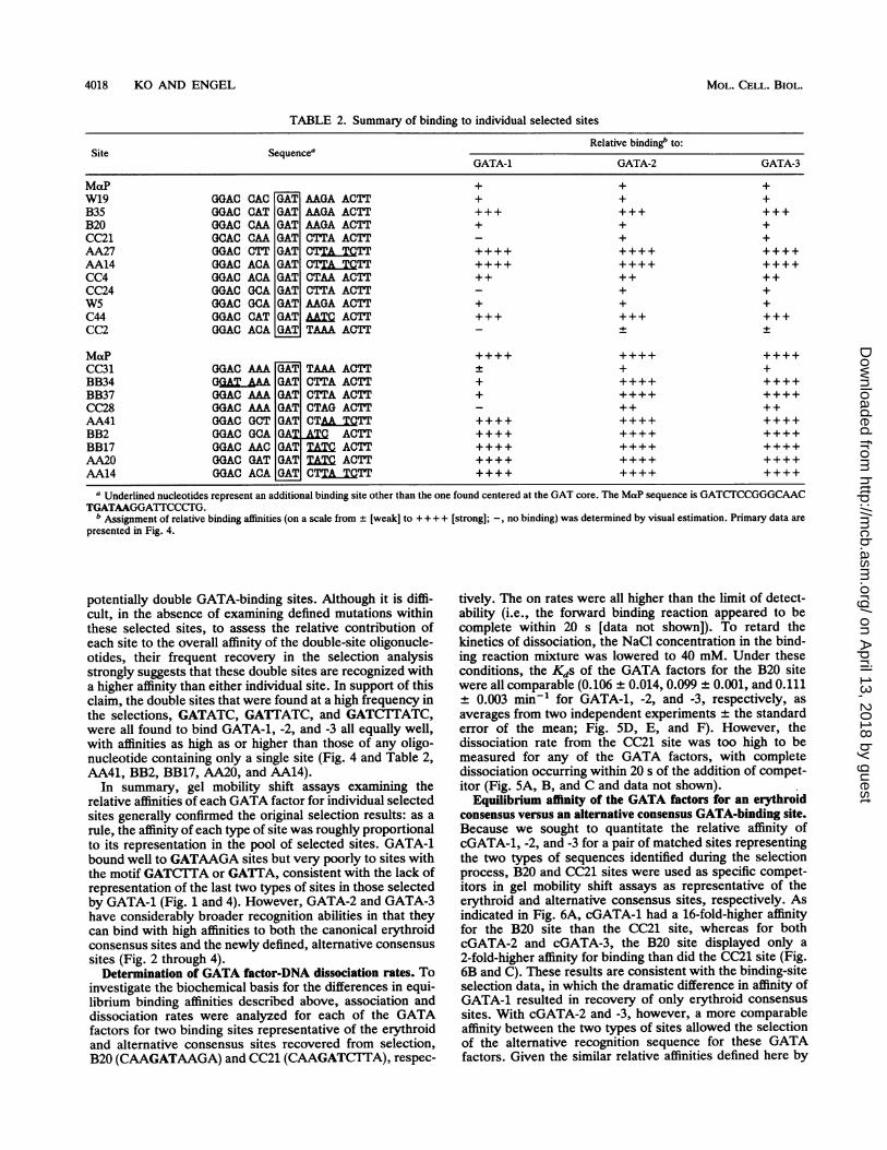
TABLE 2. Summary of binding to individual selected sites
Relative binding" to:Site Sequencea
GATA-1 GATA-2 GATA-3
MaP + + +W19 GGAC CAC GAT AAGA ACTT + + +B35 GGAC CAT GAT AAGA ACTT +++ +++ +++B20 GGAC CAA GAT AAGA ACTT + + +CC21 GCAC CAA GAT CTTA ACTT + +AA27 GGAC CTT GAT CTT C..ITT ++++ ++++ ++++AA14 GGAC ACA GAT CTTATCTT ++++ ++++ ++++CC4 GGAC ACA GAT CTAA ACTT ++ ++ ++CC24 GGAC GCA GAT CTTA ACTT + +W5 OGAC GCA GAT AAGA ACTT + + +C44 GGAC CAT GAT MTC ACTT +++ +++ +++CC2 GGAC ACA GAT TAAA ACTT -4+ +
MaP _ ++CC31 GGAC AAA GAT TAAA ACTT + + +BB34 G=_AA GAT CTTA ACTT + ++++ ++++BB37 GGAC AAA GAT CTTA ACTT + ++++ ++++CC28 GGAC AAA GAT CTAG ACTT ++ ++AA41 GGAC GCT GAT CTAAMTCTT ++++ ++++ ++++BB2 OGAC GCA GAT XC ACTT ++++ ++++ ++++BB17 GGAC AAC GAT TATC ACTT ++++ ++++ ++++AA20 GGAC GAT GAT TATC ACTT ++++ ++++ ++++AA14 GGAC ACA GAT CTTATCTT ++++ ++++ ++++
a Underlined nucleotides represent an additional binding site other than the one found centered at the GAT core. The MaP sequence is GATCTCCGGGCAACTGATAAGGATTCCCrG.
b Assignment of relative binding affinities (on a scale from + [weak] to + + + + [strong]; -, no binding) was determined by visual estimation. Primary data arepresented in Fig. 4.
potentially double GATA-binding sites. Although it is diffi-cult, in the absence of examining defined mutations withinthese selected sites, to assess the relative contribution ofeach site to the overall affinity of the double-site oligonucle-otides, their frequent recovery in the selection analysisstrongly suggests that these double sites are recognized witha higher affinity than either individual site. In support of thisclaim, the double sites that were found at a high frequency inthe selections, GATATC, GATTATC, and GATC1`TATC,were all found to bind GATA-1, -2, and -3 all equally well,with affinities as high as or higher than those of any oligo-nucleotide containing only a single site (Fig. 4 and Table 2,AA41, BB2, BB17, AA20, and AA14).
In summary, gel mobility shift assays examining therelative affinities of each GATA factor for individual selectedsites generally confirmed the original selection results: as arule, the affinity of each type of site was roughly proportionalto its representation in the pool of selected sites. GATA-1bound well to GATAAGA sites but very poorly to sites withthe motif GATC1TA or GATTA, consistent with the lack ofrepresentation of the last two types of sites in those selectedby GATA-1 (Fig. 1 and 4). However, GATA-2 and GATA-3have considerably broader recognition abilities in that theycan bind with high affinities to both the canonical erythroidconsensus sites and the newly defined, alternative consensussites (Fig. 2 through 4).
Determination of GATA factor-DNA dissociation rates. Toinvestigate the biochemical basis for the differences in equi-librium binding affinities described above, association anddissociation rates were analyzed for each of the GATAfactors for two binding sites representative of the erythroidand alternative consensus sites recovered from selection,B20 (CAAGATAAGA) and CC21 (CAAGATCTTA), respec-
tively. The on rates were all higher than the limit of detect-ability (i.e., the forward binding reaction appeared to becomplete within 20 s [data not shown]). To retard thekinetics of dissociation, the NaCl concentration in the bind-ing reaction mixture was lowered to 40 mM. Under theseconditions, the Kds of the GATA factors for the B20 sitewere all comparable (0.106 + 0.014, 0.099 ± 0.001, and 0.111± 0.003 min-' for GATA-1, -2, and -3, respectively, asaverages from two independent experiments ± the standarderror of the mean; Fig. 5D, E, and F). However, thedissociation rate from the CC21 site was too high to bemeasured for any of the GATA factors, with completedissociation occurring within 20 s of the addition of compet-itor (Fig. 5A, B, and C and data not shown).
Equilibrium affinity of the GATA factors for an erythroidconsensus versus an alternative consensus GATA-binding site.Because we sought to quantitate the relative affinity ofcGATA-1, -2, and -3 for a pair of matched sites representingthe two types of sequences identified during the selectionprocess, B20 and CC21 sites were used as specific compet-itors in gel mobility shift assays as representative of theerythroid and alternative consensus sites, respectively. Asindicated in Fig. 6A, cGATA-1 had a 16-fold-higher affinityfor the B20 site than the CC21 site, whereas for bothcGATA-2 and cGATA-3, the B20 site displayed only a2-fold-higher affinity for binding than did the CC21 site (Fig.6B and C). These results are consistent with the binding-siteselection data, in which the dramatic difference in affinity ofGATA-1 resulted in recovery of only erythroid consensussites. With cGATA-2 and -3, however, a more comparableaffinity between the two types of sites allowed the selectionof the alternative recognition sequence for these GATAfactors. Given the similar relative affinities defined here by
MOL. CELL. BIOL.
on April 13, 2018 by guest
http://mcb.asm
.org/D
ownloaded from

VOL.13,1993~~~~~GATAFACTOR FAMILY BINDING SITE SPECIFICITY 4019
A 820 CC21 B B20 CC210 1253 10 20 40Eq 0 0 2 5 10 20 40 Eq 0 1 2 5 IO 20Eq 0 12 510 20 Eq
~~-ksae~ b. z-flUwSS'm I
1 2 3 4 5 6 7 S 9 10 11 12 13 14 15 16 1 2 3 4 5 6 7 8 9 10 11 12 13 14
isD
-0.6 -
-0.6- -
r.4.9
kd= 0.106±0.014
E
S
V.
.2
F-1.2-
r.
-.4
0 2 4 60 10
1 2 0 2 4 6 810
12 0 2 4 6
010 1 2
time (roin) time (mi;.) those (moin)
FIG. 5. Dissociation rate constant determination for the B20 and CC21 sites. Four nanograms of probe was used in binding reactions withcGATA-1 (A), cGATA-2 (B), and cGATA-3 (C). First, the radiolabeled binding sites plus factor were allowed to come to binding equilibrium.A 100-fold excess of unlabeled MaP30 was then added to the reaction, and at the timnes indicated (in minutes) above each lane, aliquots of thereaction mixture were loaded onto a native polyacrylamide gel during electrophoresis. The migration of the free probe is indicated by thearrow, and markers 1, 2, and 3 show the GATA factor-DNA complexes of GATA-1, -2, and -3, respectively, with each probe. Lanes Eq, theendpoint of the competitions in binding reactions with competitor after each was allowed to reach equilibrium. (D), E, and F) Quantitativeevaluation of the results of GATA-1 (D), GATA-2 (E), and GATA-3 (F) dissociation from B20. The Kd shown is the average of twoindependent determinations ± the standard error of the mean. See Materials and Methods for details of the kinetic analysis.
GATA-2 and -3, why would cGATA-2 not select the alter-native (GATCT) sites as frequently as cGATA-3 (Table 1)?One possibility is that the analysis of only the B20 and CC21sites is too limited and that these results are not be easilyextrapolated to sites with differing identities at the -1 to -3
positions. Thus, GATA-2 could recognize the erythroidconsensus site at a much higher relative affinity comparedwith that of the alternative site if both binding sites containedresidues different from those within the two selected sitesexamined here.
B20 CC21
25 50 100 200 50 100 200 400 1600
B B20 CC21
25 50 100 200 400 50 100 200 400 800
C B20 CC21
25 50 100 200 400 50 100 200 400 800
4i' .& 2- N40# 3- *
~~t 1",~~~
1 2 3 4 5 6 7 8 9 10 1 2 3 4 5 6 7 8 9 10 1 2 3 4 5 6 7 8 9 10
FIG. 6. Relative affinities of cGATA-1, -2, and -3 for sites B20 and CC21. Binding reactions were performed with GATA-1 (A), -2 (B), and
-3 (C); the MaLP3 probe; and increasing amounts (indicated in nanograms above each lane) of either B20 or CC21 binding-site competitor. The
migration of the free MocP3 probe is indicated by the arrow, and markers 1, 2, and 3 show the GATA factor-DNA complexes of GATA-1,
-2, and -3, respectively.
C B20 CC210 1 2 5 10 20 Eq 0 1 2 5 10 20 Eq
A
VOL. 13, 1993
on April 13, 2018 by guest
http://mcb.asm
.org/D
ownloaded from

4020 KO AND ENGEL
DISCUSSION
Utilizing a gel mobility shift selection and PCR amplifica-tion strategy with randomized oligonucleotides and bacteri-ally expressed proteins, we have determined the preferredDNA-binding sites for the chicken GATA-1, -2, and -3proteins. While all three factors bind to double sites (espe-cially in particular arrangements of inverted orientations)with the highest affinity, determination of the relative affinityfor preferred single sites was also possible. All three factorsselected an erythroid consensus NNAGATAANN bindingsite, in accord with the canonical recognition site of WGA-TAR (13). Surprisingly, there is no selection for, and morelikely is a selection against, a G at the +2 position as wouldhave been predicted by the previously defined consensus(WGATAR). We have identified sequences that can discrim-inate between GATA-1 and GATA-2 or -3 binding; se-quences containing an AGATCTTA motif are bound byGATA-2 and -3 with high affinity, while GATA-1 binding isvery weak. Although the positions bordering the GAT corewere most critical to determining the ability of a site to berecognized, the positions beyond these central nucleotidesshowed preferences for specific nucleotides as well.The results presented here are consistent with those of
Plumb et al. (29) in which various synthetic oligonucleotideswith mutations in the mouse a-globin promoter GATA site,MaP, were assessed for their ability to compete for bindingwith a wild-type MaP site. Mutation of the two adenines atpositions +1 and +2 were shown to dramatically reducecompetition of binding (20- and 4-fold, respectively), but the+3 site could be altered with no difference in ability tocompete with the wild type, consistent with the presence ofan N at this position in the consensus derived here. Further-more, competition with an MacP site mutated to a GATAGand the GATAG site from the chicken p3H-globin promoterwas shown to compete four- to sixfold less well than thewild-type GATAA MaP site.
Previously identified GATAG sites have been found in thechicken a-globin enhancer, the chicken pH-globin promoter,the human 3-globin enhancer and A-y-globin promoter, andthe chicken P-globin enhancer (12, 13, 20, 24, 29, 33, 46); thelast two sites have also been shown to be required for fullerythroid transcriptional activity of these elements (23, 32).If GATAG is a relatively low-affinity binding site, why is itfound in functionally identified GATA factor-regulated cis-regulatory elements? One possible explanation is that thereexist other cofactors which allow GATA factor binding tothese sites in vivo that are not present in the bacteriallyexpressed purified proteins used here. Alternatively, thesesites may have evolved so that the site may be more easilyregulated, for example, by concentration changes in GATAfactors which have been shown to occur during erythroidcell differentiation and development (22, 47) and as havebeen proposed in the mechanism for regulation of thechicken p-globin gene (26). The observation that nature hasevolved less than optimal pairings of transcription factorsand their cognate sites has been made for other transcriptionfactors (e.g., for CACC and c-myc [14, 16]).
In addition to showing that all three factor family membershave similar preferences for an erythroid consensus GATA-binding site, we have also defined new recognition consen-sus sequences for GATA-2 and GATA-3. The sequenceAGATCTTA is a high-affinity site while AGATTA is arelatively low-affinity site for both GATA-2 and GATA-3;GATA-1 does not bind to either type of site well. Althoughthe precise nucleotide identities of positions -1, + 1, and +2
were the most critical determinants of recognition specificityand affinity, specific changes of nucleotides at the otherpositions could modulate binding by all three proteins, andall to similar degrees (Fig. 4). In vivo footprinting data of anmGATA-1 promoter consensus site revealed strong protec-tion of the G of the GAT core and weaker, but clearlydetectable, protection of the G at position +3 (43). That thenucleotides several positions away from the central GAT canaffect GATA factor function through that site is evidencedby recent studies of the cGATA-1 promoter region. Adeletion of the nucleotide at the -3 position of the GATAsite at -139 of the promoter results in an increased dissoci-ation rate of DNA-protein complex formation and a 30%diminution of promoter activity (36).
While it remains a formal possibility that there are GATAfactor-binding sites totally unrelated to any identified here(i.e., without a GAT sequence), these would be predicted tobe generally of lower affinity than the GAT-containing sites.In the majority (11 of 15) of selected oligonucleotides inwhich the GAT core was disrupted by mutation generated asa PCR artifact, a GAT sequence was found elsewhere (seeResults), strongly suggesting that the highest-affinity bindingsites for all the GATA factors include this GAT motif.The similar binding specificities of cGATA-2 and -3 dis-
tinct from that of their highly related family member,cGATA-1, was not unanticipated; in the 107 amino acids ofhomology in the fingers and adjacent basic region (which isrequired for cGATA-1 DNA binding [49]) of all three factors,there are only three residues which differ between cGATA-2and -3, but there are 18 differences in the cGATA-1 sequencecompared with the cGATA-2 sequence (11, 48). In ananalysis of the function of the two zinc fingers in mouse andchicken GATA-1, the C-terminal finger has been identified ascontaining the critical DNA-binding function, while theN-terminal finger plays a role in stabilizing protein-DNAcomplex formation (23, 49). Five of the amino acid differ-ences between cGATA-1 and cGATA-2 are found in theC-terminal finger, and notably, one of the differences isfound between the first pair of cysteines in the C-terminalfinger (at position 165 in cGATA-1 [11]). Significantly, asingle amino acid change in this "knuckle" region betweenthe conserved cysteines in the steroid hormone receptorshas been shown to be sufficient to alter DNA-binding spec-ificity (7, 44).The highest-affinity sites defined in this study were double
sites in inverted orientation, to which all three factorsstrongly bind as monomeric proteins. Although the gelmobility shift assays with individual sites did not reveal anydifferences between the ability of the three factors to recog-nize the three types of inverted binding sites, the fact thatcertain types of double sites appeared to be selected by eachGATA factor implies that there may be some preference fora defined spacing between overlapping inverted sites. Forexample, GATA-1 more frequently selected GATC1`TATC(Fig. 1B), GATA-2 reproducibly selected GATATC (Fig. 2Aand B), and GATA-3 often selected GATIATC (Fig. 3A).The existence of multiple GATA factors in the erythroid
cell environment (22, 48) poses an interesting question as tothe functional roles of cGATA-1, -2, and -3. The necessity ofmouse GATA-1 expression for development of the erythroidlineage has been previously demonstrated (28, 37), andGATA-1 in chickens is presumed to be analogously required.It is interesting to note that cGATA-3 expression seems tocorrelate with later stages of differentiation in erythrocytesand nervous system tissues (1Sa, 22, 48). Given the broaderrecognition ability of GATA-3, it is conceivable that upon its
MOL. CELL. BIOL.
on April 13, 2018 by guest
http://mcb.asm
.org/D
ownloaded from

GATA FACTOR FAMILY BINDING SITE SPECIFICITY 4021
induction during late erythroid differentiation, GATA-3could replace GATA-1 at some erythroid consensus bindingsites, maintaining GATA factor function at those targetgenes but additionally recognizing another battery of alter-native sites which regulate genes expressed later in differen-tiation. The identification in this study of a high-affinity,noncanonical binding site for GATA-2 and GATA-3, AGATCTTA, may facilitate the identification of downstream targetgenes for these factors in erythroid as well as nonerythroidcells (3).The determination of which GATA factor acts at a given
consensus site appears to be dictated by some aspect offactor function beyond simple differences in site recognitionproperties. The octamer binding proteins Oct-1 and Oct-2,which also have identical binding specificities, have beenproposed to display promoter selectivity by interactions oftheir activation domains with other proteins at the transcrip-tional initiation complexes (40). A similar model, in whichspecificity is localized to regions outside the DNA-bindingdomain, has also been explored for the differential activationabilities of MRF4 and myogenin on the muscle creatininekinase enhancer (5). A mechanism imparting differentialspecificity may also be operative in this case as well;experiments are currently under way to test this hypothesis.
ACKNOWLEDGMENTS
We thank Brett Andres for indispensable technical assistance;Mark Leonard, Zhuoying Yang, and Jon Widom for technical adviceand valuable discussions; and Katie George, Kevin Foley, and MattRoth for critical reading of the manuscript. We also thank ChristinaKo for the contribution of reagents critical to the successful com-pletion of this project.
This work was supported by an NIH NRSA training grant awardto Northwestern University (L.J.K.; GM 08061) and an NIH re-search grant (GM 28896).
REFERENCES1. Blackwell, T. K., L. Kretzner, E. M. Blackwood, R. N. Eisen-
man, and H. Weintraub. 1990. Sequence-specific DNA bindingby the c-Myc protein. Science 250:1149-1151.
2. Blackwell, T. K., and H. Weintraub. 1990. Differences andsimilarities in DNA-binding preferences of MyoD and E2Aprotein complexes revealed by binding site selection. Science250:1104-1110.
3. Briegel, K., K.-C. Lrm, C. Plank, H. Beug, J. D. Engel, and M.Zenke. Ectopic expression of a conditional GATA-2/estrogenreceptor chimera arrests erythroblast differentiation in a hor-mone-dependent manner. Genes Dev., in press.
4. Catala, F., E. deBoer, G. Habets, and F. Grosveld. 1989. Nuclearprotein factors and erythroid transcription of the human A_globin gene. Nucleic Acids Res. 17:3811-3826.
5. Chakraborty, T., and E. N. Olson. 1991. Domains outside of theDNA-binding domain impart target gene specificity to myogeninand MRF4. Mol. Cell. Biol. 11:6103-6108.
6. Choi, O.-R., and J. D. Engel. 1986. A 3' enhancer is required fortemporal and tissue-specific transcriptional activation of thechicken adult 3-globin gene. Nature (London) 323:731-734.
7. Danielsen, M., L. Hinck, and G. M. Ringold. 1989. Two aminoacids within the knuckle of the first zinc finger specify DNAresponse element activation by the glucocorticoid receptor. Cell57:1131-1138.
8. Dessain, S., C. T. Gross, M. A. Kuziora, and W. McGinnis. 1992.Antp-type homeodomains have distinct DNA binding specifici-ties that correlate with their different regulatory functions inembryos. EMBO J. 11:991-1002.
9. Ekker, S. C., D. P. von Kessler, and P. A. Beachy. 1992.Differential DNA sequence recognition is a determinant ofspecificity in homeotic gene action. EMBO J. 11:4059-4072.
10. Emerson, B. M., J. M. Nickol, P. D. Jackson, and G. Felsenfeld.
1987. Analysis of the tissue-specific enhancer at the 3' end of thechicken adult j-globin gene. Proc. Natl. Acad. Sci. USA84:4786-4790.
11. Evans, T., and G. Felsenfeld. 1989. The erythroid-specific tran-scription factor Eryfl: a new finger protein. Cell 58:877-885.
12. Evans, T., and G. Felsenfeld. 1991. trans-activation of a globinpromoter in nonerythroid cells. Mol. Cell. Biol. 11:843-853.
13. Evans, T., M. Reitman, and G. Felsenfeld. 1988. An erythrocyte-specific DNA-binding factor recognizes a regulatory sequencecommon to all chicken globin genes. Proc. Natl. Acad. Sci.USA 85:5976-5980.
14. Fisher, D. E., L. A. Parent, and P. A. Sharp. 1993. High affinityDNA-binding Myc analogs: recognition by an a helix. Cell72:467-476.
15. Gallarda, J. L., K. P. Foley, Z. Yang, and J. D. Engel. 1989. The,B-globin stage selector element factor is erythroid-specific pro-moter/enhancer binding protein NF-E4. Genes Dev. 3:1845-1859.
15a.George, K., J. Kornhauser, and J. D. Engel. Unpublishedobservations.
16. Hartzog, G. A., and R. M. Myers. 1993. Discrimination amongpotential activators of the ,3-globin CACCC element by corre-lation of binding and transcriptional properties. Mol. Cell. Biol.13:44-56.
17. Ho, I.-C., L.-H. Yang, G. Morle, and J. M. Leiden. 1989. AT-cell-specific transcriptional enhancer element 3' of Ca in thehuman T-cell receptor a locus. Proc. Natl. Acad. Sci. USA86:6714-6718.
18. Ito, E., T. Toki, H. Ishihara, H. Ohtani, L. Gu, M. Yokoyama,J. D. Engel, and M. Yamamoto. 1993. Erythroid transcriptionfactor GATA-1 is abundantly transcribed in mouse testis. Na-ture (London) 362:466-469.
19. Joulin, V., D. Bories, J.-F. Eleouet, M.-C. Labastie, S. Chretien,M.-G. Mattei, and P.-H. Romeo. 1991. A T-cell specific TCR 8DNA binding protein is a member of the human GATA family.EMBO J. 10:1809-1816.
20. Knezetic, J. A., and G. Felsenfeld. 1989. Identification andcharacterization of a chicken a-globin enhancer. Mol. Cell.Biol. 9:893-901.
21. Ko, L. J., M. Yamamoto, M. W. Leonard, K. M. George, P.Ting, and J. D. Engel. 1991. Murine and human T-lymphocyteGATA-3 factors mediate transcription through a cis-regulatoryelement within the human T-cell receptor 8 gene enhancer. Mol.Cell. Biol. 11:2778-2784.
21a.Kornhauser, J., K. George, and J. D. Engel. Unpublishedobservations.
22. Leonard, M. W., K.-C. Lim, and J. D. Engel. Expression of theGATA transcription factor family during early erythroid devel-opment and differentiation. Submitted for publication.
23. Martin, D. I. K., and S. H. Orkin. 1990. Transcriptionalactivation and DNA-binding by the erythroid factor GF-1/NF-El/Eryfl. Genes Dev. 4:1886-1898.
24. Martin, D. I. K., S.-F. Tsai, and S. H. Orkin. 1989. Increased-y-globin expression in a nondeletion HPFH mediated by anerythroid-specific DNA-binding factor. Nature (London) 338:435-438.
25. Martin, D. I. K., L. I. Zon, G. Mutter, and S. H. Orkin. 1990.Expression of an erythroid transcription factor in megakaryo-cytic and mast cell lineages. Nature (London) 344:444 447.
26. Minie, M., T. Kimura, and G. Felsenfeld. 1992. The develop-mental switch in embryonic p-globin expression is correlatedwith erythroid lineage-specific differences in transcription factorlevels. Development 115:1149-1164.
27. Naar, A. M., J.-M. Boutin, S. M. Lipkin, V. C. Yu, J. M.Holloway, C. K. Glass, and M. G. Rosenfeld. 1991. The orien-tation and spacing of core DNA-binding motifs dictate selectivetranscriptional responses to three nuclear receptors. Cell 85:1267-1279.
28. Pevny, L., M. C. Simon, E. Robertson, W. H. Klein, S.-F. Tsai,V. D'Agati, S. H. Orkin, and F. Costantini. 1991. Erythroiddifferentiation in chimaeric mice blocked by a targeted mutationin the gene for transcription factor GATA-1. Nature (London)349:257-260.
VOL. 13, 1993
on April 13, 2018 by guest
http://mcb.asm
.org/D
ownloaded from

4022 KO AND ENGEL
29. Plumb, M., J. Frampton, H. Wainwright, M. Walker, K. Ma-cleod, G. Goodwin, and P. Harrison. 1989. GATAAG; a cis-control region binding an erythroid-specific nuclear factor witha role in globin and non-globin gene expression. Nucleic AcidsRes. 17:73-92.
30. Redondo, J. M., S. Hata, C. Brocklehurst, and M. S. Krangel.1990. A T cell-specific transcriptional enhancer within thehuman T cell receptor 8 locus. Science 247:1225-1229.
31. Redondo, J. M., J. L. Pfohl, and M. S. Krangel. 1991. Identifi-cation of an essential site for transcriptional activation withinthe human T-cell receptor 8 enhancer. Mol. Cell. Biol. 11:5671-5680.
32. Reitman, M., and G. Felsenfeld. 1988. Mutational analysis of thechicken 3-globin enhancer reveals two positive-acting domains.Proc. Natl. Acad. Sci. USA 85:6267-6271.
33. Reitman, M., E. Lee, H. Westphal, and G. Felsenfeld. 1990.Site-independent expression of the chicken 03A-globin gene intransgenic mice. Nature (London) 348:749-752.
34. Romeo, P.-H., M.-H. Prandini, V. Joulin, V. Mignotte, M.Prenant, W. Vainchenker, G. Marguerie, and G. Uzan. 1990.Megakaryocytic and erythrocytic lineages share specific tran-scription factors. Nature (London) 344:447-449.
35. Sanger, F., S. Nicklen, and A. R. Coulson. 1977. DNA sequenc-ing with chain-terminating inhibitors. Proc. Natl. Acad. Sci.USA 74:5463-5467.
36. Schwartzbauer, G., K. Schlesinger, and T. Evans. 1992. Interac-tion of the erythroid transcription factor cGATA-1 with acritical autoregulatory element. Nucleic Acids Res. 20:4429-4436.
37. Simon, M. C., L. Pevny, M. V. Wiles, G. Keller, F. Costantini,and S. H. Orkin. 1992. Rescue of erythroid development in genetargeted GATA-1-mouse embryonic stem cells. Nat. Genet.1:92-98.
38. Smith, D. B., and K. S. Johnson. 1988. Single-step purification ofpolypeptides expressed in Escherichia coli as fusions withglutathionine S-transferase. Gene 67:31-40.
39. Studier, F. W., A. H. Rosenberg, J. J. Dunn, and J. W.Dubendorf. 1990. Use of T7 RNA polymerase to direct expres-sion of cloned genes. Methods Enzymol. 185:60-89.
40. Tanaka, M., J.-S. Lai, and W. Herr. 1992. Promoter-selectiveactivation domains in Oct-1 and Oct-2 direct differential activa-tion of an snRNA and mRNA promoter. Cell 68:755-767.
41. Trainor, C. D., S. J. Stamler, and J. D. Engel. 1987. Erythroid-specific transcription of the chicken histone H5 gene is directedby a 3' enhancer. Nature (London) 328:827-830.
42. Tsai, S.-F., D. I. K. Martin, L. I. Zon, A. D. D'Andrea, G. G.Wong, and S. H. Orkin. 1989. Cloning of cDNA for the majorDNA-binding protein of the erythroid lineage through expres-sion in mammalian cells. Nature (London) 339:446-451.
43. Tsai, S.-F., E. Strauss, and S. H. Orkin. 1991. Functionalanalysis and in vivo footprinting implicate the erythroid tran-scription factor GATA-1 as a positive regulator of its ownpromoter. Genes Dev. 5:919-931.
44. Umesono, K., and R. M. Evans. 1989. Determinants of targetgene specificity for steroid/thyroid hormone receptors. Cell57:1139-1146.
45. Umesono, K., K. K. Murakami, C. C. Thompson, and R. M.Evans. 1991. Direct repeats as selective response elements forthe thyroid hormone, retinoic acid, and vitamin D3 receptors.Cell 65:1255-1266.
46. Wall, L., E. deBoer, and F. Grosveld. 1988. The human [B-globingene 3' enhancer contains multiple binding sites for an eryth-roid-specific protein. Genes Dev. 2:1089-1100.
47. Whitelaw, E., S.-F. Tsai, P. Hogben, and S. H. Orkin. 1990.Regulated expression of globin chains and the erythroid tran-scription factor GATA-1 during erythropoiesis in the developingmouse. Mol. Cell. Biol. 10:6596-6606.
48. Yamamoto, M., L. J. Ko, M. W. Leonard, H. Beug, S. H. Orkin,and J. D. Engel. 1990. Activity and tissue-specific expression ofthe transcription factor NF-E1 multigene family. Genes Dev.4:1650-1662.
49. Yang, H.-Y., and T. Evans. 1992. Distinct roles for the twocGATA-1 finger domains. Mol. Cell. Biol. 12:4562-4570.
50. Yang, Z., M. W. Leonard, L. J. Ko, K. M. George, M.Yamamoto, and J. D. Engel. 1991. Transcription factors impli-cated in ,-globin gene switching, p. 249-265. In G. Stamatoyan-nopoulos and A. W. Nienhuis (ed.), The regulation of hemoglo-bin switching. Johns Hopkins University Press, Baltimore.
MOL. CELL. BIOL.
on April 13, 2018 by guest
http://mcb.asm
.org/D
ownloaded from