dna biosensors and microarrays - school of engineering · pdf filedna biosensors and...
TRANSCRIPT
DNA Biosensors and Microarrays
Audrey Sassolas, Beatrice D. Leca-Bouvier, and Loıc J. Blum*
Laboratoire de Genie Enzymatique et Biomoleculaire, Institut de Chimie et Biochimie Moleculaires et Supramoleculaires, 43 Boulevard du 11Novembre 1918, Villeurbanne F-69622, France, UMR5246, Centre National de La Recherche Scientifque, Villeurbanne F-69622, France,Universite de Lyon, Lyon F-69622, France, Universite Lyon 1, Lyon F-69622, France, Institut National des Sciences Appliquees de Lyon,
EÄ cole d’Ingenieurs, Villeurbanne F-69621, France, and EÄ cole Superieure Chimie Physique EÄ lectronique de Lyon, Villeurbanne F-69616, France
Received June 21, 2007
Contents1. Introduction 1092. DNA Immobilization 111
2.1. Immobilization Techniques Used To DevelopDNA Biosensors and Microarrays
111
2.1.1. Adsorption 1112.1.2. Covalent Immobilization 1122.1.3. Avidin (or Streptavidin)−Biotin Interactions 113
2.2. DNA Microarrays 1132.2.1. In Situ Synthesis of DNA Microarrays 1132.2.2. Spotted Microarrays 1142.2.3. Nanogen Technology 114
2.3. Immobilization Technique Specific to DNABiosensors: Entrapment
114
3. Nucleic Acid Hybridization Detection 1153.1. Optical DNA Biosensors and DNA
Microarrays115
3.1.1. Fluorescence Detection 1153.1.2. Surface-Enhanced Raman Scattering
(SERS) Spectroscopy120
3.1.3. Chemiluminescent Detection 1213.1.4. Colorimetric Detection 1223.1.5. Dual Polarization Interferometry 1223.1.6. Surface-Plasmon-Based Detection 122
3.2. Electrochemical DNA Biosensors andMicroarrays
123
3.2.1. Enzyme Label 1233.2.2. Ferrocene 1253.2.3. Interacting Electroactive Substances 1263.2.4. Metal Nanoparticles for DNA Labeling 1283.2.5. Label-Free Electrochemical Detection 129
3.3. Gravimetric DNA Biosensors 1313.3.1. Quartz Crystal Microbalance Sensors 1313.3.2. Microcantilever Sensors 132
4. Conclusion 1325. List of Abbreviations 1346. References 135
1. IntroductionDevelopment of DNA biosensors and DNA microarrays
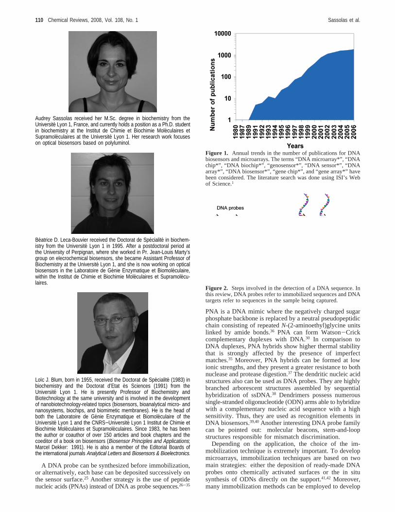
has increased tremendously over the past few years asdemonstrated by the large number of scientific publicationsin this area (Figure 1).
Recent progress in the development of DNA biosensorsand microarrays is summarized in this review. It is importantto point out that reviews dealing with DNA biosensorsmainly focus on electrochemical transduction.2-10 Very fewpapers have been written describing DNA biosensors includ-ing not only electrochemical but also optical and piezoelectrictransduction modes.11 Moreover, the recent evolution of DNAmicroarrays is also studied in this work. DNA probe immobi-lization and hybridization detection are largely reported.
In recent years, the interest for DNA-based diagnostic testshas been growing. The development of systems allowingDNA detection is motivated by applications in many fields:DNA diagnostics, gene analysis, fast detection of biologicalwarfare agents, and forensic applications. Detection ofgenetic mutations at the molecular level opens up thepossibility of performing reliable diagnostics even before anysymptom of a disease appears.
Numerous DNA detection systems based on the hybridiza-tion between a DNA target and its complementary probe,which is present either in solution or on a solid support, havebeen described. Homogeneous assays allowing the determi-nation of DNA sequences have been developed. Thesesystems can be based on optical12-15 or electrochemical16,17
detection. However, they do not allow easily continuousmonitoring and miniaturization. DNA biosensors and DNAmicroarrays offer promising alternatives to these methods.They allow continuous, fast, sensitive, and selective detectionof DNA hybridization, and they also can be reused.
DNA biosensors (also called genosensors) and DNAmicroarrays (commonly called gene chips, DNA chips, orbiochips) exploit the preferential binding of complementarysingle-stranded nucleic acid sequences. This system usuallyrelies on the immobilization of a single-stranded DNA(ssDNA) probe onto a surface to recognize its complementaryDNA target sequence by hybridization (Figure 2). Trans-duction of hybridization of DNA can be measured optically,electrochemically, or using mass-sensitive devices.
In the case of DNA biosensors and contrary to DNAmicroarrays, the immobilization of a DNA probe is achieveddirectly onto a transducer surface. DNA microarrays aremade from glass, plastic, or silicon supports and areconstituted of tens to thousands of 10-100µm reaction zonesonto which individual oligonucleotide sequences have beenimmobilized.18,19 The exact number of DNA probes variesin accordance with the application. Contrary to DNAbiosensors that allow single-shot measurements, DNA mi-croarrays allow multiple parallel detection and analysis ofthe patterns of expression of thousands of genes in a singleexperiment.20-24
* Author to whom correspondence should be addressed. Telephone:+33472 43 13 97. Fax:+33 472 44 79 70. E-mail: [email protected].
109Chem. Rev. 2008, 108, 109−139
10.1021/cr0684467 CCC: $71.00 © 2008 American Chemical SocietyPublished on Web 12/21/2007
A DNA probe can be synthesized before immobilization,or alternatively, each base can be deposited successively onthe sensor surface.25 Another strategy is the use of peptidenucleic acids (PNAs) instead of DNA as probe sequences.26-35
PNA is a DNA mimic where the negatively charged sugarphosphate backbone is replaced by a neutral pseudopeptidicchain consisting of repeatedN-(2-aminoethyl)glycine unitslinked by amide bonds.36 PNA can form Watson-Crickcomplementary duplexes with DNA.30 In comparison toDNA duplexes, PNA hybrids show higher thermal stabilitythat is strongly affected by the presence of imperfectmatches.35 Moreover, PNA hybrids can be formed at lowionic strengths, and they present a greater resistance to bothnuclease and protease digestion.37 The dendritic nucleic acidstructures also can be used as DNA probes. They are highlybranched arborescent structures assembled by sequentialhybridization of ssDNA.38 Dendrimers possess numeroussingle-stranded oligonucleotide (ODN) arms able to hybridizewith a complementary nucleic acid sequence with a highsensitivity. Thus, they are used as recognition elements inDNA biosensors.39,40Another interesting DNA probe familycan be pointed out: molecular beacons, stem-and-loopstructures responsible for mismatch discrimination.
Depending on the application, the choice of the im-mobilization technique is extremely important. To developmicroarrays, immobilization techniques are based on twomain strategies: either the deposition of ready-made DNAprobes onto chemically activated surfaces or the in situsynthesis of ODNs directly on the support.41,42 Moreover,many immobilization methods can be employed to develop
Audrey Sassolas received her M.Sc. degree in biochemistry from theUniversite Lyon 1, France, and currently holds a position as a Ph.D. studentin biochemistry at the Institut de Chimie et Biochimie Moleculaires etSupramoleculaires at the Universite Lyon 1. Her research work focuseson optical biosensors based on polyluminol.
Beatrice D. Leca-Bouvier received the Doctorat de Specialite in biochem-istry from the Universite Lyon 1 in 1995. After a postdoctoral period atthe University of Perpignan, where she worked in Pr. Jean-Louis Marty’sgroup on elecrochemical biosensors, she became Assistant Professor ofBiochemistry at the Universite Lyon 1, and she is now working on opticalbiosensors in the Laboratoire de Genie Enzymatique et Biomoleculaire,within the Institut de Chimie et Biochimie Moleculaires et Supramolecu-laires.
Loıc J. Blum, born in 1955, received the Doctorat de Specialite (1983) inbiochemistry and the Doctorat d’Etat es Sciences (1991) from theUniversite Lyon 1. He is presently Professor of Biochemistry andBiotechnology at the same university and is involved in the developmentof nanobiotechnology-related topics (biosensors, bioanalytical micro- andnanosystems, biochips, and biomimetic membranes). He is the head ofboth the Laboratoire de Genie Enzymatique et Biomoleculaire of theUniversite Lyon 1 and the CNRS−Universite Lyon 1 Institut de Chimie etBiochimie Moleculaires et Supramoleculaires. Since 1983, he has beenthe author or coauthor of over 150 articles and book chapters and thecoeditor of a book on biosensors (Biosensor Principles and Applications;Marcel Dekker: 1991). He is also a member of the Editorial Boards ofthe international journals Analytical Letters and Biosensors & Bioelectronics.
Figure 1. Annual trends in the number of publications for DNAbiosensors and microarrays. The terms “DNA microarray*”, “DNAchip*”, “DNA biochip*”, “genosensor*”, “DNA sensor*”, “DNAarray*”, “DNA biosensor*”, “gene chip*”, and “gene array*” havebeen considered. The literature search was done using ISI’s Webof Science.1
Figure 2. Steps involved in the detection of a DNA sequence. Inthis review, DNA probes refer to immobilized sequences and DNAtargets refer to sequences in the sample being captured.
110 Chemical Reviews, 2008, Vol. 108, No. 1 Sassolas et al.
both DNA microarrays and DNA biosensors: adsorption,covalent immobilization, and (strept-)avidin-biotin interac-tion.43,44
The traditional method of labeling is radioactivity.45,46
Although this method is one of the most sensitive, the useof radioisotopes, such as32P or 125I, presents seriousdisadvantages. However, optical and electrochemical tech-niques have been developed based on various labels. Forexample, fluorescent dyes, and more recently quantum dots(QDs), are largely used for optical detection.47 Electrochem-istry has received considerable attention recently2-5,48-53 forthe detection of DNA hybridization. These systems offersome advantages, such as low cost, simple design, or smalldimensions. The label can be an enzyme, an electroactiveindicator, such as ferrocene (Fc), cationic metal complexesor intercalating organic compounds (e.g., methylene blue),or a nanoparticle. A direct labeling strategy can be usedwhere the immobilized DNA probe hybridizes with thelabeled DNA target (Figure 3a). However, a sandwich-typeternary complex can be formed. The immobilized DNAprobe hybridizes to a part of the target whereas the otherpart of the target is complementary to a signaling DNAsequence that serves to label the target upon hybridization(Figure 3b). In a few cases, a competitive system can beused with a competition between the target and a labeledsequence both complementary to a DNA probe.54
Label-free electrochemical detection of hybridizationrepresents an attractive alternative approach for detectingDNA sequences. In this case, the detection is based onmodifications of properties, such as capacitance, or anintrinsic electrochemical response due to DNA (e.g., oxida-tion of guanines). Gravimetric DNA biosensors are able todetect label-free ODNs.
It can be highlighted that regeneration of the surface-immobilized probe is possible, allowing the reuse of the DNAbiosensor or the microarray without a significant loss ofhybridization activity. For that, a thermal or chemicalregeneration step is necessary. Biotinylated ODN probes havebeen immobilized on the core surface of a multimode optical
fiber.55 After the hybridization reaction, the two types ofregeneration have been tested. For thermal separation ofprobe/target duplexes, the surface has been washed withhybridization buffer at 70°C for 2 min whereas the chemicalregeneration has been done by pumping a 4 Murea solutionthrough the flow cell for 2 min. Regeneration of an opticalbiosensor system has also been achieved based on fluores-cence excitation and detection in the evanescent field of aquartz fiber by thermal and chemical treatment.56 In this case,the sensor surface has been heated to a temperature of 68.5°C or treated by 50% (w/w) aqueous urea solution. Regen-eration has also been achieved by a flash of 50 mM NaOH.57
More than 60 hybridization-regeneration cycles have beenperformed with a 10% loss of reproducibility. In the sameway, strand regeneration of the DNA hybrid has beendescribed by the addition of 10 mM NaOH for 1 min.58
Microarray surfaces also have been regenerated by a treat-ment with 50 mM NaOH/0.1% sodium dodecyl sulfate(SDS).59 HCl also has been reported at 160 or 10 mM.61
Two aspects are essential when developing hybridizationbiosensors and microarrays: the sensitivity and the selectiv-ity. It is important to be able to detect low DNA concentra-tions and to detect a point mutation. Thus, two types ofsystems can be developed: systems for DNA hybridizationand systems for detection of DNA damage.62 A perfect matchin the target sequence produces very stable double-strandedDNA (dsDNA), whereas one or more base mismatchesdecreases the stability, causing a signal modification.
2. DNA ImmobilizationDNA probes are short ODNs (12-40-mer) able to hybrid-
ize with specific target sequences.The immobilization step for the DNA probe is essential
to develop a whole range of biosensors and microarrays. Theachievement of high sensitivity and selectivity requiresminimization of nonspecific adsorption and stability ofimmobilized biomolecules. The control of this step isessential to ensure high reactivity, orientation, accessibility,and stability of the surface-confined probe and to avoidnonspecific binding.
2.1. Immobilization Techniques Used To DevelopDNA Biosensors and Microarrays
DNA can be immobilized on sensor surfaces with methodssimilar to those used for enzyme-based biosensors: adsorp-tion, covalent immobilization, and avidin (or streptavidin)-biotin interaction.63 These immobilization techniques also canbe used to develop DNA microarrays.41
2.1.1. Adsorption
Adsorption is the simplest immobilization method becauseit does not require any nucleic acid modification.
Immobilization has been reported based on ionic interac-tions occurring between the negatively charged groupspresent on the DNA probe and positive charges coveringthe surface. For instance, a chitosan film was used for theimmobilization of ssDNA on a glassy carbon electrode(GCE).64,65 Chitosan is a cationic polymer that can form astable complex with the negatively charged phosphate groupsof the DNA. A DNA-based diagnostic quartz crystal mi-crobalance (QCM) sensor was also developed.66 The sensinglayer was prepared according to different methods, amongwhich was adsorption by electrostatic interactions. DNA
Figure 3. Main types of labeled DNA biosensors and micro-arrays: (a) direct label between immobilized DNA probe andlabeled DNA target; (b) sandwich-type system. A sandwich-typeternary complex is formed between immobilized DNA probe, target,and signaling DNA probe
DNA Biosensors and Microarrays Chemical Reviews, 2008, Vol. 108, No. 1 111
probes were adsorbed on the outer layer of poly(allylamine)-hydrochloride (PAAH)/sodium poly(styrenesulfonate) (PSS)/PAAH film. The coupling between the negatively chargedphosphate backbone of the DNA probe and a positivelycharged film surface also allowed the development of DNAmicroarrays.67 For example, short ODN probes were im-mobilized to a positively charged amino-silanized glasssurface.68,69
DNA also can be linked by physical adsorption. Forexample, a screen-printed electrode (SPE) was immersedovernight in a DNA-containing solution before rinsing it toremove unadsorbed DNA.70 The electrochemical DNAbiosensor can detect 6× 10-16 M of target. The DNA probewas adsorbed on a polished basal plane pyrolytic graphite(BPPG) electrode.71 DNA was also immobilized on goldmicroelectrodes,72 which were modified by dropping a smallvolume of DNA on their sensor surface before an overnightair-drying. More recently, the probe was immobilized ontopreoxidized GCE by physical adsorption.73
ssDNA is often immobilized by applying a potential toan electrode. The electrode surface is sometimes electro-chemically pretreated to increase its hydrophobicity and itsroughness. Then, the controlled-potential adsorption of thessDNA is achieved.74-78 This potential applied duringimmobilization (generally+0.5 V vs Ag/AgCl) enhances thestability of the probe through the electrostatic attractionbetween the positively charge surface and the negativelycharged sugar-phosphate backbone of DNA.
2.1.2. Covalent ImmobilizationDNA immobilization by covalent attachment is often
used.79-83 Many different methods for covalent immobiliza-tion of DNA probes on different supports have been reportedin the literature and are briefly described below.
2.1.2.1. ChemisorptionThiol-metal interactions are frequently used to bind
biomolecules covalently onto gold surfaces. The strongaffinity of the thiol groups for noble metal surfaces enablesthe formation of covalent bonds between the sulfur and goldatoms.
On the basis of this principle (chemisorption), biosensorshave been developed using thiol-modified DNA probes.81,84-87
In the same way, DNA probes were immobilized onto gold-interdigitated ultramicroelecrode arrays by self-assembly ofthiol-modified ODNs.81 DNA strands also were attached togold micropads deposited on a silicon surface.86
2.1.2.2. Covalent Attachment of a Modified Probe onFunctionalized Surfaces
Covalent reactions often use carbodiimide as a reagent,with or without N-hydroxysuccinimide (NHS). 1-Ethyl-3-(3-dimethylaminopropyl)carbodiimide (EDC) is the mostfrequently used activation coupling reagent (Figure 4).
For example, self-assembled carbon nanotube (CNT)layers were formed on gold substrates.88 Carboxylic acidgroups were introduced to CNTs that formed covalent bondswith amino groups at the 5′ ends of DNA probes in thepresence of EDC. Different covalent immobilization tech-niques also were tested.82 According to the authors, the one-step EDC reaction was the most efficient for ODN immo-bilization. A DNA probe was immobilized onto a carboxylate-terminated 4-aminobenzoic acid monolayer via EDC and
NHS.89 Aminated or carboxylated DNA were also im-mobilized to the respective carboxylated or aminated single-walled carbon nanotube (SWNT) multilayer films usingEDC.90 DNA was also attached to an aminosilane film.91
For that purpose, the phosphorylated DNA reacted with EDCto form anO-phosphoryl isourea intermediate that reactedwith the aminated surface to produce a phosphoramidatelinkage.
Covalent immobilization of DNA probes on electrodesmodified with a conducting polymer also has been described.For example, an amino-substituted ODN was immobilizedonto the surface of the conducting copolymer of pyrrole and4-(3-pyrrolyl)butanoic acid (PBA) via EDC.92 Another sortof conductive polymer was used by polymerization ofterthiophene monomers having a carboxyl group.93 DNAprobes were immobilized on the polymer using an EDC-mediated reaction. The immobilization of probes also wasreported on a film of a polyacrylamide-based electronconductive redox hydrogel on a vitreous carbon electrode.94
Probes were covalently attached to hydrazide functions ofthe hydrogel by carbodiimide coupling. EDC was also usedto immobilize aminated probes onto polyaniline/polyacrylate(PAn/PAA)-modified boron-doped diamond electrodes pos-sessing a high density of carboxylic groups.95
Situma and co-workers produced functional scaffolds onpolymers for the covalent immobilization of ODN probesfor DNA microarray applications.96,97They developed a UVphotomodification protocol using poly(methyl methacrylate)and polycarbonate, producing surface-confined carboxylatefunctional groups that allow covalent immobilization ofamino probes to these surfaces through carbodiimide cou-pling.
The optimization of covalent immobilization of dsDNAwas reported on self-assembled monolayer (SAM)-modifiedgold electrodes.98 dsDNA was covalently immobilized onamino-, hydroxyl-, or carboxyl-terminated SAM/Au surfacesobtained under the activation of EDC.
Other covalent methods for DNA immobilization also havebeen described. For example, Fuentes and co-workersproposed the use of a polyaldehyde-aspartic acid dextranto covalently immobilize probes. First, electrostatic adsorp-tion of functional dextran on aminated plates99 or onsuperparamagnetic nanoparticles100 containing amino groupswas realized. Aminated probes were then covalently attachedto the support using a reaction between the aldehyde groupsin the dextran and the amino groups on the surface.
ODN probes were grafted on a copolymer of 3-acetic acidpyrrole and 3-N-hydroxyphthalimide pyrrole.101-103 A direct
R-SH + Au f R-S-Au + e- + H+
Figure 4. Schematic representation of DNA immobilization usingEDC coupling.
112 Chemical Reviews, 2008, Vol. 108, No. 1 Sassolas et al.
chemical substitution of theN-hydroxyphthalimide leavinggroup by an ODN bearing a terminal amino group on its 5′phosphorylated position allowed the covalent immobilizationof probes onto the copolymer.
ssDNA was covalently immobilized onto cantilevers usingglutaraldehyde to develop an atomic force microscopy(AFM)-based DNA sensor.104 After silanization of thesurface, glutaraldehyde allows reaction between aminogroups of both the solid support and ssDNA. This bridgingagent also has been used to develop DNA chips.105
A method was also described for the preparation of DNAmicroarrays based on disulfide-modified ODNs immobilizedonto a mercaptosilane-modified glass surface.106
The 20-mer probes also were attached on microbeads thatwere adsorbed as arrays on glass surfaces that were firstsilanized with 3-glycidoxypropyltrimethoxysilane (GOPS).107
The DNA immobilization relied on a reaction between theepoxy group of GOPS and an amino linker of ODNs.
Recently, a new immobilization strategy based on anilinederivative electroaddressing has been investigated, creatinga covalent linkage with a conducting material surface.108,109
First, the diazotation reaction of an aniline derivative easilyleads to the formation of an aryl diazonium. Then, theelectrochemical reduction of this latter species generates anaryl radical, which attacks the surface and forms an X-Cbond (where X is the electrode material, namely, Au, C, Cu,Si) between the aryl group and the electrode material. Thisstrategy has been used to develop an ODN-functionalizedbiochip.110 A 20-mer sequence from a “hot spot” of exon 8of the p53 tumor suppressor gene was functionalized at its5′ end with a 4-aminobenzylamine aniline derivative toprovide oriented grafting. It was electroaddressed to be usedas a stationary-phase probe sequence for hybridization testingof a biotinylated target sequence.
2.1.3. Avidin (or Streptavidin)−Biotin Interactions
The formation of avidin (or streptavidin)-biotin com-plexes is useful in a wide variety of applications.111-113 Thisspecific binding is largely used to immobilize enzymes,antibodies, or DNA. Biotin is a small molecule that bindswith a very high affinity to the avidin or streptavidin bindingsites (Ka ) 1015 M-1). Moreover, avidin and streptavidin aretetrameric proteins that have four identical binding sites forbiotin. Streptavidin with an isoelectric point (pI) equal to 5is thus preferably used over avidin, which has a pI of 10.5,to avoid nonspecific interactions.
The avidin (or streptavidin)-biotin interaction is oftenused to develop DNA biosensors. For example, ODNs werebound to a SAM of 2-mercaptoethanol and 11-mercaptoun-decanoic acid through streptavidin-biotin interactions.114
Avidin was also adsorbed onto a silica surface beforeimmobilizing a biotinylated molecular beacon (MB).115 Asystem based on biotin covalently linked to pyrrole mono-mers also was described.116 Polypyrrole (PPy) was formedon the electrode, and the biotin units attached to the filmwere used as anchoring points for the avidin immobilization.Three sites still remained free on the avidin to react withbiotinylated DNA probes.
Photobiotin was used as a biotin derivative.117 An activa-tion step based on photolithographic techniques was neces-sary to initiate attachment of a photoactive biotin moleculeto a poly(dimethylsiloxane) (PDMS) chip. Then, avidinbinding enabled immobilization of the biotinylated DNAprobe to the surface. Microelectrode-based DNA chips were
also produced using streptavidin-biotin interaction.118 Strepta-vidin was immobilized on the surface of carbon or gold metalelectrodes within an electrodeposited polymer of 7-hydroxy-6-methoxy-coumarin (scopoletin). Biotinylated DNA probeswere then immobilized on top of the modified electrodes.
2.2. DNA MicroarraysMicroarrays can be categorized as either: complementary
DNA (cDNA) arrays, usually using probes constructed withpolymerase chain reaction (PCR) products of up a fewthousands base pairs, or ODN arrays, using either short(25-30-mer) or long ODN (60-70-mer) probes.
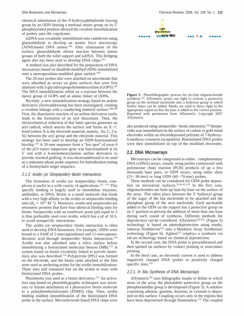
Three methods can be considered for ODN probe deposi-tion on microarray surfaces.25,41,43,119 In the first case,oligonucleotides are built up base-by-base on the surface ofthe array. This takes place between the 5′ hydroxyl groupof the sugar of the last nucleotide to be attached and thephosphate group of the next nucleotide. Each nucleotideadded to the ODN on the support has a protective group onits 5′ position to prevent the addition of more than one baseduring each round of synthesis. Different methods fordeprotection can be considered. Affymetrix120,121(Figure 5)technology is based on photodeprotection using masks,whereas NimbleGen122 uses a Maskless Array Synthesizertechnology (Figure 6). Agilent123 employs a synthesis viaink-jet technology based on chemical deprotection.
In the second case, the DNA probe is presynthesized andthen spotted on surfaces by contact printing or noncontactprinting.
In the third case, an electronic current is used to addressnegatively charged DNA probes to positively chargedspecific sites.124
2.2.1. In Situ Synthesis of DNA MicroarraysAffymetrix120 uses lithographic masks to define in which
areas of the array the photolabile protective group on thephosphoramidite group is decomposed (Figure 5). A solutioncontaining adenine, guanine, thymine, or cytosine is depos-ited on this surface. Coupling occurs only in the regions thathave been deprotected through illumination.125 The coupled
Figure 5. Photolithographic process for on-chip oligonucleotidesynthesis.120 Affymetrix arrays use light to convert a protectivegroup on the terminal nucleotide into a hydroxyl group to whichfurther bases can be added. Masks are used to direct light to theappropriate region so that the base is added to the correct position.Reprinted with permission from Affymetrix. Copyright 2007Affymetrix.
DNA Biosensors and Microarrays Chemical Reviews, 2008, Vol. 108, No. 1 113
nucleotide also bears a light-sensitive protecting group sothat the cycle can be repeated. In this way, the microarrayis built as the probes are synthesized through repeated cyclesof deprotection and coupling. The process is repeated untilthe probes reach their full length, usually 25 nucleotides.
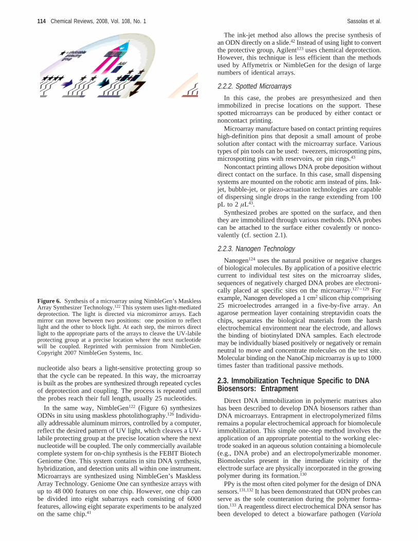
In the same way, NimbleGen122 (Figure 6) synthesizesODNs in situ using maskless photolithography.126 Individu-ally addressable aluminum mirrors, controlled by a computer,reflect the desired pattern of UV light, which cleaves a UV-labile protecting group at the precise location where the nextnucleotide will be coupled. The only commercially availablecomplete system for on-chip synthesis is the FEBIT BiotechGeniome One. This system contains in situ DNA synthesis,hybridization, and detection units all within one instrument.Microarrays are synthesized using NimbleGen’s MasklessArray Technology. Geniome One can synthesize arrays withup to 48 000 features on one chip. However, one chip canbe divided into eight subarrays each consisting of 6000features, allowing eight separate experiments to be analyzedon the same chip.41
The ink-jet method also allows the precise synthesis ofan ODN directly on a slide.42 Instead of using light to convertthe protective group, Agilent123 uses chemical deprotection.However, this technique is less efficient than the methodsused by Affymetrix or NimbleGen for the design of largenumbers of identical arrays.
2.2.2. Spotted Microarrays
In this case, the probes are presynthesized and thenimmobilized in precise locations on the support. Thesespotted microarrays can be produced by either contact ornoncontact printing.
Microarray manufacture based on contact printing requireshigh-definition pins that deposit a small amount of probesolution after contact with the microarray surface. Varioustypes of pin tools can be used: tweezers, microspotting pins,microspotting pins with reservoirs, or pin rings.43
Noncontact printing allows DNA probe deposition withoutdirect contact on the surface. In this case, small dispensingsystems are mounted on the robotic arm instead of pins. Ink-jet, bubble-jet, or piezo-actuation technologies are capableof dispersing single drops in the range extending from 100pL to 2 µL43.
Synthesized probes are spotted on the surface, and thenthey are immobilized through various methods. DNA probescan be attached to the surface either covalently or nonco-valently (cf. section 2.1).
2.2.3. Nanogen Technology
Nanogen124 uses the natural positive or negative chargesof biological molecules. By application of a positive electriccurrent to individual test sites on the microarray slides,sequences of negatively charged DNA probes are electroni-cally placed at specific sites on the microarray.127-129 Forexample, Nanogen developed a 1 cm2 silicon chip comprising25 microelectrodes arranged in a five-by-five array. Anagarose permeation layer containing streptavidin coats thechips, separates the biological materials from the harshelectrochemical environment near the electrode, and allowsthe binding of biotinylated DNA samples. Each electrodemay be individually biased positively or negatively or remainneutral to move and concentrate molecules on the test site.Molecular binding on the NanoChip microarray is up to 1000times faster than traditional passive methods.
2.3. Immobilization Technique Specific to DNABiosensors: Entrapment
Direct DNA immobilization in polymeric matrixes alsohas been described to develop DNA biosensors rather thanDNA microarrays. Entrapment in electropolymerized filmsremains a popular electrochemical approach for biomoleculeimmobilization. This simple one-step method involves theapplication of an appropriate potential to the working elec-trode soaked in an aqueous solution containing a biomolecule(e.g., DNA probe) and an electropolymerizable monomer.Biomolecules present in the immediate vicinity of theelectrode surface are physically incorporated in the growingpolymer during its formation.130
PPy is the most often cited polymer for the design of DNAsensors.131,132It has been demonstrated that ODN probes canserve as the sole counteranion during the polymer forma-tion.133 A reagentless direct electrochemical DNA sensor hasbeen developed to detect a biowarfare pathogen (Variola
Figure 6. Synthesis of a microarray using NimbleGen’s MasklessArray Synthesizer Technology.122 This system uses light-mediateddeprotection. The light is directed via micromirror arrays. Eachmirror can move between two positions: one position to reflectlight and the other to block light. At each step, the mirrors directlight to the appropriate parts of the arrays to cleave the UV-labileprotecting group at a precise location where the next nucleotidewill be coupled. Reprinted with permission from NimbleGen.Copyright 2007 NimbleGen Systems, Inc.
114 Chemical Reviews, 2008, Vol. 108, No. 1 Sassolas et al.
major virus) using ultrathin films of the conducting PPydoped with an ODN probe.134 ssDNA was entrapped withinPPy formed on a platinum electrode.135 In the same way,oligonucleotide probes were immobilized during the elec-tropolymerization of PPy onto multiwalled CNT-modifiedelectrodes.136 Electropolymerization by cyclic voltammetry(CV) also was reported to entrap DNA sequences into a PPyfilm.137,138
More rarely, other electrically conductive films have beenused to entrap DNA probes. Polyaniline, polydiaminoben-zene, and poly(3,4-ethylenedioxythiophene)139,140allowed theimmobilization of DNA probes to develop electrochemicalbiosensors.
3. Nucleic Acid Hybridization DetectionTables 1-3 present the performances obtained with
different DNA biosensors or microarrays. Nucleic acidhybridization can be detected according to different tech-niques, based on optical (Table 1), electrochemical (Table2), or gravimetric (QCM) (Table 3) detection.
3.1. Optical DNA Biosensors and DNAMicroarrays
DNA hybridization can be optically detected using fluo-rescence, surface plasmon resonance (SPR), chemilumines-cence, colorimetry, interferometry, or surface-enhancedRaman scattering (SERS) spectroscopy (Table 1).
3.1.1. Fluorescence Detection
Table 1 presents the performances obtained with somefluorescent-based biosensors or microarrays.56,115,141-151
When the DNA target is labeled with a fluorophore, suchas fluorescein isothiocyanate (FITC), its hybridization witha probe can be easily measured with an imaging fluorescenceapparatus. p(dA) probes (18-mer) or H-ras wild-type probes(10-mer) were immobilized with succinimidyl ester residues
in acrylamide-based polymer matrixes deposited on an opticalfiber.142 This sensor also was able to identify one pointmutation in the Ras oncogene PCR products. It can detectpoint mutations at DNA concentrations of 2× 10⁻¹⁰-1.96× 10⁻⁷ M following a 20 min hybridization (Table 1). Probeswere immobilized on self-assembled DNA-conjugated poly-mer of PAA.152 The complementary target was labeled withFITC. A biotinylated probe also was immobilized on anoptical fiber via avidin.56 The hybridization with a fluorescein-labeled complementary target (16-mer) was monitored in realtime by fluorescence detection. In this case, the detectionlimit was 2 × 10-13 M (Table 1). A multiplexed DNAhybridization system also was developed using encoded Nimicroparticles.141 These particles were coated with a SAMof 16-mercaptohexadecanoic acid and were treated withavidin. Biotinylated DNA probes were immobilized on theparticle having a specific code via avidin-biotin interaction.The detection limit was estimated to be around 1× 10⁻¹⁰
M (Table 1). Recently, organic-dye-doped silica nanoparticleswere used to detect DNA.153 A large number of fluorophoreswas encapsulated inside a single nanoparticle, which pro-duced a strong fluorescence signal when it was properlyexcited. Therefore, when one probe DNA was labeled withone dye-doped silica nanoparticle, the signal was greatlyamplified as compared to that with one fluorophore. Throughthe use of this strategy, DNA target molecules could bedetected at a concentration as low as 8× 10-13 M.154
Fluorescence detection using a fluorophore as a label alsohas been employed in DNA chip technology. Amino-modified ODNs were immobilized on an aldehyde-function-alized polyacrylamide gel.155 A chip also was developedbased on the incorporation of biotinylated DNA probes intobead microreactors.143 In both cases, a fluorophore-conju-gated DNA target was complementary to the capture strand.Ali et al.143 used pyramidal wells to confine the sensor beadsin a central position. It seems that the porous beads’ internalmicroenvironment was more suitable for DNA hybridization.
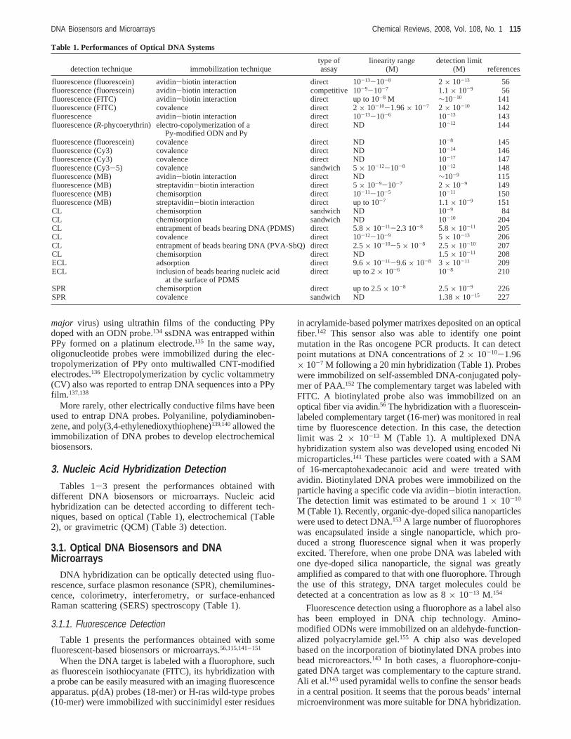
Table 1. Performances of Optical DNA Systems
detection technique immobilization techniquetype ofassay
linearity range(M)
detection limit(M) references
fluorescence (fluorescein) avidin-biotin interaction direct 10-13-10-8 2 × 10-13 56fluorescence (fluorescein) avidin-biotin interaction competitive 10-9-10-7 1.1× 10⁻⁹ 56fluorescence (FITC) avidin-biotin interaction direct up to 10-8 M ∼10-10 141fluorescence (FITC) covalence direct 2× 10⁻¹⁰-1.96× 10⁻⁷ 2 × 10⁻¹⁰ 142fluorescence avidin-biotin interaction direct 10-13-10-6 10-13 143fluorescence (R-phycoerythrin) electro-copolymerization of a
Py-modified ODN and Pydirect ND 10-12 144
fluorescence (fluorescein) covalence direct ND 10-8 145fluorescence (Cy3) covalence direct ND 10-14 146fluorescence (Cy3) covalence direct ND 10-17 147fluorescence (Cy3-5) covalence sandwich 5× 10-12-10-8 10-12 148fluorescence (MB) avidin-biotin interaction direct ND ∼10-9 115fluorescence (MB) streptavidin-biotin interaction direct 5× 10⁻⁹-10-7 2 × 10⁻⁹ 149fluorescence (MB) chemisorption direct 10-11-10-5 10-11 150fluorescence (MB) streptavidin-biotin interaction direct up to 10-7 1.1× 10⁻⁹ 151CL chemisorption sandwich ND 10-9 84CL chemisorption sandwich ND 10-10 204CL entrapment of beads bearing DNA (PDMS) direct 5.8× 10⁻¹¹-2.3 10-8 5.8× 10⁻¹¹ 205CL covalence direct 10-12-10-9 5 × 10-13 206CL entrapment of beads bearing DNA (PVA-SbQ) direct 2.5× 10⁻¹⁰-5 × 10⁻⁸ 2.5× 10⁻¹⁰ 207CL chemisorption direct ND 1.5× 10⁻¹¹ 208ECL adsorption direct 9.6× 10⁻¹¹-9.6× 10⁻⁸ 3 × 10-11 209ECL inclusion of beads bearing nucleic acid
at the surface of PDMSdirect up to 2× 10⁻⁶ 10-8 210
SPR chemisorption direct up to 2.5× 10⁻⁸ 2.5× 10⁻⁹ 226SPR covalence sandwich ND 1.38× 10⁻¹⁵ 227
DNA Biosensors and Microarrays Chemical Reviews, 2008, Vol. 108, No. 1 115
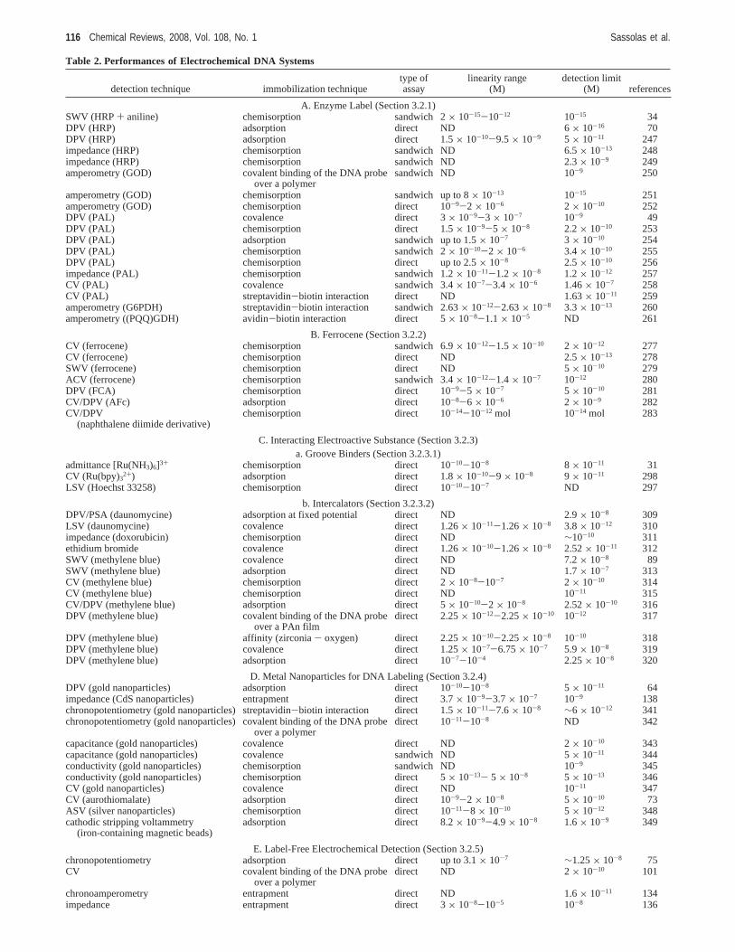
Table 2. Performances of Electrochemical DNA Systems
detection technique immobilization techniquetype ofassay
linearity range(M)
detection limit(M) references
A. Enzyme Label (Section 3.2.1)SWV (HRP+ aniline) chemisorption sandwich 2× 10-15-10-12 10-15 34DPV (HRP) adsorption direct ND 6× 10-16 70DPV (HRP) adsorption direct 1.5× 10⁻¹⁰-9.5× 10⁻⁹ 5 × 10-11 247impedance (HRP) chemisorption sandwich ND 6.5× 10⁻¹³ 248impedance (HRP) chemisorption sandwich ND 2.3× 10⁻⁹ 249amperometry (GOD) covalent binding of the DNA probe
over a polymersandwich ND 10-9 250
amperometry (GOD) chemisorption sandwich up to 8× 10-13 10-15 251amperometry (GOD) chemisorption direct 10-9-2 × 10⁻⁶ 2 × 10⁻¹⁰ 252DPV (PAL) covalence direct 3× 10⁻⁹-3 × 10⁻⁷ 10-9 49DPV (PAL) chemisorption direct 1.5× 10⁻⁹-5 × 10⁻⁸ 2.2× 10⁻¹⁰ 253DPV (PAL) adsorption sandwich up to 1.5× 10⁻⁷ 3 × 10⁻¹⁰ 254DPV (PAL) chemisorption sandwich 2× 10⁻¹⁰-2 × 10⁻⁶ 3.4× 10⁻¹⁰ 255DPV (PAL) chemisorption direct up to 2.5× 10⁻⁸ 2.5× 10⁻¹⁰ 256impedance (PAL) chemisorption sandwich 1.2× 10⁻¹¹-1.2× 10⁻⁸ 1.2× 10⁻¹² 257CV (PAL) covalence sandwich 3.4× 10⁻⁷-3.4× 10⁻⁶ 1.46× 10⁻⁷ 258CV (PAL) streptavidin-biotin interaction direct ND 1.63× 10⁻¹¹ 259amperometry (G6PDH) streptavidin-biotin interaction sandwich 2.63× 10⁻¹²-2.63× 10⁻⁸ 3.3× 10⁻¹³ 260amperometry ((PQQ)GDH) avidin-biotin interaction direct 5× 10⁻⁸-1.1× 10⁻⁵ ND 261
B. Ferrocene (Section 3.2.2)CV (ferrocene) chemisorption sandwich 6.9× 10⁻¹²-1.5× 10⁻¹⁰ 2 × 10-12 277CV (ferrocene) chemisorption direct ND 2.5× 10⁻¹³ 278SWV (ferrocene) chemisorption direct ND 5× 10⁻¹⁰ 279ACV (ferrocene) chemisorption sandwich 3.4× 10⁻¹²-1.4× 10⁻⁷ 10-12 280DPV (FCA) chemisorption direct 10-9-5 × 10⁻⁷ 5 × 10⁻¹⁰ 281CV/DPV (AFc) adsorption direct 10-8-6 × 10⁻⁶ 2 × 10⁻⁹ 282CV/DPV
(naphthalene diimide derivative)chemisorption direct 10-14-10-12 mol 10-14 mol 283
C. Interacting Electroactive Substance (Section 3.2.3)a. Groove Binders (Section 3.2.3.1)
admittance [Ru(NH3)6]3+ chemisorption direct 10-10-10-8 8 × 10-11 31CV (Ru(bpy)32+) adsorption direct 1.8× 10⁻¹⁰-9 × 10⁻⁸ 9 × 10-11 298LSV (Hoechst 33258) chemisorption direct 10-10-10-7 ND 297
b. Intercalators (Section 3.2.3.2)DPV/PSA (daunomycine) adsorption at fixed potential direct ND 2.9× 10⁻⁸ 309LSV (daunomycine) covalence direct 1.26× 10⁻¹¹-1.26× 10⁻⁸ 3.8× 10⁻¹² 310impedance (doxorubicin) chemisorption direct ND ∼10-10 311ethidium bromide covalence direct 1.26× 10⁻¹⁰-1.26× 10⁻⁸ 2.52× 10⁻¹¹ 312SWV (methylene blue) covalence direct ND 7.2× 10⁻⁸ 89SWV (methylene blue) adsorption direct ND 1.7× 10⁻⁷ 313CV (methylene blue) chemisorption direct 2× 10⁻⁸-10-7 2 × 10⁻¹⁰ 314CV (methylene blue) chemisorption direct ND 10-11 315CV/DPV (methylene blue) adsorption direct 5× 10⁻¹⁰-2 × 10⁻⁸ 2.52× 10⁻¹⁰ 316DPV (methylene blue) covalent binding of the DNA probe
over a PAn filmdirect 2.25× 10⁻¹²-2.25× 10⁻¹⁰ 10-12 317
DPV (methylene blue) affinity (zirconia- oxygen) direct 2.25× 10⁻¹⁰-2.25× 10⁻⁸ 10-10 318DPV (methylene blue) covalence direct 1.25× 10⁻⁷-6.75× 10⁻⁷ 5.9× 10⁻⁸ 319DPV (methylene blue) adsorption direct 10-7-10-4 2.25× 10⁻⁸ 320
D. Metal Nanoparticles for DNA Labeling (Section 3.2.4)DPV (gold nanoparticles) adsorption direct 10-10-10-8 5 × 10-11 64impedance (CdS nanoparticles) entrapment direct 3.7× 10⁻⁹-3.7× 10⁻⁷ 10-9 138chronopotentiometry (gold nanoparticles) streptavidin-biotin interaction direct 1.5× 10⁻¹¹-7.6× 10⁻⁸ ∼6 × 10-12 341chronopotentiometry (gold nanoparticles) covalent binding of the DNA probe
over a polymerdirect 10-11-10-8 ND 342
capacitance (gold nanoparticles) covalence direct ND 2× 10⁻¹⁰ 343capacitance (gold nanoparticles) covalence sandwich ND 5× 10-11 344conductivity (gold nanoparticles) chemisorption sandwich ND 10-9 345conductivity (gold nanoparticles) chemisorption direct 5× 10-13- 5 × 10⁻⁸ 5 × 10-13 346CV (gold nanoparticles) covalence direct ND 10-11 347CV (aurothiomalate) adsorption direct 10-9-2 × 10⁻⁸ 5 × 10⁻¹⁰ 73ASV (silver nanoparticles) chemisorption direct 10-11-8 × 10⁻¹⁰ 5 × 10-12 348cathodic stripping voltammetry
(iron-containing magnetic beads)adsorption direct 8.2× 10⁻⁹-4.9× 10⁻⁸ 1.6× 10⁻⁹ 349
E. Label-Free Electrochemical Detection (Section 3.2.5)chronopotentiometry adsorption direct up to 3.1× 10⁻⁷ ∼1.25× 10⁻⁸ 75CV covalent binding of the DNA probe
over a polymerdirect ND 2× 10⁻¹⁰ 101
chronoamperometry entrapment direct ND 1.6× 10⁻¹¹ 134impedance entrapment direct 3× 10⁻⁸-10-5 10-8 136
116 Chemical Reviews, 2008, Vol. 108, No. 1 Sassolas et al.
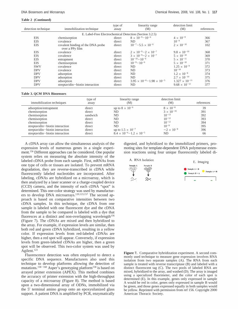
A cDNA array can allow the simultaneous analysis of theexpression levels of numerous genes in a single experi-ment.156 Different approaches can be considered. One popularsystem relies on measuring the absolute intensity of thelabeled cDNA probe from each sample. First, mRNAs fromone type of cells or tissues are isolated. To prevent mRNAdegradation, they are reverse-transcribed in cDNA whilefluorescently labeled nucleotides are incorporated. Afterlabeling, cDNAs are hybridized on a microarray, which isthen analyzed by a laser scanner or a charge-coupled device(CCD) camera, and the intensity of each cDNA “spot” isdetermined. This one-color strategy was used by manufactur-ers to develop DNA microarrays.120,123,157The second ap-proach is based on comparative intensities between twocDNA samples. In this technique, the cDNA from onesample is labeled with one fluorescent dye and the cDNAfrom the sample to be compared is labeled with a dye thatfluoresces at a distinct and non-overlapping wavelength156
(Figure 7). The cDNAs are mixed and then hybridized tothe array. For example, if expression levels are similar, thenboth red and green cDNA hybridized, resulting in a yellowcolor. If expression levels from red-labeled cDNAs arehigher, then a red spot will appear. Conversely, if expressionlevels from green-labeled cDNAs are higher, then a greenspot will be observed. This two-color system was used byAgilent.123
Fluorescence detection was often employed to detect aspecific DNA sequence. Manufacturers also used thistechnique to develop platforms allowing the detection ofmutations.158-160 Asper’s genotyping platform158 is based onarrayed primer extension (APEX). This method combinesthe accuracy of primer extension with the high-throughputcapacity of a microarray (Figure 8). The method is basedupon a two-dimensional array of ODNs, immobilized viathe 5′ terminal amino group onto an epoxysilanized glasssupport. A patient DNA is amplified by PCR, enzymatically
digested, and hybridized to the immobilized primers, pro-moting sites for template-dependent DNA polymerase exten-sion reactions using four unique fluorescently labeled di-
Table 2 (Continued)
detection technique immobilization techniquetype ofassay
linearity range(M)
detection limit(M) references
E. Label-Free Electrochemical Detection (Section 3.2.5)EIS chemisorption direct 8× 10⁻⁹-10-6 4 × 10⁻⁹ 366EIS covalence direct ND 10-9 367EIS covalent binding of the DNA probe
over a PPy filmdirect 10-7-5.5× 10⁻⁶ 2 × 10⁻¹⁰ 102
EIS covalence direct 2× 10⁻⁹-2 × 10⁻⁷ 9.8× 10⁻¹⁰ 368EIS covalence direct 3× 10⁻⁹-2 × 10⁻⁷ 5 × 10⁻¹⁰ 369EIS entrapment direct 10-10-10-6 5 × 10-11 370EIS chemisorption direct 10-9-10-6 5 × 10⁻¹⁰ 371SWV covalence direct ND 1.25× 10⁻⁹ 372DPV covalence direct ND 10-10 373DPV adsorption direct ND 1.2× 10⁻⁸ 374DPV adsorption direct ND 2.7× 10⁻¹⁰ 375DPV adsorption direct 3.95× 10⁻⁶-1.98× 10⁻⁵ 1.327× 10⁻¹¹ 376DPV streptavidin-biotin interaction direct ND 9.68× 10⁻¹² 377
Table 3. QCM DNA Biosensors
immobilization techniquestype ofassay
linearity range(M)
detection limit(M) references
adsorption/entrapment direct up to 8× 10⁻⁶ 8 × 10⁻⁸ 39chemisorption direct ND 3× 10-16 391chemisorption sandwich ND 10-13 392chemisorption direct ND 10-12 393chemisorption direct ND 10-12 394streptavidin-biotin interaction direct ND 10-9 395streptavidin-biotin interaction direct up to 1.5× 10⁻⁷ ∼2 × 10⁻⁸ 396streptavidin-biotin interaction direct 8.4× 10⁻⁶-1.2× 10⁻⁵ ND 66
Figure 7. Comparative hybridization experiment. A second com-monly used technique to measure gene expression involves RNAisolation from two separate samples (A). The RNA from eachsample is treated with reverse transcriptase (B) and labeled with adistinct fluorescent tag (C). The two pools of labeled RNA aremixed, hybridized to the array, and washed (D). The array is imagedusing a specialized fluorimeter, and the color of each spot isdetermined (E). In this example, genes only expressed in sampleA would be red in color, genes only expressed in sample B wouldbe green, and those genes expressed equally in both samples wouldbe yellow. Reprinted with permission from ref 156. Copyright 2000American Thoracic Society.
DNA Biosensors and Microarrays Chemical Reviews, 2008, Vol. 108, No. 1 117
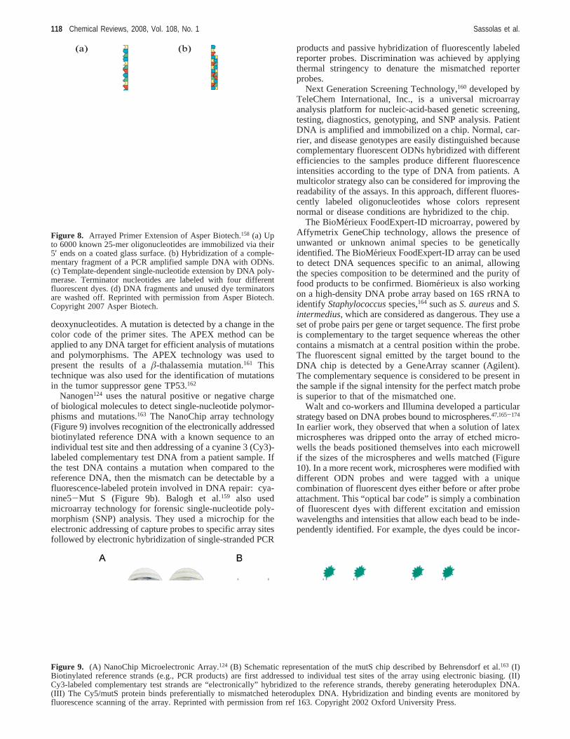
deoxynucleotides. A mutation is detected by a change in thecolor code of the primer sites. The APEX method can beapplied to any DNA target for efficient analysis of mutationsand polymorphisms. The APEX technology was used topresent the results of aâ-thalassemia mutation.161 Thistechnique was also used for the identification of mutationsin the tumor suppressor gene TP53.162
Nanogen124 uses the natural positive or negative chargeof biological molecules to detect single-nucleotide polymor-phisms and mutations.163 The NanoChip array technology(Figure 9) involves recognition of the electronically addressedbiotinylated reference DNA with a known sequence to anindividual test site and then addressing of a cyanine 3 (Cy3)-labeled complementary test DNA from a patient sample. Ifthe test DNA contains a mutation when compared to thereference DNA, then the mismatch can be detectable by afluorescence-labeled protein involved in DNA repair: cya-nine5-Mut S (Figure 9b). Balogh et al.159 also usedmicroarray technology for forensic single-nucleotide poly-morphism (SNP) analysis. They used a microchip for theelectronic addressing of capture probes to specific array sitesfollowed by electronic hybridization of single-stranded PCR
products and passive hybridization of fluorescently labeledreporter probes. Discrimination was achieved by applyingthermal stringency to denature the mismatched reporterprobes.
Next Generation Screening Technology,160 developed byTeleChem International, Inc., is a universal microarrayanalysis platform for nucleic-acid-based genetic screening,testing, diagnostics, genotyping, and SNP analysis. PatientDNA is amplified and immobilized on a chip. Normal, car-rier, and disease genotypes are easily distinguished becausecomplementary fluorescent ODNs hybridized with differentefficiencies to the samples produce different fluorescenceintensities according to the type of DNA from patients. Amulticolor strategy also can be considered for improving thereadability of the assays. In this approach, different fluores-cently labeled oligonucleotides whose colors representnormal or disease conditions are hybridized to the chip.
The BioMerieux FoodExpert-ID microarray, powered byAffymetrix GeneChip technology, allows the presence ofunwanted or unknown animal species to be geneticallyidentified. The BioMe´rieux FoodExpert-ID array can be usedto detect DNA sequences specific to an animal, allowingthe species composition to be determined and the purity offood products to be confirmed. Biome´rieux is also workingon a high-density DNA probe array based on 16S rRNA toidentify Staphylococcusspecies,164 such asS. aureusandS.intermedius, which are considered as dangerous. They use aset of probe pairs per gene or target sequence. The first probeis complementary to the target sequence whereas the othercontains a mismatch at a central position within the probe.The fluorescent signal emitted by the target bound to theDNA chip is detected by a GeneArray scanner (Agilent).The complementary sequence is considered to be present inthe sample if the signal intensity for the perfect match probeis superior to that of the mismatched one.
Walt and co-workers and Illumina developed a particularstrategy based on DNA probes bound to microspheres.47,165-174
In earlier work, they observed that when a solution of latexmicrospheres was dripped onto the array of etched micro-wells the beads positioned themselves into each microwellif the sizes of the microspheres and wells matched (Figure10). In a more recent work, microspheres were modified withdifferent ODN probes and were tagged with a uniquecombination of fluorescent dyes either before or after probeattachment. This “optical bar code” is simply a combinationof fluorescent dyes with different excitation and emissionwavelengths and intensities that allow each bead to be inde-pendently identified. For example, the dyes could be incor-
Figure 8. Arrayed Primer Extension of Asper Biotech.158 (a) Upto 6000 known 25-mer oligonucleotides are immobilized via their5′ ends on a coated glass surface. (b) Hybridization of a comple-mentary fragment of a PCR amplified sample DNA with ODNs.(c) Template-dependent single-nucleotide extension by DNA poly-merase. Terminator nucleotides are labeled with four differentfluorescent dyes. (d) DNA fragments and unused dye terminatorsare washed off. Reprinted with permission from Asper Biotech.Copyright 2007 Asper Biotech.
Figure 9. (A) NanoChip Microelectronic Array.124 (B) Schematic representation of the mutS chip described by Behrensdorf et al.163 (I)Biotinylated reference strands (e.g., PCR products) are first addressed to individual test sites of the array using electronic biasing. (II)Cy3-labeled complementary test strands are “electronically” hybridized to the reference strands, thereby generating heteroduplex DNA.(III) The Cy5/mutS protein binds preferentially to mismatched heteroduplex DNA. Hybridization and binding events are monitored byfluorescence scanning of the array. Reprinted with permission from ref 163. Copyright 2002 Oxford University Press.
118 Chemical Reviews, 2008, Vol. 108, No. 1 Sassolas et al.
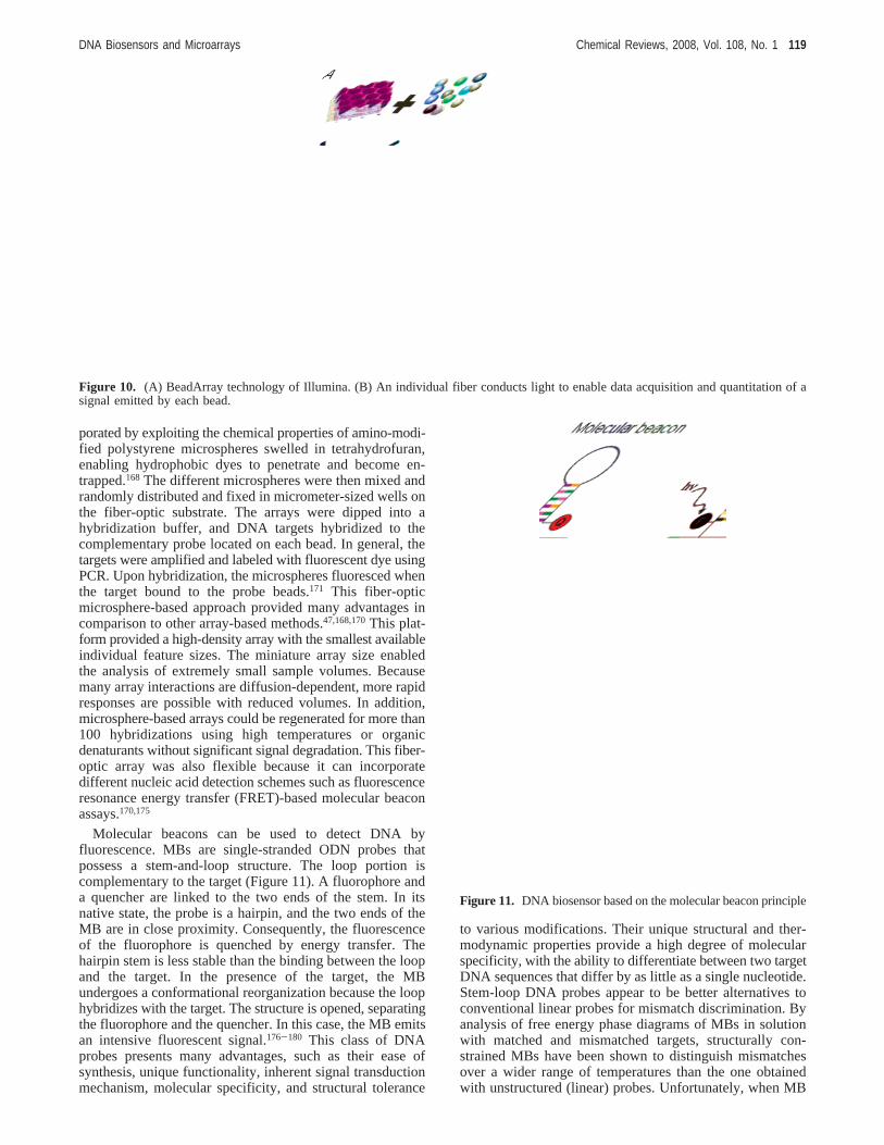
porated by exploiting the chemical properties of amino-modi-fied polystyrene microspheres swelled in tetrahydrofuran,enabling hydrophobic dyes to penetrate and become en-trapped.168 The different microspheres were then mixed andrandomly distributed and fixed in micrometer-sized wells onthe fiber-optic substrate. The arrays were dipped into ahybridization buffer, and DNA targets hybridized to thecomplementary probe located on each bead. In general, thetargets were amplified and labeled with fluorescent dye usingPCR. Upon hybridization, the microspheres fluoresced whenthe target bound to the probe beads.171 This fiber-opticmicrosphere-based approach provided many advantages incomparison to other array-based methods.47,168,170This plat-form provided a high-density array with the smallest availableindividual feature sizes. The miniature array size enabledthe analysis of extremely small sample volumes. Becausemany array interactions are diffusion-dependent, more rapidresponses are possible with reduced volumes. In addition,microsphere-based arrays could be regenerated for more than100 hybridizations using high temperatures or organicdenaturants without significant signal degradation. This fiber-optic array was also flexible because it can incorporatedifferent nucleic acid detection schemes such as fluorescenceresonance energy transfer (FRET)-based molecular beaconassays.170,175
Molecular beacons can be used to detect DNA byfluorescence. MBs are single-stranded ODN probes thatpossess a stem-and-loop structure. The loop portion iscomplementary to the target (Figure 11). A fluorophore anda quencher are linked to the two ends of the stem. In itsnative state, the probe is a hairpin, and the two ends of theMB are in close proximity. Consequently, the fluorescenceof the fluorophore is quenched by energy transfer. Thehairpin stem is less stable than the binding between the loopand the target. In the presence of the target, the MBundergoes a conformational reorganization because the loophybridizes with the target. The structure is opened, separatingthe fluorophore and the quencher. In this case, the MB emitsan intensive fluorescent signal.176-180 This class of DNAprobes presents many advantages, such as their ease ofsynthesis, unique functionality, inherent signal transductionmechanism, molecular specificity, and structural tolerance
to various modifications. Their unique structural and ther-modynamic properties provide a high degree of molecularspecificity, with the ability to differentiate between two targetDNA sequences that differ by as little as a single nucleotide.Stem-loop DNA probes appear to be better alternatives toconventional linear probes for mismatch discrimination. Byanalysis of free energy phase diagrams of MBs in solutionwith matched and mismatched targets, structurally con-strained MBs have been shown to distinguish mismatchesover a wider range of temperatures than the one obtainedwith unstructured (linear) probes. Unfortunately, when MB
Figure 10. (A) BeadArray technology of Illumina. (B) An individual fiber conducts light to enable data acquisition and quantitation of asignal emitted by each bead.
Figure 11. DNA biosensor based on the molecular beacon principle
DNA Biosensors and Microarrays Chemical Reviews, 2008, Vol. 108, No. 1 119
probes are immobilized onto solid supports, they displaylower sensitivities than in solution, so various solid supportshave been explored, and appropriate probes have beendesigned. In this case, the MBs have been used widely todevelop biosensors for DNA detection.181,182
Some authors reported the use of a biotinylated ssDNAMB.115,151 The 5′ end was linked to a fluorophore, tetra-methylrhodamine (TMR), and the 3′ end was attached to thequencher, 4-(4-(dimethylamino)phenyl)azo)benzoic acid (DAB-CYL). The MB was immobilized on a solid surface via anavidin-biotin linkage. The MBs also were immobilized onsilica microspheres through avidin-biotin interactions.183TheMBs were labeled using carboxytetra-methylrhodamine (TAMRA) and DABCYL. However, theability of metals was noted to quench fluorescence.184 It wasdemonstrated that fluorophore-tagged DNA hairpins attachedto gold films can function as highly sensitive sensors forODNs. The substrate material itself was used as the quench-ing agent. Rhodamine-labeled MBs are specific for genesknown to confer methicillin resistance toS. aureusand wereimmobilized onto gold films. In the same way, ability of agold-surface-immobilized MB to distinguish single-basemismatches was studied.185 Apparently, MBs are veryspecific and can distinguish targets that differ by only singlenucleotides. This specificity is due to their ability to form astem-and-loop structure.186 A MB fiber-optic DNA array alsowas developed.170,175 In this work, MBs were immobilizedon optically encoded microspheres that can be made byentrapping different dyes inside polystyrene microspheres.Fluorescein and DABCYL were used as the fluorophore andthe quencher, respectively, for each MB. These immobilizedMBs were incubated with the unlabeled target, and thefluorescence was monitored in real time. However, MBs wereimmobilized on an agarose film-coated slide, and the sensorwas compared with a conventional system that directlyimmobilizes MBs on a glutaraldehyde-derived glass slide.187
The MB array can identify a single-nucleotide difference ina 16-mer target DNA sequence. A streptavidin-biotin bridgealso was used to immobilize MB on a glass slide.149 Theresponse of the MB biosensor to its 19-mer target DNA islinear from 5× 10⁻⁹ to 10-7 M (Table 1).
Quantum dots also could be used to detect DNA sequencesinstead of fluorophores. A quantum dot is a semiconductorparticle (e.g., ZnS, CdSe, and CdS) that can be used as afluorophore.188-190 The size and the shape of these structuresand the number of electrons that they contain can becontrolled precisely. Colloidal semiconductors are veryattractive for the labeling of biomolecules. They are candi-dates for replacing conventional fluorescent markers suchas rhodamine in biodetection assays.
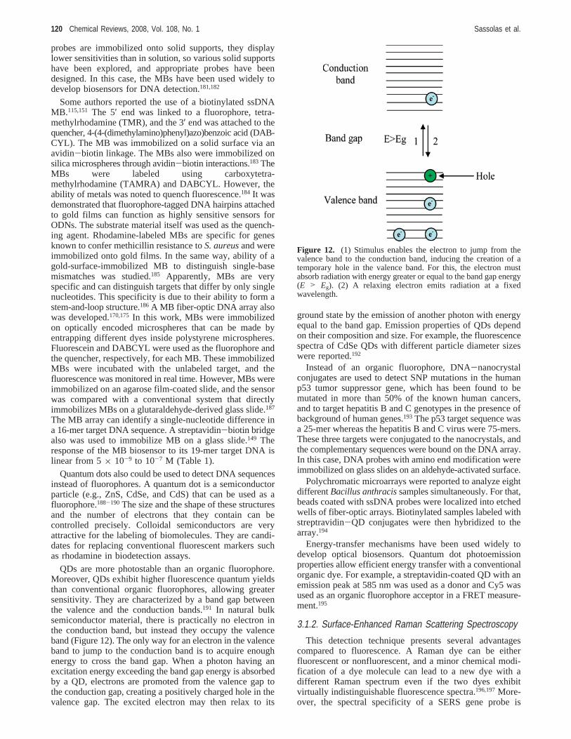
QDs are more photostable than an organic fluorophore.Moreover, QDs exhibit higher fluorescence quantum yieldsthan conventional organic fluorophores, allowing greatersensitivity. They are characterized by a band gap betweenthe valence and the conduction bands.191 In natural bulksemiconductor material, there is practically no electron inthe conduction band, but instead they occupy the valenceband (Figure 12). The only way for an electron in the valenceband to jump to the conduction band is to acquire enoughenergy to cross the band gap. When a photon having anexcitation energy exceeding the band gap energy is absorbedby a QD, electrons are promoted from the valence gap tothe conduction gap, creating a positively charged hole in thevalence gap. The excited electron may then relax to its
ground state by the emission of another photon with energyequal to the band gap. Emission properties of QDs dependon their composition and size. For example, the fluorescencespectra of CdSe QDs with different particle diameter sizeswere reported.192
Instead of an organic fluorophore, DNA-nanocrystalconjugates are used to detect SNP mutations in the humanp53 tumor suppressor gene, which has been found to bemutated in more than 50% of the known human cancers,and to target hepatitis B and C genotypes in the presence ofbackground of human genes.193 The p53 target sequence wasa 25-mer whereas the hepatitis B and C virus were 75-mers.These three targets were conjugated to the nanocrystals, andthe complementary sequences were bound on the DNA array.In this case, DNA probes with amino end modification wereimmobilized on glass slides on an aldehyde-activated surface.
Polychromatic microarrays were reported to analyze eightdifferentBacillus anthracissamples simultaneously. For that,beads coated with ssDNA probes were localized into etchedwells of fiber-optic arrays. Biotinylated samples labeled withstreptravidin-QD conjugates were then hybridized to thearray.194
Energy-transfer mechanisms have been used widely todevelop optical biosensors. Quantum dot photoemissionproperties allow efficient energy transfer with a conventionalorganic dye. For example, a streptavidin-coated QD with anemission peak at 585 nm was used as a donor and Cy5 wasused as an organic fluorophore acceptor in a FRET measure-ment.195
3.1.2. Surface-Enhanced Raman Scattering Spectroscopy
This detection technique presents several advantagescompared to fluorescence. A Raman dye can be eitherfluorescent or nonfluorescent, and a minor chemical modi-fication of a dye molecule can lead to a new dye with adifferent Raman spectrum even if the two dyes exhibitvirtually indistinguishable fluorescence spectra.196,197More-over, the spectral specificity of a SERS gene probe is
Figure 12. (1) Stimulus enables the electron to jump from thevalence band to the conduction band, inducing the creation of atemporary hole in the valence band. For this, the electron mustabsorb radiation with energy greater or equal to the band gap energy(E > Eg). (2) A relaxing electron emits radiation at a fixedwavelength.
120 Chemical Reviews, 2008, Vol. 108, No. 1 Sassolas et al.
excellent in comparison to that of the fluorescence method.For example, the spectral bandwidths of cresyl fast violet(CFV) in UV absorption and fluorescence are broad whereasthe bandwidth of the SERS spectrum of the same CFV dyeis narrower.198 One of the major difficulties in the develop-ment of the SERS technique for genomics applications isthe production of surfaces or media that can be readilyadapted to the assay formats, e.g., DNA hybridization. TheSERS substrates must have an easily controlled protrusionsize and reproducible structures. Roughened metal electrodesand metal colloids were among the first SERS-active mediaused. A variety of solid-surface SERS substrates weredeveloped on metal-covered nanoparticles.199 However, DNAbiosensors or microarrays that did not use a metallic SERSsubstrate also have been reported,197,200but in this case SERSdetection is possible using ODN labeled with Au or Agnanoparticles.
The SERS technique was reported as a tool for detectingspecific nucleic acid sequences.201 For that, the SERS-labeledprobes were hybridized to fragments of DNA attached onnitrocellulose. After hybridization, probes and DNA frag-ments were transferred on a SERS-active substrate foranalysis. A negative control that consisted of labeled DNAthat was not complementary to the DNA probe exhibited noSERS signal. However, after hybridization between the CFV-labeled target and the probe, SERS peaks were observed.The SERS technique also was applied to detect DNAfragments of the human immunodeficiency virus (HIV).198
In the same way, the development of SERS-active substratesfor cancer gene detection was described.202 The thiolatedssDNA probe was immobilized on a silver surface that wasincubated in the presence of rhodamine-B-labeled DNAtarget. A signal was observed on the SERS spectrum onlyafter the hybridization. The authors also demonstrated thatthe addition of silver colloids to the surface enhanced theRaman signal.
SERS was used to monitor DNA hybridization of afragment of the BRCA1 breast cancer gene on modifiedsilver surfaces, which were prepared by depositing a layerof silver onto glass slides, forming a microwell platform.203
The ODN probe was covalently immobilized on the silver-coated glass slide covered with a SAM. In this work, theSERS-active dye, rhodamine B, labeled the DNA target.
Nanoparticles functionalized with ODNs and Ramanlabels, coupled with SERS spectroscopy, can be used toperform multiplexed detection of ODN targets.197A sandwichcomplex was formed between the immobilized DNA probe,the target, and a DNA signaling sequence attached to Cy3-labeled Au nanoparticles. Then, the chip was treated withAg enhancement solution. Before Ag enhancement, noRaman scattering signal was detectable. After the treatment,the Ag particles could grow around the Cy3-labeled nano-particle probes, leading to large Raman scattering enhance-ments. Other dyes than Cy3 also were used to create a largenumber of probes with distinct and measurable SERS signalsfor multiplexed detection. Six dissimilar DNA targets withsix Raman labeled nanoparticle probes were distinguished.The current unoptimized detection limit of this method was2 × 10-14 M.
3.1.3. Chemiluminescent Detection
Table 1 presents some performances of chemilumines-cence- and electrochemiluminescence-based biosensors ormicroarrays.84,204-210
Luminescent reactions can be catalyzed by a biomolecule,such as hemin or horseradish peroxidase (HRP), or triggeredby the application of a potential between the workingelectrode and a pseudo-reference. Luminol and derivativesare often used for chemiluminescent (CL) or electrochemi-luminescent (ECL) reactions.
The chemiluminescent properties of luminol were firstreported by Albrecht.211 Luminol oxidation leads to theformation of an aminophthalate ion in an excited state, whichthen emits light on return to the ground state. The peroxidase-catalyzed chemiluminescent oxidation of luminol was largelyused to detect analytes212 or DNA concentration.
DNA-sensitive biochips that could be used to detect abiotin-labeled sequence were described.207 The ODN-chargedbeads were entrapped in a poly(vinyl alcohol) bearingstyrylpyridinium groups (PVA-SbQ) photopolymer, and theprobes were hybridized with biotinylated d(T)22 that couldreact with HRP-labeled streptavidin. The chemiluminescentreaction is catalyzed by this enzyme in the presence ofluminol, H2O2, andp-iodophenol. This microarray allowedDNA detection with a detection limit of 25× 10-11 M (Table1). In the same way, beads bearing DNA were spotted ontoa poly(vinyl chloride) (PVC) master and were transferredthen to a PDMS interface.205 This biochip enabled thequantitative detection of biotinylated ODN concentrationsfrom 5.8 × 10⁻¹¹ to 2.3 × 10⁻⁸ M (Table 1). However, adetection system for specific nucleic acid sequences wasdeveloped using a chemiluminescent signal based on alkalinephosphatase (PAL).208 After immobilization of the DNAprobe on a gold surface, the biotinylated target was hybrid-ized with this sequence. Therefore, detection was possibledue to interaction with avidin-PAL. 3-(2′-Spiroadamantane)-4-methoxy-4-(3′′-phosphoryloxy)phenyl-1,2-dioxetane (AMP-PD) was used as a direct chemiluminescent substrate for thisenzyme.213 In this case, the detection limit was 15× 10-12
M (Table 1).A DNAzyme capable of forming a supramolecular G-qua-
druplex structure with hemin was used to detect the DNAtarget.84 First, a thiolated probe complementary to the 5′ endof the target (34-mer) was immobilized on a gold surface.The free part of the target hybridized a nucleic acid thatincluded the complementary sequence and a biocatalyticsequence that can form a supramolecular complex withhemin. This hemin G-quadruplex structure acted as a bio-catalyst for the chemiluminescence in the presence of H2O2
and luminol. The detection limit was 10-9 M (Table 1).DNAzyme-functionalized gold nanoparticles also were re-ported as catalytic labels.204 In this case, nanoparticles weremodified with nucleic acids including the biocatalytic se-quence and the complementary sequence. The detection limitfor the DNA target (36-mer) is 0.1× 10⁻⁹ M (Table 1).
Mallard et al. also developed an opto-electronic DNA chipat the level of 1 pixel.206 The probes were grafted on thesurface of a low-cost complementary metal oxide semicon-ductor (CMOS) photodetector. After hybridization of theHRP-labeled target, a chemiluminescent substrate of theenzyme is added. The photons produced are captured by aphotodiode that converts them into electrons. The detectionlimit of this system was around 5× 10-13 M.
Beads bearing glucose-oxidase-labeled DNA were im-mobilized at the surface of PDMS.210 The biochip coulddetect DNA due to ECL reaction of luminol with H2O2
enzymatically produced by the oxidase. In this case, thedetection limit was 20 fmol (10-8 M) (Table 1). N-(4-
DNA Biosensors and Microarrays Chemical Reviews, 2008, Vol. 108, No. 1 121

Aminobutyl)-N-ethylisoluminol (ABEI) is a derivative ofluminol that can be used as a chemiluminescence marker tolabel an ODN sequence. Electropolymerization on a GCEof a copolymer of pyrrole units and pyrrole monomersfunctionalized with an ODN also was described.214 In thiswork, the ECL label was a biotinylated ABEI grafted to thetarget (16-mer) through an avidin-biotin link. An ABEI-labeled DNA probe also was used to recognize the targetssDNA immobilized on a PPy-modified electrode.209 In thiscase, the intensity of the ECL was linearly related to theconcentration of the complementary sequence (24-mer) inthe range of 9.6× 10⁻¹¹-9.6× 10⁻⁸ M (Table 1). A noveldetection method for DNA hybridization based on the ECLof Ru(bpy)32+ (Tris-(2,2′-bipyridyl)dichloro-ruthenium(II)hexahydrate) using DNA-binding intercalators has beendeveloped.215 DNA probes were immobilized on the cylindri-cal gold surface of a through-hole array, which was designedto prevent interference from the electric field of the electrodesand to focus luminescence from each electrode by separatedholes. After hybridization to the DNA target, the surface wasincubated with the intercalator containing 1 mM Ru(bpy)3
2+.When a voltage of+1.19 V vs Ag/AgCl was applied to theworking electrode, Ru(bpy)3
2+ was oxidized to Ru(bpy)33+
and consequently converted into its excited form by a redoxreaction with an intercalator near the electrode. Then, thecompound generated an orange light when it returned to theground state. The amount of light generated was proportionalto the amount of intercalator and thus to the concentrationof the target DNA after hybridization.
Applied Biosystems216 developed Expression Array Sys-tem Microarray assays based on a chemiluminescent detec-tion of digoxigenin-labeled targets. An immobilized ODNwas hybridized with a digoxigenin-labeled cDNA target.Then, the anti-digoxigenin antibody-PAL conjugate inter-acted with digoxigenin. Interaction of the enhancer, PAL,and the chemiluminescent substrate produces light with anemission maximum at 458 nm.
3.1.4. Colorimetric Detection
A bead sensor was described based on the two-dimensionalaggregation of single-stranded ODN-modified gold nano-particle probes upon hybridization with the complementarytarget.217 Two types of gold nanoparticles carrying differentDNA sequences complementary to the target were adsorbedon a lipid layer by electrostatic forces. This lipid layer formedon an organic or an inorganic substrate allowed the nano-particles to move along the surface. The sensor was thenincubated with the target. The color change described as acolorimetric signal in the three-dimensional (3D) system218-220
cannot be used with the two-dimensional (2D) system, wheredetection is based on the desorption properties of thenanoparticles by adding chemical species to the solution.Dextran sulfate was added to the solution after hybridization.When hybridization was carried out with a noncomplemen-tary target, dextran sulfate induced complete nanoparticledesorption. Consequently, a discoloration of the sample wasobserved. However, when the hybridization was carried outwith complementary DNA, no desorption occurred. Thus,the sample remained red. This 2D colorimetric DNA sensorwas highly specific and allowed the detection of DNAmismatches and damages.221
A two-color labeling of an ODN array was described toanalyze two different DNA targets in a solution.222 Theimmobilized DNA probe, the ODN-functionalized nanopar-
ticle labels, and the targets to be detected were designed tocohybridize in a three-component sandwich assay (Figure13). The 50 and 100 nm diameter gold nanoparticlesfunctionalized with ODNs were used to identify two differenttarget sequences. Green light was observed when 50 nm Auparticles were attached to the surface, and orange light wasobserved only for attached 100 nm particles.
3.1.5. Dual Polarization InterferometryDual polarization interferometry (DPI) is a new approach
for label-free, quantitative, and real-time measurement ofinteractions. DPI uses nondiffractive optics to interrogate andresolve the size and density of a biomolecule at a solidsolution interface in real time.223-225 This technique wasemployed previously to characterize the immobilization ofa single-stranded DNA probe by simple adsorption on asilanized surface or via an avidin-biotin bridge and toexamine the effect of probe concentration on hybridizationefficiency.225 More recently, the covalent immobilization ofa ssDNA probe and the selective detection of target DNAhybridization were investigated using DPI. In this work, twoimmobilization protocols were employed to immobilize probemolecules on an amino-derivatized surface. In the first case,probes were attached directly via the formation of a Schiff’sbase whereas in the second case a 1,2-homo-bifunctionalcross-linker molecule was employed to attach an amine-modified probe to the surface. DPI allowed the determinationof probe orientation and the measurement of probe coverageat different stages of the immobilization process in real timeand in a single experiment. The results showed that a probemolecule attached to the surface via a cross-linker group hadgreater mobility to hybridize to target DNA than one attachedby the direct method.
3.1.6. Surface-Plasmon-Based DetectionPerformances of SPR-based biosensors or microarrays are
presented in Table 1.226,227
SPR is an optical technique that investigates what happensat the interface of a thin metal-coated prism in contact witha solution. SPR is used for determining refractive indexchanges at a surface. When light is incident on the prismside at a particular angle called the resonance angle, theintensity of the reflected light is at its minimum. In thepresence of biomolecules on the metal (gold) surface, thisangle variation is very sensitive. Changes in reflectivity givea signal that is proportional to the mass of the biomoleculesbound to the surface. To detect one molecule, such as a DNAtarget, ligand (e.g., DNA probe) is immobilized onto thesurface. As the analyte binds to the ligand, the mass and therefractive index increase. As SPR can detect the binding ofthe analyte on a surface without any label,228-231 thistechnique has been used frequently to develop biosensors,
Figure 13. Two-color labeling of oligonucleotide arrays via size-selective scattering of nanoparticle probes. Reprinted from ref 222.Copyright 2001 American Chemical Society.
122 Chemical Reviews, 2008, Vol. 108, No. 1 Sassolas et al.
especially to detect DNA-DNA hybridization. SAMs con-stituted of either COOH-terminated thiol molecules232 orbranched probe DNA-containing single- and double-strandedportions233 were reported. In the same way, a DNA SAMmade of a probe containing dsDNA and ssDNA portions wasconstructed.85 A DNA-based SPR biosensor also was de-veloped by immobilizing thiolated ODNs onto suitablephotolithographic patterned gold substrates.234 Two differentimmobilization approaches were reported for the analysis ofODNs: DNA amplified by PCR and enzymatically digestedgenomic DNA.226 In this work, two instruments based onSPR were used: Biacore X and Spreeta. When thiolatedprobes were immobilized on a gold surface, a detection limitof 2.5× 10⁻⁹ M was observed for the P35S target (25-mer)using Biacore X, whereas the detection limit was 10× 10-9
M with Spreeta. Jiang et al.235 reported a method for detectingTP53 mutations using the commercially available Spreeta.In this work, the thiolated probe immobilized on a bare goldsensor surface was hybridized to its complementary (26-mer)or mismatched sequence. Recently, SPR spectroscopy wasalso used to study DNA assembly, DNA hybridization, andprotein-DNA interactions on a planar (2D) streptavidin anda Biacore streptavidin chip (3D).236 The first chip was abiotin-containing thiol-treated gold disk onto which strepta-vidin molecules were adsorbed through one or two biotinlinkages. In the second case, the streptavidin was covalentlyimmobilized on a 50 nm dextran matrix through aminecoupling.
However, if low-molecular-weight molecules such asshort-chain DNA molecules are involved in binding or ifthe packing density of the film is very small, then theresonance angle shifts are very slight and SPR is no longersensitive enough to monitor these binding events or interac-tion accurately. Thus, there is a need to incorporate signalamplifications that can be used to monitor all of theseinterfacial interactions. Fluorescence tagging of moleculescan be combined with the resonant excitation of surfaceplasmons.
Surface plasmon field-enhanced fluorescence spectroscopy(SPFS) has become a powerful tool for the detection andthe quantitative evaluation of interfacial binding reac-tions.237-240 For example, biotinylated probes were boundto free binding pockets of streptavidin molecules at the sensorsurface. The hybridization of the labeled target to thesesurface-attached capture probe ODNs brought the fluoro-phore into the evanescent optical field of the surface plasmonmode excited at the gold/dielectric interface.
Surface plasmon microscopy (SPM)241-243 allows theimaging of systems without any addition of fluorescent dyes.However, surface plasmon field-enhanced fluorescence mi-croscopy (SPFM) also was used to detect hybridizationreactions between targets, which are labeled with organicdyes or with semiconducting QDs, and electrochemicallyimmobilized ODN probes.244 This detection method allowedfor the observation of ODN hybridization between labeledtargets to a complementary probe immobilized via a mono-layer of streptavidin-SH chemisorbed to a gold surface.245
3.2. Electrochemical DNA Biosensors andMicroarrays
Electrochemical transducers have often been used fordetecting DNA hybridization due to their high sensitivity,small dimensions, low cost, and compatibility with micro-manufacturing technology.53,246There are numerous labeled
electrochemical DNA biosensors where the tag can be anenzyme, ferrocene, an interactive electroactive substance (agroove binder, such as Hoechst 33258, or an intercalator),or nanoparticles. Other label-free electrochemical DNAbiosensors also have been reported (Table 2).
3.2.1. Enzyme Label
Some performances of enzyme-labeled biosensors andmicroarrays are presented in Table 2A.34,49,70,247-261
The hybridization event is commonly detected by labelingthe target DNA sequence or the reporter DNA probe with aredox-active enzyme (e.g., HRP or glucose oxidase, GOD).
A DNA biosensor was described using a peroxidase labelthat converts theo-phenylenediamine (OPD) into 2,2′-diaminobenzene (DAA), which is a chromophore and alsoan electroactive product.70 Thus, this molecule can bedetected by differential pulse voltammetry (DPV). Theelectrochemical system was 83-fold more sensitive than thecolorimetric hybridization system (detection limit of 6×10-16 M, Table 2A).
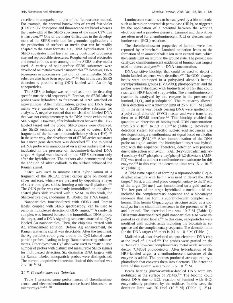
An electrochemical detection of 26-mer DNA sequenceswas performed using Faradaic impedance spectroscopy.249
A thiolated ODN containing the sequence complementaryto a part of the target was immobilized on a gold electrode.The probe was hybridized to a duplex constituted of the targetand a biotinylated signaling ODN forming a sandwichstructure. This double-stranded assembly was then treatedwith avidin-labeled HRP. The association of the enzyme labelwith the assembly enabled the biocatalytic oxidation of4-chloro-1-naphthol by H2O2 and formed a precipitate at theelectrode surface, which acted as a barrier for interfacialelectron transfer. The sensor can detect a DNA target at aconcentration of 2.3× 10⁻⁹ M (Table 2A). Liposomeslabeled with HRP also were used in connection with Faradaicimpedance spectroscopy.248 The first steps were the same asthose described by Patolsky et al.249 However, in this case,the bifunctional double-stranded assembly on the electrodesurface reacted with biotinylated HRP-labeled liposomes viaan avidin-biotin bridge (Figure 14). The DNA target wasdetected at a concentration of 6.5× 10⁻¹³ M (Table 2A).
Pividori and co-workers used a nylon membrane modifiedwith a DNA target integrated onto a transducer based ongraphite-epoxy composite (GEC) using different formats.In all cases, hybridization was detected electrochemicallywith a labeling system using an HRP-streptavidin conjugatein the presence of H2O2 and hydroquinone as a mediator.262-265
Encapsulation of streptavidin in siloxane-poly(propyleneoxide) (PPO) films also was reported.266 Biotinylated probeswere immobilized via streptavidin-biotin interactions. Afterhybridization, an avidin-peroxidase conjugate allowed thedetection of the target DNA by amperometric measurement.The HRP-biocatalyzed oxidation of 4-chloro-1-naphtol alsowas used.267 Electropolymerization involved a conductingpolymer bearing an ODN unit. HRP was linked through amolecular assembly using a streptavidin-biotin bridge. Theenzyme-catalyzed oxidation of 4-chloro-1-naphtol was fol-lowed by precipitation of the insoluble product, 4-chloro-1-naphton, on the support. This local deposition caused achange in conductivity, which was detected by scanningelectrochemical microscopy (SECM).
Cationic redox polymers containing osmium-bipyridinecomplexes coated on the electrode to improve the electron-transfer efficiency between the enzyme reaction and theelectrode were used for the first time by Heller and
DNA Biosensors and Microarrays Chemical Reviews, 2008, Vol. 108, No. 1 123
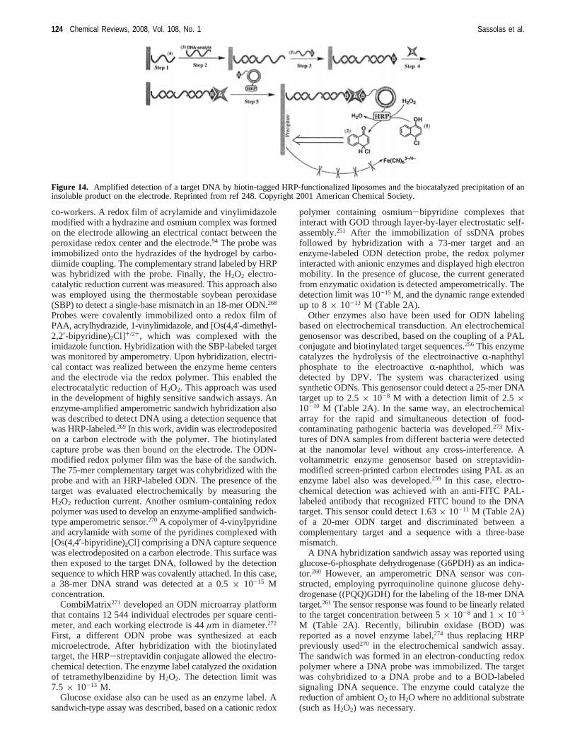
co-workers. A redox film of acrylamide and vinylimidazolemodified with a hydrazine and osmium complex was formedon the electrode allowing an electrical contact between theperoxidase redox center and the electrode.94 The probe wasimmobilized onto the hydrazides of the hydrogel by carbo-diimide coupling. The complementary strand labeled by HRPwas hybridized with the probe. Finally, the H2O2 electro-catalytic reduction current was measured. This approach alsowas employed using the thermostable soybean peroxidase(SBP) to detect a single-base mismatch in an 18-mer ODN.268
Probes were covalently immobilized onto a redox film ofPAA, acrylhydrazide, 1-vinylimidazole, and [Os(4,4′-dimethyl-2,2′-bipyridine)2Cl]+/2+, which was complexed with theimidazole function. Hybridization with the SBP-labeled targetwas monitored by amperometry. Upon hybridization, electri-cal contact was realized between the enzyme heme centersand the electrode via the redox polymer. This enabled theelectrocatalytic reduction of H2O2. This approach was usedin the development of highly sensitive sandwich assays. Anenzyme-amplified amperometric sandwich hybridization alsowas described to detect DNA using a detection sequence thatwas HRP-labeled.269 In this work, avidin was electrodepositedon a carbon electrode with the polymer. The biotinylatedcapture probe was then bound on the electrode. The ODN-modified redox polymer film was the base of the sandwich.The 75-mer complementary target was cohybridized with theprobe and with an HRP-labeled ODN. The presence of thetarget was evaluated electrochemically by measuring theH2O2 reduction current. Another osmium-containing redoxpolymer was used to develop an enzyme-amplified sandwich-type amperometric sensor.270 A copolymer of 4-vinylpyridineand acrylamide with some of the pyridines complexed with[Os(4,4′-bipyridine)2Cl] comprising a DNA capture sequencewas electrodeposited on a carbon electrode. This surface wasthen exposed to the target DNA, followed by the detectionsequence to which HRP was covalently attached. In this case,a 38-mer DNA strand was detected at a 0.5× 10⁻¹⁵ Mconcentration.
CombiMatrix271 developed an ODN microarray platformthat contains 12 544 individual electrodes per square centi-meter, and each working electrode is 44µm in diameter.272
First, a different ODN probe was synthesized at eachmicroelectrode. After hybridization with the biotinylatedtarget, the HRP-streptavidin conjugate allowed the electro-chemical detection. The enzyme label catalyzed the oxidationof tetramethylbenzidine by H2O2. The detection limit was7.5 × 10-13 M.
Glucose oxidase also can be used as an enzyme label. Asandwich-type assay was described, based on a cationic redox
polymer containing osmium-bipyridine complexes thatinteract with GOD through layer-by-layer electrostatic self-assembly.251 After the immobilization of ssDNA probesfollowed by hybridization with a 73-mer target and anenzyme-labeled ODN detection probe, the redox polymerinteracted with anionic enzymes and displayed high electronmobility. In the presence of glucose, the current generatedfrom enzymatic oxidation is detected amperometrically. Thedetection limit was 10-15 M, and the dynamic range extendedup to 8× 10-13 M (Table 2A).
Other enzymes also have been used for ODN labelingbased on electrochemical transduction. An electrochemicalgenosensor was described, based on the coupling of a PALconjugate and biotinylated target sequences.256 This enzymecatalyzes the hydrolysis of the electroinactiveR-naphthylphosphate to the electroactiveR-naphthol, which wasdetected by DPV. The system was characterized usingsynthetic ODNs. This genosensor could detect a 25-mer DNAtarget up to 2.5× 10⁻⁸ M with a detection limit of 2.5×10⁻¹⁰ M (Table 2A). In the same way, an electrochemicalarray for the rapid and simultaneous detection of food-contaminating pathogenic bacteria was developed.273 Mix-tures of DNA samples from different bacteria were detectedat the nanomolar level without any cross-interference. Avoltammetric enzyme genosensor based on streptavidin-modified screen-printed carbon electrodes using PAL as anenzyme label also was developed.259 In this case, electro-chemical detection was achieved with an anti-FITC PAL-labeled antibody that recognized FITC bound to the DNAtarget. This sensor could detect 1.63× 10⁻¹¹ M (Table 2A)of a 20-mer ODN target and discriminated between acomplementary target and a sequence with a three-basemismatch.
A DNA hybridization sandwich assay was reported usingglucose-6-phosphate dehydrogenase (G6PDH) as an indica-tor.260 However, an amperometric DNA sensor was con-structed, employing pyrroquinoline quinone glucose dehy-drogenase ((PQQ)GDH) for the labeling of the 18-mer DNAtarget.261 The sensor response was found to be linearly relatedto the target concentration between 5× 10⁻⁸ and 1× 10⁻⁵
M (Table 2A). Recently, bilirubin oxidase (BOD) wasreported as a novel enzyme label,274 thus replacing HRPpreviously used270 in the electrochemical sandwich assay.The sandwich was formed in an electron-conducting redoxpolymer where a DNA probe was immobilized. The targetwas cohybridized to a DNA probe and to a BOD-labeledsignaling DNA sequence. The enzyme could catalyze thereduction of ambient O2 to H2O where no additional substrate(such as H2O2) was necessary.
Figure 14. Amplified detection of a target DNA by biotin-tagged HRP-functionalized liposomes and the biocatalyzed precipitation of aninsoluble product on the electrode. Reprinted from ref 248. Copyright 2001 American Chemical Society.
124 Chemical Reviews, 2008, Vol. 108, No. 1 Sassolas et al.
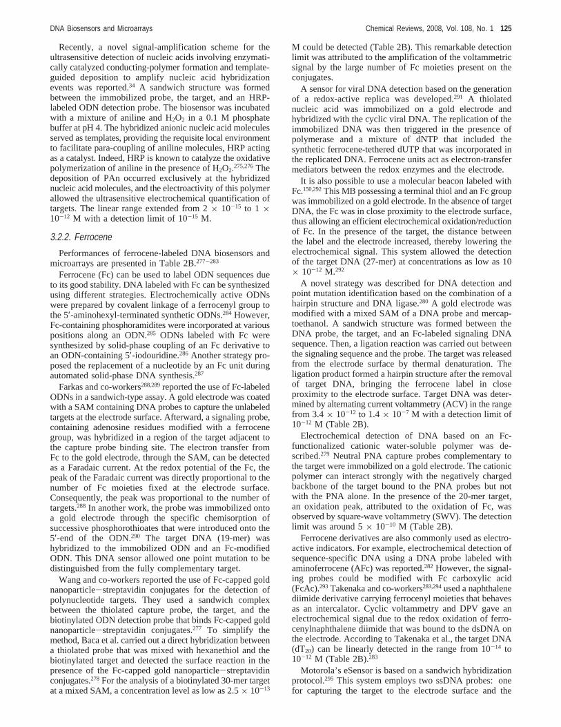
Recently, a novel signal-amplification scheme for theultrasensitive detection of nucleic acids involving enzymati-cally catalyzed conducting-polymer formation and template-guided deposition to amplify nucleic acid hybridizationevents was reported.34 A sandwich structure was formedbetween the immobilized probe, the target, and an HRP-labeled ODN detection probe. The biosensor was incubatedwith a mixture of aniline and H2O2 in a 0.1 M phosphatebuffer at pH 4. The hybridized anionic nucleic acid moleculesserved as templates, providing the requisite local environmentto facilitate para-coupling of aniline molecules, HRP actingas a catalyst. Indeed, HRP is known to catalyze the oxidativepolymerization of aniline in the presence of H2O2.275,276Thedeposition of PAn occurred exclusively at the hybridizednucleic acid molecules, and the electroactivity of this polymerallowed the ultrasensitive electrochemical quantification oftargets. The linear range extended from 2× 10-15 to 1 ×10-12 M with a detection limit of 10-15 M.
3.2.2. Ferrocene
Performances of ferrocene-labeled DNA biosensors andmicroarrays are presented in Table 2B.277-283
Ferrocene (Fc) can be used to label ODN sequences dueto its good stability. DNA labeled with Fc can be synthesizedusing different strategies. Electrochemically active ODNswere prepared by covalent linkage of a ferrocenyl group tothe 5′-aminohexyl-terminated synthetic ODNs.284 However,Fc-containing phosphoramidites were incorporated at variouspositions along an ODN.285 ODNs labeled with Fc weresynthesized by solid-phase coupling of an Fc derivative toan ODN-containing 5′-iodouridine.286 Another strategy pro-posed the replacement of a nucleotide by an Fc unit duringautomated solid-phase DNA synthesis.287
Farkas and co-workers288,289reported the use of Fc-labeledODNs in a sandwich-type assay. A gold electrode was coatedwith a SAM containing DNA probes to capture the unlabeledtargets at the electrode surface. Afterward, a signaling probe,containing adenosine residues modified with a ferrocenegroup, was hybridized in a region of the target adjacent tothe capture probe binding site. The electron transfer fromFc to the gold electrode, through the SAM, can be detectedas a Faradaic current. At the redox potential of the Fc, thepeak of the Faradaic current was directly proportional to thenumber of Fc moieties fixed at the electrode surface.Consequently, the peak was proportional to the number oftargets.288 In another work, the probe was immobilized ontoa gold electrode through the specific chemisorption ofsuccessive phosphorothioates that were introduced onto the5′-end of the ODN.290 The target DNA (19-mer) washybridized to the immobilized ODN and an Fc-modifiedODN. This DNA sensor allowed one point mutation to bedistinguished from the fully complementary target.
Wang and co-workers reported the use of Fc-capped goldnanoparticle-streptavidin conjugates for the detection ofpolynucleotide targets. They used a sandwich complexbetween the thiolated capture probe, the target, and thebiotinylated ODN detection probe that binds Fc-capped goldnanoparticle-streptavidin conjugates.277 To simplify themethod, Baca et al. carried out a direct hybridization betweena thiolated probe that was mixed with hexanethiol and thebiotinylated target and detected the surface reaction in thepresence of the Fc-capped gold nanoparticle-streptavidinconjugates.278 For the analysis of a biotinylated 30-mer targetat a mixed SAM, a concentration level as low as 2.5× 10⁻¹³
M could be detected (Table 2B). This remarkable detectionlimit was attributed to the amplification of the voltammetricsignal by the large number of Fc moieties present on theconjugates.
A sensor for viral DNA detection based on the generationof a redox-active replica was developed.291 A thiolatednucleic acid was immobilized on a gold electrode andhybridized with the cyclic viral DNA. The replication of theimmobilized DNA was then triggered in the presence ofpolymerase and a mixture of dNTP that included thesynthetic ferrocene-tethered dUTP that was incorporated inthe replicated DNA. Ferrocene units act as electron-transfermediators between the redox enzymes and the electrode.
It is also possible to use a molecular beacon labeled withFc.150,292This MB possessing a terminal thiol and an Fc groupwas immobilized on a gold electrode. In the absence of targetDNA, the Fc was in close proximity to the electrode surface,thus allowing an efficient electrochemical oxidation/reductionof Fc. In the presence of the target, the distance betweenthe label and the electrode increased, thereby lowering theelectrochemical signal. This system allowed the detectionof the target DNA (27-mer) at concentrations as low as 10× 10-12 M.292
A novel strategy was described for DNA detection andpoint mutation identification based on the combination of ahairpin structure and DNA ligase.280 A gold electrode wasmodified with a mixed SAM of a DNA probe and mercap-toethanol. A sandwich structure was formed between theDNA probe, the target, and an Fc-labeled signaling DNAsequence. Then, a ligation reaction was carried out betweenthe signaling sequence and the probe. The target was releasedfrom the electrode surface by thermal denaturation. Theligation product formed a hairpin structure after the removalof target DNA, bringing the ferrocene label in closeproximity to the electrode surface. Target DNA was deter-mined by alternating current voltammetry (ACV) in the rangefrom 3.4× 10⁻¹² to 1.4× 10⁻⁷ M with a detection limit of10-12 M (Table 2B).
Electrochemical detection of DNA based on an Fc-functionalized cationic water-soluble polymer was de-scribed.279 Neutral PNA capture probes complementary tothe target were immobilized on a gold electrode. The cationicpolymer can interact strongly with the negatively chargedbackbone of the target bound to the PNA probes but notwith the PNA alone. In the presence of the 20-mer target,an oxidation peak, attributed to the oxidation of Fc, wasobserved by square-wave voltammetry (SWV). The detectionlimit was around 5× 10⁻¹⁰ M (Table 2B).
Ferrocene derivatives are also commonly used as electro-active indicators. For example, electrochemical detection ofsequence-specific DNA using a DNA probe labeled withaminoferrocene (AFc) was reported.282 However, the signal-ing probes could be modified with Fc carboxylic acid(FcAc).293 Takenaka and co-workers283,294used a naphthalenediimide derivative carrying ferrocenyl moieties that behavesas an intercalator. Cyclic voltammetry and DPV gave anelectrochemical signal due to the redox oxidation of ferro-cenylnaphthalene diimide that was bound to the dsDNA onthe electrode. According to Takenaka et al., the target DNA(dT20) can be linearly detected in the range from 10-14 to10-12 M (Table 2B).283
Motorola’s eSensor is based on a sandwich hybridizationprotocol.295 This system employs two ssDNA probes: onefor capturing the target to the electrode surface and the
DNA Biosensors and Microarrays Chemical Reviews, 2008, Vol. 108, No. 1 125
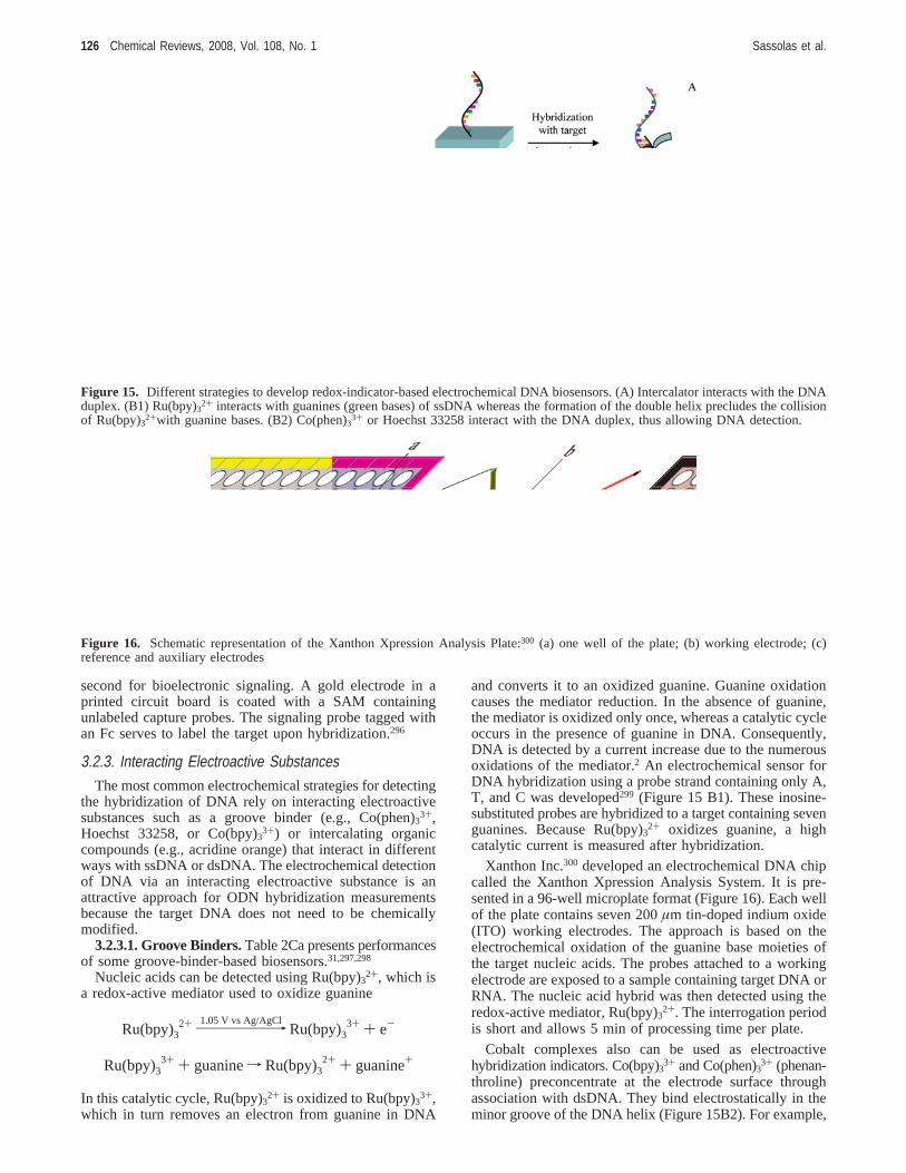
second for bioelectronic signaling. A gold electrode in aprinted circuit board is coated with a SAM containingunlabeled capture probes. The signaling probe tagged withan Fc serves to label the target upon hybridization.296
3.2.3. Interacting Electroactive SubstancesThe most common electrochemical strategies for detecting
the hybridization of DNA rely on interacting electroactivesubstances such as a groove binder (e.g., Co(phen)3
3+,Hoechst 33258, or Co(bpy)3
3+) or intercalating organiccompounds (e.g., acridine orange) that interact in differentways with ssDNA or dsDNA. The electrochemical detectionof DNA via an interacting electroactive substance is anattractive approach for ODN hybridization measurementsbecause the target DNA does not need to be chemicallymodified.
3.2.3.1. Groove Binders.Table 2Ca presents performancesof some groove-binder-based biosensors.31,297,298
Nucleic acids can be detected using Ru(bpy)32+, which is
a redox-active mediator used to oxidize guanine
In this catalytic cycle, Ru(bpy)32+ is oxidized to Ru(bpy)33+,
which in turn removes an electron from guanine in DNA
and converts it to an oxidized guanine. Guanine oxidationcauses the mediator reduction. In the absence of guanine,the mediator is oxidized only once, whereas a catalytic cycleoccurs in the presence of guanine in DNA. Consequently,DNA is detected by a current increase due to the numerousoxidations of the mediator.2 An electrochemical sensor forDNA hybridization using a probe strand containing only A,T, and C was developed299 (Figure 15 B1). These inosine-substituted probes are hybridized to a target containing sevenguanines. Because Ru(bpy)3
2+ oxidizes guanine, a highcatalytic current is measured after hybridization.
Xanthon Inc.300 developed an electrochemical DNA chipcalled the Xanthon Xpression Analysis System. It is pre-sented in a 96-well microplate format (Figure 16). Each wellof the plate contains seven 200µm tin-doped indium oxide(ITO) working electrodes. The approach is based on theelectrochemical oxidation of the guanine base moieties ofthe target nucleic acids. The probes attached to a workingelectrode are exposed to a sample containing target DNA orRNA. The nucleic acid hybrid was then detected using theredox-active mediator, Ru(bpy)3
2+. The interrogation periodis short and allows 5 min of processing time per plate.
Cobalt complexes also can be used as electroactivehybridization indicators. Co(bpy)3
3+ and Co(phen)33+ (phenan-
throline) preconcentrate at the electrode surface throughassociation with dsDNA. They bind electrostatically in theminor groove of the DNA helix (Figure 15B2). For example,
Figure 15. Different strategies to develop redox-indicator-based electrochemical DNA biosensors. (A) Intercalator interacts with the DNAduplex. (B1) Ru(bpy)32+ interacts with guanines (green bases) of ssDNA whereas the formation of the double helix precludes the collisionof Ru(bpy)32+with guanine bases. (B2) Co(phen)3
3+ or Hoechst 33258 interact with the DNA duplex, thus allowing DNA detection.
Figure 16. Schematic representation of the Xanthon Xpression Analysis Plate:300 (a) one well of the plate; (b) working electrode; (c)reference and auxiliary electrodes
Ru(bpy)32+98
1.05 V vs Ag/AgClRu(bpy)3
3+ + e-
Ru(bpy)33+ + guaninef Ru(bpy)3
2+ + guanine+
126 Chemical Reviews, 2008, Vol. 108, No. 1 Sassolas et al.
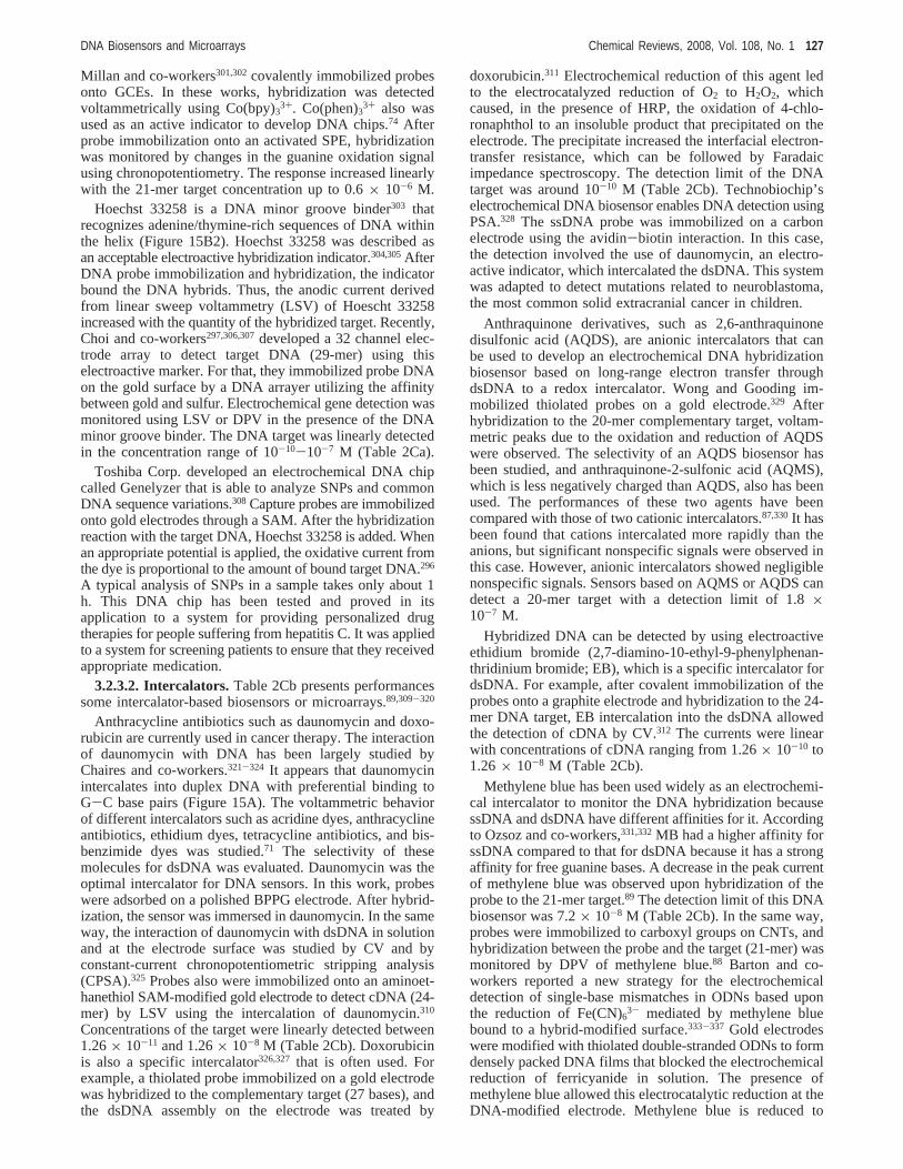
Millan and co-workers301,302covalently immobilized probesonto GCEs. In these works, hybridization was detectedvoltammetrically using Co(bpy)3
3+. Co(phen)33+ also wasused as an active indicator to develop DNA chips.74 Afterprobe immobilization onto an activated SPE, hybridizationwas monitored by changes in the guanine oxidation signalusing chronopotentiometry. The response increased linearlywith the 21-mer target concentration up to 0.6× 10⁻⁶ M.
Hoechst 33258 is a DNA minor groove binder303 thatrecognizes adenine/thymine-rich sequences of DNA withinthe helix (Figure 15B2). Hoechst 33258 was described asan acceptable electroactive hybridization indicator.304,305AfterDNA probe immobilization and hybridization, the indicatorbound the DNA hybrids. Thus, the anodic current derivedfrom linear sweep voltammetry (LSV) of Hoescht 33258increased with the quantity of the hybridized target. Recently,Choi and co-workers297,306,307developed a 32 channel elec-trode array to detect target DNA (29-mer) using thiselectroactive marker. For that, they immobilized probe DNAon the gold surface by a DNA arrayer utilizing the affinitybetween gold and sulfur. Electrochemical gene detection wasmonitored using LSV or DPV in the presence of the DNAminor groove binder. The DNA target was linearly detectedin the concentration range of 10-10-10-7 M (Table 2Ca).
Toshiba Corp. developed an electrochemical DNA chipcalled Genelyzer that is able to analyze SNPs and commonDNA sequence variations.308 Capture probes are immobilizedonto gold electrodes through a SAM. After the hybridizationreaction with the target DNA, Hoechst 33258 is added. Whenan appropriate potential is applied, the oxidative current fromthe dye is proportional to the amount of bound target DNA.296
A typical analysis of SNPs in a sample takes only about 1h. This DNA chip has been tested and proved in itsapplication to a system for providing personalized drugtherapies for people suffering from hepatitis C. It was appliedto a system for screening patients to ensure that they receivedappropriate medication.
3.2.3.2. Intercalators.Table 2Cb presents performancessome intercalator-based biosensors or microarrays.89,309-320
Anthracycline antibiotics such as daunomycin and doxo-rubicin are currently used in cancer therapy. The interactionof daunomycin with DNA has been largely studied byChaires and co-workers.321-324 It appears that daunomycinintercalates into duplex DNA with preferential binding toG-C base pairs (Figure 15A). The voltammetric behaviorof different intercalators such as acridine dyes, anthracyclineantibiotics, ethidium dyes, tetracycline antibiotics, and bis-benzimide dyes was studied.71 The selectivity of thesemolecules for dsDNA was evaluated. Daunomycin was theoptimal intercalator for DNA sensors. In this work, probeswere adsorbed on a polished BPPG electrode. After hybrid-ization, the sensor was immersed in daunomycin. In the sameway, the interaction of daunomycin with dsDNA in solutionand at the electrode surface was studied by CV and byconstant-current chronopotentiometric stripping analysis(CPSA).325 Probes also were immobilized onto an aminoet-hanethiol SAM-modified gold electrode to detect cDNA (24-mer) by LSV using the intercalation of daunomycin.310
Concentrations of the target were linearly detected between1.26× 10-11 and 1.26× 10⁻⁸ M (Table 2Cb). Doxorubicinis also a specific intercalator326,327 that is often used. Forexample, a thiolated probe immobilized on a gold electrodewas hybridized to the complementary target (27 bases), andthe dsDNA assembly on the electrode was treated by
doxorubicin.311 Electrochemical reduction of this agent ledto the electrocatalyzed reduction of O2 to H2O2, whichcaused, in the presence of HRP, the oxidation of 4-chlo-ronaphthol to an insoluble product that precipitated on theelectrode. The precipitate increased the interfacial electron-transfer resistance, which can be followed by Faradaicimpedance spectroscopy. The detection limit of the DNAtarget was around 10-10 M (Table 2Cb). Technobiochip’selectrochemical DNA biosensor enables DNA detection usingPSA.328 The ssDNA probe was immobilized on a carbonelectrode using the avidin-biotin interaction. In this case,the detection involved the use of daunomycin, an electro-active indicator, which intercalated the dsDNA. This systemwas adapted to detect mutations related to neuroblastoma,the most common solid extracranial cancer in children.
Anthraquinone derivatives, such as 2,6-anthraquinonedisulfonic acid (AQDS), are anionic intercalators that canbe used to develop an electrochemical DNA hybridizationbiosensor based on long-range electron transfer throughdsDNA to a redox intercalator. Wong and Gooding im-mobilized thiolated probes on a gold electrode.329 Afterhybridization to the 20-mer complementary target, voltam-metric peaks due to the oxidation and reduction of AQDSwere observed. The selectivity of an AQDS biosensor hasbeen studied, and anthraquinone-2-sulfonic acid (AQMS),which is less negatively charged than AQDS, also has beenused. The performances of these two agents have beencompared with those of two cationic intercalators.87,330It hasbeen found that cations intercalated more rapidly than theanions, but significant nonspecific signals were observed inthis case. However, anionic intercalators showed negligiblenonspecific signals. Sensors based on AQMS or AQDS candetect a 20-mer target with a detection limit of 1.8×10⁻⁷ M.
Hybridized DNA can be detected by using electroactiveethidium bromide (2,7-diamino-10-ethyl-9-phenylphenan-thridinium bromide; EB), which is a specific intercalator fordsDNA. For example, after covalent immobilization of theprobes onto a graphite electrode and hybridization to the 24-mer DNA target, EB intercalation into the dsDNA allowedthe detection of cDNA by CV.312 The currents were linearwith concentrations of cDNA ranging from 1.26× 10⁻¹⁰ to1.26× 10-8 M (Table 2Cb).
Methylene blue has been used widely as an electrochemi-cal intercalator to monitor the DNA hybridization becausessDNA and dsDNA have different affinities for it. Accordingto Ozsoz and co-workers,331,332MB had a higher affinity forssDNA compared to that for dsDNA because it has a strongaffinity for free guanine bases. A decrease in the peak currentof methylene blue was observed upon hybridization of theprobe to the 21-mer target.89 The detection limit of this DNAbiosensor was 7.2× 10⁻⁸ M (Table 2Cb). In the same way,probes were immobilized to carboxyl groups on CNTs, andhybridization between the probe and the target (21-mer) wasmonitored by DPV of methylene blue.88 Barton and co-workers reported a new strategy for the electrochemicaldetection of single-base mismatches in ODNs based uponthe reduction of Fe(CN)6
3- mediated by methylene bluebound to a hybrid-modified surface.333-337 Gold electrodeswere modified with thiolated double-stranded ODNs to formdensely packed DNA films that blocked the electrochemicalreduction of ferricyanide in solution. The presence ofmethylene blue allowed this electrocatalytic reduction at theDNA-modified electrode. Methylene blue is reduced to
DNA Biosensors and Microarrays Chemical Reviews, 2008, Vol. 108, No. 1 127
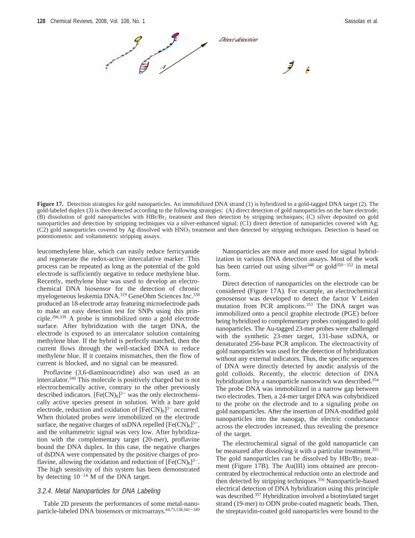
leucomethylene blue, which can easily reduce ferricyanideand regenerate the redox-active intercalative marker. Thisprocess can be repeated as long as the potential of the goldelectrode is sufficiently negative to reduce methylene blue.Recently, methylene blue was used to develop an electro-chemical DNA biosensor for the detection of chronicmyelogeneous leukemia DNA.319 GeneOhm Sciences Inc.338
produced an 18-electrode array featuring microelectrode padsto make an easy detection test for SNPs using this prin-ciple.296,339 A probe is immobilized onto a gold electrodesurface. After hybridization with the target DNA, theelectrode is exposed to an intercalator solution containingmethylene blue. If the hybrid is perfectly matched, then thecurrent flows through the well-stacked DNA to reducemethylene blue. If it contains mismatches, then the flow ofcurrent is blocked, and no signal can be measured.
Proflavine (3,6-diaminoacridine) also was used as anintercalator.340 This molecule is positively charged but is notelectrochemically active, contrary to the other previouslydescribed indicators. [Fe(CN)6]3- was the only electrochemi-cally active species present in solution. With a bare goldelectrode, reduction and oxidation of [Fe(CN)6]3- occurred.When thiolated probes were immobilized on the electrodesurface, the negative charges of ssDNA repelled [Fe(CN)6]3-,and the voltammetric signal was very low. After hybridiza-tion with the complementary target (20-mer), proflavinebound the DNA duplex. In this case, the negative chargesof dsDNA were compensated by the positive charges of pro-flavine, allowing the oxidation and reduction of [Fe(CN)6]3-.The high sensitivity of this system has been demonstratedby detecting 10-14 M of the DNA target.
3.2.4. Metal Nanoparticles for DNA Labeling
Table 2D presents the performances of some metal-nano-particle-labeled DNA biosensors or microarrays.64,73,138,341-349
Nanoparticles are more and more used for signal hybrid-ization in various DNA detection assays. Most of the workhas been carried out using silver348 or gold350-352 in metalform.
Direct detection of nanoparticles on the electrode can beconsidered (Figure 17A). For example, an electrochemicalgenosensor was developed to detect the factor V Leidenmutation from PCR amplicons.353 The DNA target wasimmobilized onto a pencil graphite electrode (PGE) beforebeing hybridized to complementary probes conjugated to goldnanoparticles. The Au-tagged 23-mer probes were challengedwith the synthetic 23-mer target, 131-base ssDNA, ordenaturated 256-base PCR amplicon. The electroactivity ofgold nanoparticles was used for the detection of hybridizationwithout any external indicators. Thus, the specific sequencesof DNA were directly detected by anodic analysis of thegold colloids. Recently, the electric detection of DNAhybridization by a nanoparticle nanoswitch was described.354
The probe DNA was immobilized in a narrow gap betweentwo electrodes. Then, a 24-mer target DNA was cohybridizedto the probe on the electrode and to a signaling probe ongold nanoparticles. After the insertion of DNA-modified goldnanoparticles into the nanogap, the electric conductanceacross the electrodes increased, thus revealing the presenceof the target.
The electrochemical signal of the gold nanoparticle canbe measured after dissolving it with a particular treatment.355
The gold nanoparticles can be dissolved by HBr/Br2 treat-ment (Figure 17B). The Au(III) ions obtained are precon-centrated by electrochemical reduction onto an electrode andthen detected by stripping techniques.356 Nanoparticle-basedelectrical detection of DNA hybridization using this principlewas described.357 Hybridization involved a biotinylated targetstrand (19-mer) to ODN probe-coated magnetic beads. Then,the streptavidin-coated gold nanoparticles were bound to the
Figure 17. Detection strategies for gold nanoparticles. An immobilized DNA strand (1) is hybridized to a gold-tagged DNA target (2). Thegold-labeled duplex (3) is then detected according to the following strategies: (A) direct detection of gold nanoparticles on the bare electrode;(B) dissolution of gold nanoparticles with HBr/Br2 treatment and then detection by stripping techniques; (C) silver deposited on goldnanoparticles and detection by stripping techniques via a silver-enhanced signal; (C1) direct detection of nanoparticles covered with Ag;(C2) gold nanoparticles covered by Ag dissolved with HNO3 treatment and then detected by stripping techniques. Detection is based onpotentiometric and voltammetric stripping assays.
128 Chemical Reviews, 2008, Vol. 108, No. 1 Sassolas et al.
captured target before a dissolution process with HBr/Br2.Finally, the dissolved gold tags were detected at a disposablethick-film carbon electrode by PSA.
After hybridization, silver deposition on the gold nano-particles can be done to enhance the electrochemical signal(Figure 17C).64,342,355,358-362 In this case, gold nanoparticlescan be treated with a silver enhancer solution containing areducing agent such as hydroquinone. In its presence,colloidal gold could catalyze the chemical reduction of silverions (from silver lactate or silver acetate) to metallic silveron the nanoparticle surface.64 Nevertheless, the reducingagent that triggers silver precipitation is difficult to control,and nonspecific silver deposition on the substrate cannot beavoided. To eliminate this drawback, the chemical silverreduction can be replaced by an electrochemical depositionprocess,363 which has been reported previously for otherapplications related to trace metal quantitations.364,365Aftersilver deposition using a reducing agent or an electrochemicalpotential, two techniques can be considered for DNAdetection. In the first case, the silver deposition on the goldnanoparticles and the strategy based on the metal dissolutionhave been combined (Figure 17C2). For example, a bioti-nylated oligomer target was hybridized to the biotinylatedDNA probe immobilized on a streptavidin-coated magneticlatex sphere.362 Streptavidin-coated colloidal gold was thenbound to the target. Silver metal was deposited on the goldnanoparticles before being dissolved in a solution containingHNO3 and detected at a disposable thick-carbon electrodeusing PSA. The use of an ODN-functionalized electrocon-ducting polymer coupled with a gold nanoparticle-basedhybridization indicator was reported.342 Poly(2-aminobenzoicacid) was formed on an ITO electrode and was used tocovalently immobilize an amino-modified probe. Signaltransduction was achieved by binding the gold nanoparticlelabel to the hybridized target. The amount of silver depositedon the gold nanoparticle label was determined by measuringthe electrochemical oxidative silver dissolution responseduring a PSA scan. The sensor could linearly detect a 16-mer target from 10× 10-12 to 10× 10-9 M (Table 2D). Inthe second case, silver deposition on the gold nanoparticlewas successful but not the acidic dissolution (Figure 17C1).An electrochemical detection method based on the catalyticprecipitation of silver onto the gold nanoparticle label alsowas reported.64 In this work, a 32-mer target DNA wasadsorbed on a chitosan-modified GCE. After silver depositionon the gold nanoparticles, hybridization was monitored byDPV. The sensor response showed a linear relationship tothe DNA target from 10-10 to 10-8 M (Table 2D). The silver-deposition technique also was exploited to construct a DNAsensor based on conductivity measurements. The target DNAwas detected at concentrations as low as 5× 10-13 M (Table2D).346
Numerous protocols rely on the use of colloidal gold tags.Nevertheless, silver nanoparticles can be used instead of goldones.348 The 30-base target DNA was immobilized on goldcolloid particles associated with a cysteamine monolayer onthe gold electrode surface. Then, the target was hybridizedwith a silver nanoparticle-ODN DNA probe. The silverparticles, which were bound to the hybrids, were dissolvedby HNO3 treatment. Anodic stripping voltammetry (ASV)allowed the detection of the DNA target in the range from10-11 to 8 × 10⁻¹⁰ M (Table 2D).
It is also possible to use polymeric microbeads carryinggold nanoparticle tags. For instance, polystyrene beadscarrying numerous gold nanoparticles were reported.341 The
DNA target (19-mer) labeled with the gold nanoparticlecarrier beads were hybridized with probes captured onmagnetic beads. The hybridization process was followed bya nanoparticle-promoted precipitation of gold. A gold dis-solution process was achieved prior to electrochemicalstripping detection. The detection limit was around 6× 10-12
M (Table 2D).Iron-containing particles were used for electrochemical
detection of DNA hybridization.349 Two strategies have beencontemplated. The first possibility involved probes labeledwith iron-gold core-shell nanoparticles. The biotinylatedtarget, immobilized onto streptavidin-coated polystyrenebeads, was hybridized to the iron-gold-particle-labeledprobe. The second protocol relied on the use of commercialiron-containing magnetic beads. The probes were im-mobilized onto the chitosan-modified microwell and werethen hybridized with the magnetic-bead-labeled target. Inboth cases, the captured iron-containing particles weredissolved, and the released iron was quantified by cathodicstripping voltammetry (CSV) in the presence of the 1-nitroso-2-naphthol ligand and a bromate catalyst. The 32-mer DNAtarget detection, based on gold-coated iron nanoparticle tags,enabled a detection limit of around 4.7× 10⁻⁹ M. The 37-base DNA detection based on magnetic-sphere redox tagspresented a detection limit of 1.6× 10⁻⁹ M (Table 2D).
Quantum dots also can be used to electrochemically detectthe DNA target. After the ODN sample entrapment into aPPy film, the surface was exposed to complementary probesor CdS-tagged ODN probes.137,138 According to them, theuse of CdS nanoparticle-labeled ODNs resulted in a highersensitivity and a significantly improved detection limit ofthe sensor. In this case, the impedance signal was linear tothe logarithm of ODN concentration between 3.7× 10⁻⁹
and 3.7× 10⁻⁷ M (Table 2D).
3.2.5. Label-Free Electrochemical Detection
Table 2E presents performances of some label-free DNAbiosensors or microarrays.75,101,102,134,136,366-377
DNA detection can be based on the natural electroactivityof the nucleotide residues present in DNA. This has beenexploited in particular by Palecek group who showed thatDNA and RNA are electroactive compounds producingreduction and oxidation signals after hybridization.378 Signalsof adenine, cytosine, and guanine can be observed onoscillopolarograms of ssDNA, whereas these signals areabsent with dsDNA. Guanine was described as the mostredox-active nitrogeneous base in DNA.
Immobilized guanine-free probes can be used to developa label-free electrochemical DNA hybridization biosensor.Inosine can be substituted with guanine.75,379 While theinosine moiety preferentially forms a base pair with the targetcytosine residue, its oxidation signal is well-separated fromthe guanine response. After hybridization, the appearanceof the guanine oxidation signal of the 29-mer target allowedthe detection of the DNA hybrid by PSA. The detection limitwas estimated to be around 1.25× 10⁻⁸ M (Table 2E).
Pividori and co-workers also exploited the guanine oxida-tion signal for detecting duplex formation.79 For example,an inosine-substituted probe was immobilized by wet adsorp-tion on the GEC.376 In this case, the 23-mer target wasdetected by DPV using the oxidation signal of guanine. Thelinear range for target detection was observed from 3.95×10⁻⁶ to 1.98× 10⁻⁵ M (Table 2E). Recently, a genomagneticassay was developed using a GEC electrode containing a
DNA Biosensors and Microarrays Chemical Reviews, 2008, Vol. 108, No. 1 129
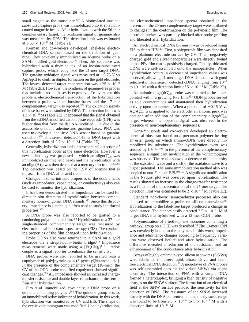
small magnet as the transducer.377 A biotinylated inosine-substituted capture probe was immobilized onto streptavidin-coated magnetic beads. After hybridization with the 56-mercomplementary target, the oxidation signal of guanine alsowas measured by DPV. The detection limit was estimatedat 9.68× 10⁻¹² M (Table 2E).
Kerman and co-workers developed label-free electro-chemical DNA sensors based on the oxidation of gua-nine. They covalently immobilized an adenine probe on aSAM-modified gold electrode.372 Then, this sequence washybridized with a thymine tag of an inosine-substitutedcapture probe, which recognized the 21-mer target DNA.The guanine oxidation signal was measured at+0.73 V vsAg/AgCl to confirm duplex formation on the gold electrode.The lowest detected target concentration was 1.25× 10⁻⁹
M (Table 2E). However, the synthesis of guanine-free probesthat includes inosine bases is expensive. To overcome thisproblem, electrochemical transduction of the hybridizationbetween a probe without inosine bases and the 17-mercomplementary target was reported.374 The oxidation signalsof these bases were studied by DPV. The detection limit was1.2× 10-8 M (Table 2E). It appeared that the signal obtainedfrom the ssDNA-modified carbon paste electrode (CPE) washigher than that from the dsDNA-modified CPE due to theaccessible unbound adenine and guanine bases. PNA wasused to develop a label-free DNA sensor based on guanineoxidation.375 This sensor detected 14-mer DNA target witha detection limit of 2.7× 10⁻¹⁰ M (Table 2E).
Generally, hybridization and electrochemical detection ofthis hybridization occur at the same electrode. However, anew technology was proposed in which an oligo(T)25 wasimmobilized on magnetic beads and the hybridization withan oligo(A)25 was then detected at a mercury electrode.380,381
The detection was based on the CSV of adenine that isreleased from DNA after acid treatment.
Changes in some intrinsic properties of the double helix(such as impedance, capacitance, or conductivity) also canbe used to monitor the hybridization.
It has been demonstrated that impedance can be used fordirect in situ detection of hybridization between comple-mentary homo-oligomer DNA strands.382 Since this discov-ery, impedance is a technique often used to study interfacialproperties.383
A DNA probe was also reported to be grafted to aconducting polythiophene film.384 Hybridization to a 37-mersingle-stranded complementary target was measured byelectrochemical impedance spectroscopy (EIS). The conduct-ing properties of the film changed upon hybridization.
Probe ODNs also were attached to a SAM on a goldelectrode via a streptavidin-biotin bridge.114 Impedancemeasurements were made using a [Fe(CN)6]4-/3- redoxcouple as a signal reporter to enhance the sensitivity.
DNA probes were also reported to be grafted onto acopolymer of poly[pyrrole-co-4-(3-pyrrolyl)butanoic acid].In the presence of the complementary target (18-mer), theCV of the ODN probe-modified copolymer showed signifi-cant changes.385 AC impedance showed an increased charge-transfer resistance and double-layer capacitance of the sensorfilm after hybridization.
Piro et al. immobilized, covalently, a DNA probe on aquinone-containing polymer.386 The quinone group acts asan immobilized redox indicator of hybridization. In this work,hybridization was monitored by CV and EIS. The shape ofthe cyclic voltammogram was modified. Upon hybridization,
the electrochemical impedance spectra obtained in thepresence of the 20-mer complementary target were attributedto changes in the conformation on the polymeric film. Theelectrode surface was partially blocked after probe graftingand liberated after hybridization.
An electrochemical DNA biosensor was developed usingEIS to detect HIV.371 First, a polypyrrole film was depositedon a platinum electrode surface by CV. Then, negativelycharged gold and silver nanoparticles were directly boundonto a PPy film that is positively charged. Finally, thiolatedODNs were self-assembled onto the nanoparticles. Whenhybridization occurs, a decrease of impedance values wasobserved, allowing 21-mer target DNA detection with goodselectivity. This sensor detected cDNA ranging from 10-9
to 10-6 M with a detection limit of 5× 10⁻¹⁰ M (Table 2E).
An anionic oligo(dG)20 probe was reported to be incor-porated within a growing PPy film.133 ODN probes servedas sole counteranions and maintained their hybridizationactivity upon entrapment. When a potential of+0.15 V vsAg/AgCl was applied to the electrode, an anionic peak wasobtained after addition of the complementary oligo(dC)20
target whereas the opposite signal was observed in thepresence of noncomplementary sequences.
Korri-Youssoufi and co-workers developed an electro-chemical biosensor based on a precursor polymer bearingan ester group on which amino-ODNs were directly im-mobilized by substitution. The hybridization event wasstudied by CV.101,103In the presence of the complementarysequence, a significant modification in the voltammogramwas observed. The results showed a decrease of the intensityof the oxidation wave and a shift of the oxidation wave to ahigher potential. The same immobilization method also wascoupled to non-Faradaic EIS.102,103A significant modificationin the Nyquist plot was observed upon hybridization. Theresults showed an increase in the charge-transfer resistanceas a function of the concentration of the 25-mer target. Thedetection limit was estimated to be 2× 10⁻¹⁰ M (Table 2E).
Standard “top-down” semiconductor processes also canbe used to immobilize a probe on silicon nanowires.387
Hybridization to the label-free target produced a change inconductance. The authors easily detected 2.5× 10⁻¹¹ M oftarget DNA that hybridized with a 12-mer ODN probe.
Polymerization of a terthiophene monomer containing acarboxyl group on a GCE was described.93 The 19-mer ODNwas covalently bound to the polymer. In this work, imped-ance and admittance changes according to frequency varia-tion were observed before and after hybridization. Thedifference revealed a reduction of the resistance and anenhancement of the conductance after hybridization.
Arrays of highly ordered n-type silicon nanowires (SiNWs)were fabricated for direct rapid, ultrasensitive, and label-free electrical DNA detection.33 A monolayer of PNA probeswas self-assembled onto the individual SiNWs via silanechemistry. The interaction of PNA with a sample DNAformed a heteroduplex, bringing a high density of negativecharges on the SiNW surface. The formation of an electricalfield at the SiNW surface provided the sensitivity for thedetection of DNA. The resistance of the SiNW increasedlinearly with the DNA concentration, and the dynamic rangewas found to be from 2.5× 10⁻¹⁴ to 5 × 10-12 M with adetection limit of 10-14 M.
130 Chemical Reviews, 2008, Vol. 108, No. 1 Sassolas et al.
3.3. Gravimetric DNA Biosensors
3.3.1. Quartz Crystal Microbalance Sensors
A QCM sensor is a mass-sensitive sensor capable ofmeasuring very small mass changes.388 It consists of a thinquartz disc sandwiched between a pair of electrodes. Quartzis a piezoelectric material that deforms when an electric fieldis applied across the electrode. The quartz crystal has aresonant frequency dependent on the total oscillating mass.This frequency increases with an increase in material on theQCM surface. In addition to electrochemical and opticaldetection of DNA, mass sensors are also capable of detectinglabel-free ODNs.389,390 Table 3 presents performances ofsome QCM-based biosensors or microarrays.39,66,391-396
Willner and co-workers described a method to amplifyDNA detection involving the use of functional lipo-somes.391,392,397They described two strategies to amplify theODN-DNA processes.392 In the first case, a thiolated primerwas assembled on a gold-quartz crystal, followed byhybridization with the 27-mer analyte DNA. Then, thisdouble-stranded assembly interacted with a liposome func-tionalized with another ODN complementary to one part ofthe target. The detection limit was estimated to be 5× 10-12
M. In the second configuration, the double-stranded assemblyinteracted with a biotinylated ODN. The resulting assemblyinteracted with avidin and then with a biotin-functionalizedliposome. The use of liposomes containing ODNs allowedthe formation of dendritic structures. The lower limit ofdetection for the target by this amplification method was10-13 M (Table 3). Willner et al. described three methodsfor the amplified detection of a single-base mismatch in DNAwhere a guanine in the mutant gene substituted for an adeninein the normal gene.391 In the three cases, a thiolated primercomplementary to the analyte was assembled on a gold-quartz crystal. After hybridization, a biotinylated dCTP wasincorporated to the mutation site in the presence of DNApolymerase I. Thus, the biotin label indicated the presenceof the single-base mutation in the sequence. As previouslydescribed, the first strategy involved the association of avidin-and biotin-labeled liposomes. The second method used anAu nanoparticle-avidin conjugate whereas the third protocolrequired an avidin-PAL bioconjugate that bound the biotinlabel introduced in the polymerase-induced process. Thisenzyme catalyzed the oxidative hydrolysis of 5-bromo-4-chloro-3-indolyl phosphate to an insoluble product thatprecipitated on the gold-quartz crystal. The protocol usingan Au nanoparticle-avidin conjugate is the most sensitivemethod (detection limit of 3× 10-16 M, Table 3).
A system was developed for the detection of a pointmutation in DNA.395 For that, the authors immobilized assDNA probe on a QCM surface via streptavidin-biotininteraction. After hybridization, the MutS protein was usedto recognize mutation. The MutS application caused afrequency change that can be attributed to the MutS binding.DNA containing a single T:G mismatch and one to fourunpaired mutation(s) has been discriminated from perfectlymatched DNA down to target concentrations of 1× 10⁻⁹ M(Table 3).
A QCM DNA sensor was developed for the detection ofEscherichia coliO157:H7 using a nanoparticle amplificationmethod.393 A thiolated ssDNA probe specific to theE. coliO157:H7 eaeA gene was immobilized on the QCM surface.After bovine serum albumin blocking, the probe was exposedto the biotinylated target DNA; however, the frequency
change was negligible. Therefore, to amplify this frequencychange, streptavidin conjugated Fe3O4 nanoparticles wereattached to the target. This sensor could then detect DNAconcentrations as low as 10-12 M (Table 3).
Okahata and co-workers largely have used QCM to detectDNA. They immobilized a DNA probe that was comple-mentary to the EcoRI binding site of single-stranded M13phage DNA.394 In the presence of the target, the frequencydecreased with time. The minimum concentration that couldbe detected was 10-12 M (Table 3). A biotinylated DNAprobe also could be immobilized either via avidin, whichwas covalently attached to the QCM surface, or via elec-trostatic interaction with poly(allylamine hydrochloride).398
The authors also formed multilayer films of DNA bysuccessive deposition of avidin and poly(styrenesulfonate)to improve nucleic acid detection. A commercially available27 MHz QCM also was used to study the in situ binding ofavidin to a modified electrode, the immobilization of thesingle-stranded biotin DNA, and the hybridization with thecomplementary target.399 It appeared that the 27 MHz QCMwas 10 times more sensitive than the commonly used 9 MHzQCM. The same 27 MHz QCM also was used by im-mobilizing a 10-30-mer ODN on the surface.400 The effectof 1, 2, or 3 mismatching bases in the target sequence onhybridization was studied. When the number of mismatchingbases increased, the frequency increased, showing that thebinding amount clearly decreased.
DNA dendrimers also were used with numerous peripheralsingle-stranded arms as a probe to detectCryptosporidiumDNA.39 Adsorption and electropolymeric entrapment wereused to immobilize DNA dendrimers on the crystal. For bothimmobilization techniques, the response increased linearlywith the 38-mer target concentration up to 8× 10⁻⁶ M (100µg/mL) with a detection limit around 8× 10⁻⁸ M (1 µg/mL) (Table 3).
A DNA genetic sensor was developed for the diagnosisof â-thalassaemia using a QCM technique.66 An 18-mer DNAprobe complementary to the site of geneticâ-thalassaemiamutations was immobilized on the QCM electrodes. Differentmethods for immobilizing the DNA probe were tested.Mono- or multilayered DNA probes were formed by avidin-biotin interaction, chemisorption, or electrostatic adsorptionon a polyelectrolyte film. The DNA sensor based on theimmobilization of biotinylated DNA probe to avidin providesfast sensor response and high hybridization efficiency. Theoptimum concentration range of the target DNA sequenceis 8.4 × 10⁻⁶ M (50 µg/mL)-1.2 × 10-5 M (70 µg/mL)(Table 3).
A biotinylated single strand ODN was immobilized onstreptavidin, which is covalently attached to a dextran-modified thiol layer on the piezoelectric crystal.401 Thissystem could detect DNA that was complementary to theimmobilized probe with good selectivity and high reproduc-ibility.
Technobiochip328 described a QCM DNA sensor for thedetection of genetically modified organisms (GMOs). Aspreviously described, the gold-quartz surface is functionalizedby a biotinylated DNA probe layer through its high affinitywith streptavidin deposited on the surface.402 This DNAsensor calledµLibra is a reliable, fast, and cost-effectivemethod for GMO screening in food samples.
Although not very common, piezoelectric devices can befound in a multiarray format. A piezoelectric microarray wasdeveloped to detect the hepatitis B virus quantitatively in
DNA Biosensors and Microarrays Chemical Reviews, 2008, Vol. 108, No. 1 131
clinical samples.403 A bis-PNA probe was designed andimmobilized on the surface of the microarray. Every crystalcould oscillate independently without interfering with theothers.
3.3.2. Microcantilever SensorsMicrocantilever sensors recently have emerged as a
promising tool to detect biomolecular interactions.404-407
These systems offer many advantages compared to conven-tional sensors: label-free detection, high precision, reliability,reduced size, easy manufacture of multielement sensor arrays,and small thermal mass. Microcantilever sensors are basedon a response due to either surface stress variation or massloading. Interaction between an immobilized ligand (e.g., aDNA probe) and an analyte (e.g., a DNA target) causes achange of the surface stress of the cantilever and can bedetected as changes in the cantilever deflection (∆x).
Thiolated ODNs were covalently immobilized on the gold-covered side of the cantilevers408 (Figure 18). One cantileverwas functionalized with a 12-mer ODN, whereas the otherwas functionalized with a 16-mer ODN (Figure 18A). Thearrays were brought to equilibrium in a hybridization bufferuntil the differential signal became stable. The complemen-tary 16-mer sequence (Figure 18B) and the complementary12-mer ODN (Figure 18C) were then injected into the liquidcell. Hybridization of the target to the matching ODNsimmobilized on the cantilever surfaces occurred, causing thecantilever deflection.
Hansen’s group developed microcantilever-based sensorsfor DNA detection. They immobilized a 5′-thiol-modifiedprobe on the Au-coated cantilever surface. The functionalizedcantilever was then exposed to a solution containing comple-mentary target ssDNA. They demonstrated that cantileverdeflection varied with the complementary oligomer length.409
A change in deflection was caused by hybridization of assDNA probe (20-mer) complementary to the distal end oftarget ssDNA of four different lengths (9-, 10-, 15-, and 20-mers). They also could discriminate full and partial comple-mentary sequences.410 For example, hybridization of 10-mercomplementary target resulted in net positive deflectionwhereas hybridization with sequences containing one or twointernal mismatches resulted in net negative deflection.
Covalently immobilization of ssDNA onto AFM tips alsowas reported.104 Interaction between the cantilever and thetarget was quantified by obtaining the percent separationdistance (PSD)
where L1 is the separation distance (nm) for the buffersolution andL2 is the separation distance (nm) for the samebuffer solution containing the target ssDNA. They found acorrelation between the concentration of the target in themedium and the PSD value. After hybridization between theimmobilized probe and its complementary sequence, the PSDvalues significantly increased.
4. ConclusionDifferent strategies used to develop DNA biosensors and
DNA microarrays have been reported in this review. Acritical step is the DNA probe immobilization on a surface.To develop microarrays, the probes can be made base-by-base on the support or presynthesized and then spotted onthe surface. Adsorption, covalent immobilization, and avi-din-biotin interactions can be contemplated for the develop-ment of DNA microarrays but also for the manufacture ofDNA biosensors. Regeneration of a surface-immobilizedprobe also has been described, allowing the reuse of DNAbiosensors and microarrays without the loss of hybridizationactivity. Three requirements have to be taken into accountwith probe immobilization: the immobilization chemistryneeds to be stable, the probes have to remain functional afterattachment, and biomolecules have to be immobilized withan appropriate orientation and configuration.178 None of theimmobilization techniques is ideal, and each one obviouslypresents some drawbacks. For example, covalent attachmentis often used because immobilized probe stability is high,even if a time-consuming surface treatment is needed andimmobilization yields are rather low. Associated with theDNA probe immobilization, most of the detection methods,which are used to develop DNA biosensors and microarrays,are open to criticism.411 Optical, electrochemical, or gravi-metric techniques can be used for DNA detection. Frequently,the detection methods are based on a tagging approach.Among them, the fluorescence-based technique is often useddue to its high sensitivity. But the entire optical systemincluding the laser diode, photodiode, and filter is not verysuited for miniaturization and appears very costly. Electro-chemistry is a promising tool often employed to developDNA biosensors and DNA microarrays. Labels, such asenzymes, redox-active indicators, or nanoparticles, can beused, but label-free electrochemical detection of hybridization
Figure 18. Cantilever sensor principle. (A) Each cantilever isfunctionalized on one side with a different oligonucleotide basesequence (red or blue). (B) After injection of the first complemen-tary oligonucleotide (green), hybridization occurs on the cantileverthat provides the matching sequence (red), increasing the differentialsignal,∆x. (C) Injection of the second complementary oligonucleo-tide (yellow) causes the cantilever functionalized with the secondoligonucleotide (blue) to bend. Reprinted with permission fromScience(http://www.aaas.org), ref 408. Copyright 2000 AmericanAssociation for the Advancement of Science.
PSD) (L2 - L1) × 100
132 Chemical Reviews, 2008, Vol. 108, No. 1 Sassolas et al.
is also a very attractive approach. Moreover, the electro-chemistry-based approach should be more appropriate foron-site testing with portable analyzers.
Sensitivity and specificity are important features toconsider. Molecular beacon probes are known to offer highspecificity and appear as better alternatives to linear probesfor mismatch discrimination. However, PNA also appearsto be a good candidate for DNA detection, presentingremarkable sequence specificity, including the detection ofsingle-base mismatches. DNA dendrimers also can be usedto obtain a higher sensitivity related to an increase inhybridization capacity. On the basis of these different kindsof DNA probes or on more conventional linear DNAsequences, two types of biosensors and microarrays havebeen developed: systems for DNA hybridization detectionand systems for detection of DNA mutations. Moreover,electrochemical DNA biosensors able to detect low-molec-ular-weight compounds, such as toxins, pollutants, or drugswith affinity for DNA, have been described. The determi-nation of such compounds is measured by their effect onthe oxidation signal of the guanine peak of DNA immobilizedon an electrode surface and investigated by chronopotentio-metric or voltammetric analysis. For example, Mascini andco-workers developed a DNA biosensor allowing the detec-tion of aflatoxin, which is among the most potent environ-mental mutagens and is known as a liver carcinogen, andpolychlorinated biphenyls (PCBs), which are environmentalpollutants. Single-stranded calf thymus DNA was im-mobilized on the electrode surface by applying a fixedpotential.309,412The guanine base in ssDNA was reported tobe more available for oxidation than that in dsDNA, andthe peak area of guanine decreased in the presence ofaflatoxine or PCB concentrations. Detection limits of 0.2 and10 mg/mL were obtained for PCB and aflatoxin, respectively.An electrochemical DNA biosensor also was reported as ascreening device for the rapid detection of toxic compoundsin water and wastewater samples.413 Double-stranded calfthymus DNA was immobilized onto a screen-printed elec-trode surface. Potentially, toxic compounds, such as aromaticamines or 4-nitroquinoline-N-oxide present in samples, wereevaluated by changes in the electrochemical signals ofguanines. In the same way, the effects of four aromaticcompounds (1,2-diaminoanthraquinone, 2-anthramine, 2-naph-thylamine, and acridine orange) on dsDNA-coated electrodeswere compared.414 A decrease of the guanine signal wasobserved probably due to the surface accessibility of guanineupon intercalative binding of the aromatic amines to dsDNA.Submicromolar detection limits have been obtained formolecules after a 2 min accumulation. An SPR-based DNAbiosensor also was described for the analysis of bioactivecompounds with applications in drug and herbal drugscreening.415 Wang and co-workers also detected environ-mental and toxic compounds using a DNA biosensor.416 Theyreported a dsDNA-coated CPE as a sensitive biosensor forthe detection of hydrazine compounds.417 The detection limitfor methyl- and dimethylhydrazines was 0.5µg/L.
Biosensors and microarrays both appear as highly efficientdevices with enormous potential. Both are rapid and sensitiveand give results in real time. Biosensors can be used forsingle-shot measurements whereas microarrays allow mul-tiple simultaneous detections. Some issues still remain to beconsidered. Biosensors are difficult to commercialize dueto their inherent instability. Although a diversity of microar-rays for diagnostic and therapeutic applications has been
described in research laboratories worldwide, only some ofthese chips have entered the clinical market, and more chipsare awaiting commercialization. DNA microarray technology(section 2.2) and sample preparation have limitations thatinfluence sensitivity, accuracy, specificity, and reproducibilityof the results.418-420 Gene-expression profiling relies on thepreservation of small amounts of mRNA species from thetissue of interest. In situ synthesis of DNA probes (section2.2.1) is also a critical parameter to consider. In this case,ODN probes are built up base-by-base on the surface throughrepeated cycles of deprotection and coupling that must bewell controlled to prevent any mismatch that would lead toan incorrect hybridization. Splice variants also can introducedifficulties in microarray analysis. It is estimated that at leasthalf of the human genes are alternatively spliced. Thus,measurements can reflect the concentration of either all splicevariants present in the sample or a specific splice variant ofa given gene. A cross-hybridization signal, produced bytargets that are not fully complementary to the probe buthave an important sequence similarity with it, also canexplain the discrepancies in microarray measurements. Whenworking with DNA microarrays, users have to consider thesignal-to-noise ratio instead of the absolute signal. DNAmicroarrays can allow the comprehensive measurement ofthe expression levels of hundreds of genes simultaneously,giving a large amount of data that makes analysis complex.Thus, statistic and data processing technologies are necessary.There are many techniques in bioinformatics for the analysisof DNA microarray data.421-423 For example, Affymetrixdeveloped the GeneChip Scanner 3000, which is a 16-bitsophisticated opto-mechanical confocal scanner measuringfluorescence intensity emitted by the labeled cRNA boundto the probe arrays. It can detect as few as 400 phycoerythrinmolecules in a 20× 20 µm probe site. A computerworkstation running the GeneChip Operating Softwareconverts the hybridization intensity data into a presence/absence call for each gene using appropriate algorithms.Statistical techniques (e.g., algorithms) are also used to modelthe hybridization and cross-hybridization processes.424-426
The commercial array platforms differ in probe preparationmethods and array surface chemistry. Thus, the availabilityof such multiple platforms raises the question of cross-platform agreement in gene-expression measurements.427-435
Several comparative studies have been carried out showingpoor correlation.429-431,436 More especially, a widely citedstudy429 achieved by Cam and co-workers reported the resultsof gene-expression measurements generated from identicalRNA preparations using three commercially available mi-croarray platforms. They compared Affymetrix, Agilent, andAmersham (Codelink) microarrays. They showed a Venndiagram of overlapping circles representing the number ofgenes that were the most or least active on each device. Froma set of 185 common genes selected, only four behavedconsistently on all three platforms.
Although results raise doubts about the repeatability,reproducibility, and comparability of microarray technology,several studies also have been recently published showingincreased reproducibility of microarray data generated atdifferent test sites and/or using a different platform.432,437-439
For example, two different arrays (Affymetrix GeneChipsand spotted long ODN arrays) were used to analyze geneexpression in two human RNA samples.432 Expressionmeasurements for 7344 genes that were represented in bothtypes of arrays were compared. Strong correlations between
DNA Biosensors and Microarrays Chemical Reviews, 2008, Vol. 108, No. 1 133
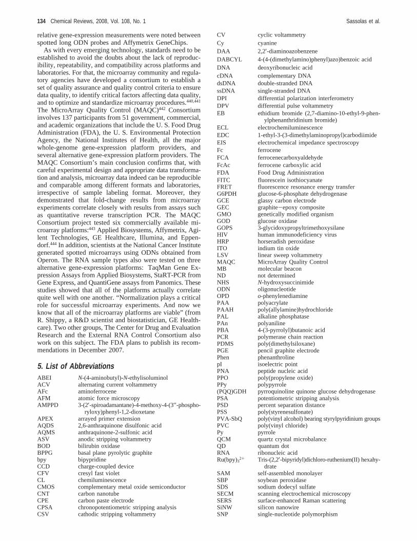
relative gene-expression measurements were noted betweenspotted long ODN probes and Affymetrix GeneChips.
As with every emerging technology, standards need to beestablished to avoid the doubts about the lack of reproduc-ibility, repeatability, and compatibility across platforms andlaboratories. For that, the microarray community and regula-tory agencies have developed a consortium to establish aset of quality assurance and quality control criteria to ensuredata quality, to identify critical factors affecting data quality,and to optimize and standardize microarray procedures.440,441
The MicroArray Quality Control (MAQC)442 Consortiuminvolves 137 participants from 51 government, commercial,and academic organizations that include the U. S. Food DrugAdministration (FDA), the U. S. Environmental ProtectionAgency, the National Institutes of Health, all the majorwhole-genome gene-expression platform providers, andseveral alternative gene-expression platform providers. TheMAQC Consortium’s main conclusion confirms that, withcareful experimental design and appropriate data transforma-tion and analysis, microarray data indeed can be reproducibleand comparable among different formats and laboratories,irrespective of sample labeling format. Moreover, theydemonstrated that fold-change results from microarrayexperiments correlate closely with results from assays suchas quantitative reverse transcription PCR. The MAQCConsortium project tested six commercially available mi-croarray platforms:443 Applied Biosystems, Affymetrix, Agi-lent Technologies, GE Healthcare, Illumina, and Eppen-dorf.444 In addition, scientists at the National Cancer Institutegenerated spotted microarrays using ODNs obtained fromOperon. The RNA sample types also were tested on threealternative gene-expression platforms: TaqMan Gene Ex-pression Assays from Applied Biosystems, StaRT-PCR fromGene Express, and QuantiGene assays from Panomics. Thesestudies showed that all of the platforms actually correlatequite well with one another. “Normalization plays a criticalrole for successful microarray experiments. And now weknow that all of the microarray platforms are viable” (fromR. Shippy, a R&D scientist and biostatistician, GE Health-care). Two other groups, The Center for Drug and EvaluationResearch and the External RNA Control Consortium alsowork on this subject. The FDA plans to publish its recom-mendations in December 2007.
5. List of AbbreviationsABEI N-(4-aminobutyl)-N-ethylisoluminolACV alternating current voltammetryAFc aminoferroceneAFM atomic force microscopyAMPPD 3-(2′-spiroadamantane)-4-methoxy-4-(3′′-phospho-
ryloxy)phenyl-1,2-dioxetaneAPEX arrayed primer extensionAQDS 2,6-anthraquinone disulfonic acidAQMS anthraquinone-2-sulfonic acidASV anodic stripping voltammetryBOD bilirubin oxidaseBPPG basal plane pyrolytic graphitebpy bipypridineCCD charge-coupled deviceCFV cresyl fast violetCL chemiluminescenceCMOS complementary metal oxide semiconductorCNT carbon nanotubeCPE carbon paste electrodeCPSA chronopotentiometric stripping analysisCSV cathodic stripping voltammetry
CV cyclic voltammetryCy cyanineDAA 2,2′-diaminoazobenzeneDABCYL 4-(4-(dimethylamino)phenyl)azo)benzoic acidDNA deoxyribonucleic acidcDNA complementary DNAdsDNA double-stranded DNAssDNA single-stranded DNADPI differential polarization interferometryDPV differential pulse voltammetryEB ethidium bromide (2,7-diamino-10-ethyl-9-phen-
ylphenanthridinium bromide)ECL electrochemiluminescenceEDC 1-ethyl-3-(3-dimethylaminopropyl)carbodiimideEIS electrochemical impedance spectroscopyFc ferroceneFCA ferrocenecarboxyaldehydeFcAc ferrocene carboxylic acidFDA Food Drug AdministrationFITC fluorescein isothiocyanateFRET fluorescence resonance energy transferG6PDH glucose-6-phosphate dehydrogenaseGCE glassy carbon electrodeGEC graphite-epoxy compositeGMO genetically modified organismGOD glucose oxidaseGOPS 3-glycidoxypropyltrimethoxysilaneHIV human immunodeficiency virusHRP horseradish peroxidaseITO indium tin oxideLSV linear sweep voltammetryMAQC MicroArray Quality ControlMB molecular beaconND not determinedNHS N-hydroxysuccinimideODN oligonucleotideOPD o-phenylenediaminePAA polyacrylatePAAH poly(allylamine)hydrochloridePAL alkaline phosphatasePAn polyanilinePBA 4-(3-pyrrolyl)butanoic acidPCR polymerase chain reactionPDMS poly(dimethylsiloxane)PGE pencil graphite electrodePhen phenanthrolinepI isoelectric pointPNA peptide nucleic acidPPO poly(propylene oxide)PPy polypyrrole(PQQ)GDH pyrroquinoline quinone glucose dehydrogenasePSA potentiometric stripping analysisPSD percent separation distancePSS poly(styrenesulfonate)PVA-SbQ poly(vinyl alcohol) bearing styrylpyridinium groupsPVC poly(vinyl chloride)Py pyrroleQCM quartz crystal microbalanceQD quantum dotRNA ribonucleic acidRu(bpy)32+ Tris-(2,2′-bipyridyl)dichloro-ruthenium(II) hexahy-
drateSAM self-assembled monolayerSBP soybean peroxidaseSDS sodium dodecyl sulfateSECM scanning electrochemical microscopySERS surface-enhanced Raman scatteringSiNW silicon nanowireSNP single-nucleotide polymorphism
134 Chemical Reviews, 2008, Vol. 108, No. 1 Sassolas et al.
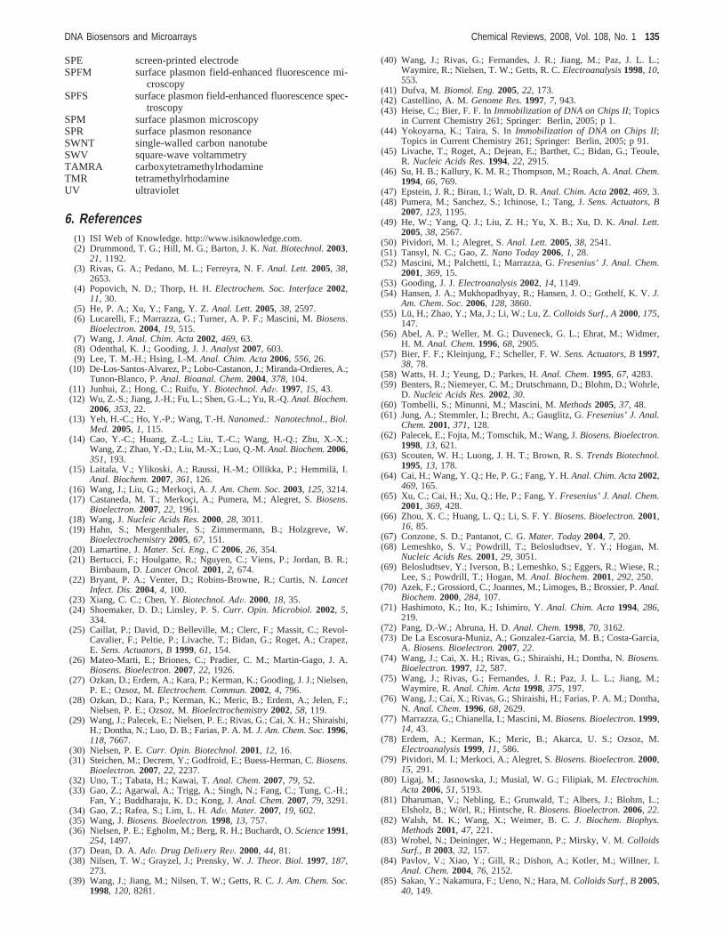
SPE screen-printed electrodeSPFM surface plasmon field-enhanced fluorescence mi-
croscopySPFS surface plasmon field-enhanced fluorescence spec-
troscopySPM surface plasmon microscopySPR surface plasmon resonanceSWNT single-walled carbon nanotubeSWV square-wave voltammetryTAMRA carboxytetramethylrhodamineTMR tetramethylrhodamineUV ultraviolet
6. References(1) ISI Web of Knowledge. http://www.isiknowledge.com.(2) Drummond, T. G.; Hill, M. G.; Barton, J. K.Nat. Biotechnol.2003,
21, 1192.(3) Rivas, G. A.; Pedano, M. L.; Ferreyra, N. F.Anal. Lett.2005, 38,
2653.(4) Popovich, N. D.; Thorp, H. H.Electrochem. Soc. Interface2002,
11, 30.(5) He, P. A.; Xu, Y.; Fang, Y. Z.Anal. Lett.2005, 38, 2597.(6) Lucarelli, F.; Marrazza, G.; Turner, A. P. F.; Mascini, M.Biosens.
Bioelectron.2004, 19, 515.(7) Wang, J.Anal. Chim. Acta2002, 469, 63.(8) Odenthal, K. J.; Gooding, J. J.Analyst2007, 603.(9) Lee, T. M.-H.; Hsing, I.-M.Anal. Chim. Acta2006, 556, 26.
(10) De-Los-Santos-Alvarez, P.; Lobo-Castanon, J.; Miranda-Ordieres, A.;Tunon-Blanco, P.Anal. Bioanal. Chem.2004, 378, 104.
(11) Junhui, Z.; Hong, C.; Ruifu, Y.Biotechnol. AdV. 1997, 15, 43.(12) Wu, Z.-S.; Jiang, J.-H.; Fu, L.; Shen, G.-L.; Yu, R.-Q.Anal. Biochem.
2006, 353, 22.(13) Yeh, H.-C.; Ho, Y.-P.; Wang, T.-H.Nanomed.: Nanotechnol., Biol.
Med.2005, 1, 115.(14) Cao, Y.-C.; Huang, Z.-L.; Liu, T.-C.; Wang, H.-Q.; Zhu, X.-X.;
Wang, Z.; Zhao, Y.-D.; Liu, M.-X.; Luo, Q.-M.Anal. Biochem.2006,351, 193.
(15) Laitala, V.; Ylikoski, A.; Raussi, H.-M.; Ollikka, P.; Hemmila¨, I.Anal. Biochem.2007, 361, 126.
(16) Wang, J.; Liu, G.; Merkoc¸ i, A. J. Am. Chem. Soc.2003, 125, 3214.(17) Castaneda, M. T.; Merkoc¸ i, A.; Pumera, M.; Alegret, S.Biosens.
Bioelectron.2007, 22, 1961.(18) Wang, J.Nucleic Acids Res.2000, 28, 3011.(19) Hahn, S.; Mergenthaler, S.; Zimmermann, B.; Holzgreve, W.
Bioelectrochemistry2005, 67, 151.(20) Lamartine, J.Mater. Sci. Eng., C2006, 26, 354.(21) Bertucci, F.; Houlgatte, R.; Nguyen, C.; Viens, P.; Jordan, B. R.;
Birnbaum, D.Lancet Oncol.2001, 2, 674.(22) Bryant, P. A.; Venter, D.; Robins-Browne, R.; Curtis, N.Lancet
Infect. Dis.2004, 4, 100.(23) Xiang, C. C.; Chen, Y.Biotechnol. AdV. 2000, 18, 35.(24) Shoemaker, D. D.; Linsley, P. S.Curr. Opin. Microbiol. 2002, 5,
334.(25) Caillat, P.; David, D.; Belleville, M.; Clerc, F.; Massit, C.; Revol-
Cavalier, F.; Peltie, P.; Livache, T.; Bidan, G.; Roget, A.; Crapez,E. Sens. Actuators, B1999, 61, 154.
(26) Mateo-Marti, E.; Briones, C.; Pradier, C. M.; Martin-Gago, J. A.Biosens. Bioelectron.2007, 22, 1926.
(27) Ozkan, D.; Erdem, A.; Kara, P.; Kerman, K.; Gooding, J. J.; Nielsen,P. E.; Ozsoz, M.Electrochem. Commun.2002, 4, 796.
(28) Ozkan, D.; Kara, P.; Kerman, K.; Meric, B.; Erdem, A.; Jelen, F.;Nielsen, P. E.; Ozsoz, M.Bioelectrochemistry2002, 58, 119.
(29) Wang, J.; Palecek, E.; Nielsen, P. E.; Rivas, G.; Cai, X. H.; Shiraishi,H.; Dontha, N.; Luo, D. B.; Farias, P. A. M.J. Am. Chem. Soc.1996,118, 7667.
(30) Nielsen, P. E.Curr. Opin. Biotechnol.2001, 12, 16.(31) Steichen, M.; Decrem, Y.; Godfroid, E.; Buess-Herman, C.Biosens.
Bioelectron.2007, 22, 2237.(32) Uno, T.; Tabata, H.; Kawai, T.Anal. Chem.2007, 79, 52.(33) Gao, Z.; Agarwal, A.; Trigg, A.; Singh, N.; Fang, C.; Tung, C.-H.;
Fan, Y.; Buddharaju, K. D.; Kong, J.Anal. Chem.2007, 79, 3291.(34) Gao, Z.; Rafea, S.; Lim, L. H.AdV. Mater. 2007, 19, 602.(35) Wang, J.Biosens. Bioelectron.1998, 13, 757.(36) Nielsen, P. E.; Egholm, M.; Berg, R. H.; Buchardt, O.Science1991,
254, 1497.(37) Dean, D. A.AdV. Drug DeliVery ReV. 2000, 44, 81.(38) Nilsen, T. W.; Grayzel, J.; Prensky, W.J. Theor. Biol.1997, 187,
273.(39) Wang, J.; Jiang, M.; Nilsen, T. W.; Getts, R. C.J. Am. Chem. Soc.
1998, 120, 8281.
(40) Wang, J.; Rivas, G.; Fernandes, J. R.; Jiang, M.; Paz, J. L. L.;Waymire, R.; Nielsen, T. W.; Getts, R. C.Electroanalysis1998, 10,553.
(41) Dufva, M.Biomol. Eng.2005, 22, 173.(42) Castellino, A. M.Genome Res.1997, 7, 943.(43) Heise, C.; Bier, F. F. InImmobilization of DNA on Chips II; Topics
in Current Chemistry 261; Springer: Berlin, 2005; p 1.(44) Yokoyarna, K.; Taira, S. InImmobilization of DNA on Chips II;
Topics in Current Chemistry 261; Springer: Berlin, 2005; p 91.(45) Livache, T.; Roget, A.; Dejean, E.; Barthet, C.; Bidan, G.; Teoule,
R. Nucleic Acids Res.1994, 22, 2915.(46) Su, H. B.; Kallury, K. M. R.; Thompson, M.; Roach, A.Anal. Chem.
1994, 66, 769.(47) Epstein, J. R.; Biran, I.; Walt, D. R.Anal. Chim. Acta2002, 469, 3.(48) Pumera, M.; Sanchez, S.; Ichinose, I.; Tang, J.Sens. Actuators, B
2007, 123, 1195.(49) He, W.; Yang, Q. J.; Liu, Z. H.; Yu, X. B.; Xu, D. K.Anal. Lett.
2005, 38, 2567.(50) Pividori, M. I.; Alegret, S.Anal. Lett.2005, 38, 2541.(51) Tansyl, N. C.; Gao, Z.Nano Today2006, 1, 28.(52) Mascini, M.; Palchetti, I.; Marrazza, G.Fresenius’ J. Anal. Chem.
2001, 369, 15.(53) Gooding, J. J.Electroanalysis2002, 14, 1149.(54) Hansen, J. A.; Mukhopadhyay, R.; Hansen, J. O.; Gothelf, K. V.J.
Am. Chem. Soc.2006, 128, 3860.(55) Lu, H.; Zhao, Y.; Ma, J.; Li, W.; Lu, Z.Colloids Surf., A2000, 175,
147.(56) Abel, A. P.; Weller, M. G.; Duveneck, G. L.; Ehrat, M.; Widmer,
H. M. Anal. Chem.1996, 68, 2905.(57) Bier, F. F.; Kleinjung, F.; Scheller, F. W.Sens. Actuators, B1997,
38, 78.(58) Watts, H. J.; Yeung, D.; Parkes, H.Anal. Chem.1995, 67, 4283.(59) Benters, R.; Niemeyer, C. M.; Drutschmann, D.; Blohm, D.; Wohrle,
D. Nucleic Acids Res.2002, 30.(60) Tombelli, S.; Minunni, M.; Mascini, M.Methods2005, 37, 48.(61) Jung, A.; Stemmler, I.; Brecht, A.; Gauglitz, G.Fresenius’ J. Anal.
Chem.2001, 371, 128.(62) Palecek, E.; Fojta, M.; Tomschik, M.; Wang, J.Biosens. Bioelectron.
1998, 13, 621.(63) Scouten, W. H.; Luong, J. H. T.; Brown, R. S.Trends Biotechnol.
1995, 13, 178.(64) Cai, H.; Wang, Y. Q.; He, P. G.; Fang, Y. H.Anal. Chim. Acta2002,
469, 165.(65) Xu, C.; Cai, H.; Xu, Q.; He, P.; Fang, Y.Fresenius’ J. Anal. Chem.
2001, 369, 428.(66) Zhou, X. C.; Huang, L. Q.; Li, S. F. Y.Biosens. Bioelectron.2001,
16, 85.(67) Conzone, S. D.; Pantanot, C. G.Mater. Today2004, 7, 20.(68) Lemeshko, S. V.; Powdrill, T.; Belosludtsev, Y. Y.; Hogan, M.
Nucleic Acids Res.2001, 29, 3051.(69) Belosludtsev, Y.; Iverson, B.; Lemeshko, S.; Eggers, R.; Wiese, R.;
Lee, S.; Powdrill, T.; Hogan, M.Anal. Biochem.2001, 292, 250.(70) Azek, F.; Grossiord, C.; Joannes, M.; Limoges, B.; Brossier, P.Anal.
Biochem.2000, 284, 107.(71) Hashimoto, K.; Ito, K.; Ishimiro, Y.Anal. Chim. Acta1994, 286,
219.(72) Pang, D.-W.; Abruna, H. D.Anal. Chem.1998, 70, 3162.(73) De La Escosura-Muniz, A.; Gonzalez-Garcia, M. B.; Costa-Garcia,
A. Biosens. Bioelectron.2007, 22.(74) Wang, J.; Cai, X. H.; Rivas, G.; Shiraishi, H.; Dontha, N.Biosens.
Bioelectron.1997, 12, 587.(75) Wang, J.; Rivas, G.; Fernandes, J. R.; Paz, J. L. L.; Jiang, M.;
Waymire, R.Anal. Chim. Acta1998, 375, 197.(76) Wang, J.; Cai, X.; Rivas, G.; Shiraishi, H.; Farias, P. A. M.; Dontha,
N. Anal. Chem.1996, 68, 2629.(77) Marrazza, G.; Chianella, I.; Mascini, M.Biosens. Bioelectron.1999,
14, 43.(78) Erdem, A.; Kerman, K.; Meric, B.; Akarca, U. S.; Ozsoz, M.
Electroanalysis1999, 11, 586.(79) Pividori, M. I.; Merkoci, A.; Alegret, S.Biosens. Bioelectron.2000,
15, 291.(80) Ligaj, M.; Jasnowska, J.; Musial, W. G.; Filipiak, M.Electrochim.
Acta 2006, 51, 5193.(81) Dharuman, V.; Nebling, E.; Grunwald, T.; Albers, J.; Blohm, L.;
Elsholz, B.; Worl, R.; Hintsche, R.Biosens. Bioelectron.2006, 22.(82) Walsh, M. K.; Wang, X.; Weimer, B. C.J. Biochem. Biophys.
Methods2001, 47, 221.(83) Wrobel, N.; Deininger, W.; Hegemann, P.; Mirsky, V. M.Colloids
Surf., B2003, 32, 157.(84) Pavlov, V.; Xiao, Y.; Gill, R.; Dishon, A.; Kotler, M.; Willner, I.
Anal. Chem.2004, 76, 2152.(85) Sakao, Y.; Nakamura, F.; Ueno, N.; Hara, M.Colloids Surf., B2005,
40, 149.
DNA Biosensors and Microarrays Chemical Reviews, 2008, Vol. 108, No. 1 135
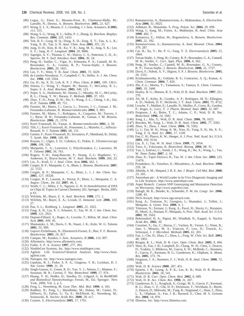
(86) Legay, G.; Finot, E.; Meunier-Prest, R.; Cherkaoui-Malki, M.;Latruffe, N.; Dereux, A.Biosens. Bioelectron.2005, 21, 627.
(87) Wong, E. L. S.; Mearns, F. J.; Gooding, J. J.Sens. Actuators, B2005,111, 515.
(88) Wang, S. G.; Wang, R. L.; Sellin, P. J.; Zhang, Q.Biochem. Biophys.Res. Commun.2004, 325, 1433.
(89) Teh, H. F.; Gong, H. Q.; Dong, X. D.; Zeng, X. T.; Tan, A. L. K.;Yang, X. H.; Tan, S. N.Anal. Chim. Acta2005, 551, 23.
(90) Jung, D.-H.; Kim, B. H.; Ko, Y. K.; Jung, M. S.; Jung, S. K.; Lee,S. Y.; Jung, H.-T.Langmuir2004, 20, 8886.
(91) Saprigin, A. V.; Thomas, C. W.; Dulcey, C. S.; Patterson, C. H., Jr.;Spector, M. S.Surf. Interface Anal.2004, 36, 24.
(92) Peng, H.; Soeller, C.; Vigar, N.; Kilmartin, P. A.; Cannell, M. B.;Bowmaker, G. A.; Cooney, R. P.; Travas-Sejdic, J.Biosens.Bioelectron.2005, 20, 1821.
(93) Lee, T. Y.; Shim, Y. B.Anal. Chem.2001, 73, 5629.(94) de Lumley-Woodyear, T.; Campbell, C. N.; Heller, A.J. Am. Chem.
Soc.1996, 118, 5504.(95) Gu, H.; Su, X. D.; Loh, K. P.J. Phys. Chem. B2005, 109, 13611.(96) Situma, C.; Wang, Y.; Hupert, M.; Barany, F.; McCarley, R. L.;
Soper, S. A.Anal. Biochem.2005, 340, 123.(97) Soper, S. A.; Hashimoto, M.; Situma, C.; Murphy, M. C.; McCarley,
R. L.; Cheng, Y. W.; Barany, F.Methods2005, 37, 103.(98) Zhao, Y.-D.; Pang, D.-W.; Hu, S.; Wang, Z.-L.; Cheng, J.-K.; Dai,
H.-P. Talanta1999, 49, 751.(99) Fuentes, M.; Mateo, C.; Garcia, L.; Tercero, J. C.; Guisan, J. M.;
Fernandez-Lafuente, R.Biomacromolecules2004, 5, 883.(100) Fuentes, M.; Mateo, C.; Rodriguez, A.; Casqueiro, M.; Tercero, J.
C.; Riese, H. H.; Fernandez-Lafuente, R.; Guisan, J. M.Biosens.Bioelectron.2006, 21, 1574.
(101) Korri-Youssoufi, H.; Yassar, A.Biomacromolecules2001, 2, 58.(102) Tlili, C.; Korri-Youssoufi, H.; Ponsonnet, L.; Martelet, C.; Jaffrezic-
Renault, N. J.Talanta2005, 68, 131.(103) Garnier, F.; Korri-Youssoufi, H.; Srivastava, P.; Mandrand, B.; Delair,
T. Synth. Met.1999, 100, 89.(104) Kocum, C.; Ulgen, S. D.; Cubukcu, E.; Piskin, E.Ultramicroscopy
2006, 106, 326.(105) Marquette, C. A.; Lawrence, I.; Polychronakos, C.; Lawrence, M.
F. Talanta2002, 56, 763.(106) Rogers, Y.-H.; Jiang-Baucom, P.; Huang, Z.-J.; Bogdanov, V.;
Anderson, S.; Boyce-Jacino, M. T.Anal. Biochem.1999, 266, 23.(107) Liu, X.; Krull, U. J.Anal. Chim. Acta2006, 562, 1.(108) Corgier, B. P.; Marquette, C. A.; Blum, L.Biosens. Bioelectron.2007,
22, 1522.(109) Corgier, B. P.; Marquette, C. A.; Blum, L. J.J. Am. Chem. Soc.
2005, 127, 18328.(110) Corgier, B. P.; Laurent, A.; Perriat, P.; Blum, L.; Marquette, C. A.
Angew. Chem., Int. Ed.2007, 46, 1.(111) Smith, C. L.; Milea, J. S.; Nguyen, G. H. InImmobilization of DNA
on Chips II; Topics in Current Chemistry 261; Springer: Berlin, 2005;p 63.
(112) Wilchek, M.; Bayer, E. A.Anal. Biochem.1988, 171, 1.(113) Wilchek, M.; Bayer, E. A.; Livnah, O.Immunol. Lett.2006, 103,
27.(114) Pan, S. L.; Rothberg, L.Langmuir2005, 21, 1022.(115) Fang, X. H.; Liu, X. J.; Schuster, S.; Tan, W. H.J. Am. Chem. Soc.
1999, 121, 2921.(116) Dupont-Filliard, A.; Roget, A.; Livache, T.; Billon, M.Anal. Chim.
Acta 2001, 449, 45.(117) Shamansky, L. M.; Davis, C. B.; Stuart, J. K.; Kuhr, W. G.Talanta
2001, 55, 909.(118) Gajovic-Eichelmann, N.; Ehrentreich-Forster, E.; Bier, F. F.Biosens.
Bioelectron.2003, 19, 417.(119) Campas, M.; Katakis, I.Sens. Actuators, B2006, 114, 897.(120) Affymetrix. http://www.affymetrix.com.(121) Fodor, S. P. A.Science1997, 277, 393.(122) NimbleGen Systems, Inc. http://www.nimblegen.com.(123) Agilent: Life Sciences/Chemical Analysis. http://www.chem.
agilent.com.(124) Nanogen, Inc. http://www.nanogen.com.(125) Lipshutz, R. J.; Fodor, S. P. A.; Gingeras, T. R.; Lockhart, D. J.
Nat. Genet.1999, 21, 20.(126) Singh-Gasson, S.; Green, R. D.; Yue, Y. J.; Nelson, C.; Blattner, F.;
Sussman, M. R.; Cerrina, F.Nat. Biotechnol.1999, 17, 974.(127) Huang, Y. H.; Hodko, D.; Smolko, D.; Lidgard, G. InBioMEMS
and Biomedical Nanotechnology; Ferrari, M., Ed.; Springer: NewYork, 2006; Vol. 2, p 3.
(128) Feng, L.; Nerenberg, M.Gene Ther. Mol. Biol.1999, 4, 183.(129) Radtkey, R.; Feng, L.; Muralhidar, M.; Duhon, M.; Canter, D.;
DiPierro, D.; Fallon, S.; Tu, E.; McElfresh, K.; Nerenberg, M.;Sosnowski, R.Nucleic Acids Res.2000, 28, e17.
(130) Cosnier, S.Electroanalysis2005, 17, 1701.
(131) Ramanavicius, A.; Ramanaviciene, A.; Malinauskas, A.Electrochim.Acta 2006, 51, 6025.
(132) Adhikari, B.; Majumdar, S.Prog. Polym. Sci.2004, 29, 699.(133) Wang, J.; Jiang, M.; Fortes, A.; Mukherjee, B.Anal. Chim. Acta
1999, 402, 7.(134) Komarova, E.; Aldissi, M.; Bogomolova, A.Biosens. Bioelectron.
2005, 21, 182.(135) Ramanaviciene, A.; Ramanavicius, A.Anal. Bioanal. Chem.2004,
379, 287.(136) Cai, H.; Xu, Y.; He, P. G.; Fang, Y. Z.Electroanalysis2003, 15,
1864.(137) Travas-Sejdic, J.; Peng, H.; Cooney, R. P.; Bowmaker, G. A.; Cannell,
M. B.; Soeller, C.Curr. Appl. Phys.2006, 6, 562.(138) Peng, H.; Soeller, C.; Cannell, M. B.; Bowmaker, G. A.; Cooney,
R. P.; Travas-Sejdic, J.Biosens. Bioelectron.2006, 21, 1727.(139) Davis, F.; Nabok, A. V.; Higson, S. P. J.Biosens. Bioelectron.2005,
20, 1531.(140) Krishnamoorthy, K.; Gokhale, R. S.; Contractor, A. Q.; Kumar, A.
Chem. Commun.2004, 7, 820.(141) Zhi, Z.-L.; Morita, Y.; Yamamura, S.; Tamiya, E.Chem. Commun.
2005, 19, 2448.(142) Healey, B. G.; Matson, R. S.; Walt, D. R.Anal. Biochem.1997, 251,
270.(143) Ali, M. F.; Kirby, R.; Goodey, A. P.; Rodriguez, M. D.; Ellington,
A. D.; Neikirk, D. P.; McDevitt, J. T.Anal. Chem.2003, 75, 4732.(144) Livache, T.; Maillart, E.; Lassalle, N.; Mailley, P.; Corso, B.; Guedon,
P.; Roget, A.; Levy, Y.J. Pharm. Biomed. Anal.2003, 32, 687.(145) Ferguson, J. A.; Boles, T. C.; Adams, C. P.; Walt, D. R.Nat.
Biotechnol.1996, 14, 1681.(146) Song, L.; Ahn, S.; Walt, D. R.Anal. Chem.2006, 78, 1023.(147) Bowden, M.; Song, L.; Walt, D. R.Anal. Chem.2005, 77, 5583.(148) Zhou, X.; Zhou, J.Anal. Chem.2004, 76, 5302.(149) Li, J.; Tan, W. H.; Wang, K. M.; Xiao, D.; Yang, X. H.; He, X. X.;
Tang, Z. Q.Anal. Sci.2001, 17, 1149.(150) Fan, C. H.; Plaxco, K. W.; Heeger, A. J.Proc. Natl. Acad. Sci. U.S.A.
2003, 100, 9134.(151) Liu, X. J.; Tan, W. H.Anal. Chem.1999, 71, 5054.(152) Taira, S.; Yokoyama, K.Biotechnol. Bioeng.2004, 88, 35.(153) Yan, J.; Este´vez, C.; Smith, J. E.; Wang, K.; He, X.; Wang, L.; Tan,
W. Nano Today2007, 2, 44.(154) Zhao, X.; Tapec-Dytioco, R.; Tan, W.J. Am. Chem. Soc.2003, 125,
11474.(155) Proudnikov, D.; Timofeev, E.; Mirzabekov, A.Anal. Biochem.1998,
259, 34.(156) Albelda, S. M.; Shepard, J. R. E.Am. J. Respir. Cell Mol. Biol.2000,
23, 265.(157) Amersham plc: A World Leader in In-Vivo Diagnostic Imaging and
in Life Sciences. http://www.amersham.com.(158) Asper Biotech: Custom SNP Genotyping and Mutatation Detection
on Microarray. http://www.asperbio.com.(159) Balogh, M. K.; Bender, K.; Schneider, P. M.Int. Congr. Ser.2006,
1288, 43.(160) NGS-ArrayIt. http://www.ngs-arrayit.com.(161) Kurg, A.; Tonisson, N.; Georgiou, I.; Shumaker, J.; Tollett, J.;
Metspalu, A.Genet. Test.2000, 4, 1.(162) Tonisson, N.; Zernant, J.; Kurg, A.; Pavel, H.; Slavin, G.; Roomere,
H.; Meiel, A.; Hainaut, P.; Metspalu, A.Proc. Natl. Acad. Sci. U.S.A.2002, 99, 5503.
(163) Behrensdorf, H. A.; Pignot, M.; Windhab, N.; Kappel, A.NucleicAcids Res.2002, 30.
(164) Couzinet, S.; Jay, C.; Barras, C.; Vachon, R.; Vernet, G.; Ninet, B.;Jane, I.; Minazio, M. A.; Francois, P.; Lew, D.; Troesch, A.;Schrenzel, J.J. Microbiol. Methods2005, 61, 201.
(165) Fan, J.; Che, D.; Zhao, C.; Zhou, L.; Feng, W.Chin. Sci. Bull.2003,48, 1903.
(166) Brogan, K. L.; Walt, D. R.Curr. Opin. Chem. Biol.2005, 9, 494.(167) Shen, R.; Fan, J. B.; Campbell, D.; Chang, W. H.; Chen, J.; Doucet,
D.; Yeakley, J.; Bibikova, M.; Garcia, E. W.; McBride, C.; Steemers,F.; Garcia, F.; Kermani, B. G.; Gunderson, K.; Oliphant, A.Mutat.Res.2005, 573, 70.
(168) Ferguson, J. A.; Steemers, F. J.; Walt, D. R.Anal. Chem.2000, 72,5618.
(169) Walt, D. R.Science2000, 287, 451.(170) Epstein, J. R.; Leung, A. P. K.; Lee, K. H.; Walt, D. R.Biosens.
Bioelectron.2003, 18, 541.(171) Walt, D. R.Curr. Opin. Chem. Biol.2002, 6, 689.(172) Walt, D. R.Acc. Chem. Res.1998, 31, 267.(173) Gunderson, K. L.; Kruglyak, S.; Graige, M. S.; Garcia, F.; Kermani,
B. G.; Zhao, C. F.; Che, D. P.; Dickinson, T.; Wickham, E.; Bierle,J.; Doucet, D.; Milewski, M.; Yang, R.; Siegmund, C.; Haas, J.; Zhou,L. X.; Oliphant, A.; Fan, J. B.; Barnard, S.; Chee, M. S.GenomeRes.2004, 14, 870.
(174) Illumina, Inc. http://www.illumina.com.
136 Chemical Reviews, 2008, Vol. 108, No. 1 Sassolas et al.
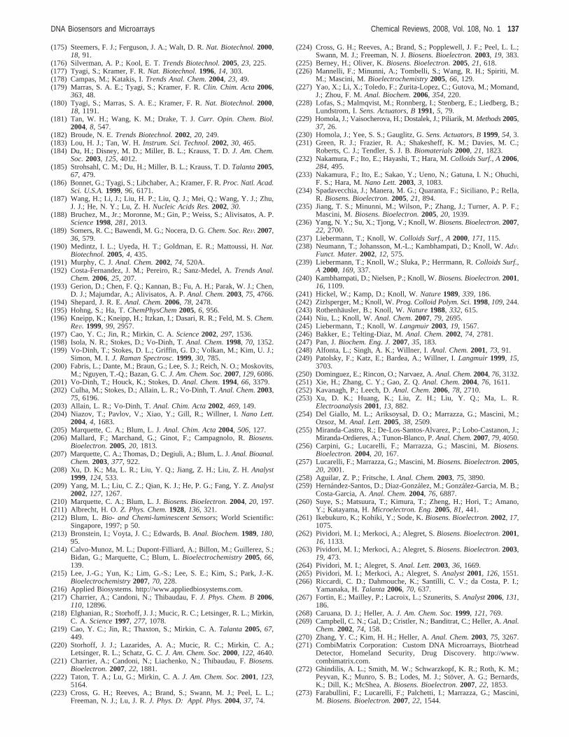
(175) Steemers, F. J.; Ferguson, J. A.; Walt, D. R.Nat. Biotechnol.2000,18, 91.
(176) Silverman, A. P.; Kool, E. T.Trends Biotechnol.2005, 23, 225.(177) Tyagi, S.; Kramer, F. R.Nat. Biotechnol.1996, 14, 303.(178) Campas, M.; Katakis, I.Trends Anal. Chem.2004, 23, 49.(179) Marras, S. A. E.; Tyagi, S.; Kramer, F. R.Clin. Chim. Acta2006,
363, 48.(180) Tyagi, S.; Marras, S. A. E.; Kramer, F. R.Nat. Biotechnol.2000,
18, 1191.(181) Tan, W. H.; Wang, K. M.; Drake, T. J.Curr. Opin. Chem. Biol.
2004, 8, 547.(182) Broude, N. E.Trends Biotechnol.2002, 20, 249.(183) Lou, H. J.; Tan, W. H.Instrum. Sci. Technol.2002, 30, 465.(184) Du, H.; Disney, M. D.; Miller, B. L.; Krauss, T. D.J. Am. Chem.
Soc.2003, 125, 4012.(185) Strohsahl, C. M.; Du, H.; Miller, B. L.; Krauss, T. D.Talanta2005,
67, 479.(186) Bonnet, G.; Tyagi, S.; Libchaber, A.; Kramer, F. R.Proc. Natl. Acad.
Sci. U.S.A.1999, 96, 6171.(187) Wang, H.; Li, J.; Liu, H. P.; Liu, Q. J.; Mei, Q.; Wang, Y. J.; Zhu,
J. J.; He, N. Y.; Lu, Z. H.Nucleic Acids Res.2002, 30.(188) Bruchez, M., Jr.; Moronne, M.; Gin, P.; Weiss, S.; Alivisatos, A. P.
Science1998, 281, 2013.(189) Somers, R. C.; Bawendi, M. G.; Nocera, D. G.Chem. Soc. ReV. 2007,
36, 579.(190) Medintz, I. L.; Uyeda, H. T.; Goldman, E. R.; Mattoussi, H.Nat.
Biotechnol.2005, 4, 435.(191) Murphy, C. J.Anal. Chem.2002, 74, 520A.(192) Costa-Fernandez, J. M.; Pereiro, R.; Sanz-Medel, A.Trends Anal.
Chem.2006, 25, 207.(193) Gerion, D.; Chen, F. Q.; Kannan, B.; Fu, A. H.; Parak, W. J.; Chen,
D. J.; Majumdar, A.; Alivisatos, A. P.Anal. Chem.2003, 75, 4766.(194) Shepard, J. R. E.Anal. Chem.2006, 78, 2478.(195) Hohng, S.; Ha, T.ChemPhysChem2005, 6, 956.(196) Kneipp, K.; Kneipp, H.; Itzkan, I.; Dasari, R. R.; Feld, M. S.Chem.
ReV. 1999, 99, 2957.(197) Cao, Y. C.; Jin, R.; Mirkin, C. A.Science2002, 297, 1536.(198) Isola, N. R.; Stokes, D.; Vo-Dinh, T.Anal. Chem.1998, 70, 1352.(199) Vo-Dinh, T.; Stokes, D. L.; Griffin, G. D.; Volkan, M.; Kim, U. J.;
Simon, M. I.J. Raman Spectrosc.1999, 30, 785.(200) Fabris, L.; Dante, M.; Braun, G.; Lee, S. J.; Reich, N. O.; Moskovits,
M.; Nguyen, T.-Q.; Bazan, G. C.J. Am. Chem. Soc.2007, 129, 6086.(201) Vo-Dinh, T.; Houck, K.; Stokes, D.Anal. Chem.1994, 66, 3379.(202) Culha, M.; Stokes, D.; Allain, L. R.; Vo-Dinh, T.Anal. Chem.2003,
75, 6196.(203) Allain, L. R.; Vo-Dinh, T.Anal. Chim. Acta2002, 469, 149.(204) Niazov, T.; Pavlov, V.; Xiao, Y.; Gill, R.; Willner, I.Nano Lett.
2004, 4, 1683.(205) Marquette, C. A.; Blum, L. J.Anal. Chim. Acta2004, 506, 127.(206) Mallard, F.; Marchand, G.; Ginot, F.; Campagnolo, R.Biosens.
Bioelectron.2005, 20, 1813.(207) Marquette, C. A.; Thomas, D.; Degiuli, A.; Blum, L. J.Anal. Bioanal.
Chem.2003, 377, 922.(208) Xu, D. K.; Ma, L. R.; Liu, Y. Q.; Jiang, Z. H.; Liu, Z. H.Analyst
1999, 124, 533.(209) Yang, M. L.; Liu, C. Z.; Qian, K. J.; He, P. G.; Fang, Y. Z.Analyst
2002, 127, 1267.(210) Marquette, C. A.; Blum, L. J.Biosens. Bioelectron.2004, 20, 197.(211) Albrecht, H. O.Z. Phys. Chem.1928, 136, 321.(212) Blum, L. Bio- and Chemi-luminescent Sensors; World Scientific:
Singapore, 1997; p 50.(213) Bronstein, I.; Voyta, J. C.; Edwards, B.Anal. Biochem.1989, 180,
95.(214) Calvo-Munoz, M. L.; Dupont-Filliard, A.; Billon, M.; Guillerez, S.;
Bidan, G.; Marquette, C.; Blum, L.Bioelectrochemistry2005, 66,139.
(215) Lee, J.-G.; Yun, K.; Lim, G.-S.; Lee, S. E.; Kim, S.; Park, J.-K.Bioelectrochemistry2007, 70, 228.
(216) Applied Biosystems. http://www.appliedbiosystems.com.(217) Charrier, A.; Candoni, N.; Thibaudau, F.J. Phys. Chem. B2006,
110, 12896.(218) Elghanian, R.; Storhoff, J. J.; Mucic, R. C.; Letsinger, R. L.; Mirkin,
C. A. Science1997, 277, 1078.(219) Cao, Y. C.; Jin, R.; Thaxton, S.; Mirkin, C. A.Talanta 2005, 67,
449.(220) Storhoff, J. J.; Lazarides, A. A.; Mucic, R. C.; Mirkin, C. A.;
Letsinger, R. L.; Schatz, G. C.J. Am. Chem. Soc.2000, 122, 4640.(221) Charrier, A.; Candoni, N.; Liachenko, N.; Thibaudau, F.Biosens.
Bioelectron.2007, 22, 1881.(222) Taton, T. A.; Lu, G.; Mirkin, C. A.J. Am. Chem. Soc.2001, 123,
5164.(223) Cross, G. H.; Reeves, A.; Brand, S.; Swann, M. J.; Peel, L. L.;
Freeman, N. J.; Lu, J. R.J. Phys. D: Appl. Phys.2004, 37, 74.
(224) Cross, G. H.; Reeves, A.; Brand, S.; Popplewell, J. F.; Peel, L. L.;Swann, M. J.; Freeman, N. J.Biosens. Bioelectron.2003, 19, 383.
(225) Berney, H.; Oliver, K.Biosens. Bioelectron.2005, 21, 618.(226) Mannelli, F.; Minunni, A.; Tombelli, S.; Wang, R. H.; Spiriti, M.
M.; Mascini, M. Bioelectrochemistry2005, 66, 129.(227) Yao, X.; Li, X.; Toledo, F.; Zurita-Lopez, C.; Gutova, M.; Momand,
J.; Zhou, F. M.Anal. Biochem.2006, 354, 220.(228) Lofas, S.; Malmqvist, M.; Ronnberg, I.; Stenberg, E.; Liedberg, B.;
Lundstrom, I.Sens. Actuators, B1991, 5, 79.(229) Homola, J.; Vaisocherova, H.; Dostalek, J.; Piliarik, M.Methods2005,
37, 26.(230) Homola, J.; Yee, S. S.; Gauglitz, G.Sens. Actuators, B1999, 54, 3.(231) Green, R. J.; Frazier, R. A.; Shakesheff, K. M.; Davies, M. C.;
Roberts, C. J.; Tendler, S. J. B.Biomaterials2000, 21, 1823.(232) Nakamura, F.; Ito, E.; Hayashi, T.; Hara, M.Colloids Surf., A2006,
284, 495.(233) Nakamura, F.; Ito, E.; Sakao, Y.; Ueno, N.; Gatuna, I. N.; Ohuchi,
F. S.; Hara, M.Nano Lett.2003, 3, 1083.(234) Spadavecchia, J.; Manera, M. G.; Quaranta, F.; Siciliano, P.; Rella,
R. Biosens. Bioelectron.2005, 21, 894.(235) Jiang, T. S.; Minunni, M.; Wilson, P.; Zhang, J.; Turner, A. P. F.;
Mascini, M. Biosens. Bioelectron.2005, 20, 1939.(236) Yang, N. Y.; Su, X.; Tjong, V.; Knoll, W.Biosens. Bioelectron.2007,
22, 2700.(237) Liebermann, T.; Knoll, W.Colloids Surf., A2000, 171, 115.(238) Neumann, T.; Johansson, M.-L.; Kambhampati, D.; Knoll, W.AdV.
Funct. Mater.2002, 12, 575.(239) Liebermann, T.; Knoll, W.; Sluka, P.; Herrmann, R.Colloids Surf.,
A 2000, 169, 337.(240) Kambhampati, D.; Nielsen, P.; Knoll, W.Biosens. Bioelectron.2001,
16, 1109.(241) Hickel, W.; Kamp, D.; Knoll, W.Nature1989, 339, 186.(242) Zizlsperger, M.; Knoll, W.Prog. Colloid Polym. Sci.1998, 109, 244.(243) Rothenha¨usler, B.; Knoll, W.Nature1988, 332, 615.(244) Niu, L.; Knoll, W. Anal. Chem.2007, 79, 2695.(245) Liebermann, T.; Knoll, W.Langmuir2003, 19, 1567.(246) Bakker, E.; Telting-Diaz, M.Anal. Chem.2002, 74, 2781.(247) Pan, J.Biochem. Eng. J.2007, 35, 183.(248) Alfonta, L.; Singh, A. K.; Willner, I.Anal. Chem.2001, 73, 91.(249) Patolsky, F.; Katz, E.; Bardea, A.; Willner, I.Langmuir1999, 15,
3703.(250) Dominguez, E.; Rincon, O.; Narvaez, A.Anal. Chem.2004, 76, 3132.(251) Xie, H.; Zhang, C. Y.; Gao, Z. Q.Anal. Chem.2004, 76, 1611.(252) Kavanagh, P.; Leech, D.Anal. Chem.2006, 78, 2710.(253) Xu, D. K.; Huang, K.; Liu, Z. H.; Liu, Y. Q.; Ma, L. R.
Electroanalysis2001, 13, 882.(254) Del Giallo, M. L.; Ariksoysal, D. O.; Marrazza, G.; Mascini, M.;
Ozsoz, M.Anal. Lett.2005, 38, 2509.(255) Miranda-Castro, R.; De-Los-Santos-Alvarez, P.; Lobo-Castanon, J.;
Miranda-Ordieres, A.; Tunon-Blanco, P.Anal. Chem.2007, 79, 4050.(256) Carpini, G.; Lucarelli, F.; Marrazza, G.; Mascini, M.Biosens.
Bioelectron.2004, 20, 167.(257) Lucarelli, F.; Marrazza, G.; Mascini, M.Biosens. Bioelectron.2005,
20, 2001.(258) Aguilar, Z. P.; Fritsche, I.Anal. Chem.2003, 75, 3890.(259) Herna´ndez-Santos, D.; Diaz-Gonza´lez, M.; Gonza´lez-Garcia, M. B.;
Costa-Garcia, A.Anal. Chem.2004, 76, 6887.(260) Suye, S.; Matsuura, T.; Kimura, T.; Zheng, H.; Hori, T.; Amano,
Y.; Katayama, H.Microelectron. Eng.2005, 81, 441.(261) Ikebukuro, K.; Kohiki, Y.; Sode, K.Biosens. Bioelectron.2002, 17,
1075.(262) Pividori, M. I.; Merkoci, A.; Alegret, S.Biosens. Bioelectron.2001,
16, 1133.(263) Pividori, M. I.; Merkoci, A.; Alegret, S.Biosens. Bioelectron.2003,
19, 473.(264) Pividori, M. I.; Alegret, S.Anal. Lett.2003, 36, 1669.(265) Pividori, M. I.; Merkoci, A.; Alegret, S.Analyst2001, 126, 1551.(266) Riccardi, C. D.; Dahmouche, K.; Santilli, C. V.; da Costa, P. I.;
Yamanaka, H.Talanta2006, 70, 637.(267) Fortin, E.; Mailley, P.; Lacroix, L.; Szunerits, S.Analyst2006, 131,
186.(268) Caruana, D. J.; Heller, A.J. Am. Chem. Soc.1999, 121, 769.(269) Campbell, C. N.; Gal, D.; Cristler, N.; Banditrat, C.; Heller, A.Anal.
Chem.2002, 74, 158.(270) Zhang, Y. C.; Kim, H. H.; Heller, A.Anal. Chem.2003, 75, 3267.(271) CombiMatrix Corporation: Custom DNA Microarrays, Biotrhead
Detector, Homeland Security, Drug Discovery. http://www.combimatrix.com.
(272) Ghindilis, A. L.; Smith, M. W.; Schwarzkopf, K. R.; Roth, K. M.;Peyvan, K.; Munro, S. B.; Lodes, M. J.; Sto¨ver, A. G.; Bernards,K.; Dill, K.; McShea, A.Biosens. Bioelectron.2007, 22, 1853.
(273) Farabullini, F.; Lucarelli, F.; Palchetti, I.; Marrazza, G.; Mascini,M. Biosens. Bioelectron.2007, 22, 1544.
DNA Biosensors and Microarrays Chemical Reviews, 2008, Vol. 108, No. 1 137
(274) Zhang, Y. C.; Pothukuchy, A.; Shin, W.; Kim, Y.; Heller, A.Anal.Chem.2004, 76, 4093.
(275) Liu, W.; Kumar, J.; Tripathy, S. K.; Senecal, K. J.; Samuelson, L.J.Am. Chem. Soc.1999, 121, 71.
(276) Nagarajan, R.; Liu, W.; Kumar, J.; Tripathy, S. K.Macromolecules2001, 34, 3921.
(277) Wang, J.; Li, J. H.; Baca, A. J.; Hu, J. B.; Zhou, F. M.; Yan, W.;Pang, D. W.Anal. Chem.2003, 75, 3941.
(278) Baca, A. J.; Zhou, F. M.; Wang, J.; Hu, J. B.; Li, J. H.; Wang, J. X.;Chikneyan, Z. S.Electroanalysis2004, 16, 73.
(279) Le Floch, F.; Ho, H. A.; Harding-Lepage, P.; Bedard, M.; Neagu-Plesu, R.; Leclerc, M.AdV. Mater. 2005, 17, 1251.
(280) Wu, Z.-S.; Jiang, J.-H.; Shen, G.-L.; Yu, R.-Q.Hum. Mutat.2007,28, 630.
(281) Cai, H.; Xu, C.; He, P. G.; Fang, Y. Z.J. Electroanal. Chem.2001,510, 78.
(282) Xu, C.; Cai, H.; He, P. G.; Fang, Y. Z.Analyst2001, 126, 62.(283) Takenaka, S.; Yamashita, K.; Takagi, M.; Uto, Y.; Kondo, H.Anal.
Chem.2000, 72, 1334.(284) Ihara, T.; Maruo, Y.; Takenaka, S.; Takagi, M.Nucleic Acids Res.
1996, 24, 4273.(285) Yu, C. J.; Wang, H.; Yowanto, H.; Kim, J. C.; Donilon, L. H.; Tao,
C.; Strong, M.; Chong, Y.J. Org. Chem.2001, 66, 2937.(286) Beilstein, A. E.; Grinstaff, M. W.Chem. Commun.2000, 509.(287) Navarro, A. E.; Spinelli, N.; Chaix, C.; Moustrou, C.; Mandrand,
B.; Brisset, H.Bioorg. Med. Chem. Lett.2004, 14, 2439.(288) Umek, R. M.; Lin, S. W.; Vielmetter, J.; Terbrueggen, R. H.; Irvine,
B.; Yu, C. J.; Kayyem, J. F.; Yowanto, H.; Blackburn, G. F.; Farkas,D. H.; Chen, Y. P.J. Mol. Diagn.2001, 3, 74.
(289) Farkas, D. H.J. Assoc. Lab. Autom.1999, 4 (5), 20.(290) Nakayama, M.; Ihara, T.; Nakano, K.; Maeda, M.Talanta2002, 56,
857.(291) Patolsky, F.; Weizmann, Y.; Willner, I.J. Am. Chem. Soc.2002,
124, 770.(292) Thorp, H. H.Trends Biotechnol.2003, 21, 522.(293) Liepold, P.; Wieder, H.; Hillebrandt, H.; Friebel, A.; Hartwich, G.
Bioelectrochemistry2005, 67, 143.(294) Takenaka, S.; Uto, Y.; Saita, H.; Yokoyama, M.; Kondo, H.; Wilson,
W. D. Chem. Commun.1998, 1111.(295) Motorola. http://www.motorola.com.(296) Kerman, K.; Kobayashi, M.; Tamiya, E.Meas. Sci. Technol.2004,
15, R1.(297) Choi, Y. S.; Lee, K. S.; Park, D. H.Curr. Appl. Phys.2006, 6, 777.(298) Yan, F.; Erdem, A.; Meric, B.; Kerman, K.; Ozsoz, M.; Sadik, O.
A. Electrochem. Commun.2001, 3, 224.(299) Napier, M. E.; Loomis, C. R.; Sistare, M. F.; Kim, J.; Eckhardt, A.
E.; Thorp, H. H.Bioconjugate Chem.1997, 8, 906.(300) Popovich, N. D.IVD Technol.2001, 7, 36.(301) Millan, K. M.; Mikkelsen, S. R.Anal. Chem.1993, 65, 2317.(302) Millan, K. M.; Spurmanis, A. J.; Mikkelsen, S. R.Electroanalysis
1992, 4, 929.(303) Pjura, P. E.; Grzeskowiak, K.; Dickerson, R. E.J. Mol. Biol. 1987,
197, 257.(304) Ito, K.; Hashimoto, K.; Ishimori, Y.Anal. Chim. Acta1996, 327,
29.(305) Hashimoto, K.; Ito, K.; Ishimori, Y.Anal. Chem.1994, 66, 3830.(306) Choi, Y. S.; Lee, K. S.; Park, D. H.J. Micromech. Microeng.2005,
15, 1938.(307) Choi, Y.-S.; Park, D. H.J. Korean Phys. Soc.2004, 44, 1556.(308) Toshiba: Press Releases 20 June 2003. http://www.toshiba.co.jp/
about/press/2003_06/pr2001.htm.(309) Marrazza, G.; Chianella, I.; Mascini, M.Anal. Chim. Acta1999, 387,
297.(310) Sun, X. Y.; He, P. G.; Liu, S. H.; Ye, J. N.; Fang, Y. Z.Talanta
1998, 47, 487.(311) Patolsky, F.; Katz, E.; Willner, I.Angew. Chem., Int. Ed.2002, 41,
3398.(312) Liu, S. H.; Ye, J. N.; He, P. G.; Fang, Y. H.Anal. Chim. Acta1996,
335, 239.(313) Pournaghi-Azar, M. H.; Hejazi, M. S.; Alipour, E.Anal. Chim. Acta
2006, 570, 144.(314) Jin, Y.; Yao, X.; Liu, Q.; Li, J.Biosens. Bioelectron.2007, 22, 1126.(315) Liu, S. F.; Li, Y. F.; Li, J. R.; Jiang, L.Biosens. Bioelectron.2005,
21, 789.(316) Li, J.; Liu, Q.; Liu, Y. J.; Liu, S. C.; Yao, S. Z.Anal. Biochem.
2005, 346, 107.(317) Zhu, N. N.; Chang, Z.; He, P. G.; Fang, Y. Z.Electrochim. Acta
2006, 51, 3758.(318) Zhu, N. N.; Zhang, A. P.; Wang, Q. J.; He, P. G.; Fang, Y. Z.Anal.
Chim. Acta2004, 510, 163.(319) Lin, X.-H.; Wu, P.; Chen, W.; Zhang, Y.-F.; Xia, X.-H.Talanta2007,
72, 468.(320) Ren, Y.; Jiao, K.; Sun, W.; Gao, H.Electroanalysis2005, 17, 2182.
(321) Chaires, J. B.Biophys. Chem.1990, 35, 191.(322) Chaires, J. B.Biochemistry1983, 22, 4204.(323) Chaires, J. B.Biopolymers1985, 24, 403.(324) Chaires, J. B.; Dattagupta, N.; Crothers, D. M.Biochemistry1982,
21, 3933.(325) Wang, J.; Ozsoz, M.; Cai, X. H.; Rivas, G.; Shiraishi, H.; Grant, D.
H.; Chicharro, M.; Fernandes, J.; Palecek, E.Bioelectrochem.Bioenerg.1998, 45, 33.
(326) Zeman, S. M.; Phillips, D. R.; Crothers, D. M.Proc. Natl. Acad.Sci. U.S.A.1998, 95, 11561.
(327) Yang, M. S.; Yau, H. C. M.; Chan, H. L.Langmuir1998, 14, 6121.(328) Technobiochip. http://www.technobiochip.com.(329) Wong, E. L. S.; Gooding, J. J.Anal. Chem.2003, 75, 3845.(330) Wong, E. L. S.; Erohkin, P.; Gooding, J. J.Electrochem. Commun.
2004, 6, 648.(331) Kerman, K.; Ozkan, D.; Kara, P.; Meric, B.; Gooding, J. J.; Ozsoz,
M. Anal. Chim. Acta2002, 462, 39.(332) Erdem, A.; Kerman, K.; Meric, B.; Ozsoz, M.Electroanalysis2001,
13, 219.(333) Kelley, S. O.; Barton, J. K.; Jackson, N. M.; Hill, M. G.Bioconjugate
Chem.1997, 8, 31.(334) Kelley, S. O.; Boon, E. M.; Barton, J. K.; Jackson, N. M.; Hill, M.
G. Nucleic Acids Res.1999, 27, 4830.(335) Boon, E. M.; Barton, J. K.; Bhagat, V.; Nersissian, M.; Wang, W.;
Hill, M. G. Langmuir2003, 19, 9255.(336) Boon, E. M.; Ceres, D. M.; Drummond, T. G.; Hill, M. G.; Barton,
J. K. Nat. Biotechnol.2000, 18, 1318.(337) Boal, A. K.; Barton, J. K.Bioconjugate Chem.2005, 16, 312.(338) BD GeneOhm Rapid Molecular Assays. http://www.geneohm.com.(339) Moeller, R.; Fritzsche, W.IEE Proc.: Nanobiotechnol.2005, 152,
47.(340) Park, N.; Hahn, J. H.Anal. Chem.2004, 76, 900.(341) Kawde, A. N.; Wang, J.Electroanalysis2004, 16, 101.(342) Li, L. L.; Cai, H.; Lee, T. M. H.; Barford, J.; Hsing, I. M.
Electroanalysis2004, 16, 81.(343) Moreno-Hagelsieb, L.; Lobert, P. E.; Pampin, R.; Bourgeois, D.;
Remacle, J.; Flandre, D.Sens. Actuators, B2004, 98, 269.(344) Moreno-Hagelsieb, L.; Foultier, B.; Laurent, G.; Pampin, R.; Remacle,
J.; Raskin, J.-P.; Flandre, D.Biosens. Bioelectron.2007, 22, 2199.(345) Cheng, Y. T.; Pun, C. C.; Tsai, C. Y.; Chen, P. H.Sens. Actuators,
B 2005, 109, 249.(346) Park, S. J.; Taton, T. A.; Mirkin, C. A.Science2002, 295, 1503.(347) Moses, S.; Brewer, S. H.; Kraemer, S.; Fuierer, R. R.; Lowe, L. B.;
Agbasi, C.; Sauthier, M.; Franzen, S.Sens. Actuators, B2007, 125,574.
(348) Wang, M. J.; Sun, C. Y.; Wang, L. Y.; Ji, X. H.; Bai, Y. B.; Li, T.J.; Li, J. H.J. Pharm. Biomed. Anal.2003, 33, 1117.
(349) Wang, J.; Liu, G.; Merkoci, A.Anal. Chim. Acta2003, 482, 149.(350) Daniel, M. C.; Astruc, D.Chem. ReV. 2004, 104, 293.(351) Thaxton, C. S.; Georganopoulo, D. G.; Mirkin, C. A.Clin. Chim.
Acta 2006, 363, 120.(352) Castan˜eda, M. T.; Alegret, S.; Merkoc¸ i, A. Electroanalysis2007,
19, 743.(353) Ozsoz, M.; Erdem, A.; Kerman, K.; Ozkan, D.; Tugrul, B.; Topc-
uoglu, N.; Ekren, H.; Taylan, M.Anal. Chem.2003, 75, 2181.(354) Ah, C. S.; Yun, Y. J. Y.; Park, H. J.; Jung, S. K.; Kim, W.-J.; Ha,
D. H.; Yun, W. S.Curr. Appl. Phys.2006, 6S1, e157.(355) Wang, J.Anal. Chim. Acta2003, 500, 247.(356) Merkoci, A.; Aldavert, M.; Marin, S.; Alegret, S.TrAC, Trends Anal.
Chem.2005, 24, 341.(357) Wang, J.; Xu, D. K.; Kawde, A. N.; Polsky, R.Anal. Chem.2001,
73, 5576.(358) Csaki, A.; Kaplanek, P.; Mo¨ller, R.; Fritzsche, W.Nanotechnology
2003, 14, 1262.(359) Lee, T. M. H.; Cai, H.; Hsing, I. M.Electroanalysis2004, 16, 1628.(360) Moller, R.; Powell, R. D.; Hainfeld, J. F.; Fritzsche, W.Nano Lett.
2005, 5, 1475.(361) Taton, T. A.; Mirkin, C. A.; Letsinger, R. L.Science2000, 289, 1757.(362) Wang, J.; Polsky, R.; Xu, D. K.Langmuir2001, 17, 5739.(363) Cai, H.; Shang, C.; Hsing, I. M.Anal. Chim. Acta2004, 523, 61.(364) Herna´ndez-Santos, D.; Gonza´lez-Garcia, M. B.; Costa-Garcia, A.
Electrochim. Acta2000, 46, 607.(365) Herna´ndez-Santos, D.; Gonza´lez-Garcia, M. B.; Costa-Garcia, A.
Electroanalysis2000, 12, 1461.(366) Fu, Y. Z.; Yuan, R.; Xu, L.; Chai, Y. Q.; Liu, Y.; Tang, D. P.; Zhang,
Y. J. Biochem. Biophys. Methods2005, 62, 163.(367) Lillis, B.; Manning, M.; Hurley, E.; Berney, H.; Duane, R.;
Mathewson, A.; Sheehan, M. M.Biosens. Bioelectron.2007, 22,1289.
(368) Peng, H.; Soeller, C.; Vigar, N. A.; Caprio, V.; Travas-Sejdic, J.Biosens. Bioelectron.2007, 22, 1868.
(369) Peng, H.; Soeller, C.; Travas-Sejdic, J.Macromolecules2007, 40,909.
138 Chemical Reviews, 2008, Vol. 108, No. 1 Sassolas et al.
(370) Xu, Y.; Jiang, Y.; Cai, H.; He, P.-G.; Fang, Y.-Z.Anal. Chim. Acta2004, 516, 19.
(371) Fu, Y.; Yuan, R.; Chai, Y.; Zhou, L.; Zhang, Y.Anal. Lett.2006,39, 467.
(372) Kerman, K.; Morita, Y.; Takamura, Y.; Tamiya, E.Electrochem.Commun.2003, 5, 887.
(373) Xu, Y.; Jiang, Y.; Yang, L.; He, P.-G.; Fang, Y.-Z.Chin. J. Chem.2005, 23, 1665.
(374) Meric, B.; Kerman, K.; Ozkan, D.; Kara, P.; Ozsoz, M.Electroanaly-sis 2002, 14, 1245.
(375) Kerman, K.; Ozkan, D.; Kara, P.; Erdem, A.; Meric, B.; Nielsen, P.E.; Ozsoz, M.Electroanalysis2003, 15, 667.
(376) Erdem, A.; Pividori, M. I.; del Valle, M.; Alegret, S.J. Electroanal.Chem.2004, 567, 29.
(377) Erdem, A.; Pividori, M. I.; Lermo, A.; Bonanni, A.; del Valle, M.;Alegret, S.Sens. Actuators, B2006, 114, 591.
(378) Palecek, E.Nature1960, 188, 656.(379) Wang, J.; Kawde, A.-N.Anal. Chim. Acta2001, 431, 219.(380) Palecek, E.; Billova, S.; Havran, L.; Kizek, R.; Miculkova, A.; Jelen,
F. Talanta2002, 56, 919.(381) Palecek, E.; Fojta, M.; Jelen, F.Bioelectrochemistry2002, 56, 85.(382) Souteyrand, E.; Cloarec, J. P.; Martin, J. R.; Wilson, C.; Lawrence,
I.; Mikkelsen, S.; Lawrence, M. F.J. Phys. Chem. B1997, 101, 2980.(383) Pejcic, B.; De Marco, R.Electrochim. Acta2006, 51, 6217.(384) Gautier, C.; Cougnon, C.; Pilard, J. F.; Casse, N.J. Electroanal.
Chem.2006, 587, 276.(385) Travas-Sejdic, J.; Peng, H.; Kilmartin, P. A.; Cannell, M. B.;
Bowmaker, G. A.; Cooney, R. P.; Soeller, C.Synth. Met.2005, 152,37.
(386) Piro, B.; Haccoun, J.; Pham, M. C.; Tran, L. D.; Rubin, A.; Perrot,H.; Gabrielli, C.J. Electroanal. Chem.2005, 577, 155.
(387) Li, Z.; Chen, Y.; Li, X.; Kamins, T. I.; Nauka, K.; Williams, R. S.Nano Lett.2004, 4, 245.
(388) O’Sullivan, C. K.; Guilbault, G. G.Biosens. Bioelectron.1999, 14,663.
(389) Janshoff, A.; Galla, H. J.; Steinem, C.Angew. Chem., Int. Ed.2000,39, 4004.
(390) Fawcett, N. C.; Evans, J. A.; Chien, L.-C.; Flowers, N.Anal. Lett.1988, 21, 1099.
(391) Willner, I.; Patolsky, F.; Weizmann, Y.; Willner, B.Talanta2002,56, 847.
(392) Patolsky, F.; Lichtenstein, A.; Willner, I.J. Am. Chem. Soc.2000,122, 418.
(393) Mao, X. L.; Yang, L. J.; Su, X. L.; Li, Y. B.Biosens. Bioelectron.2006, 21, 1178.
(394) Okahata, Y.; Matsunobu, Y.; Ijiro, K.; Mukae, M.; Murakami, A.;Makino, K. J. Am. Chem. Soc.1992, 114, 8299.
(395) Su, X. D.; Robelek, R.; Wu, Y. J.; Wang, G. Y.; Knoll, W.Anal.Chem.2004, 76, 489.
(396) Su, X. D.; Wu, Y. J.; Knoll, W.Biosens. Bioelectron.2005, 21, 719.(397) Patolsky, F.; Lichtenstein, A.; Willner, I.J. Am. Chem. Soc.2001,
123, 5194.(398) Caruso, F.; Rodda, E.; Furlong, D. F.; Niikura, K.; Okahata, Y.Anal.
Chem.1997, 69, 2043.(399) Caruso, F.; Furlong, D. N.; Niikura, K.; Okahata, Y.Colloids Surf.,
B 1998, 10, 199.(400) Okahata, Y.; Kawase, M.; Niikura, K.; Ohtake, F.; Furusawa, H.;
Ebara, Y.Anal. Chem.1998, 70, 1288.(401) Storri, S.; Santoni, T.; Mascini, M.Anal. Lett.1998, 31, 1795.(402) Passamano, M.; Pighini, M.Sens. Actuators, B2006, 118, 177.(403) Chen, M.; Liu, M. H.; Yu, L. L.; Cai, G. R.; Chen, Q. H.; Wu, R.;
Wang, F.; Zhang, B.; Jiang, T. L.; Fu, W. L.J. Nanosci. Nanotechnol.2005, 5, 1266.
(404) Sepaniak, M.; Datskos, P.; Lavrik, N.; Tipple, C.Anal. Chem.2002,74, 568A.
(405) Hansen, K. M.; Thundat, T.Methods2005, 37, 57.(406) Ziegler, C.Anal. Bioanal. Chem.2004, 379, 946.(407) Lang, H. P.; Hegner, M.; Gerber, C.Mater. Today2005, 8, 30.(408) Fritz, J.; Baller, M. K.; Lang, H. P.; Rothuizen, H.; Vettiger, P.;
Meyer, E.; Guntherodt, H. J.; Gerber, C.; Gimzewski, J. K.Science2000, 288, 316.
(409) Wu, G. H.; Ji, H. F.; Hansen, K.; Thundat, T.; Datar, R.; Cote, R.;Hagan, M. F.; Chakraborty, A. K.; Majumdar, A.Proc. Natl. Acad.Sci. U.S.A.2001, 98, 1560.
(410) Hansen, K. M.; Ji, H. F.; Wu, G. H.; Datar, R.; Cote, R.; Majumdar,A.; Thundat, T.Anal. Chem.2001, 73, 1567.
(411) Khanna, V. K.Biotechnol. AdV. 2007, 25, 85.(412) Mascini, M.Pure Appl. Chem.2001, 73, 23.(413) Lucarelli, F.; Kicela, A.; Palchetti, I.; Marrazza, G.; Mascini, M.
Bioelectrochemistry2002, 58, 113.(414) Chiti, G.; Marrazza, G.; Mascini, M.Anal. Chim. Acta2001, 427,
155.(415) Minunni, M.; Tombelli, S.; Mascini, M.; Bilia, A.; Bergonzi, M. C.;
Vincieri, F. F.Talanta2005, 65, 578.(416) Wang, J.; Rivas, G.; Cai, X.; Palecek, E.; Nielsen, P.; Shiraishi, H.;
Dontha, N.; Luo, D.; Parrado, C.; Chicharro, M.; Farias, P. A. M.;Valera, F. S.; Grant, D. H.; Ozsoz, M.; Flair, M. N.Anal. Chim.Acta 1997, 347, 1.
(417) Wang, J.; Chicharro, M.; Rivas, G.; Cai, X.; Dontha, N.; Farias, P.A. M.; Shiraishi, H.Anal. Chem.1996, 68, 2251.
(418) Draghici, S.; Khatri, P.; Eklund, A. C.; Szallasi, Z.Trends Genet.2006, 22, 101.
(419) Abdullah-Sayani, A.; Bueno-de-Mesquita, J.; Van de Vijver, M. J.Nat. Clin. Pract.2006, 3, 501.
(420) Hofman, P.Nephron Physiol.2005, 99, 85.(421) Leung, Y. F.; Cavalieri, D.Trends Genet.2003, 19, 649.(422) Hanai, T.; Hamada, H.; OkamotoJ. Biosci. Bioeng.2006, 101, 377.(423) Beckers, G. J. M.; Conrath, U.Trends Plant Sci.2006, 11, 322.(424) Tu, Y.; Stolovitzly, G.; Klein, U.Proc. Natl. Acad. Sci. U.S.A.2002,
99, 14031.(425) Kaderali, L.; Schliep, A.Bioinformatics2002, 18, 1340.(426) Vikalo, H.; Hassibi, B.; Hassibi, A.IEEE Trans. Signal Process.
2006, 54, 2444.(427) Marshall, E.Science2004, 306, 630.(428) Park, P. J.; Cao, Y. A.; Lee, S. Y.; Kim, J.-W.; Chang, M. S.; Hart,
R.; Choi, S.J. Biotechnol.2004, 112, 225.(429) Tan, P. K.; Downey, T. J.; Spitznagel, E. L.; Xu, P.; Fu, D.; Dimitrov,
D. S.; Lempicki, R. A.; Raaka, B. M.; Cam, M. C.Nucleic AcidsRes.2003, 32, 5676.
(430) Li, J.; Pankratz, M.; Johnson, J. A.Toxicol. Sci.2002, 69, 383.(431) Kothapalli, R.; Yoder, S. J.; Mane, S.; Loughran, T. P.BMC Bioinf.
2002, 3, 22.(432) Barczak, A.; Rodriguez, M. W.; Hanspers, K.; Koth, L. L.; Tai, Y.
C.; Bolstad, B. M.; Speed, T. P.; Erle, D. J.Genome Res.2003, 13,1775.
(433) Held, G. A.; Duggar, K.; Stolovitzky, G.J. Integr. Biol.2006, 10,532.
(434) Lee, J. K.; Bussey, K. J.; Gwadry, F. G.; Reinhold, W.; Riddick, G.;Pelletier, S. L.; Nishizuka, S.; Szakacs, G.; Annereau, J. P.;Shankavaram, U.; Lababidi, S.; Smith, L. H.; Gottesman, M. M.;Weinstein, J. N.Genome Biol.2003, 4, R82.
(435) Patterson, T. A.; Lobenhofer, E. K.; Fulmer-Smentek, S. B.; Collins,P. J.; Chu, T. M.; Bao, W. J.; Fang, H.; Kawasaki, E. S.; Hager, J.;Tikhonova, I. R.; Walker, S. J.; Zhang, L. A.; Hurban, P.; deLongueville, F.; Fuscoe, J. C.; Tong, W. D.; Shi, L. M.; Wolfinger,R. D. Nat. Biotechnol.2006, 24, 1140.
(436) Kuo, W. P.; Jenssen, T. K.; Butte, A. J.; Ohno-Machado, L.; Kohane,I. S. Bioinformatics2002, 18, 405.
(437) Irizarry, R. A.; Warren, D.; Spencer, F.; Kim, I. F.; Biswal, S.; Frank,B. C.; Gabrielson, E.; Garcia, J. G. N.; Geoghegan, J.; Germino, G.;Griffin, C.; Hilmer, S. C.; Hoffman, E.; Jedlicka, A. E.; Kawasaki,E.; Martinez-Murillo, F.; Morsberger, L.; Lee, H.; Petersen, D.;Quackenbush, J.; Scott, A.; Wilson, M.; Yang, Y. Q.; Ye, S. Q.; Yu,W. Nat. Methods2005, 2, 345.
(438) Petersen, D.; Chandramouli, G. V. R.; Geoghegan, J.; Hilburn, J.;Paarlberg, J.; Kim, C. H.; Munroe, D.; Gangi, L.; Han, J.; Puri, R.;Staudt, L.; Weinstein, J.; Barrett, J. C.; Green, J.; Kawasaki, E. S.BMC Genomics2005, 6, 63.
(439) Dobbin, K. K.; Beer, D. G.; Meyerson, M.; Yeatman, T. J.; Gerald,W. L.; Jacobson, J. W.; Conley, B.; Buetow, K. H.; Heiskanen, M.;Simon, R. N.; Minna, J. D.; Girard, L.; Misek, D. E.; Taylor, J. M.G.; Hanash, S.; Naoki, K.; Hayes, D. N.; Ladd-Acosta, C.; Enkemann,S. A.; Viale, A.; Giordano, T. J.Clin. Cancer Res.2005, 11, 565.
(440) Casciano, D. A.; Woodcock, J.Nat. Biotechnol.2006, 24, 1103.(441) Lipp, E.Genet. Eng. News2006, 26, 27.(442) NCTR Center for Toxicoinformatics: MAQC Project. http://www.
fda.gov/nctr/science/centers/toxicoinformatics/maqc/.(443) MAQC Consortium.Nat. Biotechnol.2006, 24, 1151.(444) Eppendorf International. http://www.eppendorf.com.
CR0684467
DNA Biosensors and Microarrays Chemical Reviews, 2008, Vol. 108, No. 1 139