dna damage tolerance in hematopoietic stem and progenitor ... · dna damage tolerance (ddt) enables...
TRANSCRIPT

DNA damage tolerance in hematopoietic stem andprogenitor cells in miceBas Pilzeckera,1, Olimpia Alessandra Buoninfantea,1, Paul van den Berka,1, Cesare Lancinib, Ji-Ying Songc,Elisabetta Citteriob, and Heinz Jacobsa,2
aDivision of Tumor Biology and Immunology, The Netherlands Cancer Institute, 1066 CX Amsterdam, The Netherlands; bDivision of Molecular Genetics, TheNetherlands Cancer Institute, 1066 CX Amsterdam, The Netherlands; and cDivision of Experimental Animal Pathology, The Netherlands Cancer Institute,1066 CX Amsterdam, The Netherlands
Edited by Tasuku Honjo, Graduate School of Medicine, Kyoto University, Kyoto, Japan, and approved July 5, 2017 (received for review April 20, 2017)
DNA damage tolerance (DDT) enables bypassing of DNA lesionsduring replication, thereby preventing fork stalling, replicationstress, and secondary DNA damage related to fork stalling. Threemodes of DDT have been documented: translesion synthesis (TLS),template switching (TS), and repriming. TLS and TS depend on site-specific PCNA K164 monoubiquitination and polyubiquitination,respectively. To investigate the role of DDT in maintaining hema-topoietic stem cells (HSCs) and progenitors, we used PcnaK164R/K164R
mice as a unique DDT-defective mouse model. Analysis of the com-position of HSCs and HSC-derived multipotent progenitors (MPPs)revealed a significantly reduced number of HSCs, likely owing toincreased differentiation of HSCs toward myeloid/erythroid-associated MPP2s. This skewing came at the expense of the numberof lymphoid-primed MPP4s, which appeared to be compensated forby increased MPP4 proliferation. Furthermore, defective DDT de-creased the numbers of MPP-derived common lymphoid progenitor(CLP), common myeloid progenitor (CMP), megakaryocyte-erythroidprogenitor (MEP), and granulocyte-macrophage progenitor (GMP)cells, accompanied by increased cell cycle arrest in CMPs. The HSCand MPP phenotypes are reminiscent of premature aging andstressed hematopoiesis, and indeed progressed with age and wereexacerbated on cisplatin exposure. Bone marrow transplantationsrevealed a strong cell intrinsic defect of DDT-deficient HSCs in recon-stituting lethally irradiated mice and a strong competitive disadvan-tage when cotransplanted with wild-type HSCs. These findingsindicate a critical role of DDT in maintaining HSCs and progenitorcells, and in preventing premature aging.
proliferating cell nuclear antigen K164 ubiquitination | DNA damage tolerance |hematopoietic stem and progenitor cells | translesion synthesis |template switching
Hematopoietic stem cells (HSCs) are able to maintain a steadypopulation level over long periods through self-renewal. In
addition, HSCs are pluripotent and can give rise to most special-ized hematopoietic lineages (1, 2). Functionally distinct hemato-poietic precursor subsets have been identified based on expressionmarkers and functional transplantation analyses (3–5). These sub-sets are defined as long-term HSC (LT-HSC), short-term HSC(ST-HSC), multipotent progenitor 2–4 (MPP2, MPP3, and MPP4),common lymphoid progenitor (CLP), common myeloid progeni-tor (CMP), megakaryocyte-erythroid progenitor (MEP), andgranulocyte-macrophage progenitor (GMP) (Table 1). The Line-age−, Sca-1+, cKit+ (LSK) subset contains LT-HSC, ST-HSC,MPP2, MPP3, and MPP4. The hematopoietic stem and pro-genitor cell (HSPC) compartment comprises LT-HSC, ST-HSC,MPP2, and MPP3. The Lineage−, cKit+, Sca-1− (LKS−) subsetincludes CMP, MEP, and GMP.During aging, the HSC potential declines, and HSC differenti-
ation is skewed toward the erythroid/myeloid-associated MPP2lineage, seemingly at the expense of lymphoid-associated MPP4and CLP (6). Consequently, lymphocyte production decreases andthe functionality of the lymphoid system declines with aging. Thisage-related phenomenon of decreased lymphoid functionality,
termed immunosenescence, is thought to be initiated in agingHSCs (7, 8).Defects in DNA damage repair pathways progressively impair
the fitness of HSCs and are linked to premature aging (7, 9–12). Inaddition, replicative stress has been implicated in HSC decline andaging (13). During S phase, DNA is copied by replicative poly-merases epsilon and delta on the leading and lagging strands,respectively (14, 15). However, these replicative polymerases canbe stalled by replication blocks, such as DNA lesions, G4 stacks,ribonucleotide misincorporation, and RNA/DNA hybrids thatpersist into or arise during S phase (16-18). To bypass such rep-lication blocking lesions or structures and prevent secondary DNAdamage due to prolonged fork stalling, three principle modes ofDNA damage tolerance (DDT) are distinguished: translesionsynthesis (TLS), template switching (TS), and repriming (19–23).PCNA K164-specific modifications are key to the ability toefficiently switch between a replicative mode and a damage-tolerant mode of DNA replication. In mammals, PCNA K164can be sumoylated; however, sumoylation is not K164-specific(24). In contrast, DNA damage-induced monoubiquitinationat lysine 164 of PCNA (PCNA-Ub) is site-specific and highlyconserved.PCNA-Ub facilitates efficient polymerase switching to a dam-
age tolerant Y-family TLS polymerase that can accommodatenon-Watson/Crick base pairs within their enlarged catalytic cen-ters, enabling replication to continue across a lesion, albeit moreerror-prone. PCNA K164 polyubiquitination (PCNA-Ubn) signalsTS when the intact genetic information of the sister chromatidprovides the template for an error-free bypass of the fork-stalling
Significance
The DNA damage response entails both DNA repair and DNAdamage tolerance (DDT). DDT enables replicative bypass offork-stalling DNA lesions and structures. Although DNA repairis known to be key in maintaining stem cells and tissue ho-meostasis, the contribution of DDT in stem cell maintenanceremained to be defined. Using DDT-deficient PcnaK164R/K164R
mice we here reveal a critical role of DDT in maintaining he-matopoietic stem cells (HSCs). Defective DDT results in pro-gressive impairment of HSCs, identifying DDT as a key player inpreserving HSC fitness and prohibiting premature aging.
Author contributions: B.P., O.A.B., P.v.d.B., E.C., and H.J. designed research; B.P., O.A.B.,P.v.d.B., C.L., and J.-Y.S. performed research; E.C. contributed new reagents/analytic tools;B.P., O.A.B., P.v.d.B., C.L., and J.-Y.S. analyzed data; and B.P., O.A.B., P.v.d.B., and H.J.wrote the paper.
The authors declare no conflict of interest.
This article is a PNAS Direct Submission.
Freely available online through the PNAS open access option.1B.P., O.A.B., and P.v.d.B. contributed equally to this work.2To whom correspondence should be addressed. Email: [email protected].
This article contains supporting information online at www.pnas.org/lookup/suppl/doi:10.1073/pnas.1706508114/-/DCSupplemental.
www.pnas.org/cgi/doi/10.1073/pnas.1706508114 PNAS | Published online July 31, 2017 | E6875–E6883
GEN
ETICS
PNASPL
US
Dow
nloa
ded
by g
uest
on
Dec
embe
r 29
, 201
9

lesion. PCNA K164-independent mechanisms of TLS recruitment(e.g., the Y-family TLS polymerase REV1) can recruit otherY-family TLS polymerases to stalled forks (25-28). In Saccharo-myces cerevisiae, a DDT-independent role of PCNA K164 modi-fication has been suggested to play a minor role in lagging strandsynthesis under unperturbed conditions (29), although that studycould not exclude differential DDT activity on the leading vs.lagging strand in response to endogenous DNA damageand replication blocking lesions (19). In mammals, TLS is thepredominant pathway, and PCNA-Ub–mediated TLS can bereadily observed on DNA damage induction, whereas PCNA-
Ubn–mediated TS seems less frequent (30, 31). Recent re-search has highlighted the relevance of PCNA K164-dependentDDT in genome maintenance and its critical activity within theDNA damage response network (32–38); however, the overallrelevance of these DDT pathways in the rapidly renewing he-matopoietic system remained to be defined.In this study, we investigated the role of DDT in HSC and pro-
genitor cells. The contribution of DDT in the maintenance of HSCswas determined by analyzing the bone marrow (BM) of DDT-deficient PcnaK164R/K164R mice (34). Detailed analyses of the he-matopoietic compartment of DDT-defective PcnaK164R/K164R mice
Table 1. Hematopoietic progenitor and HSC subsets and their markers
Subset Subpopulations Markers
LSK HSPC, LT-HSC, ST-HSC, MPP2, MPP3, MPP4 Lineage−, Sca-1+, cKit+
HSPC LT-HSC, ST-HSC, MPP2, MPP3 Lineage−, Sca-1+, cKit+, CD135−
LT-HSC Lineage−, Sca-1+, cKit+, CD135−, CD150+, CD48−
ST-HSC Lineage−, Sca-1+, cKit+, CD135−, CD150−, CD48−
MPP2 Lineage−, Sca-1+, cKit+, CD135−. CD150+, CD48+
MPP3 Lineage−, Sca-1+, cKit+, CD135−, CD150−, CD48+
MPP4 Lineage−, Sca-1+, cKit+, CD135+
LKS- CMP, GMP, MEP Lineage−, Sca-1−, cKit+
CMP Lineage−, Sca-1−, cKit+, CD34int, CD16/32int
GMP Lineage−, Sca-1−, cKit+, CD34+, CD16/32+
MEP Lineage−, Sca-1−, cKit+, CD34−, CD16/32−
CLP Lineage−, Sca-1int, cKitint, CD135+, CD127+
HSPC
MPP4
LKS- LSK
Sca-1, cKit subset
D
B
A
E
CCD150
CD
135
CD150
CD
48
CD34
CD
16/3
2
Pcn
aK
164R
/K16
4RW
T
LKS- LSK
Sca-1, cKit subset
Sca-1
cKit
WT PcnaK164R/K164R
CLP0
500
1,000
1,500
2,000
2,500
Nuc
leat
ed c
ells
per f
emur
*
HSPC
MPP4
LT-HSC
MPP2MPP3
ST-HSC
LT-HSC
MPP2MPP3
ST-HSC
CMP
GMP
MEP
CMP
GMP
MEP
CLP
CLP
CD135
CD
127
LSKHSPC
MPP40
20,000
40,000
60,000
Nuc
leat
ed c
ells
per f
emur
**** ****
LT-H
SC
ST-HSC
MPP2MPP3
0
5,000
10,000
Nuc
leat
ed c
ells
per f
emur
**** ****
LKS-
CMPGMP
MEP0
50,000
100,000
150,000
200,000
Nuc
leat
ed c
ells
per f
emur
******* ********
Fig. 1. PCNA K164R mutation leads to a HSC and progenitor defect. (A) Gating of hematopoietic precursor and HSC subsets in 2-mo-old WT and PcnaK164R/K164R
mice. (B–E) Quantification of hematopoietic subsets in WT and PcnaK164R/K164R femora. Merged data from two experiments are shown, with a total of six mice pergenotype. The t test was applied to calculate P values. *P > 0.05; ***P > 0.001; ****P > 0.0001.
E6876 | www.pnas.org/cgi/doi/10.1073/pnas.1706508114 Pilzecker et al.
Dow
nloa
ded
by g
uest
on
Dec
embe
r 29
, 201
9

revealed a critical contribution of DDT in determining the fitnessof HSC and their progeny. A selective skewing of hematopoiesistoward the myeloid/erythroid-biased MPP2 in the LSK compart-ment indicated that defective DDT greatly accelerates aging of thehematopoietic compartment in PcnaK164R/K164R mice. These find-ings highlight the relevance and critical contribution of DDTanalogous to DNA repair within the DNA damage response net-work, as well as the importance of DDT in safeguarding long-termtissue homeostasis.
ResultsDDT Is Required to Maintain HSCs and Progenitor Cells. To investigatethe relevance of DDT in maintaining HSCs and progenitors, weanalyzed DDT-deficient mice with a PcnaK164R/K164R mutation.The distinct hematopoietic subsets were quantified using definedgating strategies and markers (3) (Table 1 and Fig. S1A). Fol-lowing this strategy, the MPP1 subset is included in LT-HSCs.In young adult mice (age 2 mo), the total number of nucleated
cells per femur was comparable in WT and PcnaK164R/K164R mice;however, significant differences were found in various hemato-poietic subsets. The LSK population in the BM was decreased by2.1-fold, from 41 × 103 in WT compared with 19 × 103 cells perfemur in PcnaK164R/K164R mice (Fig. 1 A and B). In the femora ofPcnaK164R/K164R mice, LT-HSC was decreased by 1.4-fold, ST-HSCwas decreased by 5.3-fold, and MPP4 was decreased by 4.4-fold. Incontrast, the MPP2 subset was selectively increased by 2.1-fold inthe PcnaK164R/K164R mice (Fig. 1 B and C).The more differentiated LKS− progenitor subset was also de-
creased, by 2.5-fold, in the PcnaK164R/K164R mice (Fig. 1D and Fig.S1A). Compared with WT, in PcnaK164R/K164R mice the CMP
compartment decreased by 2.1-fold, GMP decreased by 1.9-fold,and MEP decreased by 4.0-fold. Furthermore, the CLP com-partment decreased by 2.4-fold (Fig. 1E). MPP4 and CLP con-tribute primarily to lymphocytes (3). Given the decreasedMPP4 and CLP in the PcnaK164R/K164R mice, we examined B and Tlymphocyte development in these mice (Fig. S1 B–D). Using well-defined markers to trace lymphocyte differentiation (39), wefound no major effects on B cell or T cell development. Similarly,splenic B cell and T cell populations remained largely unaffected,although B cells were slightly reduced.To establish whether the defects in HSCs and other progenitor
populations are due to increased DNA damage, we assayed thepercentage of γH2AX-positive cells per subset (Figs. S2 A–F andS3). Because γH2AX increases during S/G2, we corrected forcell cycle status; i.e., percentages were calculated as γH2AX-positive cells in S/G2 of all cells in S/G2. γH2AX was slightlyincreased in all populations, although to a significant extent onlyin PcnaK164R/K164R LSK S/G2 cells, compared with WT.In summary, the foregoing data indicate an important function of
DDT in maintaining the HSC and progenitor populations in the BM.Furthermore, the decreases in ST-HSC and MPP4 combined withthe increase of MPP2 is highly reminiscent of previously reportedfindings of hematopoietic regeneration and premature aging (3, 6).
DDT Deficiency Is Associated with Increased Proliferation and Cell CycleArrest in Distinct Hematopoietic Progenitor Subsets. Based on ourfindings of decreased numbers of cells in progenitor compart-ments and equal total BM cell numbers, we reasoned that the LSKand LKS− progenitor compartments should increase proliferationto compensate for the decrease in progenitor cells. To examine
WT PcnaK164R/K164R
LSKHSPC
MPP4LK
S-CMP
GMPMEP
0
20
40
60
% E
dU p
ostiv
e ce
lls
**** **** * **** *** ** **
A
B
LSKHSPC
LT-H
SC
ST-HSC
MPP2MPP3
MPP4LK
S-CMP
GMP0
10
20
30
40
50
% S
/G2
cells
**** *************
Fig. 2. Defective DDT leads to increased proliferation and cell cycle arrest in different hematopoietic subsets. (A) Percentage of S/G2 cells in WT and PcnaK164R/K164R
in 2-mo-old mice. Combined data from two experiments are shown. (B) Percentage of EdU-positive cells in WT and PcnaK164R/K164R BM. Mice were treated with EdUfor 24 h. Pooled data of two experiments are shown. The t test was used to calculate P values. *P > 0.05; **P > 0.01; ***P > 0.001; ****P > 0.0001.
Pilzecker et al. PNAS | Published online July 31, 2017 | E6877
GEN
ETICS
PNASPL
US
Dow
nloa
ded
by g
uest
on
Dec
embe
r 29
, 201
9

whether the compromised early hematopoiesis in PcnaK164R/K164R
mice leads to a compensatory proliferation or DDT deficiency-related cell cycle arrest at S/G2, we measured the chromatincontent in HSCs and progenitor cells. In PcnaK164R/K164R BM cells,the percentage of cells in S/G2 was increased by 1.6-fold in theLSK population, by 1.6-fold in the HSPC population, and by 1.7-fold in the MPP4 population (Fig. 2A and Fig. S4). The increasedpercentage of S/G2 cells in the LSK and HSPC of PcnaK164R/K164R
is most likely related to a selective increase in MPP2 cells perfemur, which had a very high percentage of cells in S/G2. Incontrast, within the HSPC population, no subset differed signifi-cantly in terms of percentage of cells in S/G2.In the case of MPP4, the increase in S/G2 cells could be caused
by increased proliferation or cell cycle arrest. To distinguish be-tween these possibilities, we injected mice with the thymidineanalog 5-ethynyl-2′-deoxyuridine (EdU) for 16 h. EdU incorpo-ration into DNA enables quantification of actively proliferatingcells. Following this approach, we observed that the frequency ofEdU-positive cells in the MPP4 compartment was increased inPcnaK164R/K164R mice, supporting that idea that increased pro-liferation compensates for the lack of MPP4s (Fig. 2B and Fig.S5). The increased EdU incorporation in PcnaK164R/K164R LSK andHSPC compared with WT is likely relates to the presence of highlyproliferative MPP2 precursors and their increased cell numbers inPcnaK164R/K164R mice.In contrast to LSK, LKS− (1.2-fold) and CMP (1.3-fold)
populations exhibited an increased S/G2 percentage and a re-duced frequency of EdU-positive cells in PcnaK164R/K164R mice(Fig. 2 A and B and Fig. S5). These data suggest that the failureto tolerate endogenous DNA damage leads to an increased cellcycle arrest in the LKS− compartment of PcnaK164R/K164R mice.In summary, these distinct alterations in the percentage of
cells in S/G2 and EdU incorporation in LSK and LKS− subsetscaused by the PcnaK164R/K164R mutation suggest that the BMsubsets of these mice have differ in terms of endogenous repli-cation impediments, sensitivity to DNA damage, and response toDNA damage signaling.
The Hematopoietic System of DDT-Deficient Mice Is ExtraordinarilySensitive to Cisplatin. Primary cell lines derived from PcnaK164R/K164R
mice are highly sensitive to cisplatin (CsPt) (30, 35, 40). To quantifythe sensitivity of PcnaK164R/K164R BM to interstrand and intrastrandcross-links, mice were injected i.v. with 0.8 mg/kg CsPt or mock-treated with PBS, and the BM was analyzed after 2 d. The doseused here was relatively low, because the maximal tolerable doseused in C57BL/6J mice is 6 mg/kg CsPt, which is 7.5-fold lower.In WT mice, the total number of BM cells remained un-
affected on low-dose CsPt treatment (Fig. 3 A and B). Patho-logical analysis of BM revealed that administration of low-doseCsPt at a concentration of 0.8 mg/kg did not visibly affect BMhematopoiesis in WT mice. Likewise, vehicle controls in bothWT and PcnaK164R/K174R mutant mice showed no visible changeswhen mock-treated with PBS. In contrast, the PcnaK164R/K164R
mutant mice showed massively disturbed myelopoiesis, erythro-poiesis, and thrombopoiesis (Fig. 3A and Fig. S6). In particular,the erythropoietic population was greatly depleted.Flow cytometry analysis of HSCs and progenitor cells revealed
that in WT mice, only the CMP (1.3-fold decrease) and MEPpopulations (3-fold decrease) were affected on CsPt exposure,indicating that the myeloid/erythroid lineage is particularly sensi-tive to CsPt in WT mice (Fig. 3 and Fig. S6).In line with the pathological analysis, counting total BM cells in
PcnaK164R/K164R mice, CsPt treatment reduced the number of nu-cleated BM cells per femur from 18 × 106 in mock-treated mice to10 × 106 in CsPt-treated mice, an 1.8-fold decrease (Fig. 3B andFig. S6). Most remarkably, HSCs and progenitor cells were sig-nificantly affected. The LSK (57-fold), HSPC (41-fold), LT-HSC(52-fold), ST-HSC (335-fold), MPP2 (4-fold), MPP3 (32-fold),
and MPP4 (102-fold) populations were severely diminished afterCsPt treatment in PcnaK164R/K164R mice compared with CsPt-treatedWT mice (Fig. 3 C and D and Fig. S6A). In the LKS− progenitors,the CMP (940-fold), GMP (219-fold), and MEP (1,755-fold) pop-ulations were all significantly reduced in PcnaK164R/K164R mice aftertreatment compared with the treated WT control mice (Fig. 3E andFig. S6A). As expected, the subsets most strongly affected by CsPtwere those with a higher S/G2 percentage, with the exception ofST-HSC and MPP2. ST-HSCs were decreased the most andMPP2 was decreased the least, suggesting that ST-HSCs canrapidly differentiate to MPP2 on stress to secure erythropoiesis.
A
B
WT PBS PcnaK164R/K164R PBS WT Cspt PcnaK164R/K164R Cspt
Total
BM
0.0
5.0 106
1.0 107
1.5 107
2.0 107
2.5 107
Nuc
leat
ed c
ells
per
fem
ur
***
***
LSK
HSPCMPP4
0
10000
20000
30000
40000
Nuc
leat
ed c
ells
per
fem
ur
* *** *** ** **
*** *** ****
C
PcnaK164R/K164RWT
CsP
tPB
S
LKS-
CMPGMP
MEP 0
50000
100000
150000
Nuc
leat
ed c
ells
per
fem
ur
* **** **** *** **** **
**** ** ** ***
** * **
LT H
SC
ST HSC
MPP2MPP3
0
2000
4000
6000
8000
10000
Nuc
leat
ed c
ells
per
fem
ur
**** *** *** ** * **
** ** ** ***D
E
Fig. 3. The hematopoietic system strongly depends on DDT for toleratingcross-linking agents. (A) H&E-stained sternum of WT and PcnaK164R/K164R in-jected with PBS or CsPt. (Original magnification, 20×; scale bar, 50 μm.) (B–E)Number of nucleated cells per femur at 2 d after injection of 0.8 mg/kg CsPtor PBS. One representative experiment out of two experiments is shown.*P > 0.05; **P > 0.01; ***P > 0.001; ****P > 0.0001.
E6878 | www.pnas.org/cgi/doi/10.1073/pnas.1706508114 Pilzecker et al.
Dow
nloa
ded
by g
uest
on
Dec
embe
r 29
, 201
9
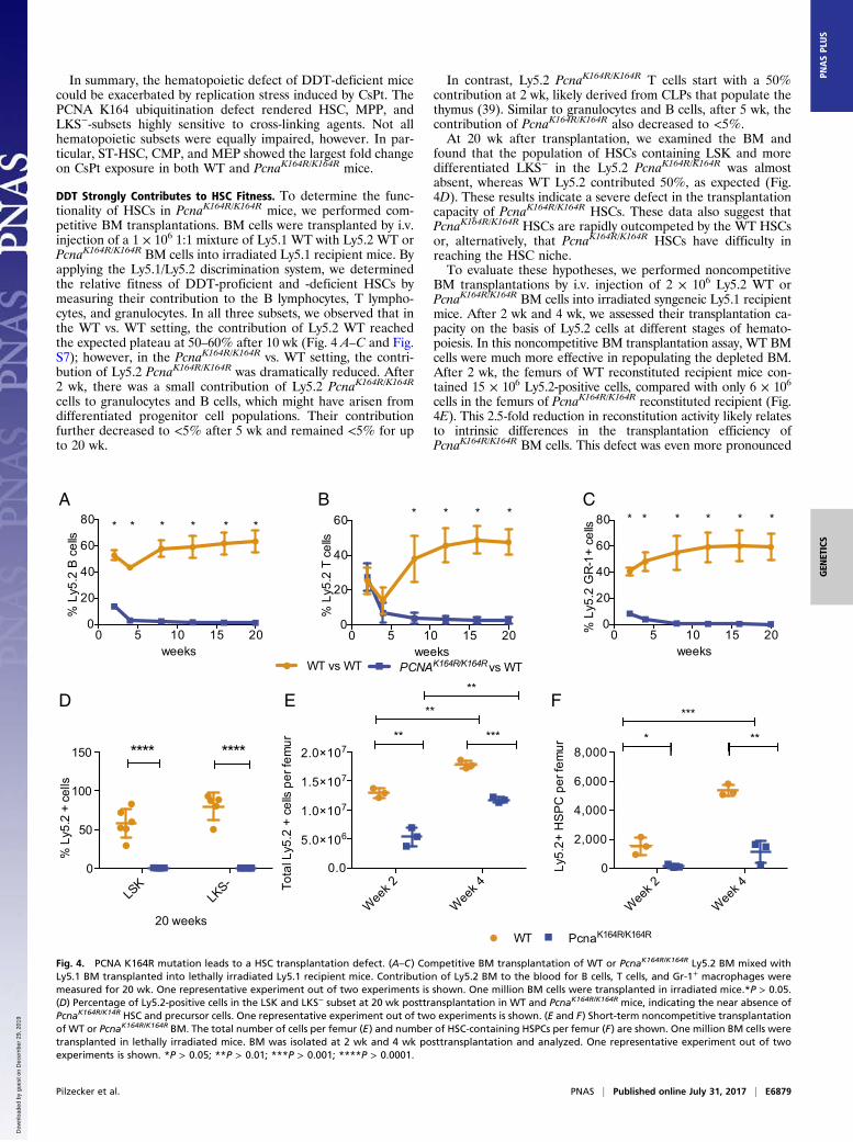
In summary, the hematopoietic defect of DDT-deficient micecould be exacerbated by replication stress induced by CsPt. ThePCNA K164 ubiquitination defect rendered HSC, MPP, andLKS−-subsets highly sensitive to cross-linking agents. Not allhematopoietic subsets were equally impaired, however. In par-ticular, ST-HSC, CMP, and MEP showed the largest fold changeon CsPt exposure in both WT and PcnaK164R/K164R mice.
DDT Strongly Contributes to HSC Fitness. To determine the func-tionality of HSCs in PcnaK164R/K164R mice, we performed com-petitive BM transplantations. BM cells were transplanted by i.v.injection of a 1 × 10⁶ 1:1 mixture of Ly5.1 WT with Ly5.2 WT orPcnaK164R/K164R BM cells into irradiated Ly5.1 recipient mice. Byapplying the Ly5.1/Ly5.2 discrimination system, we determinedthe relative fitness of DDT-proficient and -deficient HSCs bymeasuring their contribution to the B lymphocytes, T lympho-cytes, and granulocytes. In all three subsets, we observed that inthe WT vs. WT setting, the contribution of Ly5.2 WT reachedthe expected plateau at 50–60% after 10 wk (Fig. 4 A–C and Fig.S7); however, in the PcnaK164R/K164R vs. WT setting, the contri-bution of Ly5.2 PcnaK164R/K164R was dramatically reduced. After2 wk, there was a small contribution of Ly5.2 PcnaK164R/K164R
cells to granulocytes and B cells, which might have arisen fromdifferentiated progenitor cell populations. Their contributionfurther decreased to <5% after 5 wk and remained <5% for upto 20 wk.
In contrast, Ly5.2 PcnaK164R/K164R T cells start with a 50%contribution at 2 wk, likely derived from CLPs that populate thethymus (39). Similar to granulocytes and B cells, after 5 wk, thecontribution of PcnaK164R/K164R also decreased to <5%.At 20 wk after transplantation, we examined the BM and
found that the population of HSCs containing LSK and moredifferentiated LKS− in the Ly5.2 PcnaK164R/K164R was almostabsent, whereas WT Ly5.2 contributed 50%, as expected (Fig.4D). These results indicate a severe defect in the transplantationcapacity of PcnaK164R/K164R HSCs. These data also suggest thatPcnaK164R/K164R HSCs are rapidly outcompeted by the WT HSCsor, alternatively, that PcnaK164R/K164R HSCs have difficulty inreaching the HSC niche.To evaluate these hypotheses, we performed noncompetitive
BM transplantations by i.v. injection of 2 × 10⁶ Ly5.2 WT orPcnaK164R/K164R BM cells into irradiated syngeneic Ly5.1 recipientmice. After 2 wk and 4 wk, we assessed their transplantation ca-pacity on the basis of Ly5.2 cells at different stages of hemato-poiesis. In this noncompetitive BM transplantation assay, WT BMcells were much more effective in repopulating the depleted BM.After 2 wk, the femurs of WT reconstituted recipient mice con-tained 15 × 106 Ly5.2-positive cells, compared with only 6 × 10⁶
cells in the femurs of PcnaK164R/K164R reconstituted recipient (Fig.4E). This 2.5-fold reduction in reconstitution activity likely relatesto intrinsic differences in the transplantation efficiency ofPcnaK164R/K164R BM cells. This defect was even more pronounced
A B C
D
WT vs WT PCNAK164R/K164R vs WT
0 5 10 15 200
20
40
60
80
weeks
% L
y5.2
B c
ells
** * * * *
0 5 10 15 200
20
40
60
weeks
% L
y5.2
T c
ells
* * * *
0 5 10 15 200
20
40
60
80
weeks
% L
y5.2
GR
-1+
cells
* * * * * *
LSK
LKS-
0
50
100
150
20 weeks
% L
y5.2
+ c
ells
**** ****
E
Wee
k 2
Wee
k 40.0
5.0 106
1.0 107
1.5 107
2.0 107
Tota
l Ly5
.2 +
cel
ls pe
r fem
ur ** ***
**
**
Wee
k 2
Wee
k 40
2,000
4,000
6,000
8,000
Ly5.
2+ H
SP
C p
er fe
mur
* **
***
PcnaK164R/K164RWT
F
Fig. 4. PCNA K164R mutation leads to a HSC transplantation defect. (A–C) Competitive BM transplantation of WT or PcnaK164R/K164R Ly5.2 BM mixed withLy5.1 BM transplanted into lethally irradiated Ly5.1 recipient mice. Contribution of Ly5.2 BM to the blood for B cells, T cells, and Gr-1+ macrophages weremeasured for 20 wk. One representative experiment out of two experiments is shown. One million BM cells were transplanted in irradiated mice.*P > 0.05.(D) Percentage of Ly5.2-positive cells in the LSK and LKS− subset at 20 wk posttransplantation in WT and PcnaK164R/K164R mice, indicating the near absence ofPcnaK164R/K14R HSC and precursor cells. One representative experiment out of two experiments is shown. (E and F) Short-term noncompetitive transplantationof WT or PcnaK164R/K164R BM. The total number of cells per femur (E) and number of HSC-containing HSPCs per femur (F) are shown. One million BM cells weretransplanted in lethally irradiated mice. BM was isolated at 2 wk and 4 wk posttransplantation and analyzed. One representative experiment out of twoexperiments is shown. *P > 0.05; **P > 0.01; ***P > 0.001; ****P > 0.0001.
Pilzecker et al. PNAS | Published online July 31, 2017 | E6879
GEN
ETICS
PNASPL
US
Dow
nloa
ded
by g
uest
on
Dec
embe
r 29
, 201
9

when analyzing HSCs containing LSK and HSPCs (Fig. 4F andFig. S8 A–F). In addition, the LKS−, CMP, GMP, and MEPprogenitor population numbers were much lower after 2 wk inmice reconstituted with PcnaK164R/K164R BM. Even though the BMcellularity of PcnaK164R/K164R reconstituted recipients improvedsubstantially after 4 wk, major differences remained in terms ofdefined BM precursor subsets. Of note, the observed differencesat 2 wk after transplantation are not likely explained by preexistingdifferences in the initial subset composition of transplanted BMcells, because these differences greatly exceeded the preexistingdifferences.In summary, these data indicate a severe intrinsic repopulation
defect of PcnaK164R/K164R HSCs. This defect likely explains ourobservations in the competitive BM reconstitution assays.
DDT Deficiency Results in Premature Aging of the Early HematopoieticCompartment. During aging, the number of MPP2 cells increasesand the number of MPP4 cells progressively decreases in the BM(6). This suggests a priority for producing MPP2 at the expense ofMPP4, because MPP2 cells are important precursors for the es-sential myeloid/erythrocyte lineage, and MPP4s are the precursorsof the less important lymphoid lineage. In this way, oxygen supply issafeguarded. Interestingly, already after 2 mo, PcnaK164R/K164R miceshowed an increased in MPP2 and a decrease in MPP4 cells. Thisphenotype caused by defective DDT is consistent with acceleratedaging, presumably owing to increased replication stress. If thishypothesis is correct, then this phenotype would be expected to be
further enhanced during aging. Thus, we compared the cellularityof the hematopoietic subsets of WT and PcnaK164R/K164R at age9–10 mo (Fig. 5 A–E). Comparing 2-mo-old and 9- to 10-mo-oldPcnaK164R/K164R and WT mice revealed further increases in foldchanges in the ST-HSC, MPP4, and CLP subsets; however, MPP2remained higher in the PcnaK164R/K164R mice throughout this period(Fig. S9). In contrast, in the LKS− subsets, aging did not increasethe initial difference. This suggests that during stressed hemato-poiesis, the maintenance of LKS− cells is a priority, because thesecells contain the essential erythroid progenitors.In summary, defective DDT leads to accelerated aging of the
hematopoietic system, characterized by an increased selectiveskewing toward the MPP2 subset in LSK, primarily at the ex-pense of the MPP4 subset.
DiscussionAlthough the role of DNA repair in HSC maintenance has beenestablished, the contribution of DDT to HSC and progenitormaintenance has remained unclear. Here we report an elegantmodel for studying stressed hematopoiesis and demonstrate acritical cell-intrinsic role for DDT in maintaining adult HSCs andearly hematopoiesis. PcnaK164R/K164R cells are defective in TLSand TS, leading to increased replication stress and sensitivity tofork-stalling DNA lesions. In 2-mo-old mice, the PcnaK164R/K164R
mutation caused reduced cellularity of LSK, LT-HSC, ST-HSC, MPP4, LKS−, CMP, GMP, MEP, and CLP subsets inthe BM of mice. In contrast, the lack of PCNA K164-dependent
CD150
CD
135
CD150
CD
48
CD34
CD
16/3
2
CD135
CD
127
HSPC
MPP4
LT-HSC
MPP2MPP3
ST-HSC
HSPC-
MPP4
LT-HSC
MPP2MPP3
ST-HSC
CMP
GMP
MEP
CMP
GMP
MEP
Pcn
aK16
4R/K
164R
WT
Age
d m
ice
CLP
CLP
LKS- LSK
Sca-1, cKit subset
LKS- LSK
Sca-1, cKit subset
B C
D E
A
Sca-1
cKit
aged WT aged PcnaK164R/K164R
LSKHSPC
MPP40
10,000
20,000
30,000
40,000
Nuc
leat
ed c
ells
per f
emur
*** ** ***
LKS-
CMPGMP
MEP0
100,000
200,000
300,000
Nuc
leat
ed c
ells
per f
emur
** ******
CLP0
500
1,000
1,500
Nuc
leat
ed c
ells
per f
emur
****
LT-H
SC
ST-HSC
MPP2MPP3
0
5,000
10,000
Nuc
leat
ed c
ells
per
fem
ur
** ****
Fig. 5. Hematopoietic phenotype of DDT impaired mice is progressive with age. (A) FACS plots of 9- to 10-mo-old WT and PcnaK164R/K164R mice. (B–E) Numberof nucleated cells per femur of different progenitors and HSC subsets in young (2 mo old) and aged (9–10 mo old) mice. *P > 0.05; **P > 0.01; ***P > 0.001;****P > 0.0001.
E6880 | www.pnas.org/cgi/doi/10.1073/pnas.1706508114 Pilzecker et al.
Dow
nloa
ded
by g
uest
on
Dec
embe
r 29
, 201
9

DDT was associated with a selective increase in the myeloid/erythroid–associated MPP2 in PcnaK164R/K164R mice. This in-crease is likely due to increased stress-induced differentiationof HSCs, which reduces the number of HSCs, toward MPP2 ata cost of the number of MPP4 cells (3, 41, 42).Our cell cycle and cell proliferation studies reveal an increased
percentage of S/G2 cells and increased EdU incorporation inMPP4, suggestive of compensatory proliferation to counteractthe strong loss of these lymphoid-primed progenitor cells. Be-cause MPP4 mainly contributes to the lymphoid lineage, theincreased proliferation could explain the unaffected B cell andT cell development in PcnaK164R/K164R mice. In contrast, LKS−
and CMP progenitors had an increased proportion of S/G2 cellscombined with lower EdU incorporation in PcnaK164R/K164R
mice, pointing to a cell cycle arrest caused by increased forkstalling and secondary DNA damage due to defective DDT.To determine whether the BM defect of PcnaK164R/K164R mice
could be enhanced by exogenous DNA damage, mice were ex-posed to CsPt. Although WT BM subsets were marginally af-fected, PcnaK164R/K164R HSC and progenitor subsets were almostdepleted. This marked hypersensitivity to DNA damage in micedeficient for PCNA K164-dependent DDT further highlights therelevance of DDT in maintaining hematopoiesis and HSCs.Aging is characterized by a selective skewing of hematopoiesis
toward the myeloid/erythroid-associated MPP2 in the LSK subsetsin aged mice (6). Therefore, the skewing toward MPP2 in the LSKcompartment of PcnaK164R/K164R mice is likely related to accelera-
ted aging induced by increased replication stress due to defectiveDDT. We hypothesize that stress-induced regeneration, like rep-lication stress, of the hematopoietic system results in a shifteddifferentiation toward MPP2. This model is in line with previousreports documenting a shift toward MPP2 in the LSK compart-ment on stress-induced regeneration of the hematopoietic system(3). This skewing is further increased during aging. While the dif-ferentiation bias was already evident in 2-mo-old PcnaK164R/K164R
mice, this bias increased further at age 9–10 mo. This findingsupports the notion of accelerated aging in the PcnaK164R/K164R
BM. Apparently, the capacity to tolerate DNA damage and pre-vent replication stress is critical to maintaining homeostasis in theBM compartment. The failure to tolerate DNA damage stronglyaccelerates aging of the BM compartment, indicating an importantfunction of PCNA K164-dependent DDT in HSC maintenance.Defects in DNA repair pathways (9) and increased replicationstress (13) have been identified as potent drivers of prematureHSC aging, which is in line with our findings.In our model of stressed hematopoiesis due to deficient DDT
in PcnaK164R/K164R mice, HSCs shifted differentiation frommainly to MPP4 in WT to MPP2 in PcnaK164R/K164R (Fig. 6).Of note, Rad18-deficient mice do not show a decrease of LSK
cells (43). This likely is related to the existence of alternativeE3 ligases targeting PCNA K164 (44, 45). Alternatively, an un-known PCNA K164 modification may play a role in maintainingHSCs and progenitor cells. An additional phenotype of PcnaK164R/K164R mice is infertility due to the complete absence of germ cells
MEP
GMP
CMP
MPP4
MPP3
MPP2
CLP
ST-HSCLT-HSC
Hematopoiesis in WT mice
megakaryocyteserythrocytes
lymphocytes
granulocytesmacrophage
MEP
GMP
CMP
MPP4
MPP3
CLP
ST-HSCLT-HSC
Hematopoiesis in DDT defient micegranulocytesmacrophage
megakaryocyteserythrocytes
lymphocytes
MPP2
Compensatory proliferation
Increased cell cycle arrest
Fig. 6. Model for effect of DDT deficiency on HSC and early progenitors. Steady-state hematopoiesis in WT mice is indicated. In DDT-deficient PcnaK164R/K164R
mice, replication stress-induced differentiation of HSC toward myeloid/erythroid-associated MPP2 in PcnaK164R/K164R is indicated by a red arrow. Compensatoryproliferation for MPP4 is specified, as is cell cycle arrest in CMP and GMP subsets. Arrows indicate the direction of differentiation. The blue dots indicate LSKsubsets, and the green dots indicate LKS− subsets. Each dot represents 500 nucleated cells per femur.
Pilzecker et al. PNAS | Published online July 31, 2017 | E6881
GEN
ETICS
PNASPL
US
Dow
nloa
ded
by g
uest
on
Dec
embe
r 29
, 201
9

(34). The infertility, the BM phenotype, and the sensitivity to cross-linking agents are shared phenotypes with Fanconi anemia (FA)mouse models (46–48). Future research should examine the con-tribution of PCNA K164-dependent DDT in the FA pathway.The FA pathway is involved in the repair of interstrand cross-
links (49, 50). DDT pathways, on the other hand, likely tolerate amyriad of replication blocks. These replication blocks may in-clude base damages such as deamination, methylation, and oxi-dation; intrastrand and interstrand cross-links induced byendogenous and exogenous genotoxins; G4 stacks; RNA-DNAhybrids; and ribonucleotide misincorporation (16-18, 51, 52).Consequently, DDT-impaired HSCs are likely to be sensitized tothis complex spectrum of lesions. For survival, PCNA K164-deficient DDT HSCs are assumed to depend on alternativeDDT pathways, such as REV1-dependent DDT. Given the im-pact of a DDT defect on HSCs, it is likely that other stem cells,especially those in highly proliferative compartments, are sensi-tive as well. Further research is needed to examine the sensitivityof other tissue stem cells and the relevance of DDT for tissuehomeostasis.In conclusion, this study reports the relevance of DDT in
preventing premature aging in the hematopoietic compartmentand safeguarding HSC functionality. These data highlight DDTas important arm of the DNA damage response network.
MethodsMice and Breeding. The PcnaK164R/K164R knock-in mouse model has been de-scribed previously (34). All mice were kept on C57BL/J6 background underspecific pathogen-free conditions. PcnaK164R mice were maintained hetero-zygous. All experiments were approved by the Animal Experimental Com-mission of the Netherlands Cancer Institute and performed in accordancewith Dutch and European guidelines.
Antibodies. Antibody specifications are listed in Table S1.
Flow Cytometry.Hematopoietic precursor subset analysis. Mice were killed at the indicated age(2 mo or 9–10 mo), and BM from femora was flushed out using a 21-gaugesyringe with cold PBEA buffer (1× PBS 0.5% BSA, 2 mM EDTA, and 0.02%sodium azide). The samples were kept on ice. We used 5 × 10⁶ cells perstaining. BM cells were first stained with a biotinylated lineage + antibodymix for 30 min and then washed twice in PBEA buffer. For quantifying stemcells and MPP populations, cKit-APC, Sca-1-PerCp/Cy5.5, CD48-FITC, CD135-PE, CD150-PE/Cy7, and streptavidin-APC/Cy7 were used. For quantifying LKS−
progenitor populations, cKit-APC, CD34-FITC, CD16/32-PE/Cy7, streptavidin-APC/Cy7, and Sca-1-Pacific Blue were used. To quantify CLPs, cKit-APC,CD127-PerCp/Cy5.5, CD135-PE, streptavidin-APC/Cy7, and Sca-1-Pacific Bluewere used. Cells were washed twice with PBEA and then resuspended in 400μL of PBEA. For stem cells and MPP, DAPI was used as life/dead staining,whereas for LKS− and CLP, propidium iodide (PI) was used. All measurementswere performed with a BD LSRFortessa cell analyzer (BD Biosciences).Analyses were performed using FlowJo version 10.0.8r1.Cell cycle analysis of BM populations. Cell surface staining was performed asdescribed above. Samples were incubated in Cytofix/Cytoperm (BD Biosciences)for 15min. Cellswerewashedusing Permwash andharvested in PBEA containing10 μg/mL DAPI. Analyses were performed using FlowJo version 10.0.8r1.Assessing yH2AX levels of BM populations. Cell surface staining was performedas described above. BM cells were first labeled with biotinylated Lin+ anti-
body mix. Subsequently, LSK populations were stained with CD135-PE, SCA-1-PerCp/Cy5.5, cKit-APC, and streptavidin-APC/Cy7. Progenitor populationswere stained with SCA-1-PerCpCy5.5, CD16/32-PE/Cy7, cKit-APC, andstreptavidin-APC/Cy7.
Cells were fixed and permeabilized using Cytofix/Cytoperm (BD Biosci-ences), then stained with γH2AX antibody for 30 min at room temperature.Anti-mouse IgG-AF488 was used for the secondary staining. DAPI (10 μg/mL)was used for chromatin labeling. A nonspecific IgG isotype control served asa negative control. Analyses were performed using FlowJo version 10.0.8r1.B and T development. The lymphocyte composition and precursor subsets inthymus and bone marrow were analyzed as described previously (19).In vivo EdU incorporation assay. Here 200 μL of 10 mM EdU in PBS was injectedi.p. After 16 h, BM was isolated as described above. The Click-iT Plus EdUFlow Cytometry Kit AF488 (Thermo Fisher Scientific) protocol was followed.BM was stained using biotinylated Lineage+ antibody mix, cKit-APC, Sca-1-PerCp/Cy5.5, CD135-PE, CD150-PE/Cy7, and streptavidin-APC/Cy7, followedby Alexa Fluor 488 picolyl azide staining. A non–EdU-treated mouse servedas a negative control. Analyses were performed using FlowJo version10.0.8r1.
Competitive BM Reconstitution. BM was isolated from three Ly5.2 2-mo-oldWT mice and three 2-mo-old Ly5.2 PCNAK164RK164R mice and mixed1:1 with the BM isolated from one Ly5.1 WT mouse. Thereafter, 1 × 10⁶
Ly5.1/Ly5.2 mixed BM cells were transplanted into lethally irradiated (twodoses of 5.5 Gy, separated by a 3-h interval) Ly5.1 recipient mice. Blood(50 μL) was obtained after 2, 4, 8, 12, 16, and 20 wk. The contribution in theblood of Ly5.1 and Ly5.2 cells was assessed using Ly5.1-PE and Ly5.2-PE/Cy7antibodies. Cells were also stained with Gr1-APC/Cy7, CD3-FITC, CD19-APC,and CD11b-PerCp/Cy5.5 for subset analysis. DAPI was used for life/deathstaining. At 20 wk posttransplantation, the mice were killed, and the BMwas isolated for analysis, as described above using SCA-1-PerCp/Cy5.5, cKit-APC, streptavidin APC/Cy7, Ly5.1-PE, and Ly5.2-PE-Cy7. Irradiated mice weretreated with Enrobactin for the first 4 wk after irradiation. Analyses wereperformed using FlowJo version 10.0.8r1.
Noncompetitive BM Transplantation. The BM of Ly5.2 WT and PcnaK164R/K164R
mice was isolated. Recipient Ly5.1 mice were lethally irradiated (two dosesof 5.5 Gy, separated by a 3-h interval) and transplanted with 1 × 10⁶ WT orPcnaK164R/K164R BM cells. At 2 wk and 4 wk posttransplantation, BM andblood were isolated to assess the contribution of the transplanted BM, asdescribed above. Analyses were performed using FlowJo version 10.0.8r1.
In Vivo CsPt Sensitivity Assay. Mice were injected i.v. with 0.8 mg/kg cisplatinor PBS. After 2 d, the BM was isolated and analyzed as described above.
Calculations and Statistics. We determined the number of viable cells persubset by DAPI or PI staining and based on the ratio of subset of interest andviable population times the number of cells per femur. The number of cellsper femur was standardized to 20 × 106 when no difference in total BMnumbers was found between WT and PcnaK164R/K164R. The t tests were per-formed using GraphPad Prism version 6.0. In the figures, *P > 0.05; **P > 0.01;***P > 0.001; ****P > 0.0001.
ACKNOWLEDGMENTS.We thank H. te Riele and N. Wit for comments on themanuscript, A. Pfauth, F. van Diepen, and M. van Baalen for cell sorting andassistance during flow cytometry, and the animal caretakers of the NKI-AVLfor biotechnical assistance. This project was made possible by generoussupport from the Dutch Cancer Foundation (Grants KWF NKI-2012-5243 andKWF NKI-2016-10032, to H.J.).
1. Orford KW, Scadden DT (2008) Deconstructing stem cell self-renewal: Genetic insightsinto cell-cycle regulation. Nat Rev Genet 9:115–128.
2. Morrison SJ, Uchida N, Weissman IL (1995) The biology of hematopoietic stem cells.Annu Rev Cell Dev Biol 11:35–71.
3. Pietras EM, et al. (2015) Functionally distinct subsets of lineage-biased multipotentprogenitors control blood production in normal and regenerative conditions. CellStem Cell 17:35–46.
4. Cabezas-Wallscheid N, et al. (2014) Identification of regulatory networks in HSCs andtheir immediate progeny via integrated proteome, transcriptome, and DNA methyl-ome analysis. Cell Stem Cell 15:507–522.
5. Wilson A, et al. (2008) Hematopoietic stem cells reversibly switch from dormancy toself-renewal during homeostasis and repair. Cell 135:1118–1129.
6. Young K, et al. (2016) Progressive alterations in multipotent hematopoietic progen-itors underlie lymphoid cell loss in aging. J Exp Med 213:2259–2267.
7. Geiger H, de Haan G, Florian MC (2013) The ageing haematopoietic stem cell com-partment. Nat Rev Immunol 13:376–389.
8. Le Saux S, Weyand CM, Goronzy JJ (2012) Mechanisms of immunosenescence: Lessonsfrom models of accelerated immune aging. Ann N Y Acad Sci 1247:69–82.
9. Rossi DJ, et al. (2007) Deficiencies in DNA damage repair limit the function of hae-matopoietic stem cells with age. Nature 447:725–729.
10. Parmar K, et al. (2010) Hematopoietic stem cell defects in mice with deficiency ofFancd2 or Usp1. Stem Cells 28:1186–1195.
11. Prasher JM, et al. (2005) Reduced hematopoietic reserves in DNA interstrand crosslinkrepair-deficient Ercc1−/− mice. EMBO J 24:861–871.
12. Rossi DJ, Jamieson CH, Weissman IL (2008) Stems cells and the pathways to aging andcancer. Cell 132:681–696.
13. Flach J, et al. (2014) Replication stress is a potent driver of functional decline in ageinghaematopoietic stem cells. Nature 512:198–202.
E6882 | www.pnas.org/cgi/doi/10.1073/pnas.1706508114 Pilzecker et al.
Dow
nloa
ded
by g
uest
on
Dec
embe
r 29
, 201
9
14. Nick McElhinny SA, Gordenin DA, Stith CM, Burgers PM, Kunkel TA (2008) Division oflabor at the eukaryotic replication fork. Mol Cell 30:137–144.
15. Miyabe I, Kunkel TA, Carr AM (2011) Themajor roles of DNA polymerases epsilon and deltaat the eukaryotic replication fork are evolutionarily conserved. PLoS Genet 7:e1002407.
16. Sarkies P, Reams C, Simpson LJ, Sale JE (2010) Epigenetic instability due to defectivereplication of structured DNA. Mol Cell 40:703–713.
17. Helmrich A, Ballarino M, Nudler E, Tora L (2013) Transcription-replication encounters,consequences and genomic instability. Nat Struct Mol Biol 20:412–418.
18. Nick McElhinny SA, et al. (2010) Genome instability due to ribonucleotide in-corporation into DNA. Nat Chem Biol 6:774–781.
19. Pilzecker B, et al. (2016) PrimPol prevents APOBEC/AID family-mediated DNA muta-genesis. Nucleic Acids Res 44:4734–4744.
20. Bianchi J, et al. (2013) PrimPol bypasses UV photoproducts during eukaryotic chro-mosomal DNA replication. Mol Cell 52:566–573.
21. García-Gómez S, et al. (2013) PrimPol, an archaic primase/polymerase operating inhuman cells. Mol Cell 52:541–553.
22. Sale JE, Lehmann AR, Woodgate R (2012) Y-family DNA polymerases and their role intolerance of cellular DNA damage. Nat Rev Mol Cell Biol 13:141–152.
23. Moldovan GL, Pfander B, Jentsch S (2007) PCNA, the maestro of the replication fork.Cell 129:665–679.
24. Gali H, et al. (2012) Role of SUMO modification of human PCNA at stalled replicationfork. Nucleic Acids Res 40:6049–6059.
25. Guo C, et al. (2003) Mouse Rev1 protein interacts with multiple DNA polymerasesinvolved in translesion DNA synthesis. EMBO J 22:6621–6630.
26. Ohashi E, et al. (2004) Interaction of hREV1 with three human Y-family DNA poly-merases. Genes Cells 9:523–531.
27. Tissier A, et al. (2004) Co-localization in replication foci and interaction of humanY-family members, DNA polymerase pol eta and REVl protein. DNA Repair (Amst) 3:1503–1514.
28. Acharya N, Haracska L, Prakash S, Prakash L (2007) Complex formation of yeastRev1 with DNA polymerase eta. Mol Cell Biol 27:8401–8408.
29. Becker JR, et al. (2015) Genetic interactions implicating postreplicative repair inOkazaki fragment processing. PLoS Genet 11:e1005659.
30. Wit N, et al. (2015) Roles of PCNA ubiquitination and TLS polymerases κ and η in thebypass of methyl methanesulfonate-induced DNA damage. Nucleic Acids Res 43:282–294.
31. Krijger PH, et al. (2011) HLTF and SHPRH are not essential for PCNA poly-ubiquitination, survival and somatic hypermutation: Existence of an alternativeE3 ligase. DNA Repair (Amst) 10:438–444.
32. Stelter P, Ulrich HD (2003) Control of spontaneous and damage-induced mutagenesisby SUMO and ubiquitin conjugation. Nature 425:188–191.
33. Hoege C, Pfander B, Moldovan GL, Pyrowolakis G, Jentsch S (2002) RAD6-dependentDNA repair is linked to modification of PCNA by ubiquitin and SUMO. Nature 419:135–141.
34. Langerak P, Nygren AO, Krijger PH, van den Berk PC, Jacobs H (2007) A/T mutagenesisin hypermutated immunoglobulin genes strongly depends on PCNAK164 modifica-tion. J Exp Med 204:1989–1998.
35. Hendel A, et al. (2011) PCNA ubiquitination is important, but not essential fortranslesion DNA synthesis in mammalian cells. PLoS Genet 7:e1002262.
36. Edmunds CE, Simpson LJ, Sale JE (2008) PCNA ubiquitination and REV1 define tem-porally distinct mechanisms for controlling translesion synthesis in the avian cell lineDT40. Mol Cell 30:519–529.
37. Bienko M, et al. (2005) Ubiquitin-binding domains in Y-family polymerases regulatetranslesion synthesis. Science 310:1821–1824.
38. Kannouche PL, Wing J, Lehmann AR (2004) Interaction of human DNA polymerase ηwith monoubiquitinated PCNA: A possible mechanism for the polymerase switch inresponse to DNA damage. Mol Cell 14:491–500.
39. Rodewald HR, Fehling HJ (1998) Molecular and cellular events in early thymocytedevelopment. Adv Immunol 69:1–112.
40. Krijger PH, et al. (2011) PCNA ubiquitination-independent activation of polymerase ηduring somatic hypermutation and DNA damage tolerance. DNA Repair (Amst) 10:1051–1059.
41. Santos MA, et al. (2014) DNA-damage-induced differentiation of leukaemic cells as ananti-cancer barrier. Nature 514:107–111.
42. Wang J, et al. (2016) Per2 induction limits lymphoid-biased haematopoietic stem cellsand lymphopoiesis in the context of DNA damage and ageing. Nat Cell Biol 18:480–490.
43. Yang Y, et al. (2016) Rad18 confers hematopoietic progenitor cell DNA damage tol-erance independently of the Fanconi Anemia pathway in vivo. Nucleic Acids Res 44:4174–4188.
44. Simpson LJ, et al. (2006) RAD18-independent ubiquitination of proliferating-cell nu-clear antigen in the avian cell line DT40. EMBO Rep 7:927–932.
45. Terai K, Abbas T, Jazaeri AA, Dutta A (2010) CRL4(Cdt2) E3 ubiquitin ligase mono-ubiquitinates PCNA to promote translesion DNA synthesis. Mol Cell 37:143–149.
46. Bakker ST, de Winter JP, te Riele H (2013) Learning from a paradox: Recent insightsinto Fanconi anaemia through studying mouse models. Dis Model Mech 6:40–47.
47. Parmar K, D’Andrea A, Niedernhofer LJ (2009) Mouse models of Fanconi anemia.Mutat Res 668:133–140.
48. Garaycoechea JI, Patel KJ (2014) Why does the bone marrow fail in Fanconi anemia?Blood 123:26–34.
49. Kottemann MC, Smogorzewska A (2013) Fanconi anaemia and the repair of Watsonand Crick DNA crosslinks. Nature 493:356–363.
50. Deans AJ, West SC (2011) DNA interstrand crosslink repair and cancer. Nat Rev Cancer11:467–480.
51. Lindahl T, Barnes DE (2000) Repair of endogenous DNA damage. Cold Spring HarbSymp Quant Biol 65:127–133.
52. Ciccia A, Elledge SJ (2010) The DNA damage response: Making it safe to play withknives. Mol Cell 40:179–204.
Pilzecker et al. PNAS | Published online July 31, 2017 | E6883
GEN
ETICS
PNASPL
US
Dow
nloa
ded
by g
uest
on
Dec
embe
r 29
, 201
9