dna diagnostics: nanotechnology-enhanced electrochemical
TRANSCRIPT

DNA Diagnostics: Nanotechnology-Enhanced ElectrochemicalDetection of Nucleic Acids
FANG WEI, PETER B. LILLEHOJ, AND CHIH-MING HO
Mechanical and Aerospace Engineering Department, School of Engineering and Applied Science, University of California, Los Angeles,California 90095
ABSTRACT: The detection of mismatched base pairs in DNA plays acrucial role in the diagnosis of genetic-related diseases and conditions,especially for early stage treatment. Among the various biosensors thathave been used for DNA detection, EC sensors show great promisebecause they are capable of precise DNA recognition and efficientsignal transduction. Advancements in micro- and nanotechnologies,specifically fabrication techniques and new nanomaterials, have en-abled for the development of highly sensitive, highly specific sensorsmaking them attractive for the detection of small sequence variations.Furthermore, the integration of sensors with sample preparation andfluidic processes enables for rapid, multiplexed DNA detection essentialfor POC clinical diagnostics. (Pediatr Res 67: 458–468, 2010)
The recent discovery and sequencing of the human genomehas provided valuable insight into understanding how
genetic factors contribute to the development of disease.Specifically, the detection of DNA sequence variations playsan important role in the diagnosis of genetic-related diseasesand conditions, especially for early stage treatment and mon-itoring. Among the different types of diseases caused by DNAalterations, sequence-specific mismatch has the most impor-tance, yet is extremely difficult to detect (1), especially forsingle-nucleotide polymorphism (SNP). Furthermore, se-quence-specific detection has great importance in variousmedical and scientific applications such as the diagnosis ofinherited diseases and the study of pathogen response andbacterial/viral detection.
Because of the complex nature of DNA, the detection ofsingle or small numbers of base mismatches requires highsensitivity and specificity (2–4). Current detection methodsrely on sample amplification combined with meticulous ex-perimental stringency control (5). For example, polymerasechain reaction (PCR) requires careful primer design and ac-curate temperature control to obtain sensitivities in the fMrange with single-base mismatch specificity (1,3,4). Althoughthese conventional technologies provide the golden standardfor laboratory-based DNA diagnostics, they cannot meet therequirements of POC clinical diagnostics (4).
EC sensors, initially developed to detect biomolecules in alaboratory setting, have recently found extensive applications foron-site biosensing and detection (6,7), especially for medical andclinical diagnostics (8–12). While offering simplicity in operationand sample manipulation, the contemporary EC biosensor alsoprovides highly sensitive and specific measurements for a broadspectrum of biomolecules (13–17). The sample size required forcurrent EC sensors is small, ranging from several microliters tohundreds of nanoliters, which includes the sample pretreatmentreagents. Additionally, the detection time is relatively fast, vary-ing from a few minutes to tens of seconds. However, the mostimportant feature of EC sensors is their potential to be easilytransformed from a laboratory-based instrument to a commer-cializable POC device. Because of all these advantages, ECbiosensing for DNA diagnostics is becoming a very promisingarea of research and development.
Recently, micro- and nanotechnologies have shown emergingpotential in EC DNA diagnostics. EC sensors offer a perfectinterface for incorporating these technologies, which includes avariety of new materials and fabrication processes. Nanomateri-als can be used in various aspects of the detection systemincluding capture probes, reporting molecules, electrode fabrica-tion, and electrode coatings (18–25). These materials offer im-proved biocompatibility, additional binding sites and higher sig-nal intensities (via enhanced electrical properties) compared withtraditional materials in EC sensors (17,21,25–28). Nanofabrica-tion allows for miniaturization of the sensor, which improves thesensitivity and reduces the sample and reagent volumes, makingthe detection process more efficient. Although nanomaterials andnanofabrication are described here as two separated categories,recent trends combine both of these elements in the design of newEC sensors for DNA diagnostics (Fig. 1). With contributionsfrom microfluidics and MEMS technologies, EC sensors can beintegrated onto portable platforms incorporating all the necessarypreparation and fluidic processes (10,29,30), giving way to com-mercializable devices for clinical diagnostics (31,32). Ultimately,the end goal of EC sensor development is to construct a totalanalysis system for rapid DNA biosensing, which incorporatessample pretreatment, sample delivery, and detection.Received November 16, 2009; accepted January 4, 2010.
Correspondence: Chih-Ming Ho, Ph.D., Mechanical and Aerospace Engineering De-partment, School of Engineering and Applied Science, 420 Westwood Plaza, Universityof California, Los Angeles, CA 90095; e-mail: [email protected]
Supported by NASA/National Space Biomedical Research Institute (NSBRI)(TD01301), NIH/National Institute of Dental and Craniofacial Research (NIDCR) (U01DE017790), NIH/NIDCR (DE007296), and NIH/Nanomedicine Development Center(NDC) (5PN2ey018228:03).
Abbreviations: CNT, carbon nanotube; EBL, electron-beam lithography;EC, electrochemical; EIS, electrochemical impedance spectroscopy; MEMS,micro-electro-mechanical systems; POC, point-of-care; SiNW, siliconnanowire; SNR, signal-to-noise ratio; Vm, melting potential
0031-3998/10/6705-0458PEDIATRIC RESEARCH
Vol. 67, No. 5, 2010
Copyright © 2010 International Pediatric Research Foundation, Inc.Printed in U.S.A.
458

Principles for EC DNA Sensors
EC detection. The EC detection of biologic species is basedon EC reactions that occur during biorecognition processes(33). These reactions can be exhibited as changes of ECproperties (i.e. current/potential, redox kinetics, impedance,etc.) or changes of non-EC properties (i.e. conformationchanges, mass transportation, van der Waals interactions,etc.), resulting in fluctuations of an EC signal. Such fluctua-tions, which usually contribute to high background noise, arenot sequence-specific and need to be suppressed during thedetection process. The resultant signal readouts can take theform as electrical currents, potentials, or impedances in steadystate or changes in these parameters during the recognitionprocess, which correspond to the kinetics of recognition (34).Currently, ex situ EC sensors, in which sample pretreatmentand fluidic processing are performed “off-chip,” are mostcommonly used because they generate a better SNR, resultingfrom the detection of purified, concentrated biomolecules.However, these sensors have limited applications in POCdiagnostics. Therefore, in situ EC sensors, which incorporateall the sample processing steps “on-chip,” are more desirablefor clinical application; however, they require higher sensitiv-ity and specificity for non-pretreated samples. Additionally, insitu EC sensors can monitor changes of EC properties, whichis more desirable for studying biologic processes during DNArecognition (35).
EC DNA sensors. A typical EC DNA sensor consists of anelectrode, capture probe and reporter probe. A capture probe isan element used to recognize and bind to the target DNA andis usually immobilized onto a solid substrate, such as theelectrode surface. However, they can also be immobilized onnanomaterials or other biomolecules. A reporter probe is amolecule that generates the EC signal in response to ECreactions. Both the capture probe and reporter probe arecreated with high specificity to the target DNA. Additionalcomponents, such as electrode coatings and intermediate mo-lecular linkers, are also commonly integrated for improvedsensor performance. Common molecules used as probes (cap-ture and reporter) include single-stranded oligonucleotides,aptamers, peptides, and DNA-related proteins (14). In somesensors, the capture and reporter probes are combinedtogether as a single unit for improved integration. Probe,target, and reporter molecules can all be modified or linkedwith properly integrated nanomaterials, as shown in Figure2. Because of their high surface-to-volume ratios and biologiccompatibilities, nanomaterials not only increase the signalintensity but also help to accumulate/separate specific DNAmolecules during EC reactions, which greatly improves theSNR, especially for sequence-specific recognition (28). Awide variety of nanomaterials can be applied, where themost common include metal nanoparticles, cadmium sul-fide nanoparticles, CNTs and SiNWs. An extensive and
Figure 1. Schematic illustration demonstratingthe integration of nanomaterials and micro/nanofabrication technologies for constructingEC DNA sensors.
Figure 2. Common nanomaterials used in ECbiosensors for DNA/RNA diagnostics; (A) nano-materials for electrode coatings, (B) nanomate-rials for probe labeling, (C) nanomaterials fortarget labeling, and (D) nanomaterials for signalreporting. Reprinted from Xu K et al. 2009 Sen-sors 9:5534–5557. Copyright © 2009 by authors,with permission.
459ELECTROCHEMICAL DNA SENSORS

detailed review on the applications of nanomaterials forDNA biosensors can be found in literature (21,28,36).
Current EC sensors for DNA diagnostics include two schemesfor biologic recognition. The most common scheme, hybridiza-tion-based DNA detection, uses nucleotides as the probe andtargets elements (Fig. 3A–D) (37). The performance of ECsensors based on this method is highly dependent on the affinitybetween the probe and target molecules, which can be tunedby the probe design, environmental conditions, and additionalamplification processing. This detection system can either usea one probe versus one target scheme, a multiple probe versussingle target scheme, or vice versa. For example, EC sensorsbased on a sandwich detection mode are composed of one captureprobe and one reporter probe for each DNA target. Ultimately, nomatter what method is used for detection, the output signal iscaused by changes in EC properties or EC-related properties.
The second detection scheme, which has emerged in recentyears, is enzymatic-based DNA detection (Fig. 3E and F)(16,38,39). In this scheme, DNA-related enzymes are intro-duced into the biorecognition system and changes in amountof these enzymes correlate to specific biologic process (i.e.deletion/fusion of the target DNA). For example, when an ECsensor experiences a specific process, the enzyme level eitherincreases or decreases, resulting in amplification or reduction ofthe signal. The enzymatic process is highly specific to a DNAsequence, which makes it ideal for DNA mismatch detection.
Advantages of EC sensors for DNA diagnostics. EC sen-sors offer several advantages over other detection methods,making them attractive for DNA biosensing. In addition tobeing highly specific and sensitive, EC sensors are extremelyefficient, in terms of fast detection times and low power
consumption. EC DNA sensors largely rely on nucleotidehybridization during the detection process, which involvesspecific electrostatic charge distributions and strong hydrogenbonding. Because the backbone of a nucleotide is composed ofphosphoric acids and base units, the entire molecule is heavilycharged with a negative potential. Therefore, hybridizationbetween nucleotides needs to overcome the strong repulsionforce between each other. In traditional DNA detection meth-ods, temperature and chemicals are used to reduce the repul-sion of these molecules; however, both of these modulationsare not very effective. Based on the thermodynamics of DNAhybridization/denaturation, the Gibbs free energy for suchprocesses is in the range of 1 to 10 kcal/mol. Therefore, veryhigh temperatures or ion buffer concentrations are required toovercome this energy barrier and such conditions are likely tointerfere with the bio-system. In contrast, EC sensors arecapable of producing strong electrical fields, where only sev-eral hundred millivolts of potential can overcome the reactionbarrier (10,40). Similar to the melting temperature (Tm) fortraditional temperature control, the melting potential (Vm),which denotes the voltage at which 50% hybridization/denaturation occurs, is an important parameter useful forcharacterizing different DNA strands and provides an addi-tional tool for controlling the specificity (41).
A second advantage of EC sensors is their simplicity inmanipulating molecules within the sample fluid, which isperformed through electrical fields generated by the electrode.Thorough mixing and precise manipulation of molecules arecrucial for achieving high hybridization/denaturation effi-ciency. Traditional temperature or chemical-based controlschemes require additional mixing and separation procedures,which greatly hinders advancement toward a POC DNA di-agnostic platform. Specifically, the speed of these proceduresis limited by chemical reaction times and heat/mass transferprocesses within the solution. In contrast, EC sensors cangenerate high electrical fields within a very short time (34). Byusing this scheme, molecules near the electrodes can bemanipulated by applying different electrical profiles. Addi-tionally, thin dielectric double layers generated in high inten-sity electric fields can be used to enhance mixing and samplemanipulation. Such accurate control circumvents the need foradditional components and greatly simplifies the detectionprocess, making in situ DNA detection possible.
A third advantage of EC sensors is their ability to achieveprecise DNA recognition due to localization effects. Tradi-tional detection methods, based on temperature or chemicalcontrol schemes, lack precision because their effects are dis-persed within the entire sample solution rather than localizednear the DNA molecules. In addition, the strong hydrogenbonds within DNA makes precise control over hybridization/denaturation quite difficult. In contrast, EC sensors can gen-erate well-defined, localized electrical fields within the elec-trode domains where DNA recognition occurs. Additionally,nano-sized electrodes can produce electric fields that areconcentrated within a small region surrounding the electrode,which allows for even greater precision and localization.Nanoelectrodes also require much smaller electrical potentialsand reduces the overall power consumption of the sensor.
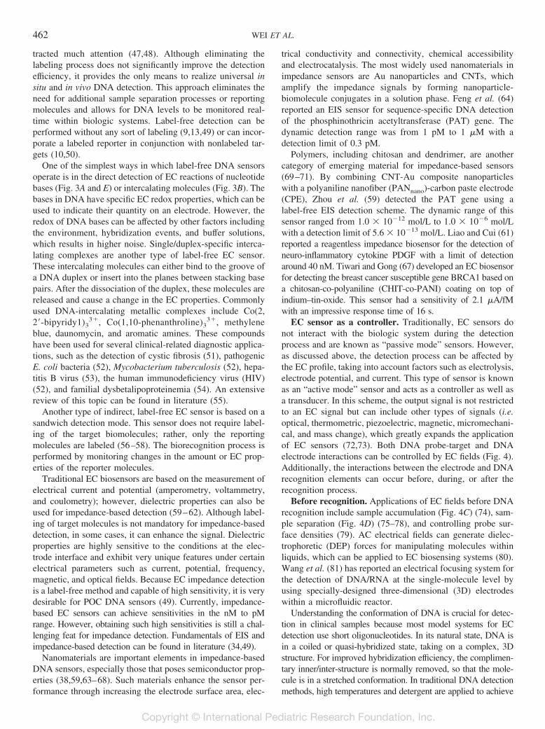
Figure 3. Schematic representation illustrating the principles for EC DNAsensors. A, Direct oxidation/reduction of nucleotide bases. B, Detection ofintercalating complex for single/duplex stands. C, Detection of specific DNAwith labeled reporting molecules. D, Detection of specific DNA with inte-grated capture probe and reporter probe. E, Direct detection after specificDNA enzymatic process. F, Detection of extra labeled reporter after specificDNA enzymatic process.
460 WEI ET AL.

Because of the advantages listed above, there are severalkey features that EC sensors offer for DNA diagnostics:
1. Detection speed: the detection of DNA involves severalprocesses including DNA recognition, sample manipula-tion, and signal readout. By using EC sensors, DNA rec-ognition can occur within minutes and in some cases,several seconds. The entire detection process, includingsample preparation, sample delivery, and signal readout,can be completed in several minutes. Such rapid detectionis ideal for POC clinical diagnostics.
2. Sensitivity: with appropriate electrical field profiles, ECDNA sensors can achieve sensitivities down to several fMand in some cases, aM for short oligonucleotides. Suchsensitivities can be achieved without sample amplification(i.e. PCR), which is a major advantage for POC systems.Additionally, the total amount of sample required for de-tection is on the orders of several microliters.
3. Specificity: the stringency of EC-controlled DNA recogni-tion enables for single-base mismatch specificity, even inclinical samples.
4. Convenience: with contributions from MEMS and nanotech-nology, EC sensors can be integrated onto portable platforms,enabling for POC monitoring and on-site biosensing.
5. Multiplexing: nowadays, a single biomarker is not suffi-cient for high specificity detection in clinical samples. Bycombining several biomarkers, the detection accuracy canbe greatly improved, making multiplex detection for DNAdiagnostics very important. Because the electrical fieldsgenerated by electrodes are highly localized, they can beused for simultaneous detection within a miniaturized plat-form, where the detection condition for each DNA targetcan be optimized separately (32,42).
EC DNA Sensor Types
Traditional EC biosensors are based on detecting changesof EC properties, such as current/potential, redox kinetics, orelectrical impedance (34). However, because most biorecog-nition is influenced by non-EC properties (mass transportationor conformational changes) in addition to EC reactions, ECsensors can also be based on controlling the biorecognitionprocess. From these two aspects, we will distinguish EC DNAsensors in two categories: EC biosensors as transducers andEC biosensors as controllers. In the following section, eachtype of sensor will be discussed in further detail.
EC sensor as a transducer. Most EC biosensors directlydetect changes in EC signals, in which case, the sensor acts asa transducer. We separate EC sensors into two categories:labeled detection and label-free detection. Usually EC sensorsuse labeled reporters, which can result in high levels ofbackground noise because of mass transportation processes oreven conformational changes. Therefore, prelabeled reporterscan help to increase the SNR. The second category, label-freedetection, usually refers to detection without any sort oflabeling. This scheme also includes detection which incorpo-rates a labeled reporter in conjunction with nonlabeled targetsbecause the final detection process is label-free.
Labeled detection. Labeling molecules are electrochemi-cally active in that they exhibit specific EC properties, whichcorrelate to the status of DNA targets on the electrodes.Usually, the amount of the labeling molecules indicates theamount of DNA targets in the sample either in a directdetecting mode or a competitive detecting mode (Fig. 3C andF). Nanomaterials, because of their high surface-to-volumeratios, provide more binding sites for nucleic acids. Addition-ally, applying nanomaterials to electrode coatings and/or la-beling tags greatly improves the signal intensity. Metal andmagnetic nanoparticles are commonly used because they canbe easily accumulated onto the sensor surface via electrical ormagnetic fields. By incorporating Au nanoparticles to DNAprobes, Ozsoz et al. (24) detected Leiden mutations with asensitivity down to 0.78 fmol. Castaneda et al. (25) success-fully detected the single-base mismatched BRCA1 breastcancer gene and a cystic fibrosis-related gene with a combi-nation of sandwich reactions of Au nanoparticles and mag-netic beads. Using magnetic nanoparticles as labels also im-proves the separation efficiency, where specific DNA/RNAtargets can be accumulated while removing nonspecific mol-ecules (25,43,44).
Similar to a direct detection mode, a competitive detectionmode measures changes in the output signal, which indicatesincreased DNA target levels. Liao and Ho (23) recently de-tected enterohemorrhagic Escherichia coli O157, a verocyto-toxin (VT1/2)-producing pathogen, using a competitive ECsensor. The electrodes were modified with Au nanoparticlesand a self-assembled monolayer (SAM) of thiol-capped sin-gle-stranded DNA (capture probe) for the detection of the rfbEgene, which is specific to E. coli O157. This assay is based ona competition between the target gene and reporter DNA-tagged liposomes. The sensitivity of detection for the rfbEgene was 0.75 aM.
In addition to detection based on changes in the amount ofDNA targets, the detection process can be based on confor-mation changes of the labeling molecules (Fig. 3D). DuringDNA recognition, conformational change of the DNA mole-cule alters the distance between the labeling molecules and theelectrode, which affects the EC signal. Fan et al. (45) firstreported an ultrasensitive EC sensor based on DNA foldingduring hybridization. The DNA probe is designed as a hairpinstructure with a ferrocene-tag at one end, which remains in aclosed configuration before hybridization. After hybridizingwith a complementary strand, the hairpin probe opens up,enlarging the distance between the ferrocene and the elec-trode, which generates a significant EC current. The sensitiv-ity of this sensor is approximately 10 pM, even in bodily fluids(45,46). By combining this folding process with an additionalamplification process, Wei et al. (10,11,20) detected an oralcancer mRNA gene in whole saliva. In their approach, DNAdendrimers were applied to the surface probe for improvedbiocompatibility and sensitivities as low as 3.9 fM of mRNAcould be obtained.
Label-free detection. Label-based EC sensors require aprelabeling process, which greatly limits the variance of avail-able reporters and the possibility for in situ detection. Re-cently, label-free EC sensors using nanomaterials have at-
461ELECTROCHEMICAL DNA SENSORS
tracted much attention (47,48). Although eliminating thelabeling process does not significantly improve the detectionefficiency, it provides the only means to realize universal insitu and in vivo DNA detection. This approach eliminates theneed for additional sample separation processes or reportingmolecules and allows for DNA levels to be monitored real-time within biologic systems. Label-free detection can beperformed without any sort of labeling (9,13,49) or can incor-porate a labeled reporter in conjunction with nonlabeled tar-gets (10,50).
One of the simplest ways in which label-free DNA sensorsoperate is in the direct detection of EC reactions of nucleotidebases (Fig. 3A and E) or intercalating molecules (Fig. 3B). Thebases in DNA have specific EC redox properties, which can beused to indicate their quantity on an electrode. However, theredox of DNA bases can be affected by other factors includingthe environment, hybridization events, and buffer solutions,which results in higher noise. Single/duplex-specific interca-lating complexes are another type of label-free EC sensor.These intercalating molecules can either bind to the groove ofa DNA duplex or insert into the planes between stacking basepairs. After the dissociation of the duplex, these molecules arereleased and cause a change in the EC properties. Commonlyused DNA-intercalating metallic complexes include Co(2,2�-bipyridy1)3
3�, Co(1,10-phenanthroline)33�, methylene
blue, daunomycin, and aromatic amines. These compoundshave been used for several clinical-related diagnostic applica-tions, such as the detection of cystic fibrosis (51), pathogenicE. coli bacteria (52), Mycobacterium tuberculosis (52), hepa-titis B virus (53), the human immunodeficiency virus (HIV)(52), and familial dysbetalipoproteinemia (54). An extensivereview of this topic can be found in literature (55).
Another type of indirect, label-free EC sensor is based on asandwich detection mode. This sensor does not require label-ing of the target biomolecules; rather, only the reportingmolecules are labeled (56–58). The biorecognition process isperformed by monitoring changes in the amount or EC prop-erties of the reporter molecules.
Traditional EC biosensors are based on the measurement ofelectrical current and potential (amperometry, voltammetry,and coulometry); however, dielectric properties can also beused for impedance-based detection (59–62). Although label-ing of target molecules is not mandatory for impedance-baseddetection, in some cases, it can enhance the signal. Dielectricproperties are highly sensitive to the conditions at the elec-trode interface and exhibit very unique features under certainelectrical parameters such as current, potential, frequency,magnetic, and optical fields. Because EC impedance detectionis a label-free method and capable of high sensitivity, it is verydesirable for POC DNA sensors (49). Currently, impedance-based EC sensors can achieve sensitivities in the nM to pMrange. However, obtaining such high sensitivities is still a chal-lenging feat for impedance detection. Fundamentals of EIS andimpedance-based detection can be found in literature (34,49).
Nanomaterials are important elements in impedance-basedDNA sensors, especially those that poses semiconductor prop-erties (38,59,63–68). Such materials enhance the sensor per-formance through increasing the electrode surface area, elec-
trical conductivity and connectivity, chemical accessibilityand electrocatalysis. The most widely used nanomaterials inimpedance sensors are Au nanoparticles and CNTs, whichamplify the impedance signals by forming nanoparticle-biomolecule conjugates in a solution phase. Feng et al. (64)reported an EIS sensor for sequence-specific DNA detectionof the phosphinothricin acetyltransferase (PAT) gene. Thedynamic detection range was from 1 pM to 1 �M with adetection limit of 0.3 pM.
Polymers, including chitosan and dendrimer, are anothercategory of emerging material for impedance-based sensors(69–71). By combining CNT-Au composite nanoparticleswith a polyaniline nanofiber (PANnano)-carbon paste electrode(CPE), Zhou et al. (59) detected the PAT gene using alabel-free EIS detection scheme. The dynamic range of thissensor ranged from 1.0 � 10�12 mol/L to 1.0 � 10�6 mol/Lwith a detection limit of 5.6 � 10�13 mol/L. Liao and Cui (61)reported a reagentless impedance biosensor for the detection ofneuro-inflammatory cytokine PDGF with a limit of detectionaround 40 nM. Tiwari and Gong (67) developed an EC biosensorfor detecting the breast cancer susceptible gene BRCA1 based ona chitosan-co-polyaniline (CHIT-co-PANI) coating on top ofindium–tin-oxide. This sensor had a sensitivity of 2.1 �A/fMwith an impressive response time of 16 s.
EC sensor as a controller. Traditionally, EC sensors donot interact with the biologic system during the detectionprocess and are known as “passive mode” sensors. However,as discussed above, the detection process can be affected bythe EC profile, taking into account factors such as electrolysis,electrode potential, and current. This type of sensor is knownas an “active mode” sensor and acts as a controller as well asa transducer. In this scheme, the output signal is not restrictedto an EC signal but can include other types of signals (i.e.optical, thermometric, piezoelectric, magnetic, micromechani-cal, and mass change), which greatly expands the applicationof EC sensors (72,73). Both DNA probe-target and DNAelectrode interactions can be controlled by EC fields (Fig. 4).Additionally, the interactions between the electrode and DNArecognition elements can occur before, during, or after therecognition process.
Before recognition. Applications of EC fields before DNArecognition include sample accumulation (Fig. 4C) (74), sam-ple separation (Fig. 4D) (75–78), and controlling probe sur-face densities (79). AC electrical fields can generate dielec-trophoretic (DEP) forces for manipulating molecules withinliquids, which can be applied to EC biosensing systems (80).Wang et al. (81) has reported an electrical focusing system forthe detection of DNA/RNA at the single-molecule level byusing specially-designed three-dimensional (3D) electrodeswithin a microfluidic reactor.
Understanding the conformation of DNA is crucial for detec-tion in clinical samples because most model systems for ECdetection use short oligonucleotides. In its natural state, DNA isin a coiled or quasi-hybridized state, taking on a complex, 3Dstructure. For improved hybridization efficiency, the complimen-tary inner/inter-structure is normally removed, so that the mole-cule is in a stretched conformation. In traditional DNA detectionmethods, high temperatures and detergent are applied to achieve
462 WEI ET AL.

this goal. By using EC sensors, this process can be accomplishedby applying an electric potential, which is faster and moreefficient. Within minutes, coiled DNA can be stretched out andready for subsequent hybridization. For EC sensors with surfaceimmobilized DNA probes, the prerecognition electrical field alsoarranges the probe in a more uniform angle to the surface (Fig.4A and B) (82).
During recognition. There are numerous examples of ECfield-assisted hybridization in literature (10,11,20,40,41,83,84). Aspreviously discussed, a positive potential improves the hybridiza-tion efficiency in a short time and with a localized pattern (Fig.4E) (40). In addition, the distance between the reporter and theelectrode surface can also be controlled by the EC field. Basedon this property, the surface of a metallic electrode can act asa quencher for the output signal. For example, in a sandwichconfiguration with a hairpin probe, the reporter near theelectrode cannot bind with the amplifier and prevents thegeneration of an output signal. Only when the reporter is farfrom the surface it can bind with the amplifier and generateoutput signals (11). Analogously, optical EC sensors are basedon the quenching of fluorescent signals from optical labels bythe surfaces of metallic electrodes. By adjusting the EC field,the optical signal can be easily modulated. In addition, thespecificity of EC DNA sensors can be guaranteed by applyingthe exact electrical profile corresponding to Vm of specificDNA (i.e. only probes and targets with the same Vm canhybridize) (41,84). Based on this concept, mismatched se-quences, which have lower Vm, are differentiated from the“normal” matched sequences.
Furthermore, multiplexed detection is advantageous for theanalysis of DNA and DNA-containing biomolecules (10). Inthis scheme, an electrode array is used where each electrode
contains a specific DNA probe/target that has a different Vm,which requires a different electrical profile. This method ismore precise than a temperature-based control, where theheating domain is widespread, making clearly defined bound-aries difficult to achieve.
After recognition. EC fields applied after DNA recognitioncan transport nonspecific species away from the electrode,resulting in higher SNRs. When a positive potential is appliedto accumulate DNA near the electrode, it also collects non-specific molecules, which increases the background noise. Byapplying a specific negative EC field after DNA recognition,nonspecific adsorbed molecules can be removed from theelectrode surface while allowing the target molecules to re-main, thereby increasing the SNR (Fig. 4F).
Controlling the EC field during DNA recognition can beperformed throughout the entire detection process and is notlimited to certain stages or steps. The combinational effects ofmanipulating molecules and assisting DNA hybridization fromEC fields results in fast and effective sensing. For example, aproperly designed EC profile with turn-overs between positiveand negative potentials enables for thorough mixing (11,20). Thepositive potential push negatively charged DNA toward theelectrode whereas the negative potential cause DNA to repel.Thorough mixing before and during detection is important be-cause it improves the reaction possibility between the probes andtargets. For samples containing biomolecules other than DNA(i.e. peptides, proteins, etc.), the mixing process is mainly drivenby ions inside the solution. Ultimately, appropriate EC fieldprofiles allow for the entire recognition process to be completedwithin seconds with high SNRs (10,20).
Sensor Fabrication
Recent advancements in micro- and nanofabrication technol-ogies have allowed for the development of EC DNA sensorswhich can precisely detect, convert, and amplify signals usingvarious electrode configurations and nanomaterials. Adaptedfrom the manufacturing of integrated circuits (IC) and semicon-ductors, surface micromachining and nanofabrication offer manyadvantages for DNA sensors including device miniaturization,high precision, and batch-fabrication capabilities. Additionally,microfluidics and MEMS technologies enable for sensors to beintegrated into total analytical systems, allowing for rapid mul-tiplexed detection on a portable platform for the eventual real-ization of POC clinical diagnostics. The following section willpresent an overview on technologies used in the fabrication of ECsensors, mainly for DNA biosensing applications. In-depth re-views on micro/nanofabrication technologies for EC sensors canbe found in literature (85,86).
MEMS fabrication and micromachining. The fabricationof EC sensors is largely influenced by semiconductor andMEMS manufacturing due to their abilities to create highquality, high precision structures and devices. These tech-niques heavily rely on surface micromachining, a top-downfabrication scheme, in which a bulk material is shaped throughsubsequent patterning, cutting, and etching using externallycontrolled machines and processes. Sensors constructed usingthis approach are traditionally fabricated on rigid substrates
Figure 4. Schematic illustration depicting the various electrical field effectsduring DNA recognition, including (A) orientation changes, (B) conforma-tional changes, (C) separation from interferents, (D) accumulation to localdomain, (E) hybridization with complementary sequence, and (F) denatur-ation of nonspecific sequence.
463ELECTROCHEMICAL DNA SENSORS

where silicon, silicon-on-insulator (SOI), and glass are themost common materials due to their compatibility with exist-ing micromachining and nanofabrication processes. Recently,microfabricated devices are widely adapting plastics as thesubstrate material because they are more cost effective andsuitable for batch-fabrication (87). However, plastics are notcompatible with many MEMS and nanofabrication processesand therefore cannot achieve very high precision or nanoscaleresolution.
Photolithography. Photolithography is one of the mostfundamental processes in semiconductor and MEMS manu-facturing, combining high precision patterning with batch-fabrication capabilities. Ultraviolet (UV) light passes througha mask onto a substrate, which is coated with a UV-sensitivephotoresist. The light exposes the photoresist, which transfersthe pattern from the mask to the substrate. The photoresist isdeveloped in a chemical etchant, resulting in a polymer struc-ture. This structure is commonly used as a mask for subse-quent processing steps, such as metal deposition or etching, orcan be used as a mold for fabricating channels, wells, andspacers for the entire sensor. Conventional MEMS-based pho-tolithography is capable of producing features down to severalmicrons, which is primarily dictated by the resolution of themask and the wavelength of the UV light.
Alternative lithography. Smaller feature can be patternedthrough alternative lithography techniques, enabling for thefabrication of nanoelectrodes. By miniaturizing electrodes to asimilar size scale as the molecules of interest (i.e. DNA, RNA,proteins), higher sensitivity can be achieved compared withmacro-sized electrodes. Nanoelectrodes provide smaller effec-tive surface areas for concentrating probe and target mole-cules, which aids in reducing the background noise. Similar tophotolithography, deep UV lithography and x-ray lithographyuse UV lasers and x-rays, respectively, to expose the resist,which are capable of nanometer resolution. Chua et al. (47)fabricated SiNW arrays by first patterning the electrode fin-gers using x-ray lithography and performing subsequent ther-mal oxidation and wet etching to further define their profile(Fig. 5B). EBL is a mask-less approach for patterning resist,which is capable of producing feature sizes down to tens ofnanometers. Rather than using light, a beam of electrons scansacross the resist, exposing those regions. EBL is commonlyused to pattern SiNWs electrodes (88–90), which is usuallyfollowed by thermal oxidation and wet etching. SiNW elec-trodes fabricated on SOI substrates enable for simplified fab-rication and improved integration with semiconductor-basedsignal processing and communication circuits. In an alterna-tive sensor configuration, Lee et al. (91) fabricated orientednanowell (ONW) arrays within an Au electrode, which wasdesigned so that each well could only accommodate for one ora few biomolecules (Fig. 5C). The nanowells were preciselypatterned using EBL whereas the remainder of the electrodesurface was passivated with a layer of resist, preventingnonspecific binding and enhancing the signal sensitivity.Analogous to EBL, ion-beam lithography uses a focused beamof ions to expose the resist. Such maskless approaches arerelatively slow and require additional photolithographic pro-
cessing for patterning larger sensor elements, such as thecontact pads and electrical leads.
Metal deposition. Thin-film electrodes can be fabricated bydepositing metals onto a patterned resist mask through evapora-tion or sputtering. Metal on top of the mask is removed byselectively etching the underlying resist whereas metal that isdeposited directly onto the substrate remains. Most noble metalscan be patterned using this approach, making it useful for fabri-cating a variety of electrodes. Au is commonly used for fabricat-ing the sensing electrode because of its high electrical conduc-tivity, which enables for enhanced sensitivity. Additionally, Au isextremely biocompatible and its surfaces can be easily modifiedusing SAMs, allowing for the direct immobilization of thiolated-probes (92). Electrodes that are fabricated using this approachcan have thicknesses ranging from tens of nanometers to severalmicrons, which can be precisely controlled through the deposi-tion process. Electrodeposition, an EC process widely used forindustrial metal plating, is an alternative approach more com-monly used for fabricating thick-film electrodes. An electricalcurrent is applied to an electrolyte bath containing the substrateand source material. Cations from the source material are reducedand deposited onto a conductive seed layer, which is patternedusing lithography to define the shape of the electrode. Alter-natively, metals can be deposited on top of screen-printedcarbon electrodes (23,93) or silver wool substrates (94). Awide variety of metals can be deposited using electrodeposi-tion (Au, Ag, Ni, Ti, Pt, etc.) while having the capability ofproducing a wide range of electrode thickness.
Nanofabrication. Electrodes fabricated using lithographictechniques are usually restricted to flat, two-dimensional (2D)
Figure 5. Images of nanomaterials and nanoelements used for EC sensors. A,Transmission electron microscopy (TEM) image of a CdTe-Au multi-segmentnanowire. Reprinted from Wang and Ozkan, Nano Lett, 2008;8:398–404 Copy-right © 2009 American Chemical Society, with permission. B, Scanning electronmicroscopy (SEM) image of patterned SiNWs, which are individually address-able by oxidepassivated metal contact lines. Reprinted from Chua et al., AnalChem, 2009;81:6266–6271 Copyright © 2009 American Chemical Society, withpermission. C, Atomic force microscopy (AFM) image of probe ssDNA immo-bilized inside an ONW array. Reprinted from Lee et al., Appl Phys Lett,2006;89:113901 Copyright © 2006 American Institute of Physics, with permis-sion. D and E, SEM images of multi-walled CNT (MWCNT) arrays patternedusing UV lithography and e-beam lithography, respectively. Reprinted from Li etal., Nano Lett, 2003;3:597–602 Copyright © 2003 American Chemical Society,with permission. F, SEM image of a CNT-poly-l-lysine film on top of a CPE.Reproduced from Jiang et al., Electrochim Acta, 2008;53:2917–2924 Copyright© 2007 Elsevier Ltd., with permission.
464 WEI ET AL.

structures. Free-standing electrodes offer larger working sur-face areas compared with planar electrodes, which enhancesthe diffusion of redox species/target molecules to the surface,enabling for more pronounced hybridization signals. 3D nano-electrodes can be constructed using bottom-up approaches, inwhich nano-sized components and molecules are self-guidedand assembled to form the final structure. This approach cangenerate electrodes with highly-ordered, defect-free atomicstructures, enhancing their electrical properties for highersensitivity measurements. Free-standing nanowires can befabricated by “nanocasting,” where materials are electroplatedor deposited within a mold containing nanopores. Selectiveetching is performed to remove the outer mold, thereby ex-posing the enclosed nanowires. Floating multi-segmentnanowires consisting of CdTe-Au-CdTe segments were fab-ricated by sequential electrodeposition of metals within analumina oxide template (Fig. 5A) (95). Alternatively, free-standing gold nanowires were fabricated through electrolessdeposition within a polycarbonate membrane followed bycontrolled plasma etching, allowing for the wires to remainsecurely embedded in the membrane (96). Nanocasting canalso be used to fabricate nonmetallic nanotubes; Chang et al.(97) fabricated polyaniline (PANI) electrodes through poly-merization within a thin nanoporous aluminum film. Thealumina film was initially sputtered on top of a graphiteelectrode and subsequently etched in H2S04, resulting in ver-tically oriented PANI nanotubes arrays.
Recently, CNTs have shown great potential as an electrodematerial for EC DNA detection due to its superior mechanicaland electrical properties (98,99). Specifically, CNTs demon-strate rapid electron transport, amplifying the detection signaland making them effective transducers. Additionally, the well-defined chemistry of CNTs allows for precise immobilizationof probe molecules by adsorption or chemical grafting. CNTscan be grown using several processes, including arc discharge,laser ablation, and chemical vapor deposition (CVD). Ofthese, plasma-enhanced CVD is the most common method forfabricating CNT electrodes due to its ability to preciselypattern nanotubes with specific orientations (Fig. 5D and E).The growth of CNTs is initiated by metal nanostructurecatalysts (Ni, Co, or Fe), which are typically patterned vialithography. This technique commonly results in a bundled,forest-like CNT configuration (Fig. 5D) (100,101), whichincreases the effective surface area; however, lacks the spatialresolution for single molecule detection (102). Improved sen-sitivity can be achieved by embedding CNT arrays within aSiO2 matrix, which enhances mechanical stability and electri-cal isolation of the electrodes (66,103,104).
Printing. Screen printing is a thick-film patterning/deposition technique capable of large-scale sensor production.Screen printed electrodes (SPEs) have been used for variousEC DNA sensors due to their straightforward fabrication, highuniformity, and material versatility (105,106). A paste or inkis spread over an emulsion or steel screen, containing theelectrode pattern. The paste is usually a mixture of an organicbinder, a solvent and the electrode material, which commonlyare metallic (Au, Pt, Ag), ceramic (Al2O3, ZrO2), or carbonnanoparticles. The pattern is transferred onto the substrate by
forcing the paste through the screen’s openings. The paste isthen set to dry, removing the solvents, followed by firing toburn off the organic binder. Alternatively, inks can be depos-ited using a printer for enhanced automation and precision(96,107,108). EC sensors commonly use CPEs composed ofcarbon nanomaterials (i.e. graphite, carbon fibers, CNTs),which does not require high-temperature processing, allowingthem to be fabricated on plastic substrates. Screen printing canproduce electrodes with thicknesses of several millimeterswith minimum features of �100 �m, without the use ofexpensive equipment or a clean room facility.
Surface chemical property modification. Recent advance-ments in surface modification technologies have led to signif-icant improvements in sensor performance. EC sensors com-monly use multilayer electrodes, which consist of thin layersof polymers, nanoparticles, or nanoparticle-polymer compos-ites stacked on top of the electrode. Such films can enhancethe sensitivity and specificity of the sensor by acting as a 3Dmatrix for entrapping nucleotide probes and reducing theinterference from nonspecific molecules, which can contributeto background noise. Additionally, conductive coatings canenhance the signal of redox species and minimize the loss ofsignal from the electrode to the electrical circuitry. Because oftheir high electrical conductivity and enlarged surface area,carbon and metallic nanoparticles are commonly used forparticle-based coatings (59,64,109–111). In this approach, asolution containing nanoparticles is dispensed on the electrodesurface and allowed to dry. As the solvent evaporates, thenanoparticles form SAMs, which are held together throughintermolecular interactions (i.e. Van der Waals, electrostatic,etc.).
Alternatively, nanoparticles can be incorporated withinpolymer films, which allows for the particles to remain se-curely embedded within rigid matrix for enhanced robustness.Nanoparticles are commonly integrated into the polymer so-lution before polymerization, which can be initialized by heat,light, plasma, or electrical current. A CNT-poly-l-lysine filmwas fabricated by depositing a layer of CNTs on top of a CPEfollowed by application of poly-l-lysine solution and subse-quent electro-polymerizing (Fig. 5F) (112). Additionally, con-ductive polymers [i.e. polypyrrol, polypyrrol propylic acid(PPA), PAN] can also be used as electrode coatings for signalenhancement (60,64,67,72,113). By applying pulsed electricfields, polymers can be locally electro-polymerized directly onthe electrode surface, without the need for additional pattern-ing procedures. Nonconductive polymer films, such as poly-amidoamine dendrimers, can be deposited through variouschemical bonding schemes (69,71).
Prospects
The future direction of EC DNA sensors is focused on thedevelopment of POC systems, which seek to integrate samplehandling, fluidic processing, and detection on a portable plat-form. Although EC biosensors have been widely developedfor laboratory-based detection within the past several years,there are very few successful POC devices for clinical diag-nostics that are currently commercialized (i.e. glucometers).
465ELECTROCHEMICAL DNA SENSORS

Clinical applications for EC DNA sensors are still far fromreality due to several important issues. Although much workhas been done to improve the performance of EC DNAsensors, the sensitivity/specificity is still a key issue. Specif-ically, the detection of clinical samples requires high sensitiv-ity/specificity as well as high repeatability/reliability, which isstill an unresolved problem. To address these issues, newnanomaterials with effective and stable performance are re-quired along with higher stringency control during manufac-turing. Additionally, the accuracy for clinical detection can beenhanced though bio-statistic support based on multiple DNAbiomarkers. To improve the application of EC sensors for realclinical tests, a simple detection process is desired, whichincorporates automatic sample processing or in situ detection.This can be achieved through using micro/nanotechnologies,which offers new materials and sensor fabrication processes.Furthermore, the safety of nanomaterials is becoming a sig-nificant issue, especially as applications for these materialsbecome more widespread.
Sensitivity and specificity. Current EC DNA sensors requirelabeling to achieve high sensitivity and specificity. Althoughlabel-free technology provides convenience for low cost andin situ detection, it suffers from high noise and false positives,which pose a serious problem for achieving high specificity. Inmost cases, signal amplification is necessary and in particular,specific-signal amplification is required for achieving a goodSNR (10). Alternatively, new nanomaterials with DNA-specific binding properties can enhance signal amplificationand improve overall sensor performance (114–116).
Repeatability and reliability. The ability to produce repeat-able and highly reliable measurements is one of the mostimportant challenges facing EC DNA sensors, especially forclinical diagnostics and commercial usage. Clinical samplesare prone to a high degree of variability, which results fromphysiologic and lifestyle differences between patients. Addi-tionally, slight variations in the actual sensor (i.e. electrodegeometry, uniformity of coatings and probes, etc.) can lead toinconsistencies in measurements, particularly for the detectionof small sequence variations. In addition to improving thedetection sensitivity and specificity through the use of nano-materials and nanoelectrodes, optimized fabrication processesand higher stringency control during manufacturing can en-hance the overall repeatability and reliability of the sensor.
Biostatistical support. The performance of EC DNA sen-sors is largely dependent on the accuracy of the targetingbiomarker(s). However, DNA biomarkers for clinical diagnos-tics still lack the accuracy needed for highly specific detection.Because of the complexity and nonlinearity of the humanbody, sequence mutations for single DNA/RNA do not nec-essarily correspond to one specific disease. Recently, a panelof multiple biomarkers resulted in improved accuracy forclinical diagnostics (10,117,118). Therefore, multiplexed de-tection will be very dominant in the future development of ECnanosensors for sequence-specific detection.
Sample pretreatment. Clinical samples are complex mix-tures, which contain a multitude of components and biologicspecies. Even in vitro detection of body fluids (i.e. blood,urine, and saliva) presents great challenges for simple detec-
tion systems. Usually, several pretreatment processes are re-quired before detection, such as separation, purification, ac-cumulation, and amplification. Application of nanomaterialscan greatly simplify and improve the efficiency of such pre-treatment processes. Additionally, MEMS and nanofabrica-tion technologies enable for the construction of portable,automated devices with batch-fabrication capabilities. Currentresearch is focused on total system integration where severalpromising devices have already been demonstrated(10,12,119,120).
Toxicity of nanomaterials. Although EC DNA sensor per-formance is greatly enhanced by nanomaterials, their cross-linked structures and associated organic reagents can presentserious toxicity problems in biomedical systems (121,122).Therefore, new nanomaterials with low toxicity are in highdemand. Recently, new biocompatible nanomaterials havebeen developed (123–125), including ones which containbiologic backbones, which have attracted extensive attention(20,126,127).
CONCLUSIONS
EC sensors show great potential for DNA biosensing,offering high sensitivity and specificity essential for single-base mismatch detection. Advancements in micro/nanotechnologies, specifically fabrication techniques andnew nanomaterials, are largely responsible for improve-ments in EC sensors. In particular, the detection sensitivityis enhanced through highly-specific molecular recognition (byappropriately-designed targets and probes), improved EC sig-nal generation, transduction and amplification, and enhancedelectrical conductivity for minimized background noise. Ad-ditionally, EC sensors are extremely efficient, in terms of fastdetection times, low power consumption, and electrode mul-tifunctionality (i.e. sample manipulation, polymer electro-polymerization, and DNA detection). Contributions from mi-crofluidics and MEMS fabrication allow for EC sensors to beintegrated with relevant sample handling and fluidic processeson a portable diagnostic platform, which enables for rapid,multiplexed, and high throughput analysis. With further de-velopment and integration of emerging technologies, EC DNAsensors will become more prominent clinical diagnostic toolsfor detecting a broad spectrum of genetic-related diseases andconditions.
Acknowledgment. We thank Dr. T.S. Wong for his usefulcomments in reviewing the manuscript.
REFERENCES
1. Yang S, Rothman RE 2004 PCR-based diagnostics for infectious diseases: uses,limitations, and future applications in acute-care settings. Lancet Infect Dis 4:337–348
2. Beaudet AL, Belmont JW 2008 Array-based DNA diagnostics: let the revolutionbegin. Annu Rev Med 59:113–129
3. Roberts DG, Morrison TB, Liu-Cordero SN, Cho J, Garcia J, Kanigan TS,Munnelly K, Brenan CJ 2009 A nanoliter fluidic platform for large-scale singlenucleotide polymorphism genotyping. Biotechniques 46:IX–XIII
4. Tost J, Gut IG 2005 Genotyping single nucleotide polymorphisms by MALDI massspectrometry in clinical applications. Clin Biochem 38:335–350
5. Murphy KM, Berg KD 2003 Mutation and single nucleotide polymorphism detec-tion using temperature gradient capillary electrophoresis. Expert Rev Mol Diagn3:811–818
466 WEI ET AL.

6. Zhang J, Wan Y, Wang LH, Song SP, Fan CH 2007 The electrochemical DNAbiosensor. Progr Chem 19:1576–1584
7. Cagnin S, Caraballo M, Guiducci C, Martini P, Ross M, SantaAna M, Danley D,West T, Lanfranchi G 2009 Overview of electrochemical DNA biosensors: newapproaches to detect the expression of life. Sensors 9:3122–3148
8. Ahmed MU, Hossain MM, Tamiya E 2008 Electrochemical biosensors for medicaland food applications. Electroanalysis 20:616–626
9. Ricci F, Bonham AJ, Mason AC, Reich NO, Plaxco KW 2009 Reagentless,electrochemical approach for the specific detection of double- and single-strandedDNA binding proteins. Anal Chem 81:1608–1614
10. Wei F, Patel P, Liao W, Chaudhry K, Zhang L, Arellano-Garcia M, Hu S, ElashoffD, Zhou H, Shukla S, Shah F, Ho CM, Wong DT 2009 Electrochemical sensor formultiplex biomarkers detection. Clin Cancer Res 15:4446–4452
11. Wei F, Wang JH, Liao W, Zimmermann BG, Wong DT, Ho CM 2008 Electro-chemical detection of low-copy number salivary RNA based on specific signalamplification with a hairpin probe. Nucleic Acids Res 36:e65
12. Gau V, Wong D 2007 Oral fluid nanosensor test (OFNASET) with advancedelectrochemical-based molecular analysis platform. Ann NY Acad Sci 1098:401–410
13. Liepold P, Wieder H, Hillebrandt H, Friebel A, Hartwich G 2005 DNA-arrays withelectrical detection: a label-free low cost technology for routine use in life sciencesand diagnostics. Bioelectrochemistry 67:143–150
14. Lucarelli F, Capponcelli S, Marrazza G, Sangiorgi L, Mascini M 2009 Splithybridisation probes for electrochemical typing of single-nucleotide polymor-phisms. Analyst 134:52–59
15. Pohlmann C, Wang YR, Humenik M, Heidenreich B, Gareis M, Sprinzl M 2009Rapid, specific and sensitive electrochemical detection of foodborne bacteria.Biosens Bioelectron 24:2766–2771
16. Wakai J, Takagi A, Nakayama M, Miya T, Miyahara T, Iwanaga T, Takenaka S,Ikeda Y, Amano M 2004 A novel method of identifying genetic mutations using anelectrochemical DNA array. Nucleic Acids Res 32:e141
17. Zhang J, Song SP, Zhang LY, Wang LH, Wu HP, Pan D, Fan C 2006 Sequence-specific detection of femtomolar DNA via a chronocoulometric DNA sensor(CDS): effects of nanoparticle-mediated amplification and nanoscale control ofDNA assembly at electrodes. J Am Chem Soc 128:8575–8580
18. Lord H, Kelley SO 2009 Nanomaterials for ultrasensitive electrochemical nucleicacids biosensing. J Mater Chem 19:3127–3134
19. Radwan SH, Azzazy HM 2009 Gold nanoparticles for molecular diagnostics.Expert Rev Mol Diagn 9:511–524
20. Wei F, Liao W, Xu Z, Yang Y, Wong DT, Ho CM 2009 Bio/abiotic interfaceconstructed from nanoscale DNA dendrimer and conducting polymer for ultrasen-sitive biomolecular diagnosis. Small 5:1784–1790
21. Pandey P, Datta M, Malhotra BD 2008 Prospects of nanomaterials in biosensors.Anal Lett 41:159–209
22. Kerman K, Morita Y, Takamura Y, Ozsoz M, Tamiya E 2004 Modification ofEscherichia coli single-stranded DNA binding protein with gold nanoparticles forelectrochemical detection of DNA hybridization. Anal Chim Acta 510:169–174
23. Liao WC, Ho JA 2009 Attomole DNA electrochemical sensor for the detection ofEscherichia coli O157. Anal Chem 81:2470–2476
24. Ozsoz M, Erdem A, Kerman K, Ozkan D, Tugrul B, Topcuoglu N, Ekren H, TaylanM 2003 Electrochemical genosensor based on colloidal gold nanoparticles for thedetection of Factor V Leiden mutation using disposable pencil graphite electrodes.Anal Chem 75:2181–2187
25. Castaneda MT, Merkoci A, Pumera M, Alegret S 2007 Electrochemical genosen-sors for biomedical applications based on gold nanoparticles. Biosens Bioelectron22:1961–1967
26. Mao X, Liu GD 2008 Nanomaterial based electrochemical DNA biosensors andbioassays. J Biomed Nanotechnol 4:419–431
27. Wang F, Hu SS 2009 Electrochemical sensors based on metal and semiconductornanoparticles. Mikrochim Acta 165:1–22
28. Xu K, Huang JR, Ye ZZ, Ying YB, Li YB 2009 Recent development of nano-materials used in DNA biosensors. Sensors 9:5534–5557
29. Ferguson BS, Buchsbaum SF, Swensen JS, Hsieh K, Lou XH, Soh HT 2009Integrated microfluidic electrochemical DNA sensor. Anal Chem 81:6503–6508
30. Pedrero M, Campuzano S, Pingarron JM 2009 Electroanalytical sensors anddevices for multiplexed detection of foodborne pathogen microorganisms. Sensors9:5503–5520
31. Kallioniemi OP 2001 Biochip technologies in cancer research. Ann Med 33:142–147
32. Pavlovic E, Lai RY, Wu TT, Ferguson BS, Sun R, Plaxco KW, Soh HT 2008Microfluidic device architecture for electrochemical patterning and detection ofmultiple DNA sequences. Langmuir 24:1102–1107
33. Pohanka M, Skladai P 2008 Electrochemical biosensors—principles and applica-tions. J Appl Biomed 6:57–64
34. Bard AJ, Faulkner LR 1980 Electrochemical Methods: Fundamentals and Appli-cations. John Wiley & Sons, New York, pp 1–44, 137–156
35. Wei F, Sun B, Guo Y, Zhao XS 2003 Monitoring DNA hybridization on alkylmodified silicon surface through capacitance measurement. Biosens Bioelectron18:1157–1163
36. Sadik OA, Aluoch AO, Zhou AL 2009 Status of biomolecular recognition usingelectrochemical techniques. Biosens Bioelectron 24:2749–2765
37. Wang J 1999 Towards genoelectronics: electrochemical biosensing of DNA hy-bridization. Chem Eur J 5:1681–1685
38. Jin Y 2009 Label-free monitoring of site-specific DNA cleavage by EcoRI endo-nuclease using cyclic voltammetry and electrochemical impedance. Anal ChimActa 634:44–48
39. Tang H, Yang X, Wang K, Tan W, Li H, He L, Liu B 2008 RNA-templatedsingle-base mutation detection based on T4 DNA ligase and reverse molecularbeacon. Talanta 75:1388–1393
40. Heaton RJ, Peterson AW, Georgiadis RM 2001 Electrostatic surface plasmonresonance: direct electric field-induced hybridization and denaturation in mono-layer nucleic acid films and label-free discrimination of base mismatches. Proc NatlAcad Sci USA 98:3701–3704
41. Wei F, Chen CL, Zhai L, Zhang N, Zhao XS 2005 Recognition of single nucleotidepolymorphisms using scanning potential hairpin denaturation. J Am Chem Soc127:5306–5307
42. Neugebauer S, Zimdars A, Liepold P, Gebala M, Schuhmann W, Hartwich G 2009Optimization of an electrochemical DNA assay by using a 48-electrode array andredox amplification studies by means of scanning electrochemical microscopy.Chembiochem 10:1193–1199
43. Kuramitz H 2009 Magnetic microbead-based electrochemical immunoassays. AnalBioanal Chem 394:61–69
44. Loaiza OA, Campuzano S, Pedrero M, Pividori MI, Garcia P, Pingarron JM 2008Disposable magnetic DNA sensors for the determination at the attomolar level ofa specific enterobacteriaceae family gene. Anal Chem 80:8239–8245
45. Fan CH, Plaxco KW, Heeger AJ 2003 Electrochemical interrogation of conforma-tional changes as a reagentless method for the sequence-specific detection of DNA.Proc Natl Acad Sci USA 100:9134–9137
46. Lai RY, Plaxco KW, Heeger AJ 2007 Aptamer-based electrochemical detection ofpicomolar platelet-derived growth factor directly in blood serum. Anal Chem79:229–233
47. Chua JH, Chee RE, Agarwal A, Wong SM, Zhang GJ 2009 Label-free electricaldetection of cardiac biomarker with complementary metal-oxide semiconductor-compatible silicon nanowire sensor arrays. Anal Chem 81:6266–6271
48. Maehashi K, Matsumoto K 2009 Label-free electrical detection using carbonnanotube-based biosensors. Sensors 9:5368–5378
49. Daniels JS, Pourmand N 2007 Label-free impedance biosensors: opportunities andchallenges. Electroanalysis 19:1239–1257
50. Pohlmann C, Humenik M, Sprinzl M 2009 Detection of bacterial 16S rRNA usingmultivalent dendrimer-reporter enzyme conjugates. Biosens Bioelectron 24:3383–3386
51. Millan KM, Mikkelsen SR 1993 Sequence-selective biosensor for DNA-based onelectroactive hybridization indicators. Anal Chem 65:2317–2323
52. Wang J, Rivas G, Cai X, Palecek E, Nielsen P, Shiraishi H, Dontha N, Luo D,Parrado C, Chicharro M, Farias PA, Valera FS, Grant DH, Ozsoz M, Flair MN1997 DNA electrochemical biosensors for environmental monitoring. A review.Anal Chim Acta 347:1–8
53. Erdem A, Kerman K, Meric B, Akarca US, Ozsoz M 1999 DNA electrochemicalbiosensor for the detection of short DNA sequences related to the hepatitis B virus.Electroanalysis 11:586–588
54. Mascini M, Palchetti I, Marrazza G 2001 DNA electrochemical biosensors. Frese-nius J Anal Chem 369:15–22
55. Belluzo MS, Ribone ME, Lagier CM 2008 Assembling amperometric biosensorsfor clinical diagnostics. Sensors 8:1366–1399
56. Hajdukiewicz J, Boland S, Kavanagh P, Nowicka A, Stojek Z, Leech D 2009Enzyme-amplified amperometric detection of DNA using redox mediating films ongold microelectrodes. Electroanalysis 21:342–350
57. Wang K, Chen JH, Chen J, Liu AL, Li GW, Luo HB, Lin XH, Chen YZ 2009 Asandwich-type electrochemical biosensor for detection of BCR/ABL fusion geneusing locked nucleic acids on gold electrode. Electroanalysis 21:1159–1166
58. Zuo X, Xiao Y, Plaxco K 2009 High specificity, electrochemical sandwich assaysbased on single aptamer sequences and suitable for the direct detection of small-molecule targets in blood and other complex matrices. J Am Chem Soc 131:6944–6945
59. Zhou N, Yang T, Jiang C, Du M, Jiao K 2009 Highly sensitive electrochemicalimpedance spectroscopic detection of DNA hybridization based on Au-nano-CNT/PAN(nano) films. Talanta 77:1021–1026
60. Liao W, Randall BA, Alba NA, Cui XT 2008 Conducting polymer-based impedi-metric aptamer biosensor for in situ detection. Anal Bioanal Chem 392:861–864
61. Liao W, Cui XT 2007 Reagentless aptamer based impedance biosensor for mon-itoring a neuro-inflammatory cytokine PDGF. Biosens Bioelectron 23:218–224
62. Vamvakaki V, Chaniotakis NA 2008 DNA stabilization and hybridization detectionon porous silicon surface by EIS and total reflection FT-IR spectroscopy. Electro-analysis 20:1845–1850
63. Suni II 2008 Impedance methods for electrochemical sensors using nanomaterials.Trends Anal Chem 27:604–611
64. Feng Y, Yang T, Zhang W, Jiang C, Jiao K 2008 Enhanced sensitivity fordeoxyribonucleic acid electrochemical impedance sensor: gold nanoparticle/polyaniline nanotube membranes. Anal Chim Acta 616:144–151
65. K’Owino IO, Sadik OA 2005 Impedance spectroscopy: a powerful tool for rapidbiomolecular screening and cell culture monitoring. Electroanalysis 17:2101–2113
66. Li J, Koehne JE, Cassell AM, Chen H, Ng HT, Ye Q, Fan W, Han J, MeyyappanM 2005 Inlaid multi-walled carbon nanotube nanoelectrode arrays for electroanaly-sis. Electroanalysis 17:15–27
67. Tiwari A, Gong SQ 2009 Electrochemical detection of a breast cancer susceptiblegene using cDNA immobilized chitosan-co-polyaniline electrode. Talanta77:1217–1222
68. Deng CY, Chen JH, Nie Z, Wang MD, Chu XC, Chen XL, Xiao XL, Lei CY, YaoSZ 2009 Impedimetric aptasensor with femtomolar sensitivity based on the en-largement of surface-charged gold nanoparticles. Anal Chem 81:739–745
467ELECTROCHEMICAL DNA SENSORS

69. Li G, Li X, Wan J, Zhang S 2009 Dendrimers-based DNA biosensors for highlysensitive electrochemical detection of DNA hybridization using reporter probeDNA modified with Au nanoparticles. Biosens Bioelectron 24:3281–3287
70. Pan C, Guo M, Nie Z, Xiao X, Yao S 2009 Aptamer-based electrochemical sensorfor label-free recognition and detection of cancer cells. Electroanalysis 21:1321–1326
71. Zhang Z, Yang W, Wang J, Yang C, Yang F, Yang X 2009 A sensitive impedi-metric thrombin aptasensor based on polyamidoamine dendrimer. Talanta78:1240–1245
72. Dong H, Cao XD, Li CM, Hu WH 2008 An in situ electrochemical surface plasmonresonance immunosensor with polypyrrole propylic acid film: comparison betweenSPR and electrochemical responses from polymer formation to protein immuno-sensing. Biosens Bioelectron 23:1055–1062
73. Lucarelli F, Tombelli S, Minunni M, Marrazza G, Mascini M 2008 Electrochemicaland piezoelectric DNA biosensors for hybridisation detection. Anal Chim Acta609:139–159
74. Stelzle M, Durr M, Cieplik M, Nisch W 2001 On-chip electrophoretic accumula-tion of DNA oligomers and streptavidin. Fresenius J Anal Chem 371:112–119
75. Hestekin CN, Jakupciak JP, Chiesl TN, Kan CW, O’Connell CD, Barron AE 2006An optimized microchip electrophoresis system for mutation detection by tandemSSCP and heteroduplex analysis for p53 gene exons 5–9. Electrophoresis 27:3823–3835
76. Griess GA, Hardies SC, Serwer P 2005 Matrix conditioning for lengthenedcapillary DNA sequencing. Electrophoresis 26:102–111
77. Nagarajan R, Liu W, Kumar J, Tripathy SK, Bruno FF, Samuelson LA 2001Manipulating DNA conformation using intertwined conducting polymer chains.Macromolecules 34:3921–3927
78. Han FT, Huynh BH, Ma YF, Lin BC 1999 High efficiency DNA separation bycapillary electrophoresis in a polymer solution with ultralow viscosity. Anal Chem71:2385–2389
79. Lubin AA, Hunt BV, White RJ, Plaxco KW 2009 Effects of probe length, probegeometry, and redox-tag placement on the performance of the electrochemicalE-DNA sensor. Anal Chem 81:2150–2158
80. Liu C, De Palma R, Reekmans G, Laureyn W, Stakenborg T, Lagae L 2009Discrimination of specific and non-specific bindings by dielectrophoretic repulsionin on-chip magnetic bio-assays. Biosens Bioelectron 24:2294–2297
81. Wang TH, Peng YH, Zhang CY, Wong PK, Ho CM 2005 Single-molecule tracingon a fluidic microchip for quantitative detection of low-abundance nucleic acids.J Am Chem Soc 127:5354–5359
82. Kelley SO, Barton JK, Jackson NM, McPherson LD, Potter AB, Spain EM, AllenMJ, Hill MG 1998 Orienting DNA helices on gold using applied electric fields.Langmuir 14:6781–6784
83. Sin ML, Gau V, Liao JC, Haake DA, Wong PK 2009 Active manipulation ofquantum dots using AC electrokinetics. J Phys Chem C 113:6561–6565
84. Wei F, Qu P, Zhai L, Chen CL, Wang HF, Zhao XS 2006 Electric potential induceddissociation of hybridized DNA with hairpin motif immobilized on silicon surface.Langmuir 22:6280–6285
85. Suzuki H 2000 Advances in the microfabrication of electrochemical sensors andsystems. Electroanalysis 12:703–715
86. Zhang S, Wright G, Yang Y 2000 Materials and techniques for electrochemicalbiosensor design and construction. Biosens Bioelectron 15:273–282
87. Becker H, Gartner C 2008 Polymer microfabrication technologies for microfluidicsystems. Anal Bioanal Chem 390:89–111
88. Gao Z, Agarwal A, Trigg AD, Singh N, Fang C, Tung CH, Fan Y, Buddharaju KD,Kong J 2007 Silicon nanowire arrays for label-free detection of DNA. Anal Chem79:3291–3297
89. Hahm J, Lieber CM 2004 Direct ultrasensitive electrical detection of DNA andDNA sequence variations using nanowire nanosensors. Nano Lett 4:51–54
90. Li Z, Chen Y, Li X, Kamins TI, Nauka K, Williams RS 2004 Sequence-specificlabel-free DNA sensors based on silicon nanowires. Nano Lett 4:245–247
91. Lee H, Park J, Kim J, Jung H, Kawai T 2006 Well-oriented nanowell array metricsfor integrated digital nanobiosensors. Appl Phys Lett 89:113901
92. Yang M, Yau H, Chan H 1998 Adsorption kinetics and ligand-binding propertiesof thiol-modified double-stranded DNA on a gold surface. Langmuir 14:6121–6129
93. Yang W, Gerasimov JY, Lai RY 2009 Folding-based electrochemical DNA sensorfabricated on a gold-plated screen-printed carbon electrode. Chem Commun(Camb) 2902–2904
94. Wu JT, Yin H, Zhou JZ, Jin L, Lin ZH 1997 Electrochemical behaviors of DNA atmercury film electrode. Bioelectrochem Bioenerg 44:151–154
95. Wang X, Ozkan CS 2008 Multisegment nanowire sensors for the detection of DNAmolecules. Nano Lett 8:398–404
96. Lapierre-Devlin MA, Asher CL, Taft BJ, Gasparac R, Roberts MA, Kelley SO2005 Amplified electrocatalysis at DNA-modified nanowires. Nano Lett 5:1051–1055
97. Chang H, Yuan Y, Shi N, Guan Y 2007 Electrochemical DNA biosensor based onconducting polyaniline nanotube array. Anal Chem 79:5111–5115
98. Baughman RH, Zakhidov AA, de Heer WA 2002 Carbon nanotubes—the routetoward applications. Science 297:787–792
99. Iijima S, Ichihashi T 1993 Single-shell carbon nanotubes of 1-nm diameter. Nature363:603–605
100. He P, Dai L 2004 Aligned carbon nanotube-DNA electrochemical sensors. ChemCommun (Camb) 348–349
101. Wang SG, Wang RL, Sellin PJ, Zhang Q 2004 DNA biosensors based onself-assembled carbon nanotubes. Biochem Biophys Res Commun 325:1433–1437
102. Heller I, Kong J, Heering HA, Williams KA, Lemay SG, Dekker C 2005 Individualsingle-walled carbon nanotubes as nanoelectrodes for electrochemistry. Nano Lett5:137–142
103. Koehne J, Chen H, Li J, Cassell AM, Ye Q, Ng HT, Han J, Meyyappan M 2003Ultrasensitive label-free DNA analysis using an electronic chip based on carbonnanotube nanoelectrode arrays. Nanotechnology 14:1239–1245
104. Li J, Ng HT, Cassell A, Fan W, Chen H, Ye Q, Koehne J, Han J, Meyyappan M2003 Carbon nanotube nanoelectrode array for ultrasensitive DNA detection. NanoLett 3:597–602
105. Azek F, Grossiord C, Joannes M, Limoges B, Brossier P 2000 Hybridization assayat a disposable electrochemical biosensor for the attomole detection of amplifiedhuman cytomegalovirus DNA. Anal Biochem 284:107–113
106. Wang J, Rivas G, Ozsos M, Grant DH, Cai XH, Parrado C 1997 Microfabricatedelectrochemical sensor for the detection of radiation-induced DNA damage. AnalChem 69:1457–1460
107. Wang J, Cai XH, Rivas G, Shiraishi H, Farias PA, Dontha N 1996 DNAelectrochemical biosensor for the detection of short DNA sequences related to thehuman immunodeficiency virus. Anal Chem 68:2629–2634
108. Ye Y, Ju H 2005 Rapid detection of ssDNA and RNA using multi-walled carbonnanotubes modified screen-printed carbon electrode. Biosens Bioelectron 21:735–741
109. Cai H, Cao XN, Jiang Y, He PG, Fang YZ 2003 Carbon nanotube-enhancedelectrochemical DNA biosensor for DNA hybridization detection. Anal BioanalChem 375:287–293
110. Yang Y, Wang Z, Yang M, Li J, Zheng F, Shen G, Yu R 2007 Electrical detectionof deoxyribonucleic acid hybridization based on carbon-nanotubes/nano zirconiumdioxide/chitosan-modified electrodes. Anal Chim Acta 584:268–274
111. Zhu NN, Chang Z, He PG, Fang YZ 2005 Electrochemical DNA biosensors basedon platinum nanoparticles combined carbon nanotubes. Anal Chim Acta 545:21–26
112. Jiang C, Yang T, Jiao K, Gao H 2008 A DNA electrochemical sensor withpoly-L-lysine/single-walled carbon nanotubes films and its application for thehighly sensitive EIS detection of PAT gene fragment and PCR amplification ofNOS gene. Electrochim Acta 53:2917–2924
113. Ramanavicius A, Ramanaviciene A, Malinauskas A 2006 Electrochemical sensorsbased on conducting polymer—polypyrrole. Electrochim Acta 51:6025–6037
114. Shen Q, Wang X, Fu D 2008 The amplification effect of functionalized goldnanoparticles on the binding of anticancer drug dacarbazine to DNA and DNAbases. Appl Surf Sci 255:577–580
115. Pinijsuwan S, Rijiravanich P, Somasundrum M, Surareungchai W 2008 Sub-femtomolar electrochemical detection of DNA hybridization based on latex/goldnanoparticle-assisted signal amplification. Anal Chem 80:6779–6784
116. Rijiravanich P, Somasundrum M, Surareungchai W 2008 Femtomolar electrochem-ical detection of DNA hybridization using hollow polyelectrolyte shells bearingsilver nanoparticles. Anal Chem 80:3904–3909
117. Malinowski DP 2007 Multiple biomarkers in molecular oncology. II. Moleculardiagnostics applications in breast cancer management. Expert Rev Mol Diagn7:269–280
118. Malinowski DP 2007 Multiple biomarkers in molecular oncology. I. Moleculardiagnostics applications in cervical cancer detection. Expert Rev Mol Diagn7:117–131
119. Liao JC, Mastali M, Li Y, Gau V, Suchard MA, Babbitt J, Gornbein J, Landaw EM,McCabe ER, Churchill BM, Haake DA 2007 Development of an advancedelectrochemical DNA biosensor for bacterial pathogen detection. J Mol Diagn9:158–168
120. Soper SA, Brown K, Ellington A, Frazier B, Garcia-Manero G, Gau V, Gutman SI,Hayes DF, Korte B, Landers JL, Larson D, Ligler F, Majumdar A, Mascini M,Nolte D, Rosenzweig Z, Wang J, Wilson D 2006 Point-of-care biosensor systemsfor cancer diagnostics/prognostics. Biosens Bioelectron 21:1932–1942
121. Nel A, Xia T, Madler L, Li N 2006 Toxic potential of materials at the nanolevel.Science 311:622–627
122. Singh N, Manshian B, Jenkins GJ, Griffiths SM, Williams PM, Maffeis TG, WrightCJ, Doak SH 2009 NanoGenotoxicology: the DNA damaging potential of engi-neered nanomaterials. Biomaterials 30:3891–3914
123. Variola F, Vetrone F, Richert L, Jedrzejowski P, Yi JH, Zalzal S, Clair S, SarkissianA, Perepichka DF, Wuest JD, Rosei F, Nanci A 2009 Improving biocompatibitityof implantable metals by nanoscale modification of surfaces: an overview ofstrategies, fabrication methods, and challenges. Small 5:996–1006
124. Zhou Y 2008 Lipid nanotubes: formation, templating nanostructures and drugnanocarriers. Crit Rev Solid State Mater Sci 33:183–196
125. Bianco A, Kostarelos K, Prato M 2008 Opportunities and challenges of carbon-based nanomaterials for cancer therapy. Expert Opin Drug Deliv 5:331–342
126. Rodriguez-Cabello JC, Prieto S, Reguera J, Arias FJ, Ribeiro A 2007 Biofunctionaldesign of elastin-like polymers for advanced applications in nanobiotechnology.J Biomater Sci Polym Ed 18:269–286
127. Banta S, Megeed Z, Casali M, Rege K, Yarmush ML 2007 Engineering protein andpeptide building blocks for nanotechnology. J Nanosci Nanotechnol 7:387–401
468 WEI ET AL.