dna nanotechnology‐based biosensors and therapeutics
TRANSCRIPT
REVIEWwww.advhealthmat.de
DNA Nanotechnology-Based Biosensors and Therapeutics
Luyao Shen, Pengfei Wang, and Yonggang Ke*
Over the past few decades, DNA nanotechnology engenders a vast variety ofprogrammable nanostructures utilizing Watson–Crick base pairing. Due totheir precise engineering, unprecedented programmability, and intrinsicbiocompatibility, DNA nanostructures cannot only interact with smallmolecules, nucleic acids, proteins, viruses, and cancer cells, but also canserve as nanocarriers to deliver different therapeutic agents. Suchaddressability innate to DNA nanostructures enables their use in variousfields of biomedical applications such as biosensors and cancer therapy. Thisreview is begun with a brief introduction of the development of DNAnanotechnology, followed by a summary of recent applications of DNAnanostructures in biosensors and therapeutics. Finally, challenges andopportunities for practical applications of DNA nanotechnology are discussed.
1. Introduction
DNA plays a key role in the Central Dogma of Molecular Biologyas a carrier of genetic information. In biology, the intrinsic prop-erties of DNA base interactions (Watson–Crick base pairing) havebeen studied since 1953.[1] The concept of programming DNAsequences for building nanostructures was proposed by NadrianC. Seeman.[2] The very first DNA nanostructures were immobi-lized four-arm branched junctions, which could be connected toform crystalline arrays via single-stranded sticky ends.[3] Later on,the branched junctions were expanded to include three-arm, five-arm, six-arm, eight-arm, and twelve-arm[4,5] (Figure 1A(a)) DNAtile motifs for building more complex lattices and a 3D DNAcube.[6] A number of other DNA tiles, such as double-crossover[7]
and triple-crossover[8] tiles, were later developed for the assemblyof 1D nanotubes[9,10] (Figure 1A(b)), well-defined 2D arrays[11,12]
(Figure 1A(c)) and 3D crystals[13,14] (Figure 1A(d)). In 2006, the
Dr. L. Shen, Prof. Y. KeWallace H. Coulter Department of Biomedical EngineeringGeorgia Institute of Technology and Emory UniversityAtlanta, GA 30322, USAE-mail: [email protected]. L. Shen, Prof. P. WangInstitute of Molecular MedicineShanghai Key Laboratory for Nucleic Acid Chemistry and NanomedicineState Key Laboratory of Oncogenes and Related GenesRenji HospitalSchool of MedicineShanghai Jiao Tong UniversityShanghai 200127, China
The ORCID identification number(s) for the author(s) of this articlecan be found under https://doi.org/10.1002/adhm.202002205
DOI: 10.1002/adhm.202002205
invention of DNA origami by Rothemundshattered the traditional paradigm of tile-based assembly: Large (≈7249 basepairs)and intricate DNA nanostructures can berationally designed and fabricated in one-pot annealing of M13 phage genome (scaf-fold) and hundreds of short syntheticDNA strands (staples).[15] Before Rothe-mund’s seminal work, Shih et al. had ex-plored the concept of DNA scaffold tobuild a DNA octahedron containing a1669-nucleotide “heavy chain” and five 40-nucleotide ‘light chains.[16] Versatile and ro-bust, DNA origami method quickly led tothe formulation of complex 2D shapes, suchas square, rectangle, star, smiley face, andtriangle (Figure 1B(a)),[15,17] and intricate
3D structures, such as DNA box,[18] tetrahedron DNA cage,[19]
slotted cross (Figure 1B(b)),[20] nanoflask (Figure 1B(c)),[21]
sphere wireframe (Figure 1B(d)),[22] and many more.[20–23] Later,scaffold-free methods, such as DNA tiles and bricks with con-catenated sticky ends, were created to form complex 2D (Fig-ure 1C(a)) and 3D shapes as well (Figure 1C(b)).[24,25] Some com-plex 3D nanosized wireframe structures were prepared based onmeshes, including icosahedrons, triangulated trusses, bottles, awaving stickman, and a Stanford bunny (Figure 1C(c)).[26,27]
The diverse DNA nanostructures and design strategies pro-vide a rich molecular repository for applications. Taking advan-tage of precise spatial addressability, flexible programmability,and sequence specificity of DNA nanostructures, research hasbeen conducted to fabricate hybrid nanostructures with pre-cisely arranged non-nucleic acid materials such as peptides,[28,29]
proteins (Figure 1D(a)),[30,31] viruses,[32,33] nanoparticles (Figure1D(b)),[34,35] and carbon nanotubes (Figure 1D(c)).[36,37] Besidesstatic nanostructures, dynamic DNA nanotechnology has beendeveloped to achieve controlled transformation by “triggers” in-cluding physical forces (Figure 1E(a)),[38] ions,[39] molecules,[40]
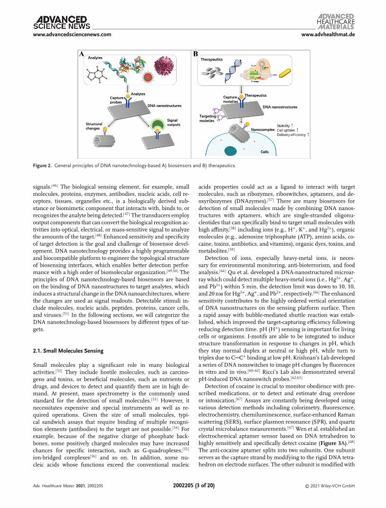
DNA/RNA (Figure 1E(b)),[41–43] proteins,[42,44] and cancer cells,[45]
which are widely targeted analytes for biosensing and targetdrugs for therapeutics. DNA nanotechnology offers a highlyprogrammable platform to engineer the biosensors, which canassemble more capture probes to increase the sensitivity. Be-sides, taking advantages of DNA unique chain reactions, theycan amplify the output signals to meet the needs of low de-tection limit. When analytes interact with DNA nanostructures,they either have structural changes or benefit from the inter-faces to have signal outputs to act as a biosensor (Figure 2A).DNA nanostructures mimic the natural biointerfaces due tointrinsic properties, which serve as biocompatible and rela-tively stable vehicles to enhance the delivery efficiency. Theycan increase the cell uptake and easily modify with therapeutic
Adv. Healthcare Mater. 2021, 2002205 © 2021 Wiley-VCH GmbH2002205 (1 of 20)

www.advancedsciencenews.com www.advhealthmat.de
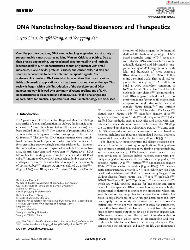
Figure 1. Structural DNA nanotechnology. A) DNA-junction nanostructures. a) 8-arm and 12-arm DNA branched junctions. Adapted with permission.[5]
Copyright 2007, American Chemistry Society. b) DNA nanotubes (double crossover). Adapted with permission.[9] Copyright 2004, American ChemistrySociety. c) 2D arrays. Adapted with permission.[12] Copyright 2004, American Chemistry Society. d) 3D crystals. Adapted with permission.[13] Copyright2009, Nature Publishing Group. B) DNA origami nanostructures. a) DNA origami square, rectangle, star, disk with three holes, triangle with rectangulardomains, sharp triangle with trapezoidal domains, and bridges. Adapted with permission.[15] Copyright 2006, Nature Publishing Group. b) DNA origamislotted cross. Adapted with permission.[20] Copyright 2009, Nature Publishing Group. c) DNA origami nanoflask. Adapted with permission.[21] Copyright2011, American Association for the Advancement of Science (AAAS). d) DNA sphere wireframe. Adapted with permission.[22] Copyright 2013, AAAS. C) a)2D structures from DNA tiles. Adapted with permission.[24] Copyright 2012, Nature Publishing Group. b) 3D structures from DNA Bricks. Adapted withpermission.[25] Copyright 2012, AAAS. c) 3D meshes of waving stickman, a bottle and a version of the Stanford bunny. Adapted with permission.[26] Copy-right 2015, Nature Publishing Group. D) hybrid nanostructures. a) 2D DNA origami-based protein-binding nanocavities. Adapted with permission.[31]
Copyright 2020, Nature Publishing Group. b) Organization of metal and metal oxide nanoclusters on DNA origami. Adapted with permission.[35] Copy-right 2019, American Chemistry Society. c) Assembly Carbon nanotube arrays with DNA origami. Adapted with permission.[37] Copyright 2020, AAAS.E) Dynamic DNA nanostructures. a) DNA origami dynamically changed by compressive depletion forces. Adapted with permission.[38] Copyright 2017,American Chemistry Society. b) DNA origami nanorobot triggered by protein displacement of aptamer locks. Adapted with permission.[42] Copyright2012, AAAS.
molecules in precisely patterns or numbers due to programma-bility. Based on that, DNA nanostructures are solid choices fortherapeutics delivery (Figure 2B). In this review, we mainly fo-cus on how DNA nanotechnologies integrate into biosensors andtherapeutics.
2. DNA Nanotechnology-Based Biosensors
Biosensors are analytical devices that incorporate a biologicalsensing element with chemophysical transducers to output com-plex bioanalytical measurements with direct and comprehensible
Adv. Healthcare Mater. 2021, 2002205 © 2021 Wiley-VCH GmbH2002205 (2 of 20)
www.advancedsciencenews.com www.advhealthmat.de
Figure 2. General principles of DNA nanotechnology-based A) biosensors and B) therapeutics.
signals.[46] The biological sensing element, for example, smallmolecules, proteins, enzymes, antibodies, nucleic acids, cell re-ceptors, tissues, organelles etc., is a biologically derived sub-stance or biomimetic component that interacts with, binds to, orrecognizes the analyte being detected.[47] The transducers employoutput components that can convert the biological recognition ac-tivities into optical, electrical, or mass-sensitive signal to analyzethe amounts of the target.[48] Enhanced sensitivity and specificityof target detection is the goal and challenge of biosensor devel-opment. DNA nanotechnology provides a highly programmableand biocompatible platform to engineer the topological structureof biosensing interfaces, which enables better detection perfor-mance with a high order of biomolecular organization.[49,50] Theprinciples of DNA nanotechnology-based biosensors are basedon the binding of DNA nanostructures to target analytes, whichinduces a structural change in the DNA nanoarchitectures, wherethe changes are used as signal readouts. Detectable stimuli in-clude molecules, nucleic acids, peptides, proteins, cancer cells,and viruses.[51] In the following sections, we will categorize theDNA nanotechnology-based biosensors by different types of tar-gets.
2.1. Small Molecules Sensing
Small molecules play a significant role in many biologicalactivities.[52] They include hostile molecules, such as carcino-gens and toxins, or beneficial molecules, such as nutrients ordrugs, and devices to detect and quantify them are in high de-mand. At present, mass spectrometry is the commonly usedstandard for the detection of small molecules.[53] However, itnecessitates expensive and special instruments as well as re-quired operations. Given the size of small molecules, typi-cal sandwich assays that require binding of multiple recogni-tion elements (antibodies) to the target are not possible.[54] Forexample, because of the negative charge of phosphate back-bones, some positively charged molecules may have increasedchances for specific interaction, such as G-quadruplexes,[55]
ion-bridged complexes[56] and so on. In addition, some nu-cleic acids whose functions exceed the conventional nucleic
acids properties could act as a ligand to interact with targetmolecules, such as ribozymes, riboswitches, aptamers, and de-oxyribozymes (DNAzymes).[57] There are many biosensors fordetection of small molecules made by combining DNA nanos-tructures with aptamers, which are single-stranded oligonu-cleotides that can specifically bind to target small molecules withhigh affinity,[58] including ions (e.g., H+, K+, and Hg2+), organicmolecules (e.g., adenosine triphosphate (ATP), amino acids, co-caine, toxins, antibiotics, and vitamins), organic dyes, toxins, andmetabolites.[59]
Detection of ions, especially heavy-metal ions, is neces-sary for environmental monitoring, anti-bioterrorism, and foodanalysis.[64] Qu et al. developed a DNA-nanostructured microar-ray which could detect multiple heavy-metal ions (i.e., Hg2+, Ag+,and Pb2+) within 5 min, the detection limit was down to 10, 10,and 20 nm for Hg2+, Ag+, and Pb2+, respectively.[66] The enhancedsensitivity contributes to the highly ordered vertical orientationof DNA nanostructures on the sensing platform surface. Thena rapid assay with bubble-mediated shuttle reaction was estab-lished, which improved the target-capturing efficiency followingreducing detection time. pH (H+) sensing is important for livingcells or organisms. I-motifs are able to be integrated to inducestructure transformation in response to changes in pH, whichthey stay normal duplex at neutral or high pH, while turn totriplex due to C─C+ binding at low pH. Krishnan’s Lab developeda series of DNA nonswitches to image pH changes by fluorencesin vitro and in vivo.[60–62] Ricci’s Lab also demonstrated severalpH-induced DNA nanoswitch probes.[62,63]
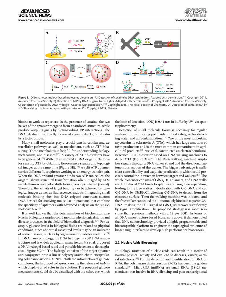
Detection of cocaine is crucial to monitor obedience with pre-scribed medications, or to detect and estimate drug overdoseor intoxication.[67] Assays are constantly being developed usingvarious detection methods including colorimetry, fluorescence,electrochemistry, chemiluminescence, surface-enhanced Ramanscattering (SERS), surface plasmon resonance (SPR), and quartzcrystal microbalance measurements.[67] Wen et al. established anelectrochemical aptamer sensor based on DNA tetrahedron tohighly sensitively and specifically detect cocaine (Figure 3A).[68]
The anti-cocaine aptamer splits into two subunits. One subunitserves as the capture strand by modifying to the rigid DNA tetra-hedron on electrode surfaces. The other subunit is modified with
Adv. Healthcare Mater. 2021, 2002205 © 2021 Wiley-VCH GmbH2002205 (3 of 20)
www.advancedsciencenews.com www.advhealthmat.de
Figure 3. DNA nanotechnology-based molecules biosensors. A) Detection of cocaine by DNA tetrahedron. Adapted with permission.[68] Copyright 2011,American Chemical Society. B) Detection of ATP by DNA origami traffic lights. Adapted with permission.[71] Copyright 2017, American Chemical Society.C) Detection of glucose by DNA hydrogel. Adapted with permission.[77] Copyright 2018, The Royal Society of Chemistry. D) Detection of ochratoxin A bya DNA walking machine. Adapted with permission.[81] Copyright 2019, Elsevier.
biotins to work as reporters. In the presence of cocaine, the twohalves of the aptamer merge to form a sandwich structure, whileproduce output signals by biotin-avidin-HRP interactions. TheDNA tetrahedrons directly increased signal-to-background ratioby a factor of four.
Many small molecules play a crucial part in cellular and ex-tracellular pathways as well as metabolism, such as ATP Mea-suring. These metabolites is helpful for understanding biology,metabolism, and diseases.[69] A variety of ATP biosensors havebeen generated.[70] Walter et al. showed a DNA origami platformfor sensing ATP by obtaining fluorescence signals and topologi-cal images at the same time (Figure 3B).[71] A split ATP aptamercarries different fluorophores working as an energy transfer pair.When the DNA origami aptamer binds two ATP molecules, theorigami shows structural transformation when imaged by AFMand its fluorescence color shifts from green (open) to red (closed).Therefore, the activity of target binding can be achieved by topo-logical images as well as fluorescence readouts. Integrating smallmolecule binding sites into DNA origami constructs customDNA devices for studying molecular interactions that combinethe specificity of aptamers with advanced analysis on the single-molecule level.[42]
It is well known that the determination of biochemical ana-lytes in biological samples could monitor physiological status anddisease processes in the field of biomedical diagnosis.[74] For ex-ample, glucose levels in biological fluids are related to physicalconditions, since abnormal measured levels may be an indicatorof some diseases, such as hypoglycemia or diabetes mellitus.[75]
In DNA nanotechnology, the DNA hydrogel is a 3D DNA nanos-tructure and is widely applied in many fields. Ma et al. proposeda DNA hydrogel-based rapid and portable biosensor to detect glu-cose (Figure 3C).[77] The hydrogel consists of the target aptamerand conjugated onto a linear polyacrylamide chain encapsulat-ing gold nanoparticles (AuNPs). With the introduction of glucosecomplexes, the hydrogel collapses, causing the release of AuNPswhich displays a red color in the solution. The proposed glucosemeasurements could also be visualized with the naked eye, which
the limit of detection (LOD) is 0.44 mm in buffer by UV–vis spec-trophotometry.
Detection of small molecule toxins is necessary for regularanalysis, for monitoring pollutants in food safety, or for detect-ing water and air contamination.[78] One of the most importantmycotoxins is ochratoxin A (OTA), which has large amounts oftoxin production and is the most common contaminant in agri-cultural products.[80] Wei et al. constructed an electrochemilumi-nescence (ECL) biosensor based on DNA walking machines todetect OTA (Figure 3D).[81] The DNA walking machine ampli-fies signals through a DNA walker strand and the directional au-tonomous motion of the walker. The biggest advantage is suffi-cient controllability and exquisite predictability which could pre-cisely control the interaction between targets and walkers.[82] Thewhole biosensor consists of CdS QDs, aptamers, and DNA walk-ers. Introduced OTA binds to aptamers causing their separation,leading to the free walker hybridization with Cy5-DNA and cutCy5-DNA by Nb.BbvCI, allowing Cy5-DNA to detach from theelectrode surface. Then the walking machine was initiated, andthe free walker continued to autonomously bind subsequent Cy5-DNA, making the ECL signal of CdS QDs recover significantlyby signal amplification. The proposed strategy was more sen-sitive than previous methods with a 12 pm LOD. In terms ofall DNA nanostructure-based biosensors above, it demonstratedthat DNA nanotechnology provided a highly programmable andbiocompatible platform to engineer the topological structure ofbiosensing interfaces to develop high performance biosensors.
2.2. Nucleic Acids Biosensing
In biology, mutation of nucleic acids can result in disorder ofnormal physical activity and can lead to diseases, cancer, or vi-ral infections.[83] For the detection and identification of DNA orRNA, the polymerase chain reaction (PCR) has been the goldenstandard.[84] MicroRNA (miRNA) are small RNAs (18–24 nu-cleotides) that involve in RNA silencing and post-transcriptional
Adv. Healthcare Mater. 2021, 2002205 © 2021 Wiley-VCH GmbH2002205 (4 of 20)

www.advancedsciencenews.com www.advhealthmat.de
Figure 4. DNA nanotechnology-based nucleic acids biosensors. A) Detection of miRNA by DNA Tetrahedron. Adapted with permission.[90] Copyright2018, American Chemical Society. B) A field-free 3D DNA nanostructure-based enzyme-free ECL biosensor for ultrasensitive detection of miRNA in asingle rapid step. Adapted with permission.[92] Copyright 2018, American Chemical Society. C) Detection of miRNAs by a DNA nanocube. Adapted withpermission.[93] Copyright 2019, American Chemical Society. D) Enzymatic polymerization on DNA modified gold nanowire for label-free detection ofpathogen DNA. Adapted with permission.[99] Copyright 2015, MDPI.
regulation of gene expression in plants, animals, and humans.The expression profiles of miRNA is considered as diagnostic andprognostic biomarkers and serve as therapeutic strategies for can-cer therapy.[85] At present, detection of miRNA is mainly basedon real-time quantitative polymerase chain reaction (real time-qPCR), microarray analysis, and Northern blotting.[86] However,challenges are that miRNA has short lengths, high sequence sim-ilarities and low abundance. They are hard to extract from cellsas well.[87,88] Thus, a simple and sensitive detection platform formiRNA analysis has attracted much attention.
Many sensitive methods based on DNA nanotechnology formiRNA detection have been developed.[88,89] Nie et al. establisheda low-fouling and sensitive SPR sensor for miRNA detectionby integrating DNA tetrahedron probes (DTPs) into gold filmalong with the catalytic enlargement of AuNPs (Figure 4A).[90]
When target miRNA let-7a appears in solution, a sandwichstructure forms through the DNA hybridization of target let-7a,DNA-linked AuNPs, and DTPs–Au film. As a result, SPR sig-nal increases through electronic coupling along with the sizes ofAuNPs expanding. This biosensor has a sensitive detection limitof 0.8 fm and showed superior specificity by distinguishing let-7afrom its homologous family. Integration of the DNA tetrahedron
with gold surfaces significantly weakened nonspecific adsorptionin a complex matrix, which enables it to detect miRNA in 100%human serum and cancer cell lysates. Furthermore, designing aprogrammable and autonomous molecular computing machinecould enhance the detection of miRNA to the molecular level. Forexample, Wang et al. established DNA origami-based logic gates(YES and AND gate) for miRNA diagnostics.[91]
Besides improving high sensitivity, researchers are also pur-suing facile, rapid detection method for miRNA. Zhang et al. es-tablished an ultrasensitive and rapid enzyme-free ECL miRNAbiosensor based on field-free 3D DNA nanostructures (Fig-ure 4B).[92] The 3D DNA nanostructure is constructed by azoben-zene (azo)-functionalized DNA nippers, which includes ECLemitter (Ru(bpy)2
2+) and quencher (Alexa Fluor, AF) modifiedprobes. When the target miRNA appear, the hybridization ofmiRNA with the nippers produces increasing ECL signals caus-ing by the adjacency between Ru(bpy)2
2+ and AF. After irradi-ation with UV or visible light, the DNA nanomachine under-goes trans–cis conversions through the interaction of azo moi-eties with the DNA nippers, which enables to regenerate thesensing system. Changing of the ECL signals leads the auto-mated movements of the 3D DNA nanomachine, which could
Adv. Healthcare Mater. 2021, 2002205 © 2021 Wiley-VCH GmbH2002205 (5 of 20)

www.advancedsciencenews.com www.advhealthmat.de
ultra-sensitively detect miRNA-21 with the LOD at 6.6 fm in rapidone step within 10 min. Since miRNA plays crucial roles in manybiological activities, it is important to develop a sensitive miRNAbiosensor with high accuracy in living cells. Liu et al. developed aFRET-based hairpin-DNA cascade amplifier (HDCA) for mRNAdetection (Figure 4C).[93] Two metastable hairpin DNAs (H1 andH2) labeled with Cy3 and Cy5 as a FRET pair modified on a DNAnanocube. A target miRNA triggers a chain-reaction growth oflong DNA duplexes assembled from H1 and H2 to produce am-plified FRET signals. Taking advantage of the spatial-limitationeffects in the restricted space of DNA nanocubes, the speed ofthe miRNA-stimulated HDCA reaction greatly expedited (7 timesfaster), as well as the reaction efficiency was enhanced (2.6 timeshigher). In addition, DNA nanocubes offer enhanced cell perme-ability and nuclease resistance. Moreover, it provides the capabil-ity of avoiding false-positive signals, which ensures reliable miR-NAs detection in living cells.
Detecting and quantifying small amount of DNA targets stillmainly relies on PCR.[94] Many PCR-free DNA detection assayshave been developed such as molecular beacons,[95] microfluidicDNA microarray,[96] nanomaterial based method,[97] and so on.Arun et al. established a simple DNA nanoswitch to detect DNAfragments using standard gel electrophoresis.[98] When the targetDNA hybrid with nanoswitch, the scaffold forms a loop, whichcan be distinguished by electrophoretic mobility shift assays.Based on hybrid dDNA nanostructures, Jeong et al. proposeda label-free biosensor to detect single-stranded pathogen DNAbased on the target-triggered gelation between gold nanowires(AuNWs) and the primer DNAs (Figure 4D).[99] The biosensorconstructed by DNA circularization in the presence of the targetDNA by T4 DNA ligase, and a macroscopic hydrogel was fabri-cated by the prolonged DNA with AuNWs for efficient gelation,whereas scattered particles were formed in the absence of thetarget DNA. The proposed biosensor can achieve label-free andsimple DNA detection without sophisticated operations or instru-ments.
The Human Genome Project and other major genomic se-quencing projects thrusted DNA sequencing into the spot-light of modern biology and completely transformed thefield of genetics.[100] Many sequencing techniques have beendeveloped.[101] At present, nanopore-based DNA sequencingtechnologies have drawn worldwide attention.[102] Farimani et al.constructed a DNA origami–graphene heterostructure nanoporeto detect DNA.[104] The association of discernible dwell time andionic current with the slower speed of translocation makes the hy-brid DNA origami–graphene nanopore could distinguish amongthe four types of bases with high sensitivity and specificity. Hav-ing mentioned all of above, DNA nanostructures-based biosen-sors not only can detect nucleic acids in low concentrations butalso can sequence them in bases.
2.3. Protein Biosensing
Proteins are vital components of organisms and participate in al-most every process within cells. They may function as enzymes,support the cell as its cytoskeleton, facilitate cell signaling, trig-ger immune responses, enable cell adhesion, and direct the cellcycle.[105] Particular expressed proteins function as biomarkers
for activities in organisms or cells, of which abnormal expressionof biomarkers often relates to the presence of diseases. Since pro-teins serve as the working unit for many cellular activities, andalso as the crucial biomarkers for indicators, the precise and ac-curate determination of proteins is necessary for not only funda-mental research but also clinical diagnosis. Until now, proteinsare detected mostly by antibodies in analytical formats such asenzyme-linked immunosorbent assay (ELISA), immunobead as-say, western blotting, microarrays and also SPR, SERS, colorimet-ric, electrochemical, and fluorescence biosensors.[106]
Using dynamic DNA nanotechnology, a DNA tweezer-like nanodevice was established to trigger and character-ize the enzyme activity of glucose-6-phosphate dehydrogenase(G6pDH)/NAD+ enzyme/cofactor pair.[107] The rigid and adapt-able DNA tetrahedron is widely used as a biosensing platform.Zhou et al. established a DNA tetrahedral probe to detect methyl-transferase by fluorescence (Figure 5A).[108] The DNA tetrahe-dron consists of strands labeled with fluorescence dyes andquenchers. When the substrate is exposed to target methyltrans-ferase, the methylated sites could be cut to collapse the DNA tetra-hedron, causing fluorescence recovery. Assisted by DNA tetra-hedron, this biosensor can detect Dam methyltransferase witha detection limit of 0.045 U mL−1 and has a good performancein a real sample as well. Moreover, we could study and analyzeenzymes at single-molecule level through DNA nanotechnology,Tintoré et al. constructed a protein recognition biosensor by DNAorigami to characterize enzymatic DNA repair activity of HumanO6-alkylguanine-DNA alkyltransferase.[109]
Protein biomarkers detection is crucial for the diagnosis ofvarious diseases. Many biosensors for biomarker detection havebeen developed based on DNA nanotechnologies.[49] For exam-ple, prostate-specific antigen (PSA) is the most notable serumbiomarker for diagnosing and monitoring for prostate cancer.Yan et al. established a sensitive and selective biosensor to detectPSA based on rolling circle amplification (RCA)-based DNA beltsand magnetic bead-based ELISA (Figure 5B).[110] DNA nanos-tructures are fabricated by RCA and folded into DNA belts withbiotin modified staples for signal amplification. Then the high-order nanostructures employ to magnetic bead-based ELISA forPSA detection. This strategy can achieve high selectivity and sen-sitivity with a detection limit of 50 am, which benefits from theRCA amplification and precise maneuverability of DNA origami.Some other strategies like target-responsive structurally transi-tioning DNA hydrogels are also powerful tools in bioanalysis.Zhang et al. designed an aptamer-functionalized DNA hydrogelencapsulating AuNPs as a target-responsive material to detectthrombin.[111] To improve the sensitivity of biomarkers detection,many signal amplification strategies have been developed, suchas RCA, catalyze hairpin assembly, hybridization chain reaction(HCR) and toehold-triggered strand displacement reaction.[112]
Qin et al. proposed a DNA nanostructure assembled by target-responsive netlike HCR (nHCR) to detect cytokine interferon-gamma (IFN-𝛾) with high sensitivity (Figure 5C).[113] When tar-get IFN-𝛾 were present, the aptamer recognition hairpin probescan have conformational changes and release the nHCR initiatorstrands, triggering the nHCR process to generate netlike DNAnanostructures. The construction of the DNA nanostructures in-troduced exceptionally amplified FRET signals to detect IFN-𝛾with a detection limit of 1.2 pm via enzyme-free approaches.
Adv. Healthcare Mater. 2021, 2002205 © 2021 Wiley-VCH GmbH2002205 (6 of 20)

www.advancedsciencenews.com www.advhealthmat.de
Figure 5. DNA nanotechnology-based protein biosensors. A) Detection of DNA methyltransferase activity by DNA tetrahedron. Adapted withpermission.[108] Copyright 2017, American Chemical Society. B) Detection of PSA by DNA origami belt. Adapted with permission.[110] Copyright 2014,American Chemical Society. C) Detection of INF-𝛼 by netlike DNA nanostructures. Adapted with permission.[113] Copyright 2019, The Royal Soci-ety of Chemistry. D) Detection of CRP by DNA origami coupled with nanopores. Adapted with permission.[114] Copyright 2020, Nature PublishingGroup.
Furthermore, Raveendran et al. developed a biosensor platformusing DNA origami coupled with a nanopore read-out for quan-titative single molecule biosensing (Figure 5D).[114] The hollowrectangle DNA origami sheet is modified with a target-specificDNA aptamer to human C-reactive protein (CRP) in its center,which enable to distinguish the DNA origami with or withouta target protein in peak shape, amplitude, and dwell time whentranslocating through the nanopore. This biosensor has a LOD of3 nm and can be applied to detect CRP in clinically relevant flu-ids. Interestingly, this biosensing method is based on countingindividual biomarkers instead of an averaged signal. Arun et al.established a programmable DNA nanoswitch platform to multi-plexed detect biomarkers at once,[115] which it will be detected bygel electrophoresis when M13 scaffold interacted with target pro-teins to have a loop formation. Above all, DNA nanostructuresmeets the requirements of all needs of protein detection frommicrocosmic to macrocosmic viewpoint.
2.4. Tumor Cell Detection
Circulating tumor cells (CTCs) are cancer cells that have beencome off from the primary or metastatic tumor and enterthe blood circulation, and are important biomarkers of tu-mor progression, prognosis, metastasis, and relapse.[116] How-ever, detection of CTCs is very demanding due to their lowconcentrations.[118] The demand for facile and sensitive biosen-
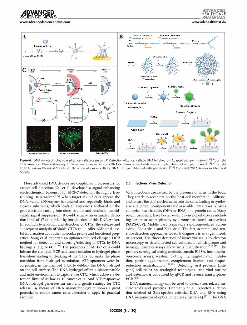
sors for CTCs detection is urgent. Enzyme-free DNA amplifi-cation reactions, such as HCR, has been applied to detect low-concentration tumor cells. Zhou et al. established a biosensorfor detection of breast cancer cells (MCF-7) with high sensitiv-ity and specificity using a multibranched HCR (mHCR) productcaptured on DNA tetrahedral surfaces (Figure 6A).[122] The ap-tamer of Epithelial cell adhesion molecule (EpCAM) hybridizingto an HCR initiator served as a trigger to begin the mHCR, whichcan generate multiple biotins for signal amplification, and mean-while multiple branched arms can multivalently bind target cellson the DNA tetrahedral gold electrode surface. The cooperativeeffect of mHCR amplification and multivalent binding bringsexcellent sensitivity with the detection limit of only four cancercells. After guaranteeing high sensitivity, some simple, fast, andquick feedback point-of-care biosensing techniques have beenestablished.[123] Zhao et al. constructed a biosensing platformfor human cervical cancer cells (HeLa) using a DNA dendrimer–streptavidin (SA) nanocomplex signal amplifier (Figure 6B).[124]
The DNA dendrimer–SA nanocomplex was self-assembled andfunctionalized with targeting aptamers and dyes. For target cellsensing, the quartz glass slide is modified with the same tar-geting aptamers to capture HeLa cells, the aptamer functional-ized nanocomplex is introduced to further bind with the capturedcells, which generates an increasing fluorescence as the outputsignal with a detection limit of 4.4 × 10³ cells mL−1. Given thedendrimer DNA nanostructures, this biosensor needs no extrasteps for separation or amplification.
Adv. Healthcare Mater. 2021, 2002205 © 2021 Wiley-VCH GmbH2002205 (7 of 20)
www.advancedsciencenews.com www.advhealthmat.de
Figure 6. DNA nanotechnology-based cancer cells biosensors. A) Detection of cancer cells by DNA tetrahedron. Adapted with permission.[122] Copyright2014, American Chemical Society. B) Detection of cancer cells by a DNA dendrimer–streptavidin nanocomplex. Adapted with permission.[124] Copyright2017 American Chemical Society. C) Detection of cancer cells by DNA hydrogel. Adapted with permission.[126] Copyright 2017, American ChemicalSociety
More advanced DNA devices are coupled with biosensors forcancer cell detection. Cai et al. developed a signal enhancingelectrochemical biosensor for MCF-7 detection through a free-running DNA walker.[125] When target MCF-7 cells appear, theDNA walker (DNAzyme) is released and repeatedly binds andcleaves substrates, which leads all sequences anchored on thegold electrode cutting into short strands and results in consid-erable signal suppression. It could achieve an estimated detec-tion limit of 47 cells mL−1 by introduction of this DNA walker.In addition to isolation and detection of CTCs, the release andsubsequent analysis of viable CTCs could offer additional use-ful information about the molecular profile and functional prop-erties. Song et al. reported an aptamer-induced clamped HCRmethod for detection and covering/releasing of CTCs by DNAhydrogels (Figure 6C).[126] The presences of MCF-7 cells couldinitiate the clamped HCR and cause solution to hydrogel phasetransition leading to cloaking of the CTCs. To make the phasetransition from hydrogel to solution, ATP aptamers were in-corporated in the clamped HCR to deblock the DNA hydrogelon the cell surface. The DNA hydrogel offers a biocompatibleand mild environment to capture live CTC, which achieve a de-tection limit of as low as 10 cancer cells. And ATP-responsiveDNA hydrogel generates an easy and gentle strategy for CTCrelease. By means of DNA nanotechnology, it shows a greatpotential to enable tumor cells detection to apply in practicalsamples.
2.5. Infectious Virus Detection
Viral infections are caused by the presence of virus in the body.They attach to receptors on the host cell membrane, infiltrate,and release the viral nucleic acids into the cells, leading to synthe-size viral protein components and assemble new virions. Virusescomprise nucleic acids (DNA or RNA) and protein coats. Manyrecent pandemic have been caused by enveloped viruses includ-ing severe acute respiratory syndrome-associated coronavirus(SARS-CoV), Middle East respiratory syndrome-related coron-avirus, Ebola virus, and Zika virus. The fast, accurate, and sen-sitive detection approaches for early diagnosis is an urgent need.At present, The direct detection of intact viruses is by electronmicroscopy or virus-infected cell cultures, in which plaque andhemagglutination assays allow virus quantification.[127,128] Theprimary serological testing methods contain ELISA, immunoflu-orescence assays, western blotting, hemagglutination inhibi-tion, particle agglutination, complement fixation, and plaque-reduction neutralization.[128,129] Detecting viral proteins (anti-gens) still relies on serological techniques. And viral nucleicacid detection is conducted by qPCR and reverse transcription-PCR.[130]
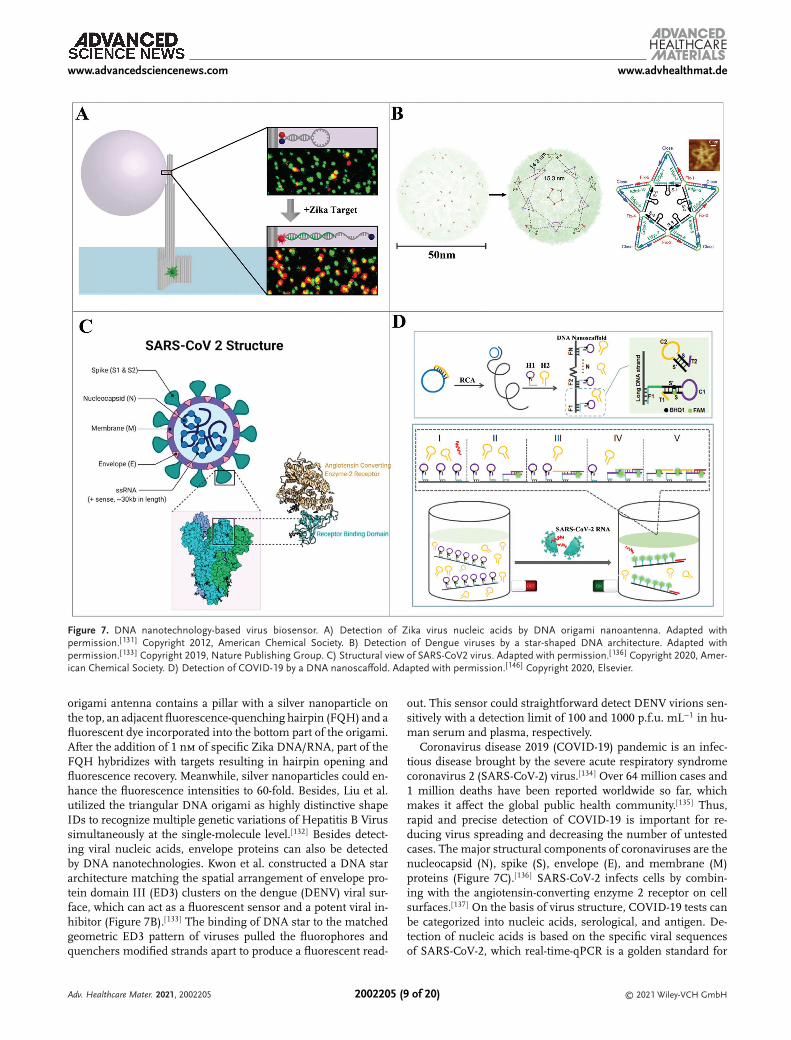
DNA nanotechnology can be used to detect virus-related nu-cleic acids and proteins. Ochmann et al. reported a detec-tion method of Zika-specific artificial DNA and RNA usingDNA origami-based optical antennas (Figure 7A).[131] The DNA
Adv. Healthcare Mater. 2021, 2002205 © 2021 Wiley-VCH GmbH2002205 (8 of 20)
www.advancedsciencenews.com www.advhealthmat.de
Figure 7. DNA nanotechnology-based virus biosensor. A) Detection of Zika virus nucleic acids by DNA origami nanoantenna. Adapted withpermission.[131] Copyright 2012, American Chemical Society. B) Detection of Dengue viruses by a star-shaped DNA architecture. Adapted withpermission.[133] Copyright 2019, Nature Publishing Group. C) Structural view of SARS-CoV2 virus. Adapted with permission.[136] Copyright 2020, Amer-ican Chemical Society. D) Detection of COVID-19 by a DNA nanoscaffold. Adapted with permission.[146] Copyright 2020, Elsevier.
origami antenna contains a pillar with a silver nanoparticle onthe top, an adjacent fluorescence-quenching hairpin (FQH) and afluorescent dye incorporated into the bottom part of the origami.After the addition of 1 nm of specific Zika DNA/RNA, part of theFQH hybridizes with targets resulting in hairpin opening andfluorescence recovery. Meanwhile, silver nanoparticles could en-hance the fluorescence intensities to 60-fold. Besides, Liu et al.utilized the triangular DNA origami as highly distinctive shapeIDs to recognize multiple genetic variations of Hepatitis B Virussimultaneously at the single-molecule level.[132] Besides detect-ing viral nucleic acids, envelope proteins can also be detectedby DNA nanotechnologies. Kwon et al. constructed a DNA stararchitecture matching the spatial arrangement of envelope pro-tein domain III (ED3) clusters on the dengue (DENV) viral sur-face, which can act as a fluorescent sensor and a potent viral in-hibitor (Figure 7B).[133] The binding of DNA star to the matchedgeometric ED3 pattern of viruses pulled the fluorophores andquenchers modified strands apart to produce a fluorescent read-
out. This sensor could straightforward detect DENV virions sen-sitively with a detection limit of 100 and 1000 p.f.u. mL−1 in hu-man serum and plasma, respectively.
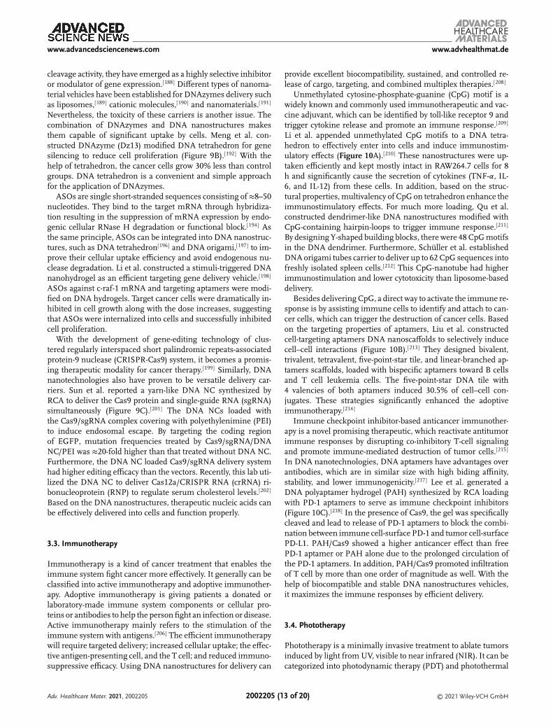
Coronavirus disease 2019 (COVID-19) pandemic is an infec-tious disease brought by the severe acute respiratory syndromecoronavirus 2 (SARS-CoV-2) virus.[134] Over 64 million cases and1 million deaths have been reported worldwide so far, whichmakes it affect the global public health community.[135] Thus,rapid and precise detection of COVID-19 is important for re-ducing virus spreading and decreasing the number of untestedcases. The major structural components of coronaviruses are thenucleocapsid (N), spike (S), envelope (E), and membrane (M)proteins (Figure 7C).[136] SARS-CoV-2 infects cells by combin-ing with the angiotensin-converting enzyme 2 receptor on cellsurfaces.[137] On the basis of virus structure, COVID-19 tests canbe categorized into nucleic acids, serological, and antigen. De-tection of nucleic acids is based on the specific viral sequencesof SARS-CoV-2, which real-time-qPCR is a golden standard for
Adv. Healthcare Mater. 2021, 2002205 © 2021 Wiley-VCH GmbH2002205 (9 of 20)
www.advancedsciencenews.com www.advhealthmat.de
COVID-19 diagnostics. Serological detection of virus-induced an-tibodies is helpful complement to nucleic acid testing, such asIgG/IgM. So far, reported antigen tests probe for the nucleocap-sid or spike proteins of SARS-CoV-2 mainly based on lateral flowor ELISA.[134,139]
Zhang et al. represented the first DNA aptamer targeting toCOVID-19 nucleocapsid protein (Np), which established a sand-wiched aptamers assay with colloidal gold immunochromato-graphic strips.[144] The best DNA sequence from selection dis-plays high binding affinity to Np with a dissociation constant of0.49 nm. Using aptamers and colloidal gold immunochromato-graphic strips, they established a sandwich assay. In the pres-ence of Np, the test line turns blue based on aptamers bindingwith 1 ng mL−1 of target proteins in human serum or urine.According to the reported aptamers, Jiao et al. reported a nu-cleic acid assay for rapid detection of SARS-CoV-2 RNA basedon DNA nanoscaffold HCR (DNHCR) (Figure 7D).[146] The pres-ence of target SARS-CoV-2 RNA initiates the cascade reaction ofhybridization along the DNA nanoscaffolds, resulting in H1 sep-arating to restore fluorescence. Given the localized accelerationof DNA probes, one target RNA can immediately acquire highlyamplified fluorescence signals of the whole nanoscaffold. Theproposed DNHCR assay was also performed in serum and salivasamples, which the detection can be finished at short respondingtime (within 10 min) and mild temperature (15–35 °C). All thesebiosensors demonstrate that DNA nanostructures can be greatlyintegrated into various types of biosensors in suitable ways.
3. DNA Nanotechnology-Based Therapeutics
DNA nanostructures have intrinsic biocompatibility and canbe easily modified. In addition, it has been shown that theycan enter cells independent of transfection agents. Therefore,DNA nanostructures have attracted strong interest to be de-veloped as delivery vehicles for therapeutical applications. Aneffective delivery vehicle should be comparatively stable un-der physiological conditions. Due to their intrinsic propertiesof nucleic acids, nature DNA molecules are susceptible to nu-clease digestion and disassembly at insufficient cation concen-trations in biological systems. Studies show that closer-packedstructures are more thermodynamicly stable and exhibit in-creased nuclease tolerance.[147] Also, paranemic crossover DNAhas dramatically enhanced resistance to nucleases compared todouble stranded DNA.[148] To increase the stability, chemicallymodified or unnatural bases are synthesized to construct DNAassemblies.[149] In addition, cationic polymers,[150] peptides,[151]
proteins,[152] and PEGylated lipid bilayers[153] based coatings pro-tects DNA nanostructures from nuclease degradation. Whileensuring the stability, specific targeting and increased cellularuptake should be fulfilled. This can be achieved by combin-ing with small molecule ligands,[154] aptamers,[155] peptides,[156]
proteins,[157] or antibodies.[158] Different targeting molecules in-duce different cellular uptake pathways of DNA nanostructures.Due to intrinsic properties of negatively charged cell mem-branes, large molecules or nanometer-sized particles (e.g., DNAnanostructures) can pass through via endocytosis,[159] includ-ing caveolin-dependent endocytosis,[160] and clathrin-mediatedendocytosis.[161] DNA nanostructures can also be internalizedby macrophages[162] and nonendocytic pathways.[163] Since most
DNA carriers and their cargos will transport to lysosomes fol-lowed by degradation through the endocytic pathways, endolyso-somal escape is important for drug delivery. Modifying differenttarget ligands on DNA nanostructures is a major strategy for effi-cient delivery of therapeutic agents. In this section, we will reviewfour types of therapeutics based on DNA nanotechnology.
3.1. Chemotherapy
Chemotherapy is widely used cancer treatments, which usesstrong chemicals to kill fast-growing cancer cells. The often-usedbroad-spectrum anticancer drug is doxorubicin (DOX), whichcan insert in the base pair of double-strand DNA. And other com-mon drugs, such as paclitaxel and camptothecin, are usually con-jugated to or encapsulated in DNA carriers.[164] Because DNAnanostructures exhibit high biocompatibility, programmability,rigidity, and ease of modification, it helps overcome the disad-vantages of inefficient delivery, systemic toxicity, and multidrugresistance in chemotherapy.[165]
Jiang et al. presented a tubular and triangular DOX-loadedDNA origami carrier (Figure 8A).[166] The DOX-origami struc-tures are effectively uptook by tumor cells and co-localized inthe cytoplasm. After entering into cells, origami-bound DOX in-duced cell death and further bypassed drug resistance in resis-tance cancer cells by enhancing the cellular uptake of DOX andpromoting endosomal escape. Meanwhile, DNA origami showedno toxicity. Loaded drugs can be released via DNA nanostruc-tures degradation under low environmental pH values or DNAenzymes. Taken together, DNA origami is a suitable drug car-rier and delivery vehicle for chemotherapy. Zhang et al. of thesame lab demonstrated triangle-shaped DNA origami loadedwith DOX had elevated tumor passive targeting and long-rangeproperties at the tumor region.[167] Another commonly used DNAnanostructure delivery platform is the DNA tetrahedron, whichresists nuclease degradation and can be uptaken by cells effi-ciently. There are many studies demonstrating its promising abil-ity as a drug delivery vehicle to overcome drug resistance in vari-ous cancer cell types.[168]
Many simpler DNA architectures have also proved to beeffective for targeted anticancer drug delivery. Several struc-tures generated by HCR or RCA such as nanotrains,[155,169]
nanocentipede,[170] and nanocruciform[171] have been developed.Zhu et al. reported the rolling circle replication based DNAnanoflowers (NFs) as a versatile biomedical platform.[172] The sta-bility of NFs is critical for biomedical applications, which they re-tained their integrity after high concentration DNase I treatmentand upon denaturation conditions or in human serum for 24 h.Owing to their stability, biocompatibility, and biodegradability,Dox-NFs induced selective cytotoxicity in target cells comparedto nonselective cytotoxicity of free Dox, which demonstrated theapplicability of NFs for targeted drug delivery. DNA-based nano-materials such as hydrogels have also been used in therapeu-tic applications. Nishikawa et al. first designed a X-shape-basedDNA hydrogel for delivery of both DOX and CpG motifs for can-cer treatment.[173] Recently, Liwinska et al. presented a disulfideoligonucleotide-based DNA nanogel for effective loading and in-ducible drug release.[174] The nanogels appeared effective, con-trolled, and prolonged drug loading and release.
Adv. Healthcare Mater. 2021, 2002205 © 2021 Wiley-VCH GmbH2002205 (10 of 20)

www.advancedsciencenews.com www.advhealthmat.de
Figure 8. DNA nanotechnology-based chemotherapy. A) Delivery of DOX by DNA origami. Adapted with permission.[166] Copyright 2012, AmericanChemical Society. B) Delivery of platinum nanodrugs by DNA icosahedron. Adapted with permission.[176] Copyright 2018, Wiley-VCH GmbH. C) Deliveryof thrombin by a DNA nanorobot. Adapted with permission.[45] Copyright 2018, Nature Publishing Group.
Since most small-molecule anticancer drugs frequently sufferfrom poor efficacy and drug resistance, nanoparticle-based drugshave been developed, such as cisplatin.[175] Ma et al. constructeda telomerase-triggered DNA icosahedron to deliver Pt nanodrugsinto cisplatin-resistance tumor cells for efficient chemotherapy(Figure 8B).[176] The encapsulated Pt nanoparticles (PtNPs) couldeffectively induce DNA damage and cell death, reducing drug re-sistance based on drug efflux. The utilization of DNA icosahe-dron could significantly reduce drug resistance and systemic tox-icity of PtNPs.
More recently, researchers have started to study the applica-tions of DNA nanodevices in vivo. Surana et al. first demon-strated the functionality of a pH-responsive DNA nanomachineinside the nematode Caenorhabditis elegans.[61] Inspired by alogic-gated DNA nanorobot for targeted transport of molecularpayloads to cells presented by Douglas,[42] recently Li et al. de-veloped a nanorobotic system for the targeted delivery of thera-peutic thrombin in vivo with tumor-bearing mouse models (Fig-ure 8C).[45] The nanorobot was a DNA origami nanotube withtargeting DNA aptamers on its outside and the thrombin an-chored inside. Aptamers were utilized as tumor targeting ligandsand a stimulus for the DNA nanorobot to expose thrombin andinduce coagulation at the tumor site. When thrombin reachestumor-associated blood vessels, it induced intravascular throm-bosis leading to tumor necrosis and growth inhibition. By thisthrombin delivery system based on a dynamic DNA nanostruc-ture, it exhibits significant in vivo antitumor efficacy in tumor-bearing mouse models. The latest research reported by Yang et al.designed a barrel-shaped DNA nanorobot for autonomous bloodanticoagulation by thrombin in human plasma.[178] A computingcore embedded in the barrel can intellectively detect the concen-tration of thrombin and actuate an autonomous anticoagulationin the presence of excess thrombin in human plasma. To sum
up, DNA nanostructures can significantly enhance the cell up-take and selectivity of anticancer drugs in chemotherapy.
3.2. Gene Therapy
Gene therapy is a medical method that utilizes therapeutic nu-cleic acids or gene regulators for disease treatment. The Oligonu-cleotide drugs include small interfering RNA (siRNA), anti-sense oligonucleotides (ASOs), DNAzyme, or whole gene edit-ing system.[179] One of the major issues of gene therapy is todevelop a nonviral vector with high delivery efficiency and cel-lular uptake. DNA nanostructures offers different types of car-riers, which can be simply loaded with therapeutic oligonu-cleotides and internalized into cells through the endocyticpathway.
siRNA is a class of double-stranded non-coding RNAmolecules, which targets and cleaves complementary mRNA.The down-regulated mRNA induced to degrade proteinexpression.[180] Bujold et al. designed a stimulative DNAnanosuitcase encapsulating siRNA.[181] The Bcl-2 and/or Bcl-XlmRNA triggers the releasement of siRNA. the nanosuitcaseincreased twofold of cytotoxicity than other controls in cell death,corresponding to the increased potency of the elongated siRNA.Other siRNA-DNA carriers are also emerging, such as DNAtetrahedrons,[182] DNA nanogel,[183] DNA nanoclew (NC),[184]
and so on. Despite so many therapeutic oligonucleotides areongoing clinical trials, only two gene therapies are approved forhuman use, one of them is gendicine, which is a recombinantadenovirus engineered to express the tumor suppressor genep53.[185] Liu et al. constructed a DNA origami kite to codeliv-ery p53 and DOX for chemo- and gene therapy of multidrugresistant breast cancer (MCF-7R) (Figure 9A).[186] The triangle
Adv. Healthcare Mater. 2021, 2002205 © 2021 Wiley-VCH GmbH2002205 (11 of 20)

www.advancedsciencenews.com www.advhealthmat.de
Figure 9. DNA nanotechnology-based gene therapy. A) Codelivery p53 and Dox by a DNA origami kite. Adapted with permission.[186] Copyright 2018,American Chemical Society. B) Delivery of DNAzyme by DNA tetrahedron. Adapted with permission.[192] Copyright 2019, American Chemical Society.C) Delivery of CRISPR–Cas9 by DNA nanoclews. Adapted with permission.[201] Copyright 2015, Wiley-VCH GmbH.
DNA origami was modified with multiple aptamers to targettumors and disulfide linkers to combine with two copies of thep53 gene and loaded with DOX as well. After treatment, theexpression of p53 mRNA increased 20-fold and tumor growthwas suppressed as well, resulting in a dramatically enhanced
growth inhibition in multidrug resistant tumors in vitro and invivo without obvious toxicities.
DNAzymes are single-stranded DNA catalysts that are able toperform a specific chemical reaction, such as RNA or DNA cleav-age and ligation or DNA phosphorylation.[187] Due to their RNA
Adv. Healthcare Mater. 2021, 2002205 © 2021 Wiley-VCH GmbH2002205 (12 of 20)
www.advancedsciencenews.com www.advhealthmat.de
cleavage activity, they have emerged as a highly selective inhibitoror modulator of gene expression.[188] Different types of nanoma-terial vehicles have been established for DNAzymes delivery suchas liposomes,[189] cationic molecules,[190] and nanomaterials.[191]
Nevertheless, the toxicity of these carriers is another issue. Thecombination of DNAzymes and DNA nanostructures makesthem capable of significant uptake by cells. Meng et al. con-structed DNAzyme (Dz13) modified DNA tetrahedron for genesilencing to reduce cell proliferation (Figure 9B).[192] With thehelp of tetrahedron, the cancer cells grow 30% less than controlgroups. DNA tetrahedron is a convenient and simple approachfor the application of DNAzymes.
ASOs are single short-stranded sequences consisting of ≈8–50nucleotides. They bind to the target mRNA through hybridiza-tion resulting in the suppression of mRNA expression by endo-genic cellular RNase H degradation or functional block.[194] Asthe same principle, ASOs can be integrated into DNA nanostruc-tures, such as DNA tetrahedron[196] and DNA origami,[197] to im-prove their cellular uptake efficiency and avoid endogenous nu-clease degradation. Li et al. constructed a stimuli-triggered DNAnanohydrogel as an efficient targeting gene delivery vehicle.[198]
ASOs against c-raf-1 mRNA and targeting aptamers were modi-fied on DNA hydrogels. Target cancer cells were dramatically in-hibited in cell growth along with the dose increases, suggestingthat ASOs were internalized into cells and successfully inhibitedcell proliferation.
With the development of gene-editing technology of clus-tered regularly interspaced short palindromic repeats-associatedprotein-9 nuclease (CRISPR-Cas9) system, it becomes a promis-ing therapeutic modality for cancer therapy.[199] Similarly, DNAnanotechnologies also have proven to be versatile delivery car-riers. Sun et al. reported a yarn-like DNA NC synthesized byRCA to deliver the Cas9 protein and single-guide RNA (sgRNA)simultaneously (Figure 9C).[201] The DNA NCs loaded withthe Cas9/sgRNA complex covering with polyethylenimine (PEI)to induce endosomal escape. By targeting the coding regionof EGFP, mutation frequencies treated by Cas9/sgRNA/DNANC/PEI was ≈20-fold higher than that treated without DNA NC.Furthermore, the DNA NC loaded Cas9/sgRNA delivery systemhad higher editing efficacy than the vectors. Recently, this lab uti-lized the DNA NC to deliver Cas12a/CRISPR RNA (crRNA) ri-bonucleoprotein (RNP) to regulate serum cholesterol levels.[202]
Based on the DNA nanostructures, therapeutic nucleic acids canbe effectively delivered into cells and function properly.
3.3. Immunotherapy
Immunotherapy is a kind of cancer treatment that enables theimmune system fight cancer more effectively. It generally can beclassified into active immunotherapy and adoptive immunother-apy. Adoptive immunotherapy is giving patients a donated orlaboratory-made immune system components or cellular pro-teins or antibodies to help the person fight an infection or disease.Active immunotherapy mainly refers to the stimulation of theimmune system with antigens.[206] The efficient immunotherapywill require targeted delivery; increased cellular uptake; the effec-tive antigen-presenting cell, and the T cell; and reduced immuno-suppressive efficacy. Using DNA nanostructures for delivery can
provide excellent biocompatibility, sustained, and controlled re-lease of cargo, targeting, and combined multiplex therapies.[208]
Unmethylated cytosine-phosphate-guanine (CpG) motif is awidely known and commonly used immunotherapeutic and vac-cine adjuvant, which can be identified by toll-like receptor 9 andtrigger cytokine release and promote an immune response.[209]
Li et al. appended unmethylated CpG motifs to a DNA tetra-hedron to effectively enter into cells and induce immunostim-ulatory effects (Figure 10A).[210] These nanostructures were up-taken efficiently and kept mostly intact in RAW264.7 cells for 8h and significantly cause the secretion of cytokines (TNF-𝛼, IL-6, and IL-12) from these cells. In addition, based on the struc-tural properties, multivalency of CpG on tetrahedron enhance theimmunostimulatory effects. For much more loading, Qu et al.constructed dendrimer-like DNA nanostructures modified withCpG-containing hairpin-loops to trigger immune response.[211]
By designing Y-shaped building blocks, there were 48 CpG motifsin the DNA dendrimer. Furthermore, Schüller et al. establishedDNA origami tubes carrier to deliver up to 62 CpG sequences intofreshly isolated spleen cells.[212] This CpG-nanotube had higherimmunostimulation and lower cytotoxicity than liposome-baseddelivery.
Besides delivering CpG, a direct way to activate the immune re-sponse is by assisting immune cells to identify and attach to can-cer cells, which can trigger the destruction of cancer cells. Basedon the targeting properties of aptamers, Liu et al. constructedcell-targeting aptamers DNA nanoscaffolds to selectively inducecell–cell interactions (Figure 10B).[213] They designed bivalent,trivalent, tetravalent, five-point-star tile, and linear-branched ap-tamers scaffolds, loaded with bispecific aptamers toward B cellsand T cell leukemia cells. The five-point-star DNA tile with4 valencies of both aptamers induced 30.5% of cell–cell con-jugates. These strategies significantly enhanced the adoptiveimmunotherapy.[214]
Immune checkpoint inhibitor-based anticancer immunother-apy is a novel promising therapeutic, which reactivate antitumorimmune responses by disrupting co-inhibitory T-cell signalingand promote immune-mediated destruction of tumor cells.[215]
In DNA nanotechnologies, DNA aptamers have advantages overantibodies, which are in similar size with high biding affinity,stability, and lower immunogenicity.[217] Lee et al. generated aDNA polyaptamer hydrogel (PAH) synthesized by RCA loadingwith PD-1 aptamers to serve as immune checkpoint inhibitors(Figure 10C).[218] In the presence of Cas9, the gel was specificallycleaved and lead to release of PD-1 aptamers to block the combi-nation between immune cell-surface PD-1 and tumor cell-surfacePD-L1. PAH/Cas9 showed a higher anticancer effect than freePD-1 aptamer or PAH alone due to the prolonged circulation ofthe PD-1 aptamers. In addition, PAH/Cas9 promoted infiltrationof T cell by more than one order of magnitude as well. With thehelp of biocompatible and stable DNA nanostructures vehicles,it maximizes the immune responses by efficient delivery.
3.4. Phototherapy
Phototherapy is a minimally invasive treatment to ablate tumorsinduced by light from UV, visible to near infrared (NIR). It can becategorized into photodynamic therapy (PDT) and photothermal
Adv. Healthcare Mater. 2021, 2002205 © 2021 Wiley-VCH GmbH2002205 (13 of 20)

www.advancedsciencenews.com www.advhealthmat.de
Figure 10. DNA nanotechnology-based immunotherapy. A) Delivery of CpG oligonucleotides by multivalent DNA tetrahedron. Adapted withpermission.[210] Copyright 2011, American Chemical Society. B) Activating immune response by cell–cell interactions via DNA nanoscaffold-multivalentaptamers. Adapted with permission.[213] Copyright 2011, Wiley-VCH GmbH. C) Immunotherapy based on Cas9-edited DNA polyaptamer hydrogel.Adapted with permission.[218] Copyright 2019, Elsevier Ltd.
therapy (PTT), where PDT kills cancer cells by reactive oxygenspecies through apoptotic mechanism while PTT kills cells bytemperature dependent necrosis.[219] Via DNA nanostructures,photosensitizers, and photothermal materials can be preciselyand effectively delivered into target tumors.
Zhuang et al. constructed a BMEPC-loaded DNA origaminanoplatform for PDT (Figure 11A).[220] Specifically, triangular-shaped DNA origami served as a nanocarrier to deliver BMEPC,a carbazole derivative photosensitizer, into regular MCF-7. TheBMEPC-DNA origami complex had better PDT functionalitiesthan carrier-free BMEPC. For an in vivo study, Pan et al. de-signed DNA nanosponges loaded with photosensitizers aimingat hypoxic environment to improve the efficacy of PDT (Fig-ure 11B).[221] After PDT treatment in HeLa tumor-bearing mice,these DNA nanoassemblies could relieve hypoxia-associated re-sistance and effectively improve PDT efficacy in vivo. With theprecise spatial addressability of DNA origami, they are used astemplates for nanoparticles fabrication such as AuNPs. In PTT,DNA origami serves as a nanocarrier to deliver photothermalnanomaterials. Du et al. assembled gold nanorods (AuNRs) onthe surface of a triangular DNA-origami structure to deliverto tumor sites enhancing PTT efficiency (Figure 11C).[222] TheAuNRs–DNA-nanostructure complex elevated the local temper-atures upon NIR irradiation in PTT and effectively inhibitedtumor regrowth. Also, it is also a sensitive optoacoustic imag-ing platform with high contrast ratio and long-lasting signals.There are some combined therapies cooperating with PTT. Forexample, Song et al. utilized a DNA origami triangle to integratechemotherapy and PTT, loading with DOX, AuNRs, and a tumor-
specific aptamer, to avoid drug resistance in MCF-7R.[223] Zhanget al. constructed a pH-responsive DNA-modified AuNPs nanoa-gent system, which can realize intracellular ATP imaging, DOXdelivery, and NIR light-irradiated PTT at the same time.[224] Theseworks demonstrated that DNA nanostructures are excellent vehi-cle to increase the efficiency of phototherapy.
4. Summary and Perspectives
With the development of DNA nanotechnology, a large amount ofresearch has demonstrated the operability of DNA as a buildingblock of nanostructures. Stimuli-responsive dynamic nanostruc-tures serve as biosensors or targeting delivery vehicles by com-bining with other biomolecules under specific conditions. Tak-ing advantage of flexible programmability, DNA nanostructurescan be applied to various biomedical applications. In this review,we specifically focused on DNA nanotechnology-based biosen-sors and therapeutics. Various analytes can be detected includ-ing small molecules, nucleic acids, proteins, cancer cells, andviruses by DNA tetrahedron, DNA origami, DNA hydrogel, RCA-based DNA assemblies, HCR-based DNA assemblies, or DNAwith other nanomaterials. Owing to the inherent biocompatibil-ity and biodegradability, DNA nanostructures are also highly in-triguing delivery vehicles for therapeutics in chemotherapy, genetherapy, immunotherapy, and photo therapy. Though a variety ofbiosensors and therapeutics based on DNA nanostructures havebeen developed, there are still several key challenges remainingto break through for DNA nanotechnology to be practical.
Adv. Healthcare Mater. 2021, 2002205 © 2021 Wiley-VCH GmbH2002205 (14 of 20)

www.advancedsciencenews.com www.advhealthmat.de
Figure 11. DNA nanotechnology-based photo therapy. A) Photodynamic therapy based on DNA origami. Adapted with permission.[220] Copyright 2016,American Chemical Society. B) Photodynamic therapy based on DNA nanosponge. Adapted with permission.[221] Copyright 2020, Wiley-VCH GmbH. C)Photothermal therapy and in vivo optoacoustic imaging based on DNA-origami–Gold-nanorod hybrids. Adapted with permission.[222] Copyright 2016,Wiley-VCH GmbH.
For applications in biosensing, only a small fraction of DNAnanostructure-based biosensors has been tested using real sam-ples. The feasibility in clinical samples remains challenging.Also, most current biosensors are available in vitro. Dynamicmonitoring biosensors in vivo are expected to be established.The major issue is the stability and reproducibility of biosen-sors in biological samples. In addition, the orientation of thefunctional groups on DNA nanostructures cannot be controlled,which may not affect that much, but will be important especiallyin detecting membrane proteins. Therefore, enhancing stabilityand controllability of biosensor manufacturing is one of the mainworks. Besides, developing low-cost rapid simple point-of-careDNA nanotechnology-based devices for use outside of the labora-tory is the next goal to pursue. Some current colorimetric assayshave the potential to be further improved,[225] which could alsocombine with paper or microfluidics. It will be extremely usefulin such a pandemic.
As with drug delivery vehicles in therapeutics, the cellular up-take process of DNA nanostructures is not completely figuredout. The possible immunogenicity and uncertain biodistributionand accumulation also raise concerns, which will induce unde-sired side effects. The kinetics and dynamics of DNA nanos-tructures in vivo are sophisticated based on different modifica-tion. Off-target issue is another problem that needs to be solved.Besides, the biosafety of DNA nanostructures still needs to becontinuously evaluated, since coatings or modifications on DNAnanostructures will have different influences. For applications inboth fields of biosensors and therapeutics, high cost and long re-action time in synthesis are the most challenging hurdles ahead
of constructing large-scale complex DNA nanostructures. Also,modification of fluorescence dyes, aptamers, antibodies, or otherfunctional groups will increase the cost. At present, bacterio-phage replication has promoted mass production. Simple DNAnanostructures assembled in vivo have been realized.[226] Eco-nomical synthesis and fabrication would profit applications thatrequire a large amount of DNA nanostructures. With increasingefforts on applying DNA nanostructures in various biomedicalapplications, we strongly believe DNA nanotechnology will bringa rosy prospect on advanced healthcare applications in the nearfuture.
AcknowledgementsY.K. thanks the support of a startup fund from Wallace H. Coulter Depart-ment of Biomedical Engineering.
Conflict of InterestThe authors declare no conflict of interest.
Keywordsbiosensors, drug delivery, nanotechnology, nucleic acid, therapeutics
Received: December 15, 2020Revised: February 19, 2021
Published online:
Adv. Healthcare Mater. 2021, 2002205 © 2021 Wiley-VCH GmbH2002205 (15 of 20)

www.advancedsciencenews.com www.advhealthmat.de
[1] J. D. Watson, F. H. C. Crick, Nature 1953, 171, 964.[2] N. C. Seeman, J. Theor. Biol. 1982, 99, 237.[3] a) N. R. Kallenbach, R.-I. Ma, N. C. Seeman, Nature 1983, 305, 829;
b) N. C. Seeman, Nano Lett. 2020, 20, 1477.[4] a) R.-I. Ma, N. R. Kallenbach, R. D. Sheardy, M. L. Petrillo, N. C.
Seeman, Nucleic Acids Res. 1986, 14, 9745; b) Y. Wang, J. E. Mueller,B. Kemper, N. C. Seeman, Biochemistry 1991, 30, 5667.
[5] X. Wang, N. C. Seeman, J. Am. Chem. Soc. 2007, 129, 8169.[6] J. Chen, N. C. Seeman, Nature 1991, 350, 631.[7] T. J. Fu, N. C. Seeman, Biochemistry 1993, 32, 3211.[8] T. H. LaBean, H. Yan, J. Kopatsch, F. Liu, E. Winfree, J. H. Reif, N. C.
Seeman, J. Am. Chem. Soc. 2000, 122, 1848.[9] P. W. K. Rothemund, A. Ekani-Nkodo, N. Papadakis, A. Kumar, D. K.
Fygenson, E. Winfree, J. Am. Chem. Soc. 2004, 126, 16344.[10] a) D. Liu, S. H. Park, J. H. Reif, T. H. LaBean, Proc. Natl. Acad. Sci. U.
S. A. 2004, 101, 717; b) A. Kuzuya, R. Wang, R. Sha, N. C. Seeman,Nano Lett. 2007, 7, 1757.
[11] E. Winfree, F. Liu, L. A. Wenzler, N. C. Seeman, Nature 1998, 394,539.
[12] B. Ding, R. Sha, N. C. Seeman, J. Am. Chem. Soc. 2004, 126, 10230.[13] J. Zheng, J. J. Birktoft, Y. Chen, T. Wang, R. Sha, P. E. Constantinou,
S. L. Ginell, C. Mao, N. C. Seeman, Nature 2009, 461, 74.[14] T. Wang, R. Sha, J. Birktoft, J. Zheng, C. Mao, N. C. Seeman, J. Am.
Chem. Soc. 2010, 132, 15471.[15] P. W. K. Rothemund, Nature 2006, 440, 297.[16] W. M. Shih, J. D. Quispe, G. F. Joyce, Nature 2004, 427, 618.[17] L. Qian, Y. Wang, Z. Zhang, J. Zhao, D. Pan, Y. Zhang, Q. Liu, C. Fan,
J. Hu, L. He, Chin. Sci. Bull. 2006, 51, 2973.[18] E. S. Andersen, M. Dong, M. M. Nielsen, K. Jahn, R. Subramani, W.
Mamdouh, M. M. Golas, B. Sander, H. Stark, C. L. P. Oliveira, J. S.Pedersen, V. Birkedal, F. Besenbacher, K. V. Gothelf, J. Kjems, Nature2009, 459, 73.
[19] Y. Ke, J. Sharma, M. Liu, K. Jahn, Y. Liu, H. Yan, Nano Lett. 2009, 9,2445.
[20] S. M. Douglas, H. Dietz, T. Liedl, B. Högberg, F. Graf, W. M. Shih,Nature 2009, 459, 414.
[21] D. Han, S. Pal, J. Nangreave, Z. Deng, Y. Liu, H. Yan, Science 2011,332, 342.
[22] D. Han, S. Pal, Y. Yang, S. Jiang, J. Nangreave, Y. Liu, H. Yan, Science2013, 339, 1412.
[23] a) H. Dietz, S. M. Douglas, W. M. Shih, Science 2009, 325, 725; b) T.Liedl, B. Högberg, J. Tytell, D. E. Ingber, W. M. Shih, Nat. Nanotech-nol. 2010, 5, 520.
[24] B. Wei, M. Dai, P. Yin, Nature 2012, 485, 623.[25] Y. Ke, L. L. Ong, W. M. Shih, P. Yin, Science 2012, 338, 1177.[26] E. Benson, A. Mohammed, J. Gardell, S. Masich, E. Czeizler, P. Or-
ponen, B. Högberg, Nature 2015, 523, 441.[27] a) E. Benson, A. Mohammed, A. Bosco, A. I. Teixeira, P. Orponen, B.
Högberg, Angew. Chem., Int. Ed. 2016, 55, 8869; b) M. Matthies, N.P. Agarwal, T. L. Schmidt, Nano Lett. 2016, 16, 2108; c) R. Veneziano,S. Ratanalert, K. Zhang, F. Zhang, H. Yan, W. Chiu, M. Bathe, Science2016, 352, 1534.
[28] a) B. A. R. Williams, K. Lund, Y. Liu, H. Yan, J. C. Chaput, Angew.Chem., Int. Ed. 2007, 46, 3051; b) A. Udomprasert, M. N. Bongio-vanni, R. Sha, W. B. Sherman, T. Wang, P. S. Arora, J. W. Canary, S.L. Gras, N. C. Seeman, Nat. Nanotechnol. 2014, 9, 537.
[29] T. Jiang, T. A. Meyer, C. Modlin, X. Zuo, V. P. Conticello, Y. Ke, J. Am.Chem. Soc. 2017, 139, 14025.
[30] a) B. Saccà, R. Meyer, M. Erkelenz, K. Kiko, A. Arndt, H. Schroeder,K. S. Rabe, C. M. Niemeyer, Angew. Chem., Int. Ed. 2010, 49, 9378;b) K. Zhou, Y. Ke, Q. Wang, J. Am. Chem. Soc. 2018, 140, 8074; c) K.Zhou, Y. Zhou, V. Pan, Q. Wang, Y. Ke, J. Am. Chem. Soc. 2020, 142,
5929; d) S. Rinker, Y. Ke, Y. Liu, R. Chhabra, H. Yan, Nat. Nanotech-nol. 2008, 3, 418.
[31] A. Aghebat Rafat, S. Sagredo, M. Thalhammer, F. C. Simmel, Nat.Chem. 2020, 12, 852.
[32] a) J. Mikkilä, A.-P. Eskelinen, E. H. Niemelä, V. Linko, M. J. Frilan-der, P. Törmä, M. A. Kostiainen, Nano Lett. 2014, 14, 2196; b) I.Kopatz, R. Zalk, Y. Levi-Kalisman, E. Zlotkin-Rivkin, G. A. Frank, S.Kler, Nanoscale 2019, 11, 10160.
[33] N. Stephanopoulos, M. Liu, G. J. Tong, Z. Li, Y. Liu, H. Yan, M. B.Francis, Nano Lett. 2010, 10, 2714.
[34] a) B. Ding, Z. Deng, H. Yan, S. Cabrini, R. N. Zuckermann, J. Bokor,J. Am. Chem. Soc. 2010, 132, 3248; b) S. Pal, Z. Deng, B. Ding, H.Yan, Y. Liu, Angew. Chem., Int. Ed. 2010, 49, 2700; c) J. A. Johnson,A. Dehankar, J. O. Winter, C. E. Castro, Nano Lett. 2019, 19, 8469;d) T. A. Meyer, C. Zhang, G. Bao, Y. Ke, Nano Lett. 2020, 20, 2799; e)C. Zhou, Y. Yang, H. Li, F. Gao, C. Song, D. Yang, F. Xu, N. Liu, Y. Ke,S. Su, P. Wang, Nano Lett. 2020, 20, 3155; f) A. Moeinian, F. N. Gür,J. Gonzalez-Torres, L. Zhou, V. D. Murugesan, A. D. Dashtestani, H.Guo, T. L. Schmidt, S. Strehle, Nano Lett. 2019, 19, 1061; g) S. Jia, J.Wang, M. Xie, J. Sun, H. Liu, Y. Zhang, J. Chao, J. Li, L. Wang, J. Lin,K. V. Gothelf, C. Fan, Nat. Commun. 2019, 10, 5597.
[35] N. Li, Y. Shang, R. Xu, Q. Jiang, J. Liu, L. Wang, Z. Cheng, B. Ding, J.Am. Chem. Soc. 2019, 141, 17968.
[36] a) H. T. Maune, S.-p. Han, R. D. Barish, M. Bockrath, W. A. G. God-dard III, P. W. K. Rothemund, E. Winfree, Nat. Nanotechnol. 2010,5, 61; b) Y. Zhang, X. Mao, F. Li, M. Li, X. Jing, Z. Ge, L. Wang, K.Liu, H. Zhang, C. Fan, X. Zuo, Angew. Chem., Int. Ed. 2020, 59, 4892;c) A.-P. Eskelinen, A. Kuzyk, T. K. Kaltiaisenaho, M. Y. Timmermans,A. G. Nasibulin, E. I. Kauppinen, P. Törmä, Small 2011, 7, 746; d)H. Atsumi, A. M. Belcher, ACS Nano 2018, 12, 7986; e) T.-G. Cha, J.Pan, H. Chen, J. Salgado, X. Li, C. Mao, J. H. Choi, Nat. Nanotechnol.2014, 9, 39.
[37] W. Sun, J. Shen, Z. Zhao, N. Arellano, C. Rettner, J. Tang, T. Cao, Z.Zhou, T. Ta, J. K. Streit, J. A. Fagan, T. Schaus, M. Zheng, S.-J. Han,W. M. Shih, H. T. Maune, P. Yin, Science 2020, 368, 874.
[38] M. W. Hudoba, Y. Luo, A. Zacharias, M. G. Poirier, C. E. Castro, ACSNano 2017, 11, 6566.
[39] Y. Li, L. Song, B. Wang, J. He, Y. Li, Z. Deng, C. Mao, Angew. Chem.,Int. Ed. 2018, 57, 6892.
[40] a) J. Zhao, J. Gao, W. Xue, Z. Di, H. Xing, Y. Lu, L. Li, J. Am. Chem.Soc. 2018, 140, 578; b) Q. Li, L. Liu, D. Mao, Y. Yu, W. Li, X. Zhao, C.Mao, J. Am. Chem. Soc. 2020, 142, 665.
[41] Y. Ke, T. Meyer, W. M. Shih, G. Bellot, Nat. Commun. 2016, 7,10935.
[42] S. M. Douglas, I. Bachelet, G. M. Church, Science 2012, 335, 831.[43] Z. Shi, C. E. Castro, G. Arya, ACS Nano 2017, 11, 4617.[44] N. A. W. Bell, U. F. Keyser, Nat. Nanotechnol. 2016, 11, 645.[45] S. Li, Q. Jiang, S. Liu, Y. Zhang, Y. Tian, C. Song, J. Wang, Y. Zou, G.
J. Anderson, J.-Y. Han, Nat. Biotechnol. 2018, 36, 258.[46] A. P. F. Turner, Chem. Soc. Rev. 2013, 42, 3184.[47] Biosensors: Fundamentals and Applications (Eds: A. Turner, I. Karube,
G. S. Wilson), Oxford University Press, Oxford, NY 1987.[48] J. Chao, D. Zhu, Y. Zhang, L. Wang, C. Fan, Biosens. Bioelectron. 2016,
76, 68.[49] R. Huang, N. He, Z. Li, Biosens. Bioelectron. 2018, 109, 27.[50] D. Ye, X. Zuo, C. Fan, Annu. Rev. Anal. Chem. 2018, 11, 171.[51] Y. Zhang, V. Pan, X. Li, X. Yang, H. Li, P. Wang, Y. Ke, Small 2019, 15,
1900228.[52] M. J. Cho, R. Juliano, Trends Biotechnol. 1996, 14, 153.[53] S. Sudsakorn, A. Phatarphekar, T. O’Shea, H. Liu, J. Chromatogr. B
2011, 879, 139.[54] K. Han, Z. Liang, N. Zhou, Sensors 2010, 10, 4541.[55] T. Simonsson, Biol. Chem. 2001, 382, 621.
Adv. Healthcare Mater. 2021, 2002205 © 2021 Wiley-VCH GmbH2002205 (16 of 20)

www.advancedsciencenews.com www.advhealthmat.de
[56] Y. Tanaka, J. Kondo, V. Sychrovský, J. Šebera, T. Dairaku, H.Saneyoshi, H. Urata, H. Torigoe, A. Ono, Chem. Commun. 2015, 51,17343.
[57] Y. Li, Y. Lu, Functional Nucleic Acids for Analytical Applications,Springer, NY 2009.
[58] D. Shangguan, Y. Li, Z. Tang, Z. C. Cao, H. W. Chen, P. Mallikaratchy,K. Sefah, C. J. Yang, W. Tan, Proc. Natl. Acad. Sci. U. S. A. 2006, 103,11838.
[59] a) P. Rothlisberger, M. Hollenstein, Adv. Drug Delivery Rev. 2018, 134,3; b) K. Sefah, D. Shangguan, X. Xiong, M. B. O’Donoghue, W. Tan,Nat. Protoc. 2010, 5, 1169; c) D. Yang, X. Liu, Y. Zhou, L. Luo, J.Zhang, A. Huang, Q. Mao, X. Chen, L. Tang, Anal. Methods 2017, 9,1976.
[60] S. Modi, S. M. G, D. Goswami, G. D. Gupta, S. Mayor, Y. Krishnan,Nat. Nanotechnol. 2009, 4, 325.
[61] S. Surana, J. M. Bhat, S. P. Koushika, Y. Krishnan, Nat. Commun.2011, 2, 340.
[62] S. Modi, S. Halder, C. Nizak, Y. Krishnan, Nanoscale 2014, 6, 1144.[63] a) A. Porchetta, A. Idili, A. Vallée-Bélisle, F. Ricci, Nano Lett. 2015, 15,
4467; b) T. Patino, A. Porchetta, A. Jannasch, A. Lladó, T. Stumpp,E. Schäffer, F. Ricci, S. Sánchez, Nano Lett. 2019, 19, 3440.
[64] a) G. Falcó, J. M. Llobet, A. Bocio, J. L. Domingo, J. Agric. Food Chem.2006, 54, 6106; b) N. Tekaya, O. Saiapina, H. Ben Ouada, F. Lagarde,H. Ben Ouada, N. Jaffrezic-Renault, Bioelectrochemistry 2013, 90, 24;c) G. L. Turdean, Int. J. Electrochem. Sci. 2011, 2011, 343125.
[65] L. Li, B. Li, Y. Qi, Y. Jin, Anal. Bioanal. Chem. 2009, 393, 2051.[66] X. Qu, F. Yang, H. Chen, J. Li, H. Zhang, G. Zhang, L. Li, L. Wang, S.
Song, Y. Tian, H. Pei, ACS Appl. Mater. Interfaces 2017, 9, 16026.[67] a) E. A. Warner, Ann. Intern. Med. 1993, 119, 226; b) I. Riezzo, C.
Fiore, D. De Carlo, N. Pascale, M. Neri, E. Turillazzi, V. Fineschi,Curr. Med. Chem. 2012, 19, 5624.
[68] A. Mokhtarzadeh, J. Ezzati Nazhad Dolatabadi, K. Abnous, M. de laGuardia, M. Ramezani, Biosens. Bioelectron. 2015, 68, 95.
[69] Y. Wen, H. Pei, Y. Wan, Y. Su, Q. Huang, S. Song, C. Fan, Anal. Chem.2011, 83, 7418.
[70] a) H. Sigel, Inorg. Chim. Acta 1992, 198, 1; b) D. H. Burke, L. Gold,Nucleic Acids Res. 1997, 25, 2020.
[71] Y. Li, J. Liu, Analyst 2020, 145, 6753.[72] H.-K. Walter, J. Bauer, J. Steinmeyer, A. Kuzuya, C. M. Niemeyer,
H.-A. Wagenknecht, Nano Lett. 2017, 17, 2467.[73] W. Ding, L. J. Smulan, N. S. Hou, S. Taubert, J. L. Watts, A. K. Walker,
Cell Metab. 2015, 22, 633.[74] T. Endoh, N. Sugimoto, Anal. Chem. 2020, 92, 7955.[75] G. G. Fraser, E. K. Harris, Crit. Rev. Clin. Lab. Sci. 1989, 27, 409.[76] D. C. Klonoff, Diabetes Care 1997, 20, 433.[77] F. Tang, X. Wang, D. Wang, J. Li, Sensors 2008, 8, 3335.[78] Y. Ma, Y. Mao, Y. An, T. Tian, H. Zhang, J. Yan, Z. Zhu, C. J. Yang,
Analyst 2018, 143, 1679.[79] X.-H. Wang, S. Wang, Sensors 2008, 8, 6045.[80] N. Alizadeh, M. Y. Memar, B. Mehramuz, S. S. Abibiglou, F. Hem-
mati, H. Samadi Kafil, J. Appl. Microbiol. 2018, 124, 644.[81] S. Amézqueta, S. Schorr-Galindo, M. Murillo-Arbizu, E. González-
Peñas, A. López de Cerain, J. P. Guiraud, Food Control 2012, 26, 259.[82] M. Wei, C. Wang, E. Xu, J. Chen, X. Xu, W. Wei, S. Liu, Food Chem.
2019, 282, 141.[83] a) Y. Yang, M. A. Goetzfried, K. Hidaka, M. You, W. Tan, H. Sugiyama,
M. Endo, Nano Lett. 2015, 15, 6672; b) M. K. Beissenhirtz, I. Willner,Org. Biomol. Chem. 2006, 4, 3392.
[84] P. I. Moreira, A. Nunomura, M. Nakamura, A. Takeda, J. C. Shenk, G.Aliev, M. A. Smith, G. Perry, Free Radicals Biol. Med. 2008, 44, 1493.
[85] A. K. Bej, M. H. Mahbubani, R. M. Atlas, Crit. Rev. Biochem. Mol.Biol. 1991, 26, 301.
[86] a) J. Lu, G. Getz, E. A. Miska, E. Alvarez-Saavedra, J. Lamb, D. Peck,A. Sweet-Cordero, B. L. Ebert, R. H. Mak, A. A. Ferrando, J. R. Down-
ing, T. Jacks, H. R. Horvitz, T. R. Golub, Nature 2005, 435, 834; b) A.Ventura, T. Jacks, Cell 2009, 136, 586; c) L. He, J. M. Thomson, M.T. Hemann, E. Hernando-Monge, D. Mu, S. Goodson, S. Powers, C.Cordon-Cardo, S. W. Lowe, G. J. Hannon, S. M. Hammond, Nature2005, 435, 828; d) C. L. Sawyers, Nature 2008, 452, 548; e) J. J. Rossi,Cell 2009, 137, 990.
[87] a) T. Tian, J. Wang, X. Zhou, Org. Biomol. Chem. 2015, 13, 2226; b)E. A. Hunt, D. Broyles, T. Head, S. K. Deo, Annu. Rev. Anal. Chem.2015, 8, 217.
[88] J. Koshiol, E. Wang, Y. Zhao, F. Marincola, M. T. Landi, Cancer Epi-demiol., Biomarkers Prev. 2010, 19, 907.
[89] Y.-X. Chen, K.-J. Huang, K.-X. Niu, Biosens. Bioelectron. 2018, 99, 612.[90] H. Peng, A. M. Newbigging, M. S. Reid, J. S. Uppal, J. Xu, H. Zhang,
X. C. Le, Anal. Chem. 2020, 92, 292.[91] W. Nie, Q. Wang, L. Zou, Y. Zheng, X. Liu, X. Yang, K. Wang, Anal.
Chem. 2018, 90, 12584.[92] D. Wang, Y. Fu, J. Yan, B. Zhao, B. Dai, J. Chao, H. Liu, D. He, Y.
Zhang, C. Fan, S. Song, Anal. Chem. 2014, 86, 1932.[93] P. Zhang, J. Jiang, R. Yuan, Y. Zhuo, Y. Chai, J. Am. Chem. Soc. 2018,
140, 9361.[94] L. Liu, Q. Rong, G. Ke, M. Zhang, J. Li, Y. Li, Y. Liu, M. Chen, X.-B.
Zhang, Anal. Chem. 2019, 91, 3675.[95] a) C. A. Heid, J. Stevens, K. J. Livak, P. M. Williams, Genome Res.
1996, 6, 986; b) Y. S. Lie, C. J. Petropoulos, Curr. Opin. Biotechnol.1998, 9, 43.
[96] a) S. Tyagi, D. P. Bratu, F. R. Kramer, Nat. Biotechnol. 1998, 16, 49;b) S. Tyagi, S. A. E. Marras, F. R. Kramer, Nat. Biotechnol. 2000, 18,1191.
[97] L. Wang, P. C. H. Li, Anal. Chim. Acta 2011, 687, 12.[98] J. Wang, D. Xu, A.-N. Kawde, R. Polsky, Anal. Chem. 2001, 73, 5576.[99] A. R. Chandrasekaran, J. Zavala, K. Halvorsen, ACS Sens. 2016, 1,
120.[100] J. Jeong, H. Kim, J. B. Lee, Int. J. Mol. Sci. 2015, 16, 13653.[101] A. Marziali, M. Akeson, Annu. Rev. Biomed. Eng. 2001, 3, 195.[102] a) M. Kircher, J. Kelso, BioEssays 2010, 32, 524; b) J. Shendure, S.
Balasubramanian, G. M. Church, W. Gilbert, J. Rogers, J. A. Schloss,R. H. Waterston, Nature 2017, 550, 345.
[103] a) G. F. Schneider, C. Dekker, Nat. Biotechnol. 2012, 30, 326; b) G.V. Soni, A. Meller, Clin. Chem. 2007, 53, 1996.
[104] a) S. Garaj, W. Hubbard, A. Reina, J. Kong, D. Branton, J. A.Golovchenko, Nature 2010, 467, 190; b) C. A. Merchant, K. Healy,M. Wanunu, V. Ray, N. Peterman, J. Bartel, M. D. Fischbein, K. Venta,Z. Luo, A. T. C. Johnson, M. Drndic, Nano Lett. 2010, 10, 2915.
[105] A. Barati Farimani, P. Dibaeinia, N. R. Aluru, ACS Appl. Mater. Inter-faces 2017, 9, 92.
[106] a) H. Hartley, Nature 1951, 168, 244; b) A. Radzicka, R. Wolfenden,Science 1995, 267, 90; c) H. Rudiger, H.-C. Siebert, D. Solís, J.Jiménez-Barbero, A. Romero, C.-W. von der Lieth, T. Diaz-Mauriño,H.-J. Gabius, Curr. Med. Chem. 2000, 7, 389.
[107] a) S. K. Arya, P. Estrela, Sensors 2018, 18, 2010; b) R. C. Zangar,D. S. Daly, A. M. White, Expert Rev. Proteomics 2006, 3, 37; c) M.Dysinger, G. Marusov, S. Fraser, J. Immunol. Methods 2017, 451, 1;d) J. C. Miller, H. Zhou, J. Kwekel, R. Cavallo, J. Burke, E. B. Butler,B. S. Teh, B. B. Haab, Proteomics 2003, 3, 56; e) J. Smolsky, S. Kaur,C. Hayashi, S. K. Batra, A. V. Krasnoslobodtsev, Biosensors 2017, 7,7.
[108] M. Liu, J. Fu, C. Hejesen, Y. Yang, N. W. Woodbury, K. Gothelf, Y. Liu,H. Yan, Nat. Commun. 2013, 4, 2127.
[109] X. Zhou, M. Zhao, X. Duan, B. Guo, W. Cheng, S. Ding, H. Ju, ACSAppl. Mater. Interfaces 2017, 9, 40087.
[110] M. Tintoré, I. Gállego, B. Manning, R. Eritja, C. Fàbrega, Angew.Chem., Int. Ed. 2013, 52, 7747.
[111] J. Yan, C. Hu, P. Wang, R. Liu, X. Zuo, X. Liu, S. Song, C. Fan, D. He,G. Sun, ACS Appl. Mater. Interfaces 2014, 6, 20372.
Adv. Healthcare Mater. 2021, 2002205 © 2021 Wiley-VCH GmbH2002205 (17 of 20)

www.advancedsciencenews.com www.advhealthmat.de
[112] L. Zhang, J. Lei, L. Liu, C. Li, H. Ju, Anal. Chem. 2013, 85, 11077.[113] R. Abolhasan, A. Mehdizadeh, M. R. Rashidi, L. Aghebati-Maleki, M.
Yousefi, Biosens. Bioelectron. 2019, 129, 164.[114] Y. Qin, D. Li, R. Yuan, Y. Xiang, Nanoscale 2019, 11, 16362.[115] M. Raveendran, A. J. Lee, R. Sharma, C. Wälti, P. Actis, Nat. Com-
mun. 2020, 11, 4384.[116] A. R. Chandrasekaran, M. MacIsaac, J. Vilcapoma, C. H. Hansen, D.
Yang, W. P. Wong, K. Halvorsen, Nano Lett. 2021, 21, 469.[117] V. Plaks, C. D. Koopman, Z. Werb, Science 2013, 341, 1186.[118] K. Pantel, C. Alix-Panabières, Trends. Mol. Med. 2010, 16, 398.[119] Z. Shen, A. Wu, X. Chen, Chem. Soc. Rev. 2017, 46, 2038.[120] W. Zhang, W. Xia, Z. Lv, C. Ni, Y. Xin, L. Yang, Cell. Physiol. Biochem.
2017, 41, 755.[121] E. Rossi, U. Basso, R. Celadin, F. Zilio, S. Pucciarelli, M. Aieta, C.
Barile, T. Sava, G. Bonciarelli, S. Tumolo, C. Ghiotto, C. Magro, A.Jirillo, S. Indraccolo, A. Amadori, R. Zamarchi, Clin. Cancer Res. 2010,16, 5233.
[122] C. P. Alix-Panabieres, K. Pantel, Cancer Discovery 2016, 6, 479.[123] G. Zhou, M. Lin, P. Song, X. Chen, J. Chao, L. Wang, Q. Huang, W.
Huang, C. Fan, X. Zuo, Anal. Chem. 2014, 86, 7843.[124] a) Y.-S. Borghei, M. Hosseini, M. Dadmehr, S. Hosseinkhani, M. R.
Ganjali, R. Sheikhnejad, Anal. Chim. Acta 2016, 904, 92; b) L. Li, Y.Zhang, L. Zhang, S. Ge, H. Liu, N. Ren, M. Yan, J. Yu, Anal. Chem.2016, 88, 5369; c) K. Wang, D. Fan, Y. Liu, E. Wang, Biosens. Bioelec-tron. 2015, 73, 1; d) X. Zhang, K. Xiao, L. Cheng, H. Chen, B. Liu, S.Zhang, J. Kong, Anal. Chem. 2014, 86, 5567.
[125] Y. Zhao, S. Hu, H. Wang, K. Yu, Y. Guan, X. Liu, N. Li, F. Liu, Anal.Chem. 2017, 89, 6907.
[126] S. Cai, M. Chen, M. Liu, W. He, Z. Liu, D. Wu, Y. Xia, H. Yang, J. Chen,Biosens. Bioelectron. 2016, 85, 184.
[127] P. Song, D. Ye, X. Zuo, J. Li, J. Wang, H. Liu, M. T. Hwang, J. Chao,S. Su, L. Wang, J. Shi, L. Wang, W. Huang, R. Lal, C. Fan, Nano Lett.2017, 17, 5193.
[128] A. Yakimovich, V. Andriasyan, R. Witte, I. H. Wang, V. Prasad, M.Suomalainen, U. F. Greber, PLoS One 2015, 10, e0138760.
[129] M. L. Killian, in Avian Influenza Virus (Ed: E. Spackman), HumanaPress, Totowa, NJ 2008, p. 47.
[130] a) A. Austria, G. Y. Wu, J. Clin. Transl. Hepatol. 2018, 6, 155; b) A.Maeda, J. Maeda, Vet. J. 2013, 195, 33.
[131] a) B. Hoffmann, M. Beer, S. M. Reid, P. Mertens, C. A. L. Oura, P.A. van Rijn, M. J. Slomka, J. Banks, I. H. Brown, D. J. Alexander, D.P. King, Vet. Microbiol. 2009, 139, 1; b) M. Clementi, S. Menzo, P.Bagnarelli, A. Manzin, A. Valenza, P. E. Varaldo, Genome Res. 1993,2, 191.
[132] S. E. Ochmann, C. Vietz, K. Trofymchuk, G. P. Acuna, B. Lalkens, P.Tinnefeld, Anal. Chem. 2017, 89, 13000.
[133] K. Liu, D. Pan, Y. Wen, H. Zhang, J. Chao, L. Wang, S. Song, C. Fan,Y. Shi, Small 2018, 14, 1701718.
[134] P. S. Kwon, S. Ren, S.-J. Kwon, M. E. Kizer, L. Kuo, M. Xie, D. Zhu, F.Zhou, F. Zhang, D. Kim, K. Fraser, L. D. Kramer, N. C. Seeman, J. S.Dordick, R. J. Linhardt, J. Chao, X. Wang, Nat. Chem. 2020, 12, 26.
[135] R. Weissleder, H. Lee, J. Ko, M. J. Pittet, Sci. Transl. Med. 2020, 12,eabc1931.
[136] COVID-19 Dashboard by the Center for Systems Science andEngineering at Johns Hopkins University March, 2021. https://coronavirus.jhu.edu/map.html.
[137] N. Bhalla, Y. Pan, Z. Yang, A. F. Payam, ACS Nano 2020, 14, 7783.[138] M. Hoffmann, H. Kleine-Weber, S. Schroeder, N. Krüger, T. Herrler,
S. Erichsen, T. S. Schiergens, G. Herrler, N.-H. Wu, A. Nitsche, M.A. Müller, C. Drosten, S. Pöhlmann, Cell 2020, 181, 271.
[139] E. J. Snijder, R. W. Limpens, A. H. De Wilde, A. W. De Jong, J. C.Zevenhoven-Dobbe, H. J. Maier, F. F. Faas, A. J. Koster, M. Bárcena,PLoS Biol. 2020, 18, e3000715.
[140] J. B. Moore, C. H. June, Science 2020, 368, 473.
[141] M. Alafeef, K. Dighe, P. Moitra, D. Pan, ACS Nano 2020, 14, 17028.[142] G. Seo, G. Lee, M. J. Kim, S.-H. Baek, M. Choi, K. B. Ku, C.-S. Lee, S.
Jun, D. Park, H. G. Kim, S.-J. Kim, J.-O. Lee, B. T. Kim, E. C. Park, S.I. Kim, ACS Nano 2020, 14, 5135.
[143] J. P. Broughton, X. Deng, G. Yu, C. L. Fasching, V. Servellita, J. Singh,X. Miao, J. A. Streithorst, A. Granados, A. Sotomayor-Gonzalez, K.Zorn, A. Gopez, E. Hsu, W. Gu, S. Miller, C.-Y. Pan, H. Guevara,D. A. Wadford, J. S. Chen, C. Y. Chiu, Nat. Biotechnol. 2020, 38,870.
[144] G. Qiu, Z. Gai, Y. Tao, J. Schmitt, G. A. Kullak-Ublick, J. Wang, ACSNano 2020, 14, 5268.
[145] L. Zhang, X. Fang, X. Liu, H. Ou, H. Zhang, J. Wang, Q. Li, H. Cheng,W. Zhang, Z. Luo, Chem. Commun. 2020, 56, 10235.
[146] R. Liu, L. He, Y. Hu, Z. Luo, J. Zhang, Chem. Sci. 2020, 11, 12157.[147] J. Jiao, C. Duan, L. Xue, Y. Liu, W. Sun, Y. Xiang, Biosens. Bioelectron.
2020, 167, 112479.[148] C. E. Castro, F. Kilchherr, D.-N. Kim, E. L. Shiao, T. Wauer, P. Wort-
mann, M. Bathe, H. Dietz, Nat. Methods 2011, 8, 221.[149] A. R. Chandrasekaran, J. Vilcapoma, P. Dey, S. W. Wong-Deyrup, B.
K. Dey, K. Halvorsen, J. Am. Chem. Soc. 2020, 142, 6814.[150] a) J. W. Conway, C. K. McLaughlin, K. J. Castor, H. Sleiman, Chem.
Commun. 2013, 49, 1172; b) T. Ono, M. Scalf, L. M. Smith, NucleicAcids Res. 1997, 25, 4581; c) A. V. Shipitsin, L. S. Victorova, E. A.Shirokova, N. B. Dyatkina, L. E. Goryunova, R. Sh. Beabealashvilli,C. J. Hamilton, S. M. Roberts, A. Krayevsky, J. Chem. Soc., PerkinTrans. 1 1999, 1039
[151] a) Y. Ahmadi, E. De Llano, I. Barišic, Nanoscale 2018, 10, 7494; b)J. K. Kiviaho, V. Linko, A. Ora, T. Tiainen, E. Järvihaavisto, J. Mikkilä,H. Tenhu, M. A. K. Nonappa, Nanoscale 2016, 8, 11674.
[152] N. Ponnuswamy, M. M. C. Bastings, B. Nathwani, J. H. Ryu, L. Y. T.Chou, M. Vinther, W. A. Li, F. M. Anastassacos, D. J. Mooney, W. M.Shih, Nat. Commun. 2017, 8, 15654.
[153] H. Auvinen, H. Zhang, A. K. Nonappa, E. H. Niemelä, S. Nummelin,A. Correia, H. A. Santos, V. Linko, M. A. Kostiainen, Adv. HealthcareMater. 2017, 6, 1700692.
[154] S. D. Perrault, W. M. Shih, ACS Nano 2014, 8, 5132.[155] J. Sudimack, R. J. Lee, Adv. Drug Delivery Rev. 2000, 41, 147.[156] G. Zhu, J. Zheng, E. Song, M. Donovan, K. Zhang, C. Liu, W. Tan,
Proc. Natl. Acad. Sci. U. S. A 2013, 110, 7998.[157] Z. Xia, P. Wang, X. Liu, T. Liu, Y. Yan, J. Yan, J. Zhong, G. Sun, D. He,
Biochemistry 2016, 55, 1326.[158] D. H. Schaffert, A. H. Okholm, R. S. Sørensen, J. S. Nielsen, T.
Tørring, C. B. Rosen, A. L. B. Kodal, M. R. Mortensen, K. V. Gothelf,J. Kjems, Small 2016, 12, 2634.
[159] M. I. Setyawati, R. V. Kutty, D. T. Leong, Small 2016, 12, 5601.[160] a) L. M. Bareford, P. W. Swaan, Adv. Drug Delivery Rev. 2007, 59, 748;
b) N. Oh, J.-H. Park, Int. J. Nanomed. 2014, 9, 51.[161] L. Liang, J. Li, Q. Li, Q. Huang, J. Shi, H. Yan, C. Fan, Angew. Chem.,
Int. Ed. 2014, 53, 7745.[162] G. Chen, D. Liu, C. He, T. R. Gannett, W. Lin, Y. Weizmann, J. Am.
Chem. Soc. 2015, 137, 3844.[163] L. Y. T. Chou, K. Zagorovsky, W. C. W. Chan, Nat. Nanotechnol. 2014,
9, 148.[164] J. Zhou, L. Sun, L. Wang, Y. Liu, J. Li, J. Li, J. Li, H. Yang, Angew.
Chem., Int. Ed. 2019, 58, 5236.[165] a) J. Zhang, Y. Guo, F. Ding, G. Pan, X. Zhu, C. Zhang, Angew. Chem.,
Int. Ed. 2019, 58, 13794; b) X. Xie, X. Shao, W. Ma, D. Zhao, S. Shi,Q. Li, Y. Lin, Nanoscale 2018, 10, 5457.
[166] X. Huang, N. T. Blum, J. Lin, J. Shi, C. Zhang, P. Huang, Mater. Horiz.2021, 8, 78
[167] Q. Jiang, C. Song, J. Nangreave, X. Liu, L. Lin, D. Qiu, Z.-G. Wang, G.Zou, X. Liang, H. Yan, B. Ding, J. Am. Chem. Soc. 2012, 134, 13396.
[168] Q. Zhang, Q. Jiang, N. Li, L. Dai, Q. Liu, L. Song, J. Wang, Y. Li, J.Tian, B. Ding, Y. Du, ACS Nano 2014, 8, 6633.
Adv. Healthcare Mater. 2021, 2002205 © 2021 Wiley-VCH GmbH2002205 (18 of 20)

www.advancedsciencenews.com www.advhealthmat.de
[169] a) K.-R. Kim, D.-R. Kim, T. Lee, J. Y. Yhee, B.-S. Kim, I. C. Kwon, D.-R.Ahn, Chem. Commun. 2013, 49, 2010; b) X. Liu, L. Wu, L. Wang, W.Jiang, Talanta 2018, 179, 356; c) M. Liu, W. Ma, Q. Li, D. Zhao, X.Shao, Q. Huang, L. Hao, Y. Lin, Cell Proliferation 2019, 52, e12511.
[170] a) Z. Xu, R. Ni, Y. Chen, Int. J. Nanomed. 2019, 14, 6831; b) L. Zhang,S. Wang, Z. Yang, S. Hoshika, S. Xie, J. Li, X. Chen, S. Wan, L. Li, S.A. Benner, W. Tan, Angew. Chem., Int. Ed. 2020, 59, 663.
[171] W. Li, X. Yang, L. He, K. Wang, Q. Wang, J. Huang, J. Liu, B. Wu, C.Xu, ACS Appl. Mater. Interfaces 2016, 8, 25733.
[172] K. Abnous, N. M. Danesh, M. Ramezani, F. Charbgoo, A. Bahreyni,S. M. Taghdisi, Expert Opin. Drug Delivery 2018, 15, 1045.
[173] G. Zhu, R. Hu, Z. Zhao, Z. Chen, X. Zhang, W. Tan, J. Am. Chem.Soc. 2013, 135, 16438.
[174] M. Nishikawa, Y. Mizuno, K. Mohri, N. Matsuoka, S. Rattanakiat, Y.Takahashi, H. Funabashi, D. Luo, Y. Takakura, Biomaterials 2011, 32,488.
[175] W. Liwinska, I. Stanislawska, M. Lyp, Z. Stojek, E. Zabost, RSC Adv.2019, 9, 13736.
[176] I. Ali, K. Salim, M. A. Rather, W. A. Wani, A. Haque, Curr. Cancer DrugTargets 2011, 11, 135.
[177] Y. Ma, Z. Wang, Y. Ma, Z. Han, M. Zhang, H. Chen, Y. Gu, Angew.Chem., Int. Ed. 2018, 57, 5389.
[178] Y. Amir, E. Ben-Ishay, D. Levner, S. Ittah, A. Abu-Horowitz, I.Bachelet, Nat. Nanotechnol. 2014, 9, 353.
[179] L. Yang, Y. Zhao, X. Xu, K. Xu, M. Zhang, K. Huang, H. Kang, H.-c.Lin, Y. Yang, D. Han, Angew. Chem., Int. Ed. 2020, 59, 17697.
[180] R. Mulligan, Science 1993, 260, 926.[181] P. A. Sharp, Genes Dev. 2001, 15, 485.[182] K. E. Bujold, J. C. C. Hsu, H. F. Sleiman, J. Am. Chem. Soc. 2016, 138,
14030.[183] K.-R. Kim, H. Jegal, J. Kim, D.-R. Ahn, Biomater. Sci. 2020, 8, 586.[184] H. Xue, F. Ding, J. Zhang, Y. Guo, X. Gao, J. Feng, X. Zhu, C. Zhang,
Chem. Commun. 2019, 55, 4222.[185] W. Ruan, M. Zheng, Y. An, Y. Liu, D. B. Lovejoy, M. Hao, Y. Zou, A.
Lee, S. Yang, Y. Lu, M. Morsch, R. Chung, B. Shi, Chem. Commun.2018, 54, 3609.
[186] Z. Peng, Hum. Gene Ther. 2005, 16, 1016.[187] J. Liu, L. Song, S. Liu, Q. Jiang, Q. Liu, N. Li, Z.-G. Wang, B. Ding,
Nano Lett. 2018, 18, 3328.[188] J. Liu, Z. Cao, Y. Lu, Chem. Rev. 2009, 109, 1948.[189] C. R. Dass, P. F. M. Choong, L. M. Khachigian, Mol. Cancer Ther.
2008, 7, 243.[190] Y. Li, R. Bhindi, Z. J. Deng, S. W. Morton, P. T. Hammond, L. M.
Khachigian, Int. J. Cardiol. 2013, 168, 3659.[191] Z. Xing, S. Gao, Y. Duan, H. Han, L. Li, Y. Yang, Q. Li, Int. J. Nanomed.
2015, 10, 5715.[192] a) I. Somasuntharam, K. Yehl, S. L. Carroll, J. T. Maxwell, M. D. Mar-
tinez, P.-L. Che, M. E. Brown, K. Salaita, M. E. Davis, Biomaterials2016, 83, 12; b) S.-R. Ryoo, H. Jang, K.-S. Kim, B. Lee, K. B. Kim,Y.-K. Kim, W.-S. Yeo, Y. Lee, D.-E. Kim, D.-H. Min, Biomaterials 2012,33, 2754.
[193] L. Meng, W. Ma, S. Lin, S. Shi, Y. Li, Y. Lin, ACS Appl. Mater. Interfaces2019, 11, 6850.
[194] T. Ren, Z. Deng, H. Liu, X. Li, J. Li, J. Yuan, Y. He, Q. Liu, Y. Yang, S.Zhong, New J. Chem. 2019, 43, 14020.
[195] R. Kole, A. R. Krainer, S. Altman, Nat. Rev. Drug Discovery 2012, 11,125.
[196] W. B. Wan, M. T. Migawa, G. Vasquez, H. M. Murray, J. G. Nichols,H. Gaus, A. Berdeja, S. Lee, C. E. Hart, W. F. Lima, E. E. Swayze, P.P. Seth, Nucleic Acids Res. 2014, 42, 13456.
[197] J. Su, F. Wu, H. Xia, Y. Wu, S. Liu, Chem. Sci. 2020, 11, 80.[198] Q. Pan, C. Nie, Y. Hu, J. Yi, C. Liu, J. Zhang, M. He, M. He, T. Chen,
X. Chu, ACS Appl. Mater. Interfaces 2020, 12, 400.
[199] J. Li, C. Zheng, S. Cansiz, C. Wu, J. Xu, C. Cui, Y. Liu, W. Hou, Y. Wang,L. Zhang, I. t. Teng, H.-H. Yang, W. Tan, J. Am. Chem. Soc. 2015, 137,1412.
[200] M. L. Maeder, C. A. Gersbach, Mol. Ther. 2016, 24, 430.[201] H.-X. Wang, M. Li, C. M. Lee, S. Chakraborty, H.-W. Kim, G. Bao, K.
W. Leong, Chem. Rev. 2017, 117, 9874.[202] W. Sun, W. Ji, J. M. Hall, Q. Hu, C. Wang, C. L. Beisel, Z. Gu, Angew.
Chem., Int. Ed. 2015, 54, 12029.[203] W. Sun, J. Wang, Q. Hu, X. Zhou, A. Khademhosseini, Z. Gu, Sci.
Adv. 2020, 6, eaba2983.[204] J. Liu, T. Wu, X. Lu, X. Wu, S. Liu, S. Zhao, X. Xu, B. Ding, J. Am.
Chem. Soc. 2019, 141, 19032.[205] F. Ding, X. Huang, X. Gao, M. Xie, G. Pan, Q. Li, J. Song, X. Zhu, C.
Zhang, Nanoscale 2019, 11, 17211.[206] J. Zhuang, J. Tan, C. Wu, J. Zhang, T. Liu, C. Fan, J. Li, Y. Zhang,
Nucleic Acids Res. 2020, 48, 8870.[207] I. Mellman, G. Coukos, G. Dranoff, Nature 2011, 480, 480.[208] a) T. A. Waldmann, Nat. Med. 2003, 9, 269; b) M. Dougan, G. Dra-
noff, in Innate Immune Regulation and Cancer Immunotherapy (Ed:R. Wang), Springer, NY 2012, p. 391.
[209] Q. Chi, Z. Yang, K. Xu, C. Wang, H. Liang, Front. Pharmacol. 2020,10, 1585.
[210] C. Yu, M. An, E. Jones, H. Liu, Pharm. Res. 2018, 35, 56.[211] J. Li, H. Pei, B. Zhu, L. Liang, M. Wei, Y. He, N. Chen, D. Li, Q. Huang,
C. Fan, ACS Nano 2011, 5, 8783.[212] Y. Qu, J. Yang, P. Zhan, S. Liu, K. Zhang, Q. Jiang, C. Li, B. Ding, ACS
Appl. Mater. Interfaces 2017, 9, 20324.[213] V. J. Schüller, S. Heidegger, N. Sandholzer, P. C. Nickels, N. A.
Suhartha, S. Endres, C. Bourquin, T. Liedl, ACS Nano 2011, 5, 9696.[214] X. Liu, H. Yan, Y. Liu, Y. Chang, Small 2011, 7, 1673.[215] a) Z. Li, Y. Hu, Y. An, J. Duan, X. Li, X.-D. Yang, Molecules 2019, 24,
478; b) B. Schrand, A. Berezhnoy, R. Brenneman, A. Williams, A.Levay, L.-Y. Kong, G. Rao, S. Zhou, A. B. Heimberger, E. Gilboa, Can-cer Immunol. Res. 2014, 2, 867; c) Y. Yang, X. Sun, J. Xu, C. Cui, H.Safari Yazd, X. Pan, Y. Zhu, X. Chen, X. Li, J. Li, W. Tan, ACS Nano2020, 14, 9562; d) D. Zhang, Y. Zheng, Z. Lin, X. Liu, J. Li, H. Yang,W. Tan, Angew. Chem., Int. Ed. 2020, 59, 12022.
[216] a) R. W. Jenkins, D. A. Barbie, K. T. Flaherty, Br. J. Cancer 2018, 118,9; b) P. Darvin, S. M. Toor, V. Sasidharan Nair, E. Elkord, Exp. Mol.Med. 2018, 50, 1.
[217] L. Gandhi, D. Rodríguez-Abreu, S. Gadgeel, E. Esteban, E. Felip,F. De Angelis, M. Domine, P. Clingan, M. J. Hochmair, S. F. Pow-ell, S. Y.-S. Cheng, H. G. Bischoff, N. Peled, F. Grossi, R. R. Jen-nens, M. Reck, R. Hui, E. B. Garon, M. Boyer, B. Rubio-Viqueira,S. Novello, T. Kurata, J. E. Gray, J. Vida, Z. Wei, J. Yang, H. Raftopou-los, M. C. Pietanza, M. C. Garassino, N. Engl. J. Med. 2018, 378,2078.
[218] S. M. Nimjee, C. P. Rusconi, B. A. Sullenger, Annu. Rev. Med. 2005,56, 555.
[219] J. Lee, Q.-V. Le, G. Yang, Y.-K. Oh, Biomaterials 2019, 218, 119359.[220] Y. Li, X. Li, F. Zhou, A. Doughty, A. R. Hoover, R. E. Nordquist, W. R.
Chen, Cancer Lett. 2019, 442, 429.[221] X. Zhuang, X. Ma, X. Xue, Q. Jiang, L. Song, L. Dai, C. Zhang, S. Jin,
K. Yang, B. Ding, P. C. Wang, X.-J. Liang, ACS Nano 2016, 10, 3486.[222] M. Pan, Q. Jiang, J. Sun, Z. Xu, Y. Zhou, L. Zhang, X. Liu, Angew.
Chem., Int. Ed. 2020, 59, 1897.[223] Y. Du, Q. Jiang, N. Beziere, L. Song, Q. Zhang, D. Peng, C. Chi, X.
Yang, H. Guo, G. Diot, V. Ntziachristos, B. Ding, J. Tian, Adv. Mater.2016, 28, 10000.
[224] L. Song, Q. Jiang, J. Liu, N. Li, Q. Liu, L. Dai, Y. Gao, W. Liu, D. Liu,B. Ding, Nanoscale 2017, 9, 7750.
[225] J. Zhang, Y.-X. Cui, X.-N. Feng, M. Cheng, A.-N. Tang, D.-M. Kong,ACS Appl. Mater. Interfaces 2019, 11, 39624.
Adv. Healthcare Mater. 2021, 2002205 © 2021 Wiley-VCH GmbH2002205 (19 of 20)
www.advancedsciencenews.com www.advhealthmat.de
[226] a) C. Ma, Z. Wu, W. Wang, Q. Jiang, C. Shi, J. Mater. Chem. B 2015,3, 2853; b) H. He, J. Dai, Z. Duan, Y. Meng, C. Zhou, Y. Long, B.Zheng, J. Du, Y. Guo, D. Xiao, Biosens. Bioelectron. 2016, 86, 985.
[227] a) J. Elbaz, P. Yin, C. A. Voigt, Nat. Commun. 2016, 7, 11179; b) Z.Li, B. Wei, J. Nangreave, C. Lin, Y. Liu, Y. Mi, H. Yan, J. Am. Chem.Soc. 2009, 131, 13093.
Luyao Shen is a postdoctoral fellow at the Wallace H. Coulter Department of Biomedical Engineer-ing at Emory University and Georgia Institute of Technology and the Institute of Molecular Medicine(IMM), Shanghai Jiao Tong University School of Medicine. She obtained her BS degree in chemistryfrom China Agricultural University and Ph.D. degree in analytical chemistry from the Institute ofChemistry, Chinese Academy of Sciences. Her current research interests focus on DNA nanotech-nology and aptamer-based self-assembly of nanostructures and its applications in biosensing anddrug delivery.
Pengfei Wang is a professor at the Institute of Molecular Medicine (IMM), Shanghai Jiao Tong Uni-versity School of Medicine, China. He obtained his B.S. and M.S. degrees in materials science andengineering from Tianjin University, China, and his Ph.D. degree in biomedical engineering from Pur-due University, USA. His research interests include nucleic acid nanotechnology and its applicationsin fabricating functional materials, disease diagnosis and therapy, and synthetic biology.
Yonggang Ke is an assistant professor at the Wallace H. Coulter Department of Biomedical Engineer-ing at Emory University and Georgia Institute of Technology. He received his Ph.D. degree in chem-istry from Arizona State University in 2009. He then worked as a postdoctoral fellow in the Depart-ment of Cancer Biology at Harvard University from 2009 to 2014. His current research focuses onprogrammable self-assembly of complex nanostructures and dynamic nanomachines using DNAand other biomolecules, functional nanomaterial self-assembly, drug delivery, and single-moleculebiophysics.
Adv. Healthcare Mater. 2021, 2002205 © 2021 Wiley-VCH GmbH2002205 (20 of 20)