dna, rna, and protein - booksite.elsevier.com · additionally, a component of rna polymerase,...
TRANSCRIPT

CHAPTER
1
2
DNA, RNA, and Protein
DNA, RNA, and Protein
2
SUMMARYThe central dogma of molecular biology states that genetic information flows from DNA to RNA and then to protein. The process of converting DNA to RNA is called transcription. A separate process, called translation, converts the information in RNA to the specific sequence of amino acids in proteins.
To understand transcription and translation, one needs to have a basic understanding of the typical gene structure. A typical gene contains three main regions: a promoter, a 5′ untranslated region (5′ UTR), and an open reading frame (ORF). The 5′ UTR contains sequences and elements needed during translation but is not actually translated into protein.
During transcription, a copy of DNA is made in the form of RNA. The major contributing enzyme is RNA polymerase. Transcription begins when RNA polymerase recognizes a specific region on the DNA called the promoter. Upon binding the promoter, transcription begins at the transcriptional start site where RNA polymerase inserts the first RNA nucleotide. Using one of the strands of the DNA double helix as a template, RNA polymerase transcribes a single-stranded RNA molecule that is complementary to the DNA template strand, with the exception that thymine is replaced with uracil in RNA. The copying of DNA into an RNA intermediary continues until RNA polymerase reaches a stop signal. The termination signal is often a hairpin loop structure that may or may not be flanked by a stretch of adenine nucleotides, which cause the DNA/RNA hybrid to become unstable. When polyadenylation is not present, a special protein called Rho unwinds the DNA/RNA hybrid to separate the two molecules.
Eukaryotic and prokaryotic genomes are arranged quite differently. Naturally, differences exist in both the mechanism and regulation of transcription between the two groups. Prokaryotic genes, particularly those for a single metabolic pathway, are often arranged next to each other and transcribed together as one unit from the same promoter. This arrangement is called an operon. In contrast, eukaryotes do not typically have operons. Eukaryotic transcription is far more complex than in prokaryotes. Prokaryotes typically have one type of RNA polymerase to transcribe all genes, but eukaryotes have three different types of RNA polymerase to transcribe different types of genes. The promoter regions of eukaryotic genes are also more complex and contain more elements, such as the initiator box, TATA box, and other regions that bind to transcription factors.
Several elements exist to ensure that RNA polymerase recognizes the correct gene to transcribe. Some genes are constitutive, meaning that they are constantly expressed and are used in the housekeeping of the cell. Other genes are expressed only at certain times in the cell cycle, under specific environmental conditions, or in the presence of some nutrients. A significant amount of control exists for transcription of many genes so that the cell is not wasting valuable energy and resources on transcribing genes that may not be necessary in a given moment. This is true for both prokaryotes and eukaryotes.
In prokaryotes, activator proteins or repressor proteins are involved in regulation of transcription. Activator proteins bind to the DNA and help RNA polymerase find the promoter. Repressor proteins are already bound to DNA and block RNA polymerase. The gene is expressed only when the repressor is removed. Additionally, a component of RNA polymerase, called a sigma factor, enables the enzyme to bind to different promoters. There are numerous sigma factors, and each one recognizes promoters for sets of genes involved in specific responses of the cell, such as housekeeping, high temperature, and the stationary phase. The classic example of regulation in prokaryotes is the lactose operon.
E. coli prefers to use glucose as a carbon source, even in the presence of other usable sources such as lactose. The genes in the lac operon are needed for the uptake and utilization of lactose. These genes include lacI, which encodes a repressor, and the structural genes lacZ (β-galactosidase), lacY (permease), and lacA (acetylase), which are transcribed as a polycistron.

ChAPteR 2
3
Since it is wasteful for the cell to express genes that are not currently needed, the lac operon is repressed by LacI, which binds to the promoter of lacZYA and prevents transcription. When lactose becomes available to the cell and all the glucose has been used, a small metabolite called allo-lactose binds to LacI and induces a shape change, which causes the repressor to dissociate from the promoter. This allows RNA polymerase to transcribe lacZYA. Additionally, levels of cyclic adenosine monophosphate (cAMP) increase when glucose is not present. Cyclic AMP binds to the cAMP receptor protein (CRP) and then to DNA to activate transcription of lacZYA. The cAMP-CRP complex is a global regulator of transcription and can modulate gene expression of many genes within the organism.
Transcription factors and epigenetic changes both contribute to the complexity that surrounds regulation of transcription in eukaryotes and even in some prokaryotes. Some transcription factors are general, which means they can initiate transcription at all promoters. Others are specific for certain genes or environmental conditions. Regardless, all are assemblies of proteins that both bind DNA and initiate RNA polymerase activity. In addition, epigenetic changes can alter the expression of genes. These changes include histone modification, DNA methylation, nucleosome remodeling, and RNA-associated silencing. DNA is wrapped around histone proteins within the membrane-bound nucleus to form nucleosomes, which may be tightly or loosely packed. The degree of chromosomal condensation plays a direct role in the expression of genes from those regions. Cells regulate the density of the chromosomes by acetylation. Acetylation loosens the chromosome structure. Also, methylating portions of the eukaryotic DNA can prevent gene expression from occurring in these regions, which is called silencing. During nucleosome remodeling, a variety of events occur at the histones. These events include sliding, removing, remodeling, and alterations to spacing. Histones are moved or remodeled to allow access to DNA. Some histones are replaced to mark specific areas of active gene expression. Lastly, noncoding RNAs can regulate epigenetic modifications, such as X-inactivation in females. Review the case study for more information on the role of RNAs in gene expression.
In prokaryotes, transcription and translation are coupled. The reason is that prokaryotes, by definition, do not contain membrane-bound organelles, including a nucleus. Once a gene is transcribed, it can be immediately and simultaneously translated into protein. Also, every portion of the prokaryotic mRNA codes for protein. The two processes in eukaryotes occur in different parts of the cell and, consequently, cannot be coupled. Eukaryotic genes are first transcribed into a primary transcript within the nucleus. The primary transcript is processed into mRNA prior to leaving the nucleus to prepare it for translation. Three major modifications of the transcript occur: addition of a 5′ cap, addition of a 3′ poly (A) tail, and excision of the noncoding regions called introns. Only the exons code for protein.
Three types of RNA are involved in translation. Messenger RNA (mRNA) is the only type that codes for protein. Transfer RNA (tRNA) carries amino acids to the ribosome, which functions as the protein factory. Ribosomal RNA (rRNA) is a structural component, along with various proteins, of the ribosome itself. The genetic code is read in triplets of nucleotides on mRNA, called codons. Each set of three bases is recognized by the anticodon region of a tRNA and represents a single amino acid. The genetic code is redundant, which means that there are more codons than amino acids. The code is also universal, although there are a few exceptions.
A ribosome consists of both a small and large subunit and contains three binding sites for tRNA molecules. The A-site accepts incoming charged tRNAs (tRNAs with amino acids). The P-site holds the tRNA bound to the growing polypeptide chain. Finally, uncharged tRNA molecules (tRNAs without amino acids) exit the ribosome at the E-site. Initiation of translation in prokaryotes begins when the small subunit of the ribosome recognizes and binds to the Shine–Dalgarno sequence, also called the ribosomal binding site, on the mRNA. The first codon to be translated is almost always AUG, which codes for a special methionine called N-formyl-methionine (fMet). The initiator tRNA carries fMet to the small ribosomal subunit,
DNA, RNA, and Protein
4
forming the initiation complex. Other protein complexes called initiation factors help with the assembly of the initiation complex. The anticodon loop of incoming charged tRNAs recognizes and binds to the next codon within the ribosome’s A-site. The ribosome catalyzes the peptide bond formation between the first and second amino acids. This activity, called peptidyl transferase, is carried out by the rRNA structural component of the ribosome and not the protein portion. Elongation factors are separate proteins that supervise the entry of charged tRNAs, the translocation of the ribosome to the next codon, and the exit of uncharged amino acids from the ribosome. Translation continues until a stop codon (UGA, UAG, UAA) is encountered by the ribosome. There are no tRNAs that recognize a stop codon. Release factors bind to the A-site of the ribosome and cause it to release the polypeptide chain. The ribosome also releases the mRNA and the two ribosomal subunits dissociate.
Slight differences exist between translation in prokaryotes and eukaryotes. Generally, more proteins are involved in the process. Specifically, there are more initiation factors in eukaryotes than prokaryotes. Also, eukaryotic mRNA does not contain a Shine–Dalgarno. Instead, eukaryotic ribosomes recognize the 5′ cap for initiation and insert an unmodified methionine as the first amino acid of the polypeptide. Additionally, the mitochondria and chloroplasts that might be present in eukaryotes contain their own genomes and direct synthesis of their own proteins. The symbiotic theory argues that mitochondria and chloroplasts were once free-living prokaryotes (aerobic, heterotroph, and cyanobacteria, respectively) that formed a symbiotic relationship with an early eukaryotic host cell. Eventually, these symbionts lost the ability to live freely from their host but have retained some ancestral identities. Size of the organelles plus genome and ribosomal similarities suggest a common ancestry with bacteria.

ChAPteR 2
5
Small RNAs and long noncoding RNAs (lncRNAs) are two classes
of functional RNAs that are known to regulate gene expression.
Small RNAs may be derived from viral sources or transposable ele-
ments. Some small RNAs work in unison with the Argonaute fam-
ily of protein effectors. The mechanism of action involves binding
of the small RNA to the effector protein, which then base-pairs to
the complementary target nucleic acid sequence. The result may
include post-transcriptional gene silencing (PTGS), in which case
the target is an RNA molecule. In PTGS, the target RNA is cleaved or
translationally repressed. Further evidence indicates that the target
might also be complementary DNAs and that the small RNA bound
to the effector protein actually targets the DNA for transcriptional
gene silencing (TGS) through DNA methylation or histone modifica-
tion. In most cases, TGS is designed to protect the integrity of the
genome. However, in some systems small RNAs moderate the exci-
sion of DNA segments during rearrangements.
Long noncoding RNAs are defined as RNA molecules over
200 bases in length that do not code for protein. The function of
lncRNAs is not well defined. Analyses of lncRNA sequences indi-
cate they are poorly conserved. Evidence from the few lncRNAs that
have been characterized indicates a role for these RNA molecules
in chromatin-level gene regulation through interactions with histone
modifiers, DNA methylation proteins, or transcriptional regulators.
In this review, the authors discuss the mechanism of action for
several small RNAs and lncRNAs, including histone modification,
DNA methylation, and DNA cleavage.
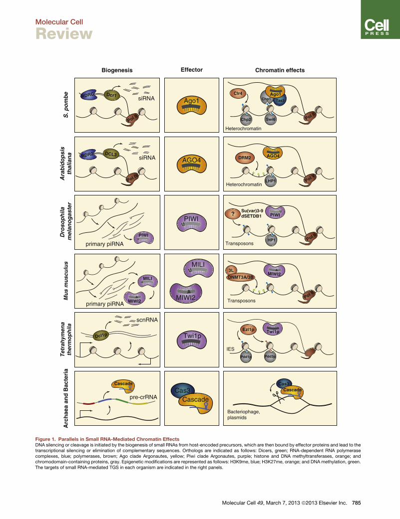
In the Schizosaccharomyces pombe system, the authors
discuss a “self-perpetuating feedforward loop” in regards to
the small RNA–mediated heterochromatin formation in the
yeast system. What is meant by this?
The centromeres of the S. pombe yeast contain a high number
of repeating sequences and mobile genetic elements. Small RNA–
mediated histone modifications repress the repeating sequences
and mobile genetic elements present in the centromeric region.
Specifically, short-interfering RNAs (siRNAs) silence this region.
These siRNAs are also derived from centromere transcripts that
have been processed by an enzyme called Dcr1. Once processed,
the siRNAs are loaded onto Ago1, a protein effector of the Argo-
naute family and, together with several other proteins, produce the
RNA-induced initiation of transcriptional silencing (RITS) complex.
The siRNA within the complex binds to target sequences at the cen-
tromere, and the result is an accumulation of several proteins at the
targeted region and TGS. Furthermore, the RITS complex recruits
the RNA-directed RNA polymerase (RDRC) complex to generate
more siRNA from the same region. In this manner, the biogenesis of
siRNA is a self-perpetuating feedforward loop.
In addition to the repressive effects of the S. pombe siRNAs,
are there other small RNA systems addressed by the authors
that induce repressive histone modifications?
Yes. Piwi proteins are a group of Argonaute proteins that func-
tion to control mobile genetic elements in flies, humans, and other
organisms. The authors discussed piRNA-directed transcriptional
silencing in flies. Piwi-interacting RNAs (piRNAs) are specific to
germline cells and function to silence the repetitive regions contain-
ing mobile DNAs by introducing and maintaining repressive histone
modifications.
How does the biogenesis of centromere-silencing siRNAs
from the S. pombe system differ from the biogenesis of piRNAs
in the germline Drosophila cells?
The siRNAs in the yeast system discussed in the paper are pro-
duced from centromere transcripts by the action of Dcr1. In con-
trast, piRNAs are derived from the transcription and processing of
piRNA clusters, which are mostly composed of inactive transposon
fragments, in a Dicer-independent manner.
What role do lncRNAs play in regulation of gene expression
in the human model?
Although the roles of lncRNAs are still quite vague, a few have
been characterized. They include Xist, which is involved in X-chro-
mosome inactivation and imprinting. Additionally, many lncRNAs
copurify with chromatin remodeling complexes, including PRC2.
This indicates a role for lncRNAs in scaffolding or targeting remod-
eling machinery to specific loci. Of the lncRNAs known to copu-
rify with PRC2, several function in human cancers. HOTAIR is an
lncRNA in humans that, when overexpressed, leads to increased
invasiveness and poor outcome for several cancers. The results
from knockdown experiments suggest that HOTAIR acts as a scaf-
fold between PRC2 and LSD1, a histone tail demethylase. In addi-
tion to HOTAIR, ANRIL is overexpressed in human leukemias and
prostate cancer due to epigenetic silencing of p15, a tumor sup-
pressor gene.
Two other lncRNAs, Air and Kcnq1ot1, are involved in genomic
imprinting and silencing of one allele in diploid human cells.
HOTAIRM1 is involved in myeloid differentiation. HOTTIP and Mis-
tral interact with chromatin modifiers that remodel the chromatin
structure and activate transcription of genes rather then repress
genes.
In terms of epigenetics, RNA-mediated modifications were
first discovered in plants but occur in a wide range of organ-
isms, including mammals. How do small RNAs and lncRNAs
guide methylation patterns in DNA?
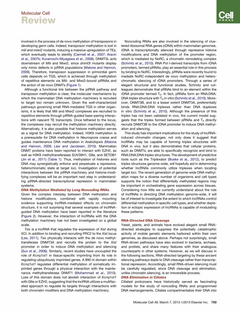
A majority of methylation in Arabidopsis, a plant model, occurs
in transposons and repetitive elements near the centromeres. A
specialized RNA polymerase produces long transcripts from these
regions. A second RNA polymerase generates a complementary
copy of the RNA transcript to produce a dsRNA molecule, which
is then processed by a ribonuclease to generate siRNAs. These
siRNAs bind to an Argonaute family effector protein that recruits
methylation enzymes to the specific region. The result is methylation
Case Study Dogma Derailed: the Many Influences of RNA on the Genome
Leah R. Sabin et al. (2013). Molecular Cell 49, 783–794.
(Continued )

DNA, RNA, and Protein
6
Case Study Dogma Derailed: the Many Influences of RNA on the Genome—cont’d
and silencing of those regions. Additionally, intergenic noncoding
transcripts produced by a recently discovered RNA polymerase act
as a scaffold between the Argonaute protein and the recently discov-
ered RNA polymerase itself.
RNA-mediated methylation also occurs in mammalian male germ
cells through the action of piRNA. The piRNAs are expressed at differ-
ent times during germ cell development and spermatogenesis. Failure
of the piRNAs to regulate DNA methylation in the developing cells
results in sterility. The exact molecular mechanism for this is not yet
understood.
DNA methylation is also mediated by lncRNAs, although the pro-
cesses have not yet been investigated on a global scale. The authors
discuss some lncRNAs that have been published. Specifically, the
Tsix lncRNA regulates expression of Xist during X chromosome inac-
tivation by interacting with chromatin-associated complex PRC2 and
recruiting it to the Xist locus. Also, Tsix recruits the methylation enzyme
to the Xist promoter, thus silencing the Xist gene. Other genes that are
influenced by noncoding RNAs include those that silence ribosomal
RNA genes through the possible action of an RNA:DNA:DNA triplex
structure, although this has just been suggested and not confirmed
in vivo.
Which organisms extensively use RNA-directed DNA cleavage
as opposed to transcriptional repression to silence regions of the
genome?
Prokaryotes (bacteria and archaea), along with a few eukaryotic
protists (ciliated protozoans), use DNA cleavage mediated by RNA
molecules to silence gene expression.
How is DNA cleavage mediated by RNA in ciliates?
Ciliates, which have two nuclei (micronucleus and macro-
nucleus), remove internal eliminated segment (IES) sequences
through the action of scnRNAs and also through the modification
of histone proteins. The scnRNAs are generated from micronucleus
sequences during conjugation and are used to scan transcripts from
the macronucleus for complementary sequences. Once found, the
sequences are degraded. Any scnRNA that is not bound to macro-
nucleus transcripts is used to find homologous sequences and target
those homologs for elimination. Elimination includes the use of Argo-
naute family effector proteins and involves a mechanism similar to
transposon silencing in S. pombe as described previously.
The CRISPR system in prokaryotes is used to help protect
against viral pathogens. How are the pathogens targeted through
the action of RNAs?
The CRISPR system retains remnants of the viral pathogens and
exogenous DNA as “memories” to help combat future infections.
These “memories” are termed CRISPR loci. CRISPR RNAs (crRNAS)
are the small RNAs that mediate immunity to the viral pathogens.
Small pieces of phage and foreign DNA are retained by the prokaryote
and integrated into the CRISPR locus. Expression of the CRISPR loci
yields long precursor RNAs that are further processed into crRNAs,
which target incoming foreign DNA. Effector proteins are recruited
when crRNAs bind to their foreign DNA targets. The result is DNA
cleavage. Several types of CRISPR systems have been characterized
and differ in the proteins used to process the long precursor RNAs
into crRNAs.
From the simplest prokaryotes to more advanced organisms,
RNA molecules play diverse roles within cells. Most often RNAs
are implicated in post-transcriptional control of gene expression.
Recently, the roles of RNAs within the cells have expanded with
the discovery that epigenetic modifications, including histone
modification, DNA methylation, and DNA cleavage, are guided by
noncoding RNAs. These RNAs control gene expression by induc-
ing changes within chromatin structure and methylation patterns.
RNA can also target DNA for rearrangement and elimination. The
consequences of RNA-mediated control of gene expression even
have health impacts for humans and represent a potential angle for
development of therapeutics.

Molecular Cell
Review
Dogma Derailed: The ManyInfluences of RNA on the Genome
Leah R. Sabin,1,2,* M. Joaquina Delas,1,2 and Gregory J. Hannon1,2,*1Watson School of Biological Sciences2Howard Hughes Medical InstituteCold Spring Harbor Laboratory, 1 Bungtown Road, Cold Spring Harbor, NY 11724, USA*Correspondence: [email protected] (L.R.S.), [email protected] (G.J.H.)http://dx.doi.org/10.1016/j.molcel.2013.02.010
Epigenetic control of gene expression is a critical component of transcriptional regulation. Remarkably, thedeposition of epigenetic modifications is often guided by noncoding RNAs. Although noncoding RNAs havebeen most often implicated in posttranscriptional gene silencing, these molecules are now emerging as crit-ical regulators of gene expression and genomic stability at the transcriptional level. Here, we review recentefforts to understand the mechanisms by which RNA controls the expression or content of DNA. We discussthe role of both small RNAs and long noncoding RNAs in directing chromatin changes through histone modi-fications and DNA methylation. Furthermore, we highlight the function of RNA in mediating DNA cleavageduring genome rearrangements and pathogen defense. In understanding the mechanisms of RNA controlover DNA, the power of RNA may one day be harnessed to impact gene expression in a therapeutic setting.
IntroductionSince each cell within an organism contains an identical copy of
the genome, regulation of the output of the genome is respon-
sible for determining cellular identity and allowing complex
organisms to develop and function. On a cellular level, organ-
isms face two main challenges: to maintain genome integrity in
the face of mutagens and mobile genetic elements, and to
express a specific repertoire of genes at the proper level and
with the appropriate timing. Disruptions of either of these two
processes can have catastrophic consequences, such as infer-
tility or malignant transformation. Therefore, organisms have
evolved elegant mechanisms to monitor the stability of the
genome and fine-tune gene expression. In recent years, it has
become increasingly evident that many of these regulatory
systems rely on RNA to mediate their effects. This review will
discuss the various classes of noncoding RNAs that exert control
over DNA, focusing on those that maintain genomic stability or
regulate DNA structure and organization through chromatin
modifications or DNA cleavage.
The catalog of functional noncoding RNAs is continuously
expanding, due in part to the development of next-generation
sequencing technologies. Two important classes of functional
RNAs responsible for mediating effects on DNA are small RNAs
and long noncoding RNAs (lncRNAs). In general, small RNAs
are generated from longer precursors, which can derive from
both endogenous and exogenous sources, including acute viral
infections and transposable elements (TEs). Following biogen-
esis, small RNAs are loaded into an Argonaute family member
within a large effector protein complex. Twoclasses of Argonaute
proteins exist in most animals: the ubiquitously expressed Argo-
naute (Ago) cladeproteins, which are definedby their relationship
to Arabidopsis AGO1, and members of the Piwi clade, which
bear similarity toDrosophila Piwi andwhose expression is largely
restricted to the germline (Hutvagner and Simard, 2008). In
many organisms, small RNAs are amplified to promote a more
robust response; this amplification can occur through a variety
of mechanisms.
The canonical role of small RNAs is to mediate posttranscrip-
tional gene silencing (PTGS) of target RNA transcripts. During
PTGS, base pairing between the small RNA, bound to its effector
complex, and the target results in target cleavage or translational
repression. However, seminal studies in plants and yeast, as well
as more recent work in other systems, have established that
small RNAs are also capable of directing transcriptional gene
silencing (TGS), which can be achieved throughDNAmethylation
or the deposition of repressive histone modifications. In these
cases, the function of TGS is often to protect genomic integrity
by maintaining a repressive heterochromatic state in repetitive
regions of the genome, most notably those regions which harbor
mobile genetic elements. Arguably themost extrememechanism
by which the content and expression of DNA can be controlled
by small RNAs is DNA elimination. In some ciliates, small RNAs
guide the excision of DNA elements, such as transposons, during
genome rearrangements. Moreover, small RNAs in bacteria
and archaea orchestrate the clustered regularly interspaced
short palindromic repeat (CRISPR) pathway, which directs
sequence-specific DNA cleavage of plasmids or invading phage.
In the following sections, wewill describe themechanistic details
of these small RNA-guided pathways and the recent advances in
our understanding of their functions.
In contrast to small RNAs, the study of lncRNAs as a defined
class of molecules is still in its relative infancy; indeed, the fact
that the human genome is pervasively transcribed, yet that
protein coding genes comprise only �10% of its content, is
a relatively recent revelation. Unlike small RNAs, there appear
to be no unifying structural, biochemical, or functional character-
istics that define a given transcript as a lncRNA; rather, the
simplest definition of a lncRNA is merely an RNA transcript
greater than 200 nucleotides in length with no coding potential
(Ponting et al., 2009). Over the last 10 years, RNA-Seq data
Molecular Cell 49, March 7, 2013 ª2013 Elsevier Inc. 783

Table 1. Chromatin Regulatory Machinery
Organism
H3K9
Methyltransferases
H3K27
Methyltransferases
H3K4
Methyltransferases
Chromodomain
Proteins De Novo DNMTs Maintenance DNMTs
S. pombe Clr4 Activity is present,
no HMT identified
Set1 Swi6 No cytosine methylation No cytosine methylation
Chp1
Chp2
Arabidopsis KYP CLF ATX1 LHP1 DRM2 MET1 (CG sites)
SUVH5 SWN ATX2 CMT3 (CHG sites)
SUVH6 MEA ATXR7
Drosophila Su(var)3-9 E(z) trx HP1 No cytosine methylation No cytosine methylation
egg
Mammals Suv39h1 EZH2 MLL CBX7 DNMT3A DNMT1
G9a DNMT3B
Tetrahymena Ezl1p Ezl1p Activity is present,
no HMT identified
Pdd1p ? ?
Pdd3p
This table lists known orthologs of histone and DNA methyltransferases, as well as chromodomain-containing proteins responsible for binding
methylated histones and nucleating protein complexes on chromatin.
Molecular Cell
Review
and chromatin maps from a staggering number of cell types and
tissues have been used in large-scale efforts to catalog thou-
sands of novel lncRNAs in organisms from plants to humans
(Hu et al., 2012). lncRNAs are often poorly conserved at the
sequence level, initially leading to uncertainty as to whether
they represent active entities or transcriptional noise. However,
as a growing number of lncRNAs are characterized, it has
been established that orthologs can be identified in other
species through synteny; although these ‘‘syntelogs’’ bear little
to no sequence similarity, their position relative to neighboring
protein coding genes has been maintained through evolution
(Ulitsky et al., 2011). Nevertheless, the functions of most of the
identified lncRNAs remain largely uncharacterized.
One theme that has emerged during large-scale characteriza-
tion efforts is that lncRNAs are commonly involved in medi-
ating chromatin-level gene regulation through interactions with
histone modifiers, the DNA methylation machinery, or transcrip-
tional regulators. Many other lncRNAs have been identified as
key regulators of essential cellular processes but are not known
to be involved in chromatin regulation and structure, and we will
omit these from our discussion. However, we invite readers to
refer to several excellent reviews on the subject (Ponting et al.,
2009; Hu et al., 2012; Rinn and Chang, 2012). In the following
sections, we highlight the functions of several diverse classes
of small and large noncoding RNAs according to the mecha-
nism by which the RNAs exert their control over DNA, spe-
cifically through histone modifications, DNA methylation, and
DNA cleavage.
RNA-Directed Histone ModificationsHistones are responsible for the organization and regulation of
DNA structure and are subject to a variety of modifications on
their N-terminal tails, such as methylation, acetylation, and ubiq-
uitination, that dynamically influence chromatin function. Histone
modifications can function in activating or repressing gene
expression; a common mark associated with active chromatin
is the trimethylation of histone 3 (H3) at lysine 4 (K4), or
784 Molecular Cell 49, March 7, 2013 ª2013 Elsevier Inc.
H3K4me3, which is often found at promoters of actively tran-
scribed genes (Black et al., 2012). Conversely, marks associated
with silenced heterochromatin include di- and trimethylated
H3K9, as well as trimethylation of H3K27 (Black et al., 2012).
Histonemethyltransferase (HMT) enzymes deposit methyl marks
onto histone tails, while chromodomain-containing proteins,
which directly bind to methylated lysines, often work in concert
with HMTs and are involved in assembling protein complexes
on DNA. Table 1 lists selected orthologs of HMTs and chromo-
domain-containing proteins operating within the organisms dis-
cussed in this review.
Small RNA-Mediated Heterochromatin Formation in
Yeast
A defining characteristic of eukaryotic centromeres is their high
density of repetitive DNA elements, such as satellite repeats
and transposons. Repressive histone modifications, particularly
methylated H3K9 (H3K9me), are essential for controlling these
mobile genetic elements within centromeric regions. The cores
of fission yeast centromeres are flanked by tRNA genes and
tandem copies of repetitive elements termed dg and dh repeats
(Castel and Martienssen, 2013). Seminal work by Volpe and
colleagues first established a role for small interfering (si) RNAs
in directing transcriptional silencing of centromeric repeats in
Schizosaccharomyces pombe. Mutants in RNA silencing factors
are defective in centromeric silencing and accumulate centro-
mere-derived sense and antisense transcripts (Volpe et al.,
2002). Furthermore, loss of RNAi components leads to a loss
of both H3K9me and the chromodomain-containing protein
Swi6 from centromeric repeats, coupled with an increase in
H3K4me3 and transcriptional activation of repeat sequences
(Volpe et al., 2002).
It is now well-established that centromeric transcripts are
processed into siRNAs by Dcr1, which are then loaded into
Ago1, the core component of the RNA-induced initiation of tran-
scriptional silencing (RITS) complex (Verdel et al., 2004; Castel
and Martienssen, 2013) (Figure 1). Other protein components
of RITS include Chp1, a centromere-associated chromodomain
Twi1p
Pol II
RDRC Dcr1 siRNA Ago1 Tas3Chp1Ago1
Pol II
Clr4
Swi6Chp2
Heterochromatin
S. p
om
be
Ara
bid
op
sis
thal
ian
a
PIWIUUUUUU
Pol II
Su(var)3-9dSETDB1?
HP1
PIWIUUUU
Transposons
PIWIUUUUUU
primary piRNA
UU
UU
Mu
s m
usc
ulu
sD
roso
ph
ila m
elan
og
aste
r
Pol II
3L
DNMT3A/3BMIWI2
AAAAAA
Transposons
MILIUUUUUU
MIWI2AAAAAA
UU
UU
MILIUUUUUU
MIWI2AAAA
primary piRNA
Dcl1p
scnRNA
Twi1p
Pdd1p
IESPdd3p
Ezl1p
Tet
rah
ymen
ath
erm
op
hila
Cascade
pre-crRNACas3
Cascade
Cas3Cascade
Bacteriophage,plasmids
AGO4 DRM2 AGO4
Pol VLHP1
Heterochromatin
DCL3RDR2siRNA
Pol IV
Arc
hae
a an
d B
acte
ria
Biogenesis Effector Chromatin effects
Figure 1. Parallels in Small RNA-Mediated Chromatin EffectsDNA silencing or cleavage is initiated by the biogenesis of small RNAs from host-encoded precursors, which are then bound by effector proteins and lead to thetranscriptional silencing or elimination of complementary sequences. Orthologs are indicated as follows: Dicers, green; RNA-dependent RNA polymerasecomplexes, blue; polymerases, brown; Ago clade Argonautes, yellow; Piwi clade Argonautes, purple; histone and DNA methyltransferases, orange; andchromodomain-containing proteins, gray. Epigenetic modifications are represented as follows: H3K9me, blue; H3K27me, orange; and DNA methylation, green.The targets of small RNA-mediated TGS in each organism are indicated in the right panels.
Molecular Cell 49, March 7, 2013 ª2013 Elsevier Inc. 785
Molecular Cell
Review

Molecular Cell
Review
protein that binds dimethylated H3K9, and Tas3, a GW domain-
containing protein (Verdel et al., 2004). GWmotifs are a common
feature of Argonaute interactors; proteins containing this domain
can also be found in Argonaute complexes in plants, flies, and
mammals (Hutvagner and Simard, 2008). According to the
current model for TGS in S. pombe, and in most other examples
of small RNA-directed TGS in other species, target identification
occurs through base pairing interactions between the bound
siRNA and nascent transcripts at the target locus; in fission
yeast, RITS associates with long, centromere-derived RNAs in
a Dcr1-dependent manner (Motamedi et al., 2004). Moreover,
RITS remains tethered to silent loci by the interaction of Chp1
with H3K9me, which is deposited by the HMT Clr4 (Noma
et al., 2004). Clr4-dependent RITS tethering to heterochromatin
is essential for maintaining transcriptional silencing and for
generating new siRNAs (Noma et al., 2004).
RITS also physically interacts with another complex named
RDRC, or the RNA-directed RNA polymerase complex, which
contains Rdp1, an RNA-dependent RNA polymerase (Motamedi
et al., 2004). The physical interaction between the RDRC and
RITS complexes requires Dcr1 and Clr4 (Motamedi et al., 2004),
suggesting that siRNA-driven RITS targeting to centromeric
repeats, and stable H3K9 binding by Chp1, allows the recruit-
ment of RDRC to chromatin. Moreover, the RDRC physically
interacts with Dcr1, suggesting that siRNA biogenesis is coupled
to dsRNA synthesis (Castel and Martienssen, 2013). Thus, the
tethering of biogenesis, effector, and amplification complexes
at the silenced loci likely organizes a self-perpetuating feedfor-
ward loop (Figure 1). The fundamental principles of siRNA-
directed centromeric silencing that have been uncovered in
S. pombe provide an excellent framework for understanding
RNA-guided heterochromatin formation in other systems.
piRNA-Directed Transcriptional Silencing in Flies
Piwi-interacting RNAs (piRNAs) are a germline-specific class of
small RNAs that bind to Piwi cladeArgonaute proteins and consti-
tute a transposon defense system in a variety of organisms,
including humans. Defects in the piRNA pathway lead to uncon-
trolled transposition ofmobile genetic elements, which can cause
genomic instability and sterility (Malone and Hannon, 2009).
piRNA-mediated transposon silencing has been studied most
extensively in the Drosophila ovary. Although the mechanisms
by which piRNAs repress transposon expression are still being
elucidated, recent work in flies suggests that piRNAs induce and
maintain heterochromatic silencing of these repetitive elements
through the deposition of repressive histone modifications.
Drosophila piRNAs are between 23 and 29 nt in length and
often bear a 50 uridine (1U), compared to endogenous siRNAs
andmiRNAs, which tend to be 20–23 nt and show little sequence
bias (Malone and Hannon, 2009). Drosophila encodes three
nonredundant Piwi clade proteins that bind piRNAs: Piwi, the
founding member of the clade, Aubergine (Aub), and Argonaute
3 (Ago3) (Hutvagner and Simard, 2008). Importantly, primary
piRNAs are not generated by a Dicer protein. Although the
precise mechanisms are only now beginning to be understood,
it is clear that piRNA biogenesis is initiated by the transcription
and processing of piRNA clusters, which are unique loci com-
prised of inactive fragments of several classes of transposons
(Brennecke et al., 2007) (Figure 1). Following primary biogenesis,
786 Molecular Cell 49, March 7, 2013 ª2013 Elsevier Inc.
piRNAs enter into the ping-pong amplification cycle, which
generates secondary piRNAs and is predominantly mediated
by Aub and Ago3 in Drosophila ovarian germ cells. During
ping-pong, a Piwi- or Aub-bound antisense piRNA recognizes
the sense strand of an active transposon, leading to transcript
cleavage. This cleavage generates the 50 end of a new sense
piRNA, which is bound by Ago3 and can recognize antisense
transposon transcripts, generating new antisense piRNAs and
thus continuing the amplification cycle (Brennecke et al., 2007;
Gunawardane et al., 2007).
This elegant ping-pong amplification mechanism silences
active transposons at the posttranscriptional level. How, then,
does transposon repression occur in the somatic follicle cells
of the ovary, where only Piwi and the primary piRNA pathway
are known to operate? Several lines of evidence now point to
a role for Piwi in mediating TGS in Drosophila. Piwi localizes to
the nucleus, and cytoplasmic retention of Piwi results in massive
transposon upregulation and sterility, which hints at a role in TGS
rather than PTGS (Klenov et al., 2011). In addition, piRNA
pathway mutants, including piwi, suppress position effect varie-
gation (Malone and Hannon, 2009). Germline Piwi knockdown or
cytoplasmic retention results in the depletion of H3K9me2 and
HP1, the Drosophila homolog of Swi6, from a specific set of
repetitive elements (Klenov et al., 2011; Wang and Elgin, 2011).
In addition, nuclear run-on experiments in Piwi-depleted ovaries
measured increased transcription of particular transposons as
compared to wild-type tissue (Shpiz et al., 2011). Unexpectedly,
nuclear run-on analysis of armitage mutants, in which Piwi does
not bind piRNAs or enter the nucleus, did not detect differences
in the transcription of repetitive elements between mutant and
control ovaries, leading to doubts surrounding the role of Piwi
in TGS (Malone and Hannon, 2009). Therefore, a direct compar-
ison of chromatin states, nascent transcription, and steady-state
transposon levels in the presence or absence of Piwi was neces-
sary to unambiguously determine its role in TGS.
Recently, Sienski and colleagues reported such a study in an
ovarian somatic follicle cell line, OSC, which does not express
Aub or Ago3 and therefore allows the dissection of the Piwi-
driven piRNA pathway in isolation. Following Piwi depletion, the
authors found that transposon transcripts are upregulated, and
Piwi-regulated transposons display enhanced Pol II occupancy
and increased rates of nascent transcription (Sienski et al.,
2012). By examining de novo insertions of individual TEs in the
OSC genome, Sienski et al. found that active elements display
Piwi-dependent H3K9me3. Moreover, two additional studies
report similar effects on chromatin states and transposon sup-
pression when Piwi is depleted in vivo (Le Thomas et al., 2013;
Rozhkov et al., 2013).These data provide strong evidence that
Piwi restricts transposon expression via TGS by directing the
deposition of repressive histone modifications on TEs. Similar
to heterochromatin formation in yeast, the recruitment of Piwi
to TEs is likely mediated by base-pairing interactions between
Piwi-bound piRNAs and nascent transposon transcripts, leading
to the recruitment of H3K9 methyltransferases and associated
chromatin binding factors to enforce a silent, heterochromatic
state (Figure 1). Whether Piwi, like the yeast RITS complex,
remains tethered to heterochromatic loci following H3K9methyl-
ation has not yet been addressed.

PRC2
Tsix
ANRIL
DNMT1
Kcnq1ot1
Kcnq1ot1Air
G9a
DNMT3A/B
pRNAsTsix
HOTTIP Mistral
MLL1 PRC2
HOTAIR
DNA methylation
H3K27meH3K9meH3K4me
LSD1/CoRESTKcnq1ot1
Figure 2. Effects of lncRNAs at theChromatin LevellncRNAs interact with several activating and re-pressive chromatin-modifying complexes. Repre-sentative lncRNAs are grouped according tothe protein complex with which they interact.Depending on the lncRNA, genomic targeting ofthese chromatin modifiers can occur in cis orin trans.
Molecular Cell
Review
Interactions of Long Noncoding RNAs with Chromatin
Remodeling Machinery
Although the diverse functions of lncRNAs are only beginning
to be uncovered, their potential ability to interact with andmodu-
late the activity of chromatin regulatory complexes may allow
lncRNAs to affect gene expression on a genome-wide scale.
Several of the well-characterized lncRNAs, such as Xist, are
involved in X chromosome inactivation (XCI) and imprinting,
which require changes in chromatin structure on amassive scale
(Lee, 2011). Moreover, many recently identified lncRNAs copur-
ify with chromatin remodeling complexes, suggesting that
they may function in targeting these complexes to genomic
loci, or may serve as molecular scaffolds for complex assembly
(Figure 2). Whether genomic targeting of protein complexes by
lncRNAs occurs in cis, through interactions with nascent lncRNA
transcripts, or in trans, perhaps through RNA:DNA duplexes or
triplexes, is an important facet of lncRNA biology that future
work must address. In a recent study, Guttman and colleagues
characterized lncRNAs bound to 12 chromatin-associated com-
plexes in mouse embryonic stem cells (Guttman et al., 2011).
Interestingly, several lncRNAs purified with more than one
complex, supporting the notion that lncRNAs serve as molecular
scaffolds that bridge multiple regulatory units. One of these
complexes, PRC2, has emerged as a common binding partner
for lncRNAs in multiple organisms (Figure 2). EZH2 is the cata-
lytic subunit of PRC2 in mammals and methylates H3K27 to
enforce repressive heterochromatin (Table 1). Studies in multiple
human and mouse cell types have found that hundreds of
lncRNAs interact with PRC2, although the specificity of each of
these interactions has not been rigorously explored (Khalil
et al., 2009; Zhao et al., 2010).
One of the most well-known PRC2-interacting lncRNAs is
HOTAIR, a 2.1 kb transcript derived from the humanHOXC locus
that represses the expression of genes within the HOXD locus,
as well as other targets throughout the genome (Rinn et al.,
2007). HOTAIR binds EZH2 and is required for PRC2-mediated
H3K27 trimethylation and silencing of theHOXD locus in humans
(Rinn et al., 2007). However, HOTAIR may play a different role
in mice, since a deletion within the HOXC locus that encom-
passesmHOTAIR has no effect on expression of theHOXD locus
(Schorderet and Duboule, 2011). HOTAIR overexpression has
been linked to increased invasiveness and poorer outcomes in
several human cancers (Rinn and Chang, 2012). Interaction of
HOTAIR with PRC2 is mediated through a region in its 50
Molecular Cell
terminus, while the 30 terminus binds
LSD1, a H3K4 demethylase that functions
within the CoREST/REST complexes
(Tsai et al., 2010). Overexpression of
HOTAIR results in global changes in PRC2 occupancy and
H3K27me3 marks (Rinn and Chang, 2012). Conversely, knock-
down of HOTAIR alters the chromatin occupancy of PRC2 and
LSD1 genome-wide, leading to reduced H3K27me3 and
increased H3K4me2 at target loci (Tsai et al., 2010). Thus,
HOTAIR appears to serve as a scaffolding molecule, bridging
PRC2 with LSD1 (Figure 2).
Another lncRNA associated with human cancers is ANRIL,
a long, antisense transcript found in the INK4a/Arf locus. ANRIL
is overexpressed in human leukemias and prostate cancers, and
its expression leads to epigenetic silencing of the nearby tumor
suppressor p15 (Yu et al., 2008; Yap et al., 2010). H3K27 trime-
thylation of the INK4a/Arf locus requiresANRIL, whichwas found
to interact with SUZ12, a component of PRC2, and CBX7, a
PRC1-associated chromodomain-containing protein (Rinn and
Chang, 2012).
The Air and Kcnq1ot1 lncRNAs are both involved in imprinting
in mammals and use similar mechanisms to induce the deposi-
tion of repressive marks at silenced alleles. Air is transcribed
from the Igf2r locus and mediates H3K9 trimethylation of the
nearby Slc22a3 promoter by recruitment of the repressive G9a
chromatin-modifying complex in cis (Ponting et al., 2009). Simi-
larly, Kcnq1ot1, which is transcribed antisense to the silenced
paternal allele of Kcnq1, binds and recruits G9a and PRC2 to
direct H3K9 and H3K27 trimethylation of the locus in cis (Pandey
et al., 2008). Defects in silencing by either of these lncRNAs
result in biallelic expression of their normally imprinted targets,
underscoring their importance inmaintainingmonoallelic hetero-
chromatic silencing.
In addition to the HOXC cluster-derived lncRNA HOTAIR,
three HOXA locus-associated lncRNAs have been described.
HOTTIP is expressed from the 50 end of the locus, Mistral is
encoded between Hoxa6 and a7, and HOTAIRM1 is located at
the distal 30 end of the cluster (Hu et al., 2012). Little is known
about HOTAIRM1, apart from the fact that it is induced during
myelopoiesis and is required for expression of HOXA genes
during myeloid differentiation. HOTTIP and Mistral, on the other
hand, mediate the transcriptional activation of HOXA genes
through physical interactions with the MLL1 complex, a H3K4
methyltransferase complex which is known to bind to HOX
gene promoters (Bertani et al., 2011; Wang et al., 2011). Deple-
tion of either HOTTIP or Mistral results in a loss of MLL1 occu-
pancy on HOXA target genes. Therefore, in the case of HOTTIP
and Mistral, interaction of the lncRNA with chromatin modifiers
49, March 7, 2013 ª2013 Elsevier Inc. 787

Molecular Cell
Review
results in the deposition of activating chromatin marks rather
than repressive marks (Figure 2).
In general, the ability of lncRNAs to recruit the activities of
protein complexes to genomic loci may allow the cell to impart
specificity to broadly acting chromatin-modifying machineries.
It is clear that much work remains to be done in order to fully
understand the complex interactions between lncRNAs and
chromatin-modifying complexes, particularly the acetylating or
ubiquitinating complexes. However, by characterizing the func-
tions of these molecules through dissecting their protein part-
ners and identifying the genomic loci with which they interact,
the regulatory power of lncRNAs may one day be harnessed
for use in chromatin-targeted therapeutic applications.
RNA-Directed DNA MethylationDNA methylation was the first RNA-guided epigenetic modifica-
tion to be discovered, and is utilized by organisms ranging from
plants to mammals. In most systems, methylation occurs on
cytosine residues in the context of a CG dinucleotide, or CpG
motif, by the action of DNA methyltransferase (DNMT) proteins.
DNA methylation is a dynamic epigenetic modification that often
functions as a repressive mark to silence transcription (Law and
Jacobsen, 2010). Two types of DNMT proteins perform DNA
methylation, and differ in their preferred substrates: de novo
DNMTs methylate completely unmethylated CpG dinucleotides,
while maintenance methyltransferases act on hemimethylated
DNA during DNA replication to methylate the newly replicated
strand (Law and Jacobsen, 2010). Table 1 lists the DNMTs
present in species relevant to this review. Notably, many species
have lost the ability to methylate their DNA; these include
S. pombe and D. melanogaster.
Small RNA-Directed DNA Methylation in Plants
RNA-directed DNA methylation (RdDM) was first described in
plants, following the observation that viroid cDNAs become spe-
cifically methylated upon integration into the tobacco genome
(Law and Jacobsen, 2010). Since its initial discovery, the path-
way has been characterized extensively in Arabidopsis and
now serves as the defining model for RdDM in eukaryotes.
Genome-wide mapping of methylation in Arabidopsis has re-
vealed that most methylation occurs on transposons and repet-
itive elements, which are concentrated near centromeres (Castel
and Martienssen, 2013). RNA Pol IV, a specialized polymerase
essential for RdDM, produces long RNA transcripts from these
heterochromatic regions (Henderson and Jacobsen, 2007). Pol
IV transcripts are substrates for RDR2, an RNA-dependent
RNA polymerase, which produces the complementary strand
to generate dsRNA (Law and Jacobsen, 2010). The RNase III
enzyme DCL3 processes the dsRNA from repetitive regions
into 24 nt heterochromatic siRNAs, which are bound by the
Argonaute family member AGO4 (Henderson and Jacobsen,
2007). AGO4 then targets the de novo methyltransferase
DRM2 to the corresponding genomic locus for methylation and
silencing (Henderson and Jacobsen, 2007) (Figure 1).
Further molecular details of this process were uncovered
following the identification of a second specialized polymerase
complex in Arabidopsis: RNA polymerase V. Wierzbicki and
colleagues found that Pol V transcribes intergenic noncoding
transcripts within heterochromatin, which serve as scaffolds
788 Molecular Cell 49, March 7, 2013 ª2013 Elsevier Inc.
for AGO4 recruitment (Wierzbicki et al., 2008). AGO4 not only
binds to nascent Pol V transcripts through siRNA-mediated
base pairing but also interacts directly with the largest Pol V
subunit, NRPE1 (Law and Jacobsen, 2010). In a sense, the
mechanism guiding AGO4 to repetitive loci parallels the recruit-
ment of S. pombe RITS or Drosophila Piwi to their targets: each
of these processes is driven by an interaction with nascent tran-
scripts at the locus.
The mechanism by which the methyltransferase DRM2 is
recruited to AGO4-targeted regions has not been fully eluci-
dated, although several recent studies hint that RDM1, a novel
regulator of RdDM, may play a role in the process (Castel and
Martienssen, 2013). RDM1 specifically binds to single-stranded
methylated DNA and is required for the accumulation of Pol V
transcripts. Astonishingly, RDM1 interacts not only with Pol V
subunits but also with AGO4 and DRM2 (Castel and Martiens-
sen, 2013). Therefore, RDM1 may recruit Pol V to heterochro-
matic loci, while mediating subsequent interactions between
components of the RNA silencing pathway and methylation
machinery at the locus. However, the sequence of these events
and the molecular mechanisms that drive them have not yet
been established.
piRNA-Mediated Methylation of Mobile Genetic
Elements in Mammals
Although many of the fundamental principles of piRNA pathway
function have been characterized in Drosophila, including the
recent discovery of piRNA-guided histone modifications, the
role of piRNAs in TGS was first described in mammals, where
piRNAs direct the methylation of transposons in order to enforce
their transcriptional repression. Mammalian piRNA pathways
operate in male germ cells, and the expression of piRNAs
occurs at two distinct stages during development. Pre-pachy-
tene piRNAs, and their interacting Piwi proteins, Mili and
Miwi2, are expressed in primordial germ cells and map to trans-
posons and repetitive elements (Malone and Hannon, 2009).
Conversely, pachytene piRNAs are expressed during adult sper-
matogenesis, and although some pachytene piRNAs align to
repetitive genomic regions, the majority map uniquely
throughout the mouse genome, and their function remains
unknown (Aravin et al., 2007a). The mouse Piwi proteins ex-
pressed at this stage are Miwi and, to a lesser extent, Mili, which
do not appear to direct transposon repression via TGS or to
engage in ping-pong amplification (Malone and Hannon, 2009).
During mammalian embryonic germ cell development, the
genome undergoes a process of global demethylation, followed
by the reestablishment of methylation at repetitive elements.
Prior to the identification of piRNAs, it was known that trans-
poson silencing in primordial germ cells was dependent upon
methylation of repetitive elements by de novo methyltrans-
ferases DNMT3A and 3B (Okano et al., 1999). DNMT3L, which
is related to DNMT3A and DNMT3B in sequence but lacks
their catalytic motifs, stimulates the activity of DNMT3A and
DNMT3B and specifically regulates transposon silencing in the
germline; DNMT3L is required for the re-establishment of DNA
methylation on repetitive elements, and its mutation results in
transposon upregulation and sterility (Malone and Hannon,
2009). Due to the overlapping phenotypes between mili, miwi2,
and dnmt3l mutants, investigators asked whether piRNAs were
Molecular Cell
Review
involved in the process of de novo methylation of transposons in
developing germ cells. Indeed, transposon methylation is lost in
mili andmiwi2mutants, inducing a massive upregulation of TEs,
which eventually leads to sterility (Carmell et al., 2007; Aravin
et al., 2007b; Kuramochi-Miyagawa et al., 2008). DNMT3L acts
downstream of Mili and Miwi2, since dnmt3l mutants display
only minor defects in piRNA populations (Malone and Hannon,
2009). Therefore, transposon suppression in primordial germ
cells depends on TGS, which is achieved through methylation
of repetitive elements via Mili- and Miwi2-bound piRNAs and
the action of de novo DNMTs (Figure 1).
Although a functional link between the piRNA pathway and
transposon methylation is clear, the molecular mechanisms by
which the mammalian DNA methylation machinery is recruited
to target loci remain unknown. Given the well-characterized
pathways governing small RNA-mediated TGS in other organ-
isms, it is likely that Mili and Miwi2 complexes are recruited to
repetitive elements through piRNA-guided base-pairing interac-
tions with nascent TE transcripts. Once tethered to the locus,
the complexes may recruit the methylation machinery directly.
Alternatively, it is also possible that histone methylation serves
as a signal for DNA methylation. Indeed, H3K9 methylation is
a prerequisite for DNA methylation in Neurospora crassa and
guides maintenance DNA methylation in Arabidopsis (Malone
and Hannon, 2009; Law and Jacobsen, 2010). Mammalian
DNMT proteins have been reported to interact with EZH2, as
well as H3K9 methyltransferases SUV39H1, G9a, and SETDB1
(Jin et al., 2011) (Table 1). Thus, methylation of histones and
DNA may synergistically enforce and perpetuate a repressive,
heterochromatic state at target loci. Investigation of potential
interactions between the piRNA machinery and histone-modi-
fying complexes will be an important next step in understand-
ing piRNA-directed transcriptional repression in mammalian
systems.
DNA Methylation Mediated by Long Noncoding RNAs
Given the complex interplay between DNA methylation and
histone modifications, combined with rapidly mounting
evidence supporting lncRNA-mediated effects on chromatin
structure, it is not surprising that several examples of lncRNA-
guided DNA methylation have been reported in the literature
(Figure 2). However, the interaction of lncRNAs with the DNA
methylation machinery has not been investigated on a global
scale.
Tsix is a lncRNA that regulates the expression of Xist during
XCI. In addition to binding and recruiting PRC2 to the Xist locus
(Lee, 2011), Tsix physically interacts with the de novo methyl-
transferase DNMT3A and recruits the protein to the Xist
promoter in order to induce DNA methylation and silencing
(Sun et al., 2006). Similarly, recent studies have uncoupled the
role of Kcnq1ot1 in tissue-specific imprinting from its role in
regulating ubiquitously imprinted genes. A 890 nt domain within
Kcnq1ot1 regulates differential methylation of somatically im-
printed genes through a physical interaction with the mainte-
nance methyltransferase DNMT1 (Mohammad et al., 2010).
Loss of this domain does not abolish interaction of Kcnq1ot1
with G9a or EZH2, suggesting that the lncRNA utilizes amultifac-
eted approach to regulate its targets through interactions with
both chromatin modifiers and DNA methylation machinery.
Noncoding RNAs are also involved in the silencing of clus-
tered ribosomal RNA genes (rDNA) within mammalian genomes.
rDNA is transcriptionally silenced through repressive histone
modifications and DNA methylation of the promoter region,
which is mediated by NoRC, a chromatin remodeling complex
(Schmitz et al., 2010). RNA Pol I-derived transcripts from rDNA
promoters, termed pRNAs, play an essential role in this process
by binding to NoRC. Interestingly, pRNAs were recently found to
mediate NoRC-independent de novo methylation and hetero-
chromatic silencing of rDNA promoters. Through a series of
elegant structural and functional studies, Schmitz and col-
leagues demonstrate that pRNAs bind to an element within the
rDNA promoter termed T0. In fact, pRNAs form an RNA:DNA:
DNA triplex structure with T0 in vitro (Schmitz et al., 2010). More-
over, DNMT3B, and to a lesser extent DNMT3A, preferentially
binds RNA:DNA:DNA triplexes rather than DNA duplexes
in vitro (Schmitz et al., 2010). Although the presence of this
triplex has not been validated in vivo, the current model sug-
gests that the triplex formed between pRNAs and T0 directly
recruits DNMT3B to the rDNA promoter to induce DNA methyl-
ation and silencing.
This study has important implications for the study of lncRNA-
induced chromatin changes; not only does it suggest that
lncRNAs may be capable of forming triplex structures with
DNA in vivo, but it also demonstrates that cellular proteins,
such as DNMTs, are able to specifically recognize and bind to
RNA:DNA:DNA triplex structures. The development of predictive
tools such as the Triplexator (Buske et al., 2012), to predict
triplex structures genome-wide, will hopefully aid in determining
whether lncRNAs commonly utilize triplexes when locating
target loci. The recent generation of genome-wide DNA methyl-
ation maps for a diverse number of organisms and cell types
supports the notion that differentially methylated regions may
be important in orchestrating gene expression across tissues.
Considering how little we currently understand about the role
of lncRNAs in directing DNA methylation genome-wide, it will
be of interest to investigate the extent to which lncRNAs control
differential methylation in specific cell types, and whether deple-
tion or ectopic expression of lncRNAs is capable of modulating
these patterns.
RNA-Directed DNA CleavageYeast, plants, and animals have evolved elegant small RNA-
directed strategies to suppress the potentially catastrophic
activity of mobile genetic elements harbored within their own
genomes, as discussed above. Perhaps not surprisingly, small
RNA-driven pathways have also evolved in bacteria, archaea,
and protists, and share many features with their analogous
counterparts in other systems. However, as we will discuss in
the following sections, RNA-directed targeting by these ancient
silencing pathways leads to DNA cleavage rather than transcrip-
tional repression. Accordingly, small RNA-driven silencing must
be carefully regulated, since DNA cleavage and elimination,
unlike chromatin silencing, is an irreversible process.
DNA Elimination in Ciliates
Ciliated protozoans have historically served as fascinating
models for the study of noncoding RNAs and programmed
DNA rearrangements. Ciliates compartmentalize their DNA into
Molecular Cell 49, March 7, 2013 ª2013 Elsevier Inc. 789

DrosophilaMammals
Oxytricha BacteriaArchaea
TetrahymenaParamecium
Figure 3. Small RNA-Mediated Genome DefenseForeign transposable and repetitive sequences, represented by blue, green, and red segments, are present in the genomes of mammals, insects, and ciliates.Mammals and Drosophila utilize piRNAs to silence active transposons, either by DNA methylation or by repressive histone modifications (gray circles). InTetrahymena and Paramecium, scnRNAs recognize IES regions, which include mobile genetic elements, and guide their genomic excision by marking them withrepressive histone modifications. Oxytricha eliminates IES sequences using an orthogonal mechanism; piRNAs correspond to the retained sequences (black).Chromatin modifications that guide this process have not yet been identified. Bacteria and archaea incorporate sequences from foreign pathogens or plasmids(colored segments) into CRISPR loci. Expression of crRNAs from these loci results in cleavage of infecting bacteriophages and plasmid DNA.
Molecular Cell
Review
two distinct nuclei: the germline micronucleus (MIC) and the
somatic macronucleus (MAC). The diploid MIC stores all of the
genetic information necessary for reproduction and develop-
ment and is transcriptionally silent during vegetative growth.
The MAC, on the other hand, contains a polyploid, stripped-
down copy of the MIC genome that functions as a transcriptional
machine to produce the cellular factors critical for vegetative
growth. Within the MAC, much of the intergenic or repetitive
content that is present in the MIC has been eliminated, and split
genes have been reassembled. During the sexual process of
conjugation, a newMIC andMAC are generated from the zygotic
nucleus. As the details of conjugation are complex and lie
beyond the scope of this review,we refer the reader to two excel-
lent reviews (Matzke andBirchler, 2005; Nowacki et al., 2011). As
the new MAC differentiates, it undergoes an extensive series of
gene rearrangements that result in elimination of a substantial
portion of the parental genome. The eliminated sequences are
termed internal eliminated segment (IES) sequences, many of
which are believed to be remnants of inactivated transposons
(Nowacki et al., 2011). Different ciliate species undergo varying
degrees of DNA elimination; Tetrahymena thermophila discards
�6,000 unique sequences, or �15% of its genome, while Para-
mecium tetraurelia expels an estimated 60,000 IES regions.
Finally, Oxytricha trifallax discards an astounding 95%–98% of
its genome during MAC differentiation while simultaneously un-
scrambling the retained segments in order to assemble full-
length, properly ordered genes from discontinuous fragments
(Nowacki et al., 2011).
How can such a process occur in a controlled, programmable
manner? The first molecular insight into this question was the
790 Molecular Cell 49, March 7, 2013 ª2013 Elsevier Inc.
observation that IES retention or elimination was a homology-
dependent phenomenon that was maternally transmitted in
both Tetrahymena and Paramecium (Duharcourt et al., 1995;
Chalker and Yao, 1996). That is, sequences present in the
parental MAC were retained in the offspring, while elements
that were absent in the parent were eliminated from the new
MAC. In short, retention of a given sequence in the new MAC
is dictated by its presence in the parental MAC.
We now know that the process of DNA elimination in Tetrahy-
mena and Paramecium is orchestrated by small RNAs, termed
scanRNAs (scnRNAs), which are expressed during conjugation
and direct chromatin modifications to mark IES sequences for
elimination. scnRNAs are generated from long, overlapping
MIC transcripts and scan the parental MAC for homologous
sequences. If a scnRNA identifies a complementary sequence
in the MAC, it is degraded, while unpaired scnRNAs are trans-
ported into the developingMAC tomark homologous sequences
for elimination (Mochizuki et al., 2002) (Figure 3). Interestingly,
scnRNA selection occurs through the scanning of long RNA
transcripts derived from the parental MAC rather than through
scanning the DNA itself (Mochizuki, 2010).
Genetic and biochemical characterization of the protein
factors that drive scnRNA-mediated DNA elimination in Tetrahy-
mena and Paramecium have revealed remarkable parallels with
cognate RNA silencing pathways in other systems (Figure 1).
For simplicity, we will now focus on the mechanisms known to
operate within Tetrahymena, although an analogous pathway
also exists in Paramecium. Similar to siRNA biogenesis in yeast,
plants, and higher organisms, the production of scnRNAs
requires the activity of a Dicer protein, Dcl1p, which generates

Molecular Cell
Review
�27–30 nt products (Malone et al., 2005; Mochizuki and Gorov-
sky, 2005). Following biogenesis, mature scnRNAs bind Twi1p,
a Piwi clade Argonaute protein (Mochizuki et al., 2002), and
therefore resemble mammalian and Drosophila piRNAs.
Similar to transposon silencing in S. pombe and Drosophila,
repressive histone modifications are a critical component of
DNA rearrangements in ciliates. Both H3K9me and H3K27me
are required for proper genome rearrangements and mark IES
sequences for elimination (Mochizuki, 2010). The HMT Ezl1p,
which is related to Drosophila E(z) and EZH2 in mammals, is
responsible for methylating both H3K9 and H3K27 in Tetrahy-
mena, and is required for DNA elimination (Liu et al., 2004;
2007). Importantly, Dcl1p and Twi1p are also required for the
methylation of H3K9 and H3K27 (Mochizuki, 2010). These
chromatin marks are bound by the chromodomain-containing
proteins Pdd1p and Pdd3p, which are required for proper
genome rearrangement (Mochizuki, 2010). Tethering Pdd1 to
an inactive IES is sufficient to induce its excision (Taverna et al.,
2002), providing strong evidence that repressive histone modifi-
cations serve as a signal to promote DNA elimination. Taken
together, these discoveries suggest a model for DNA elimination
whereby selected scnRNAs, which are generated by Dcl1p and
bound by Twi1p, enter the nucleus of the developing MAC, and
locate homologous sequences within IES regions through inter-
actions with nascent transcripts. Tethered Twi1p then recruits
Ezl1p to deposit repressive methyl marks on the proximal
histones, which are bound by Pdd1p and Pdd3p and trigger
elimination by the DNA excision machinery (Figure 1).
The process of DNA elimination inOxytricha resembles that of
Paramecium and Tetrahymena in a number of ways, but one
stark contrast between them has been the lack of evidence sup-
porting a role for small RNAs inOxytricha conjugation. However,
recent work by Fang and colleagues has uncovered a surprising
twist: Oxytricha expresses small RNAs during conjugation, but
their function is orthogonal to scnRNAs; Oxytricha small RNAs
mark complementary regions for retention, rather than elimina-
tion (Fang et al., 2012) (Figure 3). These conjugation-specific
small RNAs, or piRNAs, are 27 nt in length and display a 1U
bias, similar to piRNAs in other organisms (Fang et al., 2012; Zah-
ler et al., 2012). The Piwi protein Otiwi1 binds Oxytricha piRNAs
and is critical for the development of the newMAC. Accordingly,
injection of a synthetic piRNA complementary to an eliminated
IES programs its retention in the offspring (Fang et al., 2012). To
unscramble split genes, Oxytricha utilizes a mechanism similar
to scnRNA selection, in which transcription of the parental MAC
produces long RNA transcripts that are used as templates to
guide proper segment orientation for the reassembly of full-
length genes in the developing MAC (Nowacki et al., 2011).
These insights into DNA elimination in Oxytricha resolve many
of the uncertainties that have plagued the community for years,
but they also raise new questions. For instance, are piRNAs
generated from single- or double-stranded precursors, and
which enzyme(s) is responsible for their biogenesis? Moreover,
what are the signals deposited on macronuclear-destined
sequences that mark them for retention? Intriguingly, conjuga-
tion triggers the methylation of Oxytricha DNA (Bracht et al.,
2012). Cytosine methylation is enriched within the repetitive
sequences that are eliminated during MAC development, sug-
gesting that DNA methylation may be involved in marking
specific elements for degradation. Further investigation of DNA
methylation and other epigenetic marks present on retained
and eliminated sequences during Oxytricha conjugation will be
a critical next step.
CRISPR-Mediated Defense against Foreign DNA in
Prokaryotes
Prokaryoticorganisms faceaconstant barrageof viral pathogens;
bacteriophages are themost abundant viruses on Earth and have
the ability to rapidly evolve and adapt to their environments,
creating the need for an antiviral defense strategy that is equally
flexible and adaptable. Bacteria and archaea have responded
by employing a small RNA-driven pathway that not only protects
against invading phage but also retains a molecular memory of
the pathogen through the incorporation of small portions of the
foreign DNA into host loci termed CRISPRs. CRISPR loci can be
found in�40%of bacterial species, and inmost archaea (Marraf-
fini and Sontheimer, 2010). A CRISPR locus is a tandem array of
short direct-repeat sequences, which are separated by unique
spacer regions. On average, there are 20 repeat-spacer units in
a locus. Repeat lengths range between 21 and47 nt, while spacer
lengths can be 20–72 nt (Karginov and Hannon, 2010). CRISPR-
dependent immunity is mediated by small RNAs termed CRISPR
RNAs (crRNAs) and is achieved in three phases: adaptation,
expression, and interference (Makarova et al., 2011). CRISPR
loci are flanked by a diverse array of cas genes, which are
responsible for mediating these three phases of immunity.
During the process of adaptation, short pieces of phage and
plasmid sequences, termed protospacers, are identified and
incorporated into the CRISPR loci (Barrangou et al., 2007).
Although the molecular mechanism by which novel spacer
sequences are integrated into CRISPR loci remains unclear, in
most systems, their selection relies on the presence of a
protospacer-adjacent motif (PAM). PAMs are short sequences
encoded within the phage or plasmid genome that lie adjacent
to the region destined for integration into a CRISPR locus
(Makarova et al., 2011). These sequences likely serve as recogni-
tion motifs for the as-yet-unidentified protospacer integration
machinery, and aid in target recognition and cleavage during the
interferencephaseof immunity. The highly conservedcasproteins
Cas1andCas2aresuspected tobe involved inprotospaceracqui-
sition (Barrangou et al., 2007; Brouns et al., 2008), but no direct
evidence linking them to this process has been reported.
The expression phase of CRISPR-mediated antiviral defense
involves the transcription of CRISPR loci to yield long precursor
RNAs, which are then processed by specialized Cas proteins
into mature crRNAs (Marraffini and Sontheimer, 2010). Although
the mechanism of crRNA biogenesis varies between different
types of CRISPR systems, the expression phase ultimately
results in the generation of small RNAs bearing phage- or
plasmid-derived spacer sequences, which are then funneled
into the interference phase of the pathway (Figure 1 andFigure 3).
CRISPR-mediated interference is responsible for targeting
and cleaving the DNA of invading phage or plasmids through
crRNA-mediated base-pairing interactions, reminiscent of
miRNA-mediated seed pairing in other systems. In order to
mediate pathogen interference, crRNAs recruit effector com-
plexes to complementary sequences within the foreign DNA,
Molecular Cell 49, March 7, 2013 ª2013 Elsevier Inc. 791

Molecular Cell
Review
which are then cleaved within the spacer sequence (Wiedenheft
et al., 2012) (Figure 1 and Figure 3).
Three types of CRISPR loci have been characterized to date,
and more than one type of locus can be found within a single
organism (Makarova et al., 2011). Although the general phases
of the pathway remain the same, the cas proteins responsible
for carrying out these phases differ between CRISPR types.
The hallmark of type I CRISPR-Cas systems is the presence of
a cas3 gene, along with a large, multisubunit effector complex
known as the CRISPR-associated complex for antiviral defense,
or Cascade (Brouns et al., 2008;Makarova et al., 2011). Cascade
processes long, pre-crRNA transcripts into mature crRNAs
through endonucleolytic cleavage (Brouns et al., 2008) (Figure 1).
Next, the complex recognizes and binds a PAM motif within
the target dsDNA, and recruits Cas3, which catalyzes target
cleavage (Marraffini and Sontheimer, 2010).
Type II CRISPR-Cas systems, such as those found in
S. pyogenes, rely heavily on a large cas protein, Cas9, and
a trans-activating CRISPR RNA (tracrRNA) (Wiedenheft et al.,
2012). The tracrRNA bears 24 nt of perfect sequence comple-
mentarity to the repeat regions within the CRISPR locus, and
base pairs with crRNA precursor RNAs. This duplex is bound
by Cas9 and cleaved by a host-encoded RNase III protein to
generate mature crRNAs (Deltcheva et al., 2011). Apart from its
role in crRNA biogenesis, Cas9 was recently implicated in
crRNA-guided DNA cleavage. Cas9, in complex with a crRNA
and tracrRNA, identifies the dsDNA target and catalyzes two
ssDNA cleavage events within the target. Most importantly,
Jinek and colleagues demonstrate that Cas9 can be pro-
grammed for dsDNA cleavage using a single hybrid crRNA-
tracrRNA molecule, which combines the essential features from
both RNAs and can, in theory, be engineered to cleave any
DNA of interest (Jinek et al., 2012). Indeed, several recent studies
have demonstrated that programmedCas9 canmediate specific
DNA cleavage in human cells, providing tangible evidence that
this technologymayonedaybeusedclinically for genomeediting
or gene therapy (Cong et al., 2013; Mali et al., 2013).
The final type of CRISPR-Cas system is the type III pathway.
Present in organisms such as P. furiosis, crRNA biogenesis is
carried out by Cas6, which then transfers the small RNA to an
effector complex to mediate target recognition and cleavage
(Makarova et al., 2011). While some type III systems have been
shown to target DNA, others appear to cleave complementary
RNAs (Hale et al., 2009). By targeting RNA, these CRISPR-Cas
systems bear an even closer resemblance to eukaryotic RNA-
silencing pathways and may potentially be functioning in the
degradation of viral transcripts.
It is clear that bacteria, archaea, and viral pathogens are
engaged in adynamic interplay that has shaped their evolutionary
history. In order to gain anupper hand, bacteria andarchaeahave
developed an intricate repertoire of small RNA-driven effector
mechanisms based upon long-term immunological memory of
previous viral challenge. Moreover, it seems that viruses have re-
sponded to the restrictive pressures placed upon them by
CRISPR-Cas systems by evolving protein-based suppressors
that inactivate CRISPR-mediated defenses during infection
(Bondy-Denomyet al., 2013). Bacteriophage alsoengage in rapid
sequence evolution by shuffling their genomes in 25 nt blocks
792 Molecular Cell 49, March 7, 2013 ª2013 Elsevier Inc.
(Andersson and Banfield, 2008). Given the size of the shuffled
units, which approximate the size of many protospacers, it is
tempting to speculate that the process is in some way linked to
spacer acquisition in some CRISPR-Cas systems.
Molecular Connections between RNA and Chromatin:Future PerspectivesAs connections between noncoding RNAs and chromatin-level
regulatory mechanisms are uncovered, a number of striking
similarities between diverse species have emerged. Although
the RNAs themselves differ between organisms and cellular
pathways, they converge on a common challenge: to regulate
the content and expression of DNA. Both small and large non-
coding RNAs are able to direct chromatin-modifying machinery
to specific targets, often through base pairing with nascent tran-
scripts at the locus. In many cases, interaction with genomic
targets leads to the deposition of covalent modifications, or in
the most extreme cases, DNA cleavage. Interactions between
noncoding RNAs and chromatin-modifying machinery are key
components of these regulatory systems; lncRNAs in particular
can interact directly with both HMTs and DNMTs, which impli-
cates this novel class of molecules in physically guiding regula-
tory protein complexes to genomic targets (Figure 2). Small
RNAs, on the other hand, often associate with effector protein
complexes, which then recruit chromatin modifiers to targets
based on small RNA-mediated base pairing with nascent tran-
scripts (Figure 1). In many small RNA-driven silencing pathways,
the regulatory RNAs identify and mark potentially dangerous
‘‘nonself’’ elements for transcriptional silencing or elimination.
However, in other systems, homology between the regulatory
RNA and the target locusmarks the region as ‘‘self’’ and protects
it from elimination (Figure 3). Nevertheless, these complemen-
tary mechanisms both rely on signals directed by host-encoded
small RNAs to maintain genomic stability.
Althoughmanyof theRNA-mediatedpathways thatwedescribe
are responsible for preventing genetic lesions causedby activated
transposons, another essential facet of genome stability is the
ability to repair breaks inDNAonce theyarise.Perhapsnot surpris-
ingly, noncoding RNAs were recently implicated in double-strand
break repair in plants and animals (Francia et al., 2012; Wei et al.,
2012). Although the mechanistic details of this process have not
been fully characterized, particularly in mammals, these findings
highlight the fact that we likely have only scratched the surface
of the myriad ways in which RNA can influence DNA. While non-
coding RNAs clearly mediate the methylation of histones and
DNA, the role of RNA in directing other covalent marks, such as
acetylation or ubiquitination, remains largely unexplored. As we
expand our understanding of chromatin structure and modifica-
tions,wewill undoubtedly continue touncover excitingnewmech-
anisms of epigenetic regulation by noncoding RNAs.
ACKNOWLEDGMENTS
Given the broad scope of this review and the limitations in space, we apologizeto our colleagues whose work we were unable to describe or reference. L.R.Sis a Damon Runyon Fellow supported by the Damon Runyon Cancer ResearchFoundation (DRG-2016-12). M.J.D. is supported by a graduate studies fellow-ship from ‘‘la Caixa’’ Foundation and by the Watson School of BiologicalSciences. G.J.H. is supported by grants from the NIH and by a kind gift fromKathryn W. Davis. G.J.H. is an investigator of the HHMI.

Molecular Cell
Review
REFERENCES
Andersson, A.F., and Banfield, J.F. (2008). Virus population dynamics andacquired virus resistance in natural microbial communities. Science 320,1047–1050.
Aravin, A.A., Hannon, G.J., and Brennecke, J. (2007a). The Piwi-piRNApathway provides an adaptive defense in the transposon arms race. Science318, 761–764.
Aravin, A.A., Sachidanandam, R., Girard, A., Fejes-Toth, K., and Hannon, G.J.(2007b). Developmentally regulated piRNA clusters implicate MILI in trans-poson control. Science 316, 744–747.
Barrangou, R., Fremaux, C., Deveau, H., Richards, M., Boyaval, P., Moineau,S., Romero, D.A., and Horvath, P. (2007). CRISPR provides acquired resis-tance against viruses in prokaryotes. Science 315, 1709–1712.
Bertani, S., Sauer, S., Bolotin, E., and Sauer, F. (2011). The noncoding RNAMistral activates Hoxa6 and Hoxa7 expression and stem cell differentiationby recruiting MLL1 to chromatin. Mol. Cell 43, 1040–1046.
Black, J.C., Van Rechem, C., and Whetstine, J.R. (2012). Histone lysine meth-ylation dynamics: establishment, regulation, and biological impact. Mol. Cell48, 491–507.
Bondy-Denomy, J., Pawluk, A., Maxwell, K.L., and Davidson, A.R. (2013).Bacteriophage genes that inactivate the CRISPR/Cas bacterial immunesystem. Nature 493, 429–432.
Bracht, J.R., Perlman, D.H., and Landweber, L.F. (2012). Cytosine methylationand hydroxymethylation mark DNA for elimination in Oxytricha trifallax.Genome Biol. 13, R99. http://dx.doi.org/10.1186/gb-2012-13-10-r99.
Brennecke, J., Aravin, A.A., Stark, A., Dus, M., Kellis, M., Sachidanandam, R.,and Hannon, G.J. (2007). Discrete small RNA-generating loci as master regu-lators of transposon activity in Drosophila. Cell 128, 1089–1103.
Brouns, S.J.J., Jore, M.M., Lundgren, M., Westra, E.R., Slijkhuis, R.J.H.,Snijders, A.P.L., Dickman, M.J., Makarova, K.S., Koonin, E.V., and van derOost, J. (2008). Small CRISPR RNAs guide antiviral defense in prokaryotes.Science 321, 960–964.
Buske, F.A., Bauer, D.C., Mattick, J.S., and Bailey, T.L. (2012). Triplexator: de-tecting nucleic acid triple helices in genomic and transcriptomic data. GenomeRes. 22, 1372–1381.
Carmell,M.A.,Girard,A., vandeKant,H.J.G.,Bourc’his,D.,Bestor, T.H., deRooij,D.G., and Hannon, G.J. (2007). MIWI2 is essential for spermatogenesis andrepression of transposons in the mouse male germline. Dev. Cell 12, 503–514.
Castel, S.E., and Martienssen, R.A. (2013). RNA interference in the nucleus:roles for small RNAs in transcription, epigenetics and beyond. Nat. Rev. Genet.14, 100–112.
Chalker, D.L., and Yao, M.C. (1996). Non-Mendelian, heritable blocks to DNArearrangement are induced by loading the somatic nucleus of Tetrahymenathermophila with germ line-limited DNA. Mol. Cell. Biol. 16, 3658–3667.
Cong, L., Ran, F.A., Cox, D., Lin, S., Barretto, R., Habib, N., Hsu, P.D., Wu, X.,Jiang, W., Marraffini, L.A., and Zhang, F. (2013). Multiplex genome engineeringusing CRISPR/Cas systems. Science 339, 819–823.
Deltcheva,E.,Chylinski, K.,Sharma,C.M.,Gonzales,K.,Chao,Y.,Pirzada, Z.A.,Eckert, M.R., Vogel, J., and Charpentier, E. (2011). CRISPR RNAmaturation bytrans-encoded small RNA and host factor RNase III. Nature 471, 602–607.
Duharcourt, S., Butler, A., and Meyer, E. (1995). Epigenetic self-regulation ofdevelopmental excision of an internal eliminated sequence on Parameciumtetraurelia. Genes Dev. 9, 2065–2077.
Fang, W., Wang, X., Bracht, J.R., Nowacki, M., and Landweber, L.F. (2012).Piwi-interacting RNAs protect DNA against loss during Oxytricha genome re-arrangement. Cell 151, 1243–1255.
Francia, S., Michelini, F., Saxena, A., Tang, D., de Hoon, M., Anelli, V., Mione,M., Carninci, P., and d’Adda di Fagagna, F. (2012). Site-specific DICER andDROSHA RNA products control the DNA-damage response. Nature 488,231–235.
Gunawardane,L.S.,Saito,K.,Nishida,K.M.,Miyoshi,K.,Kawamura,Y.,Nagami,T., Siomi, H., and Siomi, M.C. (2007). A slicer-mediated mechanism for repeat-associated siRNA 50 end formation in Drosophila. Science 315, 1587–1590.
Guttman, M., Donaghey, J., Carey, B.W., Garber, M., Grenier, J.K., Munson,G., Young, G., Lucas, A.B., Ach, R., Bruhn, L., et al. (2011). lincRNAs act inthe circuitry controlling pluripotency and differentiation. Nature 477, 295–300.
Hale, C.R., Zhao, P., Olson, S., Duff, M.O., Graveley, B.R., Wells, L., Terns,R.M., and Terns, M.P. (2009). RNA-guided RNA cleavage by a CRISPRRNA-Cas protein complex. Cell 139, 945–956.
Henderson, I.R., and Jacobsen, S.E. (2007). Epigenetic inheritance in plants.Nature 447, 418–424.
Hu, W., Alvarez-Dominguez, J.R., and Lodish, H.F. (2012). Regulation ofmammalian cell differentiation by long non-coding RNAs. EMBO Rep. 13,971–983.
Hutvagner, G., and Simard, M.J. (2008). Argonaute proteins: key players inRNA silencing. Nat. Rev. Mol. Cell Biol. 9, 22–32.
Jin, B., Li, Y., and Robertson, K.D. (2011). DNAmethylation: superior or subor-dinate in the epigenetic hierarchy? Genes Cancer 2, 607–617.
Jinek, M., Chylinski, K., Fonfara, I., Hauer, M., Doudna, J.A., and Charpentier,E. (2012). A programmable dual-RNA-guided DNA endonuclease in adaptivebacterial immunity. Science 337, 816–821.
Karginov, F.V., and Hannon, G.J. (2010). The CRISPR system: small RNA-guided defense in bacteria and archaea. Mol. Cell 37, 7–19.
Khalil, A.M., Guttman, M., Huarte, M., Garber, M., Raj, A., Rivea Morales, D.,Thomas, K., Presser, A., Bernstein, B.E., van Oudenaarden, A., et al. (2009).Many human large intergenic noncoding RNAs associate with chromatin-modifying complexes and affect gene expression. Proc. Natl. Acad. Sci.USA 106, 11667–11672.
Klenov, M.S., Sokolova, O.A., Yakushev, E.Y., Stolyarenko, A.D., Mikhaleva,E.A., Lavrov, S.A., and Gvozdev, V.A. (2011). Separation of stem cell mainte-nance and transposon silencing functions of Piwi protein. Proc. Natl. Acad.Sci. USA 108, 18760–18765.
Kuramochi-Miyagawa, S., Watanabe, T., Gotoh, K., Totoki, Y., Toyoda, A.,Ikawa, M., Asada, N., Kojima, K., Yamaguchi, Y., Ijiri, T.W., et al. (2008).DNA methylation of retrotransposon genes is regulated by Piwi familymembers MILI and MIWI2 in murine fetal testes. Genes Dev. 22, 908–917.
Law, J.A., and Jacobsen, S.E. (2010). Establishing, maintaining and modifyingDNAmethylation patterns in plants and animals. Nat. Rev. Genet. 11, 204–220.
Lee, J.T. (2011). Gracefully ageing at 50, X-chromosome inactivation becomesaparadigm forRNAandchromatincontrol.Nat.Rev.Mol.CellBiol.12, 815–826.
Le Thomas, A., Rogers, A.K., Webster, A., Marinov, G.K., Liao, S.E., Perkins,E.M., Hur, J.K., Aravin, A.A., and Toth, K.F. (2013). Piwi induces piRNA-guidedtranscriptional silencing and establishment of a repressive chromatin state.Genes Dev. 27, 390–399.
Liu, Y., Mochizuki, K., and Gorovsky, M.A. (2004). Histone H3 lysine 9 methyl-ation is required for DNA elimination in developing macronuclei in Tetrahy-mena. Proc. Natl. Acad. Sci. USA 101, 1679–1684.
Liu,Y., Taverna,S.D.,Muratore, T.L., Shabanowitz, J.,Hunt,D.F., andAllis,C.D.(2007). RNAi-dependent H3K27 methylation is required for heterochromatinformation and DNA elimination in Tetrahymena. Genes Dev. 21, 1530–1545.
Makarova, K.S., Haft, D.H., Barrangou, R., Brouns, S.J.J., Charpentier, E.,Horvath, P., Moineau, S., Mojica, F.J.M., Wolf, Y.I., Yakunin, A.F., et al.(2011). Evolution and classification of the CRISPR-Cas systems. Nat. Rev.Microbiol. 9, 467–477.
Mali, P., Yang, L., Esvelt, K.M., Aach, J., Guell, M., Dicarlo, J.E., Norville, J.E.,and Church, G.M. (2013). RNA-guided human genome engineering via Cas9.Science 339, 823–826.
Malone, C.D., and Hannon, G.J. (2009). Small RNAs as guardians of thegenome. Cell 136, 656–668.
Malone, C.D., Anderson, A.M., Motl, J.A., Rexer, C.H., and Chalker, D.L.(2005). Germ line transcripts are processed by a Dicer-like protein that is
Molecular Cell 49, March 7, 2013 ª2013 Elsevier Inc. 793
Molecular Cell
Review
essential for developmentally programmed genome rearrangements of Tetra-hymena thermophila. Mol. Cell. Biol. 25, 9151–9164.
Marraffini, L.A., andSontheimer, E.J. (2010).CRISPR interference:RNA-directedadaptive immunity in bacteria and archaea. Nat. Rev. Genet. 11, 181–190.
Matzke, M.A., and Birchler, J.A. (2005). RNAi-mediated pathways in thenucleus. Nat. Rev. Genet. 6, 24–35.
Mochizuki, K. (2010). DNA rearrangements directed by non-coding RNAs inciliates. Wiley Interdiscip. Rev. RNA 1, 376–387.
Mochizuki, K., and Gorovsky, M.A. (2005). A Dicer-like protein in Tetrahymenahas distinct functions in genome rearrangement, chromosome segregation,and meiotic prophase. Genes Dev. 19, 77–89.
Mochizuki, K., Fine, N.A., Fujisawa, T., and Gorovsky, M.A. (2002). Analysis ofa piwi-related gene implicates small RNAs in genome rearrangement in tetra-hymena. Cell 110, 689–699.
Mohammad, F., Mondal, T., Guseva, N., Pandey, G.K., and Kanduri, C. (2010).Kcnq1ot1 noncoding RNAmediates transcriptional gene silencing by interact-ing with Dnmt1. Development 137, 2493–2499.
Motamedi, M.R., Verdel, A., Colmenares, S.U., Gerber, S.A., Gygi, S.P., andMoazed, D. (2004). Two RNAi complexes, RITS and RDRC, physically interactand localize to noncoding centromeric RNAs. Cell 119, 789–802.
Noma, K.-I., Sugiyama, T., Cam, H., Verdel, A., Zofall, M., Jia, S., Moazed, D.,and Grewal, S.I.S. (2004). RITS acts in cis to promote RNA interference-medi-ated transcriptional and post-transcriptional silencing. Nat. Genet. 36, 1174–1180.
Nowacki, M., Shetty, K., and Landweber, L.F. (2011). RNA-mediated epige-netic programming of genome rearrangements. Annu. Rev. Genomics Hum.Genet. 12, 367–389.
Okano, M., Bell, D.W., Haber, D.A., and Li, E. (1999). DNA methyltransferasesDnmt3a and Dnmt3b are essential for de novo methylation and mammaliandevelopment. Cell 99, 247–257.
Pandey, R.R., Mondal, T., Mohammad, F., Enroth, S., Redrup, L., Komorowski,J., Nagano, T., Mancini-Dinardo, D., and Kanduri, C. (2008). Kcnq1ot1 anti-sense noncoding RNA mediates lineage-specific transcriptional silencingthrough chromatin-level regulation. Mol. Cell 32, 232–246.
Ponting, C.P., Oliver, P.L., and Reik, W. (2009). Evolution and functions of longnoncoding RNAs. Cell 136, 629–641.
Rinn, J.L., and Chang, H.Y. (2012). Genome regulation by long noncodingRNAs. Annu. Rev. Biochem. 81, 145–166.
Rinn, J.L., Kertesz, M., Wang, J.K., Squazzo, S.L., Xu, X., Brugmann, S.A.,Goodnough, L.H., Helms, J.A., Farnham, P.J., Segal, E., and Chang, H.Y.(2007). Functional demarcation of active and silent chromatin domains inhuman HOX loci by noncoding RNAs. Cell 129, 1311–1323.
Rozhkov, N.V., Hammell, M., and Hannon, G.J. (2013). Multiple roles for Piwi insilencing Drosophila transposons. Genes Dev. 27, 400–412.
Schmitz, K.M., Mayer, C., Postepska, A., and Grummt, I. (2010). Interaction ofnoncoding RNAwith the rDNA promoter mediates recruitment of DNMT3b andsilencing of rRNA genes. Genes Dev. 24, 2264–2269.
Schorderet, P., and Duboule, D. (2011). Structural and functional differences inthe long non-coding RNA hotair in mouse and human. PLoS Genet. 7,e1002071. http://dx.doi.org/10.1371/journal.pgen.1002071.
Shpiz, S., Olovnikov, I., Sergeeva, A., Lavrov, S., Abramov, Y., Savitsky, M.,and Kalmykova, A. (2011). Mechanism of the piRNA-mediated silencing ofDrosophila telomeric retrotransposons. Nucleic Acids Res. 39, 8703–8711.
Sienski, G., Donertas, D., and Brennecke, J. (2012). Transcriptional silencing oftransposons by Piwi and maelstrom and its impact on chromatin state andgene expression. Cell 151, 964–980.
Sun, B.K., Deaton, A.M., and Lee, J.T. (2006). A transient heterochromaticstate in Xist preempts X inactivation choice without RNA stabilization. Mol.Cell 21, 617–628.
794 Molecular Cell 49, March 7, 2013 ª2013 Elsevier Inc.
Taverna, S.D., Coyne, R.S., and Allis, C.D. (2002). Methylation of histone h3at lysine 9 targets programmed DNA elimination in tetrahymena. Cell 110,701–711.
Tsai, M.-C., Manor, O., Wan, Y., Mosammaparast, N., Wang, J.K., Lan, F., Shi,Y., Segal, E., and Chang, H.Y. (2010). Long noncoding RNA as modular scaf-fold of histone modification complexes. Science 329, 689–693.
Ulitsky, I., Shkumatava, A., Jan, C.H., Sive, H., and Bartel, D.P. (2011).Conserved function of lincRNAs in vertebrate embryonic development despiterapid sequence evolution. Cell 147, 1537–1550.
Verdel, A., Jia, S., Gerber, S., Sugiyama, T., Gygi, S., Grewal, S.I.S., andMoazed, D. (2004). RNAi-mediated targeting of heterochromatin by the RITScomplex. Science 303, 672–676.
Volpe, T.A., Kidner, C., Hall, I.M., Teng, G., Grewal, S.I.S., and Martienssen,R.A. (2002). Regulation of heterochromatic silencing and histone H3 lysine-9methylation by RNAi. Science 297, 1833–1837.
Wang, S.H., and Elgin, S.C.R. (2011). Drosophila Piwi functions downstreamof piRNA production mediating a chromatin-based transposon silencingmechanism in female germ line. Proc. Natl. Acad. Sci. USA 108, 21164–21169.
Wang, K.C., Yang, Y.W., Liu, B., Sanyal, A., Corces-Zimmerman, R., Chen, Y.,Lajoie, B.R., Protacio, A., Flynn, R.A., Gupta, R.A., et al. (2011). A long noncod-ing RNA maintains active chromatin to coordinate homeotic gene expression.Nature 472, 120–124.
Wei, W., Ba, Z., Gao, M., Wu, Y., Ma, Y., Amiard, S., White, C.I., Rendtlew Dan-ielsen, J.M., Yang, Y.-G., and Qi, Y. (2012). A role for small RNAs in DNAdouble-strand break repair. Cell 149, 101–112.
Wiedenheft, B., Sternberg, S.H., and Doudna, J.A. (2012). RNA-guided geneticsilencing systems in bacteria and archaea. Nature 482, 331–338.
Wierzbicki, A.T., Haag, J.R., and Pikaard, C.S. (2008). Noncoding transcriptionby RNA polymerase Pol IVb/Pol V mediates transcriptional silencing of over-lapping and adjacent genes. Cell 135, 635–648.
Yap, K.L., Li, S., Munoz-Cabello, A.M., Raguz, S., Zeng, L., Mujtaba, S., Gil, J.,Walsh, M.J., and Zhou, M.-M. (2010). Molecular interplay of the noncodingRNA ANRIL and methylated histone H3 lysine 27 by polycomb CBX7 in tran-scriptional silencing of INK4a. Mol. Cell 38, 662–674.
Yu, W., Gius, D., Onyango, P., Muldoon-Jacobs, K., Karp, J., Feinberg, A.P.,and Cui, H. (2008). Epigenetic silencing of tumour suppressor gene p15 byits antisense RNA. Nature 451, 202–206.
Zahler, A.M., Neeb, Z.T., Lin, A., and Katzman, S. (2012). Mating of the sticho-trichous ciliate Oxytricha trifallax induces production of a class of 27 nt smallRNAs derived from the parental macronucleus. PLoS ONE 7, e42371. http://dx.doi.org/10.1371/journal.pone.0042371.
Zhao, J., Ohsumi, T.K., Kung, J.T., Ogawa, Y., Grau, D.J., Sarma, K., Song,J.J., Kingston, R.E., Borowsky, M., and Lee, J.T. (2010). Genome-wide identi-fication of polycomb-associated RNAs by RIP-seq. Mol. Cell 40, 939–953.
Note Added in ProofSince the submission of this manuscript, several additional papers havedemonstrated the use of Cas9 as a programmable nuclease for genome edit-ing in multiple organisms. These include the following:Jinek, M., East, A., Cheng, A., Lin, S., Ma, E., and Doudna, J. (2013). RNA-
programmed genome editing in human cells. Elife 2, e00471. http://dx.doi.org/10.7554/eLife.00471.Cho, S.W., Kim, S., Kim, J.M., and Kim, J.-S. (2013). Targeted genome engi-
neering in human cells with the Cas9 RNA-guided endonuclease. Nat. Bio-technol. Published online January 29, 2013. http://dx.doi.org/10.1038/nbt.2507.Hwang, W.Y., Fu, Y., Reyon, D., Maeder, M.L., Tsai, S.Q., Sander, J.D., Pe-
terson, R.T., Yeh, J.-R.J., and Joung, J.K. (2013). Efficient genome editing inzebrafish using a CRISPR-Cas system. Nat. Biotechnol. Published onlineJanuary 29, 2013. http://dx.doi.org/10.1038/nbt.2501.
Jiang, W., Bikard, D., Cox, D., Zhang, F., and Marraffini, L.A. (2013). RNA-guided editing of bacterial genomes using CRISPR-Cas systems. Nat. Bio-technol. Published online January 29, 2013. http://dx.doi.org/10.1038/nbt.2508.