dngr-1 is dispensable for cd8 + t-cell priming during respiratory...
TRANSCRIPT

2340 Lydia R. Durant et al. Eur. J. Immunol. 2014. 44: 2340–2348DOI: 10.1002/eji.201444454
DNGR-1 is dispensable for CD8+ T-cell priming duringrespiratory syncytial virus infectionLydia R. Durant, Catherine Pereira, Aime Boakye, Spyridon Makris,Fahima Kausar, Michelle Goritzka and Cecilia Johansson
Centre for Respiratory Infections, Section of Respiratory Infections, National Heart and LungInstitute, Faculty of Medicine, Imperial College London, London, UK
During respiratory syncytial virus (RSV) infection CD8+ T cells both assist in viralclearance and contribute to immunopathology. CD8+ T cells recognize viral peptides pre-sented by dendritic cells (DCs), which can directly present viral antigens when infected or,alternatively, “cross-present” antigens after endocytosis of dead or dying infected cells.Mouse CD8α+ and CD103+ DCs excel at cross-presentation, in part because they expressthe receptor DNGR-1 that detects dead cells by binding to exposed F-actin and routes inter-nalized cell debris into the cross-presentation pathway. As RSV causes death in infectedepithelial cells, we tested whether cross-presentation via DNGR-1 is necessary for CD8+
T-cell responses to the virus. DNGR-1-deficient or wild-type mice were intranasally inoc-ulated with RSV and the magnitude of RSV-specific CD8+ T-cell induction was measured.We found that during live RSV infection, cross-presentation via DNGR-1 did not havea major role in the generation of RSV–specific CD8+ T-cell responses. However, afterintranasal immunization with dead cells infected with RSV, a dependence on DNGR-1for RSV-specific CD8+ T-cell responses was observed, confirming the ascribed role of thereceptor. Thus, direct presentation by DCs may be the major pathway initiating CD8+
T-cell responses to RSV, while DNGR-1-dependent cross-presentation has no detectablerole.
Keywords: CD8+ T cell � Cross-presentation � DNGR-1 � Lung infection � Virus
� Additional supporting information may be found in the online version of this article at thepublisher’s web-site
Introduction
Respiratory syncytial virus (RSV) is an important respiratoryvirus of the single-stranded RNA virus family Paramyxoviridae.RSV disease burden is estimated at 64 million cases and up to160,000 deaths every year worldwide (www.who.int). While RSVdisease manifests as a simple common cold in the majority ofcases, between 2 and 3% of children develop severe bronchioli-tis. Although most of these children recover, they have a greaterrisk of developing recurrent wheeze and asthma-like symptomsin later childhood [1, 2]. RSV infection induces a robust virus-
Correspondence: Dr. Cecilia Johanssone-mail: [email protected]
specific CD8+ T-cell response. These T cells play a key role inviral clearance by destroying infected cells [3] although, in mousemodels, they also contribute to lung immunopathology followingRSV infection [1, 4, 5].
Antigen processing and presentation of peptides on MHC class I(MHC-I) is crucial for CD8+ T-cell responses to RSV and otherviruses. The classical pathway, whereby peptides from the cytosolare loaded onto MHC-I molecules in the ER and transported tothe cell surface, is thought to be the major mechanism by whichviral antigens are displayed on the surface of infected cells [6].However, uninfected cells can also present viral antigens in someinstances. This is termed “cross-presentation” and involves anti-gens released from virally infected cells, often in the form ofdebris of lysed cells, being taken up by bystander cells through
C© 2014 The Authors. European Journal of Immunology published by Wiley-VCH Verlag GmbH & Co. KGaA,Weinheim.
www.eji-journal.eu
This is an open access article under the terms of the Creative Commons Attribution License, whichpermits use, distribution and reproduction in any medium, provided the original work is properlycited.

Eur. J. Immunol. 2014. 44: 2340–2348 Immunity to infection 2341
endocytosis or phagocytosis and then diverted into the MHC-Ipresentation pathway [6]. The ability to cross-present exogenousantigens is particularly well developed in dendritic cells (DCs).DCs are professional antigen presenting cells (APCs) that have theability to take up antigen in the periphery and then migrate tothe lymph node where they can present antigen to naıve T cells.Cross-presentation has presumably evolved to ensure that DCs canpresent viral antigens to CD8+ T cells and prime cytotoxic T lym-phocytes (CTLs) even in situations when they are not themselvesinfected or where viruses encode inhibitors of endogenous antigenprocessing.
Several distinct subsets of DCs are found across tissues in bothmouse and man but it is unclear how their dynamics changeduring viral infection. In mouse models of RSV infection, lungCD103+ and CD11b+ DCs migrate to the lymph nodes [7, 8] whilemonocyte-derived DCs migrate into the infected lungs [9–11].Lung CD103+ DCs are similar to lymphoid tissue-resident CD8α+
DCs in having a superior ability to cross-present antigens to CD8+
T cells [12–14]. The two DC subtypes are ontogenetically relatedand share the ability to avidly phagocytose dead or dying cells andcross-present antigens extracted from the phagocytosed material.This is partly dependent on the C-type lectin receptor, DNGR-1(Dendritic cell NK Lectin Group Receptor 1) (also known asCLEC9A), which is uniquely expressed by CD103+ and CD8α+
DCs and their human equivalents [15–19]. DNGR-1 binds F-actin,which is exposed by dying cells at the point when they losemembrane integrity [20, 21]. Upon recognition of F-actin, DNGR-1signals to prevent phagosomal maturation and this facilitatesantigen preservation and subsequent cross-presentation, therebyfavoring CD8+ T-cell responses against antigens present in thecellular debris, including ones of viral origin [22–24].
RSV infection induces cell death and syncytia formation [1, 25]but it is currently not known whether virus-induced cell deathinfluences the initiation and magnitude of the ensuing immuneresponse. Our work aimed to elucidate whether DNGR-1 and,by inference, cell death contributes to CD8+ priming in vivoduring RSV infection. We found that DNGR-1-deficient micemount reduced virus-specific CD8+ T-cell responses to intranasalinoculation with dead RSV-infected cells. However, upon livevirus infection, RSV-specific CD8+ T-cell responses are equivalentbetween DNGR-1 knockout (KO) and wild-type (WT) mice. Thus,cross-presentation of epitopes from dead RSV-infected cells viaDNGR-1 can influence the magnitude of CD8+ T-cell responses butis not crucial during RSV infection, suggesting that direct presen-tation of RSV epitopes by infected DCs may be the main pathwayfor CD8+ T-cell induction during RSV infection.
Results
DNGR-1 plays no role in viral load, cell infiltration orcytokine production during RSV infection
In order to evaluate whether DNGR-1 KO mice have any defectsin innate anti-RSV immunity, WT and DNGR-1 KO mice were
intranasally inoculated with 1 × 10⁶ focus forming units (FFU) ofRSV on day 0. Copies of RSV L gene RNA were detected on day 1, 4(peak), and 8 post infection (p.i.) using qPCR (Fig. 1A). There wasno noticeable difference in viral replication or clearance betweenWT and DNGR-1 KO mice. In order to assess lung inflammation,the cellular infiltration into the bronchoalveolar lavage (BAL) wasinvestigated. The total number of cells in the BAL was not differentbetween DNGR-1 KO and WT mice (Fig. 1B). Furthermore, in bothDNGR-1 KO and WT mice, accumulation of neutrophils in the BALwas detected at day 1 p.i. and lymphocytes were detectable at day4 and were further increased by day 8 p.i. (Fig. 1B). The frequencyof macrophages was not different between DNGR-1 KO and WTmice and no eosinophils were detected in any BAL samples atany time point (data not shown). In addition, early inflammatorymediators (IFN-α, CXCL10, and IL-6) were analyzed in the BALon day 1 and 4 and all were detected at similar levels in WTand DNGR-1 KO mice (Fig. 1C). Thus, DNGR1 expression doesnot appear to be essential for the early immune response or viralclearance during RSV infection.
RSV pathology and T-cell infiltration to the lung is notdependent on DNGR-1
In the mouse model, RSV disease severity as measured by weightloss has been linked to a potent response by RSV-specific T cells[1, 3, 26]. As DNGR-1 has been shown to mediate cross-primingof CTLs during virus infection [23, 24], the lack of DNGR-1 mightlead to reduced T-cell priming and therefore decreased diseaseseverity. RSV infected WT mice showed weight loss starting onday 5 p.i. with about 15% weight loss on day 6 and recoveryon day 8 p.i. (Fig. 2A). Contrary to our hypothesis, DNGR-1 KOmice showed equal weight loss to the WT mice after RSV infection(Fig. 2A). In addition, total lung cell numbers after RSV infectionwere equivalent between DNGR-1 KO and WT mice (Fig. 2B) andthere was no difference in frequency and absolute number of CD8+
T cells (Fig. 2C and E). The absolute numbers of CD4+ T cells in thelung did not increase after infection (Fig. 2C and D) as previouslyshown [27, 28]. Finally, no difference in RSV-specific antibodieswas found after primary or secondary RSV infection in DNGR-1 KOmice compared with littermate controls (data not shown). Thus,the absence of DNGR-1 does not affect the influx of T cells intothe lungs during RSV infection or the overall pathology.
DNGR-1 deficiency does not affect the induction ofRSV-specific CD8+ T-cell responses
We further examined whether the RSV-specific CD8+ T-cellresponse was affected. First, we quantified the frequency of CD8+
T cells specific for an immunodominant epitope derived from theM2 protein [29, 30] by using Alexa Fluor-647 conjugated MHCclass I tetramer-M282-90 complexes (Fig. 3A). Tetramer+CD8+
T cells were detected in the BAL and lungs at approximately 25%of CD8+ T cells on day 8 post RSV infection (the peak of T-cell
C© 2014 The Authors. European Journal of Immunology published byWiley-VCH Verlag GmbH & Co. KGaA, Weinheim.
www.eji-journal.eu

2342 Lydia R. Durant et al. Eur. J. Immunol. 2014. 44: 2340–2348
Figure 1. Viral load, cellular infiltration,and cytokine production into the BALafter RSV infection are not different inDNGR-1 KO mice compared with WTmice. Mice were intranasally infectedwith 106 FFU of RSV A2. (A) Viral loadwas determined by measuring copies ofRSV L gene RNA on day 1, 4, and 8 postinfection (p.i.). The detection limit forthe copy numbers of L gene accordingto the standard curve is indicated by thedotted line. Data are presented as themean ± SEM (n = 4–5 mice per group)and are from a single experiment rep-resentative of at least two independentexperiments. (B) Bronchoalveolar lavage(BAL) was obtained from naive or RSV-infected WT or DNGR-1 KO mice on day1, 4, and 8 p.i. The total number of BALcells was quantified and the frequencyof neutrophils and lymphocytes weredetermined using differential cell count-ing of H&E stained cytospin slides. Dataare shown as mean ± SEM of 8–22 miceper group pooled from three to four indi-vidual experiments. (C) Levels of IFN-α,CXCL10, and IL-6 in the BAL fluid weredetermined using ELISA. Data are pre-sented as the mean + SEM of 6–8 miceper group pooled from two experiments.Differences were not statistically signif-icant by two-tailed, unpaired, nonpara-metric Mann–Whitney test.
responses) but this was similar between DNGR-1 KO and WT mice(data not shown and Fig. 3A and B). We further analyzed the RSVspecific CD8+ T-cell response on day 8 p.i. by quantifying IFN-γ+
CD8+ T cells in the lung following 4 h ex vivo restimulationwith the immunodominant peptide M282-90 (Fig. 4A). Few IFN-γ+
CD8+ T cells were detected after restimulation with medium alone(Fig. 4) or with the irrelevant DbM187–195 RSV peptide (data notshown; see Fig. 5). However, around 40% of the CD8+ T cellsproduced IFN-γ after stimulation with M282-90 peptide but, impor-tantly, there was no difference between the DNGR-1 KO and WTmice (Fig. 4B). In addition, IFN-γ levels in the BAL were examinedat day 8 p.i. and similar IFN-γ production was detected in DNGR-1KO and WT mice (Fig. 4C). Furthermore, total T-cell responseswere quantified by restimulating lung single-cell suspensions withM282-90 peptide at various concentrations (5 ng/ml; 0.5 ng/ml;0.05 ng/ml) or with RSV (MOI of 1) and measuring IFN-γlevels in culture supernatants, which again showed no differencebetween DNGR-1-deficient and WT mice (Fig. 5A).
The above analysis focused exclusively on the immunodomi-nant epitope M282-90. We performed additional restimulations oflung cells from RSV infected mice with F85–93 and F93–106 peptidescorresponding to two subdominant epitopes that, like M282-90,are presented by H-2Kd [31, 32]. Compared with restimulationin medium alone, F peptides induced IFN-γ production from lungcells above background although this response was much weakerthan that seen with M2 restimulations. Importantly, there were nodifferences in the response to either F peptide between DNGR-1
KO and WT mice (Fig. 5B). Taken together, these data indicatethat DNGR-1 expression is not critical for the induction of RSVspecific CD8+ T cells in BALB/c mice.
Lack of DNGR-1 reduces RSV-specific CD8+ T cellsafter exposure to dead virus-infected cells
In order to assess whether DNGR-1 can have any impact onthe induction of anti-RSV CD8+ T cells, mice were inoculatedintranasally with UV-treated, RSV-infected human epithelial cellline 2 (HEp-2 cells). Using this model, we minimized the directinfection of DCs and increased the likelihood that any anti-RSVCD8+ T-cell responses resulted from cross-presentation of theinoculum. There was no weight loss detected in any of the groupsafter inoculation with infected dead cells (UV-RSV HEp-2; data notshown) and the number of total cells and CD8+tetramer+ cells inthe lung were similar in DNGR-1 KO and WT mice (Fig. 6A and B).However, fewer cells were found extravasating into the airways ofDNGR-1 KO mice compared with WT controls as determined byBAL analysis (Fig. 6C). The number of RSV-specific CD8+ T cellsin BAL was also reduced (Fig. 6D) and lower levels of IFN-γ inbronchoalveolar lavage fluid (BALF) were found in DNGR-1 KOmice compared with WT mice (Fig. 6E). We conclude that DNGR-1deficiency can impact on anti-RSV immunity induced by intranasalimmunization with dead infected cells.
C© 2014 The Authors. European Journal of Immunology published byWiley-VCH Verlag GmbH & Co. KGaA, Weinheim.
www.eji-journal.eu

Eur. J. Immunol. 2014. 44: 2340–2348 Immunity to infection 2343
Figure 2. Weight loss and T-cell infiltration into the lungs are not dependent on DNGR-1. Mice were intranasally infected with 106 FFU of RSV onday 0. Uninfected WT or DNGR-1 KO mice (Naıve; Na) were used as controls and received an equivalent volume of PBS. (A) Weight was recordeddaily until day 8. The percentages of the weight compared with the original weight (%) on day 0 were calculated. Data are presented as themean ± SEM (n = 4–5 mice per group) and are from a single experiment representative of four independent experiments. (B) Total lung cellswere quantified in WT and DNGR-1 KO mice. Data are shown as mean + SEM of 10–15 mice per group, pooled from three individual experiments.(C) Representative plots of nondebris (SSC-A vs FSC-A), singlet (FSC-W vs SSC-A), live (Dead cells vs. SSC-A) cells analyzed for expression of CD3 andCD19. CD3+ cells were further gated on CD4 and CD8 expression. Frequencies and absolute numbers of (D) CD4+ T cells and (E) CD8+ T cells weredetermined by flow cytometry on day 0 and 8 after RSV infection. Data are presented as mean + SEM of 4–5 mice per group from one experimentrepresentative of three independent experiments. There were no significant differences between the groups using unpaired, two-tailed, unpairedMann–Whitney test.
Discussion
CD8+ T cells are an important part of the immune response againstRSV infection. They are important for viral clearance but at thesame time can contribute to immunopathology, at least in mousemodels. It is not clear to what extent cross-presentation of viralantigens from dead cells contributes to the CD8+ T-cell responseduring respiratory infections. As RSV is cytopathic and can causesyncytia formation in epithelial cells, we wanted to investigatewhether RSV-triggered cell death is important for the initiation ofCD8+ T-cell responses. One way to address this issue is to use micelacking DNGR-1, which is known to bind to the actin cytoskeletonfrom damaged cells and ferry associated antigens into the cross-presentation pathway [33]. DNGR-1+CD103+ DCs are present inthe lung and are thought to be one of the main migratory lungDC populations [7, 10, 11, 34, 35]. In previous studies, DNGR-1KO mice were shown to be defective in mounting specific CD8+
T-cell responses to cytopathic lung infection with herpes simplexvirus 1 (HSV-1) or skin infection with vaccinia virus [23, 24].Notably, we show a dependence on DNGR-1 for the RSV-specific
CD8+ T cells in the airways when mice are inoculated with UV-killed RSV-infected cells. This demonstrates that DNGR-1 can playa role in the anti-RSV response if the lung is exposed to infecteddead cell material but, interestingly, we only detect a differencein number of RSV-specific CD8+ T cells in the airways (BAL) andnot in the lung tissue. This could be due to a decreased inflamma-tion of the lungs of DNGR-1 KO mice with less infiltration of cellsinto the airways or that RSV-specific CD8+ T cells in WT mice aremore activated and have the ability to migrate into the airways.Independently of these considerations, we found that DNGR-1 isdispensable for the anti-RSV CD8+ T-cell response in the lungsduring live virus infection. This is in contrast to what has beenfound during HSV-1 lung infection [23]. It is possible that thequantity of virally infected dead cell material and/or the host cellfor viral replication differ during RSV and HSV infection and there-fore the two models differentially depend on cross-presentationvia DNGR-1 for mounting CD8+ T-cell responses. Alternatively,or in addition, the rapid decrease of CD103+ DC from the lungearly after RSV infection [7] may lead to the contribution of thisDC subset, and, consequently, the contribution of DNGR-1, being
C© 2014 The Authors. European Journal of Immunology published byWiley-VCH Verlag GmbH & Co. KGaA, Weinheim.
www.eji-journal.eu
2344 Lydia R. Durant et al. Eur. J. Immunol. 2014. 44: 2340–2348
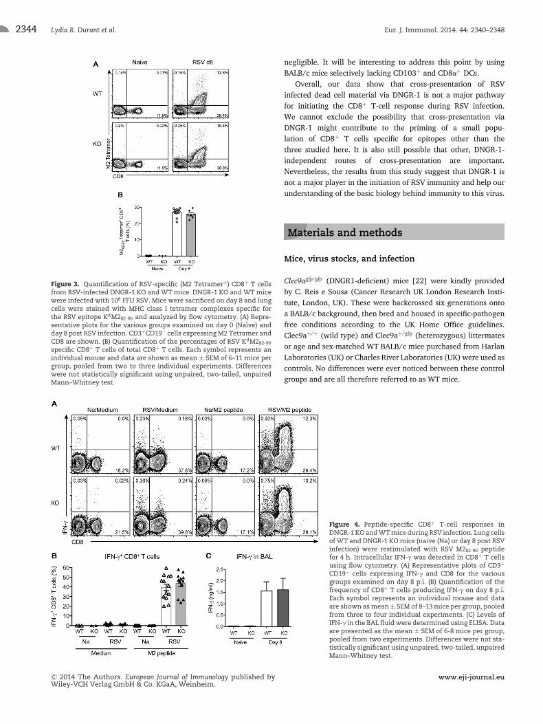
Figure 3. Quantification of RSV-specific (M2 Tetramer+) CD8+ T cellsfrom RSV-infected DNGR-1 KO and WT mice. DNGR-1 KO and WT micewere infected with 106 FFU RSV. Mice were sacrificed on day 8 and lungcells were stained with MHC class I tetramer complexes specific forthe RSV epitope KdM282-90 and analyzed by flow cytometry. (A) Repre-sentative plots for the various groups examined on day 0 (Naıve) andday 8 post RSV infection. CD3+CD19− cells expressing M2 Tetramer andCD8 are shown. (B) Quantification of the percentages of RSV KdM282-90
specific CD8+ T cells of total CD8+ T cells. Each symbol represents anindividual mouse and data are shown as mean ± SEM of 6–11 mice pergroup, pooled from two to three individual experiments. Differenceswere not statistically significant using unpaired, two-tailed, unpairedMann–Whitney test.
negligible. It will be interesting to address this point by usingBALB/c mice selectively lacking CD103+ and CD8α+ DCs.
Overall, our data show that cross-presentation of RSVinfected dead cell material via DNGR-1 is not a major pathwayfor initiating the CD8+ T-cell response during RSV infection.We cannot exclude the possibility that cross-presentation viaDNGR-1 might contribute to the priming of a small popu-lation of CD8+ T cells specific for epitopes other than thethree studied here. It is also still possible that other, DNGR-1-independent routes of cross-presentation are important.Nevertheless, the results from this study suggest that DNGR-1 isnot a major player in the initiation of RSV immunity and help ourunderstanding of the basic biology behind immunity to this virus.
Materials and methods
Mice, virus stocks, and infection
Clec9agfp/gfp (DNGR1-deficient) mice [22] were kindly providedby C. Reis e Sousa (Cancer Research UK London Research Insti-tute, London, UK). These were backcrossed six generations ontoa BALB/c background, then bred and housed in specific-pathogenfree conditions according to the UK Home Office guidelines.Clec9a+/+ (wild type) and Clec9a+/gfp (heterozygous) littermatesor age and sex-matched WT BALB/c mice purchased from HarlanLaboratories (UK) or Charles River Laboratories (UK) were used ascontrols. No differences were ever noticed between these controlgroups and are all therefore referred to as WT mice.
Figure 4. Peptide-specific CD8+ T-cell responses inDNGR-1 KO and WT mice during RSV infection. Lung cellsof WT and DNGR-1 KO mice (naive (Na) or day 8 post RSVinfection) were restimulated with RSV M282-90 peptidefor 4 h. Intracellular IFN-γ was detected in CD8+ T cellsusing flow cytometry. (A) Representative plots of CD3+
CD19− cells expressing IFN-γ and CD8 for the variousgroups examined on day 8 p.i. (B) Quantification of thefrequency of CD8+ T cells producing IFN-γ on day 8 p.i.Each symbol represents an individual mouse and dataare shown as mean ± SEM of 8–13 mice per group, pooledfrom three to four individual experiments. (C) Levels ofIFN-γ in the BAL fluid were determined using ELISA. Dataare presented as the mean ± SEM of 6-8 mice per group,pooled from two experiments. Differences were not sta-tistically significant using unpaired, two-tailed, unpairedMann–Whitney test.
C© 2014 The Authors. European Journal of Immunology published byWiley-VCH Verlag GmbH & Co. KGaA, Weinheim.
www.eji-journal.eu

Eur. J. Immunol. 2014. 44: 2340–2348 Immunity to infection 2345
Figure 5. Production of IFN-γ by RSV-specific CD8+ T cells in responseto RSV peptides M282-90, F85–93, F92-106, and RSV. Lung cells from DNGR-1KO and WT mice on day 8 post RSV infection were restimulated with(A) the immunodominant M282-90, the control (Ctrl) irrelevant M187–195
peptide and RSV (MOI = 1) for 3 days. Supernatants were quantified forlevels of IFN-γ protein by ELISA. Each symbol represents an individualmouse and data are shown as mean ± SEM of 9–21 mice per group,pooled from two to five individual experiments. (B) Lung cells were re-stimulated with the subdominant F85–93 and F92–106 peptides (500 ng/mL)for 3 days. Supernatants were quantified for levels of IFN-γ proteinby ELISA. Each symbol represents an individual mouse and data areshown as mean ± SEM of 6–8 mice per group, pooled from two indi-vidual experiments. Differences were not statistically significant usingunpaired, two-tailed, unpaired Mann–Whitney test.
Plaque-purified human RSV (A2 strain from ATCC) was grownto a high titer in HEp-2 cells. Seven- to ten-week old Clec9agfp/gfp,Clec9a+/gfp, Clec9a+/+, and WT BALB/c mice were lightly anes-thesized with isofluorane and challenged intranasally (i.n.) witha dose of 1 × 10⁶ FFU of RSV on day 0.
To prepare UV-killed RSV-infected HEp-2 cells, 80% confluentHEp2 cells were infected with RSV at an MOI of 0.5 in serum-free media (Dulbecco’s Modified Eagle medium (DMEM) supple-mented with 2 mM L-glutamine). After 2 h incubation at 37°C,serum-containing media (DMEM supplemented with 10% fetalbovine serum and 2 mM L-glutamine) was added. Infected cellswere harvested after 24 h, washed and resuspended in sterile PBSand UV-killed using 750 mJ/cm2 treatment twice in a CX-2000UV cross-linker (UVP). Cells were further rested for 2 h at roomtemperature (RT) and mice were then inoculated i.n. with 2 × 10⁶
UV-killed RSV-infected HEp-2 cells.
Cell isolation and processing
Mice were culled using a fatal dose of pentobarbital injectedintraperitonially (i.p.) according to UK home office guidelines.
BAL was collected by flushing the lungs three times with 1 mLof PBS supplemented with 0.5 mM EDTA (Life Technologies).Lung lobes were collected and digested with Collagenase D(1 mg/mL, Roche) and DNase I (30 μg/mL; Sigma-Aldrich) usinga gentleMACs cell dissociator (Miltenyi Biotech) according to themanufacturer’s protocol and incubated at 37°C for 30 min. Redblood cells were lysed by treating lung cells with ACK lysing bufferand lung cells were passed through 100 μM cell strainers to createsingle-cell suspensions. Total cell counts were determined usinghemocytometer slides and dead cells were excluded by Trypanblue staining (Sigma-Aldrich). To determine the cellular com-position in the BAL, cells were transferred onto a microscopeslide (Thermo Scientific, UK) using a Shandon Cytospin 3 Cen-trifuge and slides were stained with hematoxylin and eosin (H&E;Reagena, Gamidor, UK). Cells were categorized as macrophages,lymphocytes, neutrophils, and eosinophils based on their morphol-ogy and size under a light microscope.
Flow cytometry
For flow cytometry analysis, dead cells were discriminated usingLIVE/DEAD Fixable Aqua Dead Cell Stain (Molecular Probes, LifeTechnologies) according to the manufacturer’s instruction. Cellswere then incubated in FACS Staining Buffer (PBS containing 1%BSA and 5 mM EDTA) with antibody recognizing Fcγ II/III recep-tor (CD16/CD32; BD Biosciences). Surface receptor staining wasperformed using the following antibodies, all purchased from BDBioscience unless otherwise stated: PE-Cy7- conjugated anti-CD3(clone 145–2C11); allophycocyanin-H7 or allophycocyanin-Cy7conjugated anti-CD4 (clone GK1.5); AlexaFluor 700 conjugatedanti-CD8a (clone 53–6.7); and FITC conjugated anti-CD19 (clone1D3, eBioscience) in FACS Staining Buffer.
Biotinylated RSV M282-90 peptide monomers (H-2Kd/SYIGSINNI) were obtained from the NIH Tetramer Core Facility(Emory University Atlanta, GA, USA) and tetramerization was per-formed in-house using allophycocyanin-conjugated Streptavidin(Molecular Probes) using a protocol provided by the NIH TetramerCore Facility. M2 tetramer staining was performed on lung andBAL cells following Fc block steps and prior to surface receptorstaining. Cells were incubated with APC-conjugated M2 tetramerin FACS staining buffer for 30 min in the dark at RT. Cells werefixed for 30 min in BD Cytofix/Cytoperm fixation buffer at 4°C,washed and then analyzed on the flow cytometer.
For intracellular detection of IFN-γ, lung cells were stimu-lated in 96-well plates with 2 μg/mL of the immunodominantRSV M282-90 peptide (H-2Kd/SYIGSINNI; Synthetic Biomolecules,San Diego, CA, USA) or the irrelevant M187–195 peptide (H-2Db/NAITNAKII; Synthetic Biomolecules, San Diego, CA, USA) iden-tified to be immunodominant in C57BL/6 (H-2Db) mice [5] incomplete DMEM (cDMEM; supplemented with 10% fetal bovineserum, 2 mM L-glutamine, 100 U/mL penicillin and 100 μg/mLstreptomycin). After 1 h incubation, monensin (Golgi Stop, BD)was added. Cells were incubated a further 3 h, washed and incu-bated with live-dead fixable stain, Fcγ II/III receptor and surface
C© 2014 The Authors. European Journal of Immunology published byWiley-VCH Verlag GmbH & Co. KGaA, Weinheim.
www.eji-journal.eu

2346 Lydia R. Durant et al. Eur. J. Immunol. 2014. 44: 2340–2348
Figure 6. DNGR-1 is important for induction of RSV-specific CD8+ T cells responses after intranasal dead cell inoculation. DNGR-1 KO and WT micewere inoculated with 2 × 10⁶ UV-irradiated, RSV-infected HEp-2 cells (UV-RSV HEp-2). Mice were sacrificed on day 8 and airway (BAL) and lung cellswere enumerated and stained with MHC class I tetramer complexes specific for the RSV epitope KdM282-90 and analyzed by flow cytometry. (A) Thetotal number of lung cells was quantified in naıve or UV-RSV HEp-2 inoculated mice. (B) Quantification of the total number of RSV KdM282-90 specificCD8+ T cells in the lung of naıve or UV-RSV HEp-2 inoculated mice. (C) The total number of BAL cells was quantified in naıve or UV-RSV HEp-2inoculated mice. (D) Quantification of the total number of RSV KdM282-90 specific CD8+ T cells in the BAL of naıve or UV-RSV HEp-2 inoculated mice.(E) Levels of IFN-γ in the BAL fluid were determined using ELISA. Each symbol represents an individual mouse and data are shown as mean ± SEMof 6–10 mice pooled from two individual experiments except in (B) were data are presented as the mean ± SEM (n = 4–5 mice per group) and arefrom a single experiment representative of two independent experiments. ***p � 0.001, *p � 0.05, unpaired, two-tailed, unpaired Mann–Whitneytest.
receptor antibodies as described above. Intracellular cytokinestaining was performed using the Cytofix/Cytoperm kit (BD)according to manufacturer’s instructions. Briefly, cells were fixedin BD Cytofix/Cytoperm buffer for 30 min at 4°C, washed withFACS staining buffer and then incubated with Fcγ II/III recep-tor (CD16/CD32) antibody in BD Perm Wash Buffer for 15 min.Cells were then incubated with PE-conjugated anti-IFN-γ antibody(clone XMG1.2, BD biosciences) in Perm Wash buffer for 1 h at4°C, washed and transferred to FACS staining buffer for analy-sis. Cells were acquired on the LSR Fortessa flow cytometer (BD)and data were analyzed using Flowjo Software (Version 9.6.4).Cells were gated for nondebris, live cells, and singlets and thenanalyzed for indicated markers.
Gene Expression
Total RNA was extracted from homogenized lung tissue usingTRIzol RNA Isolation reagent in combination with a chloroformseparation, followed by washes with isopropanol and 75% ethanolas per the protocol provided (TRIzol Reagent, Ambion, Life Tech-nologies, UK). RNA purity and concentration was determinedusing the Nanodrop 1000 (Thermo Scientific) and 1–2 μg of totalRNA was reverse-transcribed to cDNA using the high capacity
RNA-to-DNA kit according to the manufacturer’s instructions(Invitrogen, Life Technologies). Real-time quantitative PCR wasperformed to determine mRNA levels of RSV L gene using primers(Invitrogen) and probes (Eurofins) [36]. Results were normalizedto Gapdh (Applied Biosystems) and the exact copy numbers of Lgene was calculated using an internal plasmid standard to give anabsolute quantification. Analysis was performed using the Quanti-tect Probe PCR Master Mix (Qiagen) and the 7500 Fast Real-timePCR System (Applied Biosystems).
Chemokine and cytokine protein detection
To quantify RSV-specific IFN-γ levels from infected lungcells, 4 × 10⁵ cells/well in 96-well round-bottomedplates were stimulated with either RSV (MOI of 1) orRSV M282-90 (H-2Kd/SYIGSINNI), F85–93 (H-2Kd/KYKNAVTEL),F92–106 (H-2Kd/ELQLLMQSTPPTNNR) or control M187–195
(H-2Db/NAITNAKII) peptide in indicated concentrations (Syn-thetic Biomolecules, San Diego, CA, USA) in cDMEM for 72 h at37°C. Supernatants were assayed using an IFN-γ ELISA kit (R&DSystems) according to the manufacturer’s instructions.
Levels of IL-6, CXCL10 (IP-10) and IFN-γ protein weremeasured in the BAL fluid using ELISA kits and following the
C© 2014 The Authors. European Journal of Immunology published byWiley-VCH Verlag GmbH & Co. KGaA, Weinheim.
www.eji-journal.eu

Eur. J. Immunol. 2014. 44: 2340–2348 Immunity to infection 2347
manufacturer’s instructions (all from R&D Systems). IFN-α levelsin the BAL were measured by ELISA as previously described [37].Data was acquired on a SpectraMax Plus plate reader (MolecularDevices) and analyzed using SoftMax software (version 5.2).
Statistical analysis
Results are presented as mean ± SEM. Statistical significancewas determined using a two-tailed, unpaired, nonparametricMann–Whitney t-test (*p � 0.05, **p � 0.01, ***p � 0.001).Values of p < 0.05 were considered significant (Prism software;Graph-Pad Software Inc.).
Acknowledgments: We thank Neil Rogers and Caetano Reis eSousa for supplying the DNGR-1 KO mice and Caetano Reis eSousa for critically reading the manuscript. We also thank theviral immunology group and especially Fiona Culley for criti-cally reading the manuscript and Peter Openshaw for help andadvice. C.J. is supported by a Career Development Award from theMedical Research Council (Grant G0800311). M. G., S. M., and F.K. are supported by PhD Fellowships from the National Heart andLung Institute Foundation (registered charity number 1048073).This work was funded by a Director’s Award to CJ from theWellcome Trust Centre for Respiratory Infection (awarded to P.Openshaw; 083567/Z/07/Z). We acknowledge the NIH TetramerCore Facility for provision of biotinylated RSV M282-90 peptidemonomers (H-2Kd/SYIGSINNI). We also thank staff of the St.Mary’s flow cytometry facility and the animal facility for theirassistance.
Conflict of interest: The authors declare no commercial or finan-cial conflict of interest.
References
1 Borchers, A. T., Chang, C., Gershwin, M. E. and Gershwin, L. J., Respira-
tory syncytial virus—a comprehensive review. Clinic. Rev. Allerg. Immunol.
2013. 45: 331–379.
2 Blanken, M. O., Rovers, M. M., Molenaar, J. M., Winkler-Seinstra, P. L.,
Meijer, A., Kimpen, J. L. L. and Bont, L., Respiratory Syncytial Virus and
Recurrent Wheeze in Healthy Preterm Infants. N. Engl. J. Med. 2013. 368:
1791–1799.
3 Openshaw, P. J. and Chiu, C., Protective and dysregulated T cell immunity
in RSV infection. Curr. Opin. Virol. 2013. 3: 468–474.
4 Tregoning, J. S., Yamaguchi, Y., Harker, J., Wang, B. and Openshaw, P. J.
M., The role of T cells in the enhancement of respiratory syncytial virus
infection severity during adult reinfection of neonatally sensitized mice.
J. Virol. 2008. 82: 4115–4124.
5 Rutigliano, J. A., Rock, M. T., Johnson, A. K., Crowe, J. E. and Graham, B.
S., Identification of an H-2D(b)-restricted CD8 +cytotoxic T lymphocyte
epitope in the matrix protein of respiratory syncytial virus. Virology 2005.
337: 335–343.
6 Blum, J. S., Wearsch, P. A. and Cresswell, P., Pathways of antigen pro-
cessing. Annu. Rev. Immunol. 2013. 31: 443–473.
7 Lukens, M. V., Kruijsen, D., Coenjaerts, F. E. J., Kimpen, J. L. L. and van
Bleek, G. M., Respiratory syncytial virus-induced activation and migra-
tion of respiratory dendritic cells and subsequent antigen presentation
in the lung-draining lymph node. J. Virol. 2009. 83: 7235–7243.
8 Ruckwardt, T. J., Malloy, A. M. W., Morabito, K. M. and Graham, B. S.,
Quantitative and qualitative deficits in neonatal lung-migratory dendritic
cells impact the generation of the CD8+ T cell response. PLoS Pathog. 2014.
10: e1003934.
9 Demoor, T., Petersen, B. C., Morris, S., Mukherjee, S., Ptaschinski, C., De
Almeida Nagata, D. E., Kawai, T. et al., IPS-1 Signaling has a nonredun-
dant role in mediating antiviral responses and the clearance of respira-
tory syncytial virus. J. Immunol. 2012. 189: 5942–5945.
10 Neyt, K. and Lambrecht, B. N., The role of lung dendritic cell subsets in
immunity to respiratory viruses. Immunol. Rev. 2013. 255: 57–67.
11 Braciale, T. J., Sun, J. and Kim, T. S., Regulating the adaptive immune
response to respiratory virus infection. Nat. Rev. Immunol. 2012. 2: 295–
305.
12 Shortman, K. and Heath, W. R., The CD8+ dendritic cell subset. Immunol.
Rev. 2010. 234: 18–31.
13 Joffre, O. P., Segura, E., Savina, A. and Amigorena, S., Cross-presentation
by dendritic cells. Nat. Rev. Immunol. 2012. 12: 557–569.
14 Desch, A. N., Randolph, G. J., Murphy, K., Gautier, E. L., Kedl, R. M.,
Lahoud, M. H., Caminschi, I. et al., CD103+ pulmonary dendritic cells
preferentially acquire and present apoptotic cell-associated antigen. J.
Exp. Med. 2011. 208: 1789–1797.
15 Sancho, D., Mourao-Sa, D., Joffre, O. P., Schulz, O., Rogers, N. C., Pen-
nington, D. J., Carlyle, J. R. et al., Tumor therapy in mice via antigen
targeting to a novel, DC-restricted C-type lectin. J. Clin. Invest. 2008. 118:
2098–2110.
16 Huysamen, C., Willment, J. A., Dennehy, K. M. and Brown, G. D., CLEC9A
is a novel activation C-type lectin-like receptor expressed on BDCA3+dendritic cells and a subset of monocytes. J. Biol. Chem. 2008. 283: 16693–
16701.
17 Caminschi, I., Proietto, A. I., Ahmet, F., Kitsoulis, S., Shin Teh, J., Lo, J. C.
Y., Rizzitelli, A. et al., The dendritic cell subtype-restricted C-type lectin
Clec9A is a target for vaccine enhancement. Blood 2008. 112: 3264–3273.
18 Poulin, L. F., Salio, M., Griessinger, E., Anjos-Afonso, F., Craciun, L., Chen,
J.-L., Keller, A. M. et al., Characterization of human DNGR-1+ BDCA3+leukocytes as putative equivalents of mouse CD8alpha+ dendritic cells.
J. Exp. Med. 2010. 207: 1261–1271.
19 Poulin, L. F., Reyal, Y., Uronen-Hansson, H., Schraml, B., Sancho, D.,
Murphy, K. M., Hakansson, U. K. et al., DNGR-1 is a specific and uni-
versal marker of mouse and human Batf3-dependent dendritic cells in
lymphoid and non-lymphoid tissues. Blood 2012. 119: 6052–6062.
20 Ahrens, S., Zelenay, S., Sancho, D., Hanc, P., Kjær, S., Feest, C., Fletcher,
G. et al., F-Actin is an evolutionarily conserved damage-associated
molecular pattern recognized by dngr-1, a receptor for dead cells. Immu-
nity 2012. 36: 635–645.
21 Zhang, J.-G., Czabotar, P. E., Policheni, A. N., Caminschi, I., Wan, S. S.,
Kitsoulis, S., Tullett, K. M. et al., The dendritic cell receptor Clec9A binds
damaged cells via exposed actin filaments. Immunity 2012. 36: 646–657.
22 Sancho, D., Joffre, O. P., Keller, A. M., Rogers, N. C., Martınez, D., Hernanz-
Falcon, P., Rosewell, I. et al., Identification of a dendritic cell recep-
tor that couples sensing of necrosis to immunity. Nature 2009. 458:
899–903.
C© 2014 The Authors. European Journal of Immunology published byWiley-VCH Verlag GmbH & Co. KGaA, Weinheim.
www.eji-journal.eu

2348 Lydia R. Durant et al. Eur. J. Immunol. 2014. 44: 2340–2348
23 Zelenay, S., Keller, A. M., Whitney, P. G., Schraml, B. U., Deddouche, S.,
Rogers, N. C., Schulz, O. et al., The dendritic cell receptor DNGR-1 controls
endocytic handling of necrotic cell antigens to favor cross-priming of
CTLs in virus-infected mice. J. Clin. Invest. 2012. 122: 1615–1627.
24 Iborra, S., Izquierdo, H. M., Martınez-Lopez, M., Blanco-Menendez, N.,
Reis e Sousa, C. and Sancho, D., The DC receptor DNGR-1 mediates cross-
priming of CTLs during vaccinia virus infection in mice. J. Clin. Invest.
2012. 122: 1628–1643.
25 Johnson, J. E., Gonzales, R. A., Olson, S. J., Wright, P. F. and Graham,
B. S., The histopathology of fatal untreated human respiratory syncytial
virus infection. Mod. Pathol. 2007. 20: 108–119.
26 Openshaw, P. J. M. and Tregoning, J. S., Immune responses and disease
enhancement during respiratory syncytial virus infection. Clin. Microbiol.
Rev. 2005. 18: 541–555.
27 Loebbermann, J., Thornton, H., Durant, L., Sparwasser, T., Webster, K.
E., Sprent, J., Culley, F. J. et al., Regulatory T cells expressing granzyme
B play a critical role in controlling lung inflammation during acute viral
infection. Mucosal. immunology 2012. 5: 161–172.
28 Loebbermann, J., Schnoeller, C., Thornton, H., Durant, L., Sweeney, N. P.,
Schuijs, M., O’Garra, A. et al., IL-10 regulates viral lung immunopathology
during acute respiratory syncytial virus infection in mice. PLoS ONE. 2012.
7: e32371.
29 Chang, J. and Braciale, T. J., Respiratory syncytial virus infection sup-
presses lung CD8+ T-cell effector activity and peripheral CD8+ T-cell
memory in the respiratory tract. Nat. Med. 2002. 8: 54–60.
30 Kulkarni, A. B., Collins, P. L., Bacik, I., Yewdell, J. W., Bennink, J. R.,
Crowe, J. E. and Murphy, B. R., Cytotoxic T cells specific for a single
peptide on the M2 protein of respiratory syncytial virus are the sole
mediators of resistance induced by immunization with M2 encoded by a
recombinant vaccinia virus. J. Virol. 1995. 69: 1261–1264.
31 Chang, J., Srikiatkhachorn, A. and Braciale, T. J., Visualization and char-
acterization of respiratory syncytial virus F-specific CD8(+) T cells during
experimental virus infection. J Immunol. 2001. 167: 4254–4260.
32 Jiang, S., Borthwick, N. J., Morrison, P., Gao, G. F. and Steward, M. W.,
Virus-specific CTL responses induced by an H-2K(d)-restricted, motif-
negative 15-mer peptide from the fusion protein of respiratory syncytial
virus. J. Gen. Virol. 2002. 83: 429–438.
33 Zelenay, S. and Sousa, C. R. E. Adaptive immunity after cell death. Trends
Immunol. 2013. 34: 329–335.
34 Helft, J., Manicassamy, B., Guermonprez, P., Hashimoto, D., Silvin, A.,
Agudo, J., Brown, B. D. et al., Cross-presenting CD103+ dendritic cells
are protected from influenza virus infection. J. Clin. Invest. 2012. 122:
4037–4047.
35 Desch, A. N., Henson, P. M. and Jakubzick, C. V., Pulmonary den-
dritic cell development and antigen acquisition. Immunol. Res. 2013. 55:
178–186.
36 Culley, F. J., Pollott, J. and Openshaw, P. J. M. Age at first viral infection
determines the pattern of T cell-mediated disease during reinfection in
adulthood. J. Exp. Med. 2002. 196: 1381–1386.
37 Asselin-Paturel, C., Boonstra, A., Dalod, M., Durand, I., Yessaad, N.,
Dezutter-Dambuyant, C., Vicari, A. et al., Mouse type I IFN-producing
cells are immature APCs with plasmacytoid morphology. Nat. Immunol.
2001. 2: 1144–1150.
Abbreviations: BAL: bronchoalveolar lavage · DNGR-1: DC NK Lectin
Group Receptor 1 · FFU: focus forming units · HEp-2: human epithe-
lial cell line 2 · HSV-1: herpes simplex virus 1 · p.i.: post infection
Full correspondence: Dr. Cecilia Johansson, Respiratory Infections,NHLI, Imperial College London, St. Mary’s Campus, Norfolk Place,London W2 1PG, U.K.Fax: +44-207-262-8913e-mail: [email protected]
Received: 8/1/2014Revised: 25/3/2014Accepted: 23/4/2014Accepted article online: 28/4/2014
C© 2014 The Authors. European Journal of Immunology published byWiley-VCH Verlag GmbH & Co. KGaA, Weinheim.
www.eji-journal.eu