dopamine agonist inhibits vascular endothelial growth
TRANSCRIPT

SHORT COMMUNICATION Open Access
Dopamine agonist inhibits vascularendothelial growth factor proteinproduction and secretion in granulosa cellsHortensia Ferrero1,2*, Carmen M. García-Pascual1,2, Nuria Pellicer1, Carlos Simón1,2, Antonio Pellicer1,3
and Raúl Gómez2
Abstract
Background: Dopamine receptor 2 agonists (D2-ags) inhibit vascular endothelial growth factor (VEGF) secretion inluteinized granulosa cells (LGCs) both in vitro and in vivo. However, the mechanism of D2 regulation of the VEGF/VEGF Receptor 2 (VEGFR-2) pathway remains to be elucidated. We sought to determine the effects of D2 signalingon VEGF transcription and translation in LGCs, with the expectation of identifying potential D2-ag-based therapiesfor ovarian hyperstimulation syndrome (OHSS).
Findings: LGCs from egg donors were cultured with chorionic gonadotropin (hCG) in the presence of Actinomycin-D(ActD) or Brefeldin-A (BFA) to evaluate the effects of a D2-ag, cabergoline (Cb2), on VEGF secretion. The contribution ofthe conventional Gi/Go, Gz and AKT/β-Arrestin pathways in the VEGF regulation was assessed by adding pertussis toxin(PTX), phorbol 12-myristate 13-acetate (PMA), or wortmannin (WT). While Cb2 inhibited VEGF secretion by interferingwith VEGF peptide translation and secretion, inhibition of conventional D2 transduction pathways did not reverse Cb2-mediated inhibition of VEGF secretion.
Conclusions: The effects of D2-ag on VEGF translation and secretion are mediated by D2 signaling pathways that haveyet to be described. We found that D2-ag inhibits VEGF secretion at the post-transcriptional level, suggesting that D2-agtreatment should be combined with therapies that inhibit VEGF transcription, such as the employment of LH or GnRHfor triggering ovulation, to improve the efficacy of OHSS prevention.
Keywords: OHSS, VEGF, Granulosa cells, Dopamine receptor 2, Dopamine receptor-2 agonist
FindingsOvarian hyperstimulation syndrome (OHSS) is an iatro-genic complication of ovarian stimulation associated withthe use of human chorionic gonadotropin (hCG), is char-acterized by an increase in vascular permeability (VP) [1].Vascular endothelial growth factor (VEGF) is an import-ant component in the development of OHSS [2, 3]. Be-sides, it has been demonstrated that hCG administrationincreases VEGF mRNA expression in luteinized granulosacells (LCGs) [4, 5]. Several studies have attempted to avoidan increase in VEGF while employing GnRH agonists toinduce ovulation [6, 7]; however, this strategy does not
totally avoid OHSS onset [8], leaving a need for treatmentsthat block VEGF/VEGF receptor 2 (VEGFR2) signalingcompletely.Several in vivo [9, 10] and in vitro studies [11] have
suggested a role for dopamine in the regulation of theVEGF/VEGFR2 pathway. Interestingly, high levels ofdopamine are present in follicular fluid and human ovar-ian biopsies [12, 13], and dopamine receptors have beenfound on human granulosa cells (GCs) [14]. Previousstudies suggest a role for dopamine receptor agonist(D2-ag) in preventing VP increases [15, 16] by inhibitingVEGF secretion [17, 18]. However, D2-ag has previouslybeen shown to block the onset of early-stage OHSS in50 % of women at risk for developing the condition, butit was not effective in preventing the late onset form[19]. Understanding the molecular mechanisms involved
* Correspondence: [email protected]ón IVI, C/ Catedrático Agustín Escardino, n°9, Paterna, Valencia46980, Spain2Instituto Universitario IVI/ INCLIVA, Valencia 46015, SpainFull list of author information is available at the end of the article
© 2015 Ferrero et al. Open Access This article is distributed under the terms of the Creative Commons Attribution 4.0International License (http://creativecommons.org/licenses/by/4.0/), which permits unrestricted use, distribution, andreproduction in any medium, provided you give appropriate credit to the original author(s) and the source, provide a link tothe Creative Commons license, and indicate if changes were made. The Creative Commons Public Domain Dedication waiver(http://creativecommons.org/publicdomain/zero/1.0/) applies to the data made available in this article, unless otherwise stated.
Ferrero et al. Reproductive Biology and Endocrinology (2015) 13:104 DOI 10.1186/s12958-015-0102-4
in D2 regulation of VEGF is critical to elucidating therole of D2-ag in OHSS prevention. Therefore, the goalof this study was to determine the molecular mechanismthrough which D2-ag inhibits VEGF secretion and toestablish which D2 signaling pathways described in D2-expressing cells (Gi/Go, Gz and AKT/β-Arrestin) [20]are involved in the regulation of VEGF secretion.LGCs were obtained from 24 egg donors [aged 25–30
years, oocytes retrieved = 10–15, estrogen (E2) <2000 pg/mL, body mass index (BMI) < 30]. Cells wereisolated by filtering [21], then washed and cultured asdescribed below in the different studies. Written in-formed consent was provided by all participants, andthe study protocol was approved by ethics committeeof IVI Valencia.
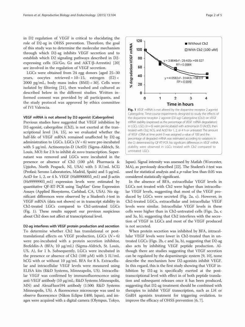
VEGF mRNA is not altered by D2-agonist (Cabergoline)Previous studies have suggested that VEGF inhibition byD2-agonist, cabergoline (Cb2), is not exerted at the tran-scriptional level [14, 15], so we evaluated whether thehalf-life of VEGF mRNA remained unaffected by D2-agadministration to LGCs. LGCs (N = 6) were pre-incubatedwith 5 μg/mL Actinomycin-D (ActD) (Sigma-Aldrich, St.Louis, MO) for 2 h to inhibit de novo transcription. Super-natant was removed and LGCs were incubated in thepresence or absence of Cb2 (100 μM; Pharmacia &Upjohn, North Peapack, NJ, USA) with 5 IU/mL hCG(Profasi; Serono Laboratories, Madrid, Spain) and 5 μg/mLActD for 1, 2, or 4 h. VEGF (Hs00900055_m1) and β-actin(Hs99999903_m1) expression levels were estimated byquantitative QF-RT-PCR using TaqMan® Gene ExpressionAssays (Applied Biosystems, Carlsbad, CA, USA). No sig-nificant differences were observed by a Student’s t-test inVEGF mRNA (data not shown) or in transcript stability inCb2-treated LGCs compared to Cb2-untreated LGCs(Fig. 1). These results support our previous suspiciousabout Cb2 does not affect at transcriptional level.
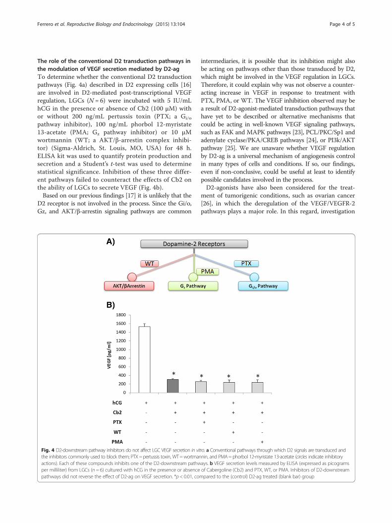
D2-ag interferes with VEGF protein production and secretionTo determine whether Cb2 has translational or post-translational effects on VEGF production, LGCs (N = 6)were pre-incubated with a protein secretion inhibitor,Brefeldin-A (BFA; 10 μg/mL) (Sigma-Aldrich, St. Louis,US, A), for 1 h. Subsequently, LGCs were incubated inthe presence or absence of Cb2 (100 μM) with 5 IU/mLhCG with or without 10 μg/mL BFA for 8 h. Extracellu-lar and intracellular VEGF levels were measured usingELISA kits (R&D Systems, Minneapolis, US). Intracellu-lar VEGF was confirmed by immunofluorescence usinganti-VEGF antibody (10 μg/mL; R&D Systems Minneapolis,MN) and AlexaFluor594 antibody (1:500; R&D SystemsMinneapolis, US). A fluorescence microscope was used toobserve fluorescence (Nikon Eclipse E400, Japan), and im-ages were acquired with a digital camera (Olympus, Tokyo,
Japan). Signal intensity was assessed by Matlab (Worcester,MA), as previously described [22]. The Student’s t-test wasused for statistical analysis and a p-value less than 0.05 wasconsidered statistically significant.In the absence of BFA, extracellular VEGF levels in
LGCs not treated with Cb2 were higher than intracellu-lar VEGF levels, suggesting that most of the VEGF pro-duced by LGCs were secreted (Fig. 2a, c). However, inCb2-treated LGCs, extracellular and intracellular VEGFlevels were similar. Intracellular VEGF levels in thesecells were higher than in Cb2-untreated cells (Figs. 2a, cand 3a, b), suggesting that Cb2 interferes with the secre-tion of VEGF in LGCs and most of the VEGF producedis not secreted.When protein secretion was inhibited by BFA, intracel-
lular VEGF levels were lower in Cb2-treated than in un-treated LGCs (Figs. 2b, c and 3a, b), suggesting that D2-agalso acts by inhibiting VEGF peptide production. Al-though there are studies suggesting that VEGF secretioncan be regulated by the dopaminergic system [9, 10], nonedescribe the mechanism how D2-agonists inhibit VEGF.In this regard, this is the first study showing that VEGF in-hibition by D2-ag is specifically exerted at the post-transcriptional level with effect in of both peptide transla-tion and subsequent releases once it has been produced,suggesting that D2-ag treatment should be combined withtherapies to inhibit VEGF transcription, such as LH orGnRH agonists treatment for triggering ovulation, toimprove the efficacy of OHSS prevention [6, 7].
Fig. 1 VEGF mRNA is not altered by the dopamine receptor 2 agonistCabergoline. Time-course experiments designed to study the effects ofthe dopamine receptor 2 agonist (D2-ag) Cabergoline (Cb2) on VEGFmRNA stability (expressed as the percentage of VEGF mRNA degradation)in LGCs. LGCs (n=6) were pre-incubated with actinomycin D (ActD), thentreated with Cb2, hCG, and ActD for 1, 2, or 4 h or untreated. The amountof VEGF cDNA at time point 0 was assigned a value of 100 and thepercentage of degraded mRNA was estimated according to variations inthe Ct determined by QF-RT-PCR. No significant differences in VEGF mRNAstability were observed in LGCs treated with Cb2 compared tountreated LGCs
Ferrero et al. Reproductive Biology and Endocrinology (2015) 13:104 Page 2 of 5
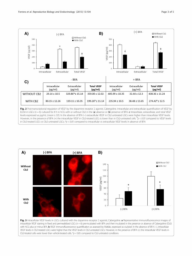
Fig. 2 Post-transcriptional regulation of VEGF by the dopamine receptor 2 agonist, Cabergoline. Intracellular and extracellular quantification of VEGF byELISA in LGCs (n = 6) cultured for 8 h in hCG with or without Cb2 in the (a) absence or (b) presence of BFA. c Intracellular, extracellular, and total VEGFlevels expressed as pg/mL (mean ± SD). In the absence of BFA (−) extracellular VEGF in Cb2-untreated LGCs were higher than intracellular VEGF levels.However, in the presence of BFA (+) the intracellular VEGF in Cb2-treated LGCs is lower than in Cb2-untreated cells. ǂp < 0.05 compared to VEGF levelsin Cb2-treated LGCs vs Cb2-untreated LGCs; *p < 0.05 compared to intracellular vs extracellular VEGF levels in absence of BFA
Fig. 3 Intracellular VEGF levels in LGCs cultured with the dopamine receptor 2 agonist, Cabergoline. a Representative immunofluorescence images ofintracellular VEGF staining in fixed and permeabilized LGCs (n=6) pre-incubated with BFA and then incubated in the presence or absence of Cabergoline (Cb2)with hCG plus or minus BFA. b VEGF immunofluorescence quantification as assessed by Matlab, expressed as eu/pixel. In the absence of BFA (−), intracellularVEGF levels in Cb2-treated LGCs were higher than the VEGF levels in Cb2-untreated LGCs. However, in the presence of BFA (+) the intracellular VEGF levels inCb2-treated cells were lower than vehicle-treated cells. *p<0.05 compared to Cb2-untreated conditions
Ferrero et al. Reproductive Biology and Endocrinology (2015) 13:104 Page 3 of 5
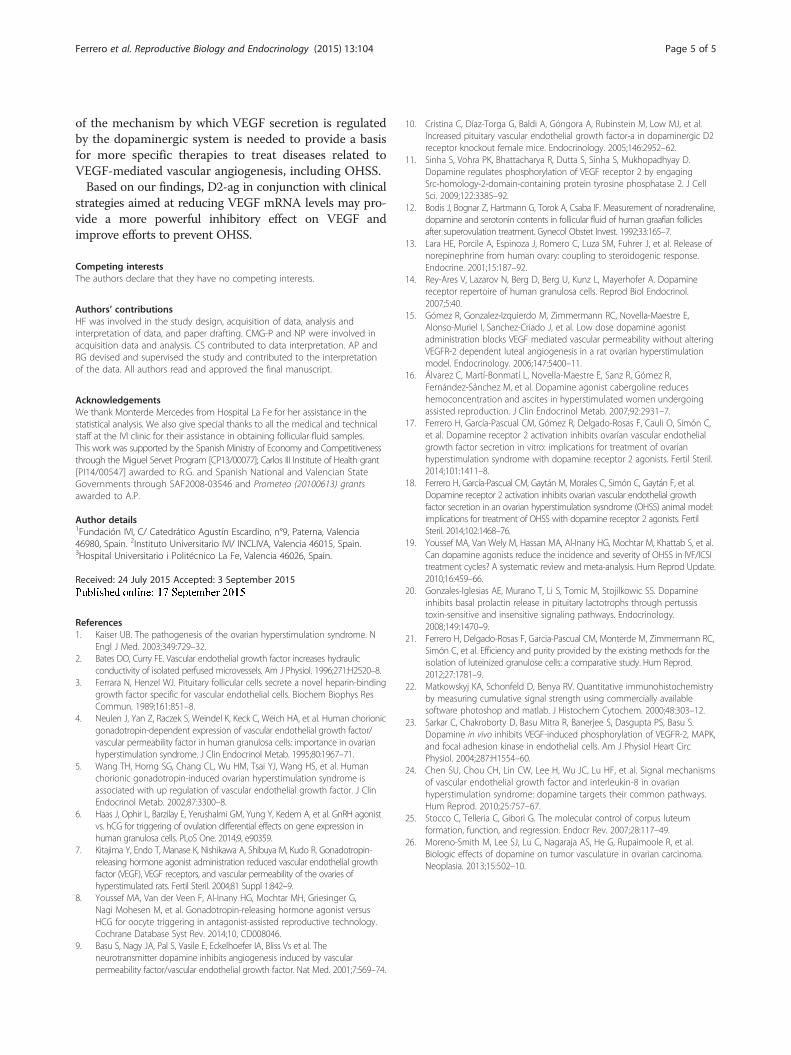
The role of the conventional D2 transduction pathways inthe modulation of VEGF secretion mediated by D2-agTo determine whether the conventional D2 transductionpathways (Fig. 4a) described in D2 expressing cells [16]are involved in D2-mediated post-transcriptional VEGFregulation, LGCs (N = 6) were incubated with 5 IU/mLhCG in the presence or absence of Cb2 (100 μM) withor without 200 ng/mL pertussis toxin (PTX; a Gi/o
pathway inhibitor), 100 ng/mL phorbol 12-myristate13-acetate (PMA; Gz pathway inhibitor) or 10 μMwortmannin (WT; a AKT/β-arrestin complex inhibi-tor) (Sigma-Aldrich, St. Louis, MO, USA) for 48 h.ELISA kit was used to quantify protein production andsecretion and a Student’s t-test was used to determinestatistical significance. Inhibition of these three differ-ent pathways failed to counteract the effects of Cb2 onthe ability of LGCs to secrete VEGF (Fig. 4b).Based on our previous findings [17] it is unlikely that the
D2 receptor is not involved in the process. Since the Gi/o,Gz, and AKT/β-arrestin signaling pathways are common
intermediaries, it is possible that its inhibition might alsobe acting on pathways other than those transduced by D2,which might be involved in the VEGF regulation in LGCs.Therefore, it could explain why was not observe a counter-acting increase in VEGF in response to treatment withPTX, PMA, or WT. The VEGF inhibition observed may bea result of D2-agonist-mediated transduction pathways thathave yet to be described or alternative mechanisms thatcould be acting in well-known VEGF signaling pathways,such as FAK and MAPK pathways [23], PCL/PKC/Sp1 andadenylate cyclase/PKA/CREB pathways [24], or PI3k/AKTpathway [25]. We are unaware whether VEGF regulationby D2-ag is a universal mechanism of angiogenesis controlin many types of cells and conditions. If so, our findings,even if non-conclusive, could be useful at least to identifypossible candidates involved in the process.D2-agonists have also been considered for the treat-
ment of tumorigenic conditions, such as ovarian cancer[26], in which the deregulation of the VEGF/VEGFR-2pathways plays a major role. In this regard, investigation
Fig. 4 D2-downstream pathway inhibitors do not affect LGC VEGF secretion in vitro. a Conventional pathways through which D2 signals are transduced andthe inhibitors commonly used to block them; PTX=pertussis toxin, WT=wortmannin, and PMA=phorbol 12-myristate 13-acetate (circles indicate inhibitoryactions). Each of these compounds inhibits one of the D2-downstream pathways. b VEGF secretion levels measured by ELISA (expressed as picogramsper milliliter) from LGCs (n = 6) cultured with hCG in the presence or absence of Cabergoline (Cb2) and PTX, WT, or PMA. Inhibitors of D2-downstreampathways did not reverse the effect of D2-ag on VEGF secretion. *p < 0.01, compared to the (control) D2-ag treated (blank bar) group
Ferrero et al. Reproductive Biology and Endocrinology (2015) 13:104 Page 4 of 5
of the mechanism by which VEGF secretion is regulatedby the dopaminergic system is needed to provide a basisfor more specific therapies to treat diseases related toVEGF-mediated vascular angiogenesis, including OHSS.Based on our findings, D2-ag in conjunction with clinical
strategies aimed at reducing VEGF mRNA levels may pro-vide a more powerful inhibitory effect on VEGF andimprove efforts to prevent OHSS.
Competing interestsThe authors declare that they have no competing interests.
Authors’ contributionsHF was involved in the study design, acquisition of data, analysis andinterpretation of data, and paper drafting. CMG-P and NP were involved inacquisition data and analysis. CS contributed to data interpretation. AP andRG devised and supervised the study and contributed to the interpretationof the data. All authors read and approved the final manuscript.
AcknowledgementsWe thank Monterde Mercedes from Hospital La Fe for her assistance in thestatistical analysis. We also give special thanks to all the medical and technicalstaff at the IVI clinic for their assistance in obtaining follicular fluid samples.This work was supported by the Spanish Ministry of Economy and Competitivenessthrough the Miguel Servet Program [CP13/00077]; Carlos III Institute of Health grant[PI14/00547] awarded to R.G. and Spanish National and Valencian StateGovernments through SAF2008-03546 and Prometeo (20100613) grantsawarded to A.P.
Author details1Fundación IVI, C/ Catedrático Agustín Escardino, n°9, Paterna, Valencia46980, Spain. 2Instituto Universitario IVI/ INCLIVA, Valencia 46015, Spain.3Hospital Universitario i Politécnico La Fe, Valencia 46026, Spain.
Received: 24 July 2015 Accepted: 3 September 2015
References1. Kaiser UB. The pathogenesis of the ovarian hyperstimulation syndrome. N
Engl J Med. 2003;349:729–32.2. Bates DO, Curry FE. Vascular endothelial growth factor increases hydraulic
conductivity of isolated perfused microvessels. Am J Physiol. 1996;271:H2520–8.3. Ferrara N, Henzel WJ. Pituitary follicular cells secrete a novel heparin-binding
growth factor specific for vascular endothelial cells. Biochem Biophys ResCommun. 1989;161:851–8.
4. Neulen J, Yan Z, Raczek S, Weindel K, Keck C, Weich HA, et al. Human chorionicgonadotropin-dependent expression of vascular endothelial growth factor/vascular permeability factor in human granulosa cells: importance in ovarianhyperstimulation syndrome. J Clin Endocrinol Metab. 1995;80:1967–71.
5. Wang TH, Horng SG, Chang CL, Wu HM, Tsai YJ, Wang HS, et al. Humanchorionic gonadotropin-induced ovarian hyperstimulation syndrome isassociated with up regulation of vascular endothelial growth factor. J ClinEndocrinol Metab. 2002;87:3300–8.
6. Haas J, Ophir L, Barzilay E, Yerushalmi GM, Yung Y, Kedem A, et al. GnRH agonistvs. hCG for triggering of ovulation differential effects on gene expression inhuman granulosa cells. PLoS One. 2014;9, e90359.
7. Kitajima Y, Endo T, Manase K, Nishikawa A, Shibuya M, Kudo R. Gonadotropin-releasing hormone agonist administration reduced vascular endothelial growthfactor (VEGF), VEGF receptors, and vascular permeability of the ovaries ofhyperstimulated rats. Fertil Steril. 2004;81 Suppl 1:842–9.
8. Youssef MA, Van der Veen F, Al-Inany HG, Mochtar MH, Griesinger G,Nagi Mohesen M, et al. Gonadotropin-releasing hormone agonist versusHCG for oocyte triggering in antagonist-assisted reproductive technology.Cochrane Database Syst Rev. 2014;10, CD008046.
9. Basu S, Nagy JA, Pal S, Vasile E, Eckelhoefer IA, Bliss Vs et al. Theneurotransmitter dopamine inhibits angiogenesis induced by vascularpermeability factor/vascular endothelial growth factor. Nat Med. 2001;7:569–74.
10. Cristina C, Díaz-Torga G, Baldi A, Góngora A, Rubinstein M, Low MJ, et al.Increased pituitary vascular endothelial growth factor-a in dopaminergic D2receptor knockout female mice. Endocrinology. 2005;146:2952–62.
11. Sinha S, Vohra PK, Bhattacharya R, Dutta S, Sinha S, Mukhopadhyay D.Dopamine regulates phosphorylation of VEGF receptor 2 by engagingSrc-homology-2-domain-containing protein tyrosine phosphatase 2. J CellSci. 2009;122:3385–92.
12. Bodis J, Bognar Z, Hartmann G, Torok A, Csaba IF. Measurement of noradrenaline,dopamine and serotonin contents in follicular fluid of human graafian folliclesafter superovulation treatment. Gynecol Obstet Invest. 1992;33:165–7.
13. Lara HE, Porcile A, Espinoza J, Romero C, Luza SM, Fuhrer J, et al. Release ofnorepinephrine from human ovary: coupling to steroidogenic response.Endocrine. 2001;15:187–92.
14. Rey-Ares V, Lazarov N, Berg D, Berg U, Kunz L, Mayerhofer A. Dopaminereceptor repertoire of human granulosa cells. Reprod Biol Endocrinol.2007;5:40.
15. Gómez R, Gonzalez-Izquierdo M, Zimmermann RC, Novella-Maestre E,Alonso-Muriel I, Sanchez-Criado J, et al. Low dose dopamine agonistadministration blocks VEGF mediated vascular permeability without alteringVEGFR-2 dependent luteal angiogenesis in a rat ovarian hyperstimulationmodel. Endocrinology. 2006;147:5400–11.
16. Álvarez C, Martí-Bonmatí L, Novella-Maestre E, Sanz R, Gómez R,Fernández-Sánchez M, et al. Dopamine agonist cabergoline reduceshemoconcentration and ascites in hyperstimulated women undergoingassisted reproduction. J Clin Endocrinol Metab. 2007;92:2931–7.
17. Ferrero H, García-Pascual CM, Gómez R, Delgado-Rosas F, Cauli O, Simón C,et al. Dopamine receptor 2 activation inhibits ovarían vascular endothelialgrowth factor secretion in vitro: implications for treatment of ovaríanhyperstimulation syndrome with dopamine receptor 2 agonists. Fertil Steril.2014;101:1411–8.
18. Ferrero H, García-Pascual CM, Gaytán M, Morales C, Simón C, Gaytán F, et al.Dopamine receptor 2 activation inhibits ovarian vascular endothelial growthfactor secretion in an ovarian hyperstimulation sysndrome (OHSS) animal model:implications for treatment of OHSS with dopamine receptor 2 agonists. FertilSteril. 2014;102:1468–76.
19. Youssef MA, Van Wely M, Hassan MA, Al-Inany HG, Mochtar M, Khattab S, et al.Can dopamine agonists reduce the incidence and severity of OHSS in IVF/ICSItreatment cycles? A systematic review and meta-analysis. Hum Reprod Update.2010;16:459–66.
20. Gonzales-Iglesias AE, Murano T, Li S, Tomic M, Stojilkowic SS. Dopamineinhibits basal prolactin release in pituitary lactotrophs through pertussistoxin-sensitive and insensitive signaling pathways. Endocrinology.2008;149:1470–9.
21. Ferrero H, Delgado-Rosas F, Garcia-Pascual CM, Monterde M, Zimmermann RC,Simón C, et al. Efficiency and purity provided by the existing methods for theisolation of luteinized granulose cells: a comparative study. Hum Reprod.2012;27:1781–9.
22. Matkowskyj KA, Schonfeld D, Benya RV. Quantitative immunohistochemistryby measuring cumulative signal strength using commercially availablesoftware photoshop and matlab. J Histochem Cytochem. 2000;48:303–12.
23. Sarkar C, Chakroborty D, Basu Mitra R, Banerjee S, Dasgupta PS, Basu S.Dopamine in vivo inhibits VEGF-induced phosphorylation of VEGFR-2, MAPK,and focal adhesion kinase in endothelial cells. Am J Physiol Heart CircPhysiol. 2004;287:H1554–60.
24. Chen SU, Chou CH, Lin CW, Lee H, Wu JC, Lu HF, et al. Signal mechanismsof vascular endothelial growth factor and interleukin-8 in ovarianhyperstimulation syndrome: dopamine targets their common pathways.Hum Reprod. 2010;25:757–67.
25. Stocco C, Telleria C, Gibori G. The molecular control of corpus luteumformation, function, and regression. Endocr Rev. 2007;28:117–49.
26. Moreno-Smith M, Lee SJ, Lu C, Nagaraja AS, He G, Rupaimoole R, et al.Biologic effects of dopamine on tumor vasculature in ovarian carcinoma.Neoplasia. 2013;15:502–10.
Ferrero et al. Reproductive Biology and Endocrinology (2015) 13:104 Page 5 of 5