dorothy crowfoot hodgkin - nobel lecture
TRANSCRIPT

D OR O T H Y C R O W F O O T H O D G K I N
The X-ray analysis of complicated molecules
Nobel Lecture, December 11, 1964
I first met the subject of X-ray diffraction of crystals in the pages of the book
W. H. Bragg wrote for school children in 1925, 'Concerning the Nature of
Things' . In this he wrote: "Broadly speaking, the discovery of X-rays has
increased the keenness of our vision over ten thousand times and we can now
'see' the individual atoms and mo1ecules." I also first learnt at the same time
about biochemistry which provided me with the molecules it seemed most
desirable to 'see' . At Oxford, seriously studying chemistry, with Robinson
and Hinshelwood among my professors, I became captivated by the edifices
chemists had raised through experiment and imagination-but sti l l I h a d a
lurking question. Would it not be better if one could really 'see' whether
molecules as complicated as the sterols, or strychnine were just as experiment
suggested? The process of 'seeing' with X-rays was clearly more difficult to
apply to such systems than my early reading of Bragg had suggested; it was
with some hesitation that I began my first piece of research work with H. M.
Powell on thallium dialkyl halides, substances remote from, yet curiously
connected with, my later subjects for research.
A series of lucky accidents (a chance meeting in a train between an old
friend of mine, Dr. A. F. Joseph and Professor Lowry was one) took me to
Cambr idge to work wi th J .D .Berna l in 1932 . There our sc ien t i f i c wor ld
ceased to know any boundaries. In a sub-department of Mineralogy, changed
during my stay into one of Physics, we explored the crystallography of a wide
variety of natural products, the structure of l iquids and particularly water,
Rochelle salt, isomorphous replacement and phase determination, metal crys-
tals and pepsin crystals, and speculated about muscular contraction. Our
closest friends were biologists and biochemists. I left Cambridge with great
reluctance to try to settle down academically and to try to solve at least one
or two of the many problems we had raised.
I do not need here to give a detailed account of the theoretical background
of structure analysis by the X-ray diffraction of crystals since this was done
l o n g a g o b y W . L . B r a g gI a n d a g a i n t w o y e a r s a g o , v e r y b e a u t i f u l l y , b y
P e r u t z a n d K e n d r e w2. The exper imenta l da ta we have to employ are the

7 2 1 9 6 4 D O R O T H Y C R O W F O O T H O D G K I N
X-ray diffraction spectra from the crystal to be studied, usually recorded
photographically and their intensities estimated by eye. These spectra corre-
spond with a series of harmonic terms which can be recombined to give us a
representation of the X-ray scattering material in the crystal , the electron
density. The calculation involves the summat i o n of a Fourier series in which
the terms have the amplitudes and phases of the observed spectra; both depend
on the positions of the atoms in the crystal, but only the amplitudes are easily
measurable. As Perutz and Kendrew explained, the introduction of additional
heavy atoms into a crystal under investigation at sites which can be found,
may make it possible to calculate phase angles directly from the observed
amplitudes of the spectra given by the isomorphous crystals. One is then in the
position that, from a sufficient number of measurements, one can calculate
directly the electron density and see the whole structure spread out before
one’s eyes. However, the feat involved in the calculations described two years
ago was prodigious - tens of thousands of reflections for five or six crystals
were measured to provide the electron-density distribution in myoglobin
and haemoglob in . More o f ten , and wi th most c rys ta l s , the condi t ions for
direct electron-density calculation are not initially met and one’s progress
towards the final answer is stepwise; if some of the atoms can be placed,
particularly the heavier atoms in the crystal , calculations, necessarily im-
perfect, of the electron density can be started from which new regions in the
crystal may be identified; the calculation is then repeated until the whole
atomic distribution is clear. At the outset of my research career, two essential
tools became available, the Patterson synthesis and Beevers and Lipson strips.
Patterson showed that a first Fourier synthesis calculated directly from the
raw data, without phase information, represented the inter-atomic vector
distribution in the crystal structure3. This was capable, in simple structures, of
showing the whole atomic arrangement and, in more complicated ones, at
least of indicating the positions of heavy atoms. Beevers and Lipson strips 4
provided the means for a poor crystallographer to start calculating - each strip
represents the wave - like distribution corresponding with a single term. I still
have the letter Beevers and Lipson wrote offering me a box for ;G 5 - I bought it.
Our early attempts at structure analysis now seem to be very primitive. The
crystal structures of cholesteryl chloride and bromide proved not sufficiently
i somorphous to so lve by d i rec t -phase de terminat ion . We moved over to
cholesteryl iodide, where the heavier atom was both easier to place in the
crystal from the Patterson synthesis (Fig. 1) and contributed more to the scat-
t e r i n g5. Harry Carlisle showed it was possible to place the atoms in three

X - R A Y A N A L Y S I S O F C O M P L I C A T E D M O L E C U L E S 73
Fig. 1. The initial stages of the X-ray analysis of cholesteryl iodide. Above Patterson pro-jection along b. The heavy peaks indicate I - I vectors. Below, electron-density projec-tion calculated with terms given the phase angles of the iodine contributions. The out-
lines of two sterol molecules related by a two-fold screw axis are drawn in.
dimensions by calculating sections and lines in the three-dimensional elec-
tron-density distribution with phases derived at first from the iodine contri-
butions alone; it took him months to make calculations on Beevers-Lipson
strips which now would take fewer hours. The atomic arrangement found
completely confirmed the sterol formula as revised by Rosenheim and King
and Wie land and Dane , fo l lowing Berna l ’ s f i r s t X - ray measurements6. W e
sought for a compound of more unknown structure.
We were encouraged to t ry our opera t ions on pen ic i l l in by Cha in and
Abraham before ever the antibiotic itself was crystallised; I grew crystals for
X-ray analysis from 3 mg of the sodium salt flown over during the war from
the Squibb Research Institute to Sir Henry Dale; the crystals were grown
under the watchful eyes of Kathleen Lonsdale, who brought them to me from
London. La ter , we a l so grew crys ta l s o f potass ium and rub id ium benzyl -
penicillin, hoping again for an isomorphous series. But first the sodium salt
was not isomorphous with the other two, then the potassium and rubidium
ions were in such positions in the structure that they did not contribute to
many of the reflections. The solution followed from a comparison of very
imperfect maps calculated for the two series. But the methods by which these
maps were obtained were more a consequence of the ingenuity of my collab-
orators, Charles Bunn and Barbara Low, combined with our low computing

7 4 1 9 6 4 D O R O T H Y C R O W F O O T H O D G K I N
power, than general processes for structure solving today7. This little structure
would now be handled quite differently, by heavy-atom methods, using one
crystalline form alone. For example, by biosynthetic methods it is easy to in-
troduce a heavy atom such as bromine into the molecule; the heavy atom can
be placed unambiguously in three dimensions by the calculated Patterson
distribution; the remaining atomic positions appear with no difficulty at all in
the following three-dimensional electron-density distribution and, on re-
finement, the atoms appear beautifully clearly. The example shown in Fig. 2
is actually bromophenoxymethylpenicill in 8, prepared for a study of the differ-
ences between benzylpenicil l in and the acid-stable penicil l ins by Dr. Mar-
greiter. But with molecules of this sort it is really not necessary to introduce an
Fig.2. p -Bromophenoxymethylpenicillin. Electron density near the atomic centresshown projected on the c plane. The contours are drawn at intervals of 1 e/A3
(K.J. Watson).
extra heavy atom at all . The sulphur atom present is i tself relatively heavy
enough to operate for structure finding purposes in sodium benzylpenicillin
on ly a l i t t l e l e ss e f fec t ive ly then bromine . As Mas len and Abrahamsson
showed, in relation to penicillin V itself 9 and cephalosporin C (ref. 10), with

X - R A Y A N A L Y S I S O F C O M P L I C A T E D M O L E C U L E S 75
only a little more trouble one can place the sulphur atom unambiguously in
the crystal structure and use the vector distribution relative to this to find the
remaining atoms. Fig. 3 shows the electron density in the crystal of cephalo-
sporin Cc, a very interesting antibiotic prepared by Abraham and Newton.
Here it is easy to see the chemical structure, a four-membered p-lactam ring
at tached to a s ix -membered su lphur -conta in ing r ing-der ived most prob-
ably l ike the corresponding penicil l in, from 6 - (α-aminoadipoyl)-csteinyl
valine but in an oxidised form. The natural antibiotic, cephalosporin C, loses
on hydrolysis an acetyl group to give the more stable Cc molecule with the
carboxyl group combined in a five-membered lactone ring. It crystallises with
one molecule of acetic acid of crystall isation, f itt ing in between the long
chains in the crystal II.
Fig.3. Cephalosporin Cc. Electron density near the atomic centres shown projectedalong a. Contours at intervals of 2 e/A 3 (R.Diamond).
The X-ray scattering effect of sulphur is roughly one sixth of that of the rest
of the molecule of cephalosporin C, approximately the same as that of cobalt
in relation to vitamin B 12, cyanocobalamin. At the time that Dr. Lester Smith
brought us his first red crystals of this, the antipemicious anaemia factor,
shortly after its f irst isolation by Dr.Folkers and his colleagues, we knew
nothing at all about the molecule. Two X-ray photographs, taken overnight,
showed that it had a molecular weight of the order of 1500. It is of such
complexity that even its analytical formula follows best from its X-ray anal-
ysis. Formally, the process of structure determination followed the course

7 6 1 9 6 4 D O R O T H Y C R O W F O O T H O D G K I N
o u t l i n e d e a r l i e r- 3D Patterson, 3D Fourier, atom sorting in rounds of calcu-
lation - the outline hardly gives an accurate impression of the stages of con-
fused half knowledge through which we passed. Again an important part in
the analysis was played by our having a view of the corrin nucleus surround-
ing the cobalt atom in quite different crystals, the cyanocobalamin crystals
and also some, or perhaps I should say one, crystal of a hexacarboxylic acid
derived by degradation from them by Cannon, Johnson and Todd12. And we
were greatly helped by friends with computers; on a particularly happy day
Kenneth Trueblood, on a casual summer visit to Oxford, walked into the
laboratory and offered to carry out any additional calculations we needed on a
fast computer in California, free and for nothing and with beautiful accuracy.
Extracts from the calculations he carried out sti l l provide some of the best
examples of the processes I have been describing, of the gradual appearance of
precise peaks marking atomic positions through stages in the electron-density
calculations 13. Examples are given in Fig. 4.
Fig.4. Elearon-density peaks over the corrinnucleus in the hexacarboxylic acid at differ-ent calculation stages. Terms phased on contributions calculated for (a) cobalt, (b) withthe nucleus atoms less C-10, (c) cobalt with all the nucleus atoms. Contours at 1 e/A3,
except over Co.
Today our best evidence for the structure of the nucleus in B12 a comes from
the X-ray analysis of cobyric acid, Factor V Ia, orange-red crystals isolated in
very small quantities from sewage sludge by Bernhauer, Wagner and Wahl
(ref. 14). The X-ray analysis was achieved by what still seems to me a remark-
able operation. The crystals are monoclinic, P2 1, with two molecules in the
unit cell , and X-ray photographs, taken of them with copper Ka -radiation

X - R A Y A N A L Y S I S O F C O M P L I C A T E D M O L E C U L E S 7 7
Fig.5. okl reflections from Factor V Ia. Note dissymmetry across line marked + +.
show very markedly the effects of anomalous dispersion - compare Fig. 5,
Fh,&,]# Fz& The effects are due to a small phase change introduced by the scat-
tering at the cobalt atom which has an absorption edge near the wave length
of copper Ka-radiation. They make it possible to use yet another method of
phase ang le de terminat ion f i r s t sugges ted by B i jvoe t , Ramachandran and
o t h e r s15 ,16 , and illustrated in Fig. 6. By measuring the intensities of both Fhkl
and f’hkl reflections, Dale and Venkatesan were able to assign rather accurate
phase angles to 1994 reflections - about half the total observed. The calculation
requires a knowledge of the cobalt atom position, easily found from a Patter-
son synthesis. The first three dimensional electron density map calculated with
just these 1994 terms showed the whole molecule and crystal structure clearly
defined; the chemical formula of cobyric acid (I) could have been written
with very little hesitation from this map alone although, strictly, it shows only
par t o f each a tom (F ig . 7a ) . Wi th fur ther rounds o f ca l cu la t ion the fu l l
electron density is introduced; even many of the hydrogen atom positions
then appear as individual peaks as in Fig. 7b. It is clear that the molecule is
present in the form of an aquo- or hydroxy-cyanide, where the cyanide group
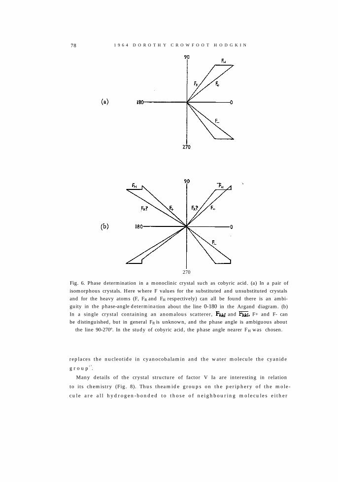
78 1 9 6 4 D O R O T H Y C R O W F O O T H O D G K I N
270
Fig. 6. Phase determination in a monoclinic crystal such as cobyric acid. (a) In a pair ofisomorphous crystals. Here where F values for the substituted and unsubstituted crystalsand for the heavy atoms (F, F R and FH respectively) can all be found there is an ambi-guity in the phase-angle determination about the line 0-180 in the Argand diagram. (b)In a single crystal containing an anomalous scatterer, F&l and G, F+ and F- canbe distinguished, but in general FR is unknown, and the phase angle is ambiguous about
the line 90-270º. In the study of cobyric acid, the phase angle nearer F H was chosen.
replaces the nucleotide in cyanocobalamin and the water molecule the cyanide
g r o u p1 7.
Many details of the crystal structure of factor V Ia are interesting in relation
to its chemistry (Fig. 8). Thus theamide groups on the periphery of the mole-
cu le a re a l l hydrogen-bonded to those o f ne ighbour ing molecu les e i ther

X - R A Y A N A L Y S I S O F C O M P L I C A T E D M O L E C U L E S 79
Formula I.
directly or through water molecules, except one, that on ring B. This isturned inwards to the hydrogen bond with the water molecule on the cobaltatom; the forces involved are sufficient to distort the position of the β-carbonatom, C-7, to which it is attached. At the same time, the amide nitrogen atomis brought close to C-8 with which it could readily react to form the lactam

80 1 9 6 4 D O R O T H Y C R O W F O O T H O D G K I N
Fig. 7b. Part of difference map Ae6, calculated for cobyric acid showing density due tohydrogen atoms attached to C-24 and C-10.
Fig. 8. The crystal structure of cobyric acid projected along a. Molecules centred at x areshown in strong lines, those at X in thin lines. Hydrogen bonds are dotted.

X - R A Y A N A L Y S I S O F C O M P L I C A T E D M O L E C U L E S 81
ring observed in the hexa acid, mentioned earlier. That the oxygen atomattached to cobalt is part of a water molecule, not a hydroxyl group, is sug-gested by the fact that these orange-red crystals separate at acid pH ( cf. thehaemoglobins and myoglobin). In our crystal this oxygen atom makes asecond contact through a water molecule with the carboxyl group attached toa neighbouring molecule. It would, in fact, be very easy to change the system
by a movement of two hydrogen atoms within the crystal; it would be inter-esting to see if this movement occurs, as in Rochelle salt, under the influence ofan electric field.
The interatomic distances in the inner ring of factor V Ia, which has beencalled the corrin ring, conform very closely with the distances proposed 12 fora structure containing six resonating double bonds, so closely as to leave al-most no doubt of the correct formulation of its chemical structure ( cf. Fig. 9).It was all the same, quite a moment in my life when J. D. Dunitz showed lastsummer at the Royal Society very closely similar figures derived by the X-rayanalysis of the nickel corrin derivative synthesized early this year by Eschen-moser and his coworkers18. The molecule as a whole is much smaller (Fig. 10)but the identity of the nucleus with that in cobyric acid is certain.
Very recently, Dunitz and Meyer have refined the nickel corrin structurethrough several more stages; their latest interatomic distances shown inFig. 11 are now so close to the earlier proposed theoretical figures that onebegins to feel that the small remaining deviations are likely to be real, e.g. inC-N 9-21 and 11-22. There is a tendency in the natural series also for thesebonds to be longer than the distances given by the simple-theory at firstproposed.
One of the features of the corrin nucleus in the natural compounds is thateven the inner ring is not quite planar. The same is true of the synthetic nu-cleus. In nickel corrin, the distortion is small but very regular and tetrahedralin character in relation to the nickel atom; alternate nitrogen atoms, bondedto it, lie above and below the least squares plane passing through the nickel andfour nitrogen atoms. The non-planarity of the system as a whole no doubtderives from the stereochemistry of the five-membered ring C-1, C-19,

1 9 6 4 D O R O T H Y C R O W F O O T H O D G K I N
Fig. 9. Interatomic distances (below) found from e6 for cobyric acid compared withthose suggested for a system containing six resonating double bonds.
N-23, Ni, N-20. In the natural corrins, the deviations are rather different incompounds with or without the nucleotide. In the unsubstituted compounds,and particularly in cobyric acid, the deviations are in the same sense as in thenickel corrin derivative and as in this molecule, C-5 and C-15, which carryC-35 and C-53 respectively, are on opposite sides of the plane containing thecobalt and four inner nitrogen atoms. In the nucleotide-containing com-pounds these two atoms are on the same side of the inner plane. In both series,the most marked deviations occur in the region of C-35 which is in a veryovercrowded situation.
Cobyric acid is the natural precursor of the most remarkable molecule ofour series, Co-5’-deoxyadenosyl-cobalamin, the coenzyme discovered byBarker, which ought, most properly to be called vitamin B12. Crystals of

X - R A Y A N A L Y S I S O F C O M P L I C A T E D M O L E C U L E S 83
Fig. 10. Electron-density peaks over the atoms in the nickel corrin derivative (Dunitzand Meyer).
this compound were grown from water in capillary tubes by Dr. Galen Len-hert in 1960 from material supplied by Dr. Barker, and X-ray photographswere taken of them in situ, in their mother liquor. Again the intensities of theX-ray diffraction spectra were measured, Patterson and electron-densitydistributions were calculated, atoms belonging to the best known parts of thecobalamin molecule being placed first in the calculations. At this point, Ishould pause to say that a great advantage of X-ray analysis as a method ofchemical structure analysis is its power to show some totally unexpected andsurprising structure with, at the same time, complete certainty. Fig. 12 illus-trates the structure we found - first, the electron-density map calculated overthe region of uncertain structure, with the known part of the moleculeplaced-then the whole atomic arrangement that is derivable from the com-pleted map. Clearly, in this molecule, cobalt was shown to be attached directto the 5’-carbon atom of the adenosyl residue as in formula II19. There fol-lowed directly an explanation of the observed great instability of the moleculeto light and cyanide ions - instability which had led to the failure of earlierinvestigations to recognise its existence and to isolate, in its place, cyano-cobalamin.
The detailed geometry of the coenzyme molecule as a whole is fascinatingin its complexity. The peculiar form of the corrin ring with the direct link

1 9 6 4 D O R O T H Y C R O W F O O T H O D G K I N
Fig.11. Interatomic distances measured in the nickel corrin derivative (Dunitz andMeyer).
between rings A and D and the position of the methyl group at C-24 makethe two sides of the corrin ring system very different stereochemically and thedifferences are reinforced by the positions of the methyl group and acetamideand propionamide residues attached at the carbon atoms (cf. Fig. 13). Ap-proach to the cobalt atom from the lower side of the molecule is hindered by

X - R A Y A N A L Y S I S O F C O M P L I C A T E D M O L E C U L E S 85
Fig. 12. (a) Electron-density peaks over the adenosyl residue calculated from X-ray dataon dimethylbenzimidazole cobamide coenzyme. The calculation illustrated was one inwhich phase angles were computed from the positions of all the atoms in the moleculeexcept those shown. Although only part of the electron density appears, the relativeweights of the atoms are in agreement with their chemical structure. (b) The atomicpositions found for the coenzyme molecule projected along the crystallographic b axis.
the methyl group C-24 and all groups attached here are rather loosely boundand easily displaced. At the upper site, on the other hand, attachment of awide variety of ligands is possible; once in position they are enclosed by non-polar groups, the methyl and methylene groups projecting normal to the planeof the ring. Here they may be positively protected from immediate reactionwith the surrounding solvent, for use when required in different biochemicaltransformations. In terms of this structure, one can begin to understand someof the uses to which the cobalamin nucleus is put in nature, for example, itspart in the transfer of methyl groups. In the laboratory, methylcobalamin canbe made by a series of reactions involving the reduction of aquocobalaminto a compound, probably cobalamin hydride, which easily exchanges withmethyl compounds such as diazomethane or methyl sulphate; in nature,reduction also seems necessary for methyl transfer; the experiments of D. D.

86 1 9 6 4 D O R O T H Y C R O W F O O T H O D G K I N
Fig. 13a. The positions found for the atoms in the coenzyme molecule projected (a) ontothe least square squares plane passing through cobalt and the four inner nitrogen atoms,
(b) and (c) across this plane.
Woods and his colleagues strongly suggest that methylcobalamin is the actualintermediate in one of the pathways by which methyl groups are transferredfrom methyltetrahydrofolate to homocysteine to form methionine20. othereffects of the B12 coenzymes are still not easy to explain in detail, particularlythe part they play in the isomerisation reactions which led to their discovery.How so complex a system can effect the simple and fundamental migrationprocess that changes methyhnalonate to succinate, for example, is still a prob-lem. One can see features of the system that might be important, the variable
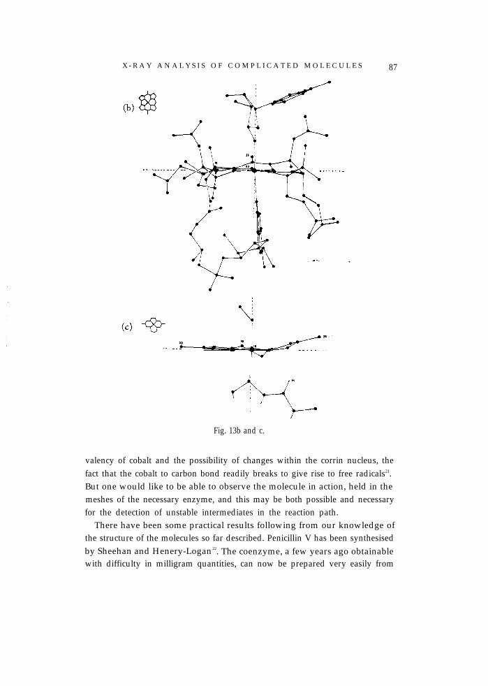
X - R A Y A N A L Y S I S O F C O M P L I C A T E D M O L E C U L E S 87
Fig. 13b and c.
valency of cobalt and the possibility of changes within the corrin nucleus, thefact that the cobalt to carbon bond readily breaks to give rise to free radicals21.But one would like to be able to observe the molecule in action, held in themeshes of the necessary enzyme, and this may be both possible and necessaryfor the detection of unstable intermediates in the reaction path.
There have been some practical results following from our knowledge ofthe structure of the molecules so far described. Penicillin V has been synthesisedby Sheehan and Henery-Logan22. The coenzyme, a few years ago obtainablewith difficulty in milligram quantities, can now be prepared very easily from

88 1 9 6 4 D O R O T H Y C R O W F O O T H O D G K I N
cyanocobalamin - Dr.Lester Smith has made crystals 0.5 cm across. Soon,and no doubt very beautifully, cyanocobalamin itself will be synthesised byR.B.Woodward. But microorganisms are even more efficient than chemistsat synthesis of molecules of this magnitude and will most likely continue toprovide the main supplies of these compounds to be used in medicine. Whatwe most hope to gain from knowledge of the structure and synthesis of thesemolecules is a complete understanding of their biogenesis and the part theyplay in metabolism. This should enable natural processes to be controlledwhen they go astray.
I should not like to leave an impression that all structural problems can besettled by X-ray analysis or that all crystal structures are easy to solve. I seemto have spent much more of my life not solving structures than solving them.I will illustrate some of the difficulties to be overcome by considering ourefforts to achieve the X-ray analysis of insulin.
Insulin is a molecule of weight about 6000, larger than any so far described,though small if considered as a protein and compared with myoglobin andhaemoglobin. Although its complete chemical structure is now known fromSanger’s researches23, it is quite unclear what exactly the molecule does thatmakes it so necessary to life. Our hope, following the kind of reasoning out-lined above, is that a complete knowledge of the molecular geometry, howthe peptide chains fit together within the molecule and the molecules withincrystals, may make it possible for us to understand and control its behaviour.It crystallises in a number of different modifications and their very differentdegrees of complexity seem significant. In acid insulin salts the molecule ap-pears to be dimeric, the two insulin molecules in one dimer being related toone another by two-fold axes of symmetry24. Crystallographic two-foldaxes also relate insulin molecules in a cubic metal-free form of insulin firstobserved by Abel25 in 1927. These crystals have so far only been obtained asextremely small rhombic dodecahedra; their habit and symmetry suggest thathere six insulin dimers may be grouped in a larger aggregate, with symmetry23, reminiscent of structures proposed for the smaller viruses26. But it is diffi-cult to get sufficient X-ray diffraction effects from these crystals to check thehypothesis. In all insulin crystals and in solutions which contain adequate zinc,or some other similar bivalent metal ions, a definite aggregate of six insulinmolecules appears; having, indeed, the molecular weight 36000 first recordedby Svedberg27 in 1935. This insulin hexamer corresponds with the unit cell inthe rhombohedral crystals first investigated and is the asymmetric unit in themonoclinic form which appears in the presence of phenol. The proportion of

X - R A Y A N A L Y S I S O F C O M P L I C A T E D M O L E C U L E S 89
\
Fig. 14. Crystals of insulin (a) cubic; (b) monoclinic and (c) rhombohedral (taken fromSchlichrkrull, Insulin Crystals, 1958).

90 1 9 6 4 D O R O T H Y C R O W F O O T H O D G K I N
zinc to insulin molecules is 2:6 and the symmetry relations strongly suggestthat the zinc is situated on the three-fold axes around which are arrangedinsulin molecules related to one another both by two- and three-fold axes.The symmetry relations have been explored by the use of the functions de-scribed by Rossman and Blow28. In the presence of halide, a slightly differentpacking is adopted with 4 Zn:6 insulin molecules. All these crystals givebeautiful X-ray diffraction effects from which it ought to be possible to solvethe structure to atomic resolution. Here the zinc present is much too light,about 0.01 in scattering effect, to be used for phase determination as we usedcobalt in B12. We need to introduce additional heavy atoms into the crystalstructure. This is not difficult to do in quantity. But it seems very difficult tolimit the crystal uptake to the one per insulin molecule which should be easyto place by X-ray methods, either by chemical reaction or by cocrystallisation,the methods adopted by Kendrew and Perutz. We are driven either to tryto solve an initially complex problem, concerned with the heavy atom distri-bution in the crystals, or to do more chemistry in the hope of binding oneheavy atom alone to each insulin molecule. In practice, I suppose, we shallattempt both. There are encouraging features of our present experiments thatlure us on - some of our heavy atom containing crystals show very markedanomalous absorption effects, from which some initial phasing evidence canbe obtained. But the electron- density maps we have so far calculated are fartoo imperfect and difficult to interpret for me to present them today*.
It will be clear from all that I have said so far that my research owes a debtI cannot adequately pay to the work of others, my colleagues who haveprovided many of the ideas I have used and many interesting examples ofsimilar analyses, my collaborators, without whose brains and hands and eyesvery little would have been done. I should also like to remember here todaymany whose friendship and encouragement I have greatly enjoyed. I willname three particularly, W. T. Astbury, I. Fankuchen and K. Linderström-Lang, because they would themselves so much have enjoyed this occasion.
* Subsequent examination suggests these maps are not so imperfect as I supposed and areinterpretable.

X - R A Y A N A L Y S I S O F C O M P L I C A T E D M O L E C U L E S 91
1. W.L.Bragg, Nobel Lectures, Chemistry, 1901-1921, Elsevier, Amsterdam, 1967, p.370.
2. M.F.Perutz and J.C.Kendrew, Nobel Lectures, Chemistry, 1942-1962, Elsevier,Amsterdam, 1964 pp. 653,676.
3. A.L.Patterson, Z.Krist., A, 90(1935)517.4. C.A.Beevers and H.Lipson, Proc.Phys.Soc. (Lordon), 48(1936)772.5. C.H.Carlisle and D.Crowfoot, Proc.Roy.Soc. (London), Ser..A, 184(1945) 64.6. J.D.Bernal, Nature, 129(1932) 277.7. D. Crowfoot, C. W.Bunn, B. W.Rogers-Low and A.Turner Jones, The Chemistry
of Penicillin, Princeton University Press, 1949, p. 310.8. K. J. Watson, to be published.9. S.Abrahamson, D.C.Hodgkin and E.N.Maslen, Biochem.J., 86(1963)514.
10. D.C.Hodgkin and E.N.Maslen, Biochem.J., 79(1961)393.11. R.D.Diamond, D.Phil. Thesis, A Crystallographic Study of the Structure of Some Anti-
biotics, Oxford, 1963.12. D. C. Hodgkin, J. Kamper, J.Lindsey, M. Mackay, J.Pickworth, J. H.Robertson,
C.B. Shoemaker, J.G. White, R. J.Prosen and K. N.Trueblood, Proc. Roy. Soc.(London), Ser.A, 242(1957) 228.
13. D. C.Hodgkin, J.Pickworth, J.H.Robertson, R. J.Prosen, R. A. Sparks and K. N.Trueblood, Proc.Roy.Soc. (London), Ser.A, 251(1959)306.
14. K.Bernhauer, F. Wagner and D.Wahl, Biochem.Z., 334(1961)279.15. A.F.Peerdeman and J.M.Bijvoet, Acta Cryst., 9(1956)1012.16. G.N.Ramachandran and S.Raman, Current Sci. (India), 25(1956) 348; S.Raman,
Proc.Indian Acad.Sci., 47(1958)1.17. D.Dale, D. C. Hodgkin and K.Venkatesan, Crystallography and Crystal Perfection,
Academic Press, London, 1963, p. 237.18. E. Bertele, H.Boos, J.D.Dunitz, F. Elsinger, A. Eschenmoser, I.Felner, H. P. Gribi,
H. Gschwend, E. F. Meyer, M. Pesaro and R. Scheffold, Angew.Chem., 76(1964)393;Angew.Chem., Intern.Ed., 3(1964)490.
19. P.G.Lenhert and D.C.Hodgkin, Nature, 192(1961)937.20. M. A. Foster, M. J.Dilworth and D. D. Woods, Nature, 201(1964)39.21. For a review see K.Bernhauer, O.Muller and F. Wagner, Angew.Chem., 3(1964)
200.22. J. C. Sheehan and K.R.Henery-Logan, J. Amer.Chem. Soc., 81(1959)3089.23. A.P.Ryle, F.Sanger, L. F. Smith and R.Kitai, Biochem.J., 60 (1955) 541.24. B.W.Low and J.R.Einstein, Nature, 156 (1960) 470.25. J. J. Abel, E. M.K. Geiling, C.A.Rouiller, F.K.Bell and O. Wintersteiner, J.Phar-
macol.Exptl.Therap., 31(1927)65.26. M.M.Harding, D.C.Hodgkin, A. F.Kennedy, A. O’Connor and P.D.J.Weitz-
mann, in preparation.27. K.Marcker, Acta Chem. Scand., 14(1960)2071.28. M.G.Rossman and D. M.Blow, Acta Cryst., 15(1962)24; E.Caller, M. M-Harding,
D. C. Hodgkin and M. G. Rossmann, in preparation.