LEFORT FRACTURES
ARTERIAL LINE WAVEFORM

RULE OF 9’S

12 LEAD EKG
SUBDURAL HEMATOMA
1) No clear delineation
2) Blood between dura and arachnoid layers
3) Usually venous in nature
4) Increased morbidity and mortality rate
a) Increases with every millimeter of brain tissue shift
5) Types
a) Acute
i) Onset within 24 hours
b) Subacute
i) Onset within 2-10 days
c) Chronic

i) Onset after 2 weeks
6) Elderly
a) Larger bleed with slowly developing symptoms due to cerebral atrophy
7) Younger person
a) Rapid onset with marked increased ICP
8) Pediatric
a) Generally occur in children < 18 months
b) Fontanelle not fully closed
c) Ssx
i) Bulging fontanelle
ii) Separation of sutures
iii) Shock
iv) Retinal hemorrhage
v) Die from bleeding to death
EPIDURAL HEMATOMA
1) Clear line of delineation
2) Lens shaped
3) Bleeding between skull and dura mater
4) Usually arterial but can be venous
5) Laceration of middle meningeal artery in temporal lobe area
6) Classic symptomology
a) Transient loss of consciousness followed by period of lucidity then decreased level of consciousness
i) Hit head
ii) Lose consciousness
iii) Regain consciousness
iv) Loss of consciousness
7) Uncal herniation
a) Will result in dilation of ipsilateral pupil with contralateral neuro deficits and posturing
8) Classification
a) Acute
i) Usually arterial bleed
ii) Onset of symptoms within a few hours
b) Subacute
i) Usually venous bleed
ii) Takes longer time for onset of symptoms
SUBARACHNOID HEMORRHAGE
1) Bleeding between arachnoid mater and pia mater
2) Trauma most common cause

3) May occur with other injuries or as only evidence of trauma
a) Rupture of aneurysm in Circle of Willis
i) Berry aneurysm rupture
b) Maintain ICP, CPP, etc
4) Aneurysmal SAH
a) Medical
b) Rupture may be self-limiting
i) 2nd rupture is 100% mortality
c) Quick loss of consciousness then dies
d) Fatal within short period of time
e) Worse headache ever
i) Die in emergency department or drop dead
f) Managed differently than all others
i) Decrease amount of perfusion to control blood pressure to prevent increased pressure
ii) Decrease heart rate
iii) Cardene
iv) Nipride
v) SPB < 140
5) Ssx
a) Presents similar to meningitis
b) Severe headache
i) Worst ever
ii) Deteriorate rapidly
c) Vomiting
d) Stiff neck
i) Bending down hurts
e) Leg pain
i) Drawing up hurts
f) Confusion
g) Lethargy
h) Loss of consciousness
i) Blood accumulates in meninges
6) Avoid lumbar puncture due to possibility of uncal herniation until CT scan verifies SAH vs meningitis
GRAVE’S DISEASE AND THYROTOXICOSIS
1) Thyroid storm
2) Autoimmune disease which causes overstimulation of TSH receptors
a) Thin hair

b) Papery skin
c) Bulging eyes
d) Hypermetabolic
3) Causes
a) Iodine
i) Amiodarone
b) Infection
c) Surgery to thyroid
d) Toxemia
4) Presentation
a) Dramatic weight loss
b) Chest pain
i) Palpitations
ii) SOB
iii) Trying to keep up with metabolic demand leading to increased cardiac output
c) Fever
d) Tremors
e) Nervousness
f) Marked tachycardia
g) Hypertension
h) Profuse sweating
5) Treatment
a) Antipyretics
i) NO ASA
b) Supportive care
c) LOTS of fluids
d) Correct electrolytes
e) Supplemental oxygen
f) Beta-blockers
i) Cardioprotective
g) PTU or MMI
i) PO
ii) NGT
h) Consider IV glucocorticoids
i) Decadron inhibits hormone production and conversion from T4 to T3
(1) Actual metabolite that causes cells to “ramp up”
MYXEDEMA COMA AND HYPOTHYROIDISM
1) Causes
a) Infection
b) Cessation of thyroid replacement

c) Thyroid removal
2) Presentation
a) Primarily women
b) Almost exclusively over 60
c) >90% of cases occur in winter
d) Fatigue
e) Weight gain
f) Cold intolerance
g) Deep voice
h) Coarse hair
i) Obese
j) Round face
k) Slow appearance
l) Officially “myxedema coma” with any change in level of consciousness
i) Hypometabolic
3) Treatment
a) Supportive during comatose states
b) IV levothyroxine
i) T4
c) Triostat
i) T3
d) Adrenal insufficiency
i) DO NOT GIVE Etomidate
(1) Can potentiate adrenal insufficiency
ADDISON’S DISEASE/ADRENAL INSUFFICIENCY
1) Causes
a) Autoimmune disease
i) Primary Addison’s
b) Decreased levels of ACTH
i) Secondary Addison’s
c) Acute withdrawal from glucocorticoid therapy
i) Prednisone
2) Presentation
a) Weakness
b) Weight loss
c) Fatigue
d) Decreased blood pressure
e) Negative ACTH test
i) No increase in serum cortisol with ACTH administration
3) Treatment

a) Supportive care
b) ABCs
c) Volume replacement
d) Correct electrolytes
e) Hydrocortisone or Decadron
f) NO Etomidate
CUSHING’S SYNDROME/HYPERALDOSTERONISM
1) Causes
a) Chronic glucocorticoid use
b) Pituitary disorder
c) Oat cell carcinoma
d) Adrenal carcinoma
2) Presentation
a) Upper body obesity with thin arms and legs
b) Rounded face
c) “Buffalo hump” on back of neck
d) Fatigue
e) Hypertension
f) Hyperglycemia
g) Facial hair on women
3) Treatment
a) Initiation or reduction of glucocorticoids
i) Causes negative feedback loop
ii) Causes adrenals to decrease production of glucocorticoids
b) Support symptoms
c) Surgery to remove some of adrenal gland
SYNDROME OF INAPPROPRIATE ANTIDIURETIC HORMONE (SIADH)
1) Released by posterior pituitary
a) Tells kidneys to hold onto water
2) LOTS of ADH produced
a) Kidneys hold excessive water
b) Not excreting water at same rate of other chemicals
i) Blood hyperosmolar
3) Causes
a) Oat cell carcinoma
b) Viral pneumonia
c) Head injury
i) CVA
ii) ICH

4) Presentation
a) Neuro changes
i) Confusion
ii) Coma
b) Cerebral edema
i) Increased ICP
c) Seizures
d) Hyponatremia
e) Urine osmolarity/specific gravity
i) Concentrated
5) Treatment
a) Restrict fluids
i) Over fluid loaded
b) Diuresis
i) Push water out of kidneys
ii) Lasix
c) Hypertonic saline
i) Treat slowly
ii) Do not raise sodium more than 0.5 mEq/liter/hr
DIABETES INSIPIDUS
1) No ADH being released or used
2) Causes
a) Head injury
i) ICH
ii) CVA
b) Dilantin toxicity
3) Presentation
a) Extreme urine output with very low urine osmolality/specific gravity
i) Looks like free water
(1) Clear
ii) Copious amounts
4) Treatment
a) Aggressive fluid resuscitation
b) Vasopressin
i) Cardiovascular effects
ii) Exogenous ADH
c) Desmopressin
i) DDAVP
ii) Does not have vasoconstricting effects of Vasopressin

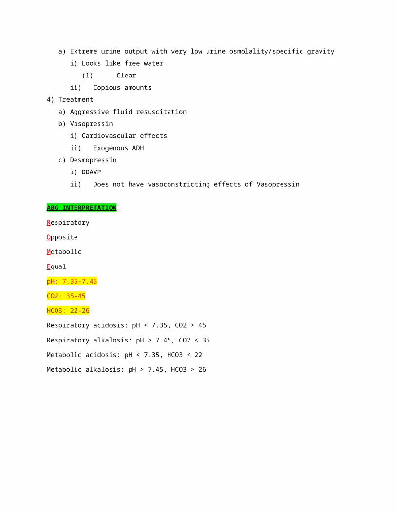
ABG INTERPRETATION
Respiratory
Opposite
Metabolic
Equal
pH: 7.35-7.45
CO2: 35-45
HCO3: 22-26
Respiratory acidosis: pH < 7.35, CO2 > 45
Respiratory alkalosis: pH > 7.45, CO2 < 35
Metabolic acidosis: pH < 7.35, HCO3 < 22
Metabolic alkalosis: pH > 7.45, HCO3 > 26
CUSHING’S TRIAD—EMMINENT UNCAL HERNIATION
ANTIDOTES FOR SELECT POISONING
Carbon monoxide 1) Oxygen: 100% for 2-6 hrs; competes with CO; causes positive neurological change
2) Hyperbarics: do NOT use if lactic acidosis present; tachycardia, hypotension, use if patient can wait 4-6 hours
Cyanide 1) Amyl or sodium nitrate2) Nathiosulfate
Organophosphates/organocarbamatos 1) Atropine2) 2-PAM
Methemoglobinemia 1) Methylene blue
Anticholinergic 1) Physostigmine
Coumadin 1) Vitamin K
Heparin 1) Protamine
Beta-blockers 1) Glucagon
Calcium channel blockers 1) Calcium
BECK’S TRIAD—PERICARDIAL TAMPONADE

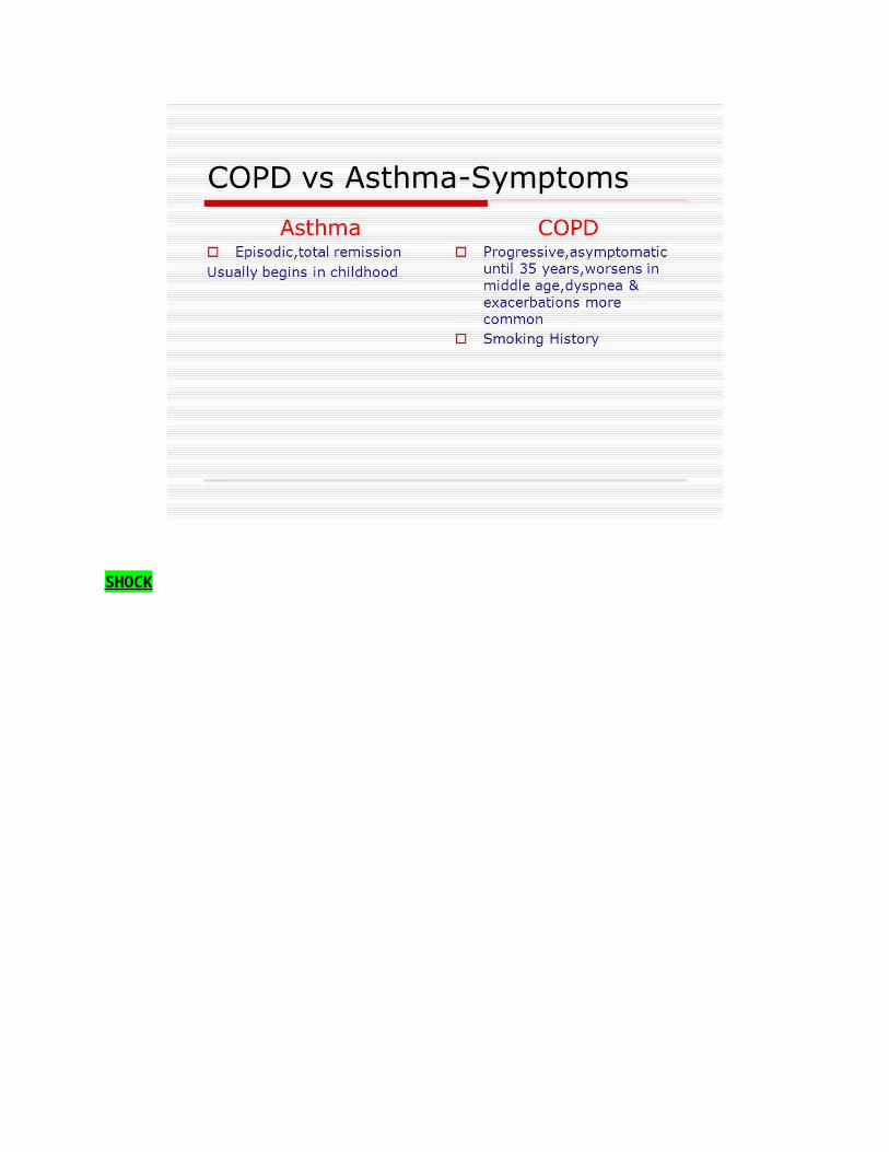
COPD VS ASTHMA
SHOCK

PLACENTAL PLACEMENT
Abruption: painful, dark red blood
Previa: painless, bright red blood
SPINAL CORD INJURIES