Araştırma Yazısı SELÇUK TIP DERGİSİSelçuk Tıp Derg 2013;29(3):108-112
Yazışma Adresi: Yüksel Çiçek, Rize Recep Tayyip Erdoğan Üniversitesi Tıp Fakültesi Kardiyoloji Anabil im Dalı, Rizee-posta: [email protected]ş Tarihi: 16.10.2012 Yayına Kabul Tarihi: 30.04.2013
Özet Abstract Çok kesit l i bi lgisayarlı tomografi (ÇKBT) koroner arter hastalığının tespit i için son zamanlarda kullanılmaya başlamıştır. Hem ÇKBT hemde koroner anjiyografi direkt olarak koroner arterleri görüntüleyebil ir. Bu çalışmanın amacı 128 kesit l i BT’ nin ciddi koroner arter hastalığının tespit inde tanısal değerini konvansiyonel koroner anjiyografi i le karşılaştırmaktır. Çalışmaya koroner arter hastalığı şüphesi olan 42 hasta prospektif olarak alındı.Hastalara 128 kesit l i BT çekildi ve sonrasında koroner lezyon ciddiyetini doğrulamak ve karşılaştırmak amacı i le koroner anjiyografi yapıldı.Kayıtlar Amerikan kalp birl iğinin 16 segment modeline göre değerlendiri ldi. Hastaların yaş ortalaması 56±12 yıl idi.İşlem öncesi ortalama kalp hızı 69±4 / dakika idi. (en düşük-en yüksek: 54-78). Toplam 672 segmentin değerlendirmeye uygun 669 segmenti değerlendiri ldi. Değerlendiri len tüm segmentler için sensit ivite, spesif i te, pozit i f prediktiv değer, negatif prediktif değer ve doğruluk oranı 128 kesit BT için sırasıyla % 91, %98.5, %93.8, % 97.8 ve %97 olarak bulundu. Bu değerler sadece proximal segmentler değerlendiri ldiğinde %97.8, %96.4, %95.7, % 98.1 ve % 97.1 olarak bulundu. Sadece distal segmentler değerlendiri ldiğinde ise yine sırasıyla %87.4, %98.8, %92.7, %97.7 ve %97 olarak bulundu. Çalışmamızın sonucunda 128 kesit l i BT’nin konvansiyonel koroner anjiyografiye hemen hemen yakın doğrulukta tanısal doğruluğu olduğunu tespit ett ik ve bu tekniğin giderek artan kullanımı i le bir l ikte gereksiz konvansiyonel koroner anjiyografi işlem sayısının azalacağını düşünmekteyiz
Anahtar kelimeler: 128 kesit, çok kesit l i bi lgisayarlı tomografi, koroner arter hastalığı, koroner anjiyografi
Multi-Slice Computed Tomography (MSCT) is a recently introduced non-invasive technique used for the diagnosis of coronary artery disease (CAD). Both MSCT and conventional coronary angiography (CAG) may provide direct visualization of the coronary arteries. To determine the diagnostic accuracy of 128-slice MSCT compared with the results of conventional CAG for the diagnosis of signif icant coronary artery stenosis. Forty-two eligible patients who underwent 128-slice MSCT with the suspicion of CAD and had any coronary lesion within the whole coronary tree were enrolled prospectively. Subsequently, CAG was performed to confirm the severity of coronary lesions. The records acquired by MSCT coronary angiography and conventional CAG were evaluated by 16-segment model of American Heart Association (AHA). Mean age of the patients was 56±12 years, and mean heart rate prior to the examination was 69±4 bpm (min-max: 54-78). Of 672 coronary segments, 669 were interpretable and evaluated in 42 patients. For all the interpretable segments, overall sensit ivity, specif icity, posit ive predictive value (PPV), negative predictive value (NPV) and the diagnostic accuracy (DA) of 128-slice MSCT to detect signif icant stenosis were 91%, 98.5%, 93.8%, 97.8% and 97%, respectively. These values for evaluation of proximal segments were 97.8%, 96.4%, 95.7%, 98.1% and 97.1% and, for distal segments were 87.4%, 98.8%, 92.7%, 97.7% and 97%, respectively. Our results show that, the diagnostic performance of MSCT appear almost equivalent to CAG, current gold standard, with excellent, sensit ivity, specif icity, posit ive and negative predictive values. We think that, 128 slice-MSCT wil l be increasingly uti l ized in future, decreasing unnecessary coronary angiography procedures.
Key words: 128-slice; multi-sl ice computed tomography, coronary artery disease, coronary angiography
INTRODUCTION Coronary artery disease (CAD), atherosclerosis of the coronary arteries, is a major source of morbidity and mortality, in developed countries (1,2). Coronary angiography is currently the gold standard imaging technique for coronary artery disease. However, due to being an
128 Kesitli Bilgisayarlı Tomografinin Ciddi Koroner Arter Hastalığının Tespitinde Tanısal Değeri
Diagnostic Performance of 128-Slice Multi-Slice Computed Tomography for the Detection of Significant Coronary Artery Lesions
Yüksel Çiçek1, M. Emre Durakoğlugil1, S. Altan Kocaman2, Turan Erdoğan1, Mustafa Özateş3, Sedat Bozkurt4, Mustafa Çetin2, Aytun Çanga2, Ömer Şatıroğlu1
¹Rize University, Medical School, Department of Cardiology, Rize²Rize Education and Research Hospital, Department of Cardiology, Rize
³Kartal Training and Research Hospital, Department of Radiology, Istanbul4Middle East Private Hospital, Department of Radiology, Sanliurfa
invasive procedure, CAG inherently carries some procedure related risks. Therefore, utilization of non-invasive diagnostic imaging methods for the early detection of CAD, especially multi-slice computed tomography (MSCT), has recently gained interest. Previously, 4, 6, 8 and 16-slice

Çiçek ve ark. Selçuk Tıp Dergisi
109
CT detectors and lately 64-slice detectors is widely used. To date, many studies have been conducted using these imaging techniques, and recent
MATERIAL and METHODS Technological advances have made 128-slice CT scans possible (3-5). However the diagnostic value and advantages of 128-slice MSCT have not been adequately studied, till now. In the present study, we aimed to investigate the diagnostic values of 128-slice MSCT scans compared to CAG, the present gold standard, and to reveal whether they can provide satisfactory image quality, even in cases with tachycardia.Study design and Patients This study, having a cross-sectional and observational design, was approved by the local ethics committee. All patients gave informed consent before the study. The patients with the complaint of typical chest pain and positive or equivalent findings on exercise stress test and/or myocardial perfusion scanning, who underwent MSCT due to suspicion of CAD at our institution between December 2008 and November 2009, were consecutively enrolled. Of them, 42 eligible patients with any coronary lesion including mild or significant narrowing on MSCT prospectively underwent CAG to confirm the severity of lesions and to perform percutaneous coronary intervention (PCI), if required. The patients with totally normal coronary arteries on MSCT were excluded from study. Baseline characteristics were recorded during the direct interview with the patient. Hypertension was defined as the active use of antihypertensive drugs or documentation of blood pressure more than 140/90 mmHg. Diabetes mellitus was acknowledged as fasting plasma glucose (FPG) levels over 126 mg/dl or glucose level over 200 mg/dl at any measurement or active use antidiabetic treatment. Smoking status was considered as current active smoking. The family history for CAD was approved with a history of CAD or sudden death in a first-degree relative before the age of 55 years for men and 65 years for women. Patients with atrial fibrillation or other rhythm disturbances, which could adversely affect the quality of the procedure, hypersensitivity to iodine-containing contrast agents, chronic kidney disease (serum creatinine ≥1.5 mg/dl), a previous history of coronary artery by-pass graft operation (CABG), coronary artery anomaly, and pregnant women were excluded from the study.
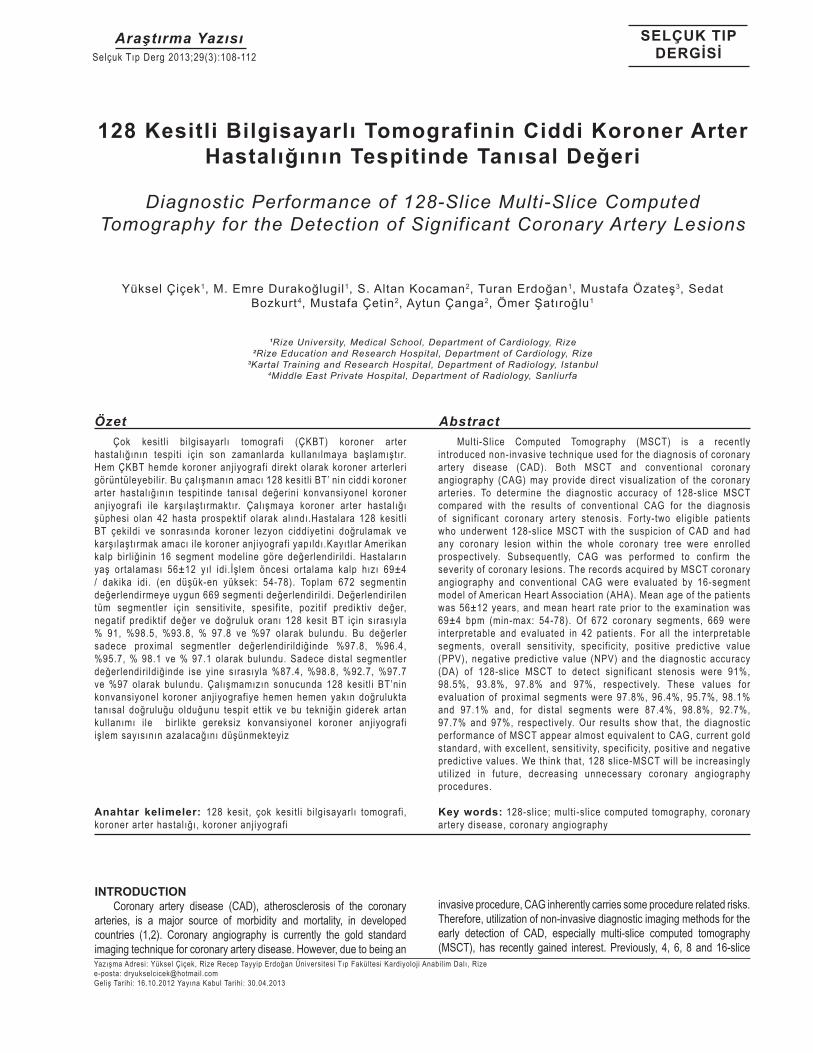
Figure 1. The simultaneous presentation of the coronary lesions on LAD and Cx with conventional CAG and 128-slice MSCT coronary angiography (On the image of conventional CAG, two critical lesions are shown (Left side) on LAD (oblique arrow) and CX (horizontal arrow). Same lesions are presented by MDCT (right side))
Multi-slice computed tomography coronary angiography Computed tomography coronary angiography was performed with a 128-slice multi-detector CT scanner (SOMATOM Definition AS, Siemens Medical Solutions, Forchheim, Germany). ECG-controlled tube current modulation technique and prospective ECG-gating were used for all examinations. Scanning protocol was as follows: tube voltage 120 kV, tube current 150–300 mAs/rotation, gantry rotation time 300 msec, collimation (128x0.6 mm slice). All images were acquired during full inspiration and breath-holding. A beta-blocker (metoprolol, 5 mg, IV) was injected intravenously if the heart rate exceeded 70 bpm prior to the examination (n=18; 42%). In five cases without sufficient decrease in HR, metoprolol dose was not repeated, but a target heart rate of ≤80 bpm was reached in every patient. Mean HR was 69±4 bpm (min-max: 54-78). A 70-80 ml of non-ionic contrast medium (Iopromide, Ultravist 370 mg I/mL, Bayer-Schering Pharma, Berlin, Germany) was injected into an antecubital vein at a rate of 5 ml/sec using a double-headed auto-injector (Covidien OptiVantage™ DH; Mallinckrodt, Cincinnati,
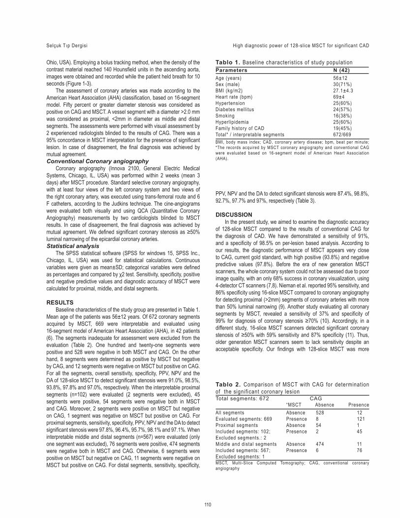
Figure 2. Demonstration of the crit ical lesion on LAD with CAG and MSCT (On the image of conventional CAG, the critical lesion is shown (Left side) on LAD (horizontal arrow). Same lesion is presented by MSCT (right side) and seen as a calcified lesion (Oblique arrow))
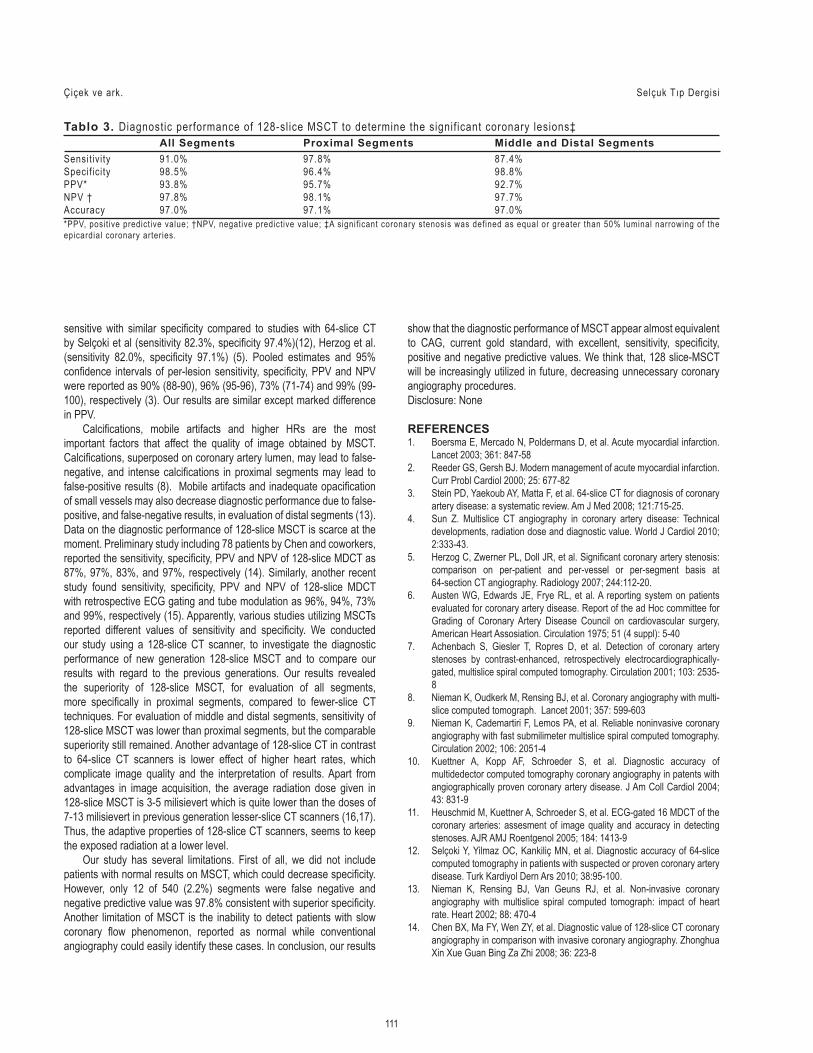
Figure 3. Demonstration of the crit ical lesion on RCA with conventional CAG and MSCT (On the image of conventional CAG, the critical lesion is shown (Left side) on RCA (horizontal arrow). Same lesion is presented by MSCT (right side) and seen as a severe lesion (Vertical arrow))
110
Selçuk Tıp Dergisi High diagnostic power of 128-slice MSCT for signif icant CAD
Tablo 1. Baseline characteristics of study population
BMI, body mass index; CAD, coronary artery disease; bpm, beat per minute; *The records acquired by MSCT coronary angiography and conventional CAG were evaluated based on 16-segment model of American Heart Association (AHA).
Ohio, USA). Employing a bolus tracking method, when the density of the contrast material reached 140 Hounsfield units in the ascending aorta, images were obtained and recorded while the patient held breath for 10 seconds (Figure 1-3). The assessment of coronary arteries was made according to the American Heart Association (AHA) classification, based on 16-segment model. Fifty percent or greater diameter stenosis was considered as positive on CAG and MSCT. A vessel segment with a diameter >2.0 mm was considered as proximal, <2mm in diameter as middle and distal segments. The assessments were performed with visual assessment by 2 experienced radiologists blinded to the results of CAG. There was a 95% concordance in MSCT interpretation for the presence of significant lesion. In case of disagreement, the final diagnosis was achieved by mutual agreement.Conventional Coronary angiography Coronary angiography (Innova 2100, General Electric Medical Systems, Chicago, IL, USA) was performed within 2 weeks (mean 3 days) after MSCT procedure. Standard selective coronary angiography, with at least four views of the left coronary system and two views of the right coronary artery, was executed using trans-femoral route and 6 F catheters, according to the Judkins technique. The cine-angiograms were evaluated both visually and using QCA (Quantitative Coronary Angiography) measurements by two cardiologists blinded to MSCT results. In case of disagreement, the final diagnosis was achieved by mutual agreement. We defined significant coronary stenosis as ≥50% luminal narrowing of the epicardial coronary arteries.Statistical analysis The SPSS statistical software (SPSS for windows 15, SPSS Inc., Chicago, IL, USA) was used for statistical calculations. Continuous variables were given as mean±SD; categorical variables were defined as percentages and compared by χ2 test. Sensitivity, specificity, positive and negative predictive values and diagnostic accuracy of MSCT were calculated for proximal, middle, and distal segments.
RESULTS Baseline characteristics of the study group are presented in Table 1. Mean age of the patients was 56±12 years. Of 672 coronary segments acquired by MSCT, 669 were interpretable and evaluated using 16-segment model of American Heart Association (AHA), in 42 patients (6). The segments inadequate for assessment were excluded from the evaluation (Table 2). One hundred and twenty-one segments were positive and 528 were negative in both MSCT and CAG. On the other hand, 8 segments were determined as positive by MSCT but negative by CAG, and 12 segments were negative on MSCT but positive on CAG. For all the segments, overall sensitivity, specificity, PPV, NPV and the DA of 128-slice MSCT to detect significant stenosis were 91.0%, 98.5%, 93.8%, 97.8% and 97.0%, respectively. When the interpretable proximal segments (n=102) were evaluated (2 segments were excluded), 45 segments were positive, 54 segments were negative both in MSCT and CAG. Moreover, 2 segments were positive on MSCT but negative on CAG, 1 segment was negative on MSCT but positive on CAG. For proximal segments, sensitivity, specificity, PPV, NPV and the DA to detect significant stenosis were 97.8%, 96.4%, 95.7%, 98.1% and 97.1%. When interpretable middle and distal segments (n=567) were evaluated (only one segment was excluded), 76 segments were positive, 474 segments were negative both in MSCT and CAG. Otherwise, 6 segments were positive on MSCT but negative on CAG, 11 segments were negative on MSCT but positive on CAG. For distal segments, sensitivity, specificity,
Parameters N (42)Age (years) 56±12Sex (male) 30(71%)BMI (kg/m2) 27.1±4.3Heart rate (bpm) 69±4Hypertension 25(60%)Diabetes mell i tus 24(57%)Smoking 16(38%)Hyperlipidemia 25(60%)Family history of CAD 19(45%)Total* / interpretable segments 672/669
Total segments: 672 CAG “MSCT Absence PresenceAll segments Absence 528 12Evaluated segments: 669 Presence 8 121Proximal segments Absence 54 1Included segments: 102; Presence 2 45Excluded segments.: 2 Middle and distal segments Absence 474 11Included segments: 567; Presence 6 76Excluded segments: 1MSCT, Multi-Slice Computed Tomography; CAG, conventional coronary angiography
Tablo 2. Comparison of MSCT with CAG for determination of the signif icant coronary lesion
PPV, NPV and the DA to detect significant stenosis were 87.4%, 98.8%, 92.7%, 97.7% and 97%, respectively (Table 3).
DISCUSSION In the present study, we aimed to examine the diagnostic accuracy of 128-slice MSCT compared to the results of conventional CAG for the diagnosis of CAD. We have demonstrated a sensitivity of 91%, and a specificity of 98.5% on per-lesion based analysis. According to our results, the diagnostic performance of MSCT appears very close to CAG, current gold standard, with high positive (93.8%) and negative predictive values (97.8%). Before the era of new generation MSCT scanners, the whole coronary system could not be assessed due to poor image quality, with an only 68% success in coronary visualization, using 4-detector CT scanners (7,8). Nieman et al. reported 95% sensitivity, and 86% specificity using 16-slice MSCT compared to coronary angiography for detecting proximal (>2mm) segments of coronary arteries with more than 50% luminal narrowing (9). Another study evaluating all coronary segments by MSCT, revealed a sensitivity of 37% and specificity of 99% for diagnosis of coronary stenosis ≥70% (10). Accordingly, in a different study, 16-slice MSCT scanners detected significant coronary stenosis of ≥50% with 59% sensitivity and 87% specificity (11). Thus, older generation MSCT scanners seem to lack sensitivity despite an acceptable specificity. Our findings with 128-slice MSCT was more
Çiçek ve ark. Selçuk Tıp Dergisi
111
All Segments Proximal Segments Middle and Distal SegmentsSensit ivity 91.0% 97.8% 87.4%Specif icity 98.5% 96.4% 98.8%PPV* 93.8% 95.7% 92.7%NPV † 97.8% 98.1% 97.7%Accuracy 97.0% 97.1% 97.0%
Tablo 3. Diagnostic performance of 128-slice MSCT to determine the signif icant coronary lesions‡
*PPV, posit ive predictive value; †NPV, negative predictive value; ‡A signif icant coronary stenosis was defined as equal or greater than 50% luminal narrowing of the epicardial coronary arteries.
sensitive with similar specificity compared to studies with 64-slice CT by Selçoki et al (sensitivity 82.3%, specificity 97.4%)(12), Herzog et al. (sensitivity 82.0%, specificity 97.1%) (5). Pooled estimates and 95% confidence intervals of per-lesion sensitivity, specificity, PPV and NPV were reported as 90% (88-90), 96% (95-96), 73% (71-74) and 99% (99-100), respectively (3). Our results are similar except marked difference in PPV. Calcifications, mobile artifacts and higher HRs are the most important factors that affect the quality of image obtained by MSCT. Calcifications, superposed on coronary artery lumen, may lead to false-negative, and intense calcifications in proximal segments may lead to false-positive results (8). Mobile artifacts and inadequate opacification of small vessels may also decrease diagnostic performance due to false-positive, and false-negative results, in evaluation of distal segments (13). Data on the diagnostic performance of 128-slice MSCT is scarce at the moment. Preliminary study including 78 patients by Chen and coworkers, reported the sensitivity, specificity, PPV and NPV of 128-slice MDCT as 87%, 97%, 83%, and 97%, respectively (14). Similarly, another recent study found sensitivity, specificity, PPV and NPV of 128-slice MDCT with retrospective ECG gating and tube modulation as 96%, 94%, 73% and 99%, respectively (15). Apparently, various studies utilizing MSCTs reported different values of sensitivity and specificity. We conducted our study using a 128-slice CT scanner, to investigate the diagnostic performance of new generation 128-slice MSCT and to compare our results with regard to the previous generations. Our results revealed the superiority of 128-slice MSCT, for evaluation of all segments, more specifically in proximal segments, compared to fewer-slice CT techniques. For evaluation of middle and distal segments, sensitivity of 128-slice MSCT was lower than proximal segments, but the comparable superiority still remained. Another advantage of 128-slice CT in contrast to 64-slice CT scanners is lower effect of higher heart rates, which complicate image quality and the interpretation of results. Apart from advantages in image acquisition, the average radiation dose given in 128-slice MSCT is 3-5 milisievert which is quite lower than the doses of 7-13 milisievert in previous generation lesser-slice CT scanners (16,17). Thus, the adaptive properties of 128-slice CT scanners, seems to keep the exposed radiation at a lower level. Our study has several limitations. First of all, we did not include patients with normal results on MSCT, which could decrease specificity. However, only 12 of 540 (2.2%) segments were false negative and negative predictive value was 97.8% consistent with superior specificity. Another limitation of MSCT is the inability to detect patients with slow coronary flow phenomenon, reported as normal while conventional angiography could easily identify these cases. In conclusion, our results
show that the diagnostic performance of MSCT appear almost equivalent to CAG, current gold standard, with excellent, sensitivity, specificity, positive and negative predictive values. We think that, 128 slice-MSCT will be increasingly utilized in future, decreasing unnecessary coronary angiography procedures.Disclosure: None
REFERENCES1. Boersma E, Mercado N, Poldermans D, et al. Acute myocardial infarction.
Lancet 2003; 361: 847-582. Reeder GS, Gersh BJ. Modern management of acute myocardial infarction.
Curr Probl Cardiol 2000; 25: 677-823. Stein PD, Yaekoub AY, Matta F, et al. 64-slice CT for diagnosis of coronary
artery disease: a systematic review. Am J Med 2008; 121:715-25.4. Sun Z. Multislice CT angiography in coronary artery disease: Technical
developments, radiation dose and diagnostic value. World J Cardiol 2010; 2:333-43.
5. Herzog C, Zwerner PL, Doll JR, et al. Significant coronary artery stenosis: comparison on per-patient and per-vessel or per-segment basis at 64-section CT angiography. Radiology 2007; 244:112-20.
6. Austen WG, Edwards JE, Frye RL, et al. A reporting system on patients evaluated for coronary artery disease. Report of the ad Hoc committee for Grading of Coronary Artery Disease Council on cardiovascular surgery, American Heart Assosiation. Circulation 1975; 51 (4 suppl): 5-40
7. Achenbach S, Giesler T, Ropres D, et al. Detection of coronary artery stenoses by contrast-enhanced, retrospectively electrocardiographically-gated, multislice spiral computed tomography. Circulation 2001; 103: 2535-8
8. Nieman K, Oudkerk M, Rensing BJ, et al. Coronary angiography with multi-slice computed tomograph. Lancet 2001; 357: 599-603
9. Nieman K, Cademartiri F, Lemos PA, et al. Reliable noninvasive coronary angiography with fast submilimeter multislice spiral computed tomography. Circulation 2002; 106: 2051-4
10. Kuettner A, Kopp AF, Schroeder S, et al. Diagnostic accuracy of multidedector computed tomography coronary angiography in patents with angiographically proven coronary artery disease. J Am Coll Cardiol 2004; 43: 831-9
11. Heuschmid M, Kuettner A, Schroeder S, et al. ECG-gated 16 MDCT of the coronary arteries: assesment of image quality and accuracy in detecting stenoses. AJR AMJ Roentgenol 2005; 184: 1413-9
12. Selçoki Y, Yilmaz OC, Kankiliç MN, et al. Diagnostic accuracy of 64-slice computed tomography in patients with suspected or proven coronary artery disease. Turk Kardiyol Dern Ars 2010; 38:95-100.
13. Nieman K, Rensing BJ, Van Geuns RJ, et al. Non-invasive coronary angiography with multislice spiral computed tomograph: impact of heart rate. Heart 2002; 88: 470-4
14. Chen BX, Ma FY, Wen ZY, et al. Diagnostic value of 128-slice CT coronary angiography in comparison with invasive coronary angiography. Zhonghua Xin Xue Guan Bing Za Zhi 2008; 36: 223-8

15. Kim JS, Choo KS, Jeong DW, et al. Step-and-shoot prospectively ECG-gated vs. retrospectively ECG-gated with tube current modulation coronary CT angiography using 128-slice MDCT patients with chest pain: diagnostic performance and radiation dose. Acta Radiol 2011; 52: 860-5.
16. Cody DD, Mahesh M. AAPM/RSNA physics tutorial for residents:technologic advances in multidetector CT with a focus on cardiac imaging. Radiographics 2007; 27: 1829-37
17. Gerber TC, Kuzo RS, Morin RL. Technigues and parameters for estimating radiation exposure and dose in cardiac computed tomoghraphy. Int J Cardiovasc Imaging 2005; 21: 165-76
Selçuk Tıp Dergisi High diagnostic power of 128-slice MSCT for signif icant CAD
112


