
Cell, Vol. 80, 413-422, February 10, 1995, Copyright © 1995 by Cell Press
a4 Integrins Mediate Lymphocyte Attachment and Rolling under Physiologic Flow
C. Berlin, 1 R. F. Bargatze, 1, 2 j . j . Campbell,1 U. H. von Andrian, 1,3 M. C. Szabo, ~ S. R. Hasslen, 4 R. D. Nelson, 5 E. L. Berg, ~,8 S. L. Erlandsen, 7 and E. C. Butcher ~ 1Laboratory of Immunology and Vascular Biology Department of Pathology Stanford University Stanford, California 94305 and the Center for Molecular Biology and Medicine Foothill Research Center Veterans Affairs Medical Center Palo Alto, California 94304 2Veterinary Molecular Biology Laboratory Montana State University Marsh Labs Bozeman, Montana 59717 4Departmentof Laboratory Medicine and Pathology SDepartment of Dermatology 7Department of Cell Biology and Neuroanatomy University of Minnesota Minneapolis, Minnesota 55455 3Center for Blood Research Boston, Massachusetts 02115 6Protein Design Labs Mountain View, California 94043
Summary
Of the several families of adhesion receptors involved in leukocyte-endothelial cell interactions, only the se- lectins have been shown to initiate leukocyte interac- tion under physiologic shear; indeed, p2 (CD18) inte- grins responsible for neutrophil arrest are unable to engage without prior selectin-mediated rolling. In con- trast, (~4 (CD49d) integrins are shown here to initiate lymphocyte contact ("tethering") in vitro under shear and in the absence of a selectin contribution. The e4 integrin ligands MAdCAM-1 and VCAM-1 support loose reversible interactions including rolling, as well as rapid sticking and arrest that is favored following inte- grin activation. Moreover, a41~7 mediates L-selectin (CD62L)-independent attachment of blood-borne lym- phocytes to lamina propria venules in situ. Scanning electron microscopy of e4p7 h~ lymphoid cells reveals that, like L-selectin, a4p7 is highly concentrated on microvillous sites of initial cellular contact, whereas the J~2 integrin LFA-1 is excluded from villi. Thus, a4 but not p2 integrins can initiate leukocyte adhesion under flow, a capacity that may be in part a function of topographic presentation on microvilli.
Introduction
The interaction of blood-borne leukocytes with venular en- dothelial cells (ECs) represents a key control point in leuko- cyte recruitment at sites of trafficking and inflammation
and, thus, is central to the regulation and maintenance of local immune and inflammatory reactions (Picker and Butcher, 1992; Mackay, 1993; Springer, 1994). An under- standing of the mechanisms involved may lead to means of suppressing and/or manipulating local immune re- sponses, for example, in autoimmune and other inflamma- tory disorders. Blood-borne leukocytes pass through ven- ules at high shear rates (Atherton and Born, 1972), so that their initial interaction with the blood vessel wall requires specialized adhesive mechanisms permitting rapid asso- ciation with vascular ligands. Initial functional "contact" is often followed by transient, reversible "rolling" along the vascular endothelium, a process that may facilitate sam- pling of the local microenvironment for activating factors that then trigger the leukocyte to activation-dependent sticking and arrest (Kishimoto et al., 1989; von Andrian et al., 1991; Bargatze and Butcher, 1993). Leukocyte ad- hesion can be regulated at any or all of these sequential steps (contact/rolling, activation, sticking), a fact that may provide for combinatorial diversity and specificity in leuko- cyte-EC interactions and leukocyte recruitment (Butcher, 1991 ; Shimizu et al., 1992).
Previous studies focusing on neutrophil interactions with inflamed venules, or with isolated vascular ligands have implicated the selectin family of adhesion receptors in initiating contact and supporting the loose interactions required for rolling, whereas neutrophi1132 integrins are unable to initiate interactions under shear and instead ap- pear specialized to support adhesion strengthening and sticking in response to activation of rolling cells (von An- drian and Arfors, 1993; Abassi et al., 1993; Springer, 1994). These observations have been interpreted to imply that selectins are uniquely specialized for initiating contact and mediating rolling under flow. Lymphocytes, however, express a4 integrins that are not shared by neutrophils. ~4~7 (lymphocyte-Peyer's patch adhesion molecule 1 [LPAM-1]) has been implicated as a lymphocyte homing receptor for the mucosal addressin cell adhesion molecule 1 (MAdCAM-1), a vascular ligand selectively expressed in gut-associated lymphoid tissues and in the intestinal lamina propria (LP) (Streeter et al., 1988a; Hu et al., 1992; Berlin et al., 1993; Briskin et ai., 1993; Hamann et al., 1994). a4J31 (very late antigen 4 [VLA-4]) is thought to participate in mononuclear cell trafficking to sites of inflam- mation through interaction with an inducible vascular li- gand, the vascular cell adhesion molecule 1 (VCAM-1) (reviewed by Postigo et al., 1993). a4137 can also bind VCAM-I. Here, we explore the ability of a4 integrins, espe- cially (z41~7, to mediate selectin-independent interaction and rolling of lymphocytes under physiologic flow in vitro and in vivo.
Results
a4p7-Mediated Attachment of TK1 Lymphoma Cells under Flow Mouse TK1 lymphoma cells, which express high levels of ~4!37 (Berlin et al., 1993), were assessed for their capacity

Cell 414
100
'~ 6o
200 -
___~ 150"
~ 100 °
50"
0
6o 7
40
20"
10"
0 2 3
wa l l shear stress (dynes/cm2)
8o 1,9 dynes/cm2 B
4O
. . . .
- - c - - an~-LFA-I 20
ant~-MAdCAM-I
2 3 4 5 6 7 8 9
t ime (min)
1.9 dynes/era2 ~ 12O
*. ~"~. " 100
...... " " TKI ~ 40 EDTA
g 2O
c 1.9 dynes/cm2 ,..~,.,,,~
~ entibodv
none anti- ~t~l]?
3 4 5 6 7 t ime (min)
1 9 1 ~ E
..... m..,,, TK1 ceils antE-L-seFectin N'dase TKldcAM.1
0 2 3 4 5 6 7 8 9 1011 12 2 3 4 5 6 7 8 9 101112
t ime (min) t ime (rain)
Figure 1. TK1 Lymphoma Cells Use *z4~7to Bind rMAdCAM-1 under Flow (A) Number of TK1 cells bound per 0.2 mm 2 field, measured 4 min after initiation of flow at the indicated WSSs (dynes/cm2). LNCs, which express lower levels of ~4~7, are shown for comparison. Both cell types were at 1.5 x 10⁸ ml. Coated tubes were treated with neuramini- dase in this experiment to exclude any contribution of L-selectin (see below), but similar results were observed on untreated rMAdCAM-1 (data not shown). Results of a single representative experiment are shown, but the results of binding under flow were surprisingly consis- tent from day to day (see for comparison Figures 2 and 3). Estimated rMAdCAM-1 site density is 200/p 2. (B and C) TK1 binding to rMAdCAM-1 under shear is blocked by anti- bodies to ~4 and MAdCAM-1, but not LFA1 (B); and by antibodies the a4~7 heterodimer (C). Background binding to serum-coated portions of the tubes was assessed in each experiment and was negligible (<2-3 cells/field at 1.9 dynes/cm 2, 0-1 at higher WSS). (D) Effect of EDTA on TK1 binding. TK1 cells washed one time in Ca2--I Mg2÷-free (CMF) Hank's balanced salt solution (HBSS), incubated in 2 mM EDTA in HBSS, and resuspended in CMF-HBSS failed to inter- act with rMAdCAM-1 (EDTA). EDTA-treated cells resuspended in HBSS with 2 mM CaCI2 and MgCI2 (EDTA+Ca/Mg) bound nearly as well as control untreated cells. (E) Lack of significant effect of anti-L-selectin MAb MEL-14, or of neur- aminidase treatment of TK1 cells or of the rMAdCAM-l-coated tube, on binding under shear.
to interact with mouse recombinant MAdCAM-1 (rMAd- CAM-l) under laminar flow conditions, rMAdCAM-1 was isolated by antibody affinity chromatography from a per- manently transfected mouse L1-2 pre-B cell line and was used to coat the interior surface of glass capillary tubes at an estimated density of - 200 sites/p 2. Freshly cultured TK1 cells were capable of interacting with rMAdCAM-1- coated surfaces, but not with control serum-coated por-
tions of the tube, up to wall shear stresses (WSSs) of >3 dynes/cm 2 (Figure 1A), within the physiologic range of ven- ular shear stresses in vivo (Perry and Granger, 1991).
Binding at 1.9 dynes/cm 2 was highly efficient, leading to rapid stable adhesion of almost all cells coming into close contact with the wall. This was well illustrated in experiments in which the frequency and velocity of nonin- teracting cells adjacent to the wall was determined. These cells, which display "tumbling" due to shear stresses near the wall, were identified by plane of focus and by lack of visible interaction with the surface. On an anti-MAdCAM-1 blocked tube (experiment illustrated in Figure 1 B) at 1.8 x 10⁶ TK1 cells/ml and 1.9 dynes/cm 2, - 11 tumbling cells/s passed through a 100 pm bar perpendicular to the plane of flow (as determined by videotape analysis); their mean velocity was 750 _.+ 106 i~/s (SD), slightly higher than that reported for human neutrophils at similar shear stress (Lawrence and Springer, 1991), as predicted from the larger size of TK1 cells. On rMAdCAM-l-coated surfaces in the same experiment, no tumbling cells were seen over a 2 min observation period, indicating that almost all cells coming in close proximity to rMAdCAM-1 bound rapidly.
Binding was blocked by anti-MAdCAM-1 monoclonal an- tibody (MAb) MECA-367 and anti-a4 integrin MAb PS2/1 (Figure 1B), by anti-~41~7 MAb DATK32 (Figure 1C), and by EDTA chelation of cations (Figure 1D): under these conditions, only rare cells interacted detectably, indicating that the treatments prevented functional contact required for local accumulation. We attempted to exclude involve- ment of selectins in the interaction. TK1 cells display little L-selectin (mean - 1% of that expressed by normal lymph node cells [LNCs]) (Berlin et al., 1993), and anti-L-selectin MAb MEL-14 had no effect on adhesion (Figure 1 E). Neur- aminidase treatment effectively destroys physiologically relevant selectin-binding carbohydrates (reviewed by Lasky, 1992): neuraminidase treatment of TK1 cells or of the rMAdCAM-l-coated tube, alone (Figure 1E) or in combination (data not shown), had no significant effect on TK1 adhesion under flow.
Normal LymphocYte Attachment through a4p7 Having established the capacity of ~4l~7 to mediate selec- tin-independent adhesion under flow, we next assessed the role of this integrin on LNCs, which express much lower levels of (~4137 than TK1 ceils but higher levels of L-selectin. L-selectin can support binding to lymph node- derived MAdCAM-1 modified in vivo by L-selectin-binding carbohydrate determinants (Berg et al., 1993), but in ear- lier studies was unable to support interaction of ~4157- lymphoid cells (L1-2 pre-B cells expressing transfected L-selectin) with the rMAdCAM-1 employed here (Berg et al., 1993). Nonetheless, to exclude rigorously a contribu- tion of L-selectin to LNC adhesion in the current studies, assays were carried out on rMAdCAM-l-coated tubes pre- treated with neuraminidase (NM-rMAdCAM-1). Addition- ally, in many experiments, LNCs were treated with low concentrations of chymotrypsin (Cx), which cleaves off >95% of L-selectin while leaving integrin-mediated adhe- sion intact (von Andrian et al., 1992; Jutila et al., 1989; confirmed for the present studies, data not shown). LNCs
e4 Integrins Mediate Adhesion under Flow 415
60"
40-
20"
....... ~.-" untreated A chymotrypsin (Cx) ,O Cx + anti-L.seleCtin ,.
2 3 4 5 6 7 8 9 10 time (min)
50-
4o-
~_ 30 +
20-
10-
0
19 dynes/crn2 /O__ B
/
% anl~-L F~
2 3 4 5 6 7 8 9 10
time {min)
6o
5(}
4o
-~ 30
~ 2o
lO
o
1.65ynes/crn2 ~ C
.~ 7 .,. '] ~#,~ ef ~ antibody
/ . . . . . . . . . . . . . . . I 2 3 4 5 6 7 8 9 1011
time (min)
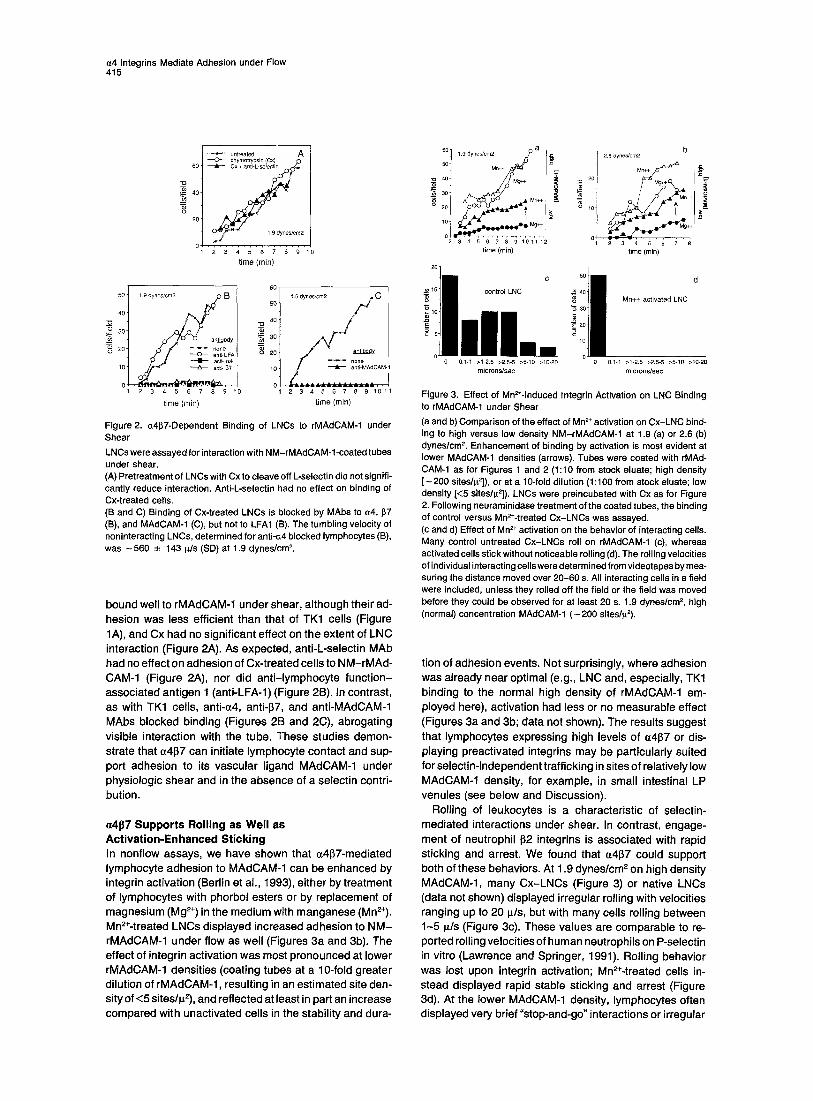
Figure 2. a4~7-Dependent Binding of LNCs to rMAdCAM-1 under Shear
LNCs were assayed for interaction with NM-rMAdCAM-l-coated tubes under shear. (A) Pret reatment o f LNCs with Cx to cleave off L-selectin did not signifi- cantly reduce interaction. Anti-L-selectin had no effect on binding of
Cx-treated cells. (B and C) Binding of Cx-treated LNCs is blocked by MAbs to ~4, ~7
(El), and MAdCAM-1 (C), but not to LFA1 (8). The tumbling velocity of noninteracting LNCs, determined for anti-c.4 blocked lymphocytes (B),
was ~ 5 6 0 __. 143 ~/s (SD) at 1.9 dynes/cm 2.
bound well to rMAdCAM-1 under shear, although their ad- hesion was less efficient than that of TK1 cells (Figure 1A), and Cx had no significant effect on the extent of LNC interaction (Figure 2A). As expected, anti-L-selectin MAb had no effect on adhesion of Cx-treated cells to NM-rMAd- CAM-1 (Figure 2A), nor did anti-lymphocyte function- associated antigen 1 (anti-LFA-1) (Figure 2B). In contrast, as with TK1 cells, anti-~4, anti-137, and anti-MAdCAM-1 MAbs blocked binding (Figures 2B and 2C), abrogating visible interaction with the tube. These studies demon- strate that ~41~7 can initiate lymphocyte contact and sup- port adhesion to its vascular ligand MAdCAM-1 under physiologic shear and in the absence of a selectin contri- bution.
a4J~7 Supports Rolling as Well as Activation-Enhanced Sticking In nonflow assays, we have shown that a4~7-mediated lymphocyte adhesion to MAdCAM-1 can be enhanced by integrin activation (Berlin et al., 1993), either by treatment of lymphocytes with phorbol esters or by replacement of magnesium (Mg 2÷) in the medium with manganese (Mn2÷). Mn2+-treated LNCs displayed increased adhesion to N M - rMAdCAM-1 under flow as well (Figures 3a and 3b). The effect of integrin activation was most pronounced at lower rMAdCAM-1 densities (coating tubes at a 10-fold greater dilution of rMAdCAM-1, resulting in an estimated site den- sity of <5 sites/12), and reflected at least in part an increase compared with unactivated cells in the stability and dura-
M ~=,_~ M
t ;
time (min) time (rain)
l, il c d
_~ 15 control LNC n ~ Mn++ activated LNC "6
l o
5 o. 0 0.1.1 >1-2.5 >2,5-5 >5-10 >10-20 o 0,1-1 >1-2.5 >2,5-5 >5-1o >10-20
microns/sec microns/see
Figure 3. Effect of Mn~-Induced Integrin Activation on LNC Binding to rMAdCAM-1 under Shear
(a and b) Compar ison of the effect of Mn 2+ activation on Cx-LNC bind- ing to high versus low density NM-rMAdCAM-1 at 1.9 (a) or 2.6 (b)
dynes/cm 2. Enhancement of binding by activation is most evident at lower MAdCAM-1 densities (arrows). Tubes were coated with rMAd- CAM-1 as for Figures 1 and 2 (1:10 from stock eluate; high density [ - 2 0 0 sites/~2]), or at a 10-fold di lut ion (1:100 from stock eluate; low density [<5 sites/~,2]). LNCs were preincubated with Cx as for Figure 2. Following neuraminidase treatment of the coated tubes, the binding of control versus Mn2+-treated Cx-LNCs was assayed. (c and d) Effect of Mn 2+ activation on the behavior of interacting ceils. Many control untreated Cx-LNCs roll on rMAdCAM-1 (c), whereas activated cells stick without noticeable rolling (d). The rolling velocities of individual interacting cells were determined from videotapes by mea- suring the distance moved over 2 0 - 6 0 s. All interacting cells in a field were included, unless they rolled off the field or the field was moved before they could be observed for at least 20 s. 1.9 dynes/cm 2, high (normal) concentration MAdCAM-1 ( - 2 0 0 sites/~2).
tion of adhesion events. Not surprisingly, where adhesion was already near optimal (e.g., LNC and, especially, TK1 binding to the normal high density of rMAdCAM-1 em- ployed here), activation had less or no measurable effect (Figures 3a and 3b; data not shown). The results suggest that lymphocytes expressing high levels of ~4137 or dis- playing preactivated integrins may be particularly suited for selectin-independent trafficking in sites of relatively low MAdCAM-1 density, for example, in small intestinal LP venules (see below and Discussion).
Rolling of leukocytes is a characteristic of selectin- mediated interactions under shear. In contrast, engage- ment of neutrophil 62 integrins is associated with rapid sticking and arrest. We found that ~4137 could support both of these behaviors. At 1.9 dynes/cm 2 on high density MAdCAM-1, many Cx-LNCs (Figure 3) or native LNCs (data not shown) displayed irregular rolling with velocities ranging up to 20 p./s, but with many cells rolling between 1-5 ~/s (Figure 3c). These values are comparable to re- ported rolling velocities of human neutrophils on P-selectin in vitro (Lawrence and Springer, 1991). Rolling behavior was lost upon integrin activation; Mn2+-treated cells in- stead displayed rapid stable sticking and arrest (Figure 3d). At the lower MAdCAM-1 density, lymphocytes often displayed very brief "stop-and-go" interactions or irregular

Cell 416
10 20 30 40 50 60 % of TK1 cells interacting
0 10 20 3o 40 50
% of lymphocytes interacting
Figure 4. ~4~7-Dependent Lymphocyte Attachment to LP Venules In Situ Fluorescence-labeled TK1 cells (A) or lymph node cells (B) were in- jected intravenously into anesthetized recipients. Cells entering LP venuies were observed in the exteriorized small intestine under epiflu- orescence microscopy, and their behavior was recorded for subse- quent video analysis. The effect of MAbs on the frequency of interac- tion was assessed either by preincubating and injecting lymphocytes with an excess of MAb (generally 0.25-0.5 mg), or by injecting MAbs after the cells, which allowed assessment of the behavior of the lym- phocytes before and after MAb blockade in the same venules. The mean frequency of interaction in three to six experiments is presented with SEM. In cases in which one or two experiments were performed, the mean is illustrated and presented with individual values. In (B), control MAbs included anti-L-seiectin (400/0 of cells interacting), anti- LFA1 (46%), corn bined treatment with anti-L-selectin and then to LFA-1 (50O/o), and neuraminidase pretreatment combined with anti-L-selectin (26%).
skipping, whereas Mn2÷-activated cells again displayed more frequent sticking. Thus, a4137 can (like the selectins) support transient interactions, which seem to be favored at lower receptor densities and which can involve either rolling or rapid stop-and-go events, as well as shear- resistant sticking favored at high receptor densities and following integrin activation.
a41~7 Mediates Lymphocyte Contact and Adhesion in LP Venules In Situ To determine whether ~4137 could support lymphocyte at- tachment to ECs under physiologic conditions in vivo, we carried out in situ videomicroscopic studies of lymphocyte interactions with venules in the small intestinal LP. These vessels were selected because they express MAdCAM-1, yet are thought to lack L-selectin-binding carbohydrates (reviewed by Picker and Butcher, 1992). The intestine was exteriorized and positioned for epifluorescence videomi- croscopic analyses. Sample lymphocytes were labeled with fluorescent dyes and injected intravenously, and the behavior of cells entering LP venules was recorded for subsequent video analyses. As we were interested in as- sessing the ability of ~4137 to contribute to cell contact and attachment in this physiologic setting, all vessel wall interactions of at least 1 s duration were scored, whether transient or leading to stable arrest.
~4l~7 h~ TK1 cells displayed a significant frequency of in- teraction in LP venules (Figure 4A), and this was corn-
pletely abrogated by anti-~4 MAbs, which prevented all visible interactions with the vessel wall. Anti-MAdCAM-1 MAb MECA-367 also blocked functional contact but, as expected from the lack of significant L-selectin expression by TK1 cells, anti-L-selectin MAb had no effect. Anti-LFA-1 MAb also failed to prevent attachment (Figure 4A).
In contrast with TK1 cells, with rare exceptions, resting LNCs failed to interact detectably with the venular wall, confirming the inability of L-selectin to mediate attachment in this site. Integrin activation with Mn 2÷, or by brief pre- treatment with phorbol myristate acetate (PMA), however, led to a substantial frequency of attachment, nearly com- parable in some experiments to that of TK1 cells; and this was also inhibited by anti-~4, anti-137, and anti-MAdCAM-1 MAbs (Figure 4B). In contrast, in separate individual exper- iments, activated lymphocytes retained the ability to inter- act well in the presence of MAbs to L-selectin, LFA-1, com- bined L-selectin plus LFA-1, or following neuraminidase pretreatment plus anti-L-selectin (data pooled as control MAb treatments in Figure 4B).
Although our focus in these in situ studies is on the ability of ~4~7 to initiate functional interactions, whether transient or stable, it is also of interest that the behavior of lymphocytes during contact with the venular wall was variable: some TK1 or activated LNCs displayed very brief (<1 s) glancing or stop-and-go contact (not scored for data analysis, but also inhibited by anti-a4137 and anti.MAdCAM-1 MAbs). Most cells, however, interacted for/>1 s, and of these, the majority displayed rapid sticking (as seen most often for activated lymphocytes and TK1 cells in vitro), although in vivo many arrested cells subsequently re- leased back to the circulation within a few seconds. Rolling and mixed behaviors were also common, however, with 15o--25% of interacting TK1 cells rolling for 1 s or longer in different experiments.
The WSS in LP venules was calculated using the maxi- mal velocity of noninteracting cells as an estimate of cen- terline blood velocity and determining mean venule diame- ters from videotape analyses. Calculated values ranged from 11-34 dynes/cm 2 (mean 18 _ 7 SD, n = 10 venules from four animals).
These studies strongly support the ability of a4137 to mediate selectin-independent contact and attachment of lymphocytes to MAdCAM-1 in LP venules in situ. They also confirm the importance of integrin activation in regulating lymphocyte interactions with ECs under physiologic condi- tions.
a41~7 Is Presented on Microvilli Leukocytes in the blood are characterized by a remarkable display of cell surface microvilli, which are thought to rep- resent the principal sites of initial contact with the vascular endothelium under flow (van Ewijk, 1980). L-selectin, which can initiate leukocyte interactions with its vascular ligands under shear (von Andrian et al., 1991 ; Berg et al., 1993; Lawrence et al., submitted), is displayed selectively on these microvillous processes (Picker et al., 1991; Erland- sen et al., 1993). Scanning electron microscopy of TK1 cells reveals the typical topography of an unactivated lym- phocyte with numerous microvilli extending out from the

(~4 Integrins Mediate Adhesion under Flow 417
Figure 5. Immunolocalization of Cell Adhe- sion Molecules on Unstimulated TK1 Cells by Low Voltage Scanning Electron Microscopy (a) Low magnification image illustrating the sur- face morphology of TK1 cells (bar = 1.0 p.m). (b) High magnification image of cells stained for the 132 (CD18) integrin LFA1 (bar = 200 nm). Note imrnunogold staining on membrane surface of cell body (arrowheads). (c and d) immunogold staining for (c) (x4 and (d) 137 (bar = 400 nm). Both (~4 and 137 are predomi- nantly detected on microvilli and membrane ruffles (arrowheads).
cell body (Figure 5a), and immunogold staining for (~4 or 137 (Figures 5c and 5d) demonstrates that, like L-selectin, the a4 and 157 integrin chains are localized primarily on these processes, with many of the gold particles at or near the tips of the microvilli. Analyses by stereoscopic examina- tion reveals that - 80% of gold particles labeling a4 or 137 were detected on microvilli ((~4: 79% on villi, n = 5 cells, 938 gold particles analyzed; 137: 79%, n = 6, 1063 gold particles). This distribution is in sharp contrast to that of LFA-1 (aLl32), which, as shown previously for the 132 inte- grin Mac-1 on unactivated neutrophils (Erlandsen et al., 1993), is largely excluded from TK1 microvilli: (132: 88% on planar cell body, n = 5 cells, 1135 gold particles) (Fig- ure 5b).
Lymphocyte Attachment through a4 Integrin- VCAM-1 but Not LFA-1 (¢tLp2)-ICAM-1 Pathways under Shear To determine whether the capacity to initiate functional contact is unique to MAdCAM-1, or is shared with the re- lated (~4 integrin ligand VCAM-1, we assessed lymphocyte interactions with VCAM-1 under flow. We also compared interactions with intercellular adhesion molecule I (ICAM-1), an LFA-1 Iigand. In the representative experiment illus- trated in Figure 6, VCAM-1 was coated at a density of - 37 sites/l~2; MAdCAM-1 was titred to yield similar densities of - 2 0 sites/~2; and ICAM-1 was used at - 4 0 0 or 1000 sites/l~ 2. The ICAM-1 was functional in static assays (data not shown), binding activated LNCs as well as or better
than MAdCAM-I. In contrast, MAdCAM-1 and VCAM-1 but not ICAM-1 supported attachment and accumulation of Mn2÷-activated LNCs at 1.9 dynes/cm 2. Detectable inter- actions with ICAM-l-coated segments of the tube were extremely infrequent.
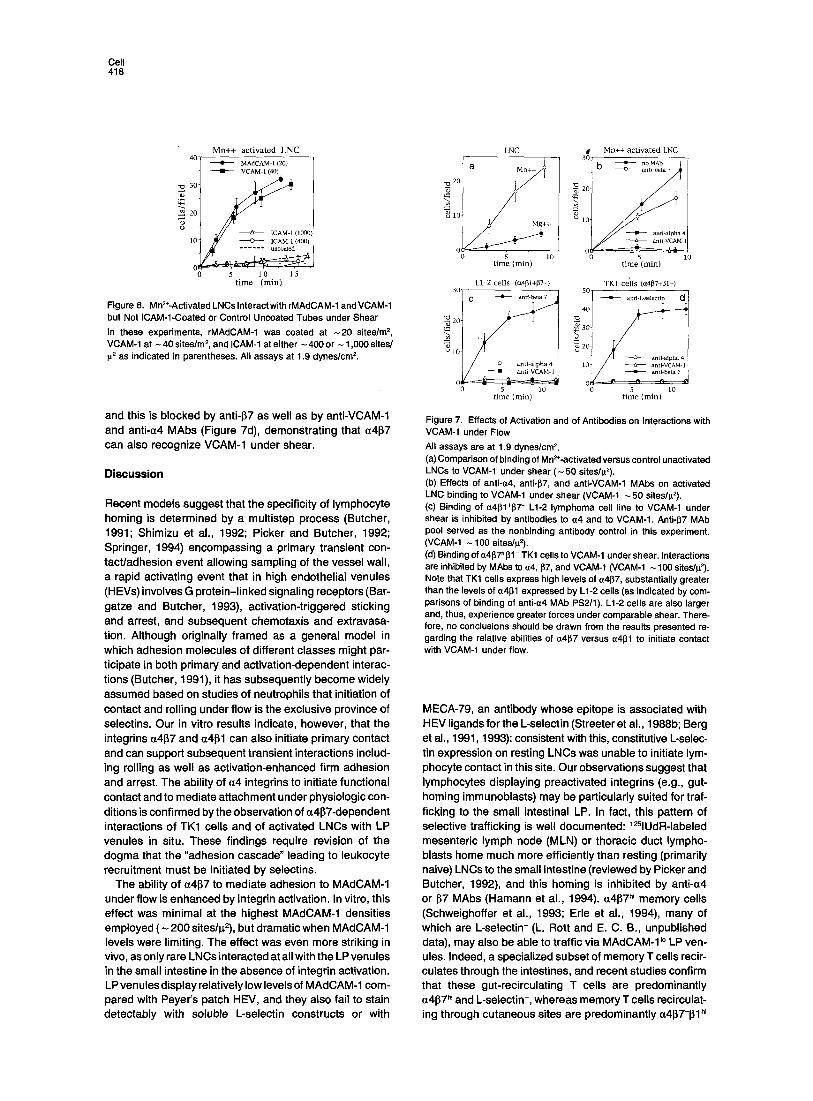
Interactions of LNCs with VCAM-l-coated tubes were greatly enhanced by Mn 2+ pretreatment (Figure 7a), indi- cating that integrin activation is important for lymphocyte contact and adhesion through VCAM-1 as well as through MAdCAM-1 under shear at these relatively low site densi- ties. As on MAdCAM-1, resting LNCs interacting with VCAM-1 displayed transient stop-and-go and irregular roll- ing behaviors, whereas rapid arrest was favored following integrin activation. As shown in Figure 7b, treatment with anti-c(4 or anti-VCAM-1 abrogated all visible interaction. Anti-l~7 MAbs, which completely blocked interaction with MAdCAM-1 assayed in parallel (data not shown), reduced binding to VCAM-1 only minimally. The efficent interaction of anti-137-treated LNCs with VCAM-1 is consistent with participation of a4131.
To confirm the ability of (~4131 to mediate binding to VCAM-1 under shear, we assessed interaction of the L1-2 lymphoma cell line, which expresses (~41~1 but not 137 or L-selectin (Andrew et al., 1994). Although L1-2 cells are considerably larger than LNCs or TK1 cells and tumble at significantly higher velocity (940 _ 160 I~/s), they none- theless interacted well, and this was inhibited by anti-(~4 and by anti-VCAM-1 MAbs (Figure 7c). TK1 cells, which express (~4137 but not (~4131, also interact with VCAM-1,
Cell 418
Mn++ act ivated LN C 40
MAdCAM- 1 (20) • VCAM-t (4O)
'~'~ 30
"~ 20
(1oo0) 10 ~ ~ ICAM-I (400)
~ ~ u n c o a t e d
5 10 15 t ime (min)
Figure 6. Mn=*-ActivatedLNCslnteractwithrMAdCAM-1 andVCAM-1 but Not ICAM-1-Coated or Control Uncoated Tubes under Shear In these experiments, rMAdCAM-1 was coated at -20 sites/m =, VCAM-1 at - 40 sites/m s, and ICAM-1 at either - 400 or - 1,000 sites/ g= as indicated in parentheses. All assays at 1.9 dynes/cmE
and this is blocked by anti-137 as well as by anti-VCAM-1 and anti-~4 MAbs (Figure 7d), demonstrating that ~4137 can also recognize VCAM-1 under shear.
Discussion
Recent models suggest that the specificity of lymphocyte homing is determined by a multistep process (Butcher, 1991; Shimizu et al., 1992; Picker and Butcher, 1992; Springer, 1994) encompassing a primary transient con- tact/adhesion event allowing sampling of the vessel wall, a rapid activating event that in high endothelial venules (HEVs) involves G protein-linked signaling receptors (Bar- gatze and Butcher, 1993), activation-triggered sticking and arrest, and subsequent chemotaxis and extravasa- tion. Although originally framed as a general model in which adhesion molecules of different classes might par- ticipate in both primary and activation-dependent interac- tions (Butcher, 1991), it has subsequently become widely assumed based on studies of neutrophils that initiation of contact and rolling under flow is the exclusive province of selectins. Our in vitro results indicate, however, that the integrins cz4137 and ~4131 can also initiate primary contact and can support subsequent transient interactions includ- ing rolling as well as activation-enhanced firm adhesion and arrest. The ability of ~4 integrins to initiate functional contact and to mediate attachment under physiologic con- ditions is confirmed by the observation of ~4137-dependent interactions of TK1 cells and of activated LNCs with LP venules in situ. These findings require revision of the dogma that the "adhesion cascade" leading to leukocyte recruitment must be initiated by selectins.
The ability of a4~7 to mediate adhesion to MAdCAM-1 under flow is enhanced by integrin activation. In vitro, this effect was minimal at the highest MAdCAM-1 densities employed (~ 200 sites/p2), but dramatic when MAdCAM-1 levels were limiting. The effect was even more striking in vivo, as only rare LNCs interacted at all with the LP venules in the small intestine in the absence of integrin activation. LP venules display relatively low levels of MAdCAM-1 com- pared with Peyer's patch HEV, and they also fail to stain detectably with soluble L-selectin constructs or with
LNC
a
"~ 11 Mg++
5 l0 t ime (min)
L1-2 ceils (a4~1+[82-) 30-
~ 20-
o ~ " , ~ 5 10
time (min)
Mn++ activated LNC 30
b • ,oMAb I 1 anti-be~ 7
-~ 2o
• anu-Npha 4 -----tr--- anti-VCAM-I
0 o 5 lO
t ime (min)
TK1 ceils (o~11~7+1~I-)
• anti-L-sele
~. so
lO
o- 5 lO
t ime (min)
Figure 7. Effects of Activation and of Antibodies on Interactions with VCAM-1 under Flow All assays are at 1.9 dynes/cm 2. (a) Comparison of binding of Mn2÷-activated versus control unactivated LNCs to VCAM-1 under shear (-50 sites/p2). (b) Effects of anti-~4, anti-1~7, and anti-VCAM-1 MAbs on activated LNC binding to VCAM-1 under shear (VCAM-1 -50 sites/p2). (c) Binding of ~4l~1÷~7 - L1-2 lymphoma cell line to VCAM-1 under shear is inhibited by antibodies to ~z4 and to VCAM-I. Anti-1~7 MAb pool served as the nonbinding antibody control in this experiment. (VCAM-1 - 100 sites/g2). (d) Binding of ~z4[~7~J31- TK1 cells to VCAM-1 under shear. Interactions are inhibited by MAbs to a4, 97, and VCAM-1 (VCAM-1 ~ 100 siteslp2). Note that TK1 cells express high levels of ~4~7, substantially greater than the levels of ~4~1 expressed by L1-2 cells (as indicated by com- parisons of binding of anti-a4 MAb PS2/1). L1-2 cells are also larger and, thus, experience greater forces under comparable shear. There- fore, no conclusions should be drawn from the results presented re- garding the relative abilities of a4~7 versus ct41~1 to initiate contact with VCAM-1 under flow.
MECA-79, an antibody whose epitope is associated with HEV ligands for the L-selectin (Streeter et al., 1988b; Berg et al., 1991, 1993): consistent with this, constitutive L-selec- tin expression on resting LNCs was unable to initiate lym- phocyte contact in this site. Our observations suggest that lymphocytes displaying preactivated integrins (e.g., gut- homing immunoblasts) may be particularly suited for traf- ficking to the small intestinal LP. In fact, this pattern of selective trafficking is well documented: 12qUdR-labeled mesenteric lymph node (MLN) or thoracic duct lympho- blasts home much more efficiently than resting (primarily naive) LNCs to the small intestine (reviewed by Picker and Butcher, 1992), and this homing is inhibited by anti-~4 or [57 MAbs (Hamann et al., 1994). ~4137 h~ memory cells (Schweighoffer et al., 1993; Erie et al., 1994), many of which are L-selectin- (L. Rott and E. C. B., unpublished data), may also be able to traffic via MAdCAM-I~° LP ven- ules. Indeed, a specialized subset of memory T cells recir- culates through the intestines, and recent studies confirm that these gut-recirculating T cells are predominantly ~4~7 h~ and L-selectin-, whereas memory T cells recirculat- ing through cutaneous sites are predominantly Q4~7-131 "~

a4 Integrins Mediate Adhesion under Flow 419
(Mackay, 1993; C. Mackay and E. C. B., unpublished data). It is important to note that our results suggest that lymphoblasts expressing preactivated integrins, and pos- sibly memory cells displaying very high levels of a4131 or ~4137, may have an ability to attach and arrest on ECs without any requirement for local, vessel-associated acti- vating signals, thus bypassing step 2 (activation) in the general multistep paradigm of leukocyte-EC recognition mentioned above. It will be important to assess the require- ment for G protein signaling in lymphoblast interactions with LP venules in situ and in subsequent diapedesis.
HEVs in the mucosal Peyer's patches and MLNs ex- press much higher levels of MAdCAM-1 than LP venules. Furthermore, these HEVs can modify MAdCAM-1 as well as other glycoproteins with L-selectin-binding carbohy- drate ligands (Berg et al., 1993), allowing participation of both L-selectin and a41~7 in lymphocyte homing to these mucosal lymphoid organs (Hamann et al., 1994). This combination of ligands may help explain the remarkably efficient homing of naive lymphocytes (which are uniformly L-selectin h~, ~4~7 ~°) to Peyer's patches: L-selectin would facilitate initial attachment, and the expression of high lev- els of MAdCAM-1 should support efficient binding even of naive a4137 t° lymphocytes. Consistent with this model, recent in vivo homing studies demonstrate that both anti-L- selectin and anti-a4 or 97 MAbs display significant inhibi- tion of short-term homing of LNCs to Peyer's patches (Ha- mann et al., 1994).
It is of interest that WSSs that support selectin and a4 integrin-mediated leukocyte contact in vitro (generally ~<4 dynes/cm 2) are at the very low end of physiologic shear stresses reported for postcapillary venules that support leukocyte rolling in vivo. In the present studies, for exam- ple, TK1 cells and activated LNCs interacted reasonably efficiently with LP venules in situ, in spite of physiologic shear rates in the range of 11-34 dynes/cm 2, In contrast, under the controlled laminar flow conditions in vitro, few if any interactions were detectable above 4 dynes/cm 2. A variety of rheological and physical parameters may oper- ate in vivo to enhance the efficiency of interactions under flow, including displacement of leukocytes from the mid- line by erythrocytes (von Andrian and Arfors, 1993), non- laminar flow conditions associated with branch points or turns in vessels and irregularities in the EC lining, and turbulence produced by previously adherent leukocytes.
Microvillous projections represent the initial site of cell- cell contact under flow, and we have shown here that a4~7 is concentrated on these projections. L-selectin, which is also highly effective at initiating contact under shear, is similarly concentrated on microvilli (Picker et al., 1991; Erlandsen et al., 1993), suggesting that this may be a com- mon feature of molecules specialized for initiating leuko- cyte-EC interactions under physiologic conditions. The microvillous distribution of L-selectin and a41~7 is in con- trast with the 132 integrins, which are concentrated on the nonvillous planar cell body (Erlandsen et al., 1993). Our present results as well as others demonstrate that 132 inte- grins are largely unable to initiate functional contact in vitro or in vivo under physiologic shear (von Andrian and Arfors, 1993; Springer, 1994). We propose that the exclu-
sion of 132 integrins from the tips of microvilli may in fact ensu re that they participate selectively in events that follow initial cellular contact in vivo, possibly including slowing of rolling as well as activation-dependent sticking. It will be important to determine the topographic distribution of other integrins implicated in EC interactions, including ~4~1 but also a61~1 (Dunon and Imhof, 1993), to define the molecular signals that target a4~7 and L-selectin to microvilli, and to ask whether the topographic distribution of receptors can be regulated independently of overall lev- els of expression. Such regulation could allow an addi- tional level of control of the ability of adhesion receptors to initiate leukocyte attachment under flow.
The ligand densities that support a4 integrin-mediated lymphocyte contact and rolling or adhesion are similar to those reported to support rolling of neutrophits on P-selectin
• (Lawrence and Springer, 1991). Interaction of ~4137 "~ TK1 cells is extremely efficient on rMAdCAM-1 at - 200 sites/ ~2, with almost all cells in the focal plane of the capillary wall attaching rapidly. At lower ligand densities, contact is less efficient and, when initiated, often leads to irregular jerky rolling and skipping behaviors. Transient irregular interactions were exaggerated in experiments at the low- est MAdCAM-1 densities (e.g., <5 sites/l~ 2 in Figure 3). It is likely that such irregular behavior reflects the making and breaking of individual microvillous contacts, perhaps involving only one or a few molecular bonds. In scanning electron microscopic images of glutaraldehyde-fixed TK1 cells, the diameters of microvilli range from 60-100 nm, and the area of a microvillous tip can be roughly approxi- mated as ~<0.01 i~ 2. The area available for interaction under flow is likely to be in this range as well (although a some- what larger area could be available if microvilli are flexed). Moreover, it appears that no more that 50-100 microvilli could reasonably contact a surface at a given instant in the absence of extensive cellular deformation. Thus at 50 sites/p. 2, each microvillus is likely to contact 0 or 1 sites on average; and as a rough approximation, the comple- ment of microvilli available for interaction at any instant could potentially contact 25-50 sites. The ability of cells to initiate functional contact and roll even if unevenly under these circumstances suggests that lymphocytes must be quite efficient at taking advantage of these potential inter- actions. The high density of a4 integrins and L-selectin receptors on the tips of microvilli may be critically im- portant in this regard.
We have focused here primarily on interactions of ~4~7 with its vascular ligand MAdCAM-1. MAdCAM-1 is a muco- sal vascular addressin, selectively expressed by venules involved in lymphocyte trafficking to mucosal tissues (Streeter et al., 1988a). The a4 integrins can also bind to VCAM-1, however, a related EC immunoglobulin family member that is induced on ECs in sites of tissue inflamma- tion and that serves as a ligand for ~41~1 and for activated ~4~7, and we have shown that LNCs and a41~1+~7 - L1-2 lymphoid cells can make contact and roll on VCAM-1 and that attachment and accumulation on VCAM-1 are en- hanced by integrin activation, a4137 on ~4137 "~ TK1 cells can also initiate adhesion to VCAM-1 but anti-137 MAbs reduce LNC interaction with VCAM-1 only slightly, sug-

Cell 420
gesting that (~4131 may predominate in LNC interactions with VCAM-1 under shear (as reported previously based on nonflow assays; Berlin et al., 1993). Our results indicate that the (x4 integrins are highly versatile, with the potential
to play a critical role in initiating physiologic interactions and rolling of lymphocytes under flow in vivo, as well as in secondary, act ivat ion-dependent sticking and arrest. This conclusion is supported by the recent demonstrat ion that (~4131 can participate in rolling of eosinophils in situ in inter- leukin-1.-activated rabbit mesenteric venules (Sriramarao et al., 1994). and by independent studies of c(4~ 1-dependent
lymphocyte attachment to VCAM-1 transfectants or pro- tein or to ECs under flow (Wolber et al., 1993; Jones et al., 1995; Alon et al., 1995; Stoolman et al., unpublished data). In the case of lym phocytes, the contribution of the (z4 integrinlVCAM-1 pathway to primary attachment is likely to be especial ly important for trafficking of immunoblasts
and/orL-select in- memory lymphocyte subsets to sites of tissue inflammation.
In conclusion, the (x4 but not ~2 integrins can mediate lymphocyte contact and adhesion under physiologic shear and in the absence of selectin involvement. Adhesion is sensitive to and can therefore be regulated physiologically by multiple parameters, including the following: the level of integrin expression, the extent of integrin activation, the density of l igand decorating target surfaces, and the local
microvascular hemodynamics. These parameters likely work in concert with engagement of selectins and of other adhesion and activating factors, to fine-tune the homing of lymphocytes to mucosal lymphoid tissues and to the intestinal wall, and to tissue sites of inflammation. Finally, our results suggest that topographic presentation on mi- crovilli may be a critical determinant of adhesion under
flow, potential ly allowing diverse adhesion receptors to engage their vascular l igands under physiologic shear
stresses in vivo.
Experimental Procedures
Antibodies MAbs utilized were the following: anti-a4 PSI2, anti-VCAM-1 MK2.7, anti-LFA-1 ~ chain FD441.8, anti-L-selectin MEL-14, anti-!CAM-1 YNI/ 1.7 (ATCC); anti-~4~7 heterodimer DATK32 and anti-137 MAbs FIB22, FIB21, FIB30, and FIB504 (Andrew et al., 1994); anti-CD44 MAb M J64 (M. Jutila and E. C. B., unpublished data); and anti-MAdCAM-1 MECA- 367 (Streeter et al., 1988a).
Lymph Node Lymphocytes and Cell Lines The AKR/Cum TK1 cell line and the C57L pre-B lymphoma line L1-2 have been described (Butcher et al., 1980; Hu et al., 1992; Berlin et al., 1993) and were cultured in RPM11640 with 5°/0 iron supplemented bovine calf serum (BCS) (Hyclone Labs, Logan, ur). TK1 variants exhibiting plastic adherence were excluded from these assays. LNCs were isolated from 6- to 8-week-old BALB/c mice, resuspended in RPMI plus 5O/o BCS 20 mM HEPES (pH 7.0) medium at room tempera- ture unless otherwise specified, and then used within 45-60 min.
Assay of Adhesion under Flow rMAdCAM-1 was isolated by MECA-367 antibody affinity chromatogra- phy as described (Berlin et al., 1993; Nakache et al., 1989; purity of isolated MAdCAM-1 is illustrated for example in the latter reference) from lysates of mouse L1-2 pre-B lymphoid cells stably transfected with pMAd7 cDNA (Briskin et al., 1993) in pMRB101 (L1-2 ~dcAu-1 cells; see Berg et al., 1993 for transfection protocols). Isolated rMAdCAM-1 in elution buffer containing 1% ~-octylglucoside was diluted below the
critical micelle concentration with phosphate buffered saline (PBS), -50 p.I was taken up into one half of a glass capillary tube (100 p_l disposable "rnicrocaps", 1.025 mm I.D., Drummond Scientific Com- pany, Broomall, PA), and the tubes were incubated in humidity at 4°C overnight. After coating, tubes were blocked with 100% BCS. Cells (1. x 106-2 x 10e/ml in different experiments) were transfused at uni- form flow rate through the capillary tubes using a syringe pump (Model 33, Harvard Instruments). Except as indicated, cells were only used for a single passage. After a 1-2 min stabilization period, the number of cells binding per randomly selected 0.2 mm 2 microscope field was determined at 30 s intervals by computerized image analysis of video- tapes. WSSs were calculated from Poiseuille's law for Newtonian flu- ids, with viscosity 0.01 P. [WSS in dynes/cm 2 = mean flow velocity (mm/s) x (8 + tube diameter (mm)) x viscosity (P)].
ICAM-1 and VCAM-1 were isolated from mouse spleen lysates by affinity chromatography on YNI/1.7- or MK2.7-conjugated Sepharose columns, and flow essays were performed as above except that for data collection six 0.2 mm 2 microscope fields were recorded as a group in rapid succession (one each 15 s) from one coated region of the tube. The microscope was then shifted to an uncoated region for two control fields and then to another coated area for six. The number of cells binding was determined in each field recorded, and the mean for each group calculated.
Ligand densities were determined by saturation binding of radiola- beled MAbs as described by Lawrence and Springer (1991). MAbs against MAdCAM-1 (MECA-367), ICAM-1 (YNl/1.7), or VCAM-1 (MK2.7) were iodinated (Iodo-Beads, Pierce, Rockford, IL) to known specific activity (1 x 10¹⁷-5 x 10¹⁷ CPM/M in different experiments) and incubated above saturation in coated capillary tubes or in control mock-coated or, in some experiments, irrelevant ligand-coated capil- lary tubes. After extensive washing, the tubes were counted for calcula- tion of site densities assuming monomeric binding (Lawrence and Springer, 1991). Site densities for a given ligand preparation showed variation from day to day within a 2-fold range. Densities for rMAd- CAM-1 as used in Figures 1-4 were determined after the experiments, using the same fraction of purified rMAdCAM-1 coated under identical conditions. Densities for Figures 6 and 7 were determined concurrently with the adhesion assays presented, on tubes coated in parallel.
Antibody, Enzyme, and Ion Treatments For in vitro antibody inhibition studies, all antibodies were used at 5- to 10-fold over saturating levels as determined by flow cytometry (10- 25 p.g/ml). Anti-lymphocyte antibodies were preincubated with test cells for 20-30 min at room temperature and were not washed away prior to assay. FIB3O was used for 1~7 inhibition in Figures 1, 2, and 4; a pool of FIB30, FIB504, and DATK32 MAbs was employed for Figures 6 and 7. Anti-MAdCAM-1 MAb MECA-367 was incubated at 50 i~g/ml in the tube for 20 min at room temperature.
For neuraminidase treatment, lymphocytes at 107 cells/ml or the coated tubes were treated with neuraminidase (sialidase) from Arthro- bacter ureafaciens (Boehringer, Mannheim, Germany) at 40 U/ml in medium for 30 min at 37°C; this treatment effectively cleaved the sLeX-associated HECA-452 epitope from U937 cells (>98% reduction in specific staining).
For chymotrypsin treatment, LNCs were incubated 5 min at 37°C in HBSS plus 10 mM HEPES (pH 7.0) containing 0.1 U/106 cells ~-chy- motrypsin (Fraction IV-S from bovine pancreas, Sigma, St. Louis), washed, and resuspended in medium for assay. Cx treatment reduced staining by anti-L-selectin MAb MEL-14 by >97%.
For activation, LNCs were incubated at room temperature in Ca2*/ Mg2+-free HBSS plus 10 mM HEPES (pH 7.0) (HBSS/HEPES) con- taining 2 mM EDTA and were then diluted 3-fold with Ca2+/Mg2+-free HBSS/HEPES, pelleted, and resuspended in HBSS/HEPES con- taining either 2 mM CaCI2 and MgCI2 (control cells), or 2 mM CaCI2 and 1 or 2 mM MnCI2 (Mn2+-activated cells).
In Situ Videomicroscopic Analyses of Lymphocyte Interactions with LP Venules In situ videomicroscopic analyses were carried out as described (Bar- gatze and Butcher, 1993) except that the exteriorized bowel segment was positioned for epifluorescence microscopy and video recording of LP venules in the small intestine. Approximately 2.5 x 10⁷ fluoro- chrome-labeled cells (either TK1 or LNC) in 0.5 ml DM EM were injected

(~4 Integrins Mediate Adhesion under Flow 421
intravenously into the tail vein. In some experiments, TK1 cells or lymphocytes were pretreated with anti-lymphocyte MAbs (100 ~g/ml) at room temperature for 20 min (not washed) before injection with an excess of MAb (250-500 p.g). In others, lymphocytes were injected and observed for 3-7 rain and then 250 or 500 ~.g of MAb was infused intravenously with or without additional cells, allowing visualization of the effect of MAb in the same vessels. Analyses were routinely initiated immediately after infusion of sample cells. When antibody was admin- istered after initial cell infusion, the antibodies were allowed to circulate and achieve saturation for 2 min (for anti-lymphocyte antibodies) or - 5 min (for anti-MAdCAM-1 MAb) prior to further analysis of cell be- havior. All cells entering the observed venules were analyzed for each treatment (range 25-100 cells per experiment). Interactions of >_-1 s were considered significant and were scored. For determination of WSS in vivo, ten venules from four different animals were analyzed. In each venule, the velocity of ten noninteracting lymphocytes was assessed. The highest velocity observed was taken as a conservative estimate of centerline blood velocities (range 2.2 to 5.0 ram/s). The mean diameter of vessels was measured by videotape analysis (range 12-42 it). WSSs were calculated from these parameters as described (von Andrian et al., 1992).
Scanning Electronmicroscopy TK1 cells were stained with primary MAbs against LFA1 (FD448.1), 64 (PS2/1), and 137 (FIB21), incubated with secondary goat anti-mouse immunoglobulin 12 nm colloidal gold, and prepared for imaging as described (Erlandsen et al., 1993). No significant staining was seen with second stage only. Samples were examined using a Hitachi S-900 field emission SEM equipped with a YAG crystal for high resolution backscatter electron detection at an accelerating voltage of 3.5 kV.
Acknowledgments
The first three authors made equivalent if independent contributions to the in vitro flow (C. B. and J. J. C.) or in vivo (R. F. B) studies reported. The authors thank M. Briskin for pMAd7 transfectants, David P. Andrew for antibodies, S. Michie for earty transmission electromi- croscopic studies of 64 localization on microvilli, L. Rott for assistance with fluorescence-activated cell sorting (FACS), A. Hamann for the gift of Fab fragments, L. McEvoy and D. Daignault for critical review of the manuscript, M. Jutila for discussions, and S. Grossman for admin- istrative assistance. Supported in part by National Institutes of Health grants GM37734, AI19957, AI37832, DK45448 (E. C. B.), and AI22374 (R. D. N.); bythe FACS facility of the Stanford Digestive Disease Center under DK38707; by funds from the Department of Veterans Affairs; and by the Minnesota Medical Foundation (S.L.E.). C. B. was supported by a fellowship of a special program to promote epidemiology and rheumatology from the Deutscher Akademischer Austauschdienst (Germany). E. L. B. was a Special Fellow of the Leukemia Society of America, U. H. v. A held a stipend from the Deutsche Forschungsgem- einschaft, and J. J. C. was supported by the Cancer, Etiology, Preven- tion, Detection, and Diagnosis training grant 5T32 CA09302 and by an individual National Institutes of Health fellowship 1F32 AI08930, and M. C. Szabo was a recipient of the Molecular and Cellular Immu- nobiology training grant 5T32 AI07290.
Received February 15, 1994; revised November 23, 1994.
References
Abassi, O., Kishimoto, T. K., Mclntire, L. V., and Smith, C. W. (1993). Neutrophil adhesion to endothelial cells. Blood Cells 19, 245-259.
Alon, R., Kassner, P. D., Carr, M. C., Finger, E. B., Hemler, M. E., and Springer, T. A. (1995). The integrin VLA-4 supports tethering and rolling in flow on VCAM-1. J. Cell Biol., in press.
Andrew, D. P., Berlin, C., Honda, S., Yoshino, T., Hamann, A., Holz- mann, B., Kilshaw, P. J., and Butcher, E. C. (1994). Distinct but overlap- ping epitopes are involved in a4J~7-mediated adhesion to VCAM-1, MAdCAM-1, fibronectin, and lymphocyte aggregation. J. Immunol. 153, 3847-3861.
Atherton, A., and Born, G. V. R. (1972). Quantitative investigations of the adhesiveness of circulating polymorphonuclear leukocytes to
blood vessel walls. J. Physiol. (Lond.) 222, 447-474.
Bargatze, R. F., and Butcher, E. C. (1993). Rapid G protein-regulated activation event involved in lymphocyte binding to high endothelial venules. J. Exp. Med. 178, 367-372.
Berg, E. L., Robinson, M. K., Warnock, R. A., and Butcher, E. C. (1991). The human peripheral lymph node vascular addressin is a ligand for LECAM-1, the peripheral lymph node homing receptor. J. Cell Biol. 114, 343-349.
Berg, E. L., McEvoy, L. M., Berlin, C., Bargatze, R. F., and Butcher, E. C. (1993). L-selectin-mediated lymphocyte rolling in MAdCAM-I. Nature 366, 695-698.
Berlin, C., Berg, E. L., Briskin, M. J , Andrew, D. P., Kilshaw, P. J., Holzmann, B., Weissman, I. L., Hamann, A., and Butcher, E. C. (1993). a4~7 integrin mediates lymphocyte binding to the mucosal vascular addressin MAdCAM-I. Cell 74, 185-195.
Briskin, M. J., McEvoy, L. M., and Butcher, E. C. (1993). MAdCAM-1 displays homology to immunoglobulin and mucin-Iike adhesion recep- tors and to IgAl. Nature 363, 461-464.
Butcher, E. C. (1991). Leukocyte-endothelial cell recognition: three (or more) steps to specificity and diversity. Cell 67, 1033-1036.
Butcher, E. C., Scollay, R., and Weissman, I. L. (1980). Organ specific- ity of lymphocyte interaction with organ-specific determinants on high endothelial venules. Eur. J. Immunol. 10, 556-561.
Dunon, D., and Imhof, B. A. (1993). Mechanisms of thymus homing. Blood 81, 1-8.
Erlandsen, S. L., Hasslen, S. R., and Nelson, R. D. (1993). Detection and spatial distribution of the 132 integrin (Mac-l) and L-selectin (LECAM-1) adherence receptors on human neutrophils by high- resolution field emission SEM. J. Histochem. Cytochem. 41,327-333. Erie, D. J., Briskin, M. J., Butcher, E. C., Garcia-Pardo, A., Lazarovits, A. I., and Tidswell, M. (1994). Expression and function of the MAd- CAM-1 receptor, integrin (14137, on human leukocytes. J. Immunol. 153, 517-528.
Hamann, A., Andrew, D. P., Jablonski-Westrich, D., Holzmann, B., and Butcher, E. C. (1994). The role of (~4-integrins in lymphocyte homing to mucosal tissues in vivo. J. Immunol. 52, 3282-3293.
Hu, M. C., Crowe, D. T., Weissman, I. L., Holzmann, B. (1992). Cloning and expression of mouse integrin 13p(137); a functional role in specific lymphocyte homing. Proc. Natl. Acad. Sci. USA 89, 8254-8258.
Jones, D. A., Mclntire, L. V., Smith, C. W., and Picker, L. J. (1995). A two-step adhesion cascade for T cell/endothelial cell adhesion under flow conditions. J. Clin. Invest., in press.
J utila, M. A., Kishimoto, T. K., and Finken, M. (1989). Low-dose chymo- trypsin treatment inhibits neutrophil migration into sites of inflamma- tion in vivo: effects on MAC-1 and M EL-14 adhesion protein expression and function. Cell. Immunol. 132, 201-214.
Kishimoto, T. K., Jutila, M. A , Berg, E. L., and Butcher, E. C. (1989). Neutrophil Mac-1 and MEL-14 adhesion proteins inversely regulated by chemotactic factors. Science 245, 1238-1241.
Lasky, L. R. (1992). Selectins: interpreters of cell-specific carbohydrate information during inflammation. Science 258, 964-959.
Lawrence, M. B., and Springer, T. A. (1991). Leukocytes roll on a selectin at physiologic flow rates: distinction from and prerequisite for adhesion through integrins. Cell 65, 859-873.
Mackay, C. R. (1993). Homing of naive, memory and effector lympho- cytes. Curr. Opin. Immunol. 5, 423-427.
Nakache, M., Berg, E. L., Streeter, P. R., and Butcher, E. C. (1988). The mucosal vascular addressin is a tissue-specific endothelial cell adhesion molecule for circulating lymphocytes. Nature 304, 32-36.
Perry, M. A., and Granger, D. N. (1991). Role of CD11/CD18 in shear rate-dependent leukocyte-endothelial cell interactions in cat mesen- teric venules. J. Clin. Invest. 87, 1798-1804.
Picker, L. J., Warnock, R. A., Burns, A. R., Doerschuk, C. M., Berg, E. L., and Butcher, E. C. (1991). The neutrophil selectin LECAM-1 presents carbohydrate ligands to the vascular selectins ELAM-1 and GMP-140. Cell 66, 921-933.
Picker, L. J., and Butcher, E. C. (1992). Physiological and molecular

Cell 422
mechanisms of lymphocyte homing. Annu. Rev. Immunol. 10, 561- 591. Postigo, A. A., Teixido, J., and Sanchez-Madrid, F. (1993). The a4J~l/ VCAM-1 adhesion pathway in physiology and disease. Res. Immunol. 144, 723-735. Schweighoffer, T., Tanaka, Y., Tidswell, M., Erie, D. J., Horgan, K. J., Luce, G. E., Lazarovits, A. I., Buck, D., and Shaw, S. (1993). Selective expression of integrin a4~7 on a subset of human CD4 + memory T cells with Hallmarks of gut-trophism. J. Immunol. 151,717-729. Shimizu, Y., Newman, W., Tanaka, Y., and Shaw, S. (1992). Lympho- cyte interactions with endothelial cells. Immunol. Today 13, 106-110.
Springer, T. (1994). Traffic signals for lymphocyte recirculation and leukocyte emigration: the multistep paradigm. Cell 76, 301-314.
Sriramarao, P., von Andrian, U. H., Butcher, E. C., Bourdon, M. A., and Broide, D. H. (1994). L-selectin and VLA-4 integrin promote eosinophil rolling at physiologic shear rates in vivo. J. Immunol. 153, 4238-4246.
Streeter, P. S., Berg, E. L., Rouse, B. T. N., Bargatze, R. F., and Butcher, E. C. (1968a). A tissue-specific endothelial cell molecule in- volved in lymphocyte homing. Nature 331, 41-46.
Streeter, P. R., Rouse, B. T. N., and Butcher, E. C. (1988b). Immunohis- tologic and functional characterization of a vascular addressin involved in lymphocyte homing into peripheral lymph nodes. J. Cell Biol. 107, 1853-1862. von Andrian, U. H., and Arfors, K.-E. (1993). Neutrophil-endothelial cell interactions in vivo: a chain of events characterized by distinct molecular mechanisms. Agents Actions Suppl. 41, 153-164.
von Andrian, U. H., Chambers, J. D., McEvoy, L., Bargatze, R. F., Arfors, K.-E., and Butcher, E. C. (1991). Two step model of leukocyte- endothelial cell interaction in inflammation: distinct roles for LECAM-1 and the leukocyte 62 integrins in vivo. Proc. Natl. Acad. Sci. USA 88, 7538---7542.
von Andrian, U. H., Hansell, P., Chambers, J. D., Berger, E. M., Filho, I. T., Butcher, E. C., and Arfors, K.-E. (1992). L-selectin function is required for ~2-integrin-mediated neurophil adhesion at physiological shear rates in vivo. Am. J. Physiol. 263, 1034-1044.
van Ewijk, W. V. (1980). Immunoelectron-microscopic characterization of lymphoid microenvironments in the lymph node and thymus. Ciba Found. Symp. 71, 21-33.
Wolber, F., Craig, R., Abassi, O., Ballew, J., Lobb, R., and Stoolman, L. M. (1993). VLA4 mediates lymphocyte binding to endothelium under shear. FASEB J. 7, 3704.


