
Acid-Base ImbalanceMetropolitan Community College
Fall 2013

Acid Base Balance
Hydrogen ions - Low concentrations but highly reactive
Concentration affects physiological functions
Alters protein and enzyme functioning Can cause cardiac, renal, pulmonary
abnormalities Alters blood clotting, Metabolization of meds

Acid and Bases
Acids – compounds that form hydrogen ion in a solution Proton donors Strong acids give up their hydrogen ion easily Weak acids hold on to their hydrogen ion more tightly
Bases – compounds that combine with hydrogen ion in a solution Proton acceptors Neutralizes
20:1 ratio (20 parts bicarbonate to one part carbonic acid)

What is pH?
pH is a measurement of the acidity or alkalinity of the blood.
It is inversely proportional to the number of hydrogen ions (H+) in the blood. The more H+ present, the lower the pH will be. The fewer H+ present, the higher the pH will
be.
Homeostasis keeps pH in a very narrow range 7.35-7.45 for optimum functioning 6.8-7.8 compatible with life

Blood pH
Blood pH < 7.40 acidosis
Blood pH > 7.40 alkalosis
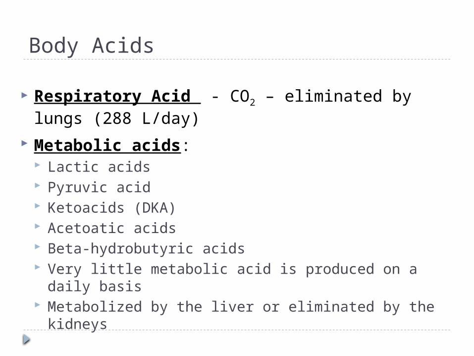
Body Acids
Respiratory Acid - CO2 – eliminated by lungs (288 L/day)
Metabolic acids: Lactic acids Pyruvic acid Ketoacids (DKA) Acetoatic acids Beta-hydrobutyric acids Very little metabolic acid is produced on a daily basis Metabolized by the liver or eliminated by the kidneys

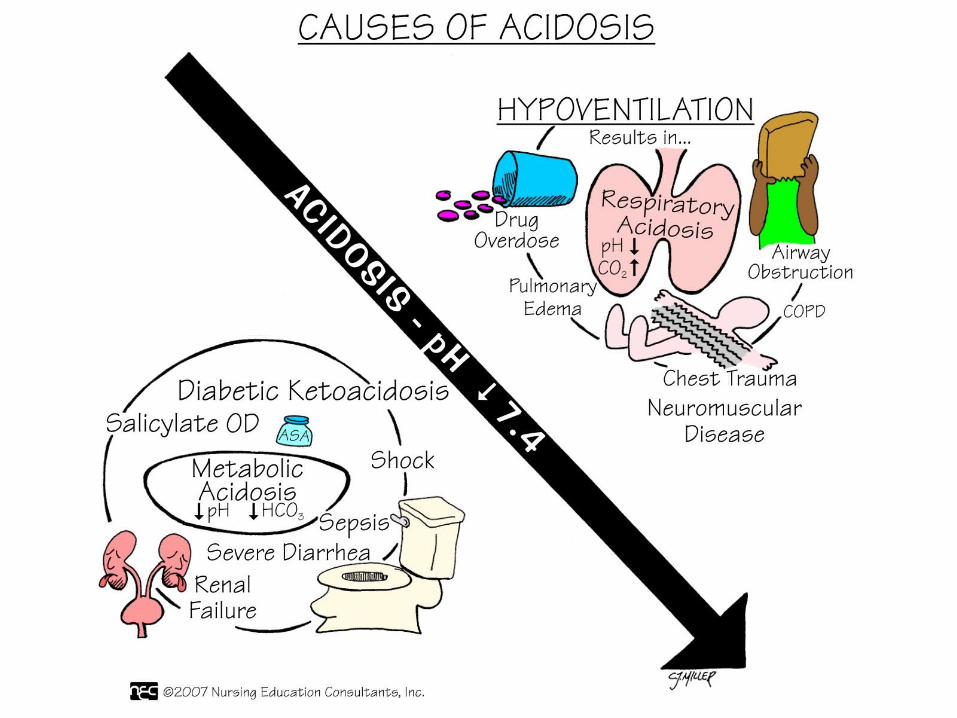
Four Basic Categories of Abnormalities Respiratory acidosis
Excess of carbon dioxide leading to an acid pH
Respiratory alkalosis Lower than normal level of carbon dioxide leading
to an alkaline pH
Metabolic acidosis Excess of hydrogen ion or a deficiency in
bicarbonate leading to an acid pH
Metabolic alkalosis Excess of bicarbonate leading to an alkaline pH

Buffer Systems
Like a sponge Soaks up extra ions Squeezed when there’s not enough
Extracellular Buffers Carbonic acid: controlled by respiration Bicarbonate: controlled by excretion
Intracellular Buffers Phosphate Buffer System
Dihydrogen phosphate (H2PO4) – hydrogen donor or acid
Hydrogen phosphate (HPO4) – hydrogen acceptor or base

Buffer Systems
Protein Buffers In the blood Plasma Proteins Hemoglobin: deoxygenated is better than
oxygenated at buffering Bones
Carbonate and phosphate salts in bone provide a long term supply of buffer.
In acute metabolic acidosis bone takes up hydrogen in exchange for calcium, sodium, and potassium.

Role of the Lungs Regulate plasma pH minute to minute
by regulating the level of Carbon Dioxide (CO2)
Carbon Dioxide is measured as a partial pressure of carbon dioxide in arterial blood PaCO2 35-45mmHg
Lungs alter rate and depth of ventilations in order to retain or excrete CO2

Minute Volume – Tidal Volume
Ventilation is measured by how much air the lungs move in one minute (minute ventilation)
Minute Ventilation is the product of respiratory rate and depth and is referred to as the TIDAL VOLUME (Vt)
Normal depth tidal volume is about 500ml Normal respiratory rate is 12 breaths per
min
12 breaths x 500 ml = 6000 ml or 6 liters
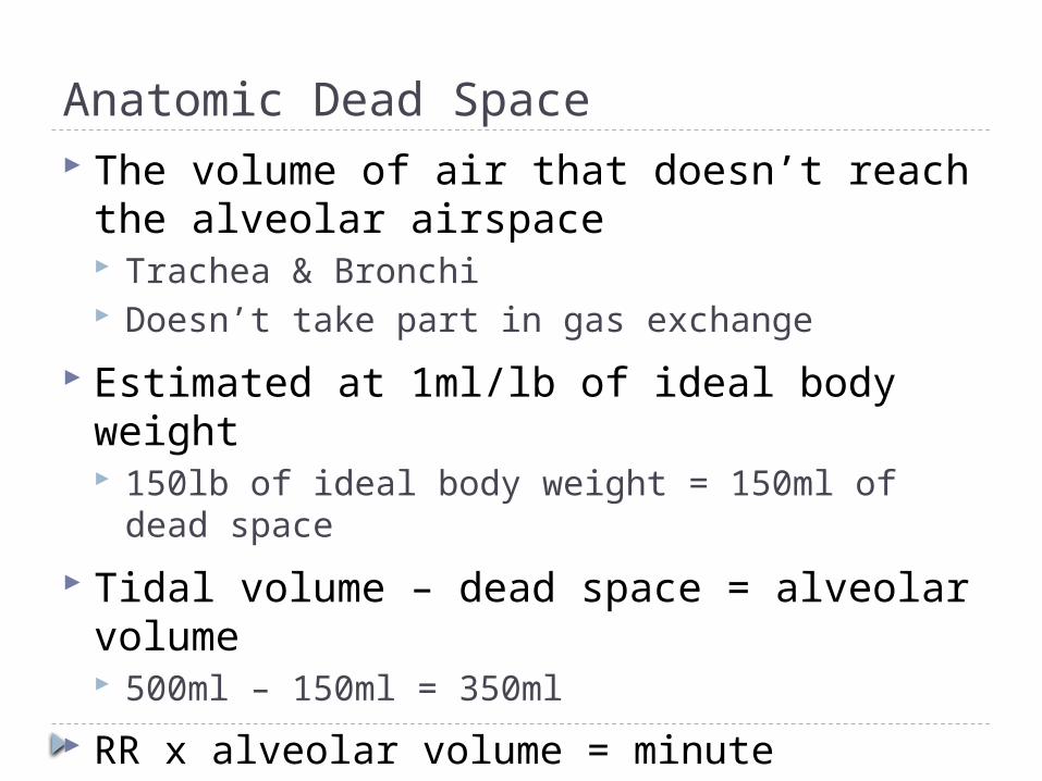
Anatomic Dead Space The volume of air that doesn’t reach the
alveolar airspace Trachea & Bronchi Doesn’t take part in gas exchange
Estimated at 1ml/lb of ideal body weight 150lb of ideal body weight = 150ml of dead
space
Tidal volume – dead space = alveolar volume 500ml – 150ml = 350ml
RR x alveolar volume = minute alveolar ventilation 12 x 350ml = 4200ml or 4.2 L/min

Hypercarbic Drive Respiratory center in the medulla controls the rate and
depth of ventilation
Responds to levels of arterial CO2, denoted as PaCO2
Chemoreceptors in the medulla come into contact with CSF
As PaCO2 rises the arterial PaCO2 reaches equilibrium with the CO2 in the CSF
The CO2 in the CSF dissociates into hydrogen ions
The hydrogen ions stimulate the chemoreceptors in the medulla which in turn stimulates the diaphragm and intercostal muscles
Respiratory rate and depth increase and CO2 is blown off

Hypoxic Drive There are also peripheral chemoreceptors
Carotid arteries Bifurcation of the common carotid and arch of
aorta
Respond to levels of Oxygen in the blood or PaO2
Hydrogen ions or pH Carbon dioxide in the blood or PaCO2
As PaO2 falls below 60 mmHg the respiratory center is stimulated to increase rate and depth

The role of the Kidneys
Two main functions to maintain acid/base Secrete hydrogen ions Restore or reclaim bicarbonate (HCO3)
In high metabolic acidosis, the kidneys can excrete ammonia as a urinary buffer.
In alkalosis - the kidneys retain hydrogen ion and excrete bicarbonate to correct the pH.
In acidosis - the kidneys excrete hydrogen ions and conserve bicarbonate to correct the pH.
Very slow process

Assessment of ACID BASE
Arterial Blood Gases (ABG) most often and the most accurate to assess acid base balances.
Serum Electrolytes can help fine tune acid base analysis
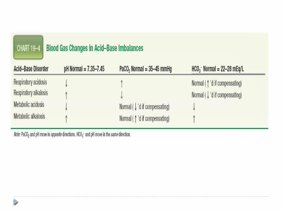
NORMAL ABG VALUES: pH = 7.35 to 7.45 PaCO2 = 35 – 45 mEq/L HCO3 = 22 – 28 mEq/L

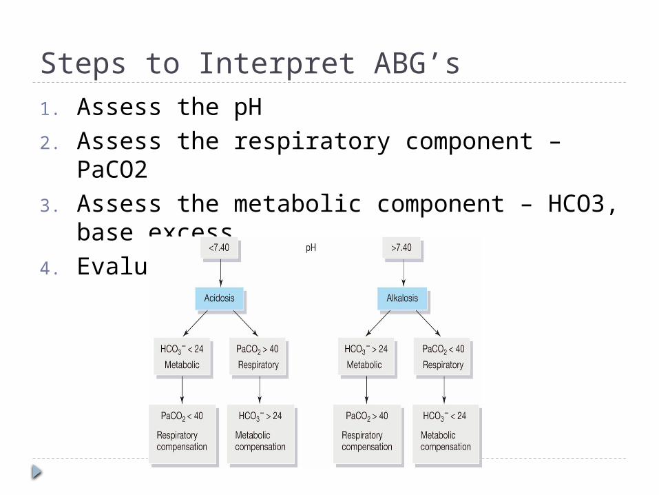
Steps to Interpret ABG’s1. Assess the pH2. Assess the respiratory component – PaCO23. Assess the metabolic component – HCO3,
base excess4. Evaluate compensation

Compensation Once the primary cause is identified look at
the other value If the value is abnormal but moving in the right
direction to bring pH back to normal compensation is occuring
If the pH value is normal than compensation is complete
Because renal compensation is slower you can infer whether respiratory abnormalities are acute or chronic If kidneys have had time to compensate is chronic If the kidneys have not had time to compensate its
acute

Respiratory Acidosis
Respiratory system fails to keep up with the body’s CO2 production
Causes (pg. 442) Acute: drug overdose, head trauma, spinal cord
injury, upper airway obstruction, pneumothorax Chronic: obesity, MS, emphysema, spinal cord
injury
Clinical Manifestations Anxiety, irritability, confusion, lethargy, increased
heart rate, warm flushed skin Mainly seen with acute causes because chronic
patients have compensated

Respiratory Acidosis Medical treatment
Treat the underlying problem Increase ventilation BiPAP Intubation Supplemental oxygen (care must be taken with chronic
pts)
Nursing care Assess PaCO2 levels and pH. Observe for signs of respiratory distress: restlessness,
anxiety, confusion, tachycardia Encourage fluid intake Position patients with head elevated 30 degrees Administer oxygen with care

Respiratory Alkalosis
Most common cause is hyperventilation caused by anxiety, panic, or pain Stroke Meningitis Head trauma
Clinical Manifestations Anxious Tachycardia Tachypnea Vertigo Forgetfulness

Respiratory Alkalosis
Medical treatment Treat underlying cause of condition Sedation may be needed
Nursing care Administer sedatives or pain medications Provide emotional support Encourage patient to breathe slowly, which will
retain carbon dioxide in the body Breath into a paper bag
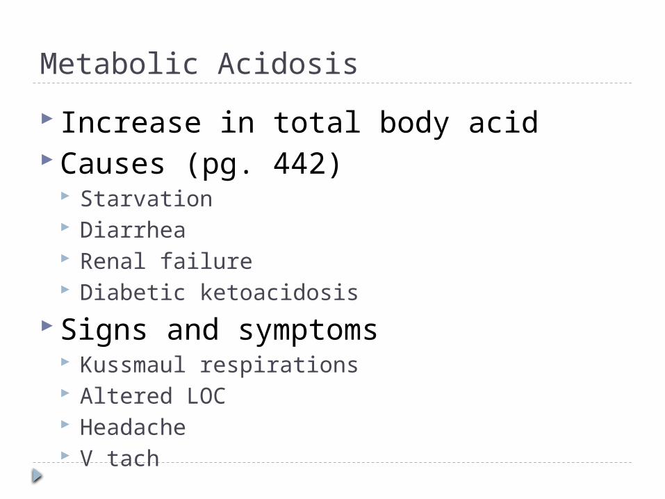
Metabolic Acidosis
Increase in total body acid Causes (pg. 442)
Starvation Diarrhea Renal failure Diabetic ketoacidosis
Signs and symptoms Kussmaul respirations Altered LOC Headache V tach

Metabolic Acidosis Medical treatment
Treat the underlying disorder
Nursing care Monitor VS & ECG Assess neurological status Provide emotional support
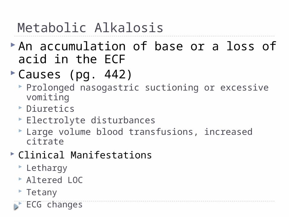
Metabolic Alkalosis An accumulation of base or a loss of
acid in the ECF Causes (pg. 442)
Prolonged nasogastric suctioning or excessive vomiting
Diuretics Electrolyte disturbances Large volume blood transfusions, increased citrate
Clinical Manifestations Lethargy Altered LOC Tetany ECG changes

Metabolic Alkalosis Medical Treatment
Treat the underlying disorder
Nursing care Monitor VS & ECG Monitor labs Accurate I&O including the amount of fluid
removed by suction Provide emotional support Use isotonic saline solutions rather than water
for irrigating NG tubes because the use of water can result in a loss of electrolytes


A client’s blood gas results are pH 7.36, PaCO2 50, HCO3 30. What do these results indicate to the nurse?
A. Respiratory acidosis, compensatedB. Metabolic acidosis, compensatedC. Metabolic acidosis, uncompensatedD. Respiratory acidosis, uncompensated

Reference Osborn, Wraa, & Watson chapter 19


