ADSORPTION OF THE PROTEIN ANTIGEN MYOGLOBIN
AFFECTS THE BINDING OF CONFORMATION-SPECIFIC
MONOCLONAL ANTIBODIES
SETH A. DARST,* CHANNING R. ROBERTSON,* AND JAY A. BERZOFSKY$*Department ofChemical Engineering, Stanford University, Stanford, California 94305; $MetabolismBranch, National Cancer Institute, National Institutes ofHealth, Bethesda, Maryland 20205
ABSTRACT Five monoclonal antibodies against sperm whale myoglobin have been used to investigate the physical stateof the antigen adsorbed onto a polydimethylsiloxane surface. The binding of each antibody is sensitive to the antigen'sconformation in solution while the locations of the antigenic sites on the myoglobin molecule for three of the antibodieshave been determined (Berzofsky, J. A., G. K. Buckenmeyer, G. Hicks, F. R. N. Gurd, R. J. Feldmann, and J. Minna.1982. J. Biol. Chem. 257:3189-3198). The binding of the fluorescein isothiocyanate-labeled IgG and Fab antibodies topreviously adsorbed myoglobin has been observed using total internal reflection fluorescence. Three of the antibodiesbind specifically to surface-adsorbed myoglobin with affinities at least 50% relative to myoglobin in solution whereastwo of the antibodies show affinities for the surface-adsorbed myoglobin diminished by at least two orders of magnituderelative to myoglobin in solution. The specific loss of certain antigenic determinants on the adsorbed myoglobin, coupledwith the retention of others, indicates a nonrandom adsorption of the myoglobin molecules.
INTRODUCTION
Proteins and other macromolecules generally adsorb ontosolid/liquid interfaces. The conformation and orientationof adsorbed proteins are thought to affect a number ofprocesses. For example, the physical state of adsorbedproteins is believed to influence cell/substrate interactions(1-3). It is recognized that the thrombogenic response ofblood towards artificial surfaces is influenced by proteinsin the adsorbed layer (4, 5). Protein adsorption accompa-nied by alterations in molecular conformation or by spatialordering on the surface may also have important implica-tions for the use of antibody-antigen assays involving theadsorption of protein antigen onto a solid surface (6). Inthis regard, it is interesting to note documented examplesof hybridoma supernatants that exhibit false positives insolid-phase immunoadsorbent screening assays. That is, amonoclonal antibody is found that reacts strongly with theantigen adsorbed to microtiter plates but is subsequentlyfound not to react well with the antigen in solution (7-10).Such antibodies sometimes bind more strongly to theantigen in solution after the antigen has been denatured byheat or by the reduction of disulfide bonds (9).
Very little direct information is available concerning therelationship between protein adsorption and conformation.Probably the most compelling evidence that conforma-tional changes can occur subsequent to the adsorption ofproteins to solid surfaces is the observation, in some cases,of alterations in the circular dichroism spectra of desorbedprotein as compared with native protein (1 1, 12). Conclu-
sions about the effects of surface-adsorption on proteinconformation have also been inferred from results obtainedusing ellipsometry and infrared spectroscopy (13). micro-calorimetry and potentiometric titrations (14), and totalinternal reflection fluorescence (15). Virtually no informa-tion is available at the molecular level concerning theconformation and orientation of an adsorbed protein.
With the goal of defining the physical state of a surface-adsorbed protein in terms of its conformation and orienta-tion, we have undertaken a study of the adsorption ofsperm whale myoglobin (Mb) to a polydimethylsiloxane(PDMS; filler-free silicone rubber) surface. A preliminarycharacterization of the adsorption behavior of Mb onPDMS has been presented (16). The Mb/PDMS systemused in this study serves as a model system of proteinadsorption onto solid surfaces. PDMS forms a relativelyhydrophobic surface and thus the noncovalent interactionsinvolved in the adsorption process and the qualitativeaspects of the results obtained herein are expected to applyto other proteins adsorbing onto hydrophobic surfaces in ageneral sense.The use of Mb offers several advantages. Mb is a
thoroughly studied and characterized protein with respectto its solution properties and structure (17, 18). In addi-tion, monoclonal antibodies that have been characterizedwith respect to their antigenic sites on the Mb molecule areavailable (19, 20). The observation that none of theseantibodies recognize denatured fragments of Mb suggestthat the binding of each antibody is sensitive to theantigen's conformation. Furthermore, the positions on the
BIOPHYS. J. © Biophysical Society * 0006-3495/88/04/533/07 $2.00Volume 53 April 1988 533-539
533
protein antigen of amino acid residues essential for thebinding of three of the antibodies have been determined.Strong arguments have been presented that these essentialamino acids are contained within the antigenic sites for thethree antibodies (20). Two of these antibodies recognizegroups of residues which are far apart in the primarysequence but close together in the folded, tertiary structureof the antigen, demonstrating the topographic, as opposedto sequential, nature of the antigenic sites. Here we presentan investigation of the binding characteristics of five ofthese monoclonal antibodies with Mb adsorbed onto aPDMS surface.
MATERIALS AND METHODS
MyoglobinSperm whale Mb (Biozyme, Batch 2, Accurate Chemical and Scientific,Hicksville, NY) was repurified by the method of Hapner et al. (21) asdescribed previously (22). The major component IV, using the notation ofGarner et al. (23), was used throughout. Mb concentration was deter-mined from the visible absorption at 540 nm using an extinction coeffi-cient of 10.4 mM-' cm-' for the ferric cyanide derivative (24). Amolecular weight of 17,800 was used in any calculations.
AntibodiesThe preparation, subclass, affinity, and binding site characterizations ofthe monoclonal anti-Mb IgG antibodies have been described previously(19, 20). Clones 1, 3.4, 4, and 5 are 'K, whereas clone 2 is UK. All weremade by fusing spleens of hyperimmunized A.SW mice with NS1, anonsecreting derivative of the P3 x 63 plasmacytoma cell line. Theantibodies were affinity purified from culture supernatant (clone 1) orascites (all others) by affinity chromatography on Mb-Sepharose, elutionwith 0.1 M glycine * HCI (pH 3), and immediate neutralization with Tris,pH 8.
For studies incorporating non-Mb-specific antibodies, purified mouseMOPC 21 myeloma protein (lot 03604; Litton Bionetics, Kensington,MD) was used.
Fab FragmentsFab fragments of each of the IgG antibodies were prepared by digestionwith papain immobilized on an agarose gel (Pierce Chemical Co.,Rockford, IL). Typically, 0.5 ml of the papain-agarose (- 130 ,ug papain)was added to 5 mg antibody dissolved in 1 ml of 20 mM sodiumphosphate, 50 mM cysteine * HCI, 10 mM sodium EDTA, pH 6.2. Themixture was incubated for 5 h at 370C with gentle rocking. Theimmobilized papain was separated by centrifugation (8,800 g for 10 min).The supernatant was desalted by size exclusion chromatography (1 x 10cm column) in 10 mM sodium phosphate, pH 8.50. The protein fractionwas then applied to a 5-ml column of protein A-Sepharose CL-4B(Pharmacia, Uppsala, Sweden) equilibrated in 10mM sodium phosphate,pH 8.50, to remove Fc fragments and undigested IgG. Although proteinA binds mouse IgG, (clones 1, 3.4, 4, and 5) only very weakly at neutralpH, IgG, and IgG2, (clone 2) antibodies are bound quantitatively andwith high affinity above pH 8.0 (25). The eluted Fab fragments werecollected and stored for further use. The protein A column was regener-ated with 0.1 M glycine * HCI, pH 2.80.The IgG digestion and Fab purification steps were followed by
size-exclusion high-performance liquid chromatography (HPLC) (Bio-Sil TSK-250 column, 7.5 x 300 mm; Bio-Rad Laboratories, Richmond,CA) in 20 mM sodium phosphate, 0.1 m Na2SO4, pH 6.80. UndigestedIgG was found to have an elution volume of 8.60 ml at the conditions used.As the papain digestion proceeded, the IgG peak, monitored by the
absorbance at 280 nm, decreased in intensity while a new peak with anelution volume of 10.2 ml appeared. This was presumably Fab and Fcfragments coeluting due to their similar molecular weights. After 5 h ofdigestion, the area under the IgG peak was <5% of the Fab-Fc peak.After the protein A-affinity chromatography, any IgG left in the samplewas too dilute to be detected by its absorbance at 280 nm.
[3H] Fluorescein Isothiocyanate AntibodiesClone 4 IgG and each of the Fab samples, prepared as described above,were labeled with fluorescein isothiocyanate (FITC) by covalent attach-ment to primary amine groups (mainly lysine residues) of the proteins(26). Antibody was incubated with a 100 times molar excess of FITC in0.1 M sodium borate, pH 9.5, for 1 h. The unreacted FITC was removedby size-exclusion chromatography (1 x 20 cm column) in 0.1 M sodiumborate, pH 9.0. The labeled protein and unreacted FITC were observed aswell-defined, yellow bands separated by more than half the column lengthwhen the protein fraction eluted from the column.The FITC-antibody was immediately tritiated by a reductive methyla-
tion technique (27). At 0°C, the FITC-antibody was mixed with a 20times molar excess of formaldehyde and a five times molar excess ofsodium [3H]borohydride (lot 2273-097, 62.0 Ci/mmol; New EnglandNuclear, Boston, MA). After 1 min, a 500 times molar excess of lysinewas added to quench the reaction. The labeled protein was separated fromlow molecular weight components of the mixture by size-exclusionchromatography (1 x 20 cm column) in 10 mM sodium phosphate, 150mM NaCl, 3 mM NaN3, pH 7.40. Each of the labeled samples behavedidentically to unlabeled samples of the same protein upon size-exclusionHPLC as described above. The labeled samples were stored at 40C andused within 1 wk.
Protein concentrations and fluorescein labeling densities were deter-mined spectrophotometrically. Extinction coefficients for IgG((276 nm= 200 mM-'cm-1) and for antibody-bound fluorescein((276= 22 mM-'cm-'and E493nm = 60 mM-'cm-' at pH 7.4) have beendetermined previously (26). An extinction coefficient of 78 mM -'cm-' at276 nm was used for Fab concentration determination (28). Fluoresceinlabeling densities were typically 1 to 2 (moles fluorescein per moleantibody). The specific activities of the labeled antibodies, determined byliquid scintillation counting, ranged from 0.5 to 1 Ci/mmol. Molecularweights for IgG and Fab of 150,000 and 50,000, respectively, were used inany calculations.
RadioimmunoassayThe immunological activities of the labeled samples of clone 4 IgG andclones 1, 2, 3.4, 4, and 5 Fab were determined by solution radioimmunoas-says. Mb was labeled with N-succinimidyl-[2,3-3H]propionate (batch 39,105 Ci/mmol; Amersham Corp., Arlington Heights, IL) as describedpreviously (19).The radiobinding assay for the labeled clone 4 IgG using polyethylene
glycol (6,000-7,000 mol w; final concentration, 10% by weight) toprecipitate the IgG plus bound antigen, was performed as describedpreviously (22). A Scatchard analysis (data not shown) indicated that thelabeled clone 4 IgG affinity for Mb was 5 x 10⁸ M-' and essentially 100%of the protein in the sample was active. The measured affinity is wellwithin experimental error of the affinity for clone 4 IgG (7.1 x 10⁸ M')determined previously (19).
Because polyethylene glycol does not precipitate Fab fragments effec-tively, a second-antibody precipitation scheme was used. Fab, carrierantibody (mouse IgG, lot 0071; Miles Scientific Div., Miles LaboratoriesInc., Naperville, IL) and [3H]Mb were mixed in 10 mM sodiumphosphate, 150 mM NaCl, 3 mM NaN3, pH 7.40. After incubation for 2h at room temperature, second antibody was added (affinity-purified goatanti-mouse IgG, F(ab')2, lot 4922; Pel Freez Biologicals, Rogers, AK).The final Fab, carrier, and second antibody concentrations were 0.20,0.67 and 7.2 uM, respectively, while the Mb concentration was varied.The final volume was 0.10 ml. The precipitation reaction was allowed toproceed for 48 h at 40C. After centrifugation at 8,800 g for 30 min, the
BIOPHYSICAL JOURNAL VOLUME 53 1988534
supernatant was removed and counted. Controls with Mb-specific Fababsent precipitated less than 5% of the radioactive Mb, whereas >95% ofthe radioactive Mb could be precipitated by the specific samples. FromScatchard analyses of the data (not shown), each labeled Fab sampleexhibited an affinity for Mb within 50% of the affinity determined for theundigested and unlabeled antibody (19, 20). At least 80% of the protein ineach sample was active. These results were taken to indicate that, for thepurposes of this study, the immunological activity of the Fab fragmentswas not significantly perturbed by the digestion and labeling procedures.
Total Internal Reflection FluorescenceApparatus and PDMS Films
A total internal reflection fluorescence (TIRF) instrument describedpreviously (29) was used. Also discussed in this prior publication are thetechniques used to deposit and characterize PDMS films on glass slides.Adsorbed protein surface concentrations were determined and related tothe TIRF signals as described elsewhere (29) with the modifications ofDarst et al. (16). For both [3H]FITC-IgG and [3H]FITC-Fab, thefluorescence signals were found to be proportional to the surface concen-tration over the range of surface concentrations encountered.
TIRF Experimental ProtocolThe same general experimental methods described previously (29) wereused for the TIRF experiments. Prior to an experiment, the proteinsolution reservoirs were incubated for several hours with a Mb or antibodysolution of equal concentration to that used in the experiment to preventloss of protein by adsorption onto the glass walls of the reservoir. Unlessotherwise specified, the following protocol was used for the TIRFexperiments. Mb was adsorbed from a flowing solution (wall shear rate 94s_') onto a PDMS film for 10 h. The Mb solution concentration was 8.5Ag/ml in 10 mM sodium phosphate, 150 mM NaCl, 3 mM NaN3, pH7.40. The temperature was controlled at 37 ± 0.50C. Under theseconditions, the Mb surface concentration was found to be 94 ± 6 ng/cm2(5.3 ± 0.3 pmol/cm2). After 10 h, the flowing Mb solution was replacedwith a solution of [3H] FITC-antibody. The adsorption of the [3H]FITC-antibody to the previously adsorbed Mb was then monitored continuouslyusing TIRF.
Nonspecific Adsorption of AntibodyTo determine the specific interactions of anti-Mb antibodies withadsorbed Mb, the extent of nonspecific antibody adsorption must beknown. The adsorption of FITC-bovine--y-globulins to adsorbed Mb havebeen investigated previously (16). For the purposes of this study, nonspe-cific adsorption of antibody was determined by exposing layers ofadsorbed Mb to various concentrations of [3H]FITC-IgG and [3H]FITC-Fab fragments of the MOPC 21 myeloma protein of P3 x 63. Thismolecule originates from the protein-secreting parent of NS1 and, beingalso 81K but without specificity for Mb, serves as a good control.When the PDMS surface was exposed to high solution concentrations
of Mb (30 ,g/ml or higher), a saturating amount of Mb was adsorbed(-130 ng/cm2). As found previously (16), the nonspecific adsorption ofantibody to the Mb-saturated surface was negligible for up to 20 h. Theamount of nonspecifically adsorbed antibody increased as the surfaceconcentration of Mb decreased. Nonspecifically adsorbed antibody thusappears to interact with surface sites distinct from the adsorbed Mbmolecules, probably vacant regions remaining on the surface when theMb surface concentration fails to reach saturation.
For the anti-Mb antibody studies reported herein, the PDMS surfaceswere -70% saturated with Mb (94 ng/cm2 vs. 130 ng/cm2 for a saturatedsurface). Exposure of such a surface to anti-Mb antibody in solution willresult in nonspecific antibody/PDMS interactions as well as the potentialantibody/adsorbed Mb interactions of interest. The amount of specifi-cally adsorbed antibody for a given antibody solution concentration wasdetermined by simply subtracting from the total amount of adsorbed
antibody the amount of nonspecifically adsorbed antibody determined atthe same solution concentration. MOPC 21 IgG adsorption was used tocorrect for the specific IgG adsorption results whereas MOPC 21 Fabadsorption was used to correct for the specific Fab results. At the highestIgG solution concentration observed (125 nM), after 2 min of antibodyadsorption to previously adsorbed Mb, the total amount of adsorbed clone4 IgG was 40 ng/cm2, whereas in a separate experiment the amount ofadsorbed MOPC 21 IgG was 4.6 ng/cm2. At lower antibody solutionconcentrations, when specific antibody/adsorbed Mb interactions wereobserved, the nonspecific antibody adsorption was always less than 10% ofthe total antibody adsorption for both IgG and Fab binding. In perform-ing these minor corrections, the assumption is made that small numbers ofamino acid changes within the binding site regions of each antibody do notsignificantly affect the nonspecific adsorption properties and thus thenonspecific binding does not vary from antibody to antibody. This issupported by the finding described above that the nonspecific bindingresults using MOPC 21 myeloma protein are essentially identical to thoseobtained using a heterogeneous bovine--y-globulins sample.
Model Used to Interpret the AntibodyBinding Results
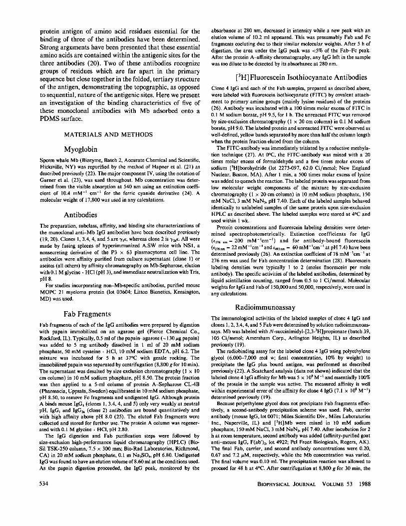
The simple model used to interpret the antibody binding results is shownschematically in Fig. 1. Nonspecific adsorption of antibody to the PDMSsurface is neglected in this model because only relatively small amounts ofnonspecific antibody binding were observed and this was taken intoaccount as described above. The model is essentially the same as thoseused to describe the equilibrium binding of antibodies or ligands to cellsurface receptors (30) with the conceptual difference that the adsorbedMb is probably not able to diffuse freely in the surface plane. Althoughthe mobility of adsorbed protein in the surface plane could affect thekinetics of the binding process, assumptions regarding surface mobilityare not required to analyze the equilibrium binding results. The binding ofan antibody is described by the linked equilibria (* indicates surfacebound species):
Ab + Mb*. Ab-l
Abr + Mb* -Ab2*
[Ab*][Ab] [Mb*]
[Ab*l[Abr] [Mb*]
(1)
(2)
where Ab denotes free antibody in solution, Mb* denotes adsorbed Mb,and Abr and Ab* denote the mono- and bivalently bound antibody,respectively. K, and K2 are the univalent and bivalent associationconstants, respectively. The quantity measured experimentally is the total
Ab
K
Ab! K2 Ab277- /j:7 Mb'PDMS SURFACE
FIGURE 1 Schematic representation of the model used to interpret theantibody binding results. *Denotes a surface-bound species. According tothis scheme Mb is bound in some unknown manner to the PDMS surface.Antibody in the bulk solution (Ab) is in equilibrium with a univalently-bound antibody/surface-adsorbed Mb complex (Ab*) with an associationconstant K1. For bivalent IgG antibodies, AbM is also in equilibrium withthe bivalently-bound species, Ab*, with an association constant K2.
DARST ET AL. Adsorption ofMyoglobin and Monoclonal Antibodies 535
bound antibody ([Ab,] + [Abf]). A surface coverage, 0, can be definedin the following manner:
[AbrI + [Ab ]I0- [M~0(3)
where [Mb*]o denotes the total surface concentration of antibody-boundand free Mb, a known quantity. 0 can be expressed as
0=K1[Ab] + K,K2[Ab] [Mb*]
1 + K,[Ab] + 2KIK2[Ab] [Mb*]'
or in the form of a Scatchard analysis:
0[Ab] = K, + KIK2[Mb*]- (K + 2K,K2[Mb*])0. (5)
For univalent Fab fragments, K2 = 0 and the expression reduces to
0[Ab] = K, - KI@ (6)
RESULTS
Clone 4 IgG and Fab Antibodies InteractStrongly with Adsorbed Mb
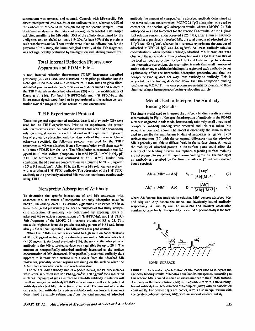
Typical experimental results for the binding of the clone 4antibody to surface-adsorbed Mb are shown in Fig. 2 (seefigure caption for details). Fab and IgG antibodies behavequalitatively the same.To derive conclusions from a Scatchard analysis, it must
be demonstrated that a reversible equilibrium exists
TIME (min)
FIGURE 2 Surface-bound antibody fluorescence signal, in units ofphotomultiplier tube current, versus time for clone 4 FITC-IgG. Mb wasallowed to adsorb onto a PDMS surface from a flowing solution for 10 hunder the conditions described in Materials and Methods. At time zero,the Mb solution was replaced by a 63-nM solution of clone 4 FITC-IgG ata fluorescent labeling density of 1 mol FITC/mol IgG. The adsorptiontime lag is due to the time required for the antibody solution to displacethe Mb solution up to the center of the flow cell and diffuse to the surfacepoint of observation. Under the conditions of this experiment, thefluoresence signals from nonspecifically adsorbed and from bulk solutionantibody are negligible. A fluoresence signal of 9 nA corresponds to anantibody surface concentration of 0.18 pmol/cm2. The Mb surfaceconcentration is 5.3 ± 0.3 pmol/cm2, resulting in a surface coverage of0.034.
between the antibody in solution and the Mb-bound spe-cies. Evidence for this arises from two observations. First,when the antibody solution concentration is stepped, eitherup or down, the new antibody surface concentration is thesame as if the experiment was performed in one step.Second, the Mb-bound antibody can be desorbed by flush-ing the flow cell with buffer or other solutions, such asbovine serum albumin. It should be noted that for highantibody solution concentrations, these equilibria condi-tions are observed as long as the Mb surface is exposed tothe antibody solution for short times. If antibody adsorp-tion is allowed to continue, a very slow increase in theantibody surface concentration occurs (in excess of nonspe-cific antibody adsorption) and the surface-antibodydesorbs at an increasingly lower rate. For instance, at anIgG solution concentration of 63 nM, the initial rapidadsorption plateaus after -5 min at a surface coverage of-0.03. The antibody surface concentration continues toslowly increase, however. After 24 h, the surface coverageof specifically adsorbed antibody is -0.13 and appears tohave attained steady state. If the antibody adsorptionprocess is interrupted after 5 h, virtually no antibodydesorption occurs for up to 20 h. This long-term processmay be due to aggregation of the surface-bound antibodybut is not entirely understood and is not addressed furtherherein. All of the isotherms presented in this work weredetermined using data from the initial plateau of theantibody binding, the time period of which depends on theantibody solution concentration.
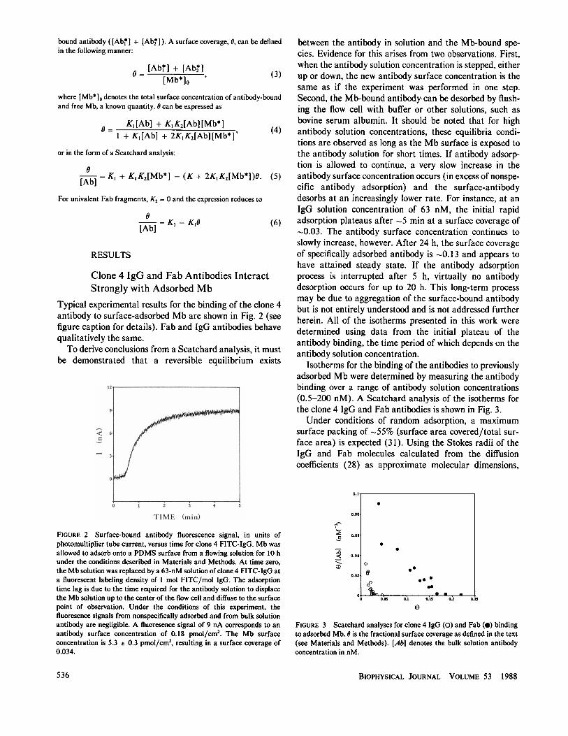
Isotherms for the binding of the antibodies to previouslyadsorbed Mb were determined by measuring the antibodybinding over a range of antibody solution concentrations(0.5-200 nM). A Scatchard analysis of the isotherms forthe clone 4 IgG and Fab antibodies is shown in Fig. 3.Under conditions of random adsorption, a maximum
surface packing of -55% (surface area covered/total sur-face area) is expected (31). Using the Stokes radii of theIgG and Fab molecules calculated from the diffusioncoefficients (28) as approximate molecular dimensions,
0.08
0
D.062
00
).04-
0
D.02- @0
.00
O-0 o .. * a b-
0 0.05 0.1 0.15 0.2 0.25
e
FIGURE 3 Scatchard analyses for clone 4 IgG (0) and Fab (0) bindingto adsorbed Mb. 0 is the fractional surface coverage as defined in the text(see Materials and Methods). [Ab] denotes the bulk solution antibodyconcentration in nM.
BIOPHYSICAL JOURNAL VOLUME 53 1988
a
a
0
0
536
this surface packing corresponds to maximum surfacecoverages (0) due to steric limitations for IgG and Fab of0.09 and 0.25, respectively. This suggests that the clone 4IgG and Fab adsorption may be limited by steric exclusionand explains the difference in maximum surface coverage
observed between IgG and Fab, although additional exper-
iments are required to confirm this point.With Eq. 6, K, can be calculated from the slope of the
Fab isotherm. The slope of the clone 4 Fab isotherm in Fig.3 is estimated to be -0.53 nM-, yielding a value for K, of5.3 x 108 M-'. This compares very favorably with thevalue of the clone 4 affinity for Mb in solution, 7.1 x 10⁸M-' (19). The limiting slope, as 0 0, of the clone 4 IgGisotherm is estimated to be -2.1 nM -'. By comparison ofEqs. 5 and 6, the steeper slope of the IgG isothermindicates that bivalent binding of the IgG moleculesoccurs. Using the value of K, determined above for theclone 4 Fab, K2 can be calculated to be 2.8 x 10"cm2/mol.
Based on a comparison of the binding rates of the clone 4antibodies to the adsorbed Mb with the results of a
convection/diffusion model appropriate for the geometryof the TIRF apparatus (32), at the wall shear rate used (94s 1) the binding rates of both the IgG and Fab antibodiesare influenced by diffusion. Thus, the intrinsic kinetics ofthe forward reaction cannot be discerned from these data.The desorption rates, which are found to be kineticallylimited, give further indication that the clone 4 IgGantibodies exhibit bivalent binding at low surface cover-
ages. An apparent desorption rate constant, kd, for theclone 4 IgG antibody was calculated assuming the follow-ing rate expression:
dtd =kd,O. (7)
The dependence of kd on 0 is tabulated in Table I. Thesignificant increase in kd with 0 indicates that for small 0,
the IgG binds bivalently to the adsorbed Mb. As 0
increases, competition among the antibodies for a limitednumber of Mb molecules causes an increase in [Ab I]/[Ab* ]. This results in an increased apparent kd. Thisphenomenon should also result in an upward concave
Scatchard plot, which is compatible with the clone 4 IgGisotherm shown in Fig. 3. However, it would be inappro-priate to compare the clone 4 IgG isotherm with Eq. 5,
TABLE IDEPENDENCE OF THE DESORPTION RATE CONSTANT,
kd, ON 0 FOR CLONE 4 IgG BOUND TOSURFACE-ADSORBED Mb
0 kd(s-' x 104)
0.020 2.10.034 2.60.045 4.3
owing to the possibility of significant steric limitationsdiscussed previously.Due to the complex binding behavior and the increased
severity of the steric limitations for the IgG antibody,investigations of the binding of clones 1, 2, 3.4, and 5 to theadsorbed Mb were conducted using Fab fragments only.
Fab Fragments of Clones 1 and 2 AlsoInteract Strongly with Adsorbed Mb
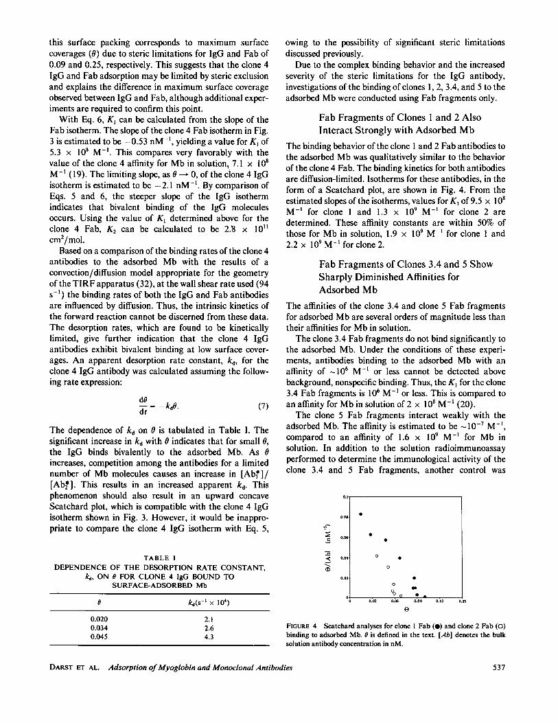
The binding behavior of the clone 1 and 2 Fab antibodies tothe adsorbed Mb was qualitatively similar to the behaviorof the clone 4 Fab. The binding kinetics for both antibodiesare diffusion-limited. Isotherms for these antibodies, in theform of a Scatchard plot, are shown in Fig. 4. From theestimated slopes of the isotherms, values for K, of 9.5 x 10⁸M-1 for clone 1 and 1.3 x 10⁹ M' for clone 2 aredetermined. These affinity constants are within 50% ofthose for Mb in solution, 1.9 x 10⁹ M` for clone 1 and2.2 x 109 M-'for clone 2.
Fab Fragments of Clones 3.4 and 5 ShowSharply Diminished Affinities forAdsorbed Mb
The affinities of the clone 3.4 and clone 5 Fab fragmentsfor adsorbed Mb are several orders of magnitude less thantheir affinities for Mb in solution.The clone 3.4 Fab fragments do not bind significantly to
the adsorbed Mb. Under the conditions of these experi-ments, antibodies binding to the adsorbed Mb with an
affinity of -106 M` or less cannot be detected abovebackground, nonspecific binding. Thus, the K, for the clone3.4 Fab fragments is 106 M` or less. This is compared toan affinity for Mb in solution of 2 x 10⁸ M-l (20).The clone 5 Fab fragments interact weakly with the
adsorbed Mb. The affinity is estimated to be 10-7 M-',compared to an affinity of 1.6 x 10⁹ M-' for Mb insolution. In addition to the solution radioimmunoassayperformed to determine the immunological activity of theclone 3.4 and 5 Fab fragments, another control was
0.03 0.06 0.09 0.12
FIGURE 4 Scatchard analyses for clone 1 Fab (0) and clone 2 Fab (0)binding to adsorbed Mb. 0 is defined in the text. [Ab] denotes the bulksolution antibody concentration in nM.
DARST ET AL. Adsorption ofMyoglobin and Monoclonal Antibodies
0.1
I08.
09
.06
1.04 O 0
0
1.02 00
I00 0 *0.~~~
°; , 1 .n1 ° ° ^
0
0.
0.
0.
0.15s
537
performed for the clone 5 Fab. It is known that the clone 4and clone 5 antibodies can bind to the Mb moleculesimultaneously in solution (33). Experiments were con-ducted in which clone 4 IgG was initially adsorbed to thePDMS surface and then exposed to Mb. Clone 5 Fab bindsrapidly to the Mb-presenting surface. The maximum Fabsurface concentration was 1.6 pmol/cm2, close to the stericlimit calculated previously. When the adsorbed clone 4IgG is exposed to bovine serum albumin rather than Mb,the subsequent adsorption of clone 5 Fab is negligible.These results indicate that the clone 5 Fab sample used inthis study is active under the conditions of these experi-ments and that the diminished binding to adsorbed Mb isdue to an effect of the adsorption process on the Mb itself.
DISCUSSION
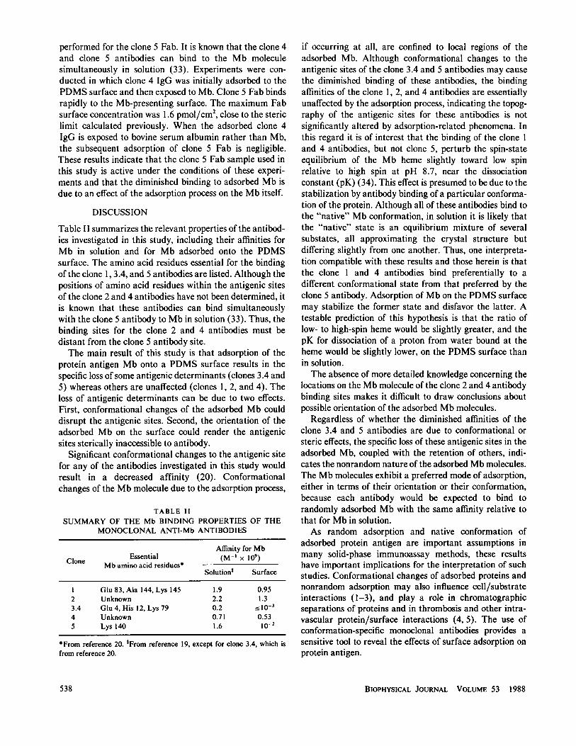
Table II summarizes the relevant properties of the antibod-ies investigated in this study, including their affinities forMb in solution and for Mb adsorbed onto the PDMSsurface. The amino acid residues essential for the bindingof the clone 1, 3.4, and 5 antibodies are listed. Although thepositions of amino acid residues within the antigenic sitesof the clone 2 and 4 antibodies have not been determined, itis known that these antibodies can bind simultaneouslywith the clone 5 antibody to Mb in solution (33). Thus, thebinding sites for the clone 2 and 4 antibodies must bedistant from the clone 5 antibody site.The main result of this study is that adsorption of the
protein antigen Mb onto a PDMS surface results in thespecific loss of some antigenic determinants (clones 3.4 and5) whereas others are unaffected (clones 1, 2, and 4). Theloss of antigenic determinants can be due to two effects.First, conformational changes of the adsorbed Mb coulddisrupt the antigenic sites. Second, the orientation of theadsorbed Mb on the surface could render the antigenicsites sterically inaccessible to antibody.
Significant conformational changes to the antigenic sitefor any of the antibodies investigated in this study wouldresult in a decreased affinity (20). Conformationalchanges of the Mb molecule due to the adsorption process,
TABLE IISUMMARY OF THE Mb BINDING PROPERTIES OF THE
MONOCLONAL ANTI-Mb ANTIBODIES
Affinity for MbClone Essential (M-' x 109)Mb amino acid residues*
Solutiont Surface
1 Glu 83,Ala 144, Lys 145 1.9 0.952 Unknown 2.2 1.33.4 Glu 4, His 12, Lys 79 0.2 --lo-4 Unknown 0.71 0.535 Lys 140 1.6 10-2
*From reference 20. tFrom reference 19, except for clone 3.4, which isfrom reference 20.
if occurring at all, are confined to local regions of theadsorbed Mb. Although conformational changes to theantigenic sites of the clone 3.4 and 5 antibodies may causethe diminished binding of these antibodies, the bindingaffinities of the clone 1, 2, and 4 antibodies are essentiallyunaffected by the adsorption process, indicating the topog-raphy of the antigenic sites for these antibodies is notsignificantly altered by adsorption-related phenomena. Inthis regard it is of interest that the binding of the clone 1and 4 antibodies, but not clone 5, perturb the spin-stateequilibrium of the Mb heme slightly toward low spinrelative to high spin at pH 8.7, near the dissociationconstant (pK) (34). This effect is presumed to be due to thestabilization by antibody binding of a particular conforma-tion of the protein. Although all of these antibodies bind tothe "native" Mb conformation, in solution it is likely thatthe "native" state is an equilibrium mixture of severalsubstates, all approximating the crystal structure butdiffering slightly from one another. Thus, one interpreta-tion compatible with these results and those herein is thatthe clone 1 and 4 antibodies bind preferentially to adifferent conformational state from that preferred by theclone 5 antibody. Adsorption of Mb on the PDMS surfacemay stabilize the former state and disfavor the latter. Atestable prediction of this hypothesis is that the ratio oflow- to high-spin heme would be slightly greater, and thepK for dissociation of a proton from water bound at theheme would be slightly lower, on the PDMS surface thanin solution.The absence of more detailed knowledge concerning the
locations on the Mb molecule of the clone 2 and 4 antibodybinding sites makes it difficult to draw conclusions aboutpossible orientation of the adsorbed Mb molecules.
Regardless of whether the diminished affinities of theclone 3.4 and 5 antibodies are due to conformational orsteric effects, the specific loss of these antigenic sites in theadsorbed Mb, coupled with the retention of others, indi-cates the nonrandom nature of the adsorbed Mb molecules.The Mb molecules exhibit a preferred mode of adsorption,either in terms of their orientation or their conformation,because each antibody would be expected to bind torandomly adsorbed Mb with the same affinity relative tothat for Mb in solution.As random adsorption and native conformation of
adsorbed protein antigen are important assumptions inmany solid-phase immunoassay methods, these resultshave important implications for the interpretation of suchstudies. Conformational changes of adsorbed proteins andnonrandom adsorption may also influence cell/substrateinteractions (1-3), and play a role in chromatographicseparations of proteins and in thrombosis and other intra-vascular protein/surface interactions (4, 5). The use ofconformation-specific monoclonal antibodies provides asensitive tool to reveal the effects of surface adsorption onprotein antigen.
BIOPHYSICAL JOURNAL VOLUME 53 1988538
This work was supported by a grant from the National Institutes ofHealth (NIH-2-RO1-HL-27187). S.A.D. was the recipient of a Kodakgraduate fellowship.
Received for publication 9 July 1987 and in final form 16 November1987.
REFERENCES
1. Grinell, F., and M. K. Feld. 1981. Adsorption characteristics ofplasma fibronectin in relationship to biological activity. J. Biomed.Mater. Res. 15:363-381.
2. Grinnell, F., and M. K. Feld. Fibronectin adsorption on hydrophilicand hydrophobic surfaces detected by antibody binding and ana-lyzed during cell adhesion in serum-containing medium. J. Biol.Chem. 257:4888-4893.
3. Grinnell, F., and T. V. Phan. 1985. Platelet attachment and spread-ing on polystyrene surfaces: Dependence on fibronectin and plasmaconcentration. Thromb. Res. 39:165-171.
4. Horbett, T. A. 1982. Biomaterials: Interfacial phenomena andapplications. Adv. Chem. Ser. 199:233-244.
5. Brash, J. L. 1983. Biocompatible Polymers, Metals, and Composites.M. Szycher, editor. Technomic Publications, Lancaster, PA. 35-52.
6. Voller, A., D. Bidwell, and A. Bartlett. 1980. in Manual of ClinicalImmunology. 2nd ed. N. R. Rose and H. Friedman, editors.American Society of Microbiology, Washington, DC. 359-371.
7. Mierendorf, R. C., Jr., and Z. L. Dimond. 1983. Functional hetero-geneity of monoclonal antibodies obtained using different screen-ing assays. Anal. Biochem. 135:221-229.
8. Miller, K. F., D. J. Bolt, and R. A. Goldsby. 1983. A rapidsolution-phase screening technique for hybridoma culture superna-tants using radiolabeled antigen and a solid-phase immunoadsor-bent. J. Immunol. Methods. 59:227-280.
9. Friguet, B., L. Djavadi-Ohaniance, and M. E. Goldberg. 1984. Somemonoclonal antibodies raised with a native protein bind preferen-tially to the denatured antigen. Mol. Immunol. 21:673-677.
10. Hollander, Z., and E. Katchalski-Katzir. 1986. Use of monoclonalantibodies to detect conformational alterations in lactate dehydro-genase isoenzyme 5 on heat denaturation and on adsorption topolystyrene plates. Mol. Immunol. 23:927-933.
11. Soderquist, M. E., and A. G. Walton. 1980. Structural changes inproteins adsorbed on polymer surfaces. J. Colloid Interface Sci.75:386-397.
12. Chan, B. M. C., and J. L. Brash, 1981. Conformational change infibrinogen desorbed from glass surface. J. Colloid Interface Sci.84:263-265.
13. Morrissey, B. W. 1977. The adsorption and conformation of plasmaproteins: a physical approach. Ann. NYAcad. Sci. 288:50-64.
14. Norde, W. 1980. Adhesion and Adsorption of Polymers. Vol. 2.L. H. Y. Lee, editor. Plenum Publishing Corp., New York. 801.
15. Burghardt, T. P., and D. Axelrod. 1983. Total internal reflectionfluorescence study of energy transfer in surface-adsorbed anddissolved bovine serum albumin. Biochemistry. 22:979-985.
16. Darst, S. A., C. R. Robertson, and J. A. Berzofsky. 1986. Myoglobin
adsorption into polydimethylsiloxane. J. Colloid Interface Sci.111:466-474.
17. Antonini, E., and M. Brunori. 1971. Hemoglobin and Myoglobin inTheir Reactions with Ligands. North-Holland Publishing Co.,Amsterdam.
18. Phillips, S. E. V. 1980. Structure and refinement of oxymyoglobin at1.6 A resolution. J. Mol. Biol. 142:531-554.
19. Berzofsky, J. A., G. Hicks, J. Fedorko, and J. Minna. 1980.Properties of monoclonal antibodies specific for determinants of aprotein antigen, myoglobin. J. Biol Chem. 255:11188-11191.
20. Berzofsky, J. A., G. K. Buckenmeyer, G. Hicks, F. R. N. Gurd, R. J.Feldmann, and J. Minna. 1982. Topographic antigenic determi-nants recognized by monoclonal antibodies to sperm whale myo-globin. J. Biol. Chem. 257:3189-3198.
21. Hapner, K. D., R. A. Bradshaw, C. R. Hartzell, and F. R. N. Gurd.1968. Comparison of myoglobins from harbor seal, porpoise, andsperm whale. J. Biol. Chem. 243:683-689.
22. Berzofsky, J. A. 1978. Genetic control of the immune response tomammalian myoglobins in mice. I. More than one I-region gene inH-2 controls the antibody response. J. Immunol. 120:360-369.
23. Garner, M. H., W. H. Garner, and F. R. N. Gurd. 1974. Recognitionof primary sequence variations among sperm whale myoglobincomponents with successive proteolysis procedures. J. Biol. Chem.249:1513-1518.
24. Berzofsky, J. A., J. Peisach, and W. E. Blumberg. 1971. Sulfhemeproteins. I. Optical and magnetic properties of sulfmyoglobin andits derivatives. J. Biol. Chem. 246:3367-3377.
25. Ey, P. L., S. J. Prowse, and C. R. Jenkin. 1978. Isolation of pureIgG1, IgG2, and IgG2b immunoglobulins from mouse serum usingprotein A-Sepharose. Immunochemistry. 15:429-436.
26. Wells, A. F., C. E. Miller, and M. K. Nadel. 1966. Rapid fluoresceinand protein assay method for fluorescent-antibody conjugates.Appl. Microbiol. 14:271-275.
27. Rice, R. H., and G. E. Means. 1971. Radioactive labeling of proteinsin vitro. J. Biol. Chem. 246:831-832.
28. Fasman, G. D., editor. 1970. Handbook of Biochemistry and Molecu-lar Biology. 2nd ed. CRC Press, Cleveland.
29. Lok, B. K., Y. L. Cheng, and C. R. Robertson. 1983. Total internalreflection fluorescence: A technique for examining interactions ofmacromolecules with solid surfaces. J. Colloid Interface Sci.91:87-103.
30. Dower, S. K., C. Delisi, J. A. Titus, and D. M. Segal. 1981.Mechanism of binding of multivalent immune complexes to Fcreceptors. I. Equilibrium binding. Biochemistry. 20:6326-6334.
31. Feder, J., and I. Giaever. 1980. Adsorption of ferritin. J. ColloidInterface Sci. 78:144-154.
32. Lok, B. K., Y. L. Cheng, and C. R. Robertson. 1983. Proteinadsorption on crosslinked polydimethylsiloxane using total internalreflection fluorescence. J. Colloid Interface Sci. 91:104-116.
33. Kohno, Y., I. Berkower, J. Minna, and J. A. Berzofsky. 1982.Idiotypes of anti-myoglobin antibodies: shared idiotypes amongmonoclonal antibodies to distinct determinants of sperm whalemyoglobin. J. Immunol. 128:1742-1748.
34. Berzofsky, J. A. 1984. Monoclonal and Anti-idiotypic Antibodies:Probes for Receptor Structure and Function. J. C. Venter, C. M.Fraser, and J. Lindstrom, editors. Alan R. Liss, New York. 1-19.
DARST ET AL. Adsorption ofMyoglobin and Monoclonal Antibodies 539


