
Anesthesia Monitoring in the
Operating Room and ICU
Dr. Mustafa Alrabayah
Department of anesthesia and intensive care
The University of Jordan
2018

Monitoring in the Past
• Visual monitoring of respiration and overall clinical appearance
• Finger on pulse
• Blood pressure

Introduction
Why do we need intraoperative monitoring???
• To maintain the normal pt physiology & homeostasis throughout anesthesia and surgery:
induction, maintenance & recovery
as much as possible. To ensure the well being of the pt.
• Surgery is a very stressful condition → severe sympathetic stimulation, HTN, tachycardia, arrhythmias.
• Most drugs used for general & regional anesthesia cause hemodynamic instability, myocardial depression, hypotension & arrhythmias.
• Under GA the pt may be hypo or hyperventilated and may develop hypothermia.
• Blood loss → anemia, hypotension. So it is necessary to recognise when the pt is in need of blood transfusion (transfusion point).

Introduction
The most critical 2 times during anesthesia are:
INDUCTION - RECOVERY.
Exactly like “flying a plane” induction (= take off) & recovery (= landing).
The aim is to achieve a smooth induction , a smooth recovery & a smooth intraoperative course.

Introduction
Any monitor consists of:
1) Sensor for data collection.
2) System data analysis.
3) System for interpretation.

Introduction
Degree of invasiveness of monitoring
Non invasive ECG
Penetrating ECHO (TEE)
Invasive Arterial cannula, CVP
Highly invasive Brain, PAC

Introduction
Limitation of monitoring
1) Delay.
2) Danger.
3) Decrease skill.
4) Doubt of results.
5) Distracting set up.

ASA Monitoring Guidelines
STANDARD I
Qualified anesthesia personnel shall be present in the room throughout the conduct of all general anesthetics, regional anesthetics and monitored anesthesia care.

ASA Monitoring Guidelines
STANDARD II
During all anesthetics, the patient’s oxygenation, ventilation, circulation and temperature shall be continually evaluated.

Respiratory system monitors
CVS monitors
Monitoring of metabolism
Neuromuscular Function
Invasive monitoring
CNS monitors

Respiratory system monitors
CVS monitors
Monitoring of metabolism
Neuromuscular Function
Invasive monitoring
CNS monitors
• Clinical monitors.• Airway pressure measurement.• Disconnection alarm.• Stethoscope• Spirometery.• O2 monitoring.• Co2 monitoring.• Anesthetic gas analysis.• H+ ions measurement.

Respiratory system monitors
CVS monitors
Monitoring of metabolism
Neuromuscular Function
Invasive monitoring
CNS monitors
• Peripheral pulse.• Tissue perfusion.• ECG.• Arterial blood pressure.• Central venous catheterization• Pulmonary artery catheterization.
• Cardiac output measurement.• TEE.• Blood loss measurement.

Respiratory system monitors
CVS monitors
Monitoring of metabolism
Neuromuscular Function
Invasive monitoring
CNS monitors
• Temperature monitoring.• Tissue oxygenation monitoring.• Indirect calorimetry.• Fluid & electrolyte status• Blood gases & acid base status
monitoring.

Respiratory system monitors
CVS monitors
Monitoring of metabolism
Neuromuscular Function
Invasive monitoring
CNS monitors
Peripheral nerve stimulation:
1) Single twitch.2) Train of four twitches.3) Tetanic stimulation.4) Double burst stimulation.

Respiratory system monitors
CVS monitors
Monitoring of metabolism
Neuromuscular Function
Invasive monitoring
CNS monitors
• Arterial line• Central venous pressure• Pulmonary artery catheterization• ICP monitoring

Respiratory system monitors
CVS monitors
Monitoring of metabolism
Neuromuscular Function
Invasive monitoring
CNS monitors
• Clinical monitoring.• EEG.• Evoked potentials.• Cranial nerve monitoring.• Cerebral blood flow• cerebral oxygenation.• depth of anesthesia.

How to select a monitor
Depend on
1) Aim.
2) Experience.
3) Type of anesthesia.
4) Facilities & availability.
5) Nature of surgery.
6) General condition of the patient.

Respiratory system monitors
Oxygenation
Ensure adequate oxygen concentration in inspired gas and blood
Methods
▪ inspired gas oxygen analyzer
▪ pulse oximetry
▪ exposure to assess color

Respiratory system monitors
O2 monitoring
(1) Monitor O2 delivery to the patient:O2 failure alarm.
O2 conc. In the gas mixture
(2) Monitor O2 delivery to the tissues• clinical monitoring:
cap. refilling, state of extremities…
• O2 transport monitoring through measurement of:
Hb level & SaO2 & PaO2
• O2 uptake monitoring through:
SvˉO2 by pulmonary artery oximetry.
serum lactic acid level.

Respiratory system monitors
Pulse Oximetry
Definition: % of oxy-Hb / oxy + deoxy-Hb.
Timing of SpO2 monitoring: throughout the surgery: before induction till after extubation & recovery.
It is the LAST monitor to be removed off the pt before the pt is transferred outside the operating room to recovery room.
SpO2 monitoring should be continued in recovery room.
❑ Optical plethysmography
▪ detects pulsatile changes in blood volume
❑ Spectrophotometry
▪ measures pulsatile hemoglobin saturation

Respiratory system monitors
Pulse Oximetry
Respiratory system monitors
Pulse Oximetry
Value:
– SpO2: arterial O2 saturation (oxygenation of the pt).– HR.– Peripheral perfusion status (loss of waveform in
hypoperfusion states: hypotension & cold extremeties).
– Gives an idea about the rhythm from the plethysmography wave (arterial waveform). (Cannot identify the type of arrhythmia but can recognize if irregularity is present).
– Cardiac arrest.

Respiratory system monitors
Pulse Oximetry
How to attach/apply saturation probe:
– To the finger or toe. The red light is applied to the nail.
Nail polish and stains should be
removed → false readings and artefacts
– Usually attached to the limb with the IV line (opposite the limb with the blood pressure cuff).

Respiratory system monitors
Pulse Oximetry
Readings:
• Normal person on room air (O2 = 21%) ˃ 96%.
• Patient under GA (100% O2) = 98-100%.
• It is not accepted for O2 saturation to ↓ below 96% with 100% O2 under GA. Must search for a cause.
• < 90% = hypoxemia.
• < 85% = severe hypoxemia.

Respiratory system monitors
Pulse Oximetry
Inaccuracies occur when:– Misplaced on the pts finger, slipped.
– Pt movement, shivering.
– Poor tissue perfusion (cold extremities)
– Poor tissue perfusion (hypotension & shock)
– Cardiac arrest.
– Interference…..• Intrinsic e.g. co-Hb, Met-Hb, I.V dyes, bilirubine, fetal Hb
• Extrinsic e.g. motion, cautery, nail bed infection, polish……

Respiratory system monitors
Ventilation
Objectiveensure adequate ventilation of patient
Methodsqualitative clinical signs➢ chest excursion➢ observation of reservoir bag➢ auscultation of breath sounds
quantitative measurement➢ end tidal carbon dioxide ➢ volume of expired gas➢ Aireay pressure

Respiratory system monitors
Capnography
–What is Capnography?Continuous CO2 measurement displayed as a waveform sampled from the patient’s airway during ventilation.
–What is EtCO2?A point on the capnogram. It is the final measurement at the endpoint of the pts expiration before inspiration begins again. It is usually the highest CO2 measurement during ventilation.

Respiratory system monitors
Capnography

Respiratory system monitors
Capnography
Phases of the capnogram
– Balseline: A-B
– Upstroke: B-C
– Plateau: C-D
– End-tidal: point D
– Downstroke

Respiratory system monitors
Capnography
Normal range: 30-35 mmHg. (Usually lower than arterial PaCO2 by 5-6 mmHg due to dilution by dead space ventilation).
Value (data gained from capnography & ETCO2):• ETT: esophageal intubation.• Ventilation: hypo & hyperventilation, curare cleft (spontaneous breathing trials).
• Pulmonary perfusion: pulmonary embolism.• Breathing circuit: disconnection, kink, leakage, obstruction, unidirectional valve dysfunction, rebreathing, exhausted soda lime.
• Cardiac arrest: adequacy of resuscitation during cardiac arrest, and prognostic value (outcome after cardiac arrest).
• metabolic state of the patient

CVS monitors
Circulation
Objective
▪ ensure adequacy of circulatory function
Methods
▪ continuous electrocardiogram monitoring
▪ arterial blood pressure
▪ heart rate

CVS monitors
ECG
Value:• Heart rate.• Rhythm (arrhythmias) usually best identified from lead II.• Ischemic changes & ST segment analysis.
Timing of ECG monitoring:Throughout the surgery: before induction until after
extubation & recovery
Types & connections of ECG cables:
– 3-leads: Red=Right YeLLow=Left Black=Apex (can read leads: I, II, III)
– 5-leads: Red=Right YeLLow=Left Black=under red Green=under yellow White=central (can read any of the 12 leads: I, II, III, avR, avL, avF, V1-V6).

CVS monitors
ECG
❑Heart rate measurement▪ R wave counting (any lead)
❑ Ischemia Monitoring▪ lead II and V5 are 90%
sensitive
▪ lead II, V5 and V4 up to 98% sensitive
❑Arrhythmia monitoring▪ lead II for supraventricular
arrhythmias
▪ all leads for ventricular arrhythmias

CVS monitors
ECG
RULES:• QRS beep ON must be heard at all times.
NO silent monitors.
• Cautery → artefacts in ECG (noise/ electrical interference) → check radial (peripheral) pulse
• Arrhythmias → check radial (peripheral) pulsations.

CVS monitors
Noninvasive Blood Pressure
❑ Methodology
▪ oscillometric algorithms
▪ automated
❑ Limitations
▪ cuff size➢ oversize erroneously low measurements
➢ too small erroneously high

CVS monitors
Noninvasive Blood Pressure
NIBP:
(non-invasive ABP monitoring = automated). Gives readings for:
systolic BP, diastolic BP & MAP
Value:
to avoid and manage extremes of hypotension & HTN.
Avoid ↓ MAP < 60 mmHg (for cerebral & renal perfusion)
avoid ↓ diastolic pressor
Risks of HTN episodes: → (CVS): myocardial ischemia, pulmonary edema,
(CNS): hemorrhagic stoke, hypertensive encephalopathy.
While hypotensive episodes: (CVS): myocardial ischemia,
(CNS): ischemic stroke, hypoperfusion state metabolic acidosis, delayed recovery, renal shutdown.

CVS monitors
Noninvasive Blood Pressure
Timing of BP monitoring: throughout the surgery: before induction till after extubation & recovery.
Frequency of measurement:– By default every 5 minutes.
– Every 3 minutes: immediately after spinal anesthesia, in conditions of hemodynamic instability, during hypotensive anesthesia.
– Every 10 minutes: eg. In awake pts under local anesthesia: “monitored anesthesia care” (minimal hemodynamic changes).

Monitoring of metabolism
Temperature
❑ Objective▪ aid in maintaining appropriate body temperature
❑ Application▪ readily available method to continuously monitor
temperature if changes are intended, anticipated or suspected
❑ Monitoring Sites• Tympanic• Esophagus• Bladder• Rectum• Blood (PA catheter)• Skin

Monitoring of metabolism
Temperature
• Normal heat loss during anesthesia averages 0.5-1 C per hour, but usually not more that 2-3 C
• Temperature below 34C may lead to significant morbidity
(complications of hypothermia):• Cardiac arrhythmias: VT & cardiac arrest.
• Myocardial depression.
• Delayed recovery (delays drug metabolism).
• Increased risk of infections.
• Metabolic acidosis (tissue hypoperfusion → anerobic glycolysis → lactic acidosis)
• hyperkalemia
• Coagulopathy.

Neuromuscular Function
Evaluation of block
Peripheral nerve stimulation:
1)Single twitch.
2)Train of four twitches.
3)Tetanic stimulation.
4)Double burst stimulation.

Neuromuscular Function
Evaluation of Reversal of Blockade
❑Clinical Criteria
▪ head lift > 5 seconds
▪ sustained hand grip
▪ negative inspiratory force
➢ at least -55 cmH2O for adults
➢ at least -32 cmH2O for children
▪ vital capacity 15 ml/kg
▪ absence of nystagmus or diplopia

Invasive monitoring
–Arterial line
–Central venous pressure
–Pulmonary artery catheter

Invasive monitoring
Arterial Line
IBP: (invasive arterial blood pressure monitoring)
It is beat to beat monitoring of ABP via an arterial cannula.
Indications:– Rapid moment to moment BP changes
– Frequent blood sampling
– Circulatory therapies: bypass, IABP, vasoactivedrugs, deliberate hypotension
– Failure of indirect BP: burns, morbid obesity

Invasive monitoring
Arterial Line
Radial Artery Cannulation
• Technically easy
• Good collateral circulation of hand
• Complications uncommon except:– Vasospastic disease
– Prolonged cannulation
– Infections
– Bleeding

Invasive monitoring
Arterial Line
Alternative Sites
Brachial:– Use longer catheter to traverse elbow joint
– Postop keep arm extended
– Collateral circulation not as good as hand
Femoral:– Use guide-wire technique
– Puncture femoral artery below inguinal ligament (easier to compress, if required)

Invasive monitoring
Arterial Line

Invasive monitoring
Arterial Line
Complications of arterial cannulation
• Hematoma.
• Vasospasm.
• Thrombosis.
• Embolization of air or thrombus.
• Skin necrosis, infection…..
• Nerve damage.
• Disconnection and fatal blood loss…..
Invasive monitoring
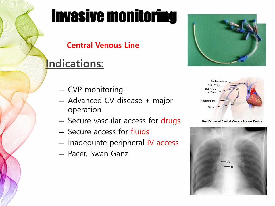
Indications:
– CVP monitoring
– Advanced CV disease + major operation
– Secure vascular access for drugs
– Secure access for fluids
– Inadequate peripheral IV access
– Pacer, Swan Ganz
Central Venous Line

Invasive monitoring
Central Venous Line
Advantages of RIJ
• Consistent, predictable anatomic location
• Readily identifiable landmarks
• Short straight course to SVC
• Easy intraop access for anesthesiologist at patient’s head
• High success rate, 90-99%

Invasive monitoring
Central Venous Line
Alternative Sites
Subclavian: – Easier to insert v. IJ if c-spine precautions– Better patient comfort v. IJ– Risk of pneumothorax - 2%
External jugular: – Easy to cannulate if visible, no risk of pneumothorax– 20%: cannot access central circulation
Femoral– High infection rate– No access for CVP readings

Invasive monitoring
Central Venous Line
• Reflects pressure at junction of vena cava + RA • CVP is driving force for filling RA + RV• CVP provides estimate of:
• Intravascular blood volume• RV preload
• Trends in CVP are very useful• Measure at end-expiration• Zero at mid-axillary line

Invasive monitoring
Central Venous Line
Component Phase of Cycle Event
a wave End diastole Atrial cont
c wave Early systole Isovol vent cont
x descent Mid systole Atrial relaxation
v wave Late systole Filling of atrium
y descent Early diastole Vent filling

Invasive monitoring
Pulmonary Artery Catheter
• Introduced by Swan + Ganz in 1970
• Allows accurate bedside measurement of important clinical variables:
CO, PAP, PCWP, CVP to estimate LV filling, guide fluid / vasoactive drug therapy and calculate core temp.

Invasive monitoring
Pulmonary Artery Catheter

Invasive monitoring
Pulmonary Artery Catheter
Complications
• Minor in 50%, e.g., arrhythmias
• Transient RBBB- 0.9-5%• External pacer if pre-existing LBBB
• Serious: 0.1-0.5%: pulmonary infarction, PA rupture (e.g., overwedge), endocarditis, structural heart damage
• Death: 0.016%

CNS monitors
BIS
• EEG analysis for frontal lobe
• Displayed as wave form and
numbers

CNS monitors
Clinical monitoring:
Clinical monitoring:
Signs of pt awareness:
• Tachycardia.
• HTN.
• Movement (facial expression).
• Pupils dilated.
• Lacrimation.

Monitoring After Extubation & Recovery
BP: within 20% of baseline.
SpO2: ˃ 92% on RA
Breathing: regular, adequate tidal volume.
Muscle power: sustained head elevation for 5 seconds, good hand grip, tongue protrusion.
Level of consciousness: fully conscious = 1) obeying orders, 2) eye opening
3) purposeful movement.
MOST IMP: Pt MUST be able to protect his own airway.

RULES NEVER to FORGET:
Never start induction with a missing monitor: ECG, BP, SpO2.
Never remove any monitors before extubation & recovery.
NEVER ignore an alarm
ALWAYS
• Remember that your clinical sense & judgement is better than & superior to any monitor.
• You are a doctor u are not a robot.
• The monitor is present to help you not to be ignored and not to cancel your brain.


