
Another case of congenital lobar
emphysema ??
Kamal MasarwehOctober 2018

Case Presentation
• born term 40 weeks GA
• spontaneous vaginal delivery
• birth weight – 3.57 kg, Apgar scores 9/10
• pregnancy:
• uneventful pregnancy
• normal antenatal ultrasound (13 and 26
weeks of gestation)
• firstborn to healthy non-consanguineous parents
• 15/2/2018
• 2 months old
• Presented in a northern hospital:
• 5 days URTI
• cough, dyspnea, cyanosis, restlessness
• afebrile

• ER: dyspneic, use of accessory muscles
• Vitals:
• tachypneic 70 BPM
• saturation 88% on room air, 98% +O2
• heart rate – 180 bpm, BP – 115/57
• Physical examination:
• moderate respiratory distress
• intercostal retractions
• decreased breath sounds on the left
• displaced heart sounds to the right side


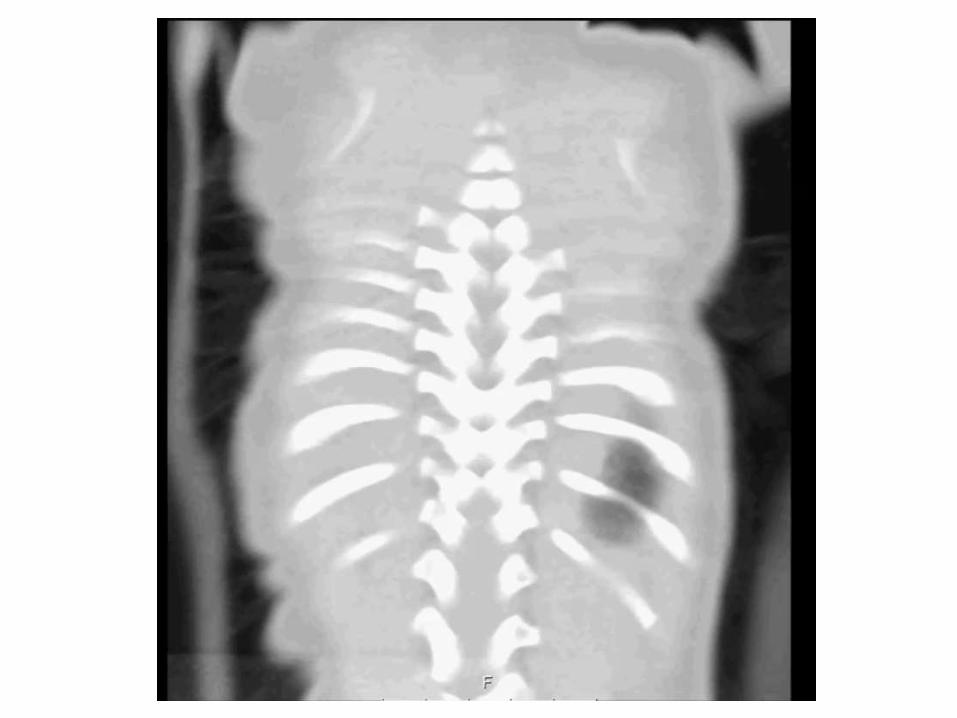

• Chest CT – hyperinflation on the left with septations, with a mediastinal shift to the right
• transferred to Rambam
• working diagnosis- CLE and significant respiratory compromise
• 16.2.2018 –
• left-sided thoracotomy
• lobectomy - left upper lobe
• uneventful surgery
• PICU
• extubated within one day
• analgesia and drainage
• post-operative pneumonia - antibiotics

Chest X-ray after surgery
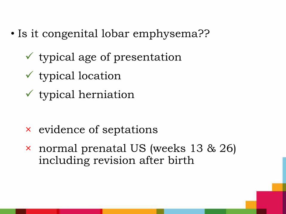
• Is it congenital lobar emphysema??
typical age of presentation
typical location
typical herniation
× evidence of septations
× normal prenatal US (weeks 13 & 26) including revision after birth
Differential diagnosis
• Congenital Lobar Emphysema
• CPAM
• Pleuropulmonary Blastoma
• Bronchogenic cyst
• Others

• macroscopically - multiple septated cystic lesion measuring 9x6x2 cm
• microscopically - cystic lesions of various sizes, lined with a monolayer of endothelial cells
x40

X100
• Markers used in immunohistochemical staining for differentiating lymphatic lesions from other etiologies:
• D2-40- monoclonal antibody to podoplanin, highly reliable lymphatic endothelial marker. Stains the endothelium of lymphatic channels but not that of blood vessels
• CD31, CD34, ERG, factor VIII-related antigen – endothelial markers
• Minato H , Solitary intrapulmonary cystic lymphangioma in an infant: a case report with literature review. Pathol Res Pract. 2010 Dec 15;206(12):851-6
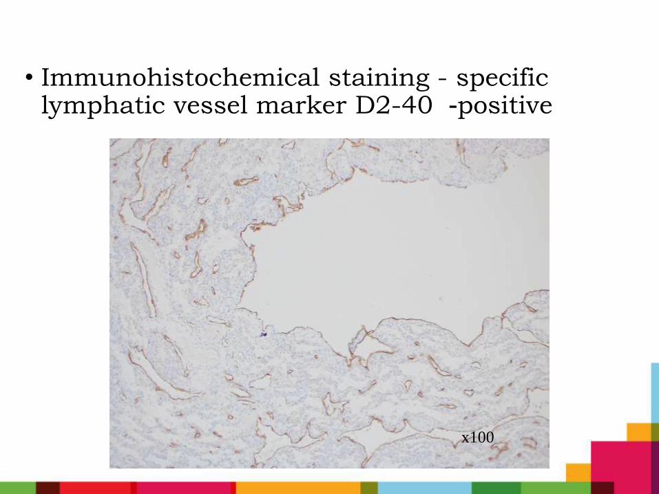
• Immunohistochemical staining - specific lymphatic vessel marker D2-40 -positive
x100
•Diagnosis:
Intrapulmonary macrocystic
lymphangioma
Pulmonary lymphatic system disorders
• rare lesions
• occur in a variety of non-neoplastic and neoplastic settings
• classification challenging
• inconsistent terminology
• mostly case reports and case series
Intrapulmonary Lymphangiomas
• extremely rare
• approximately 20 cases in literature
• patient ages – neonates (3 cases) to 54 yr
• frequently in the lower lobes of the lung, probably associated with a rich lymphatic supply

Clinical features
• symptoms vary widely depending on the age of the patient and the extent of disease
• neonates and infants - often present with pneumothorax and respiratory distress
• may remain asymptomatic until compression of vital structures
• may present as incidental findings on roentgenograms

Radiology
• Chest X-rays, US, CT scan, and MRI
• CT appearance - cystic mass with smooth margins
• no published data about antenatal US
• hypothesis that lymphangiomas become clinically significant in later stages of pregnancy or even after birth, possibly due to obstruction of lymphatic vessels

Intrapulmonary Lymphangiomas
• Treatment:
• surgical resection - frequently mandated both to confirm the diagnosis, and to prevent complications that arise from compressive effects on vital structures
• CT-guided Sclerosis
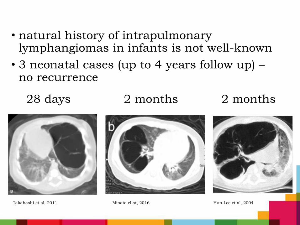
• natural history of intrapulmonary lymphangiomas in infants is not well-known
• 3 neonatal cases (up to 4 years follow up) –no recurrence
28 days 2 months 2 months
Takahashi et al, 2011 Minato el at, 2016 Hun Lee et al, 2004
Take home message
• Not all that looks like CLE is actually CLE
• Intrapulmonary lymphangioma mimics CLE and CPAM
• Does is it really matter to diagnose??
• is natural history of pulmonary lymphangiomas different than CLE/CPAM?
• is there a chance of recurrence?
• tendency for multiple lesions?
• is there a need for a different follow up?


