
Appendicular Skeleton
The Pectoral Girdle and Upper LimbThe Pelvic Girdle and Lower Limb
Nestor T. Hilvano, M.D., M.P.H.

Learning Objectives You should be able to: 1. Identify and describe the features of the
clavicle, scapula, humerus, radius, ulna, and bones of the wrist and hand, including their functions.
2. Identify and describe the features of the pelvic girdle, femur, patella, tibia, fibula, and bones of the foot, including their functions.
3. Compare the structural and functional differences between male and female pelvis.
4. Discuss common injuries of the upper and lower limbs as to structures and functions.

Pectoral (shoulder) Girdle
• Consists of:
___ attaches medially to the sternum.
___ articulates with the humerus.
a. scapula b. clavicle
• Acromioclavicular joint - easily dislocated due to loose attachment
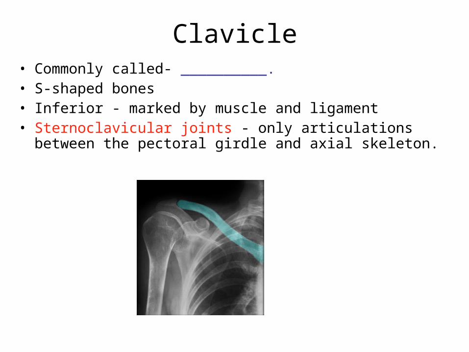
Clavicle• Commonly called- __________. • S-shaped bones• Inferior - marked by muscle and ligament • Sternoclavicular joints - only articulations between the
pectoral girdle and axial skeleton.

Scapula• Triangular plate, commonly
called ____.
• Spine ends as ___ process.
• ___ process serves for
muscle attachment. a. coracoid process c. shoulder blade
b. acromion process d. collar bone
• Subscapular, infraspinous and supraspinous fossa
• The head of humerus articulates with ___ of scapula.
a. glenoid cavity b. supraspinous fossa
c. subscapular fossa d. infraspinous fossa

Humerus• Review/study parts of humerus
• Intertubercular groove holds biceps tendon
• Surgical neck- Importance?
• Radial groove - path of radial n.
• Lateral epicondyles- attachment of forearm muscles
• Condyles consist of:
• 1. Capitulum articulates with radius.
• 2. Trochlea articulates with ulna.
• Olecranon fossa holds olecranon process of ulna.

Ulna and Radius
• Radius (lateral)– head articulates with radial notch – radial tuberosity for biceps muscle– styloid process
• Ulna (medial)– olecranon and trochlear notch – radial notch – styloid process
• Interosseous membrane – fibrous sheet
• bony prominences of wrist on medial side, ___, and lateral side, ___.
a. styloid process of radius b. styloid process of ulna

Carpal Bones
• Form the _____ bones; 2 rows (4 bones each)– proximal row = Scaphoid, Lunate, Triquetrum, Pisiform– distal row = Trapezium, Trapezoid, Capitate, Hamate
“Sally Left The Party To Take Charlie Home”

Metacarpals and Phalanges• Metacarpals are bones of the ______
– base, shaft and head
• Phalanges are bones of the ___________– thumb or pollex has proximal and distal phalanx– fingers have proximal, middle and distal phalanx a. fingers/digits
b. palms

The Pelvis • Pelvis = is a composite
structure that consists of os coxae (hip bones), sacrum, and coccyx
• Pelvic Girdle = made of os coxae (hip bones)
• Supports trunk on the legs and protects viscera
• Sacroiliac joint• Pubic symphysis
Hip Bones (os coxae) • Composed of 3 bones: 1. Ilium
– iliac crest and iliac fossa– greater sciatic notch contains
__________ which innervates the lower limb.
2. Pubis– body, superior and inferior ramus
3. Ischium– ___________ bears body weight when seated.– ischial spine– lesser sciatic notch (passage of b.v., nerves, & small muscle) – ischial ramus
• ___ - is hip joint socket
• ___ - space enclosed by collagen fibers for attachment of hip muscles.
a. obturator foramen b. acetabulum
Pelvic Inlet and Outlet
• False and True pelvis separated at pelvic brim• Pelvic inlet – enclosed space of pelvic brim• Pelvic outlet – opening bounded by coccyx,
ischial tuberosities, & inferior border of pubic symphysis
Comparison of Male and Female
Female pelvis = lighter, broader pubic angle( >100 degrees), enlarged pelvic outlet, pubic inlet wider and round, less curvature on sacrum and coccyx
Male pelvis = heavier, pubic angle (<90 degrees), small pelvic outlet, pelvic inlet heart-shaped

Femur and Patella
• Nearly spherical head and constricted neck– ligament to fovea capitis
• Greater and lesser trochanters for tendon attachment of muscles
• Posterior ridge called linea aspera attachment of hip muscles
• Medial and lateral epicondyles and condyles
• ___ = triangular sesamoid
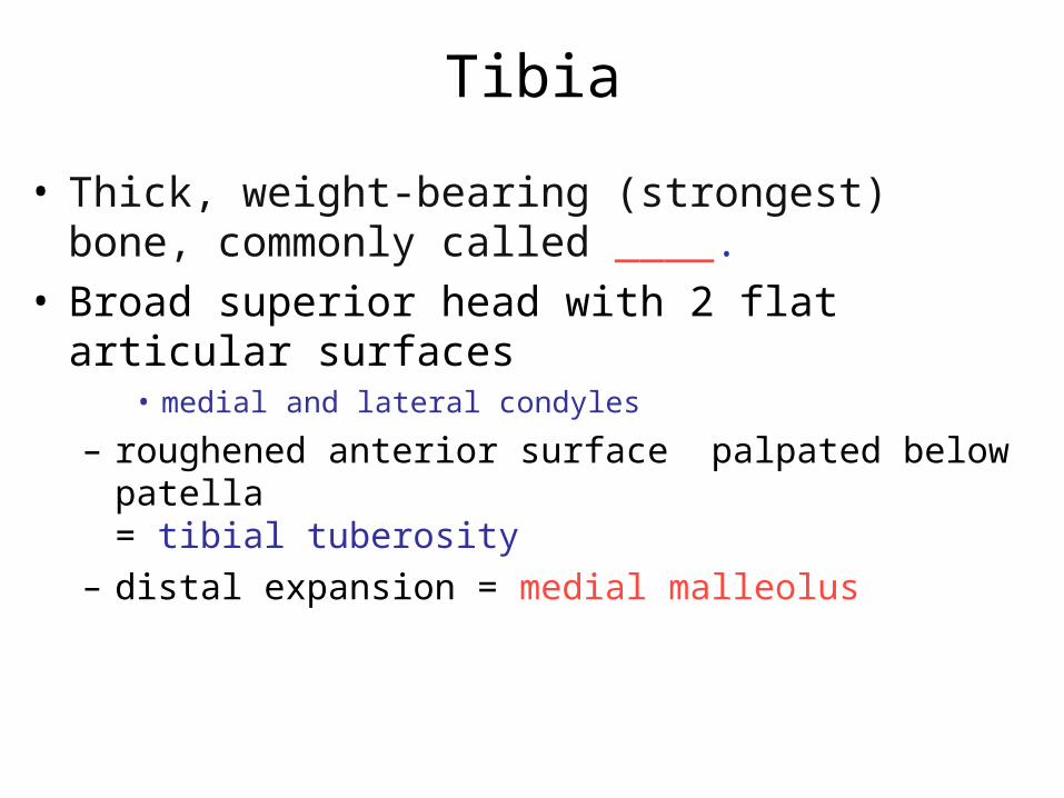
Tibia
• Thick, weight-bearing (strongest) bone, commonly called ____.
• Broad superior head with 2 flat articular surfaces• medial and lateral condyles
– roughened anterior surface palpated below patella= tibial tuberosity
– distal expansion = medial malleolus

Fibula
• Slender lateral strut• Stabilizes ankle, site for muscle attachment of leg • Does not bear any body weight
– spare bone tissue• Head = proximal end• Lateral malleolus = distal expansion• Joined to tibia by interosseous membrane. • Pott fracture – dislocate ankle (outward & backward
movement of foot), breaking both the lateral & medial malleolus.
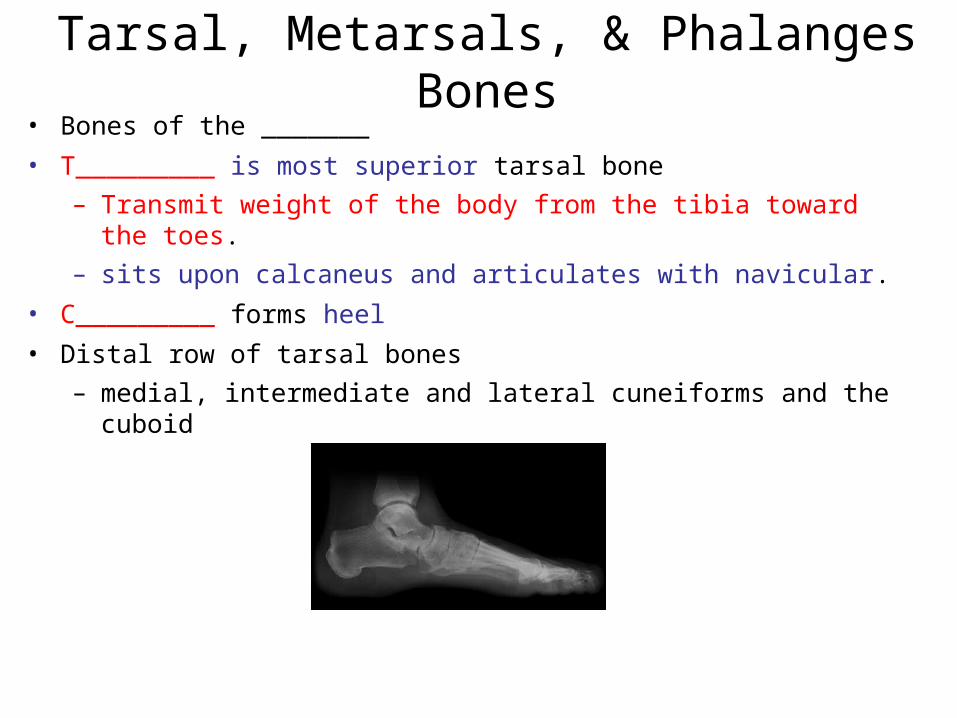
Tarsal, Metarsals, & Phalanges Bones• Bones of the _______
• T_________ is most superior tarsal bone
– Transmit weight of the body from the tibia toward the toes.
– sits upon calcaneus and articulates with navicular.
• C_________ forms heel
• Distal row of tarsal bones
– medial, intermediate and lateral cuneiforms and the cuboid
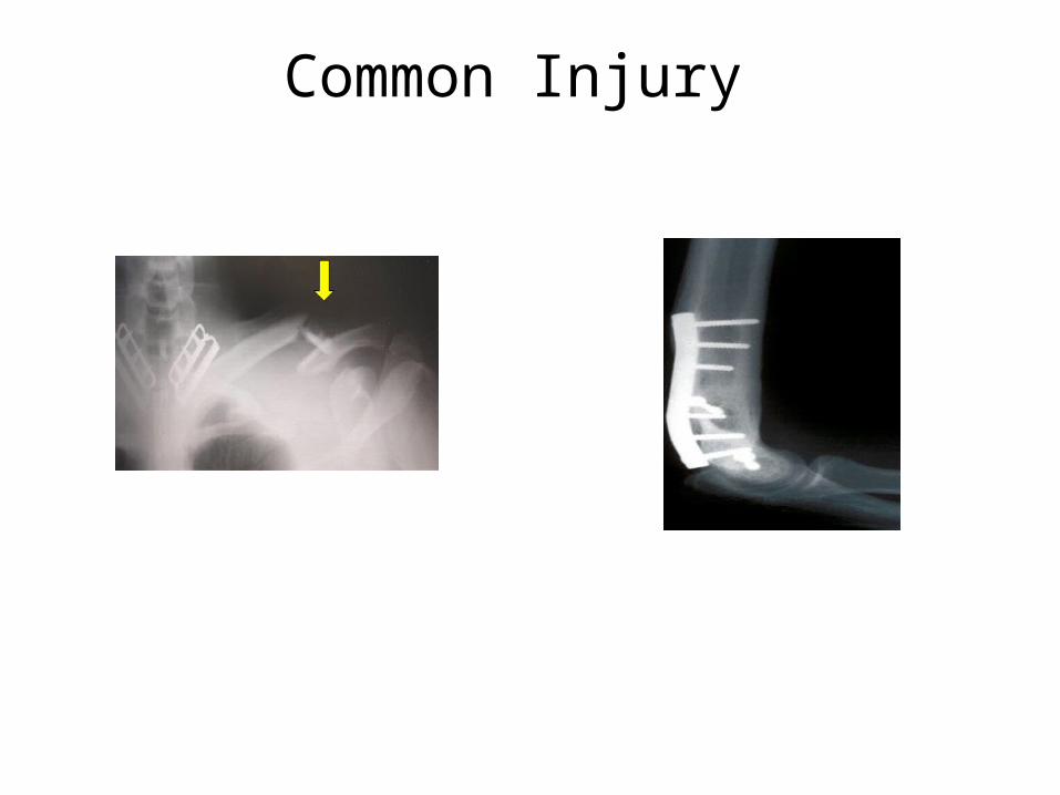
Common Injury
Homework (Self- Review) 1. Identify bone associated with the following.
a) bony prominences on medial and lateral sides of wristb) bony prominences on medial and lateral sides of anklec) rounded projections on either side of the elbowd) bearing body weight when seatede) fracture of heel f) strongest bone in the body
2. After visual inspection and measurements, what informations could the bones reveal?
3. Identify bone which articulates with the following.a) capitulum of humerus b) glenoid cavity of scapula c) acetabulum of coxal bone d) sternum (medially)
4. How is the female pelvis adapted for childbearing?


![Review Molecular and cellular basis of calpainopathy (limb ... · pathophysiological pathway for all the limb girdle muscular dystrophies [1]. Thus, when researchers discovered that](https://cdn.vdocuments.net/doc/165x107/5f83cbff01c23a1dec61ac9a/review-molecular-and-cellular-basis-of-calpainopathy-limb-pathophysiological.jpg)


