Avascular Necrosis: Causes and Treatment
Coleman D. Fowble, M.D.
Midlands Orthopaedics, P.A.
Columbia, SC
Introduction
• DefinitionLoss of blood flow to the bone leading to death of
the cellular components of bone.
Avascular Necrosis
• AVN• Osteonecrosis• Aseptic necrosis• Ischemic necrosis• Bone infarction
Bones Affected
• Femoral head – most common by far• Shoulder – humeral head• Odontoid (Neck)• Scaphoid (Wrist)• Lunate (Wrist)• Talus (Ankle)
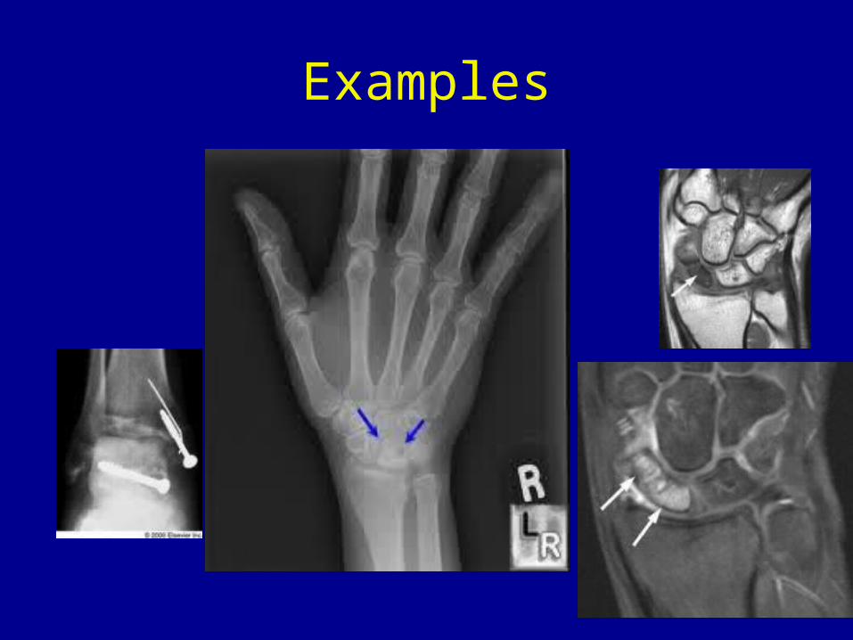
Examples
Treatment
• Frustrating• Staging very subjective in lower stages
Etiologies
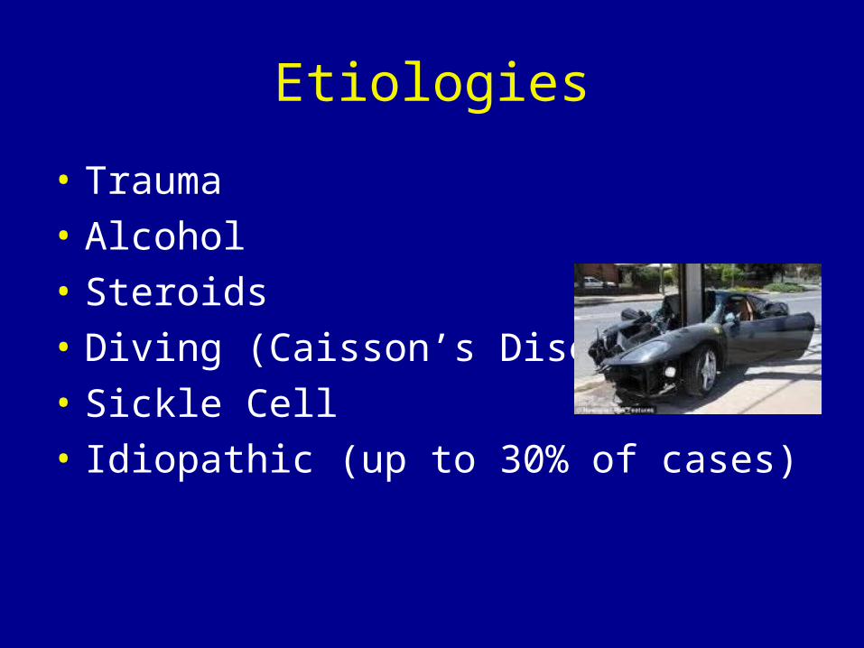
• Trauma• Alcohol• Steroids• Diving (Caisson’s Disease)• Sickle Cell• Idiopathic (up to 30% of cases)
Symptoms
• Pain• Decreased range of motion
Risk Factors
• Alcoholism• Pancreatitis• Diabetes• Gout
Staging
• Initially radiographic staging• Revised with advancement of MRI
Classification
• FicatOriginal x-ray classification of hip
• Other classifications exist for talus, scaphoid, etc.
Stage 0
• No clinical symptoms• No radiographic abnormalities• Microscopic diagnosis
Stage I
• May or may not have symptoms• Radiographs and CT are normal• MRI is abnormal as is bone scan• Microscopic exam confirms diagnosis
Stage II
• Patient is symptomatic• X-rays show osteopenia, sclerosis, cysts• No subchondral lucency or collapse• MRI confirms diagnosis
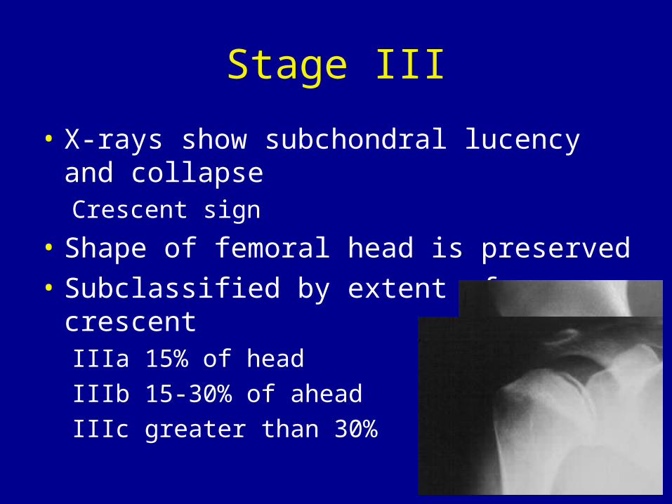
Stage III
• X-rays show subchondral lucency and collapseCrescent sign
• Shape of femoral head is preserved• Subclassified by extent of crescent
IIIa 15% of headIIIb 15-30% of aheadIIIc greater than 30%
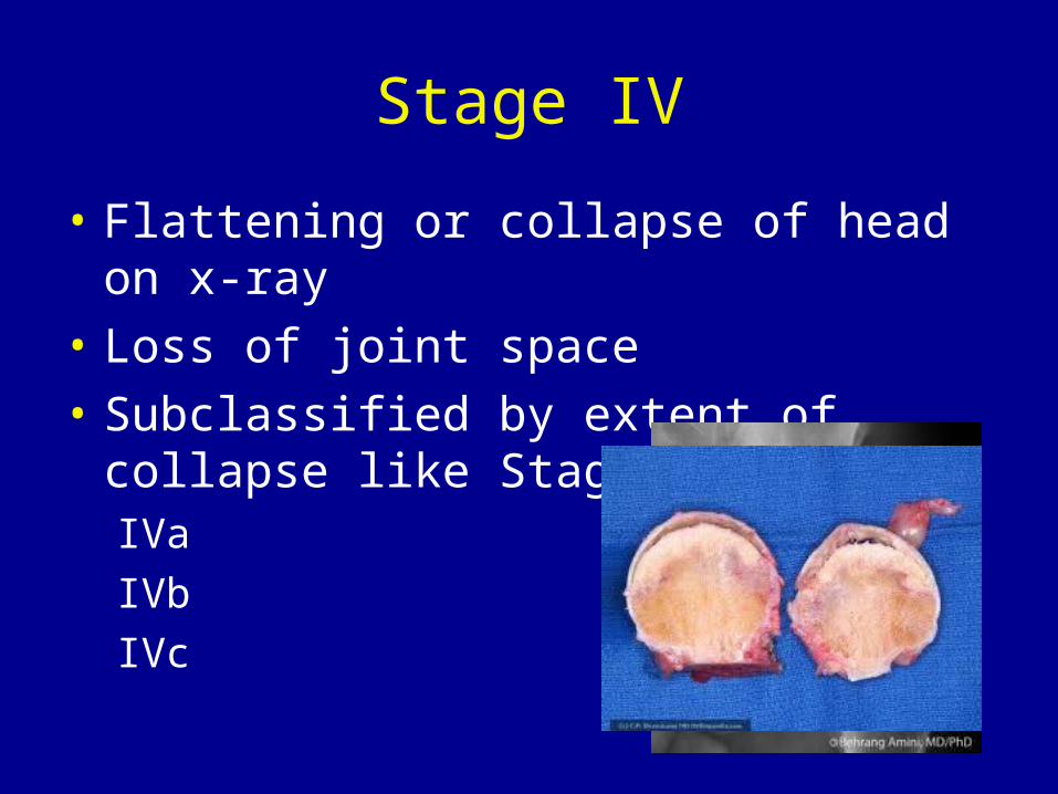
Stage IV
• Flattening or collapse of head on x-ray• Loss of joint space• Subclassified by extent of collapse like Stage III
IVaIVbIVc
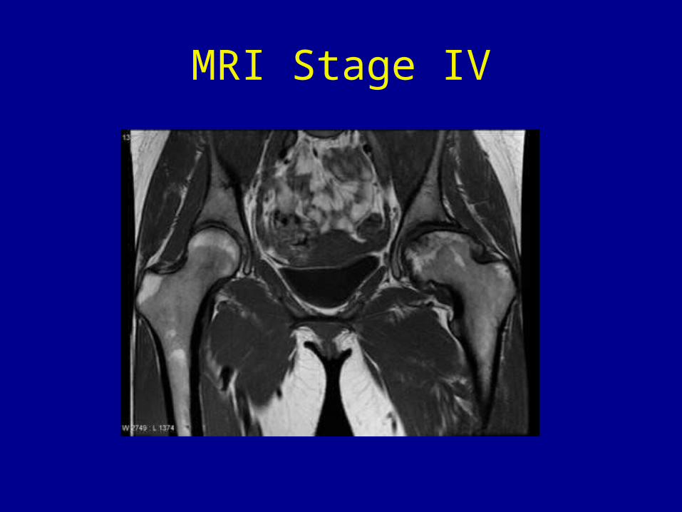
MRI Stage IV
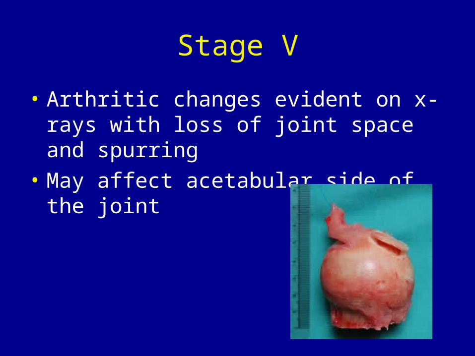
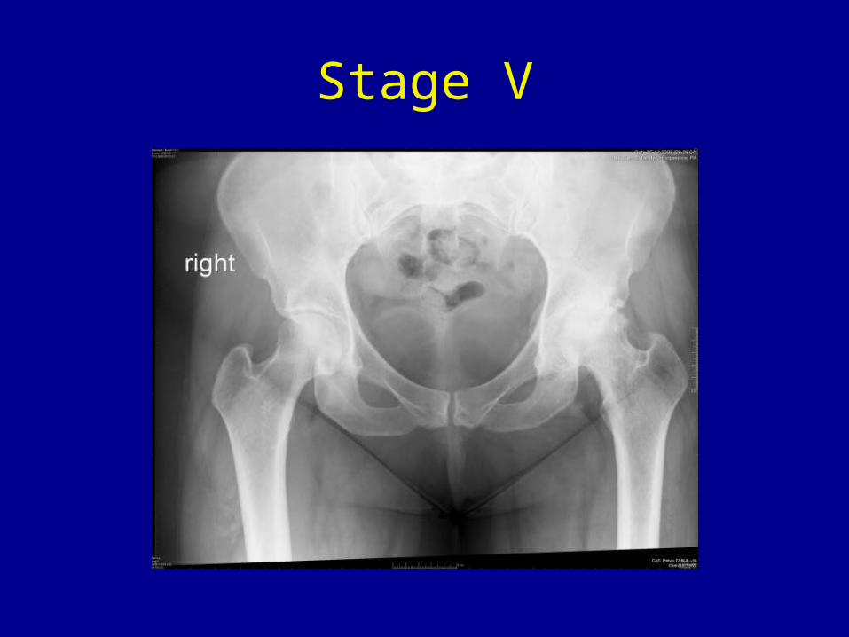
Stage V
• Arthritic changes evident on x-rays with loss of joint space and spurring
• May affect acetabular side of the joint
Stage V
Stage VI
• Extensive destruction of femoral head and joint
• May be indistinguishable from osteoarthritis
Treatment Options
• Stage dependent• Clinical signs and symptoms• Physiologic condition• Age• Medical comorbidities
Observation
• Normal x-ray• Possible abnormal MRI• No clinical signs or symptoms
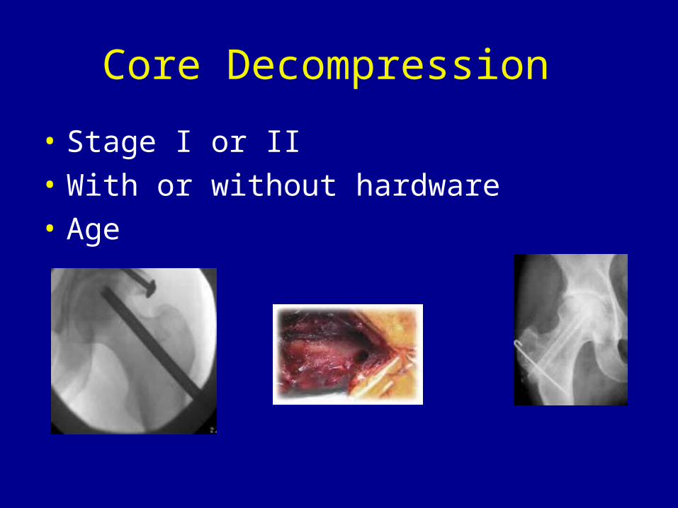
Core Decompression
• Stage I or II• With or without hardware• Age
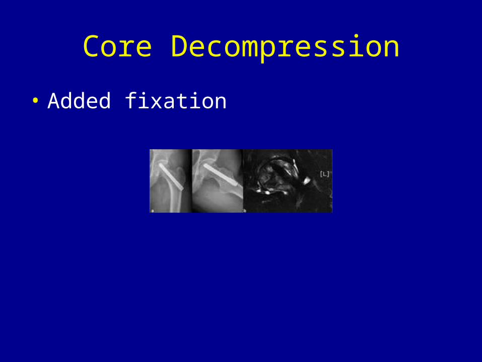
Core Decompression
• Added fixation
Free Vascularized Fibular Graft
• Pioneered in 1979 by Dr. Urbaniak at Duke• Over 2500 performed• Multidisciplinary approach• Only center with real consistent results
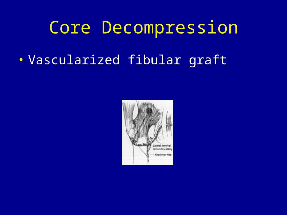
Core Decompression
• Vascularized fibular graft
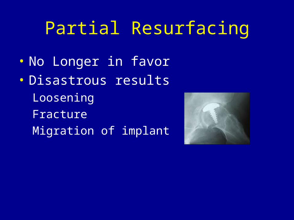
Partial Resurfacing
• No Longer in favor• Disastrous results
LooseningFractureMigration of implant
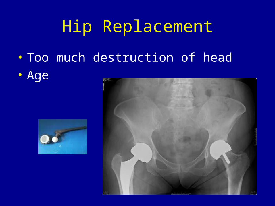
Hip Replacement
• Too much destruction of head• Age
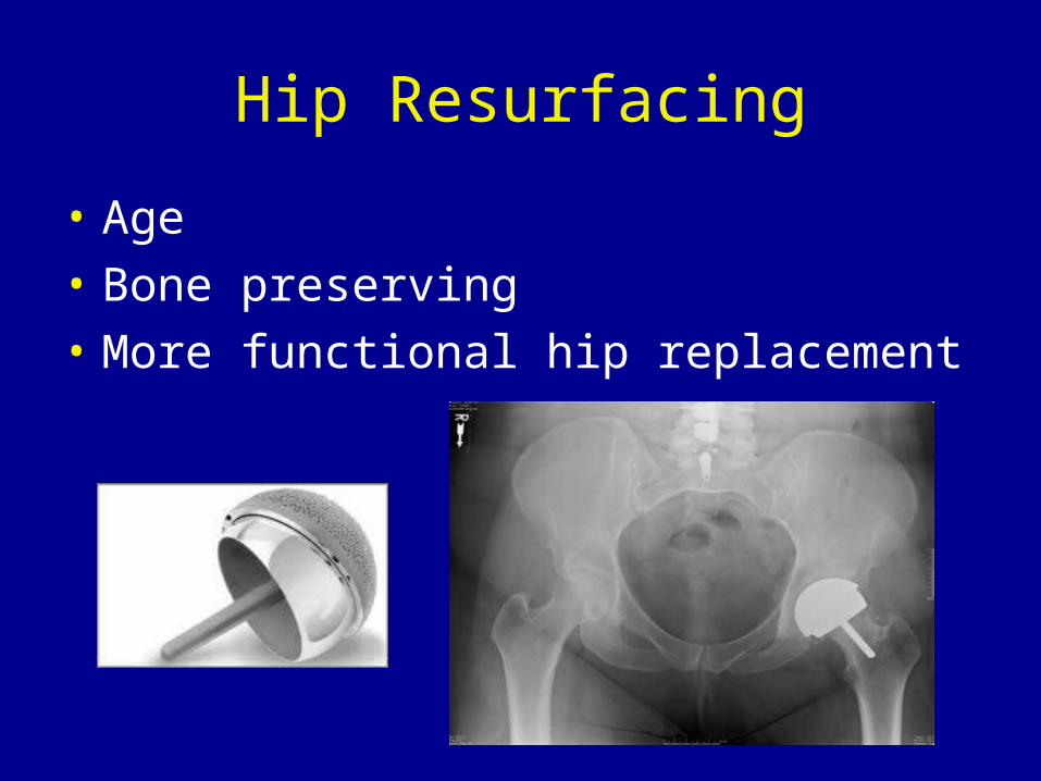
Hip Resurfacing
• Age• Bone preserving• More functional hip replacement
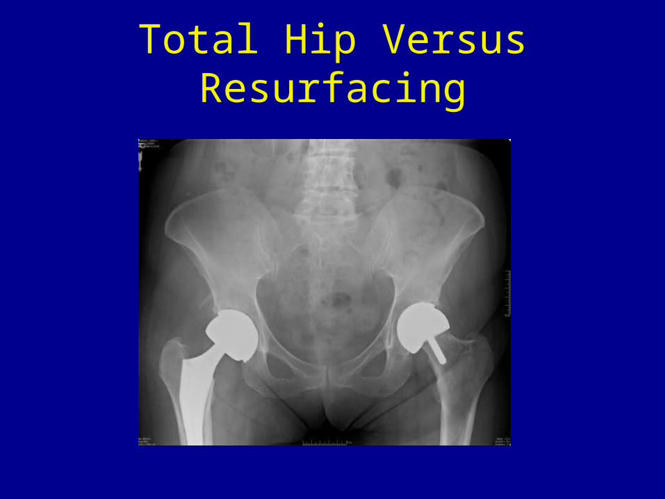
Total Hip Versus Resurfacing
Workman's Compensation
• Trauma• Secondary injury• Difficult
May take several years to show up
Femoral Neck Fracture
• Basilar neck• Transcervical• Subcapital• Intertrochanteric
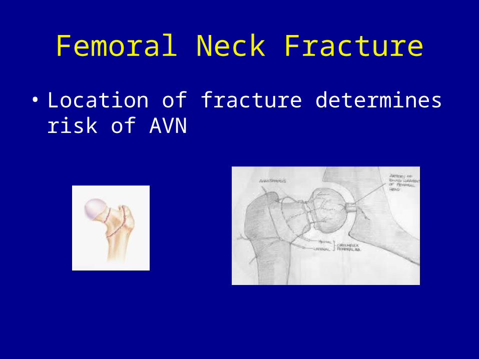
Femoral Neck Fracture
• Location of fracture determines risk of AVN
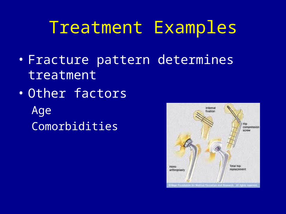
Treatment Examples
• Fracture pattern determines treatment• Other factors
AgeComorbidities
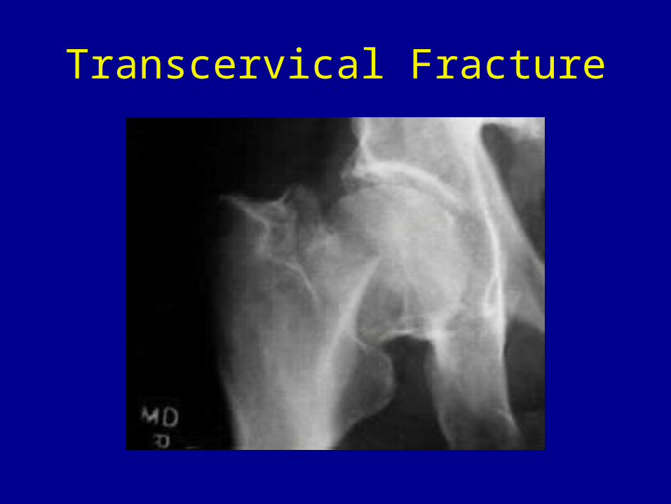
Transcervical Fracture
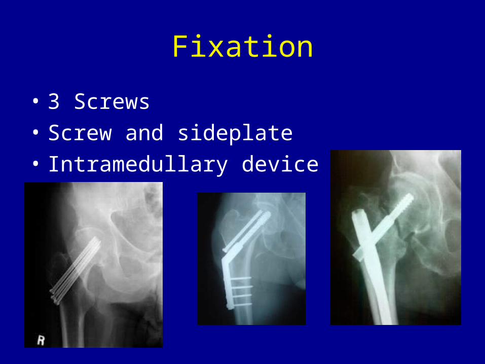
Fixation
• 3 Screws• Screw and sideplate• Intramedullary device

AVN After Treatment
• AVN can occur long after treatment
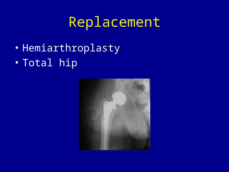
Replacement
• Hemiarthroplasty• Total hip
Summary
• AVN is the disruption of the blood supply to bone
• There are multiple causes• Diagnosis may be delayed• Treatment is dependent on stage and other
factors
Sources
Staging of Avascular Necrosis. Orthopaedia Main. In: Orthopaedia-Collaborative Orthopaedic Knowledgebase
JBJS Br. Core Decompression of the Distal Femur. Vol. 71-B. August, 1989
JBJS. Treatment of Osteonecrosis with Free Vascularized Fibular Graft. Vol 77. 1995


