
BD™ Cytometric Bead Array (CBA)
Human Inflammatory Cytokines KitInstruction Manual
Cat. No. 551811

BD flow cytometers are class I (1) laser products©2008 Becton, Dickinson and Company. All rights reserved. No part of this publication may be reproduced, transmitted, transcribed, stored in retrieval systems, or translated into any language or computer language, in any form or by any means: electronic, mechanical, magnetic, optical, chemical, manual, or otherwise, without prior written permission from BD Biosciences.For research use only. Not for use in diagnostic or therapeutic procedures. Purchase does not include or carry any right to resell or transfer this product either as a stand-alone product or as a component of another product. Any use of this product other than the permitted use without the express written authorization of Becton Dickinson and Company is strictly prohibited. BD, BD Logo and all other trademarks are property of Becton, Dickinson and Company. © 2008 BD
FCAP Array is a registered trademark of Softflow, Inc.Macintosh and Mac are trademarks of Apple Computer, Inc., registered in the US and other countries.Microsoft and Windows are registered trademarks of Microsoft Corporation.
Unless otherwise specified, all products are for Research Use Only. Not for use in diagnostic or therapeutic procedures. Not for resale.
Kit Contents
3US Orders: 877.232.8995
Kit Contents
80 Tests (50 samples and 2 standard curves)
(Store the following items at 4°C)
A1 Human IL-8 Capture Beads: 1 vial, 0.8 ml
A2 Human IL-1β Capture Beads: 1 vial, 0.8 ml
A3 Human IL-6 Capture Beads: 1 vial, 0.8 ml
A4 Human IL-10 Capture Beads: 1 vial, 0.8 ml
A5 Human TNF Capture Beads: 1 vial, 0.8 ml
A6 Human IL-12p70 Capture Beads: 1 vial, 0.8 ml
B Human Inflammatory Cytokines PE Detection Reagent: 1 vial, 4 ml
C Human Inflammatory Cytokines Standards: 2 vials, 0.2 ml lyophilized
D Cytometer Setup Beads: 1 vial, 1.5 ml
E1 PE Positive Control Detector: 1 vial, 0.5 ml
E2 FITC Positive Control Detector: 1 vial, 0.5 ml
F Wash Buffer: 1 bottle, 260 ml
G Assay Diluent: 1 bottle, 30 ml
H Serum Enhancement Buffer: 1 bottle, 10 ml

Unless otherwise specified, all products are for Research Use Only. Not for use in diagnostic or therapeutic procedures. Not for resale.
Table of Contents
4 bdbiosciences.com
Table of Contents
Introduction . . . . . . . . . . . . . . . . . . . . . . . . . . . . . . . . . . . . . . . . . . . . . . . . . . . . 6
Principle of the Test . . . . . . . . . . . . . . . . . . . . . . . . . . . . . . . . . . . . . . . . . . . . . . 7
Advantages . . . . . . . . . . . . . . . . . . . . . . . . . . . . . . . . . . . . . . . . . . . . . . . . . 7
Limitations . . . . . . . . . . . . . . . . . . . . . . . . . . . . . . . . . . . . . . . . . . . . . . . . . 8
Reagents Provided . . . . . . . . . . . . . . . . . . . . . . . . . . . . . . . . . . . . . . . . . . . . . . . 8
Bead Reagents . . . . . . . . . . . . . . . . . . . . . . . . . . . . . . . . . . . . . . . . . . . . . . . 8
Antibody and Standard Reagents . . . . . . . . . . . . . . . . . . . . . . . . . . . . . . . . 9
Buffer Reagents . . . . . . . . . . . . . . . . . . . . . . . . . . . . . . . . . . . . . . . . . . . . . . 9
Warnings and Precautions . . . . . . . . . . . . . . . . . . . . . . . . . . . . . . . . . . . . . 9
Materials Required but Not Provided . . . . . . . . . . . . . . . . . . . . . . . . . . . . . . . 10
Overview: BD CBA Human Inflammatory Cytokines Kit Assay Procedures . . 11
Culture Supernatant Assay Procedure . . . . . . . . . . . . . . . . . . . . . . . . . . . . 11
Serum/Plasma Assay Procedure . . . . . . . . . . . . . . . . . . . . . . . . . . . . . . . . . 12
Preparation of Human Inflammatory Cytokines Standards . . . . . . . . . . . . . . 12
Preparation of Mixed Human Inflammatory Cytokines Capture Beads . . . . . . 14
Preparation of Mixed Human Inflammatory Cytokines Capture Beads for Serum and Plasma Sample Analysis . . . . . . . . . . . . . . . . . . . . . . . . . . . . . . 14
Preparation of Test Samples . . . . . . . . . . . . . . . . . . . . . . . . . . . . . . . . . . . . . . . 15
BD CBA Human Inflammatory Cytokines Kit Assay Procedures . . . . . . . . . . . 15
Culture Supernatant Assay Procedure . . . . . . . . . . . . . . . . . . . . . . . . . . . . 15
Serum/Plasma Assay Procedure . . . . . . . . . . . . . . . . . . . . . . . . . . . . . . . . . 16
Cytometer Setup, Data Acquisition, and Analysis . . . . . . . . . . . . . . . . . . . . . . 17
Preparation of Cytometer Setup Beads . . . . . . . . . . . . . . . . . . . . . . . . . . . 17
Instrument Setup with BD FACSComp Software and BD Calibrite Beads . . . . . . . . . . . . . . . . . . . . . . . . . . . . . . . . . . . . . . . 18
Instrument Setup with the Cytometer Setup Beads . . . . . . . . . . . . . . . . . . 18
Data Acquisition . . . . . . . . . . . . . . . . . . . . . . . . . . . . . . . . . . . . . . . . . . . . 21
Analysis of Sample Data . . . . . . . . . . . . . . . . . . . . . . . . . . . . . . . . . . . . . . 23
Typical Data . . . . . . . . . . . . . . . . . . . . . . . . . . . . . . . . . . . . . . . . . . . . . . . . . . . 23

Unless otherwise specified, all products are for Research Use Only. Not for use in diagnostic or therapeutic procedures. Not for resale.
Table of Contents (continued)
5US Orders: 877.232.8995
Table of Contents (continued)
Performance . . . . . . . . . . . . . . . . . . . . . . . . . . . . . . . . . . . . . . . . . . . . . . . . . . . 25
Theoretical Limit of Detection . . . . . . . . . . . . . . . . . . . . . . . . . . . . . . . . . 25
Recovery . . . . . . . . . . . . . . . . . . . . . . . . . . . . . . . . . . . . . . . . . . . . . . . . . . 26
Linearity . . . . . . . . . . . . . . . . . . . . . . . . . . . . . . . . . . . . . . . . . . . . . . . . . . 27
Specificity . . . . . . . . . . . . . . . . . . . . . . . . . . . . . . . . . . . . . . . . . . . . . . . . . 28
Precision . . . . . . . . . . . . . . . . . . . . . . . . . . . . . . . . . . . . . . . . . . . . . . . . . . 29
Troubleshooting Tips . . . . . . . . . . . . . . . . . . . . . . . . . . . . . . . . . . . . . . . . . . . . 30
References . . . . . . . . . . . . . . . . . . . . . . . . . . . . . . . . . . . . . . . . . . . . . . . . . . . . 31

Unless otherwise specified, all products are for Research Use Only. Not for use in diagnostic or therapeutic procedures. Not for resale.6 bdbiosciences.com
IntroductionFlow cytometry is an analysis tool that allows for the discrimination of different particles on the basis of size and color . Multiplexing is the simultaneous assay of many analytes in a single sample . The BD™ Cytometric Bead Array (CBA) employs a series of particles with discrete fluorescence intensities to simultaneously detect multiple soluble analytes . The BD CBA is combined with flow cytometry to create a powerful multiplexed assay .
The BD CBA system uses the sensitivity of amplified fluorescence detection by flow cytometry to measure soluble analytes in a particle-based immunoassay . Each bead in a BD CBA Kit provides a capture surface for a specific protein and is analogous to an individually coated well in an ELISA plate . The BD CBA capture bead mixture is in suspension to allow for the detection of multiple analytes in a small volume sample . The combined advantages of the broad dynamic range of fluorescent detection via flow cytometry and the efficient capturing of analytes via suspended particles enable BD CBA to use fewer sample dilutions and to obtain the value of an unknown in substantially less time (compared to conventional ELISA) .
The BD CBA Human Inflammatory Cytokines Kit can be used to quantitatively measure Interleukin-8 (IL-8), Interleukin-1β (IL-1β), Interleukin-6 (IL-6), Interleukin-10 (IL-10), Tumor Necrosis Factor (TNF), and Interleukin-12p70 (IL-12p70) protein levels in a single sample . The kit performance has been optimized for analysis of specific proteins in tissue culture supernatants, EDTA plasma, and serum samples using one of two protocols depending on the sample source .

Unless otherwise specified, all products are for Research Use Only. Not for use in diagnostic or therapeutic procedures. Not for resale. 7US Orders: 877.232.8995
Principle of the TestSix bead populations with distinct fluorescence intensities have been coated with capture antibodies specific for IL-8, IL-1β, IL-6, IL-10, TNF, and IL-12p70 proteins . The six bead populations are mixed together to form the BD CBA, which is resolved in a red channel (ie, FL3 or FL4) of a flow cytometer .
Figure 1
IL-12p70
TNFIL-10 IL-6
IL-1β
IL-8
Figure 1
The capture beads, PE-conjugated detection antibodies, and recombinant standards or test samples are incubated together to form sandwich complexes . Following acquisition of sample data using the flow cytometer, the sample results are generated in graphical and tabular format using the BD CBA Analysis Software or FCAP Array™ Software . The kit provides sufficient reagents for the quantitative analysis of 50 test samples and the generation of two standard curve sets .
AdvantagesThe BD CBA provides several advantages when compared with conventional ELISA methodology:
• Therequiredsamplevolumeisapproximatelyone-sixththequantitynecessary for conventional ELISA assays due to the detection of six analytes in a single sample .
• Asinglesetofdilutedstandardsisusedtogenerateastandardcurveforeach analyte .
• ABDCBAexperimenttakeslesstimethanasingleELISAandprovidesresults that would normally require six conventional ELISAs .

Unless otherwise specified, all products are for Research Use Only. Not for use in diagnostic or therapeutic procedures. Not for resale.8 bdbiosciences.com
LimitationsThe limit of detection of the BD CBA Human Inflammatory Cytokines Kit is comparable to conventional ELISA, but due to the complexity and kinetics of this multi-analyte assay, the actual limit of detection in a given experiment may vary slightly (see Theoretical Limit of Detection and Precision information on page 25 and 29 respectively) .
The BD CBA is not recommended for use on stream-in-air instruments where signal intensities may be reduced, adversely effecting assay sensitivity . Stream-in-air instruments include the BD FACStar™ Plus and BD FACSVantage™ flow cytometers .
Serum spike recoveries for IL-1β, TNF, and IL-12p70 are lower than for the other proteins in this assay . This variation is due to assay conditions and serum proteins and may affect quantitation of these proteins in serum samples .
The sensitivity for the detection of IL-1β in this assay is less than for the other proteins measured . It is possible that, due to operator variation, instrument settings, and instrument performance, the 20 pg/ml standard curve point for IL-1β may not have signal intensity above the 0 pg/ml background level .
Quantitative results or protein levels for the same sample or recombinant protein run in ELISA and BD CBA assays may differ . A spike recovery assay can be performed using an ELISA standard followed by BD CBA analysis to assess possible differences in quantitation .
This kit is designed to be used as an integral unit . Do not mix components from different batches or kits .
Reagents ProvidedBead ReagentsHuman Inflammatory Cytokines Capture Beads (A1 – A6): The specific capture beads, having discrete fluorescence intensity characteristics, are distributed from brightest to dimmest as follows:
Bead
(Brightest) A1
A2
A3
A4
A5
(Dimmest) A6
Specificity
IL-8
IL-1β
IL-6
IL-10
TNF
IL-12p70
A single 80-test vial of each specific capture bead (A1 – A6) is included in this kit . Store at 4°C . Do not freeze .
Note: The antibody-conjugated beads will settle out of suspension over time . It is necessary to vortex the vial vigorously for 3 – 5 seconds before taking a bead suspension aliquot .
Cytometer Setup Beads (D): A single, 30-test vial of setup beads for setting the initial instrument PMT voltages and compensation settings is sufficient for 10 instrument setup procedures . The Cytometer Setup Beads are formulated for use at 50 µl/test .

Unless otherwise specified, all products are for Research Use Only. Not for use in diagnostic or therapeutic procedures. Not for resale. 9US Orders: 877.232.8995
Antibody and Standard ReagentsHuman Inflammatory Cytokines PE Detection Reagent (B): An 80-test vial of PE-conjugated anti-human IL-8, IL-1β, IL-6, IL-10, TNF, and IL-12p70 antibodies formulated for use at 50 µl/test . Store at 4°C . Do not freeze .
Human Inflammatory Cytokines Standards (C): Two vials containing lyophilized recombinant human proteins . Each vial should be reconstituted in 2 .0 ml of Assay Diluent to prepare the top standard . Store at 4°C .
Note: The Human Inflammatory Cytokines Standards vials are stable until the kit expiration date . Following reconstitution, store the freshly reconstituted 10× bulk standard at 2 – 8°C and use within 12 hours .
PE Positive Control Detector (E1): A 10-test vial of PE-conjugated antibody control formulated for use at 50 µl/test . This reagent is used with the Cytometer Setup Beads to set the initial instrument compensation settings . Store at 4°C . Do not freeze .
FITC Positive Control Detector (E2): A 10-test vial of FITC-conjugated antibody control formulated for use at 50 µl/test . This reagent is used with the Cytometer Setup Beads to set the initial instrument compensation settings . Store at 4°C . Do not freeze .
Buffer ReagentsWash Buffer (F): One 260 ml bottle of phosphate buffered saline (PBS) solution (1×), containing protein* and detergent, used for wash steps and to resuspend the washed beads for analysis . Store at 4°C .
Assay Diluent (G): A single, 30 ml bottle of a buffered protein* solution (1×) used to reconstitute and dilute the Human Inflammatory Cytokines Standards and to dilute test samples . Store at 4°C .
Serum Enhancement Buffer (H): A single, 10 ml bottle of a buffered protein* solution (1×) used to dilute mixed Capture Beads when testing serum or plasma samples . Store at 4°C .
Warnings and PrecautionsHazardous Ingredients:
Sodium Azide: Components A1 - A6, B, D, E1 - E2, F, G, and H contain 0 .09% sodium azide . Sodium azide yields a highly toxic hydrazoic acid under acidic conditions . Dilute azide compounds in running water before discharging to avoid accumulation of potentially explosive deposits in plumbing .
* Source of all serum proteins is from USDA inspected abattoirs located in the United States.

Unless otherwise specified, all products are for Research Use Only. Not for use in diagnostic or therapeutic procedures. Not for resale.10 bdbiosciences.com
Materials Required but Not ProvidedIn addition to the reagents provided in the BD CBA Human Inflammatory Cytokines Kit, the following items are also required:
• Aflowcytometerequippedwitha488nmlasercapableofdetectinganddistinguishing fluorescence emissions at 576 and 670 nm (eg, BD FACScan™ or BD FACSCalibur™ instruments) and BD CellQuest™ Software .
• 12× 75 mm sample acquisition tubes for a flow cytometer (eg, BD Falcon™ Cat . No . 352008) .
• BDCBASoftware,orFCAPArraysoftware(Cat.No.641488).Note: The BD CBA Software is no longer available for purchase but is
still supported on existing compatible systems .
• BDCalibrite™3Beads,(Cat.No.340486).

Unless otherwise specified, all products are for Research Use Only. Not for use in diagnostic or therapeutic procedures. Not for resale. 11US Orders: 877.232.8995
Overview: BD CBA Human Inflammatory Cytokines Kit Assay Procedures
Culture Supernatant Assay Procedure
1. Reconstitute Human Inflammation Standards in Assay Diluent (15 min)2. Dilute Standards by serial dilutions using the Assay Diluent
3. Mix 10 µl/test of each Human Inflammation Capture Bead suspension (vortex before aliquoting)4. Transfer 50 µl of mixed beads to each assay tube
30 minuteincubation at RT
(protect from light)
3. Add 400 µl of Wash Buffer to tubes B and C4. Add 450 µl of Wash Buffer to tube A5. Use tubes A, B and C for cytometer setup
Cytometer Setup Bead Procedure
1. Add Cytometer Setup Beads (vortex before adding) to setup tubes A, B and C (50 µl/tube) 2. Add 50 µl of FITC Positive Control to tube B and 50 µl of PE Positive Control to tube C
*
6. Add PE Detection Reagent (50 µl/test)
5. Add Standard Dilutions and test samples to the appropriate sample tubes (50 µl/tube)
7. Wash samples with 1 ml Wash Buffer and centrifuge
8. Add 300 µl of Wash Buffer to each assay tubes and analyze samples
3 Hour incubation at RT (protect from light)*

Unless otherwise specified, all products are for Research Use Only. Not for use in diagnostic or therapeutic procedures. Not for resale.12 bdbiosciences.com
Serum/Plasma Assay Procedure
1. Mix 10 µl/test of each Human Inflammatory Cytokine Capture Bead suspension (vortex briefly)2. Pellet mixed beads by centrifugaton and aspirate supernatant3. Resuspend bead pellet in Serum Enhancement Buffer (equal volume to amount removed in step 2) and vortex briefly
30 Minute incubation at RT
30 minute incubation at R T
(protect from light)
4. Transfer 50 µl of mixed beads to each assay tube5. Reconstitute Human Inflammatory Cytokine Standards in Assay Diluent (15 min)6. Dilute Standards by serial dilutions using the Assay Diluent
(protect from light)*
3. Add 400 μl of W ash Buffer to tubes B and C
4. Add 450 μl of W ash Buffer to tube A
5. Use tubes A, B and C for cytometer setup
*Cytometer Setup Bead Pr ocedur e
1. Add Cytometer Setup Beads (vortex before adding) to setup tubes A, B and C (50 μl/tube)
2. Add 50 μl of FITC Positive Control to tube B and 50 μl of
PE Positive Control to tube C
1.5 Hour incubation at RT
7. Add Standard Dilutions and test samples to the appropriate sample tubes (50 μl/tube)
8. Wash samples with 1 ml Wash Buffer and centrifuge (leave ~100 μl residual volume after aspiration)
(protect from light)*
9. Add PE Detection Reagent (50 μl/test)
10. Wash samples with 1 ml Wash Buffer and centrifuge
1.5 Hour incubation at RT (protect from light)*
11. Add 300 μl of Wash Buffer to each assay tube and analyze samples
Preparation of Human Inflammatory Cytokines StandardsThe Human Inflammatory Cytokines Standards are lyophilized and should be reconstituted and serially diluted before mixing with the Capture Beads and the PE Detection Reagent .
1 . Open one vial of lyophilized Human Inflammatory Cytokines Standards . Transfer the standard spheres to a polypropylene tube (eg, 15 ml Conical Tube, BD Falcon Cat . No . 352097) . Label the tube “Top Standard” .

Unless otherwise specified, all products are for Research Use Only. Not for use in diagnostic or therapeutic procedures. Not for resale. 13US Orders: 877.232.8995
2 . Reconstitute the standards with 2 .0 ml of Assay Diluent . Allow the reconstituted standard to equilibrate for at least 15 minutes before making dilutions . Mix reconstituted protein by pipette only. Do not vortex or mix vigorously.
3 . Label 12 × 75 mm tubes (BD Falcon Cat . No . 352008) and arrange them in the following order: 1:2, 1:4, 1:8, 1:16, 1:32, 1:64, 1:128 and 1:256 .
4 . Pipette 300 µl of Assay Diluent to each of the remaining tubes .
5 . Perform a serial dilution by transferring 300 µl from the Top Standard to the 1:2 dilution tube and mix thoroughly . Continue making serial dilutions by transferring 300 µl from the 1:2 tube to the 1:4 tube and so on to the 1:256 tube and mix thoroughly (see Figure 2) . Mix by pipette only, do not vortex . Prepare one tube containing Assay Diluent to serve as the 0 pg/ml negative control .
300 µ l 300 µ l 300 µ l 300 µ l 300 µ l 300 µ l 300 µ l 300 µ l
TopStandard
1:2
2.0 mL
Dilution T ube
1:4 Dilution
T ube
1:8 Dilution
T ube
1:16 Dilution
T ube
1:32 Dilution
T ube
1:64 Dilution
T ube
1:128 Dilution
T ube
1:256 Dilution
T ube
Figure 2. Preparation of Human Inflammatory Cytokines Standard Dilutions
The approximate concentration (pg/ml) of recombinant protein in each dilution tube is shown in Table 1 .
Table 1. Human Inflammatory Cytokines Standard concentrations after dilution
Protein
(pg/ml)
Human IL-8
Human IL-1β
Human IL-6
Human IL-10
Human TNF
Human IL-12p70
Top Standard
5000
5000
5000
5000
5000
5000
1:2 Dilution
Tube
2500
2500
2500
2500
2500
2500
1:4 Dilution
Tube
1250
1250
1250
1250
1250
1250
1:8 Dilution
Tube
625
625
625
625
625
625
1:16 Dilution
Tube
312.5
312.5
312.5
312.5
312.5
312.5
1:32 Dilution
Tube
156
156
156
156
156
156
1:64 Dilution
Tube
80
80
80
80
80
80
1:128 Dilution
Tube
40
40
40
40
40
40
1:256 Dilution
Tube
20
20
20
20
20
20

Unless otherwise specified, all products are for Research Use Only. Not for use in diagnostic or therapeutic procedures. Not for resale.14 bdbiosciences.com
Preparation of Mixed Human Inflammatory Cytokines Capture BeadsThe Capture Beads are bottled individually, and it is necessary to pool the bead reagents (A1 – A6) immediately before mixing them together with the PE Detection Reagent, standards, or samples . It is recommended that this procedure be used for preparing the mixed Human Inflammatory Cytokines Capture Beads for experiments in which cell culture supernatant samples will be analyzed . For experiments testing serum or plasma samples, refer to Preparation of Mixed Human Inflammatory Cytokines Capture Beads for Serum and Plasma Sample Analysis, below . 1 . Determine the number of assay tubes (including standards and controls)
that are required for the experiment (eg, 8 unknowns, 9 standard dilutions, and 1 negative control = 18 assay tubes) .
2 . Vigorously vortex each Capture Bead suspension for a few seconds before mixing .
3 . Add a 10 µl aliquot of each Capture Bead, for each assay tube to be analyzed, into a single tube labeled “mixed Capture Beads” (eg, 10 µl of IL-8 Capture Beads × 18 assay tubes = 180 µl of IL-8 Capture Beads required) .
4 . Vortex the Bead mixture thoroughly .
The mixed Capture Beads are now ready to be transferred to the assay tubes (50 µl of mixed Capture beads/tube) as described in BD CBA Human Inflammatory Cytokines Kit Assay Procedures, page 15 .
Note: Discard excess mixed Capture Beads . Do not store after mixing .
Preparation of Mixed Human Inflammatory Cytokines Capture Beads for Serum and Plasma Sample AnalysisIt is recommended that the following procedure be followed for preparing the mixed Human Inflammatory Cytokines Capture Beads for experiments in which serum and plasma samples will be analyzed . Use of this procedure will reduce the chances of false-positive results due to the effects of serum or plasma proteins . This procedure may also be used with cell culture supernatant samples .
1 . Follow steps 1 – 4 under Preparation of Mixed Human Inflammatory Cytokines Capture Beads, above .
2 . Centrifuge mixed Capture Beads at 200 × g for 5 minutes .
3 . Carefully aspirate and discard the supernatant .
4 . Resuspend the mixed Capture Beads pellet in Serum Enhancement Buffer (equal volume to amount removed in step 3) and vortex thoroughly .
5 . Incubate the assay tubes for 30 minutes at RT and protect from direct exposure to light .
The mixed Capture Beads are now ready to be transferred to the assay tubes (50 µl of mixed Capture Beads/tube) as described in BD CBA Human Inflammatory Cytokines Kit Assay Procedures, page 15 .
Note: Discard excess mixed Capture Beads . Do not store after mixing .

Unless otherwise specified, all products are for Research Use Only. Not for use in diagnostic or therapeutic procedures. Not for resale. 15US Orders: 877.232.8995
Preparation of Test SamplesThe standard curve for each protein covers a defined set of concentrations from 20 – 5000 pg/ml . It may be necessary to dilute test samples to ensure that their mean fluorescence values fall within the limits or range of the generated standard curve . For best results, samples that are known or assumed to contain high levels of a given protein should be diluted as described below .
1 . Dilute test sample by the desired dilution factor (ie, 1:2, 1:10, or 1:100) using the appropriate volume of Assay Diluent .
2 . Mix sample dilutions thoroughly before transferring samples to the appropriate assay tubes containing mixed Capture Beads and PE Detection Reagent .
BD CBA Human Inflammatory Cytokines Kit Assay ProceduresFollowing the preparation and dilution of the standards and mixing of the capture beads, transfer these reagents and test samples to the appropriate assay tubes for incubation and analysis . The serum/plasma assay procedure (page 16) should be used for any experiment testing serum or plasma samples . The serum/plasma assay procedure also works for culture supernatant . In order to calibrate the flow cytometer and quantitate test samples, it is necessary to run the Inflammatory Cytokines Standards and the Cytometer Setup controls in each experiment. See Table 2 for a detailed description of the reagents added to the Inflammatory Cytokines Standard control assay tubes . The Cytometer Setup procedure is described on page 17 .
Culture Supernatant Assay Procedure 1 . Add 50 µl of the mixed Capture Beads (prepared using the procedure
described in Preparation of Mixed Human Inflammatory Cytokines Capture Beads, page 14) to the appropriate assay tubes . Vortex the mixed Capture Beads before adding to the assay tubes .
2 . Add 50 µl of the Human Inflammatory Cytokines PE Detection Reagent to the assay tubes .
3 . Add 50 µl of the Human Inflammatory Cytokines Standard dilutions to the control assay tubes .
4 . Add 50 µl of each test sample to the test assay tubes .
5 . Incubate the assay tubes for 3 hours at RT and protect from direct exposure to light . During this incubation, perform the Cytometer Setup procedure described on pages 17 and 18 .
6 . Add 1 ml of Wash Buffer to each assay tube and centrifuge at 200 × g for 5 minutes .
7 . Carefully aspirate and discard the supernatant from each assay tube .
8 . Add 300 µl of Wash Buffer to each assay tube to resuspend the bead pellet .
9 . Begin analyzing samples on a flow cytometer . Vortex each sample for 3 – 5 seconds immediately before analyzing on the flow cytometer.

Unless otherwise specified, all products are for Research Use Only. Not for use in diagnostic or therapeutic procedures. Not for resale.16 bdbiosciences.com
Serum/Plasma Assay Procedure 1 . Add 50 µl of the mixed Capture Beads (prepared using the procedure
described in Preparation of Mixed Human Inflammatory Cytokines Capture Beads for Serum and Plasma Sample Analysis, page 14) to the appropriate assay tubes . Vortex the mixed Capture Beads before adding to the assay tubes .
2 . Add 50 µl of the Human Inflammatory Cytokines Standard dilutions to the control assay tubes .
3 . Add 50 µl of each test sample to the test assay tubes .
4 . Incubate the assay tubes for 1 .5 hours at RT and protect from direct exposure to light .
5 . Add 1 ml of Wash Buffer to each assay tube and centrifuge at 200 × g for 5 minutes .
6 . Carefully aspirate and discard the supernatant, leaving approximately 100 µl of liquid in each assay tube . Aspiration should be done as consistently as possible .
7 . Add 50 µl of the Human Inflammatory Cytokines PE Detection Reagent to the assay tubes . Gently agitate assay tubes to resuspend bead pellet .
8 . Incubate the assay tubes for 1 .5 hours at RT and protect from direct exposure to light . During this incubation, perform the Cytometer Setup procedure described on pages 17 and 18 .
9 . Add 1 ml of Wash Buffer to each assay tube and centrifuge at 200 × g for 5 minutes .
10 . Carefully aspirate and discard the supernatant from each assay tube .
11 . Add 300 µl of Wash Buffer to each assay tube to resuspend the bead pellet .
12 . Begin analyzing samples on a flow cytometer . Vortex each sample for 3 – 5 seconds immediately before analyzing on the flow cytometer.
Note: It is necessary to analyze CBA samples on the day of the experiment . Prolonged storage of samples, once the assay is complete, can lead to increased background and reduced sensitivity .

Unless otherwise specified, all products are for Research Use Only. Not for use in diagnostic or therapeutic procedures. Not for resale. 17US Orders: 877.232.8995
Table 2. Essential control assay tubes
Tube No.
1 (Negative Control 0 pg/ml Standards)
2 (20 pg/ml Standards)
3 (40 pg/ml Standards)
4 (80 pg/ml Standards)
5 (156 pg/ml Standards)
6 (312 pg/ml Standards)
7 (625 pg/ml Standards)
8 (1250 pg/ml Standards)
9 (2500 pg/ml Standards)
10 (5000 pg/ml Standards)
mixed Capture Beads, PE Detection Reagent, Assay Diluent
mixed Capture Beads, PE Detection Reagent, Inflammation Standards 1:256 Dilution
mixed Capture Beads, PE Detection Reagent, Inflammation Standards 1:128 Dilution
mixed Capture Beads, PE Detection Reagent, Inflammation Standards 1:64 Dilution
mixed Capture Beads, PE Detection Reagent, Inflammation Standards 1:32 Dilution
mixed Capture Beads, PE Detection Reagent, Inflammation Standards 1:16 Dilution
mixed Capture Beads, PE Detection Reagent, Inflammation Standards 1:8 Dilution
mixed Capture Beads, PE Detection Reagent, Inflammation Standards 1:4 Dilution
mixed Capture Beads, PE Detection Reagent, Inflammation Standards 1:2 Dilution
mixed Capture Beads, PE Detection Reagent, Inflammation Standards "Top Standard"
Reagents(All reagent volumes are 50 µl)
Cytometer Setup, Data Acquisition, and AnalysisThe Cytometer setup information in this section is for the BD FACScan and BD FACSCalibur flow cytometers . The BD FACSComp software is useful for setting up the flow cytometer . BD CellQuest Software is required for analyzing samples and formatting data for subsequent analysis using the BD CBA Software or FCAP Array Software .
Additional setup protocols for the BD FACSCalibur flow cytometer (dual laser), BD FACSArray™ bioanalyzer, and other BD FACS brand flow cytometers can be found at bdbiosciences.com/cbasetup .
Preparation of Cytometer Setup Beads 1 . Add 50 µl of Cytometer Setup Beads to three cytometer setup tubes labeled
A, B, and C .
2 . Add 50 µl of FITC Positive Control Detector to tube B .
3 . Add 50 µl of PE Positive Control Detector to tube C .
4 . Incubate tubes A, B, and C for 30 minutes at room temperature and protect from direct exposure to light .
5 . Add 450 µl of Wash Buffer to tube A and 400 µl of Wash Buffer to tubes B and C .

Unless otherwise specified, all products are for Research Use Only. Not for use in diagnostic or therapeutic procedures. Not for resale.18 bdbiosciences.com
Instrument Setup with BD FACSComp Software and BD Calibrite Beads 1 . Perform instrument start up .
2 . Perform flow check .
3 . Prepare tubes of BD Calibrite beads and open BD FACSComp software .
4 . Launch BD FACSComp software .
5 . Run BD FACSComp software in Lyse/No Wash mode .
6 . Proceed to Instrument Setup with the Cytometer Setup Beads .
Note: For detailed information on using BD FACSComp with BD Calibrite beads to set up the flow cytometer, refer to the BD FACSComp Software User’s Guide and the BD Calibrite Beads Package Insert . Version 4 .2 contains a BD CBA preference setting to automatically save a BD CBA calibration file at the successful completion of any Lyse/No Wash assay . The BD CBA calibration file provides the optimization for FSC, SSC, and threshold settings as described in Instrument Setup with the Cytometer Setup Beads, Steps 3 – 5 . Optimization of the fluorescence parameter settings is still required (ie, PMT and compensation settings, see Instrument Setup with the Cytometer Setup Beads, Step 6) .
Instrument Setup with the Cytometer Setup Beads 1 . Launch BD™ CellQuest Software and open the CBA Instrument Setup
template . Note: The BD CBA Instrument Setup template can be found on the
BD FACStation™ CD for Macintosh computers in the BD CBA folder . This file may also be downloaded via the internet from: bdbiosciences.com/cbatemplates
2 . Set the instrument to Acquisition mode .Note: The data will be evaluated in five parameters (FSC, SSC, FL1,
FL2, and FL3) . Turn off additional detectors .
3 . Set SSC (side light scatter) and FSC (forward light scatter) to Log mode .
4 . Decrease the SSC PMT voltage by 100 from what BD FACSComp set .
5 . Set the Threshold to SSC at 650 .
6 . In setup mode, run Cytometer Setup Beads tube A . Note: Pause and restart acquisition frequently during the instrument
setup procedure in order to reset detected values after settings adjustments .

Unless otherwise specified, all products are for Research Use Only. Not for use in diagnostic or therapeutic procedures. Not for resale. 19US Orders: 877.232.8995
Adjust gate R1 so that the singlet bead population is located in gate R1 (Figure 3a) .
Figure 3a
Adjust the FL3 PMT so that the median intensity of the top FL3 bead population is around 5000 (Figure 3b) . Adjust gate R3 as necessary so that the dim FL3 bead population is located in gate R3 (Figure 3b) . Do not adjust the R2 gate .
Bright Beads (R2) Median: 5139.70
FL1 (Median) Median: 2.53
Figure 3b
Adjust the FL1 PMT so that the median of FL1 is approximately 2 .0 – 2 .5 (Figure 3b) .
Adjust the FL2 PMT value so that the median of FL2 is approximately 2 .0 – 2 .5 (Figure 3c).
FL2 (Median) Median: 2.21
Figure 3c
Run Cytometer Setup Beads tube B to adjust the compensation settings for FL2 – %FL1 .

Unless otherwise specified, all products are for Research Use Only. Not for use in diagnostic or therapeutic procedures. Not for resale.20 bdbiosciences.com
Adjust gate R5 as necessary so that the FL1 bright bead population is located in gate R5 (Figure 3d) . Using the FL2 – %FL1 control, adjust the median of R5 to equal the median of R4 (Figure 3d) .
(R4) Median: 2.94 (R5) Median: 3.75
Figure 3d
Run Cytometer Setup Beads tube C to adjust the compensation settings for FL1 – %FL2 and FL3 – %FL2 .
Adjust gate R7 so that the FL2 bright bead population is located in gate R7 (Figure 3e) . Using the FL1 – %FL2 control, adjust the median of R7 to equal the median of R6 (Figure 3e) .
(R6) Median: 2.81 (R7) Median: 3.08
Figure 3e
Adjust gate R9 so that the FL2 bright bead population is located in gate R9 (Figure 3f) . Using the FL3 – %FL2 control, adjust the median of R9 to equal the median of R8 (Figure 3f) .
(R8) Median: 27.88 (R9) Median: 28.13
Figure 3fSet the FL2 – %FL3 to 0 .1 if necessary . Save and print the optimized instrument settings .

Unless otherwise specified, all products are for Research Use Only. Not for use in diagnostic or therapeutic procedures. Not for resale. 21US Orders: 877.232.8995
Data Acquisition 1 . Open the Acquisition template .
Note: The acquisition template may be downloaded via the internet from: bdbiosciences.com/cbatemplates
2 . Set acquisition mode and retrieve the optimized instrument settings from Instrument Setup with the Cytometer Setup Beads, page 18 .
3 . In the Acquisition and Storage window, set the resolution to 1024 .
4 . Set number of events to be counted at 1800 of R1 gated events . (This will ensure that the sample file contains approximately 300 events per Capture Bead) .
5 . Set number of events to be collected to “all events” . Saving all events collected will ensure that no true bead events are lost due to incorrect gating .
6 . In setup mode, run tube no . 1 and using the FSC vs . SSC dot plot, place the R1 region gate around the singlet bead population (see Figure 3a) .
7 . Samples are now ready to be acquired .
8 . Begin sample acquisition with the flow rate set at HIGH .
Note: Run the negative control tube (0 pg/ml standards) before any of the recombinant standard tubes . Run the control assay tubes before any unknown test assay tubes . Run the tubes in the order listed in Table 2, page 17 .
File names must be alphanumeric (ie, contain at least one letter) .

Unless otherwise specified, all products are for Research Use Only. Not for use in diagnostic or therapeutic procedures. Not for resale.22 bdbiosciences.com
Figure 4: Acquisition Template ExampleFigure 4. Acquisition Template Example

Unless otherwise specified, all products are for Research Use Only. Not for use in diagnostic or therapeutic procedures. Not for resale. 23US Orders: 877.232.8995
Analysis of Sample DataThe analysis of BD CBA data can be accomplished using BD CBA Software or FCAP Array software . For BD CBA Software, please refer to the User’s Guide for instructions . For FCAP Array software, please visit the following link for instructions: bdbiosciences.com/docs/kitanalysis
Typical Data
Figure 5: CellQuest data examplesfor standards and detectors alone
Negative ControlA. Standards 80 pg/mlB.
Standards 625 pg/mlC. Standards 5000 pg/mlD.
Figure 5. BD CellQuest Data Examples for Standards and Detectors Alone

Unless otherwise specified, all products are for Research Use Only. Not for use in diagnostic or therapeutic procedures. Not for resale.24 bdbiosciences.com
TNF
1
1 0
100
1000
10000
1 1 0 100 1000 10000
Concentration (pg/ml)
1 0 Log ScaleSemiLog ScaleLinear ScaleLog Scale
1
1 0
100
1000
10000
1 1 0 100 1000 10000
Concentration (pg/ml)
IL-10
1
1 0
100
1000
10000
1 1 0 100 1000 10000
Concentration (pg/ml)
IL-6
1
1 0
100
1000
10000
1 1 0 100 1000 10000
Concentration (pg/ml)
IL-8
1
1 0
100
1000
10000
1 1 0 100 1000 10000
Concentration (pg/ml)
IL-1β
1
1 0
100
1000
10000
1 1 0 100 1000 10000
Concentration (pg/ml)
Figure 6: BD CBA Softwareexample of standard curves
IL-12p70
Figure 6. Example of Standard Curves

Unless otherwise specified, all products are for Research Use Only. Not for use in diagnostic or therapeutic procedures. Not for resale. 25US Orders: 877.232.8995
PerformanceThe BD CBA Human Inflammatory Cytokines Kit assay has been rigorously tested for the following: theoretical limit of detection, spike recovery, dilution linearity, specificity, and intra- and inter-assay precision .
Recovery and linearity results for cell-culture supernatant are using the culture supernatant assay procedure, and the serum/plasma recovery and linearity results are using the serum/plasma assay protocol .
Theoretical Limit of DetectionThe individual standard curve range for a given protein defines the minimum and maximum quantifiable levels using the BD CBA Human Inflammatory Cytokines Kit (ie, 20 pg/ml and 5000 pg/ml) . By applying the 4-parameter curve fit option, it is possible to extrapolate values for sample intensities not falling within the limits of the standard curve . It is up to the researcher to decide the best method for calculating values for unknown samples using this assay . The theoretical limit of detection for each protein using the BD CBA Human Inflammatory Cytokines Kit is defined as the corresponding concentration at two standard deviations above the median fluorescence of 20 replicates of the negative control (0 pg/ml) .
Cytokine
IL-8
IL-1β
IL-6
IL-10
TNF
IL-12p70
Median Fluorescence
3.4
3.7
4.7
4.1
3.9
4.0
Standard Deviation
0.4
0.3
0.4
0.4
0.3
0.3
Limit of Detection(pg/ml)
3.6 pg/ml
7.2 pg/ml
2.5 pg/ml
3.3 pg/ml
3.7 pg/ml
1.9 pg/ml

Unless otherwise specified, all products are for Research Use Only. Not for use in diagnostic or therapeutic procedures. Not for resale.26 bdbiosciences.com
RecoveryIndividual protein was spiked into various matrices at three different levels within the assay range . The matrices used in these experiments were not diluted before addition of the protein . The plasma samples in these experiments were EDTA treated . Results are compared with the same concentrations of the proteins spiked in the Assay Diluent, as follows:
Matrix
Pooled Donor Sera(n = 5)
Pooled Donor Plasma(n = 5)
Cell culture supernatant
Pooled Donor Sera(n = 5)
Pooled Donor Plasma(n = 5)
Cell culture supernatant
Pooled Donor Sera(n = 5)
Pooled Donor Plasma(n = 5)
Cell culture supernatant
Pooled Donor Sera(n = 5)
Pooled Donor Plasma(n = 5)
Cell culture supernatant
Pooled Donor Sera(n = 5)
Pooled Donor Plasma(n = 5)
Cell culture supernatant
Pooled Donor Sera(n = 5)
Pooled Donor Plasma(n = 5)
Cell culture supernatant
Standard spike concentration (pg/ml)
250062580
250062580
250062580
250062580
250062580
250062580
250062580
250062580
250062580
250062580
250062580
250062580
250062580
250062580
250062580
250062580
250062580
250062580
Cytokine
IL-8
IL-8
IL-8
IL-1β
IL-1β
IL-1β
IL-6
IL-6
IL-6
IL-10
IL-10
IL-10
TNF
TNF
TNF
IL-12p70
IL-12p70
IL-12p70
Observed in given matrix (pg/ml)
214554866
205147353
270966191
165642442
177640347
281362381
171348455
225457373
264659281
201655667
230858970
244960079
150741945
185145755
286466588
136636846
175240953
254060081
% Recovery
86%88%82%82%76%67%
108%106%113%66%68%52%71%65%58%
113%100%101%69%78%69%90%92%92%
106%95%
101%81%89%84%92%94%88%98%96%99%60%67%56%74%73%69%
115%107%110%55%59%57%70%65%66%
102%96%
101%

Unless otherwise specified, all products are for Research Use Only. Not for use in diagnostic or therapeutic procedures. Not for resale. 27US Orders: 877.232.8995
LinearityIn two experiments, the following matrices were spiked with IL-8, IL-1β, IL-6, IL-10, TNF, and IL-12p70 and were then serially diluted with Assay Diluent .
Dilution
Neat1:21:41:8
1:161:321:64
1:1281:256SlopeNeat
1:21:41:8
1:161:321:64
1:1281:256SlopeNeat
1:21:41:8
1:161:321:64
1:1281:256Slope
Dilution
Neat1:21:41:8
1:161:321:64
1:1281:256SlopeNeat
1:21:41:8
1:161:321:64
1:1281:256SlopeNeat
1:21:41:8
1:161:321:64
1:1281:256Slope
ObservedIL-8 (pg/ml)
446825831363
618322129
763919
1.00449325821405
697347169
804322
0.98520025201260
602296154743919
1.01
ObservedIL-10 (pg/ml)
466224521204
559304129
793618
1.015408428422701223
648294166
8444
0.91510024941302
643329156
744019
1.01
Matrix
Pooled Donor Sera(n = 5)
Pooled Donor Plasma (n = 5)
Cell Culture Medium
Matrix
Pooled Donor Sera (n = 5)
Pooled Donor Plasma (n = 5)
Cell Culture Medium
Observed IL-1β (pg/ml)
28841815
900458248102
614626
0.88369220491130
572275138
723723
0.95580026651291
596312143
743917
1.04
ObservedTNF (pg/ml)
321320381041
519302124
833517
0.95
428422701223
648294166
844420
0.96600025971241
606320157
784523
0.99
ObservedIL-6 (pg/ml)
341020161017
486278
99693214
1.00473426541301
651315160
814319
1.00520025901280
635310150
754120
1.01Observed
IL-12p70 (pg/ml)
29621757
963470252109
673015
0.97439123591218
643309165
834221
0.97600025601283
618302154
724220
1.02
Unless otherwise specified, all products are for Research Use Only. Not for use in diagnostic or therapeutic procedures. Not for resale.28 bdbiosciences.com
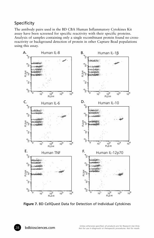
SpecificityThe antibody pairs used in the BD CBA Human Inflammatory Cytokines Kit assay have been screened for specific reactivity with their specific proteins . Analysis of samples containing only a single recombinant protein found no cross-reactivity or background detection of protein in other Capture Bead populations using this assay .
100
101
102
103
104
FL3
-H
0 101 102 103 104
FL2-H
100
101
102
103
104
FL3
-H
0 101 102 103 104
FL2-H
100
101
102
103
104
FL3
-H
0 101 102 103 104
FL2-H
100
101
102
103
104
FL3
-H
0 101 102 103 104
FL2-H
100
101
102
103
104
FL3
-H
0 101 102 103 104
FL2-H
100
101
102
103
104
FL3
-H
0 101 102 103 104
FL2-H
Figure 8: CellQuest data fordetection of individual cytokines
Human IL-8 Human IL-1β
Human IL-6 Human IL-10
Human TNF Human IL-12p70
A. B.
C. D.
E. F.
Figure 7. BD CellQuest Data for Detection of Individual Cytokines

Unless otherwise specified, all products are for Research Use Only. Not for use in diagnostic or therapeutic procedures. Not for resale. 29US Orders: 877.232.8995
PrecisionThe precision results shown for intra- and inter-assay reproducibility were generated using the serum/plasma assay procedure . Most CV values are slightly improved when using the culture supernatant assay procedure due to reduced sample handling .
Intra-assay: Ten replicates of each of three different levels of IL-8, IL-1β, IL-6, IL-10, TNF, and IL-12p70 were tested .
Cytokine
Actual Mean Conc. (pg/ml):
SD
% CV
74
3
4%
578
12
2%
2221
104
5%
69
5
7%
540
19
4%
2220
129
6%
IL-8 IL-1β
Cytokine
Actual Mean Conc. (pg/ml):
SD
% CV
77
5
6%
557
25
5%
2171
163
8%
76
5
6%
551
25
5%
2116
121
6%
IL-6 IL-10
Cytokine
Actual Mean Conc. (pg/ml):
SD
% CV
70
6
9%
527
34
6%
1971
207
10%
74
3
4%
562
19
3%
2094
132
6%
TNF IL-12p70
Inter-assay: Three different levels of IL-8, IL-1β, IL-6, IL-10, TNF, and IL-12p70 (80, 625 and 2500 pg/ml) were tested in four experiments conducted by different operators .
Cytokine
Number ofReplicates:
Actual Mean Conc. (pg/ml):
SD
% CV
8
74
3
4%
8
626
24
4%
8
2618
172
7%
8
69
9
13%
8
589
55
9%
8
2594
196
8%
IL-8 IL-1β
Cytokine
Number ofReplicates:
Actual Mean Conc. (pg/ml):
SD
% CV
8
78
6
8%
8
630
55
9%
8
2669
271
10%
8
77
6
8%
8
644
57
9%
8
2669
306
11%
IL-6 IL-10
Cytokine
Number ofReplicates:
Actual Mean Conc. (pg/ml):
SD
% CV
8
74
6
8%
8
631
94
15%
8
2607
351
13%
8
77
5
6%
8
654
49
7%
8
2563
223
9%
TNF IL-12p70
Note: The number of replicates refers to the total number of assay tubes tested at a given concentration of protein .

Unless otherwise specified, all products are for Research Use Only. Not for use in diagnostic or therapeutic procedures. Not for resale.30 bdbiosciences.com
Troubleshooting Tips
ProblemVariation between duplicate samples.
Low bead number in samples.
High background.
Little or no detection of protein in sample.
Less than six bead populations are observed during analysis or distribution is unequal.Debris (FSC/SSC) during sample acquisition.
Overlap of bead population fluorescence (FL3) during acquisition.
Standards assay tubes show low fluorescence or poor standard curve.
All samples are positive or above the high standard mean fluorescence value.
Vortex Capture Beads before pipetting. Beads can aggregate.
Avoid aspiration of beads during wash step. Do not wash or resuspend beads in volumes higher than recommended volumes.
Test various sample dilutions, the sample may be too concentrated.Remove excess Human Inflammation PE Detection Reagent by increasing the number of wash steps as the background may be due to non-specific binding.Sample may be too dilute. Try various sample dilutions.
Ensure that equal volumes of beads were added to each assay tube. Vortex Capture Bead vials before taking aliquots. Once Capture Beads are mixed, vortex to ensure that the beads are distributed evenly throughout the solution.Increase FSC threshold or further dilute samples.Increase number of wash steps if necessary. Make a tighter FSC/SSC region gate around the bead population.This may occur in samples with very high protein concentration. Ensure that instrument settings have been optimized using the Cytometer Setup Beads.Check that all components are properly prepared and stored.Use a new vial of standard with each experiment and once reconstituted, do not use after 12 hours.Ensure that incubation times were of proper length.Dilute the samples further. The samples may be too concentrated.
Suggested Solution
Biohazardous samples. It is possible to treat samples briefly with 1% paraformaldehyde before analyzing on the flow cytometer. However, this may affect assay performanceand should be validated by the user.
Note: For best performance, vortex samples immediately before analyzing on a flow cytometer .
Note: The BD CBA Human Inflammatory Cytokines Kit assay has been shown to detect IL-8, IL-6, and TNF, produced by the activation of cells from the non-human primate rhesus and cynomolgus models . Direct quantitation of proteins from the rhesus and cynomolgus models has not been validated using this kit and results may vary .

Unless otherwise specified, all products are for Research Use Only. Not for use in diagnostic or therapeutic procedures. Not for resale. 31US Orders: 877.232.8995
References
1. Bishop, J.E. and K.A. Davis. 1997. A flow cytometric immunoassay for β2-microglobulin in whole blood. J. Immunol. Methods 210:79-87.
2. Camilla C., J.P. Defoort, M. Delaage, R. Auer, J. Quintana, T. Lary, R. Hamelik, S. Prato, B. Casano, M. Martin and V. Fert. 1998. A new flow cytometry-based multi-assay system. 1. Application to cytokine immunoassays. Cytometry Suppl. 8:132.
3. Carson, R., and D. Vignali. 1999. Simultaneous quantitation of fifteen cytokines using a multiplexed flow cytometric assay. J. Immunol. Methods 227: 41-52.
4. Chen, R., L. Lowe, J.D. Wilson, E. Crowther, K. Tzeggai, J.E. Bishop and R. Varro. 1999. Simultaneous quantification of six human cytokines in a single sample using microparticle-based flow cytometric technology. Clin. Chem. 9:1693-1694.
5. Collins, D. P., B.J. Luebering and D.M. Shaut. 1998. T-lymphocyte functionality assessed by analysis of cytokine receptor expression, intracellular cytokine expression, and femtomolar detection of cytokine secretion by quantitative flow cytometry. Cytometry 33:249-55.
6. Fulton, R., R. McDade, P. Smith, L. Kienker, J. Kettman, Jr. 1997. Advanced multiplexed analysis with the FlowMetrix system.Clin. Chem. 43: 1749 -1756.
7. Kricka, LJ. 1996. Simultaneous multianalyte immunoassays. In Immunoassay. Diamandis, E.P. and T.K. Christopoulos, eds. Academic Press. pp.389-404.
8. Lund-Johansen, F., K. Davis, J. Bishop and R. de W. Malefyt. 2000. Flow cytometric analysis of immunoprecipitates: High-throughput analysis of protein phosphorylation and protein-protein interactions. Cytometry 39:250-259.
9. McHugh, T.M. 1994. Flow microsphere immunoassay for the quantitative and simultaneous detection of multiple soluble analytes. Methods in Cell Biology 42:575-595.
10. Oliver, K.G., J.R. Kettman and R.J. Fulton. 1998. Multiplexed analysis of human cytokines by use of the FlowMetrix system. Clin. Chem. 44:2057-60.
11. Stall, A., Q. Sun, R. Varro, L. Lowe, E. Crowther, B. Abrams, J. Bishop, and K. Davis. 1998. A single tube flow cytometric multibead assay for isotyping mouse monoclonal antibodies. Abstract LB77. Experimental Biology Meeting 1998 (late-breaking abstracts).
12. Cook, E.B., J.L. Stahl, L. Lowe, R. Chen, E. Morgan, J. Wilson, R. Varro, A. Chan, F.M. Graziano, and N.P. Barney. 2001. Simultaneous measurement of six cytokines in a single sample of human tears using microparticle-based flow cytometry: allergics vs. non-allergics. J. Immunol. Methods 254: 109-118.
13. Dotti, G., B. Salvoldo, S. Takahashi, T. Goltsova, M. Brown, D. Rill, C. Rooney, and M. Brenner. 2001. Adenovector-induced expression of human-CD40-ligand (hCD40L) by multiple myeloma cells: A model for immunotherapy. Exp. Hematol. 29: 952 - 961.

Notes

Notes

Notes


01-81014-551811H
BD Biosciences10975 Torreyana RoadSan Diego, CA 92121Customer/Technical ServiceTel 877.232.8995 (US)Fax 800.325.9637bdbiosciences.com
United States877.232.8995
Canada888.259.0187
Europe32.24.009.895
Japan0120.8555.90
Asia/Pacific65.6861.0633
Latin America/Caribbean55.11.5185.9995


