CASOCLÍNICODra.MaríadelaParteCardiologíainfantil
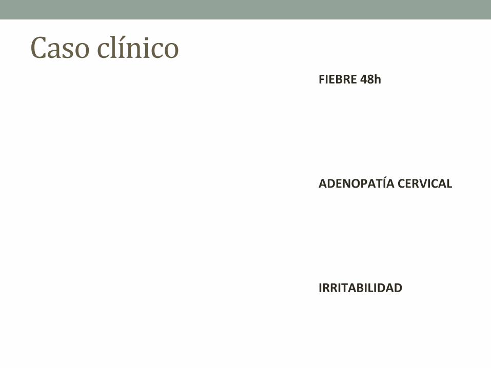
CasoclínicoFIEBRE48h
ADENOPATÍACERVICAL
IRRITABILIDAD
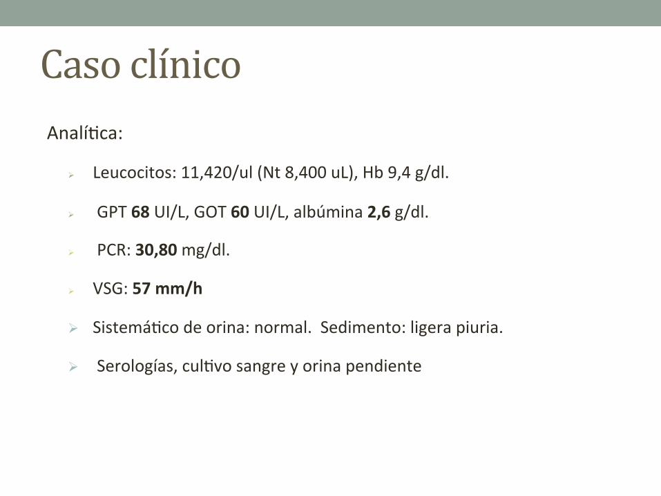
CasoclínicoAnalí&ca:
Ø Leucocitos:11,420/ul(Nt8,400uL),Hb9,4g/dl.
Ø GPT68UI/L,GOT60UI/L,albúmina2,6g/dl.
Ø PCR:30,80mg/dl.
Ø VSG:57mm/h
Ø Sistemá&codeorina:normal.Sedimento:ligerapiuria.
Ø Serologías,cul&vosangreyorinapendiente
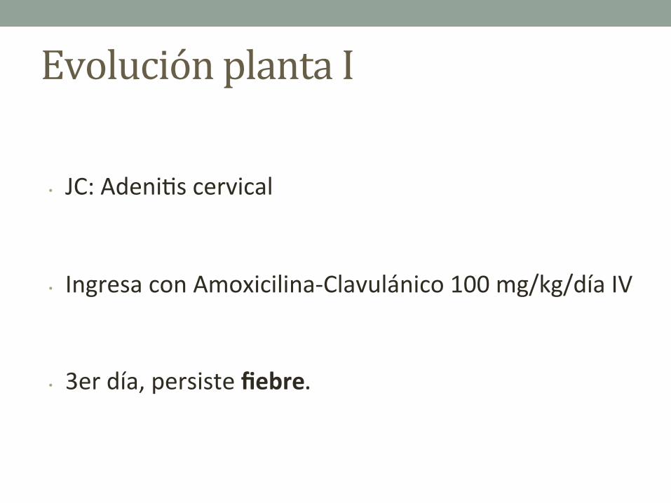
EvoluciónplantaI
• JC:Adeni&scervical
• IngresaconAmoxicilina-Clavulánico100mg/kg/díaIV
• 3erdía,persistefiebre.
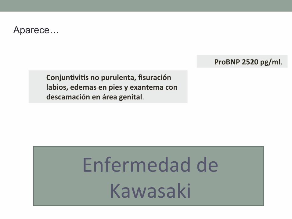
Aparece…
ProBNP2520pg/ml.
EnfermedaddeKawasaki
ConjunIviIsnopurulenta,fisuraciónlabios,edemasenpiesyexantemacondescamaciónenáreagenital.
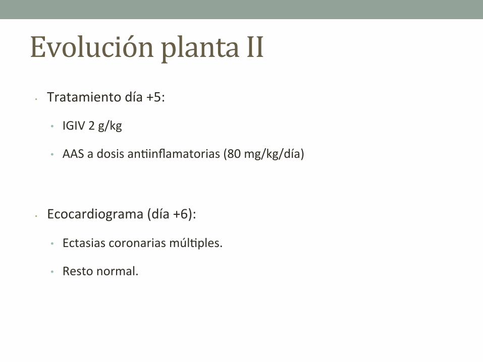
EvoluciónplantaII• Tratamientodía+5:
• IGIV2g/kg
• AASadosisan&inflamatorias(80mg/kg/día)
• Ecocardiograma(día+6):
• Ectasiascoronariasmúl&ples.
• Restonormal.
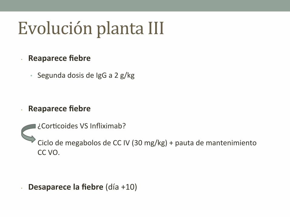
EvoluciónplantaIII• Reaparecefiebre
• SegundadosisdeIgGa2g/kg
• Reaparecefiebre
• ¿Cor&coidesVSInfliximab?
CiclodemegabolosdeCCIV(30mg/kg)+pautademantenimientoCCVO.
• Desaparecelafiebre(día+10)
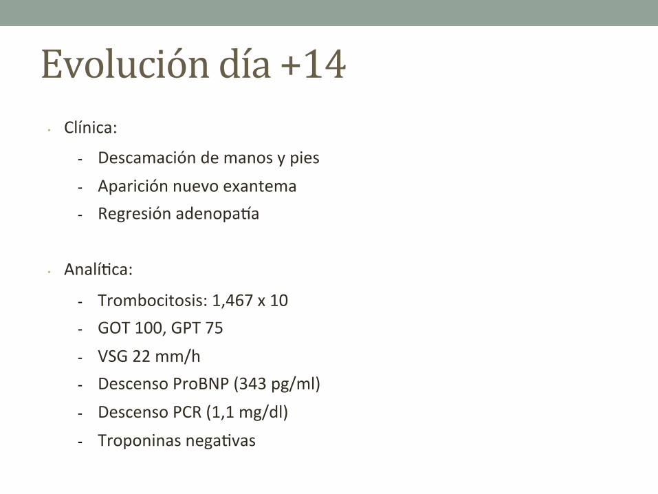
Evolucióndía+14• Clínica:
- Descamacióndemanosypies- Apariciónnuevoexantema- Regresiónadenopaca
• Analí&ca:
- Trombocitosis:1,467x10- GOT100,GPT75- VSG22mm/h- DescensoProBNP(343pg/ml)- DescensoPCR(1,1mg/dl)- Troponinasnega&vas
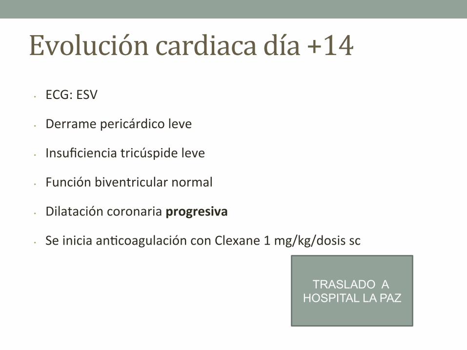
Evolucióncardiacadía+14• ECG:ESV
• Derramepericárdicoleve
• Insuficienciatricúspideleve
• Funciónbiventricularnormal
• Dilatacióncoronariaprogresiva
• Seiniciaan&coagulaciónconClexane1mg/kg/dosissc
TRASLADO A HOSPITAL LA PAZ
ENFERMEDADDE
KAWASAKI
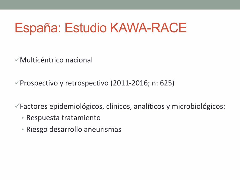
España: Estudio KAWA-RACE
ü Mul&céntriconacional
ü Prospec&voyretrospec&vo(2011-2016;n:625)
ü Factoresepidemiológicos,clínicos,analí&cosymicrobiológicos:• Respuestatratamiento• Riesgodesarrolloaneurismas
Documento de Consenso Nacional sobre diagnóstico, tratamiento y seguimiento cardiológico
de la enfermedad de Kawasaki (pendiente publicación)
Introducción• Vasculi&sagudaautolimitadavasospequeñoymedianocalibre.
• Causamáscomúnenfermedadcardiacaadquiridaenniñosenpaísesdesarrollados.
• Segundacausadevasculi&senlainfancia.
• Afectacióncoronaria:25%notratados;4%tratados
• 85%menores5años(picoincidencia18-24meses)
• Ra&o♂:♀1,5-2,1:1
• Invierno-primavera

Hipótesisetiológica
• “Respuestainmunepatológicayestereo&padaanteunoovariosfactoresambientalesoinfecciososenindividuosgenéIcamente
predispuestos”
Patogenia
Inflamaciónsistémica• Hepa&&s
• Neumoni&sinters&cial
• AfectaciónGI(abdominalgia,vómitos,diarrea,hidropsvesículabiliar…)
• Meningi&sasép&ca
• Cardíacas:miocardi&s,pericardi&s,valvulopaca
• Piuriaestéril
• Pancrea&&s
• Linfadenopaca
• Artri&s
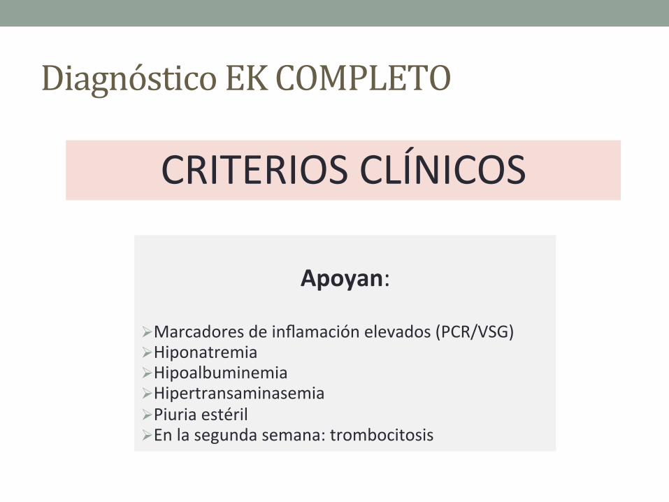
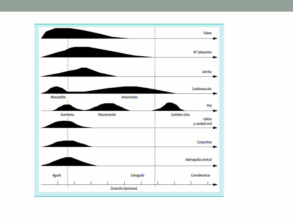
DiagnósticoEKCOMPLETO
CRITERIOSCLÍNICOS
Apoyan:
Ø Marcadoresdeinflamaciónelevados(PCR/VSG)Ø HiponatremiaØ HipoalbuminemiaØ HipertransaminasemiaØ PiuriaestérilØ Enlasegundasemana:trombocitosis

NT-proB-NP
• Marcador afectación miocárdica
• Elevado en algunos pacientes
• No suficiente capacidad para diferenciar EK
• Valores de corte no establecidos
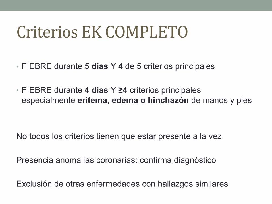
CriteriosEKCOMPLETO
• FIEBRE durante 5 días Y 4 de 5 criterios principales
• FIEBRE durante 4 días Y ≥4 criterios principales especialmente eritema, edema o hinchazón de manos y pies
No todos los criterios tienen que estar presente a la vez Presencia anomalías coronarias: confirma diagnóstico Exclusión de otras enfermedades con hallazgos similares
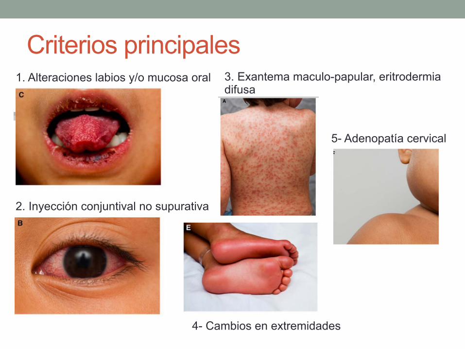
Criterios principales 1. Alteraciones labios y/o mucosa oral
2. Inyección conjuntival no supurativa
3. Exantema maculo-papular, eritrodermia difusa
4- Cambios en extremidades
5- Adenopatía cervical
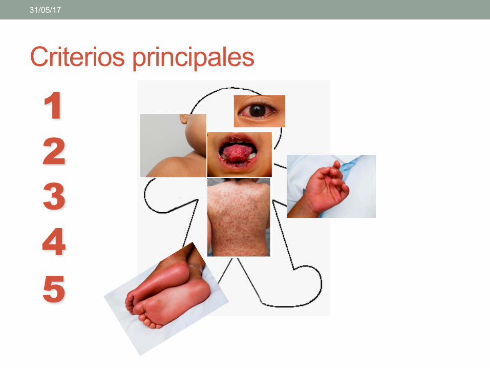
Criterios principales
31/05/17
1 2 3 4 5
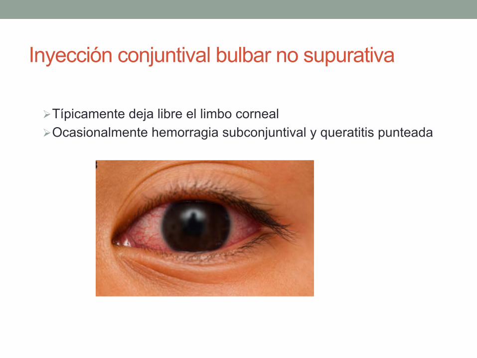
Inyección conjuntival bulbar no supurativa
Ø Típicamente deja libre el limbo corneal Ø Ocasionalmente hemorragia subconjuntival y queratitis punteada
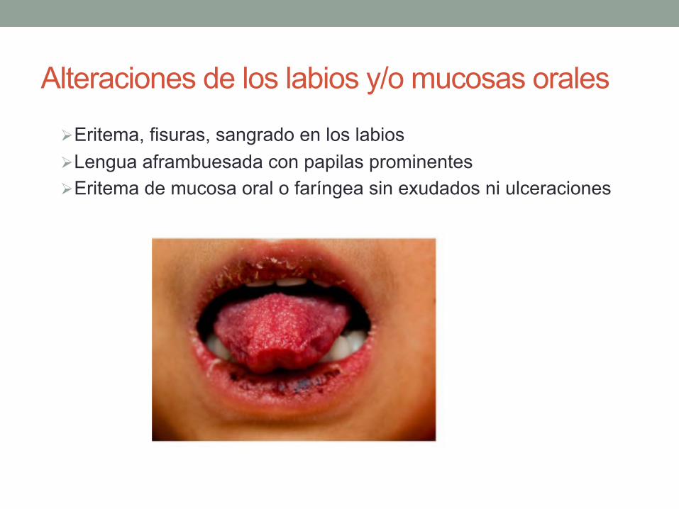
Alteraciones de los labios y/o mucosas orales
Ø Eritema, fisuras, sangrado en los labios Ø Lengua aframbuesada con papilas prominentes Ø Eritema de mucosa oral o faríngea sin exudados ni ulceraciones
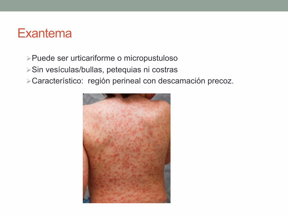
Exantema
Ø Puede ser urticariforme o micropustuloso Ø Sin vesículas/bullas, petequias ni costras Ø Característico: región perineal con descamación precoz.
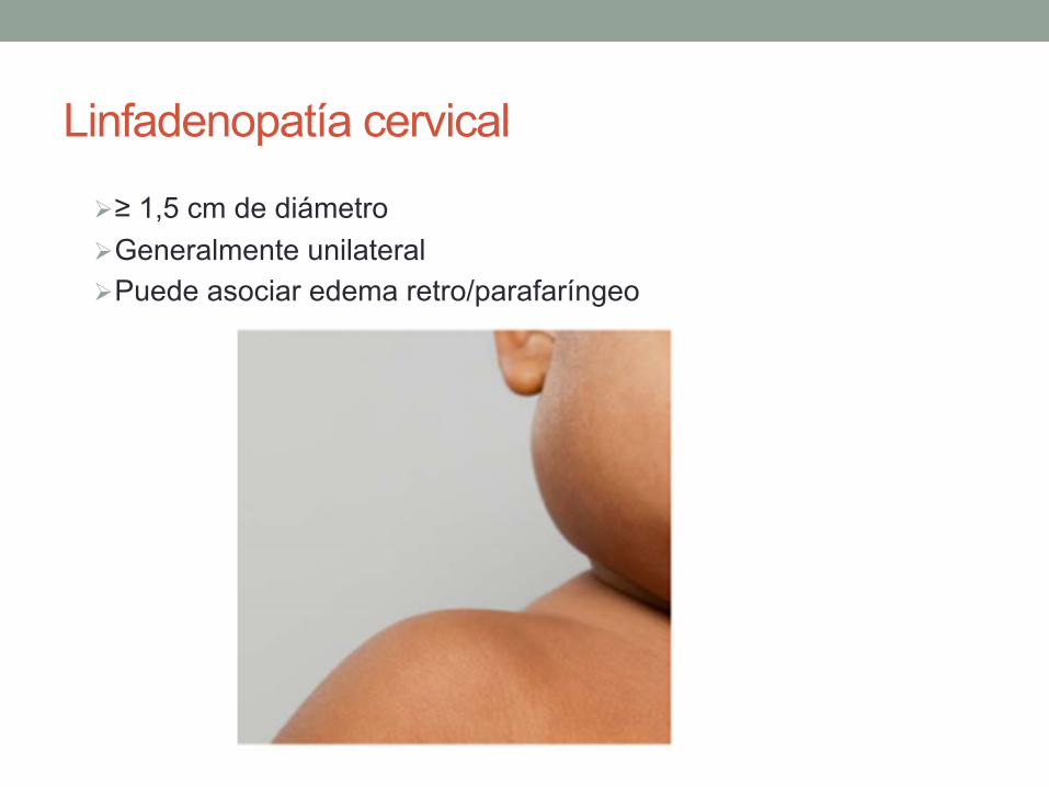
Linfadenopatía cervical
Ø ≥ 1,5 cm de diámetro Ø Generalmente unilateral Ø Puede asociar edema retro/parafaríngeo
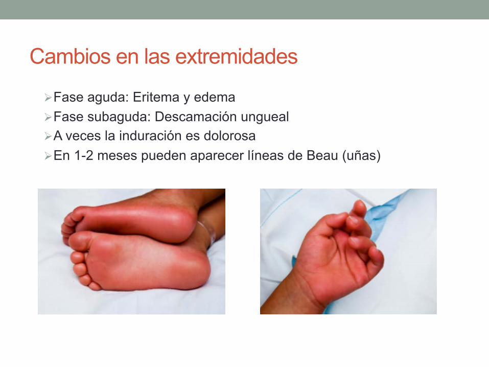
Cambios en las extremidades
Ø Fase aguda: Eritema y edema Ø Fase subaguda: Descamación ungueal Ø A veces la induración es dolorosa Ø En 1-2 meses pueden aparecer líneas de Beau (uñas)
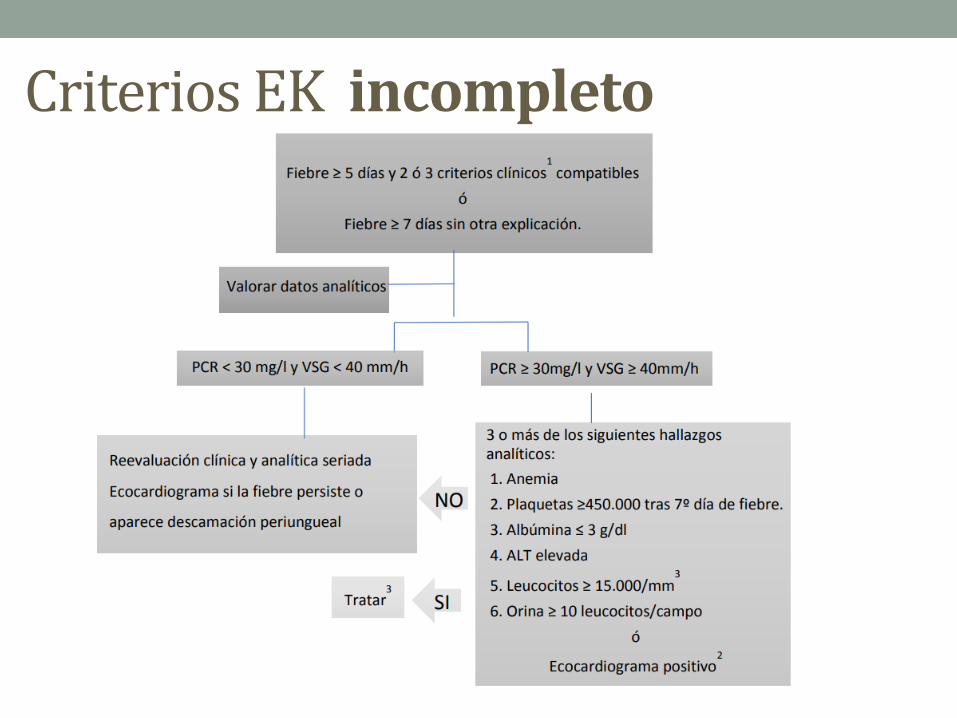
CriteriosEKincompleto
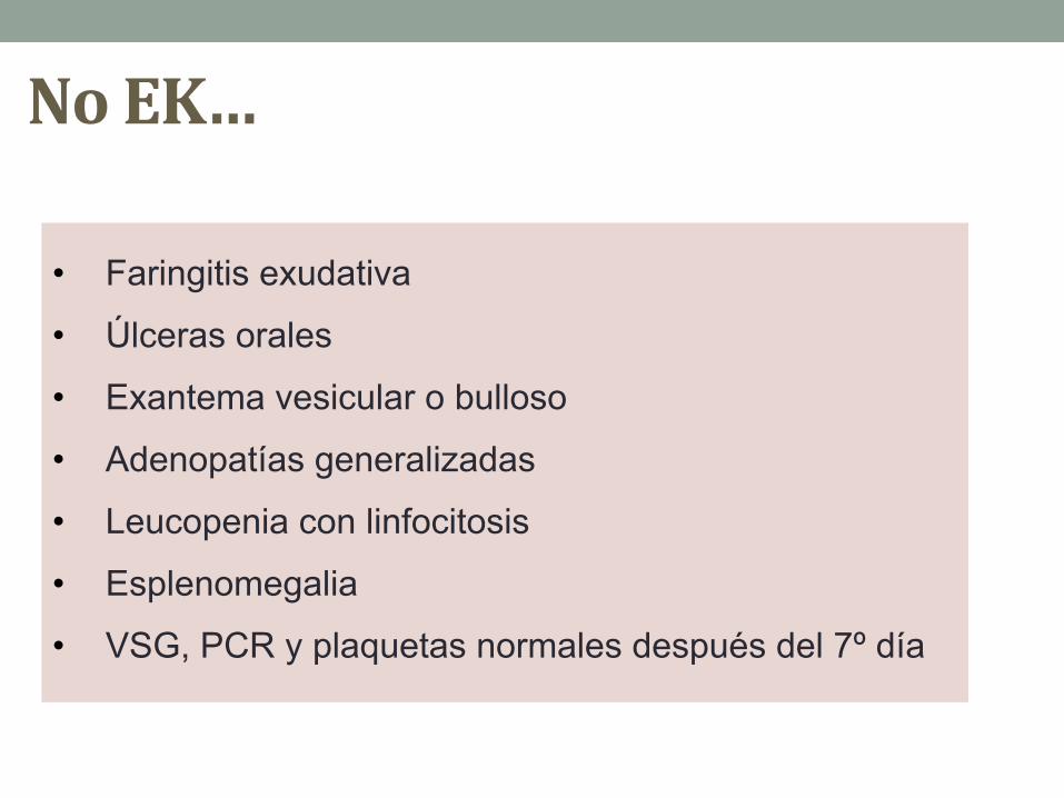
NoEK…
• Faringitis exudativa
• Úlceras orales
• Exantema vesicular o bulloso
• Adenopatías generalizadas
• Leucopenia con linfocitosis
• Esplenomegalia
• VSG, PCR y plaquetas normales después del 7º día
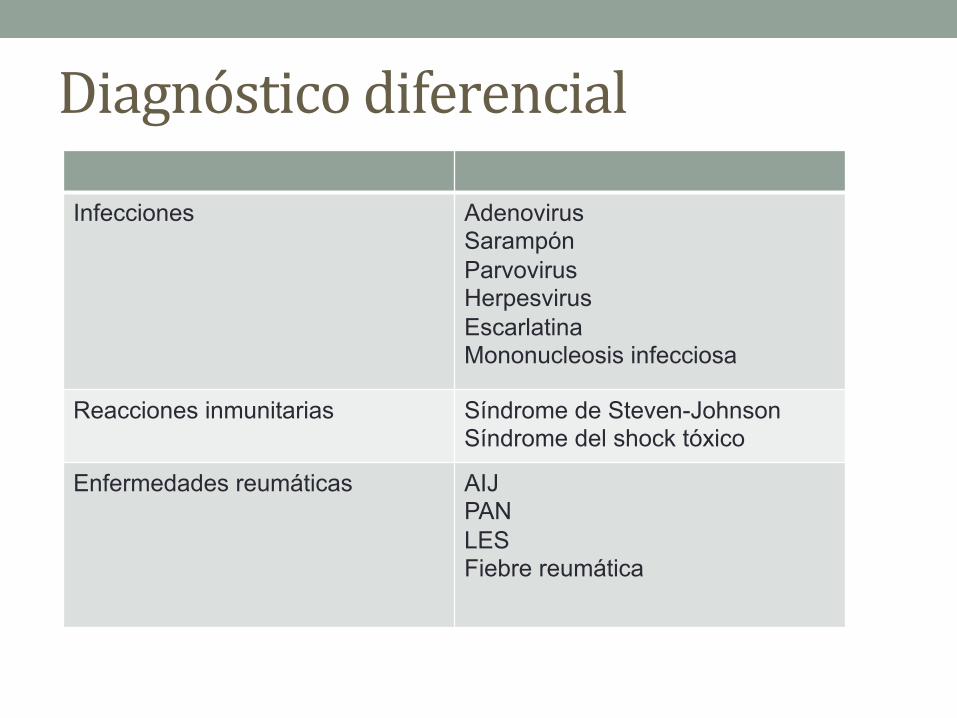
Diagnósticodiferencial
Infecciones Adenovirus Sarampón Parvovirus Herpesvirus Escarlatina Mononucleosis infecciosa
Reacciones inmunitarias Síndrome de Steven-Johnson Síndrome del shock tóxico
Enfermedades reumáticas AIJ PAN LES Fiebre reumática
Afectacióncardiológica
• Mayorcausademorbimortalidad
• Inflamaciónpericardio,miocardioyendocardio(incluyendoválvulas)yarteriascoronarias

Afectacióncardiovascular
• Shockcardiogénico
• Disfunciónmiocárdica
• Disfunciónvalvular
• Dilataciónderaizaór&ca
• Alteracionescoronarias
Diagnosis, Treatment, and Management of Kawasaki Disease
Circulation. 2017;135:00–00. DOI: 10.1161/CIR.0000000000000484 TBD, 2017 e29
CLINICAL STATEMENTS
AND GUIDELINES
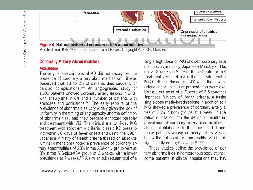
Coronary Artery AbnormalitiesPrevalenceThe original descriptions of KD did not recognize the presence of coronary artery abnormalities until it was observed that 1% to 2% of patients died suddenly of cardiac complications.259 An angiographic study of 1100 patients showed coronary artery lesions in 24%, with aneurysms in 8% and a number of patients with stenoses and occlusions.260 The early reports of the prevalence of abnormalities vary widely given the lack of uniformity in the timing of angiography and the definition of abnormalities, and they predate echocardiography and treatment with IVIG. The clinical trial of 4-day IVIG treatment with strict entry criteria (classic KD present-ing within 10 days of fever onset) and using the 1984 Japanese Ministry of Health criteria (based on absolute luminal dimensions) noted a prevalence of coronary ar-tery abnormalities of 23% in the ASA-only group versus 8% in the IVIG-plus-ASA group at 2 weeks, with a lower prevalence at 7 weeks.176 A similar subsequent trial of a
single high dose of IVIG showed coronary artery abnor-malities, again using Japanese Ministry of Health crite-ria, at 2 weeks in 9.1% of those treated with 4-day IVIG treatment versus 4.6% in those treated with high-dose IVIG (further reduced to 2.4% when those with coronary artery abnormalities at presentation were excluded).127 Using a cut point of a Z score of 2.5 together with the Japanese Ministry of Health criteria, a further trial of single-dose methylprednisolone in addition to high-dose IVIG showed a prevalence of coronary artery abnormali-ties of 30% in both groups at 1 week.176 The incorpo-ration of dilation into the definition results in a higher prevalence of coronary artery abnormalities. The prev-alence of dilation is further increased if one includes those patients whose coronary artery Z scores were below the cut point for abnormality (<2) but decreased significantly during follow-up.129,130
These studies define the prevalence of coronary ar-tery abnormalities in homogenous populations; however, some patients in clinical populations may have incom-
Figure 4. Natural history of coronary artery abnormalities. Modified from Kato258 with permission from Elsevier. Copyright © 2004, Elsevier.
by guest on May 29, 2017
http://circ.ahajournals.org/D
ownloaded from
HallazgosECG
CambiosPR,ST,QT,T,Q…
• Inflamaciónmiocárdicaclínicaosubclínica
• Afectacióncoronaria
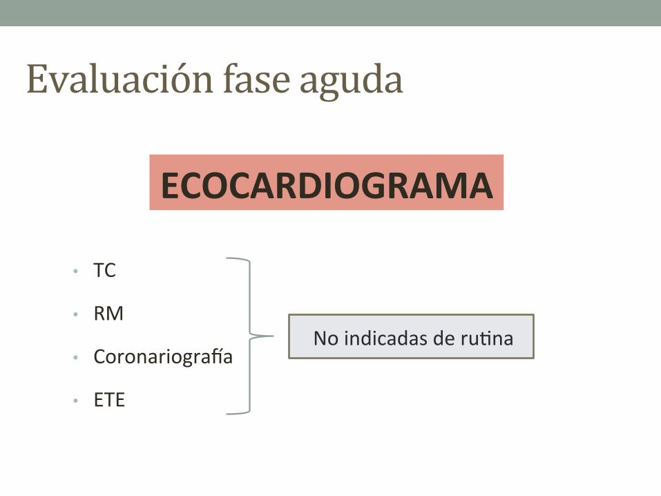
Evaluaciónfaseaguda
• TC
• RM
• Coronariograua
• ETE
*Sinodisponiblenojus&ficaretrasareltratamiento
Noindicadasderu&na
ECOCARDIOGRAMA

TRATAMIENTO
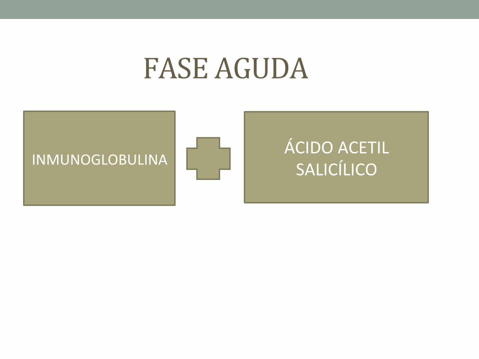
FASEAGUDA
INMUNOGLOBULINAÁCIDOACETILSALICÍLICO
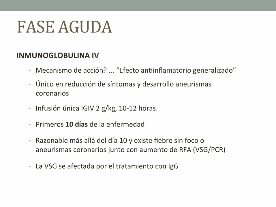
FASEAGUDAINMUNOGLOBULINAIV
• Mecanismodeacción?...“Efectoan&inflamatoriogeneralizado”
• Únicoenreduccióndesíntomasydesarrolloaneurismascoronarios
• InfusiónúnicaIGIV2g/kg,10-12horas.
• Primeros10díasdelaenfermedad
• Razonablemásalládeldía10yexistefiebresinfocooaneurismascoronariosjuntoconaumentodeRFA(VSG/PCR)
• LaVSGseafectadaporeltratamientoconIgG
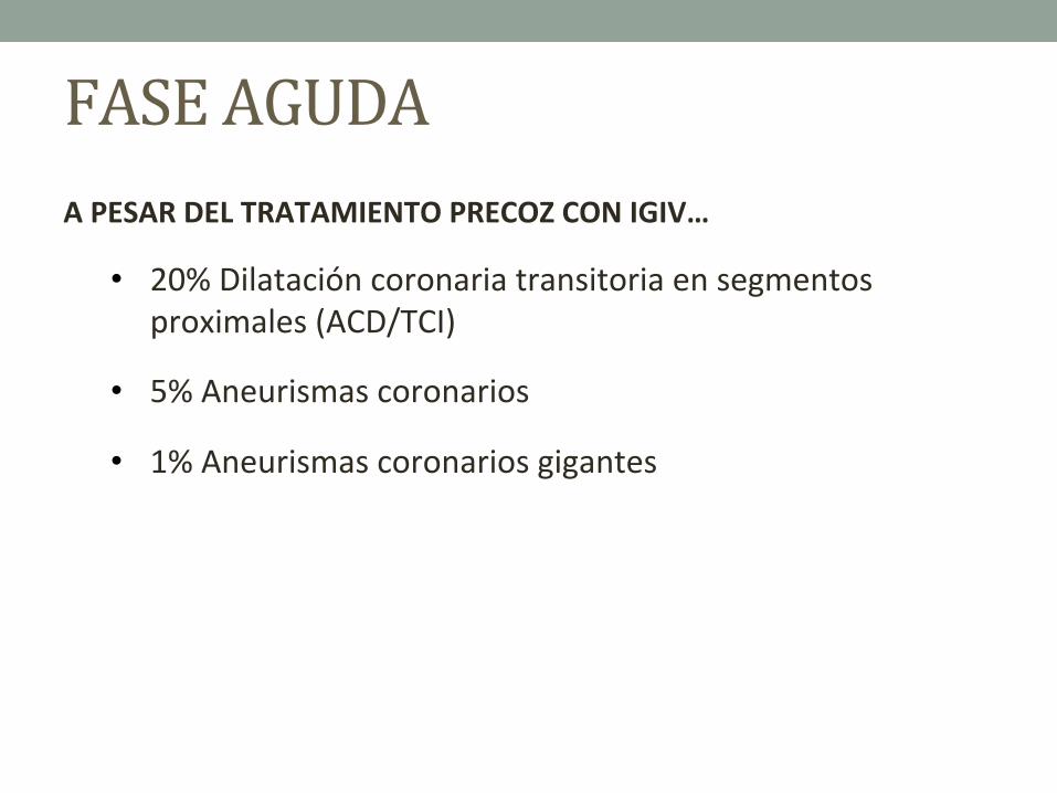
FASEAGUDAAPESARDELTRATAMIENTOPRECOZCONIGIV…
• 20%Dilatacióncoronariatransitoriaensegmentosproximales(ACD/TCI)
• 5%Aneurismascoronarios
• 1%Aneurismascoronariosgigantes
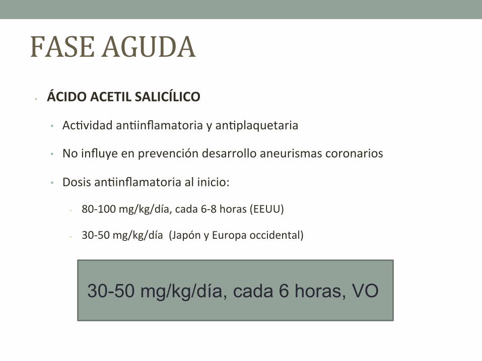
FASEAGUDA• ÁCIDOACETILSALICÍLICO
• Ac&vidadan&inflamatoriayan&plaquetaria
• Noinfluyeenprevencióndesarrolloaneurismascoronarios
• Dosisan&inflamatoriaalinicio:
• 80-100mg/kg/día,cada6-8horas(EEUU)
• 30-50mg/kg/día(JapónyEuropaoccidental)
30-50 mg/kg/día, cada 6 horas, VO
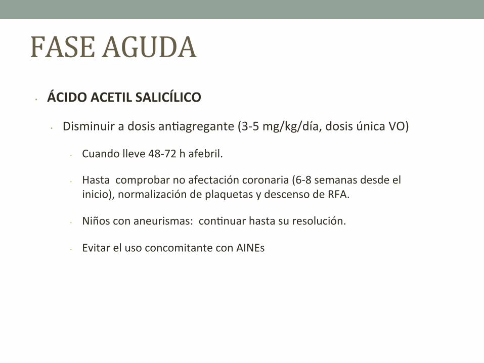
FASEAGUDA• ÁCIDOACETILSALICÍLICO
• Disminuiradosisan&agregante(3-5mg/kg/día,dosisúnicaVO)
• Cuandolleve48-72hafebril.
• Hastacomprobarnoafectacióncoronaria(6-8semanasdesdeelinicio),normalizacióndeplaquetasydescensodeRFA.
• Niñosconaneurismas:con&nuarhastasuresolución.
• EvitarelusoconcomitanteconAINEs
Tratamientocoadyuvante• Cor&coides
• Infliximab
• Etanercept
• Ciclosporina
• Anakinra
• Ciclofosfamida
• Plasmaféresis
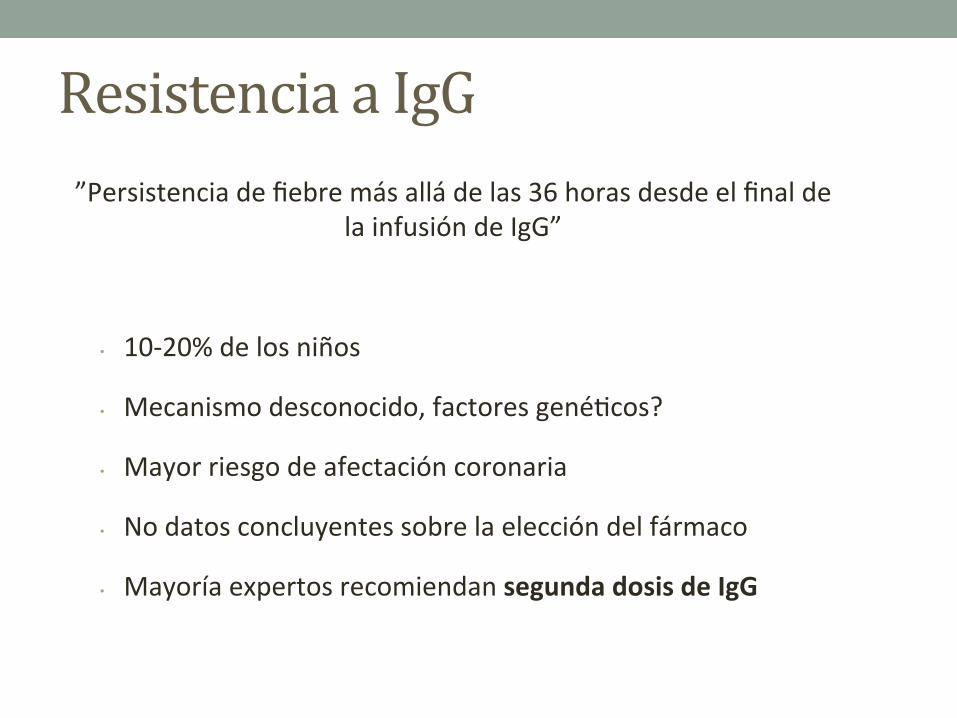
ResistenciaaIgG”Persistenciadefiebremásalládelas36horasdesdeelfinalde
lainfusióndeIgG”
• 10-20%delosniños
• Mecanismodesconocido,factoresgené&cos?
• Mayorriesgodeafectacióncoronaria
• Nodatosconcluyentessobrelaeleccióndelfármaco
• MayoríaexpertosrecomiendansegundadosisdeIgG
ResistenciaaIgG
Diagnosis, Treatment, and Management of Kawasaki Disease
Circulation. 2017;135:00–00. DOI: 10.1161/CIR.0000000000000484 TBD, 2017 e21
CLINICAL STATEMENTS
AND GUIDELINES
the basis of current information, addition of infliximab to initial therapy with IVIG is safe but does not prevent recrudescent fever.
EtanerceptA more limited experience with etanercept (soluble TNF receptor) plus IVIG for intensification of initial therapy was reported recently.198 In this prospective, open-la-bel study of 15 patients, etanercept was administered subcutaneously after IVIG infusion and again at 1 and 2 weeks later. Most patients received 0.8 mg/kg per dose. The pharmacokinetics were similar to those re-ported in older children, and there were no adverse re-actions attributable to etanercept. On the basis of these results, a phase III randomized, placebo-controlled trial was initiated and is still enrolling subjects.199 Recom-mendations for primary adjunctive treatment with etan-ercept await publication of the results of this clinical trial. The potential advantage of etanercept might be the shorter half-life if secondary infections are of con-cern. However, the soluble receptor only binds to circu-lating and not cell-bound TNF-α, which could reduce the anti-inflammatory effect.
Recommendations for Adjunctive Therapies for Primary Treatment
1. Single-dose pulse methylprednisolone should not be administered with IVIG as routine pri-mary therapy for patients with KD (Class III; Level of Evidence B).
2. Administration of a longer course of corti-costeroids (eg, tapering over 2–3 weeks), together with IVIG 2 g/kg and ASA, may be considered for treatment of high-risk patients with acute KD, when such high risk can be identified in patients before initiation of treatment (Class IIb; Level of Evidence B).
IVIG ResistanceApproximately 10% to 20% of patients with KD develop recrudescent or persistent fever at least 36 hours after the end of their IVIG infusion and are termed IVIG resis-tant.176,200,201 The immunologic basis of IVIG resistance is unknown, in part because the mechanism of action of IVIG is poorly understood. It is likely that host genetic factors, such as polymorphisms in the Fc gamma recep-tors, play a role in both the response and resistance to IVIG.61,202,203
Risk Scores for Predicting Nonresponse to IVIGApproximately 10% to 20% of patients with KD have per-sistent or recurrent fever after primary therapy with IVIG plus ASA.204,205 Many studies have shown that patients who are resistant to initial IVIG are at increased risk of developing coronary artery abnormalities.171,206,207 Thus, scoring systems have been constructed to identify pa-tients likely to be resistant to IVIG and who may benefit from more aggressive initial therapy. In 2006, 3 Japa-nese groups devised scoring systems to predict resis-tance to IVIG.187,189,208,209 However, currently available
Table 6. Treatment Options for IVIG-Resistant KD Patients
Agent Description Dose References
Most frequently administered
IVIG: Second infusion Pooled polyclonal IG 2 g/kg IV 211
IVIG + prednisolone IVIG + steroid IVIG: 2 g/kg IV + prednisolone 2 mg·kg−1·d−1 IV divided every 8 h until afebrile, then prednisone orally until CRP normalized, then taper over 2–3 wk
212
Infliximab Monoclonal antibody against TNF-α Single infusion: 5 mg/kg IV given over 2 h 194, 213, 214
Alternative treatments
Cyclosporine Inhibitor of calcineurin-NFAT pathway IV: 3 mg·kg−1·d−1 divided every 12 hPO: 4–8 mg·kg−1·d−1 divided every 12 hAdjust dose to achieve trough 50–150 ng/mL; 2-h peak level 300–600 ng/mL
215, 216
Anakinra Recombinant IL-1β receptor antagonist 2–6 mg·kg−1·d−1 given by subcutaneous injection 217, 218
Cyclophosphamide Alkylating agent blocks DNA replication 2 mg·kg−1·d−1 IV 219
Plasma exchange Replaces plasma with albumin Not applicable 220
IVIG resistance is defined as persistent or recrudescent fever at least 36 hours and <7 days after completion of first IVIG infusion. The top 3 treatments have been most commonly used, although no comparative effectiveness trial has been performed. Pulsed high-dose steroid treatment is not recommended. The alternative treatments have been used in a limited number of patients with KD.
CRP indicates C-reactive protein; IG, immunoglobulin; IL, interleukin; IV, intravenous; IVIG, intravenous immunoglobulin; KD, Kawasaki disease; NFAT, nuclear factor of activated T cells; PO, by mouth; and TNF, tumor necrosis factor.
by guest on May 29, 2017
http://circ.ahajournals.org/D
ownloaded from
Prevencióndetrombosis
• AAS(3-5mg/kg/día)hastacomprobarquenohayaafectacióncoronaria(4-6semanas)
• Pacientesconaneurismascoronariosrápidamenteprogresivosogigantes:añadiranIcoagulaciónconHBPM,warfarinaoacenocumarol
• Tripleterapia(segundoan&agregante):pacientesconaneurismasgigantesehistoriarecientedetrombosiscoronaria.
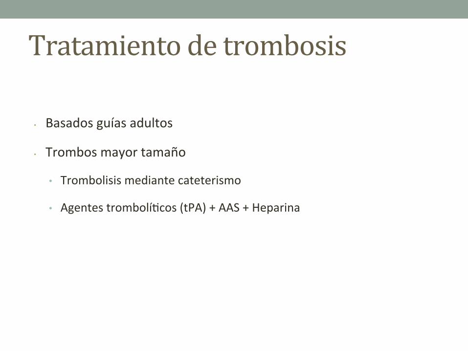
Tratamientodetrombosis
• Basadosguíasadultos
• Trombosmayortamaño
• Trombolisismediantecateterismo
• Agentestrombolí&cos(tPA)+AAS+Heparina
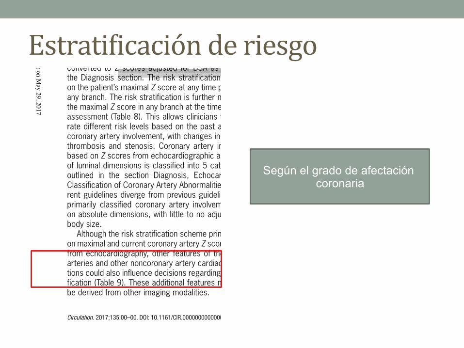
EstratiPicaciónderiesgo
Diagnosis, Treatment, and Management of Kawasaki Disease
Circulation. 2017;135:00–00. DOI: 10.1161/CIR.0000000000000484 TBD, 2017 e33
CLINICAL STATEMENTS
AND GUIDELINES
involvement should be followed, as outlined at the end of the Diagnosis section.
Echocardiography is the primary modality used to assess coronary artery luminal dimensions, which are converted to Z scores adjusted for BSA as outlined in the Diagnosis section. The risk stratification first rests on the patient’s maximal Z score at any time point and in any branch. The risk stratification is further modified by the maximal Z score in any branch at the time of current assessment (Table 8). This allows clinicians to incorpo-rate different risk levels based on the past and current coronary artery involvement, with changes in the risk of thrombosis and stenosis. Coronary artery involvement based on Z scores from echocardiographic assessment of luminal dimensions is classified into 5 categories as outlined in the section Diagnosis, Echocardiography, Classification of Coronary Artery Abnormalities. The cur-rent guidelines diverge from previous guidelines, which primarily classified coronary artery involvement based on absolute dimensions, with little to no adjustment for body size.
Although the risk stratification scheme primarily rests on maximal and current coronary artery Z scores derived from echocardiography, other features of the coronary arteries and other noncoronary artery cardiac complica-tions could also influence decisions regarding risk speci-fication (Table 9). These additional features may further be derived from other imaging modalities.
Recommendations for Risk Stratification of Coronary Artery Abnormalities
1. It is reasonable to use echocardiographic coronary artery luminal dimensions converted to BSA-adjusted Z scores to determine risk stratification (Class IIa; Level of Evidence B).
2. It is reasonable to incorporate both maximal and current coronary artery involvement in risk stratification (as per Table 8) (Class IIa; Level of Evidence C).
3. It is reasonable to incorporate the presence of additional features other than coronary artery luminal dimensions into decisions regarding risk stratification (as per Table 9) (Class IIa; Level of Evidence C).
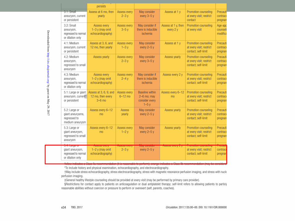
Long-Term Management of Coronary Artery AbnormalitiesOn the basis of the risk stratification scheme, specific recommendations are made regarding surveillance, car-diovascular risk factor assessment and management, medical therapy, thromboprophylaxis, physical activity, and reproductive counseling for each category of past and current coronary artery involvement. The algorithm is depicted in Tables 10 and 11. The rationale for recom-mendations in the algorithm is provided in the sections after the recommendation statements.
Risk-Stratified Recommendations for Long-Term Evaluation and ManagementNote: Long-term status is taken to be when the patient is stable after the acute illness and the coronary artery luminal dimensions are not increasing, usually at 4 to 6
Table 8. Risk Classification of Coronary Artery Abnormalities During Follow-up
Classification Description
1 No involvement at any timepoint (Z score always <2)
2 Dilation only (Z score 2 to <2.5)
3 Small aneurysm (Z score ≥2.5 to <5)
3.1 Current or persistent
3.2 Decreased to dilation only or normal luminal dimension
4 Medium aneurysm (Z score ≥5 to <10, and absolute dimension <8 mm)
4.1 Current or persistent
4.2 Decreased to small aneurysm
4.3 Decreased to dilation only or normal luminal dimension
5 Large and giant aneurysm (Z score ≥10, or absolute dimension ≥8 mm)
5.1 Current or persistent
5.2 Decreased to medium aneurysm
5.3 Decreased to small aneurysm
5.4 Decreased to dilation only or normal luminal dimension
Table 9. Additional Clinical Features That May Increase the Long-Term Risk of Myocardial Ischemia
Greater length and distal location of aneurysms that increase the risk of flow stasis
Greater total number of aneurysms
Greater number of branches affected
Presence of luminal irregularities
Abnormal characterization of the vessel wall (calcification, luminal myofibroblastic proliferation)
Presence of functional abnormalities (impaired vasodilation, impaired flow reserve)
Absence or poor quality of collateral vessels
Previous revascularization performed
Previous coronary artery thrombosis
Previous myocardial infarction
Presence of ventricular dysfunction
by guest on May 29, 2017
http://circ.ahajournals.org/D
ownloaded from
Según el grado de afectación coronaria
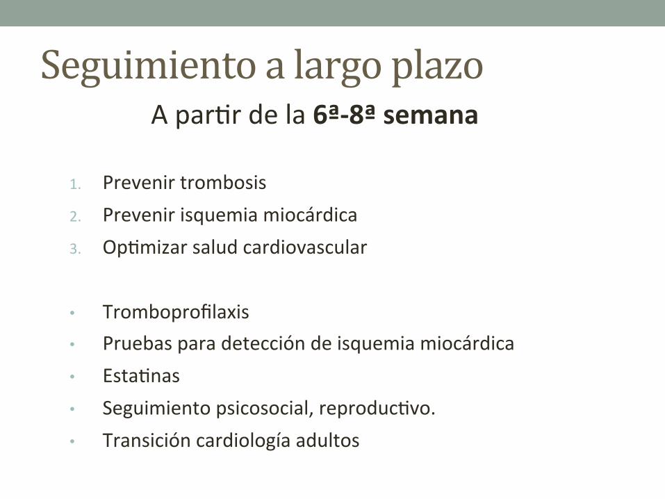
SeguimientoalargoplazoApar&rdela6ª-8ªsemana
1. Prevenirtrombosis2. Prevenirisquemiamiocárdica3. Op&mizarsaludcardiovascular
• Tromboprofilaxis• Pruebasparadeteccióndeisquemiamiocárdica• Esta&nas• Seguimientopsicosocial,reproduc&vo.• Transicióncardiologíaadultos
McCrindle et al
TBD, 2017 Circulation. 2017;135:00–00. DOI: 10.1161/CIR.0000000000000484e34
Table 10. Long-Term Assessment and Counseling Algorithm
Risk Level
Frequency of Cardiology
Assessment*
Assessment for Inducible Myocardial Ischemia†
Type and Frequency
of Additional Cardiology
Assessment
Cardiovascular Risk Factor
Assessment and Management‡
Physical Activity Counseling§
Reproductive Counseling
1: No involvement May discharge between 4 wk and
12 mo
None None Assess at 1 y Promotion counseling at every visit
Age-appropriate counseling without modification
2: Dilation only May discharge after 1 y if normal;
assess every 2–5 y if persists
None None Assess at 1 y Promotion counseling at every visit
Age-appropriate counseling without modification
3.1: Small aneurysm, current or persistent
Assess at 6 mo, then yearly
Assess every 2–3 y
May consider every 3–5 y
Assess at 1 y Promotion counseling at every visit; restrict contact
Precautions for contraception and pregnancy
3.2: Small aneurysm, regressed to normal or dilation only
Assess every 1–3 y (may omit
echocardiography)
Assess every 3–5 y
May consider if there is inducible
ischemia
Assess at 1 y, then every 2 y
Promotion counseling at every visit
Age-appropriate counseling without modification
4.1: Medium aneurysm, current or persistent
Assess at 3, 6, and 12 mo, then yearly
Assess every 1–3 y
May consider every 2–5 y
Assess at 1 y Promotion counseling at every visit; restrict contact; self-limit
Precautions for contraception and pregnancy
4.2: Medium aneurysm, regressed to small aneurysm
Assess yearly Assess every 2–3 y
May consider every 3–5 y
Assess yearly Promotion counseling at every visit; restrict contact; self-limit
Precautions for contraception and pregnancy
4.3: Medium aneurysm, regressed to normal or dilation only
Assess every 1–2 y (may omit
echocardiography)
Assess every 2–4 y
May consider if there is inducible
ischemia
Assess every 2 y Promotion counseling at every visit; restrict contact; self-limit
Precautions for contraception and pregnancy
5.1: Large or giant aneurysm, current or persistent
Assess at 3, 6, 9, and 12 mo, then every
3–6 mo
Assess every 6–12 mo
Baseline within 2–6 mo; may consider every
1–5 y
Assess every 6–12 mo
Promotion counseling at every visit; restrict contact; self-limit
Precautions for contraception and pregnancy
5.2: Large or giant aneurysms, regressed to medium aneurysm
Assess every 6–12 mo
Assess yearly
May consider every 2–5 y
Assess yearly Promotion counseling at every visit; restrict contact; self-limit
Precautions for contraception and pregnancy
5.3: Large or giant aneurysm, regressed to small aneurysm
Assess every 6–12 mo
Assess every 1–2 y
May consider every 2–5 y
Assess yearly Promotion counseling at every visit; restrict contact; self-limit
Precautions for contraception and pregnancy
5.4: Large or giant aneurysm, regressed to normal or dilation only
Assess every 1–2 y (may omit
echocardiography)
Assess every 2–3 y
May consider every 2–5 y
Assess every 2 y Promotion counseling at every visit; restrict contact; self-limit
Precautions for contraception and pregnancy
Yellow indicates a Class IIa recommendation (it is reasonable to perform); orange indicates a Class IIb recommendation (may be considered).*To include history and physical examination, echocardiography, and electrocardiography.†May include stress echocardiography, stress electrocardiography, stress with magnetic resonance perfusion imaging, and stress with nuclear medicine
perfusion imaging.‡General healthy lifestyle counseling should be provided at every visit (may be performed by primary care provider).§Restrictions for contact apply to patients on anticoagulation or dual antiplatelet therapy; self-limit refers to allowing patients to participate to their
reasonable abilities without coercion or pressure to perform or overexert (self, parents, coaches).
by guest on May 29, 2017
http://circ.ahajournals.org/D
ownloaded from
Diagnosis, Treatment, and Management of Kawasaki Disease
Circulation. 2017;135:00–00. DOI: 10.1161/CIR.0000000000000484 TBD, 2017 e35
CLINICAL STATEMENTS
AND GUIDELINES
weeks after the onset of fever. Until this point, patients should be managed in accordance with the recommen-dations in the Acute Treatment section.
No Involvement (Z Score Always <2)Frequency of cardiology assessment (to include history and physical examination, echocardiography, electrocar-diography):
1. It is reasonable to discharge patients from cardiology care at 4 to 6 weeks after KD onset, although ongoing follow-up to 12 months may be considered. Ongoing car-diology follow-up is not indicated. Patients and families should be advised to remember that having had KD is part of the patient’s
permanent medical history (Class IIa; Level of Evidence C).
Type and frequency of additional cardiology assessment (other cardiology testing):
1. It is reasonable that no additional cardiology assessment be performed (Class IIa; Level of Evidence C).
Cardiovascular risk factor assessment and management:1. It is reasonable to provide general counseling
regarding healthy lifestyle and activity pro-motion at every visit; this may be provided by the primary care provider (Class IIa; Level of Evidence C).
2. It is reasonable to assess blood pressure, fast-ing lipid profile, body mass index (and plot),
Table 11. Long-Term Thromboprophylaxis and Medical Therapy Algorithm
Risk Level Low-Dose ASAAnticoagulation
(Warfarin or LMWH)
Dual Antiplatelet Therapy
(ASA+Clopidogrel) β-Blocker Statin
1: No involvement 6–8 wk then discontinue
Not indicated Not indicated Not indicated Not indicated
2: Dilation only Continuation after 6–8 wk is
reasonable
Not indicated Not indicated Not indicated Not indicated
3.1: Small aneurysm, current or persistent
Continue May be considered May be considered as an alternative to
anticoagulation
Not indicated Empirical therapy may be considered
3.2: Small aneurysm, regressed to normal or dilation only
Continue, but discontinuation
may also be considered
Not indicated Not indicated Not indicated Empirical therapy may be considered
4.1: Medium aneurysm, current or persistent
Continue May be considered May be considered as an alternative to
anticoagulation
Not indicated Empirical therapy may be considered
4.2: Medium aneurysm, regressed to small aneurysm
Continue Not indicated May be considered Not indicated Empirical therapy may be considered
4.3: Medium aneurysm, regressed to normal or dilation only
Continue Not indicated May be considered Not indicated Empirical therapy may be considered
5.1: Large and giant aneurysm, current or persistent
Continue Reasonably indicated May be considered in addition to
anticoagulation
May be considered
Empirical therapy may be considered
5.2: Large or giant aneurysm, regressed to medium aneurysm
Continue Reasonably indicated May be considered as an alternative to
anticoagulation
May be considered
Empirical therapy may be considered
5.3: Large or giant aneurysm, regressed to small aneurysm
Continue May be considered May be considered as an alternative to
anticoagulation
May be considered
Empirical therapy may be considered
5.4: Large or giant aneurysm, regressed to normal or dilation only
Continue Not indicated May be considered as an alternative to
anticoagulation
Not indicated Empirical therapy may be considered
ASA indicates acetylsalicylic acid or aspirin; and LMWH, low-molecular-weight heparin. Green indicates a Class I recommendation (should be performed); yellow indicates a Class IIa recommendation (it is reasonable to perform); orange indicates a Class IIb recommendation (may be considered); and red indicates a Class III recommendation (should not be performed).
by guest on May 29, 2017
http://circ.ahajournals.org/D
ownloaded from
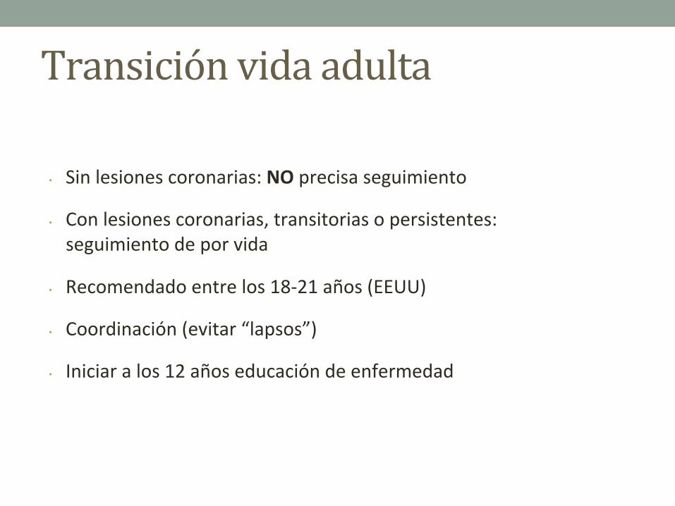
Transiciónvidaadulta
• Sinlesionescoronarias:NOprecisaseguimiento
• Conlesionescoronarias,transitoriasopersistentes:seguimientodeporvida
• Recomendadoentrelos18-21años(EEUU)
• Coordinación(evitar“lapsos”)
• Iniciaralos12añoseducacióndeenfermedad
Mayo2018
• Asintomá&co
• Riesgo5.1:Aneurismasgigantespersistentes
• AAS4mg/kg/día+ACO(Sintrom)
Conclusiones
ü Obje&voprincipal:prevenirafectacióncoronaria
ü Antesospechainiciartratamientoempírico.
ü Faltadeconsensoencasosderesistenciaatratamiento.
ü Asegurartransiciónyseguimientoóp&moencardiologíadeadultos.

FIN


