Contents lists available at ScienceDirect
Chemical Engineering Journal
journal homepage: www.elsevier.com/locate/cej
Light-responsive UiO-66-NH2/Ag3PO4 MOF-nanoparticle composites for thecapture and release of sulfamethoxazole
Xue-Yan Xua, Chun Chua, Huifen Fua, Xue-Dong Dua, Peng Wanga, Weiwei Zhengb,Chong-Chen Wanga,⁎
a Beijing Key Laboratory of Functional Materials for Building Structure and Environment Remediation, Beijing University of Civil Engineering and Architecture, Beijing100044, PR ChinabDepartment of Chemistry, Syracuse University, Syracuse, NY 13244, United States
H I G H L I G H T S
• The MOF-nanoparticle (UiO-66-NH2/Ag3PO4) composites was facilely fab-ricated.
• The light-responsive MOF-NP compo-sites for SMX capture and release wasreported.
• The SMX release from composites wascontrolled by the size of nano-Ag3PO4
on MOF.
• Mechanism of the light-triggered SMXrelease was clarified.
G R A P H I C A L A B S T R A C T
A R T I C L E I N F O
Keywords:Light-responseMOF-nanoparticle compositesDesorptionUiO-66-NH2
PPCPsMechanism
A B S T R A C T
Light-responsive materials are attracting increasing amount of attention and have great potential in many re-search fields in environmental chemistry, materials science, biology, and nanotechnology. In this work, UiO-66-NH2/Ag3PO4 (UAP-X) Metal-organic framework (MOF)-nanoparticle composites with remarkable adsorptionperformance toward sulfamethoxazole (SMX) were reported. In addition, visible light-triggered release of SMX inthe UAP-X composites was reported for the first time. It is believed that the light-triggered desorption of SMX isdue to the transformation from Ag+ to Ag0 in the light-sensitive Ag3PO4 nanoparticles (NPs) of the composites.The SMX release performance of UAP-X can be tuned by the size of Ag3PO4 NPs distributed on the UiO-66-NH2.Specifically, the smaller crystal size of Ag3PO4 NPs, which can facilitate the reduction of Ag+ to Ag0, can beachieved with an increase in relative UiO-66-NH2 content in the composites. In addition, the higher UiO-66-NH2
content of the composite could provide more deposition area to minimize the aggregation of Ag3PO4, whichcould further enhance the reduction of Ag+. The light triggered desorption provides new possibility to achievepollution-free and low-cost recyclability of adsorbents.
1. Introduction
Pharmaceuticals and personal care products (PPCPs) are widelyused and essential in daily life. However, the extensive applications and
poor elimination of PPCPs by the conventional biological wastewatertreatment plants lead to the contamination of surface water and evenground water [1,2]. The bioaccumulation of these pseudo persistentPPCPs in the aquatic life can exert serious threat to the environment
https://doi.org/10.1016/j.cej.2018.06.005Received 3 May 2018; Received in revised form 29 May 2018; Accepted 1 June 2018
⁎ Corresponding author.E-mail addresses: [email protected], [email protected] (C.-C. Wang).
Chemical Engineering Journal 350 (2018) 436–444
Available online 02 June 20181385-8947/ © 2018 Elsevier B.V. All rights reserved.
T
and ecosystem [3,4]. Sulfamethoxazole (SMX) is a type of sulfonamide(SA) that is widely used in human and veterinary pharmaceuticals toprevent and/or treat disease such as diminishing inflammation and topromote livestock growth [5]. Considering their widespread con-sumption, high stability in aquatic media, and low biodegradability,SAs are considered a substantial ecotoxicological threat to aquatic floraand fauna and to human health [6].
Up to now, various methods including photodegradation [7,8],coagulation-flocculation [9], biodegradation [10], chlorination [11],advanced oxidation processes (AOPs), ozonation [12,13] and adsorp-tion [14–19], have been adopted to eliminate PPCPs from the drinkingwater or wastewater. The removal of PPCPs by adsorption has beendrawing significant interest as it is simple and cost effective. Metal-organic frameworks (MOFs) are particularly attractive adsorbents dueto their unprecedented internal volume providing a large storage ca-pacity [20–22], and tunable framework chemistries offering a pathwayto tailor release properties [23,24]. Hill and coworkers coated an op-tical fiber with a stable UiO-66, in which an anticancer drug 5-fluor-ouracil (5-FU) was loaded using a sublimation procedure [23]. Therelease of 5-FU into the surrounding solution was triggered by 1050 nmlight. Zhou and co-workers synthesized PCN-123 to achieve reversiblealteration of CO2 capture upon photochemical and thermal treatment[25]. The combination of the high surface areas of MOFs with nano-particles are an emergent class of composite materials. The unique sizeand surface effect of nanoparticle [26,27] with desirable photo-physicalbehavior can lead enhanced properties of MOF-NP composites [28]. Forexample, Ag3PO4 semiconductor NPs are an active visible-light-drivenphotocatalyst for dye degradation and oxygen evolution from watersplitting [29,30].
It should be noted that conventional adsorption is usually a spon-taneous process, hence desorption generally needs chemical or energyinput. Recently, stimuli-responsive materials have attracted extensiveattention for their potential applications in adsorption and other pro-cesses based on molecular logic systems [31–33]. Among various sti-muli including heat, pH, light, magnetic and electric fields, light ishighly desired and has many advantages because of (1) finely tunablewith high spatial and temporal accuracy, (2) non-invasive to the en-vironment on demand, (3) free of transport limitations, (4) no by-pro-duct generation, and (5) abundant sunlight available [31–33]. How-ever, light-response MOF-NP composites are less explored and MOF-Ag3PO4 composites have not been reported to the best of our knowl-edge.
In this paper, a series of UiO-66-NH2/Ag3PO4 composites (UAP-X,X= 20mg, 35mg, 50mg, 120mg and 200mg UiO-66-NH2 in thecomposites) were synthesized in aqueous solution via an in-situ ion-exchange deposition/precipitation method using AgNO3,Na2HPO4·12H2O and UiO-66-NH2 as precursors. By the combination ofthe light-sensitive Ag3PO4 to UiO-66-NH2, the resulting UAP-X com-posites demonstrated enhanced adsorption and desorption toward SMXunder dark and visible-light conditions. To the best of our knowledge, itis the first report that MOF-NP composite was utilized to conduct light-triggered desorption toward organic matters.
2. Experimental
2.1. Materials and instruments
All chemicals were used directly as received without further pur-ification. Powder X-ray diffraction (PXRD) patterns of the samples wereobtained with a Dandonghaoyuan DX-2700B diffractometer in therange of 2θ=5°–90° with Cu Kα radiation. Thermogravimetric analysis(TGA) were performed from 70 to 800 °C in an air stream at a heatingrate of 10 °C/min on a DTU-3c thermal analyzer using α-Al2O3 as areference. The Fourier transform infrared (FTIR) spectra were recordedfrom KBr pellets on a Nicolet 6700 spectrometer in the range of4000–400 cm−1. The surface area of the sample was obtained from N2
adsorption-desorption isotherms at 77 K using the Brunauer-Emmett-Teller nitrogen absorption method (BET, BELSORP-Mini II). Themorphologies of the samples were observed using a JEM 1200EXtransmission electron microscopy (TEM) and JEOL JSM-6700F scan-ning electron microscope (SEM). X-ray photoelectron spectra (XPS)measurement was performed on a Thermo ESCALAB 250XI. An AcquityUPLC H-Class (Waters) was used to detect the concentration of the SMXin solution after adsorption-desorption experiment at 274 nm. Theanalytes were separated by a C18 (1.7 μm, 2.1×50mm) on UPLCequipped with a TUV detector. Acidified water (0.1% formic acid, v/v)and methanol were used as mobile phase A and B, respectively.Gradient was programmed as the following: 0min, 10% B; 4.0min,10% B; 5.5 min, 65% B, 6.0min 10% B. The column temperature wasmaintained at 313 K. A 6530 Q-TOF LC/MS (Agilent Technologies) wasused to detect the SMX released from the UAP-X after adsorption-des-orption. The 6530 Q-TOF LC/MS was equipped with a Dual AJS elec-trospray ionization source (ESI). Parameters for analysis were set inboth positive and negative ion modes. The optimal values of the ionsource parameters were: capillary, +3500 V; drying gas temperature,523 K; drying gas flow, 7.0 L/min; nebulizer pressure, 35 psi; shealthgas temperature, 598 K and shealth gas flow, 11.0 L/min.
2.2. Synthesis of UiO-66-NH2/Ag3PO4 composites
The UiO-66-NH2 was prepared according to a reported method byKarl Petter Lillerud and coworkers with a small modification [34].Briefly, 0.81 g (4.5 mmol) NH2-BDC and 1.05 g (4.5 mmol) ZrCl4 weredissolved in 40.0 mL DMF. Then 17.0 mL (0.3mmol) acetic acid wasadded as a modulator. After that, the suspension was transferred to aTeflon-lined stainless steel autoclave and heated at 135 °C for 24 h.After the solvothermal reaction, the autoclave was slowly cooled downto room temperature. After separation from the solution via cen-trifugation, white solid products were ultrasonically washed with dis-tilled water several times, then re-collected and dried under 60 °C in anoven.
UiO-66-NH2/Ag3PO4 (UAP-X) composites were prepared via an in-situ ion-exchange precipitation method [29,35,36]. Firstly, appropriateamount of as-prepared UiO-66-NH2 was dispersed in 100.0mL of dis-tilled water and sonicated for 30.0 min. Then, 0.10 g AgNO3 was addedand sonicated for another 30.0min. After that, 0.07 g Na2HPO4·12H2Owas dissolved in 10.0mL of distilled water and added dropwise into theabove solution under vigorous stirring. After stirring for 4 h, the finalproducts were collected via filtration, washed with distilled waterseveral times and then dried under 60 °C in an oven for further char-acterization. Series of UiO-66-NH2/Ag3PO4 composites synthesizedwith 20mg, 35mg, 50mg, 120mg, 200mg of UiO-66-NH2 weremarked as UAP-20, UAP-35, UAP-50, UAP-120, UAP-200, respectively.For comparison, pure Ag3PO4 particles were also prepared under thesame conditions without the UiO-66-NH2 particles.
2.3. Adsorption-desorption experiment
The adsorption-desorption activities of UAP-X toward sulfa-methoxazole (SMX) were carried out at 25 °C in a 50mL quartz reactorcontaining 40.0 mL 50.0 mg/L SMX aqueous solution suspended with10.0 mg UAP-X particles. After being stirred for 60min to reach ad-sorption–desorption equilibrium in the dark, the suspensions were ir-radiated by a 350mW LED lamp (PCX50A, Beijing Perfect LightTechnology Co., Ltd) to provide visible light with wavelength longerthan 420 nm (Fig. S1 in ESI†). During the adsorption-desorption processtriggered by visible light, the samples were collected at regular timeintervals using a 0.22 μm syringe filter to remove the composites par-ticles before UPLC analysis. Q-TOF LC/MS was further introduced toscan the contents of the treated samples to confirm the desorption ac-tivities of UAP-X toward SMX under visible light irradiation.
X.-Y. Xu et al. Chemical Engineering Journal 350 (2018) 436–444
437
3. Results and discussion
3.1. Characterizations of UAP-X composites
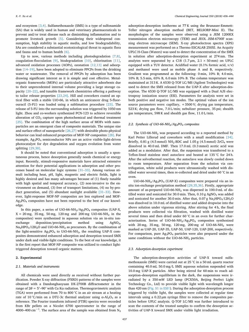
The successful preparation of UAP-X composites was confirmed byPXRD, FTIR, XPS, TGA, SEM, TEM and BET analysis. The PXRD patternsof individual Ag3PO4, UiO-66-NH2 and series UAP-X are illustrated inFig. 1a. The PXRD patterns of UiO-66-NH2 are consistent with thosereported in the literatures [37,38], demonstrating pure and well crys-tallized UiO-66-NH2 of the as-prepared samples. All 2θ peaks of UiO-66-NH2 were consistent with the UiO-66 [39], indicating that the in-troduction of –NH2 groups functionalized terephthalic acid does notaffect the skeleton of UiO-66. The diffraction peaks of as preparedAg3PO4 match perfectly with standard patterns of body-centered cubicAg3PO4 crystal (JCPDS card No. 006-0505) (Fig. S2 in ESI†). The UAP-Xcomposites exhibit almost all the characteristic PXRD peaks of UiO-66-NH2 and Ag3PO4. The slight different XRD peak intensities of thesamples might be resulted from the different relative content of UiO-66-NH2 and Ag3PO4 and the possible preferred orientation of the crystal-line UiO-66-NH2 and Ag3PO4 in the composites as well. For example,the XRD patterns of UAP-120 (Fig. 1a) show the characteristic peaks ofboth UiO-66-NH2 and Ag3PO4, indicating the introduction of UiO-66-NH2 has no effect on the crystal structure of Ag3PO4. However, noobvious characteristic diffractions for Ag3PO4 can be observed in theUAP-200, which could be ascribed to the relatively low amount ofAg3PO4. The unchanged backbones of UiO-66-NH2 and Ag3PO4 in seriesUAP-X composites were further confirmed by the similar FTIR spectraas shown in Fig. 1b. Two wide and medium adsorptions at 3507 cm−1
and 3384 cm−1 can be assigned to the aromatic amino groups in UiO-66-NH2 [37]. The adsorption peaks between 600 and 800 cm−1 are
contributed to Zr-O2 as longitudinal and transverse modes [40], whichwas relative weak at lower UiO-66-NH2 content (UAP-20 and UAP-35)for UAP-X composites. The intense doublet at 1421 and 1387 cm−1 canbe assigned to the stretching modes of the carboxylic groups in NH2-BDC ligands [41,42]. The typical P-O stretching vibrations of PO4
3−
can be observed at 554 and 1014 cm−1 [43].The successful fabrication of UAP-X (UiO-66-NH2/Ag3PO4) was
further affirmed by the surface compositions and chemical state in-vestigation via XPS analysis, as illustrated in Fig. 1c. All the bindingenergies obtained from the XPS analysis were corrected by referencingC1s to 284.8 eV. The occurrence of P 2p (132.6 eV for P(V) in Ag3PO4
[44]) and Ag 3d (374 eV and 368 eV for Ag 3d3/2 and Ag 3d5/2 [45],respectively, in Fig. S3 in ESI†) peaks in the XPS spectra of UAP-Xverified that Ag3PO4 is undoubtedly interacted to the UiO-66-NH2,which was also confirmed by both PXRD and FTIR analyses. In addition,the two peaks at 182.99 and 185.36 eV could be attributed to Zr-Oclusters coordinated with the carboxylic groups of NH2-BDC ligands inUiO-66-NH2 [46–48]. The TGA results (Fig. 1d) demonstrate that theincrease of UiO-66-NH2 content in the UAP-X composites lead to thelarger weight loss due to the loss of organic composition (NH2-BDC li-gands) from UiO-66-NH2, and the residual weight of the final residueare also consistent with the different UiO-66-NH2 content in the UAP-Xsamples.
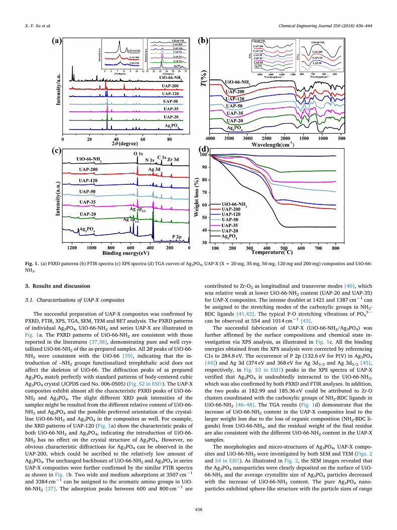
The morphologies and micro-structures of Ag3PO4, UAP-X compo-sites and UiO-66-NH2 were investigated by both SEM and TEM (Figs. 2and S4 in ESI†). As illustrated in Fig. 2, the SEM images revealed thatthe Ag3PO4 nanoparticles were clearly deposited on the surface of UiO-66-NH2 and the average crystallite size of Ag3PO4 particles decreasedwith the increase of UiO-66-NH2 content. The pure Ag3PO4 nano-particles exhibited sphere-like structure with the particle sizes of range
Fig. 1. (a) PXRD patterns (b) FTIR spectra (c) XPS spectra (d) TGA curves of Ag3PO4, UAP-X (X= 20mg, 35mg, 50mg, 120mg and 200mg) composites and UiO-66-NH2.
X.-Y. Xu et al. Chemical Engineering Journal 350 (2018) 436–444
438
from 250 to 450 nm as shown in the TEM image in Fig. 2a. Whereas, thecrystallite size of Ag3PO4 particles UAP-200 is less than 40 nm (Fig. 2f).The standard N2 adsorption measurements show the Brunauer-Emmett-Teller (BET) surface area decreased from 874.15m2 g−1 (UiO-66-NH2)to 424.4m2 g−1 (UAP-200), 191.9 m2 g−1 (UAP-120), 103.5m2 g−1
(UAP-50) and 67.2 m2 g−1 (UAP-35), which further confirm that theAg3PO4 NPs were anchored with UiO-66-NH2 (Table S1 in ESI†).
3.2. Adsorption and desorption performances of UAP-X toward SMX
3.2.1. The adsorption performances of Ag3PO4, UiO-66-NH2 and UAP-XSulfamethoxazole (SMX) is known to possess two pKa values (i.e. 1.7
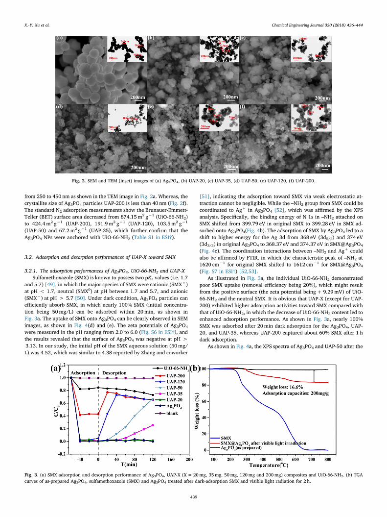
and 5.7) [49], in which the major species of SMX were cationic (SMX+)at pH < 1.7, neutral (SMX0) at pH between 1.7 and 5.7, and anionic(SMX−) at pH > 5.7 [50]. Under dark condition, Ag3PO4 particles canefficiently absorb SMX, in which nearly 100% SMX (initial concentra-tion being 50mg/L) can be adsorbed within 20min, as shown inFig. 3a. The uptake of SMX onto Ag3PO4 can be clearly observed in SEMimages, as shown in Fig. 4(d) and (e). The zeta potentials of Ag3PO4
were measured in the pH ranging from 2.0 to 6.0 (Fig. S6 in ESI†), andthe results revealed that the surface of Ag3PO4 was negative at pH >3.13. In our study, the initial pH of the SMX aqueous solution (50mg/L) was 4.52, which was similar to 4.38 reported by Zhang and coworker
[51], indicating the adsorption toward SMX via weak electrostatic at-traction cannot be negligible. While the –NH2 group from SMX could becoordinated to Ag+ in Ag3PO4 [52], which was affirmed by the XPSanalysis. Specifically, the binding energy of N 1s in –NH2 attached onSMX shifted from 399.79 eV in original SMX to 399.28 eV in SMX ad-sorbed onto Ag3PO4(Fig. 4b). The adsorption of SMX by Ag3PO4 led to ashift to higher energy for the Ag 3d from 368 eV (3d5/2) and 374 eV(3d3/2) in original Ag3PO4 to 368.37 eV and 374.37 eV in SMX@Ag3PO4
(Fig. 4c). The coordination interactions between –NH2 and Ag+ couldalso be affirmed by FTIR, in which the characteristic peak of –NH2 at1620 cm−1 for original SMX shifted to 1612 cm−1 for SMX@Ag3PO4
(Fig. S7 in ESI†) [52,53].As illustrated in Fig. 3a, the individual UiO-66-NH2 demonstrated
poor SMX uptake (removal efficiency being 20%), which might resultfrom the positive surface (the zeta potential being+9.29mV) of UiO-66-NH2 and the neutral SMX. It is obvious that UAP-X (except for UAP-200) exhibited higher adsorption activities toward SMX compared withthat of UiO-66-NH2, in which the decrease of UiO-66-NH2 content led toenhanced adsorption performance. As shown in Fig. 3a, nearly 100%SMX was adsorbed after 20min dark adsorption for the Ag3PO4, UAP-20, and UAP-35, whereas UAP-200 captured about 60% SMX after 1 hdark adsorption.
As shown in Fig. 4a, the XPS spectra of Ag3PO4 and UAP-50 after the
Fig. 2. SEM and TEM (inset) images of (a) Ag3PO4, (b) UAP-20, (c) UAP-35, (d) UAP-50, (e) UAP-120, (f) UAP-200.
Fig. 3. (a) SMX adsorption and desorption performance of Ag3PO4, UAP-X (X= 20mg, 35mg, 50mg, 120mg and 200mg) composites and UiO-66-NH2. (b) TGAcurves of as-prepared Ag3PO4, sulfamethoxazole (SMX) and Ag3PO4 treated after dark-adsorption SMX and visible light radiation for 2 h.
X.-Y. Xu et al. Chemical Engineering Journal 350 (2018) 436–444
439
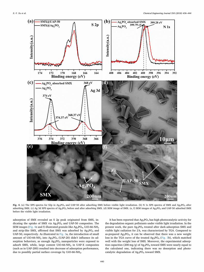
adsorption of SMX revealed an S 2p peak originated from SMX, in-dicating the uptake of SMX via Ag3PO4 and UAP-50 composites. TheSEM images (Fig. 4e and f) illustrated granule-like Ag3PO4, UiO-66-NH2
and strip-like SMX, affirmed that SMX was adsorbed by Ag3PO4 andUAP-50, respectively. As illustrated in Fig. 3a, the introduction of smallamount of UiO-66-NH2 into Ag3PO4 (UAP-20) didn’t influence its ad-sorption behaviors, as enough Ag3PO4 nanoparticles were exposed toadsorb SMX, while, large content UiO-66-NH2 in UAP-X composites(such as in UAP-200) resulted into decrease of adsorption performance,due to possibly partial surface coverage by UiO-66-NH2.
It has been reported that Ag3PO4 has high photocatalytic activity forthe degradation organic pollutants under visible light irradiation. In thepresent work, the pure Ag3PO4 treated after dark-adsorption SMX andvisible light radiation for 2 h, was characterized by TGA. Compared toas-prepared Ag3PO4, it can be observed that there was a new weightloss in the TGA curve of the treated Ag3PO4 (Fig. 3b), which matchedwell with the weight loss of SMX. Moreover, the experimental adsorp-tion capacities (200mg/g) of Ag3PO4 toward SMX were nearly equal tothe calculated one, indicating there was no desorption and photo-catalytic degradation of Ag3PO4 toward SMX.
Fig. 4. (a) The XPS spectra for S2p in Ag3PO4 and UAP-50 after adsorbing SMX before visible light irradiation. (b) N 1s XPS spectra of SMX and Ag3PO4 afteradsorbing SMX. (c) Ag 3d XPS spectra of Ag3PO4 before and after adsorbing SMX. (d) SEM image of SMX. (e, f) SEM images of Ag3PO4 and UAP-50 adsorbed SMXbefore the visible light irradiation.
X.-Y. Xu et al. Chemical Engineering Journal 350 (2018) 436–444
440
3.2.2. The release of SMX from UAP-X under visible lightLight triggered desorption is pollution-free, nearly zero-energy cost
and easy operation technique and has attracted increasing attentions. Inthis research, the series UAP-X (X ranging from 35 to 120) compositesbuilt from Ag3PO4 and UiO-66-NH2 exhibited excellent desorptionperformance under the visible light irradiation, as shown in Fig. 3a. It isworthy to noting that individual Ag3PO4 possessed good adsorption ofSMX in dark, while showed no desorption activity under visible lightirradiation. Although the individual UiO-66-NH2 exhibited poor ad-sorption and desorption behaviors, the introduction of UiO-66-NH2 intoAg3PO4 (UAP-35, 50, 120) can induce the light-triggered desorption ofSMX. Especially, the UAP-50/120 exhibited good desorption activities,in which ca. 66.9/73% SMX was released. As illustrated in Fig. 3a, the
introduction of small amount of UiO-66-NH2 into Ag3PO4 (UAP-20)didn’t influence its adsorption and desorption behaviors, as its ad-sorption activity was mainly controlled by Ag3PO4 NPs. However, largecontent UiO-66-NH2 was composited with Ag3PO4 (like UAP-200) re-sulted into decrease of adsorption performance and good desorptionactivity under the visible light irradiation.
It has been reported that the semiconductor Ag3PO4 particles can bephoto-excited for water splitting under visible light irradiation(λ < 530 nm) [54,55]. During the photo-catalytically split of watermolecules, the Ag+ ions in Ag3PO4 NPs were simultaneously beingreduced into Ag0 following the Eq. (1) [29,56]. For UAP-35, 50 and 120composites in our present work, visible light-induced transformationfrom Ag+ to Ag0 is believed as the possible mechanism for the SMX
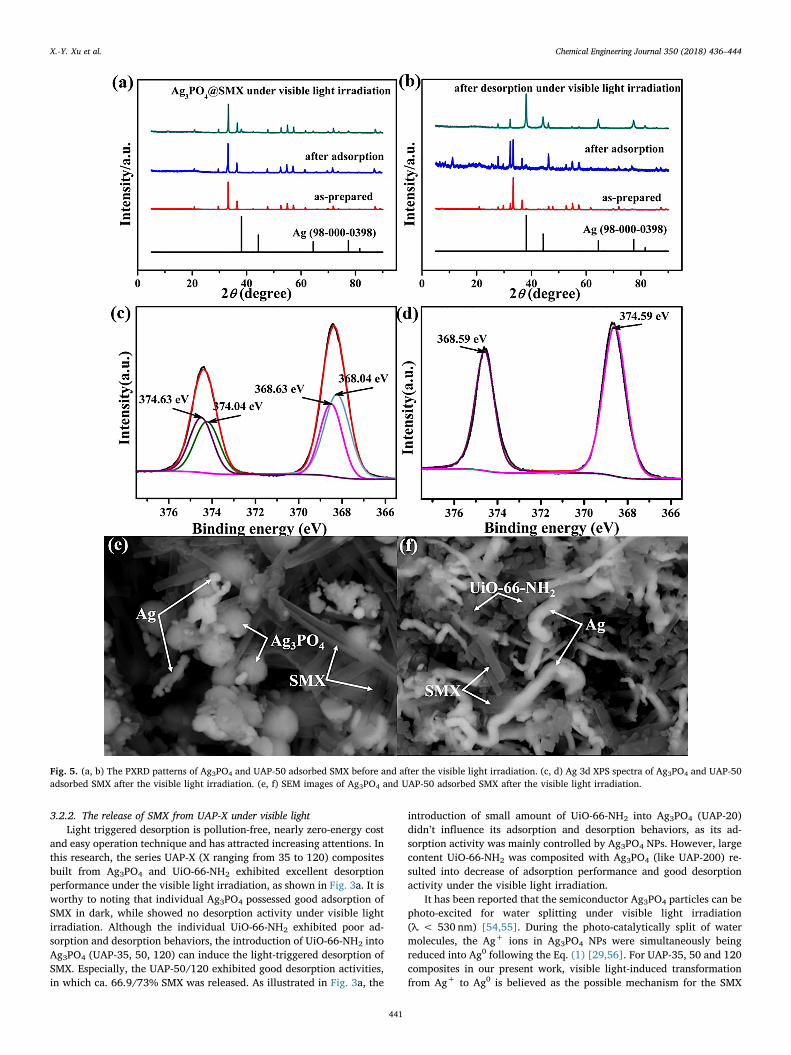
Fig. 5. (a, b) The PXRD patterns of Ag3PO4 and UAP-50 adsorbed SMX before and after the visible light irradiation. (c, d) Ag 3d XPS spectra of Ag3PO4 and UAP-50adsorbed SMX after the visible light irradiation. (e, f) SEM images of Ag3PO4 and UAP-50 adsorbed SMX after the visible light irradiation.
X.-Y. Xu et al. Chemical Engineering Journal 350 (2018) 436–444
441
release under visible light irradiation. The small size Ag3PO4 NPs fa-cilitate the reduction of Ag+, resulting in rapid decrease of adsorptivesites (Ag+) and release of adsorbed SMX. Both SEM and TEM resultsillustrated in Fig. 2 revealed that smaller Ag3PO4 NPs can be made withincreasing amount of UiO-66-NH2 and the resulting Ag3PO4 NPs aredispersed on their surface of UiO-66-NH2. Therefore, it is easy to un-derstand why UAP-120 possessed good adsorption and desorption ac-tivities. As to UAP-200, the large content of UiO-66-NH2 decreased itsadsorption performance, while smaller Ag3PO4 NPs enhanced its des-orption activity, as shown in Fig. 3a.
+ + + → + ++ −4Ag PO 6H O 12h 12e 12Ag 4H PO 3O3 4 2 3 4 2 (1)
For larger Ag3PO4 particles, the light irradiation could induce theAg+ reduction in limited extent due to its bigger particle size, whichwas affirmed by PXRD, XPS and SEM. As illustrated in Fig. 5a, thediffraction peaks at 29.70°, 33.29°, 36.59°, 55.02°, and 71.90° were thecharacteristics peaks of Ag3PO4 (JCPDS card No. 006-0505). And thediffraction peaks at 38.11°, 44.30°, 64.44°, 77.39° and 81.54° could beassigned to metallic silver (Ag0) (JCPDS card No. 98-000-0398). Aftervisible light irradiation for 2 h, the PXRD patterns of Ag3PO4 werenearly identical to both those of as-prepared Ag3PO4 and simulatedones, in which the characteristic peaks of Ag0 were difficult to be de-tected due to the minor content. In Fig. 5b, the obvious presence of Ag0
in UAP-50 could be evidenced by the occurrence of characteristic PXRDpeaks of Ag0. Furthermore, the main diffraction peak at 2θ=38.11°corresponding to Ag0 of UiO-66-NH2/Ag3PO4 composites, exhibiteddrastic enhanced peak intensity compared with that of Ag3PO4 afterdesorption and UiO-66-NH2 has good light stability (Fig. S8 in ESI†). Itcan be seen from Fig. 5c that the XPS peaks of Ag 3d of individualAg3PO4 NPs with adsorbed SMX under dark condition and Ag3PO4 NPsirradiated by visible light for 2 h could be further divided into fourdifferent peaks at 374.63, 374.04 eV and 368.63, 368.04 eV, respec-tively. The XPS peaks of Ag 3d of Ag3PO4 in UAP-50 with adsorbed SMXunder dark condition and Ag3PO4 in UAP-50 being irradiated by visiblelight for 2 h were at 368.59 and 374.59 eV (Fig. 5d). According to theresults reported by Zhang et al., the peaks at 374.6 and 368.6 eV couldbe attributed to Ag0, whereas the peaks at 374.04 and 368.04 eV areattributed to Ag+ ions in Ag3PO4 [57].
In this work, visible light-induced transform from Ag+ to Ag0 ofAg3PO4 was believed as possible mechanism for the release of SMXunder visible light irradiation. The differences in efficiency of reducedAg0 between Ag3PO4 and UAP-50 are also shown in SEM images(Fig. 5e and f). There are only small amount of long strip-like Ag re-latively and a large amount of Ag3PO4 with sphere-like structure for
treated Ag3PO4 (Fig. 5e). Whereas, almost all are long strip-like Ag fortreated UAP-50 (Fig. 5f). The presence of Ag species existed as metallicAg was further confirmed. From three SEM images shown in Fig. S9 (inESI†), long strip-like particles were not SMX molecules, UiO-66-NH2 orAg3PO4 particles, which were Ag° clusters. The difference of Ag+− Ag0
reduction efficiency could be attributed to the Ag3PO4 nanoparticlesaggregated quickly into micrometer-sized clusters in aqueous solutionsand the particle size of Ag3PO4 shows an obvious effect on photo-oxi-dative reactions [58–60]. The large surface area of small-sized particlesis expected to be beneficial for photocatalytic reactions that mostlyoccur on the surface of the catalysts. To clarify the detailed adsorption-desorption behavior of UAP-X composites toward SMX, the PXRD pat-terns of Ag3PO4 and UAP-50 adsorbing SMX under dark condition andbeing irradiated by visible light for 2 h were illustrated in Fig. 5a and b.Both Ag3PO4 and UAP-50 could keep stable after adsorbing SMXwithout light irradiation, and no Ag0 peak was observed. SMX adsorbedby UAP-50 was released along with the reduction of Ag+ to Ag0 underthe visible light irradiation. In addition, the PXRD patterns of Ag3PO4,UAP-50 and UAP-120 treated with dispersed in solvent without SMXand visible light irradiation for 2 h (Fig. S10 in ESI†) could affirm thatthe presence of SMX has no influence on the transformation from Ag+
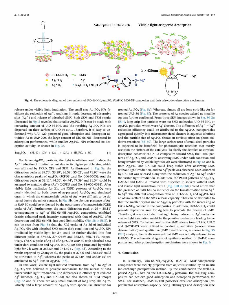
to Ag0. The different composition ratio of UiO-66-NH2 and Ag3PO4 hasan obvious effect on the SMX release capacity. That can be attributed tothat the smaller crystal size of Ag3PO4 particles with the increasing ofUiO-66-NH2 content in the composites. In addition, UiO-66-NH2 couldprovide deposition area for Ag NPs to promote the release of SMX.Therefore, it was concluded that Ag+ being reduced to Ag0 under thevisible light irradiation might be the possible mechanism leading to therelease of SMX. To further confirm the SMX release from UAP-X, UPLCand Q-TOF-MS were utilized to conduct quantitative (concentrationdetermination) and qualitative (SMX identification, as shown in Fig. S5ESI†) analysis, the results revealed that SMX was actually released fromUAP-50. The schematic diagram of synthesis method of UAP-X com-posites and adsorption-desorption mechanism were shown in Fig. 6.
4. Conclusion
In summary, UiO-66-NH2/Ag3PO4 (UAP-X) MOF-nanoparticlecomposites were facilely prepared from aqueous solution by an in-situion-exchange precipitation method. By the combination the well-dis-persed Ag3PO4 NPs on the UiO-66-NH2 platform, the resulting com-posites can achieve good adsorption and desorption performance forSMX. For instance, UAP-50/120 possesses excellent adsorption (ex-perimental adsorption capacity being 200mg/g) and desorption (the
Fig. 6. The schematic diagram of the synthesis of UiO-66-NH2/Ag3PO4 (UAP-X) MOF-NP composites and their adsorption-desorption mechanism.
X.-Y. Xu et al. Chemical Engineering Journal 350 (2018) 436–444
442
desorption amount being 134/146mg/g) activities toward SMX. Thevisible light-triggered desorption of SMX on the UAP-X composite canbe assigned to the transformation from Ag+ in Ag3PO4 to Ag0 undervisible light irradiation and was found to be heavily dependent on thecontent of UiO-66-NH2, resulting the first reported light-triggered des-orption toward organic matters of MOF-NP composites, to our bestknowledge. This work could open new opportunities for adsorption-desorption of targeted organic matters using visible light. Frankly, inthis paper, the transformation from Ag+ to Ag0 under light irradiationwas not reversible, which hindered the reutilization and potential ap-plication to remove environmental PPCPs pollutants. But, it leaves awindow open for these composites to be used in drug delivery. Furtherresearches are designed to facilely prepare similar MOFs-based com-posites to achieve their adsorption-desorption activities triggered bylight toward targeted organic matters, which will knock a door open toachieve light induced desorption with zero-pollution and low-cost re-generation.
Acknowledgements
This work was supported by National Natural Science Foundation ofChina (51578034), Great Wall Scholars Training Program Project ofBeijing Municipality Universities (CIT&TCD20180323), Project ofConstruction of Innovation Teams and Teacher Career Development forUniversities and Colleges Under Beijing Municipality (IDHT20170508),Beijing Talent Project (2017A38), and the Fundamental Research Fundsfor Beijing Universities.
Appendix A. Supplementary data
Supplementary data associated with this article can be found, in theonline version, at http://dx.doi.org/10.1016/j.cej.2018.06.005.
References
[1] D. Zhang, R.M. Gersberg, W.J. Ng, S.K. Tan, Removal of pharmaceuticals andpersonal care products in aquatic plant-based systems: a review, Environ. Pollut.184 (2014) 620–639.
[2] Q. Bu, B. Wang, J. Huang, S. Deng, G. Yu, Pharmaceuticals and personal careproducts in the aquatic environment in China: a review, J. Hazard. Mater. 262(2013) 189–211.
[3] S.-W. Nam, C. Jung, H. Li, M. Yu, J.R. Flora, L.K. Boateng, N. Her, K.-D. Zoh,Y. Yoon, Adsorption characteristics of diclofenac and sulfamethoxazole to grapheneoxide in aqueous solution, Chemosphere 136 (2015) 20–26.
[4] J. Cao, R. Jiang, J. Wang, J. Sun, Q. Feng, Z. Zhao, G. Chen, C. Zhou, E. Yin, Studyon the interaction mechanism between cefradine and Chlamydomonas reinhardtii inwater solutions under dark condition, Ecotox. Environ. Safe. 159 (2018) 56–62.
[5] S. Thiele-Bruhn, Pharmaceutical antibiotic compounds in soils – a review, J. PlantNutr. Soil Sci. 166 (2003) 145–167.
[6] I.Y. Hwang, E. Koh, H.R. Kim, W.S. Yew, M.W. Chang, Reprogrammable microbialcell-based therapeutics against antibiotic-resistant bacteria, Drug Resist. Update 27(2016) 59–71.
[7] C.C. Wang, J.R. Li, X.L. Lv, Y.Q. Zhang, G. Guo, Photocatalytic organic pollutantsdegradation in metal–organic frameworks, Energy Environ. Sci. 7 (2014)2831–2867.
[8] H.-R. Buser, T. Poiger, M.D. Müller, Occurrence and fate of the pharmaceutical drugdiclofenac in surface waters: rapid photodegradation in a lake, Environ. Sci.Technol. 32 (1998) 3449–3456.
[9] G.R. Boyd, H. Reemtsma, D.A. Grimm, S. Mitra, Pharmaceuticals and personal careproducts (PPCPs) in surface and treated waters of Louisiana, USA and Ontario,Canada, Sci. Total Environ. 311 (2003) 135–149.
[10] A. Joss, S. Zabczynski, A. Göbel, B. Hoffmann, D. Löffler, C.S. McArdell, T.A. Ternes,A. Thomsen, H. Siegrist, Biological degradation of pharmaceuticals in municipalwastewater treatment: proposing a classification scheme, Water Res. 40 (2006)1686–1696.
[11] G.R. Boyd, S. Zhang, D.A. Grimm, Naproxen removal from water by chlorinationand biofilm processes, Water Res. 39 (2005) 668–676.
[12] S. Esplugas, D.M. Bila, L.G.T. Krause, M. Dezotti, Ozonation and advanced oxidationtechnologies to remove endocrine disrupting chemicals (EDCs) and pharmaceuticalsand personal care products (PPCPs) in water effluents, J. Hazard. Mater. 149 (2007)631–642.
[13] M. Klavarioti, D. Mantzavinos, D. Kassinos, Removal of residual pharmaceuticalsfrom aqueous systems by advanced oxidation processes, Environ. Int. 35 (2009)402–417.
[14] X.D. Du, C.C. Wang, J.G. Liu, X.D. Zhao, J. Zhong, Y.X. Li, J. Li, P. Wang, Extensive
and selective adsorption of ZIF-67 towards organic dyes: performance and me-chanism, J. Colloid Interface Sci. 506 (2017) 437–441.
[15] X. Zhao, Y. Wei, H. Zhao, Z. Gao, Y. Zhang, L. Zhi, Y. Wang, H. Huang,Functionalized metal-organic frameworks for effective removal of rocephin inaqueous solutions, J. Colloid Interface Sci. 514 (2017) 234–239.
[16] J.J. Li, C.C. Wang, H.F. Fu, J.R. Cui, P. Xu, J. Guo, J.R. Li, High-performance ad-sorption and separation of anionic dyes in water using a chemically stable gra-phene-like metal-organic framework, Dalton Trans. 46 (2017) 10197–10201.
[17] D. Sheng, L. Zhu, C. Xu, C. Xiao, Y. Wang, Y. Wang, L. Chen, J. Diwu, J. Chen,Z. Chai, T.E. Albrecht-Schmitt, S. Wang, Efficient and selective uptake of TcO4
– by acationic metal-organic framework material with open Ag+ sites, Environ. Sci.Technol. 51 (2017) 3471–3479.
[18] W. He, N. Li, X. Wang, T. Hu, X. Bu, A cationic metal-organic framework based onZn4 cluster for rapid and selective adsorption of dyes, Chin. Chem. Lett. 4276(2017).
[19] L. Zhu, D. Sheng, C. Xu, X. Dai, M.A. Silver, J. Li, P. Li, Y. Wang, Y. Wang, L. Chen,C. Xiao, J. Chen, R. Zhou, C. Zhang, O.K. Farha, Z. Chai, T.E. Albrecht-Schmitt,S. Wang, Identifying the recognition site for selective trapping of 99TcO4- in a hy-drolytically stable and radiation resistant cationic metal-organic framework, J. Am.Chem. Soc. 139 (2017) 14873–14876.
[20] Y. Peng, Y. Zhang, H. Huang, C. Zhong, Flexibility induced high-performance MOF-based adsorbent for nitroimidazole antibiotics capture, Chem. Eng. J. 333 (2018)678–685.
[21] X. Zhao, H. Zhao, W. Dai, Y. Wei, Y. Wang, Y. Zhang, L. Zhi, H. Huang, Z. Gao, Ametal-organic framework with large 1-D channels and rich OH sites for high-effi-ciency chloramphenicol removal from water, J. Colloid Interface Sci. 526 (2018)28–34.
[22] Y. Li, Z. Yang, Y. Wang, Z. Bai, T. Zheng, X. Dai, S. Liu, D. Gui, W. Liu, M. Chen,L. Chen, J. Diwu, L. Zhu, R. Zhou, Z. Chai, T.E. Albrecht-Schmitt, S. Wang, A me-soporous cationic thorium-organic framework that rapidly traps anionic persistentorganic pollutants, Nat. Commun. 8 (2017) 1354.
[23] M. Nazari, M. Rubio-Martinez, G. Tobias, J.P. Barrio, R. Babarao, F. Nazari,K. Konstas, B.W. Muir, S.F. Collins, A.J. Hill, Metal-organic-framework-coated op-tical fibers as light-triggered drug delivery vehicles, Adv. Funct Mater. 26 (2016)3244–3249.
[24] T. Zheng, Z. Yang, D. Gui, Z. Liu, X. Wang, X. Dai, S. Liu, L. Zhang, Y. Gao, L. Chen,D. Sheng, Y. Wang, J. Diwu, J. Wang, R. Zhou, Z. Chai, T.E. Albrecht-Schmitt,S. Wang, Overcoming the crystallization and designability issues in the ultrastablezirconium phosphonate framework system, Nat. Commun. 8 (2017) 15369.
[25] J. Park, D. Yuan, K.T. Pham, J.-R. Li, A. Yakovenko, H.-C. Zhou, Reversible al-teration of CO2 adsorption upon photochemical or thermal treatment in a metal-organic framework, J. Am. Chem. Soc. 134 (2011) 99–102.
[26] W. Zheng, G.F. Strouse, Involvement of carriers in the size-dependent magneticexchange for Mn:CdSe quantum dots, J. Am. Chem. Soc. 133 (2011) 7482–7489.
[27] W. Zheng, Y. Liu, A. West, E.E. Schuler, K. Yehl, R.B. Dyer, J.T. Kindt, K. Salaita,Quantum dots encapsulated within phospholipid membranes: phase-dependentstructure, photostability, and site-selective functionalization, J. Am. Chem. Soc. 136(2014) 1992–1999.
[28] J. Aguilera-Sigalat, D. Bradshaw, Synthesis and applications of metal-organic fra-mework-quantum dot (QD@MOF) composites, Coord. Chem. Rev. 307 (2016)267–291.
[29] Z. Yi, J. Ye, N. Kikugawa, T. Kako, S. Ouyang, H. Stuart-Williams, H. Yang, J. Cao,W. Luo, Z. Li, An orthophosphate semiconductor with photooxidation propertiesunder visible-light irradiation, Nat. Mater. 9 (2010) 559–564.
[30] L. Luo, Y. Li, J. Hou, Y. Yang, Visible photocatalysis and photostability of Ag3PO4
photocatalyst, Appl. Surf. Sci. 319 (2014) 332–338.[31] J.-M. Schumers, C.-A. Fustin, J.-F. Gohy, Light-responsive block copolymers,
Macromol. Rapid Commu. 31 (2010) 1588–1607.[32] Y. Wei, S. Han, J. Kim, S. Soh, B.A. Grzybowski, Photoswitchable catalysis mediated
by dynamic aggregation of nanoparticles, J. Am. Chem. Soc. 132 (2010)11018–11020.
[33] Y. Shiraishi, K. Tanaka, E. Shirakawa, Y. Sugano, S. Ichikawa, S. Tanaka, T. Hirai,Light-triggered self-assembly of gold nanoparticles based on photoisomerization ofspirothiopyran, Angew. Chem. Int. Edit. 52 (2013) 8304–8308.
[34] M. Kandiah, M.H. Nilsen, S. Usseglio, S. Jakobsen, U. Olsbye, M. Tilset, C. Larabi,E.A. Quadrelli, F. Bonino, K.P. Lillerud, Synthesis and Stability of Tagged UiO-66Zr-MOFs, Chem. Mater. 22 (2010) 6632–6640.
[35] C. Cui, Y. Wang, D. Liang, W. Cui, H. Hu, B. Lu, S. Xu, X. Li, C. Wang, Y. Yang,Photo-assisted synthesis of Ag3PO4 /reduced graphene oxide/Ag heterostructurephotocatalyst with enhanced photocatalytic activity and stability under visiblelight, Appl. Catal. B: Environ. 158–159 (2014) 150–160.
[36] F.A. Sofi, K. Majid, Enhancement of the photocatalytic performance and thermalstability of an iron based metal-organic-framework functionalised by Ag/Ag3PO4,Mater. Chem. Front. (2018), http://dx.doi.org/10.1039/C8QM00051D.
[37] L. Shen, S. Liang, W. Wu, R. Liang, L. Wu, Multifunctional NH2-mediated zirconiummetal-organic framework as an efficient visible-light-driven photocatalyst for se-lective oxidation of alcohols and reduction of aqueous Cr(VI), Dalton Trans. 42(2013) 13649–13657.
[38] D. Sun, Y. Fu, W. Liu, L. Ye, D. Wang, L. Yang, X.Z. Fu, Z.H. Li, Studies on photo-catalytic CO2 reduction over NH2-Uio-66(Zr) and its derivatives: towards a betterunderstanding of photocatalysis on metal-organic frameworks, Chem. Eur. J. 19(2013) 14279–14285.
[39] P. Kanoo, K.L. Gurunatha, T.K. Maji, Versatile functionalities in MOFs assembledfrom the same building units: interplay of structural flexibilty, rigidity and reg-ularity, J. Mater. Chem. 20 (2010) 1322–1331.
[40] J. Yang, Y. Dai, X. Zhu, Z. Wang, Y. Li, Q. Zhuang, J. Shi, J. Gu, Metal–organic
X.-Y. Xu et al. Chemical Engineering Journal 350 (2018) 436–444
443
frameworks with inherent recognition sites for selective phosphate sensing throughtheir coordination-induced fluorescence enhancement effect, J. Mater. Chem. A 3(2015) 7445–7452.
[41] X. Cheng, A. Zhang, K. Hou, M. Liu, Y. Wang, C. Song, G. Zhang, X. Guo, Size- andmorphology-controlled NH2-MIL-53(Al) prepared in DMF-water mixed solvents,Dalton Trans. 42 (2013) 13698–13705.
[42] L. Valenzano, B. Civalleri, S. Chavan, S. Bordiga, M.H. Nilsen, S. Jakobsen,K.P. Lillerud, C. Lamberti, Disclosing the complex structure of UiO-66 metal organicframework: a synergic combination of experiment and theory, Chem. Mater. 23(2011) 1700–1718.
[43] M. Thomas, S. Ghosh, K. George, Characterisation of nanostructured silver ortho-phosphate, Mater. Lett. 56 (2002) 386–392.
[44] P. Ma, H. Yu, Y. Yu, W. Wang, H. Wang, J. Zhang, Z. Fu, Assembly of Ag3PO4
nanoparticles on two-dimensional Ag2S sheets as visible-light-driven photo-catalysts, Phys. Chem. Chem. Phys. 18 (2016) 3638–3643.
[45] H. Zhang, G. Wang, D. Chen, X. Lv, J. Li, Tuning photoelectrochemical perfor-mances of Ag-TiO2 nanocomposites via reduction/oxidation of Ag, Chem. Mater. 20(2008) 6543–6549.
[46] J. Long, S. Wang, Z. Ding, S. Wang, Y. Zhou, L. Huang, X. Wang, Amine-functio-nalized zirconium metal-organic framework as efficient visible-light photocatalystfor aerobic organic transformations, Chem. Commun. 48 (2012) 11656–11658.
[47] Q. Wu, D. Wu, Y. Guan, Hybrid titania–zirconia nanoparticles coated adsorbent forhighly selective capture of nucleosides from human urine in physiological condi-tion, Anal. Chem. 86 (2014) 10122–10130.
[48] R. Wang, L. Gu, J.J. Zhou, X.L. Liu, F. Tong, C.H. Li, Y.H. Shen, Y.P. Yuan, Quasi-polymeric metal-organic framework UiO-66/g-C3N4 heterojunctions for enhancedphotocatalytic hydrogen evolution under visible light irradiation, Adv. Mater.Interfaces 2 (2015) 1500037.
[49] H. Lucida, J.E. Parkin, V.B. Sunderland, Kinetic study of the reaction of sulfa-methoxazole and glucose under acidic conditions: I. Effect of pH and temperature,Int. J. Pharmaceut. 202 (2000) 47–62.
[50] D. Zhang, B. Pan, M. Wu, B. Wang, H. Zhang, H. Peng, D. Wu, P. Ning, Adsorption of
sulfamethoxazole on functionalized carbon nanotubes as affected by cations andanions, Environ. Pollut. 159 (2011) 2616–2621.
[51] D. Zhang, B. Pan, H. Zhang, P. Ning, B. Xing, Contribution of different sulfa-methoxazole species to their overall adsorption on functionalized carbon nano-tubes, Environ. Sci. Technol. 44 (2010) 3806–3811.
[52] L. Zhang, S. Yang, T. Han, L. Zhong, C. Ma, Improvement of Ag(I) adsorption ontochitosan/triethanolamine composite sorbent by an ion-imprinted technology, Appl.Surf. Sci. 263 (2012) 696–703.
[53] A. Liu, C.-C. Wang, C.-Z. Wang, H.-F. Fu, W. Peng, Y.-L. Cao, H.-Y. Chu, A.-F. Du,Selective adsorption activities toward organic dyes and antibacterial performanceof silver-based coordination polymers, J. Colloid Interface Sci. 512 (2018) 730–739.
[54] X. Yang, H. Cui, Y. Li, J. Qin, R. Zhang, H. Tang, Fabrication of Ag3PO4-graphenecomposites with highly efficient and stable visible light photocatalytic performance,ACS Catal. 3 (2013) 363–369.
[55] T. Wei, X. Li, Q. Zhao, J. Zhao, D. Zhang, In situ capture of active species andoxidation mechanism of RhB and MB dyes over sunlight-driven Ag/Ag3PO4 plas-monic nanocatalyst, Appl. Catal. B: Environ. 125 (2012) 538–545.
[56] Y. Bi, S. Ouyang, N. Umezawa, J. Cao, J. Ye, Facet effect of single-crystallineAg3PO4 sub-microcrystals on photocatalytic properties, J. Am. Chem. Soc. 133(2011) 6490–6492.
[57] H. Zhang, G. Wang, D. Chen, X. Lv, J. Li, Tuning photoelectrochemical perfor-mances of Ag−TiO2 nanocomposites via reduction/oxidation of Ag, Chem. Mater.20 (2008) 6543–6549.
[58] P.V. Kamat, Photochemistry on nonreactive and reactive (semiconductor) surfaces,Chem. Rev. 93 (1993) 267–300.
[59] C.T. Dinh, T.D. Nguyen, F. Kleitz, Large-scale synthesis of uniform silver ortho-phosphate colloidal nanocrystals exhibiting high visible light photocatalytic ac-tivity, Cheminform 47 (2011) 7797–7799.
[60] J. Ma, J. Zou, L. Li, C. Yao, T. Zhang, D. Li, Synthesis and characterization ofAg3PO4 immobilized in bentonite for the sunlight-driven degradation of Orange II,Appl. Catal. B: Environ. 134 (2013) 1–6.
X.-Y. Xu et al. Chemical Engineering Journal 350 (2018) 436–444
444
![Journal of Chromatography A · SPME is often coupled with gas chromatography (GC) via a sim-ple, robust thermal desorption interface [9]. But there are many analytes with limited](https://cdn.vdocuments.net/doc/165x107/60cac64782280b59e523e34d/journal-of-chromatography-a-spme-is-often-coupled-with-gas-chromatography-gc-via.jpg)


