www.neurovisionmedical.com
Cobra® EMG Monitoring Endotracheal Tube
Two Channel
I23-H
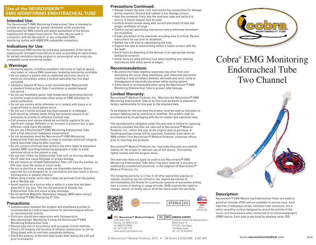
Description:Neurovision™ EMG Monitoring Endotracheal Tubes are made of
polyvinyl chloride (PVC) and are available in various sizes. Each
tube has a radiopaque stripe, ventilator tube connector, and a
silver recording surface designed to record the activity of the
vocal cord musculature when connected to an electromyographic
(EMG) device. Each tube is sterilized by ethylene oxide (EO).
Precautions Continued: • Always inspect the tube, cuff, and monitoring connections for damage
during insertion. Discard and replace if any damage occurs.
• Seat the connector firmly into the ventilator tube and verify it is
secure. A swivel adaptor may be used.
• Confirm breath sounds along with correct placement of tube and
proper ventilation of lungs.
• Confirm correct positioning and secure tube to eliminate movement
out of position.
• Proper placement of the electrode recording area is critical. Review
instructions for use prior to intubation.
• Deflate the cuff prior to repositioning the tube.
• Support the tube to avoid kinking where it makes contact with the
the teeth.
• Avoid injury by disposing of the devices in an appropriate sharps
biohazard container.
• Avoid injury by using extreme care when handling and cleaning
instruments with sharp points or edges.
Recommendations: • Be aware that false negative responses may arise from over
stimulating the nerve, deep anesthesia, poor electrode placement
resulting in lack of contact between electrode and vocal cords or
dislodgement of electrode placement while moving patient.
• A bite block is recommended when using the Neurovision™ EMG
Monitoring Endotracheal Tube to prevent tube damage.
Limited Warranty:Neurovision™ Medical Products, Inc., Warrants the Neurovision™ EMG
Monitoring Endotracheal Tube to be free from problems in material or
factory workmanship for one year at the shipment date.
To be eligible for this warranty the product must be used as indicated on
product labeling and be unaltered or modified. The product must be
accompanied by its packaging with the lot number and expiration date.
The manufacturer’s obligation under this warranty is limited to replacing
products provided that they are returned to Neurovision™ Medical
Products, Inc., within one year of the original date of purchase. A
handling/postage charge will be assessed. Customer must obtain an
RMA number from Neurovision™ Medical Products’ corporate offices
prior to returning any products.
Neurovision™ Medical Products, Inc. expressly disavows any medical
liability for the proper or improper use of this device. This liability
rightly resides with the surgeon alone.
This warranty does not apply (is void) to any Neurovision™ EMG
Monitoring Endotracheal Tube which has been repaired in any way or
modified by unauthorized personnel, in the judgment of Neurovision™
Medical Products, Inc.
The foregoing warranty is in lieu of all other warranties express or
implied, including, but not limited to, the implied warranties of
merchantability and fitness for a particular purpose of warranties arising
from a course of dealing or usage of trade. NVM reserves the rights to
change, amend, or modify any or all of the items under this warranty.
Use of the NEUROVISION™ EMG MONITORING ENDOTRACHEAL TUBE
Made inCalifornia, USA
An ISO 13485 Certified Company
© Neurov is ion ™ Medica l P roducts , 2014 • US Patent # 8 ,634 ,894 , 8 ,467 ,844
Neurovision™ Medical Products2225 Sperry Ave.
Ventura, CA 93003 U.S.A.
tel: (866) 815-6999
fax: (877) 330-1727
www.neurovisionmedical.com
EMERGO EUROPEEuropean Authorized Representation
Molenstraat 15
2513 BH, The Hague
The Netherlands
Tel: +31.70.345.8570
Intended Use:The Neurovision™ EMG Monitoring Endotracheal Tube is intended to
provide an open airway for patient ventilation while monitoring
continuously for EMG activity and status assessment of the nerves
supplying the laryngeal musculature. The tube may be used in
connection with the Nerveäna® or any compatible EMG
monitoring system with 42802 DIN compatible connectors.
Indications for Use:For continuous EMG monitoring and status assessment of the nerves
supplying the laryngeal musculature as well as providing an open airway
for patient ventilation during surgical or parasurgical care using any
compatible nerve monitoring system.
Warnings: • Paralyzing agents, including anesthetic lubricants or topical sprays,
may impair or reduce EMG responses rendering monitoring unreliable.
• Do not subject a patient with an implanted electronic device to
electrical stimulation unless a medical specialist has first been
consulted.
• Intubation beyond 24 hours is not recommended. Replace with
a standard Endotracheal Tube if ventilation is needed beyond
this period.
• Do not use flammable gases, high temperature generating devices
or electro surgical electrodes when using an EMG stimulator to
avoid combustion.
• Do not use cautery while stimulator is in contact with tissue or in
surgical fields to avoid patient burns.
• Do not use if sterile package has been opened or is damaged.
• Cuff should be inflated slowly filling the minimum amount of air
necessary to provide an effective tracheal seal.
• Cuff pressure and volume should be monitored regularly for any
significant change. Deflation or an increase in pressure due to gas
diffusion could injure the patient.
• Do not use a Neurovision™ EMG Monitoring Endotracheal Tube
with a high electrical impedance measurement.
• Do not excessively bend the Neurovision™ EMG Monitoring
Endotracheal Tube electrodes in order to maintain electrical integrity.
Check electrode integrity after insertion.
• Do not connect electrode lead wires to any other types of equipment
besides EMG recording/monitoring equipment in order to avoid
electrical shock to the patient or user.
• Do not over inflate the Endotracheal Tube cuff, as this may damage
the ET tube and cause blockage or airway damage.
• Do not remove an inflated Endotracheal Tube cuff from the trachea, as
this may injure the larynx or vocal cords.
• Do not re-sterilize or reuse single use disposable devices. Device
materials are not designed for re-sterilization and may result in device
disintegration or patient infection.
• When using a stylet, verify that it does not protrude from the patient
end or Murphy eye.
• Avoid insertion of a suction tube or stylet in a tube that has been
distorted in any way. This has the potential to damage the
Endotracheal Tube and cause airway blockage.
• Do not perform Magnetic Resonance Imaging (MRI) when using a
Neurovision™ EMG Monitoring ET Tube.
Precautions: • Communication between the surgeon and anesthesia provider is
recommended to confirm expectations for pharmacological effects
on neuromuscular activity.
• Clinicians should have experience with Intraoperative
Neurophysiologic Monitoring if using the Neurovision™ EMG
Monitoring Endotracheal Tube.
• Choose tube size in accordance with accepted clinical methods.
• Check cuff integrity and function of inflation system prior to use by
filling slowly with air and then completely deflating.
• Check the patency of the main tube lumen after testing the cuff and
prior to intubation.

www.neurovisionmedical.com
Cobra®
LaryngealElectrode
Cobra®
LaryngealElectrode
EMG GroundElectrode
Stimulator ReturnElectrode
Figure 1 Figure 2
Note the depth number on the Endotracheal Tube against the maxillary central incisors before any further positioning of the patient.
Extubation:
Extubate only after complete deflation of the cuff with a
Leur tip syringe.
Figure 3
Cobra® Electrode Item CodeEndotracheal Tube ID Size
6.0mm
7.0mm
8.0mm
LTE700DCS
LTE700DCM
LTE700DCL
2) Prior to intubation, test the cuff by slowly filling with
a Luer tip syringe. Remove syringe from valve and
check that cuff and inflation system retain air.
Reattach syringe and remove all air from cuff.
1) Choose the appropriate Endotracheal Tube size:
Intubation*:
Preparation:
2) Tape the Endotracheal Tube securely with 2 pieces of
tape by wrapping each piece first around the tube
and then securing to the upper lip. (fig. 2) A Bite
Block may also be used to secure the tube.
3) Inflate the cuff with the minimum amount of air
necessary to create an effective tracheal seal.
4) After final positioning of patient, align Endotracheal
Tube in the middle of the pharynx behind the tongue.
The posterior portion of the Endotracheal Tube should
be directly opposite the central maxillary incisor gap
at the depth number noted after initial positioning.
5) Tightly secure the ventilator circuit so the
Endotracheal Tube will not rotate or be displaced and
then verify final electrode position by laryngoscopy
with a #3 Miller Blade or with a video laryngoscope.
Support the tube to avoid kinking where it makes
contact with the teeth.
6) Attach the pairs of red and blue electrode lead wires
to the + and - terminals of the EMG recording device
and apply the EMG ground and stimulator return
electrodes to the sternum as shown in Figure 3.
* Intubation beyond 48 hours is not recommended.
Replace with a standard Endotracheal Tube if
ventilation is needed beyond this period.
1) A small amount of water-based lubricant, may be applied
to the electrode. Do not use petroleum-based lubricants.
Use of a stylet is recommended for proper placement.
Intubate using currently accepted medical techniques.
Insert the Endotracheal Tube under direct vision or with
a video laryngoscope so that each vocal cord is touching
its respective silver electrode stripe. (fig. 1) Cobra® EMG Monitoring Endotracheal Tube Instructions for Use
Reliable performance of Cobra® electrodes requires proper positioning. Please read and follow all instructions.
Caution: use of paralytics inhibits EMG nerve monitoring.


