RÚBENS PRINCE DOS SANTOS ALVES
DESENVOLVIMENTO DE UMA VACINA DE SUBUNIDADE CONTRA
O SOROTIPO 2 DO VÍRUS DENGUE BASEADA NA PROTEÍNA NÃO
ESTRUTURAL 5 (NS5).
Dissertação apresentada ao Programa de
Pós-Graduação em Microbiologia do
Instituto de Ciências Biomédicas da
Universidade de São Paulo, para obtenção
do título de Mestre em Ciências.
Área de Concentração: Microbiologia
Orientador: Prof. Dr. Luís Carlos de Souza
Ferreira
Versão original
São Paulo
2015
RESUMO
ALVES, R. P. S. Desenvolvimento de uma vacina de subunidade contra o sorotipo 2 do
vírus dengue baseada na proteína não estrutural 5 (NS5). 2015. 69 f. Dissertação de
(Mestrado em Microbiologia) – Instituto de Ciências Biomédicas, Universidade de São Paulo,
São Paulo, 2015.
A dengue é uma doença causada por quatro sorotipos do vírus da dengue (DENV 1-4), com
milhões de pessoas afetadas em todo o mundo e um número significativo de mortes. Não há
nenhum tratamento eficaz ou abordagens vacinais capazes de prevenir a infecção. As estratégias
vacinais contra a dengue baseadas em proteínas não estruturais como antígenos têm
demonstrado serem mais seguras do que as baseadas em proteínas estruturais. Além disso,
respostas imunes celulares direcionadas a proteínas não estruturais desempenham um papel
importante no controle da replicação viral. A proteína não estrutural 5 (NS5) do vírus dengue é
a proteína mais conservada entre os quatro sorotipos e desempenha um papel crucial na
replicação viral. Neste estudo, foi gerada uma forma recombinante da NS5 expressa em E. coli
em quantidades elevadas e com propriedades antigênicas preservados em relação à proteína
nativa. As condições de cultura foram optimizadas, a fim de permitir a expressão dessa proteína
na forma solúvel. A imunização de camundongos Balb/c com a NS5 sozinha ou em combinação
com um adjuvante (poli (I:C)) promoveu o aumento da sobrevida de camundongos imunizados
após desafio intracraniano com a linhagem JHA1 de DENV2. A combinação da NS5 com poli
(I:C) emulsionado em Montanide 720 induziu a expansão de linfócitos T CD8+ específicos. Em
conjunto, os resultados indicam que a proteína recombinante NS5 preserva determinantes
antigênicos da proteína nativa e pode ser uma ferramenta útil para estudos sobre a biologia do
DENV, busca de drogas anti-viriais e desenvolvimento de vacinas.
.
Palavras-chave: Dengue. Vírus dengue. NS5. Vacinas. Adjuvantes imunológicos.

ABSTRACT
ALVES, R. P. S. Development of a subunit vaccine against dengue virus serotype 2 based
on the non-structural protein 5 (NS5). 2015. 69 p. Master thesis (Microbiology) – Instituto
de Ciências Biomédicas, Universidade de São Paulo, São Paulo, 2015.
Dengue fever is a disease caused by four dengue virus serotypes (DENV 1-4), affecting millions
of people worldwide and causing a significant number of deaths. There are no effective
treatments or vaccine approaches capable of preventing such infection. Anti-DENV vaccine
strategies based on nonstructural proteins as antigens have been shown to be safer than those
based on structural proteins. In addition, anti-DENV cellular immune responses against
nonstructural proteins have shown to play a major role in controlling viral replication. The
DENV nonstructural protein 5 (NS5), the most conserved protein among the four serotypes,
plays a crucial role in viral replication. In this study, we generated a recombinant form of
DENV2 NS5 expressed in E. coli in high amounts and with preserved antigenic properties with
regard to the native protein. Culture conditions were optimized in order to allow expression of
NS5 as a soluble protein. The immunization of Balb/c mice using this protein alone or in
combination with poly (I:C) led to increased survival after intracranial challenge with the
DENV2 JHA1 strain. The combination of the protein with poly (I:C) emulsified in Montanide
720 led to the activation of NS5-specific CD8+ T lymphocytes. Altogether, the results indicate
that the recombinant NS5 protein preserves antigenic determinants of the native protein and
may be a useful tool for studies dealing with the DENV's biology, search for anti-viral drugs
and vaccine development.
Keywords: Dengue fever. Dengue virus. NS5. Vaccines. Immunologic adjuvants.
1 INTRODUÇÃO
1.1 O vírus e a doença
A febre da dengue é uma doença de caráter agudo, causada pelo virus Dengue (DENV),
um arbovírus componente do gênero Flavivirus pertencente à família Flaviviridae transmitido
por mosquitos do gênero Aedes (BHATT et al., 2013; GUZMAN et al., 2010). No ciclo urbano,
o DENV circula na forma de quatro tipos imunologicamente distinguíveis (sorotipos): DENV-
1, DENV-2, DENV-3 e DENV-4, que apresentam aproximadamente 30% de divergência na
sequência de nucleotídeos dos seus genomas (BLOK, 1985; CHAN et al., 1965; DIERCKS,
1959; MYERS et al., 1964; SMITH, 1956). A infecção por um deles confere resposta
imunológica protetora permanente contra o mesmo sorotipo e de curta duração contra os outros
sorotipos (GUZMAN et al., 2010; GUZMAN; HARRIS, 2014; SABIN, 1952; WHITEHEAD
et al., 2007).
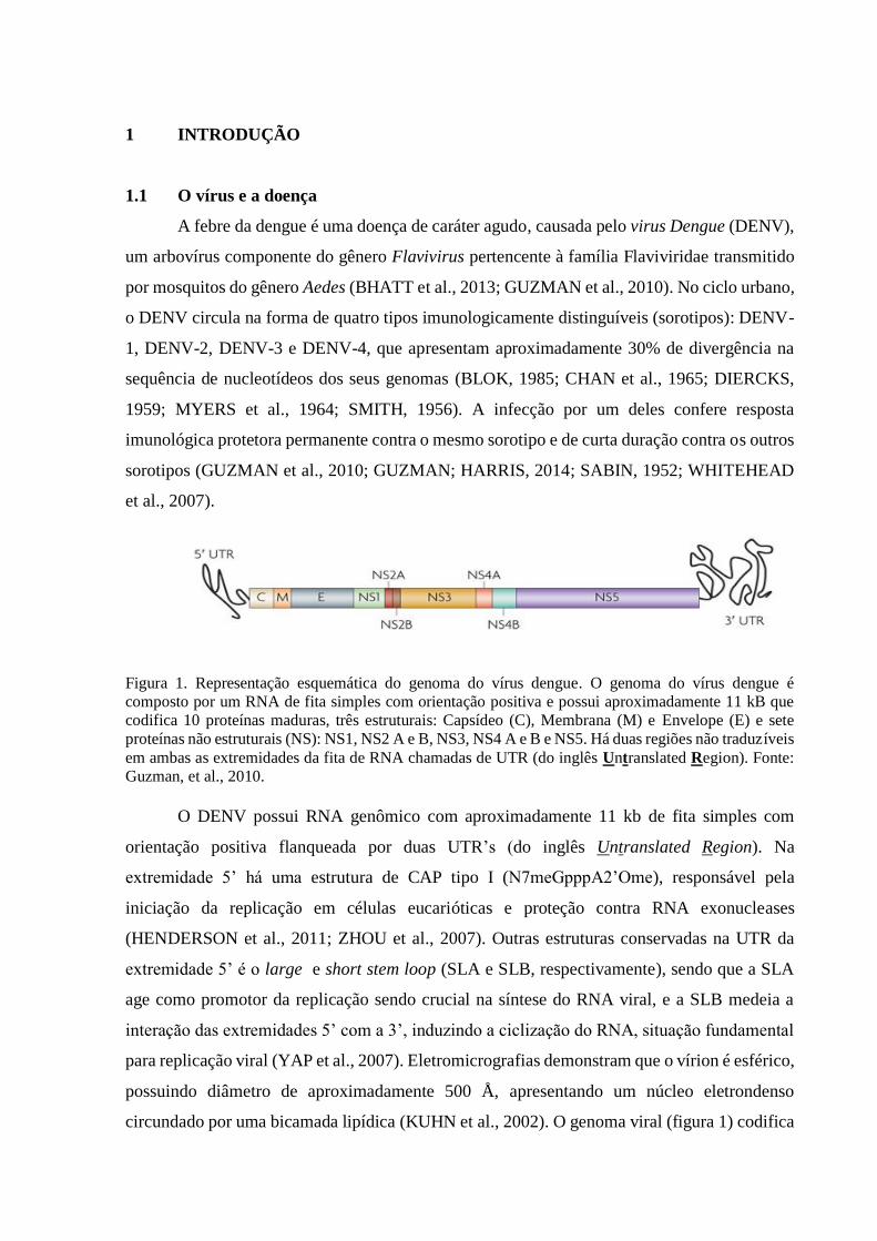
Figura 1. Representação esquemática do genoma do vírus dengue. O genoma do vírus dengue é
composto por um RNA de fita simples com orientação positiva e possui aproximadamente 11 kB que
codifica 10 proteínas maduras, três estruturais: Capsídeo (C), Membrana (M) e Envelope (E) e sete
proteínas não estruturais (NS): NS1, NS2 A e B, NS3, NS4 A e B e NS5. Há duas regiões não traduzíveis
em ambas as extremidades da fita de RNA chamadas de UTR (do inglês Untranslated Region). Fonte:
Guzman, et al., 2010.
O DENV possui RNA genômico com aproximadamente 11 kb de fita simples com
orientação positiva flanqueada por duas UTR’s (do inglês Untranslated Region). Na
extremidade 5’ há uma estrutura de CAP tipo I (N7meGpppA2’Ome), responsável pela
iniciação da replicação em células eucarióticas e proteção contra RNA exonucleases
(HENDERSON et al., 2011; ZHOU et al., 2007). Outras estruturas conservadas na UTR da
extremidade 5’ é o large e short stem loop (SLA e SLB, respectivamente), sendo que a SLA
age como promotor da replicação sendo crucial na síntese do RNA viral, e a SLB medeia a
interação das extremidades 5’ com a 3’, induzindo a ciclização do RNA, situação fundamental
para replicação viral (YAP et al., 2007). Eletromicrografias demonstram que o vírion é esférico,
possuindo diâmetro de aproximadamente 500 Å, apresentando um núcleo eletrondenso
circundado por uma bicamada lipídica (KUHN et al., 2002). O genoma viral (figura 1) codifica
três proteínas estruturais: do capsídeo (C), do envelope (E), e a proteína precursora de
membrana (prM), a qual sofre clivagem e origina a proteína de membrana (M); e sete proteínas
não-estruturais que são: NS1, NS2a, NS2b, NS3, NS4a, NS4b e NS5 (RODENHUIS-ZYBERT
et al., 2010). Quando o mosquito transfere o vírus para o homem, ocorrem as etapas do processo
infeccioso e replicação viral (figura 2). Durante a infecção natural, as principais células-alvo de
infecção pelo vírus dengue são monócitos, macrófagos e células dendríticas (HALSTEAD et
al., 1980).
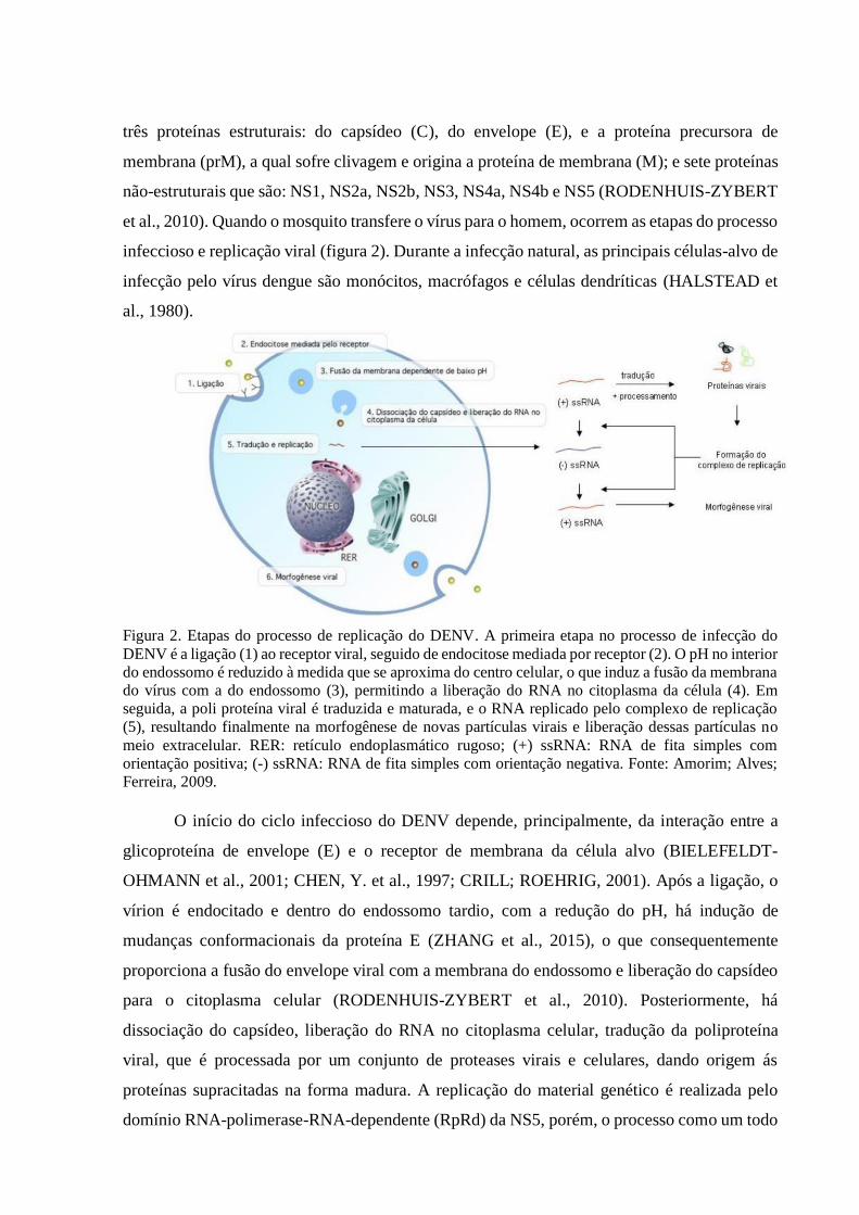
Figura 2. Etapas do processo de replicação do DENV. A primeira etapa no processo de infecção do
DENV é a ligação (1) ao receptor viral, seguido de endocitose mediada por receptor (2). O pH no interior
do endossomo é reduzido à medida que se aproxima do centro celular, o que induz a fusão da membrana
do vírus com a do endossomo (3), permitindo a liberação do RNA no citoplasma da célula (4). Em
seguida, a poli proteína viral é traduzida e maturada, e o RNA replicado pelo complexo de replicação
(5), resultando finalmente na morfogênese de novas partículas virais e liberação dessas partículas no
meio extracelular. RER: retículo endoplasmático rugoso; (+) ssRNA: RNA de fita simples com
orientação positiva; (-) ssRNA: RNA de fita simples com orientação negativa. Fonte: Amorim; Alves;
Ferreira, 2009.
O início do ciclo infeccioso do DENV depende, principalmente, da interação entre a
glicoproteína de envelope (E) e o receptor de membrana da célula alvo (BIELEFELDT-
OHMANN et al., 2001; CHEN, Y. et al., 1997; CRILL; ROEHRIG, 2001). Após a ligação, o
vírion é endocitado e dentro do endossomo tardio, com a redução do pH, há indução de
mudanças conformacionais da proteína E (ZHANG et al., 2015), o que consequentemente
proporciona a fusão do envelope viral com a membrana do endossomo e liberação do capsídeo
para o citoplasma celular (RODENHUIS-ZYBERT et al., 2010). Posteriormente, há
dissociação do capsídeo, liberação do RNA no citoplasma celular, tradução da poliproteína
viral, que é processada por um conjunto de proteases virais e celulares, dando origem ás
proteínas supracitadas na forma madura. A replicação do material genético é realizada pelo
domínio RNA-polimerase-RNA-dependente (RpRd) da NS5, porém, o processo como um todo
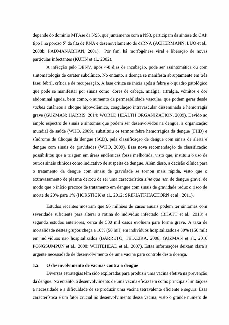
depende do domínio MTAse da NS5, que juntamente com a NS3, participam da síntese do CAP
tipo I na porção 5’ da fita de RNA e desenovelamento do dsRNA (ACKERMANN; LUO et al.,
2008b; PADMANABHAN, 2001). Por fim, há morfogênese viral e liberação de novas
partículas infectantes (KUHN et al., 2002).
A infecção pelo DENV, após 4-8 dias de incubação, pode ser assintomática ou com
sintomatologia de caráter subclínico. No entanto, a doença se manifesta abruptamente em três
fase: febril, crítica e de recuperação. A fase crítica se inicia após a febre e o quadro patológico
que pode se manifestar por sinais como: dores de cabeça, mialgia, artralgia, vômitos e dor
abdominal aguda, bem como, o aumento da permeabilidade vascular, que podem gerar desde
raches cutâneos a choque hipovolêmico, coagulação intravascular disseminada e hemorragia
grave (GUZMAN; HARRIS, 2014; WORLD HEALTH ORGANIZATION, 2009). Devido ao
amplo espectro de sinais e sintomas que podem ser desenvolvidos na dengue, a organização
mundial de saúde (WHO, 2009), substituiu os termos febre hemorrágica da dengue (FHD) e
síndrome de Choque da dengue (SCD), pela classificação de dengue com sinais de alerta e
dengue com sinais de gravidades (WHO, 2009). Essa nova recomendação de classificação
possibilitou que a triagem em áreas endêmicas fosse melhorada, visto que, instituiu o uso de
outros sinais clínicos como indicativo de suspeita de dengue. Além disso, a decisão clínica para
o tratamento da dengue com sinais de gravidade se tornou mais rápida, visto que o
extravasamento de plasma deixou de ser uma característica sine qua non de dengue grave, de
modo que o início precoce de tratamento em dengue com sinais de gravidade reduz o risco de
morte de 20% para 1% (HORSTICK et al., 2012; SRIKIATKHACHORN et al., 2011).
Estudos recentes mostram que 96 milhões de casos anuais podem ter sintomas com
severidade suficiente para alterar a rotina do indivíduo infectado (BHATT et al., 2013) e
segundo estudos anteriores, cerca de 500 mil casos evoluem para forma grave. A taxa de
mortalidade nestes grupos chega a 10% (50 mil) em indivíduos hospitalizados e 30% (150 mil)
em indivíduos não hospitalizados (BARRETO; TEIXEIRA, 2008; GUZMAN et al., 2010
PONGSUMPUN et al., 2008; WHITEHEAD et al., 2007). Estas informações deixam clara a
urgente necessidade de desenvolvimento de uma vacina para controle desta doença.
1.2 O desenvolvimento de vacinas contra a dengue
Diversas estratégias têm sido exploradas para produzir uma vacina efetiva na prevenção
da dengue. No entanto, o desenvolvimento de uma vacina eficaz tem como principais limitações
a necessidade e a dificuldade de se produzir uma vacina tetravalente eficiente e segura. Essa
característica é um fator crucial no desenvolvimento dessa vacina, visto o grande número de
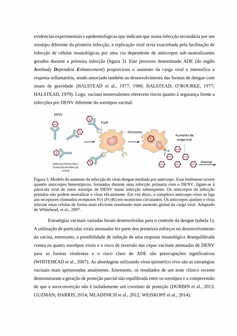
evidencias experimentais e epidemiológicas que indicam que numa infecção secundária por um
sorotipo diferente da primeira infecção, a replicação viral seria exacerbada pela facilitação de
infecção de células imunológicas por uma via dependente de anticorpos sub-neutralizantes
gerados durante a primeira infecção (figura 3). Este processo denominado ADE (do inglês
Antibody Dependent Enhancement) proporciona o aumento da carga viral e intensifica a
resposta inflamatória, sendo associado também ao desenvolvimento das formas de dengue com
sinais de gravidade (HALSTEAD et al., 1977, 1980; HALSTEAD; O’ROURKE, 1977;
HALSTEAD, 1979). Logo, vacinas monovalentes oferecem riscos quanto à segurança frente a
infecções por DENV diferente do sorotipos vacinal.
Figura 3. Modelo do aumento da infecção do vírus dengue mediada por anticorpo. Esse fenômeno ocorre
quando anticorpos heterotípicos, formados durante uma infecção primaria com o DENV, ligam-se à
partícula viral de outro sorotipo de DENV numa infecção subsequente. Os anticorpos da infecção
primária não podem neutralizar o vírus eficazmente. Em vez disso, o complexo anticorpo-vírus se liga
aos receptores chamados receptores Fcγ (FcγR) em monócitos circulantes. Os anticorpos ajudam o vírus
infectar estas células de forma mais eficiente resultando num aumento global da carga viral. Adaptado
de Whitehead, et al., 2007.
Estratégias vacinais variadas foram desenvolvidas para o controle da dengue (tabela 1).
A utilização de partículas virais atenuadas fez parte dos primeiros esforços no desenvolvimento
da vacina, entretanto, a possibilidade de indução de uma resposta imunológica desequilibrada
contra os quatro sorotipos virais e o risco de reversão das cepas vacinais atenuadas de DENV
para as formas virulentas e o risco claro de ADE são preocupações significativas
(WHITEHEAD et al., 2007). As abordagens utilizando vírus quimérico vivo são as estratégias
vacinais mais aprimoradas atualmente. Entretanto, os resultados de um teste clínico recente
demonstraram a geração de proteção parcial não equilibrada entre os sorotipos e a compreensão
de que a soroconverção não é isoladamente um correlato de proteção (DURBIN et al., 2013;
GUZMAN; HARRIS, 2014; MLADINICH et al., 2012; WEISKOPF et al., 2014).

Tabela 1. Diferentes tipos de candidatos vacinais contra a dengue e seus desenvolvedores.
Diversos grupos empenham-se no desenvolvimento de abordagens baseadas nas vacinas
de DNA (COSTA; FREIRE; ALVES, 2006; COSTA et al., 2007, 2011; WU et al., 2003a),
porém, os riscos de geração de autoimunidade bem como a geração de tolerância ao antígeno
continuam sendo os principais riscos associados a esse tipo de abordagem (KLINMAN et al.,
2000). Já as vacinas de subunidade, embora, por definição, necessitem de adjuvantes para
desencadear efeito protetor, induzem respostas imunológicas consideráveis e fornecem
flexibilidade quanto às vias de administração (AMORIM et al., 2012; PANG, 2003). Portanto,
as vacinas de subunidade, como as usadas neste trabalho, são mais seguras e de mais fácil
aprovação quando comparadas às vacinas de DNA e com o vírus.
Tipo de Candidato Vacinal Referências
1.Virus vivo atenuado
1.1 Vírus atenuado por cultura de células (BHAMARAPRAVATI; YOKSAN,
1989; VAUGHN et al., 1996;
KANESA-THASAN et al., 2003;
SANCHEZ et al., 2006)
1.2 Vírus atenuado por mutagênese (MEN et al., 1996; DURBIN et al.,
2001; TROYER et al., 2001;
WHITEHEAD; FALGOUT; et al.,
2003)
2. Vírus quimérico
2.1 Vírus quimérico com Vírus da febre amarela (CAPEDING et al., 2011; GUY;
SAVILLE; LANG, 2010; GUY et
al., 2011; MORRISON et al., 2010)
2.2 Vírus dengue inter-sorotipo (BLANEY et al., 2004, 2008;BRAY;
LAI, 1991; OSORIO et al., 2011;
WHITEHEAD et al., 2003b)
3. Vírus inteiro inativado e purificado (PUTNAK et al., 1996; SIMMONS
et al., 2010)
4. Vacinas de subunidade (CHIANG et al., 2011 CLEMENTS
et al., 2010; GUZMÁN et al., 2003;
KELLY et al., 2000)
5. Vacinas de DNA (APT et al., 2006; BECKETT et al.,
2011; DANKO; BECKETT;
PORTER, 2011; RAVIPRAKASH et
al., 2006)
6. Vetores virais expressando antígenos da dengue (BRANDLER et al., 2007;
HOLMAN et al., 2007; RAJA et al.,
2007; RAVIPRAKASH et al., 2008;
WHITE et al., 2007;)
7. VLP (ARORA et al., 2013; MANI et al.,
2013; TANG et al., 2012;)

As proteínas não estruturais são interessantes como antígenos alvos para uma vacina,
visto o alto grau de conservação entre os sorotipos, a grande imunogenicidade das mesmas, e o
risco nulo de indução de ADE, visto que essas proteínas não compões o vírion. As principais
proteínas não estruturais do DENV são: NS1, NS3 e NS5. A NS1 é uma glicoproteína de 46-
55 kDa, que pode ser associada à membrana citoplasmática, ou ainda, ser secretada para o meio
extracelular e apesar de sua função biológica não ter sido totalmente elucidada, sabe-se que é
altamente imunogênica (AVIRUTNAN et al., 2007a, 2011b; GAO et al., 2008). Dentro dos 26
anos desde o primeiro relato da NS1 em uma formulação vacinal, diversos trabalhos
demonstram a capacidade dessa proteína em gerar proteção em modelo murino (AMORIM et
al., 2012a; FALGOUT et al., 1990; HENRIQUES et al., 2013; WU et al., 2003b; ZHANG et
al., 1988). É válido ressaltar que há algumas evidencias in vitro e in vivo que em condições
altamente inflamatórias, a NS1 pode estar envolvida em fenômenos de autoimunidade, o que
torna questionável o uso dessa proteína em formulações vacinais (AMORIM et al., 2014;
AVIRUTNAN et al., 2007b, 2011a; MLADINICH et al., 2012).
A NS3 tem massa molecular de aproximadamente 70 kDa apresentando um domínio
serino-protease (~18kDa) na região N-terminal e helicase (~ 52 kDa) na região C-terminal
(LESCAR et al., 2008). A sua função biológica é vital para o ciclo viral, visto que há atuação
tanto na clivagem da poli proteína resultante da tradução do RNA viral, bem como na função
helicase que em conjunto com a NS5 promovem a replicação viral, sendo portanto, primordiais
na biologia do patógeno (LUO; XU; HUNKE; et al., 2008). A NS3H se estende entre os
aminoácidos 169-618, apresenta um sítio ATPase e por ser solúvel (diferente do domínio
protease da NS3 e da proteína inteira) pode ser mais facilmente obtida (COSTA et al., 2011).
Um fato relevante é que a NS3H conserva epítopos capazes de gerar uma resposta celular
citotóxica (SPAULDING et al., 1999a) como foi visto no uso de linhagens vacinais de
Salmonella Typhimurium como vetor para plasmídeos que continham um epítopo CTL
específico presente na NS3H que se estendia entre os aminoácidos 298 e 306.
A NS5 do DENV é uma proteína de aproximadamente 109 kDa que apresenta no
mínimo dois domínios: os resíduos de 1 a 368 da região N-terminal que compõem a 2’-O-
metiltransferase, e os resíduos de 405 a 900 que compõem uma RNA polimerase dependente
de RNA (BHATTACHARYA et al., 2009). A região inter-domínios possui duas NLS (do inglês
Nuclear Localization Sequence) bem caracterizadas que direcionam a proteína para o núcleo
(TAY et al., 2013; FRASER et al., 2014a). Ela é processada pela via de proteassoma, quando
então são produzidos e apresentados os epítopos específicos para linfócitos T, os quais são bem
conservados entre os quatro sorotipos virais. De fato, foi visto que formulações vacinais

tetravalentes com vírus dengue atenuado levam à resposta citotóxica direcionada
principalmente a essa proteína (WEISKOPF et al., 2014). A referida proteína não está associada
a partículas virais livres e, provavelmente, não induz a formação de anticorpos anti-DENV
facilitadores de infecção. Há ainda evidências relacionadas ao reconhecimento da NS5 como
importante alvo na geração resposta imune celular em macacos Rhesus infectados com o
DENV-2. Linfócitos capazes de reconhecer especificamente epítopos da NS5 exibiam perfil
polifuncional (MLADINICH et al., 2012). Esse estudo foi pioneiro no uso da NS5 na forma
recombinante como antígeno alvo em formulações vacinais contra a dengue.
1.3 Os adjuvantes vacinais
No geral, um adjuvante pode ser definido como um aditivo ou veículo que melhora a
resposta imune adaptativa ou estimula o sistema imunológico inato de forma a induzir
eficientemente os efeitos imunológicos desejados (SCHIJNS; LAVELLE, 2011). Os adjuvantes
podem ser classificados em 3 tipos baseados no seu modo de ação (BRUNNER et al., 2010):
Os adjuvantes do tipo A são, em sua maioria, derivados de patógenos e atuam por uma via
imunoestimulatória específica nas células apresentadoras de antígenos (APC’s) pela indução de
sinais de perigo. Idealmente, pela escolha do sinal correto de perigo, as APC’s podem ser
programadas a induzirem uma resposta imunológica ajustada ao patógeno ao qual pertence o
antígeno vacinal. Em contraste, os adjuvantes do tipo B promovem aumento da apresentação
de antígenos (TEMMERMAN et al., 2011). Um clássico adjuvante desse tipo é o hidróxido de
alumínio. Esse sal mineral é amplamente empregado em pesquisas e o único adjuvante viável
para vacinas administradas em seres humanos no continente americano (AGUILAR;
RODRÍGUEZ, 2007). Ainda dentro dessa classificação se têm: emulsões de óleo-em-água
(o/w) e água-em-óleo (w/o), lipossomos, nanopartículas e micropartículas. Como é sabido,
esses adjuvantes promovem o prolongamento da apresentação do antígeno via formação de
depósito, proteção contra degradação, facilitação de entrega antigência direta nos linfonodos,
aumento da captação por APCs e indução de apresentação cruzada. É importante ressaltar que
a ausência de um agente imunoestimulatório pode resultar no favorecimento da tolerância
imunológica ao antígeno (BRUNNER et al., 2010). Em contraste, os adjuvantes do tipo C
promovem um sinal de perigo, porém sem a necessidade ativação das APCs, como exemplo se
pode citar o Interferon do tipo 1 e o fator de necrose tumoral na sua forma solúvel em
combinação a antígenos (BON, LE et al., 2001; KAYAMURO et al., 2009).
O RNA dupla-fita (dsRNA) é um sinal de perigo associado à infecção viral. Diversos
dsRNAs sintéticos, como poli (I:C12U), ou ácido polinosinico-policitidilico, comumente
denominado como poli (I:C), eficientemente mimetiza o dsRNA viral. Isso faz desse composto
um potencial candidato a adjuvante do tipo 2 para vacinação contra infecções virais. Como
sabido, dsRNA são ligantes do receptor do tipo Toll (TLR) 3 que, por sua vez, se localiza na
membrana do compartimento endossomal da maioria das APCs (DESMET; ISHII, 2012;
TAKEUCHI; AKIRA, 2010).
A combinação de adjuvantes do tipo A (imunoestimulador) com B (sistema de entrega)
se mostra como uma estratégia promissora na proteção de ambos, antígeno e do
imunoestimulador, contra degradação. Inclusive, já foi sugerido que a apresentação de um
antígeno fagocitado é mais eficiente quando co-formulado com um imunoestimulador
(BLANDER; MEDZHITOV, 2006; SCHLOSSER et al., 2008). Algumas formulações já foram
desenvolvidas para a entrega do antígeno e poli (I:C). Como demonstrado em modelo murinho,
o Montanide ISA 720, cria uma emulsão w/o, com o poli (I:C) capaz de desencadear uma
resposta Th1 potente e persistente, com a proliferação de linfócitos T CD8+ antígenos-
específicos (JIN et al., 2007). O que contrasta com formulações contendo apenas o M720 como
adjuvante, onde houve uma notável polarização Th2 (JIN et al., 2007). O poli (I:C) formulado
em emulsão com o Montanide ISA 720 promoveu a indução da polarização Th1 mais
eficientemente que o poli (I:C) apenas, fenômeno atribuído à rápida degradação do poli (I:C)
livre por ribonucleases (HAFNER et al., 2013).

CONCLUSÕES
Os objetivos almejados para desenvolvimento da presente dissertação foram alcançados
e as conclusões obtidas foram:
A forma recombinante da proteína NS5 do DENV-2 foi obtida com alto
rendimento em sistema procarioto a partir da fração solúvel. A proteína preserva
epitopos conformacionais presentes na sua forma nativa.
A proteína NS5 recombinante aqui obtida é imunogênica quando
administrada pela via subcutânea, induzindo a geração de IgG antígeno-especifica,
produção de TNF-α e IFN-γ por células imunológicas e aumento de sobrevida em
modelo de desafio com DENV-2 cepa JHA1.
A imunogenicidade da NS5 foi potencializada pela adição de adjuvantes
em um protocolo vacinal, desencadeando uma resposta humoral sistêmica com perfis
de modulação adequado para cada formulação, entretanto nenhum dos adjuvantes
induziu sobrevida maior que a induzida pela proteína sozinha.
A formulação M720, que continha além da proteína os adjuvantes poli
(I:C) emulsificado em Montanide 720, foi capaz de ativar linfócitos T CD8+ específicos,
fato que não foi correlacionado com aumento na proteção ao desafio intracraniano com
a linhagem JHA1 de DENV2.

REFERÊNCIAS
ACKERMANN, M.; PADMANABHAN, R. De novo synthesis of RNA by the
dengue virus RNA-dependent RNA polymerase exhibits temperature dependence at
the initiation but not elongation phase. The Journal of biological chemistry, v. 276,
n. 43, p. 39926–39937, 2001.
AGUILAR, J. C.; RODRÍGUEZ, E. G. Vaccine adjuvants revisited. Vaccine, v. 25,
n. 19, p. 3752–3762, 2007.
AMORIM, J. H.; ALVES, R. P. D. S.; BOSCARDIN, S. B.; FERREIRA, L. C. D. S.
The dengue virus non-structural 1 protein: risks and benefits. Virus research, v. 181,
p. 53–60, 2014.
AMORIM, J. H.; DINIZ, M. O.; CARIRI, F. A M. O.; et al. Protective immunity to
DENV2 after immunization with a recombinant NS1 protein using a genetically
detoxified heat-labile toxin as an adjuvant. Vaccine, v. 30, n. 5, p. 837–845, 2012.
AMORIM, J. H.; PEREIRA BIZERRA, R. S.; SANTOS ALVES, R. P. DOS; et al.
A genetic and pathologic study of a DENV2 clinical isolate capable of inducing
encephalitis and hematological disturbances in immunocompetent mice. PloS one, v.
7, n. 9, p. e44984, 2012.
APT, D.; RAVIPRAKASH, K.; BRINKMAN, A.; et al. Tetravalent neutralizing
antibody response against four dengue serotypes by a single chimeric dengue
envelope antigen. Vaccine, v. 24, n. 3, p. 335–344, 2006.
ARORA, U.; TYAGI, P.; SWAMINATHAN, S.; KHANNA, N. Virus-like particles
displaying envelope domain III of dengue virus type 2 induce virus-specific antibody
response in mice. Vaccine, v. 31, n. 6, p. 873–878, 2013.
AVIRUTNAN, P.; HAUHART, R. E.; SOMNUKE, P.; et al. Binding of flavivirus
nonstructural protein NS1 to C4b binding protein modulates complement activation.
Journal of immunology, v. 187, n. 1, p. 424–433, 2011a.
AVIRUTNAN, P.; HAUHART, R. E.; SOMNUKE, P.; et al. Binding of flavivirus
nonstructural protein NS1 to C4b binding protein modulates complement activation.
Journal of immunology, v. 187, n. 1, p. 424–433, 2011b.
AVIRUTNAN, P.; ZHANG, L.; PUNYADEE, N.; et al. Secreted NS1 of dengue
virus attaches to the surface of cells via interactions with heparan sulfate and
chondroitin sulfate E. PLoS pathogens, v. 3, n. 11, p. e183, 2007a.
De acordo com:
ASSOCIAÇÂO BRASILEIRA DE NORMAS TÉCNICAS. NBR 6023: informação e documentação: referências: elaboração. Rio de Janeiro, 2002.

AVIRUTNAN, P.; ZHANG, L.; PUNYADEE, N.; et al. Secreted NS1 of dengue
virus attaches to the surface of cells via interactions with heparan sulfate and
chondroitin sulfate E. PLoS pathogens, v. 3, n. 11, p. e183, 2007b.
BARRETO, M. L.; TEIXEIRA, M. G. Dengue no Brasil: situação epidemiológica e
contribuições para uma agenda de pesquisa. Estudos Avançados, v. 22, n. 64, p. 53–
72, 2008.
BECKETT, C. G.; TJADEN, J.; BURGESS, T.; et al. Evaluation of a prototype
dengue-1 DNA vaccine in a Phase 1 clinical trial. Vaccine, v. 29, n. 5, p. 960–968,
2011.
BHAMARAPRAVATI, N.; YOKSAN, S. Study of bivalent dengue vaccine in
volunteers. Lancet, v. 1, n. 8646, p. 1077-1081, 1989.
BHATT, S.; GETHING, P. W.; BRADY, O. J.; et al. The global distribution and
burden of dengue. Nature, v. 496, n. 7446, p. 504–507, 2013..
BHATTACHARYA, D.; MAYURI; BEST, S. M.; et al. Protein kinase G
phosphorylates mosquito-borne flavivirus NS5. Journal of virology, v. 83, n. 18, p.
9195–9205, 2009.
BIELEFELDT-OHMANN, H.; MEYER, M.; FITZPATRICK, D. R.; MACKENZIE,
J. S. Dengue virus binding to human leukocyte cell lines: receptor usage differs
between cell types and virus strains. Virus research, v. 73, n. 1, p. 81–89, 2001.
BIEN, C. G.; BAUER, J.; DECKWERTH, T. L.; et al. Destruction of neurons by
cytotoxic T cells: a new pathogenic mechanism in Rasmussen’s encephalitis. Annals
of neurology, v. 51, n. 3, p. 311–318, 2002.
BINDER, G. K.; GRIFFIN, D. E. Immune-mediated clearance of virus from the
central nervous system. Microbes and infection / Institut Pasteur, v. 5, n. 5, p. 439–
448, 2003.
BLANDER, J. M.; MEDZHITOV, R. Toll-dependent selection of microbial antigens
for presentation by dendritic cells. Nature, v. 440, n. 7085, p. 808–812, 2006.
BLANEY, J. E.; HANSON, C. T.; FIRESTONE, C.-Y.; et al. Genetically modified,
live attenuated dengue virus type 3 vaccine candidates. The American journal of
tropical medicine and hygiene, v. 71, n. 6, p. 811–821, 2004.
BLANEY, J. E.; SATHE, N. S.; GODDARD, L.; et al. Dengue virus type 3 vaccine
candidates generated by introduction of deletions in the 3’ untranslated region (3'-
UTR) or by exchange of the DENV-3 3'-UTR with that of DENV-4. Vaccine, v. 26,
n. 6, p. 817–828, 2008.
BLOK, J. Genetic relationships of the dengue virus serotypes. The Journal of
general virology, v. 66 ( Pt 6), p. 1323–1325, 1985.
BON, A. LE; SCHIAVONI, G.; D’AGOSTINO, G.; et al. Type i interferons potently
enhance humoral immunity and can promote isotype switching by stimulating
dendritic cells in vivo. Immunity, v. 14, n. 4, p. 461–470, 2001.
BRANDLER, S.; LUCAS-HOURANI, M.; MORIS, A.; et al. Pediatric measles
vaccine expressing a dengue antigen induces durable serotype-specific neutralizing
antibodies to dengue virus. PLoS neglected tropical diseases, v. 1, n. 3, p. e96, 2007.
BRAY, M.; LAI, C. J. Construction of intertypic chimeric dengue viruses by
substitution of structural protein genes. Proceedings of the National Academy of
Sciences of the United States of America, v. 88, n. 22, p. 10342–10346, 1991.
BRIEN, J. D.; UHRLAUB, J. L.; NIKOLICH-ZUGICH, J. West Nile virus-specific
CD4 T cells exhibit direct antiviral cytokine secretion and cytotoxicity and are
sufficient for antiviral protection. Journal of immunology, v. 181, n. 12, p. 8568–
8575, 2008.
BRUNNER, R.; JENSEN-JAROLIM, E.; PALI-SCHÖLL, I. The ABC of clinical and
experimental adjuvants--a brief overview. Immunology letters, v. 128, n. 1, p. 29–
35, 2010.
CAPEDING, R. Z.; LUNA, I. A.; BOMASANG, E.; et al. Live-attenuated, tetravalent
dengue vaccine in children, adolescents and adults in a dengue endemic country:
randomized controlled phase I trial in the Philippines. Vaccine, v. 29, n. 22, p. 3863–
3872, 2011.
CHAKRABORTY, S.; NAZMI, A.; DUTTA, K.; BASU, A. Neurons under viral
attack: victims or warriors? Neurochemistry international, v. 56, n. 6-7, p. 727–735,
2010.
CHAN, Y. C.; KANAPATHIPILLAI, K.; CHEW, K. S. Isolation of two strains of
dengue virus type 3 in Singapore. Singapore medical journal, v. 5, n. 3, p. 127–132,
1965.
CHEN, C.; KUO, M.; CHIEN, L.; et al. RNA-protein interactions: involvement of
NS3, NS5, and 3’ noncoding regions of Japanese encephalitis virus genomic RNA.
Journal of Virology, v. 71, n. 5, p. 3466–3473, 1997.
CHEN, Y.; MAGUIRE, T.; HILEMAN, R. E.; et al. Dengue virus infectivity depends
on envelope protein binding to target cell heparan sulfate. Nature medicine, v. 3, n.
8, p. 866–871, 1997.
CHEVALIER, G.; SUBERBIELLE, E.; MONNET, C.; et al. Neurons are MHC class
I-dependent targets for CD8 T cells upon neurotropic viral infection. PLoS
pathogens, v. 7, n. 11, p. e1002393, 2011.
CHIANG, C.-Y.; LIU, S.-J.; TSAI, J.-P.; et al. A novel single-dose dengue subunit
vaccine induces memory immune responses. PloS one, v. 6, n. 8, p. e23319, 2011.
CLAASSEN, M. A. A.; JANSSEN, H. L. A.; BOONSTRA, A. Role of T cell
immunity in hepatitis C virus infections. Current opinion in virology, v. 3, n. 4, p.
461–467, 2013.
CLEMENTS, D. E.; COLLER, B.-A. G.; LIEBERMAN, M. M.; et al. Development
of a recombinant tetravalent dengue virus vaccine: immunogenicity and efficacy
studies in mice and monkeys. Vaccine, v. 28, n. 15, p. 2705–2715, 2010.
COSTA, S. M.; AZEVEDO, A. S.; PAES, M. V; et al. DNA vaccines against dengue
virus based on the ns1 gene: the influence of different signal sequences on the protein
expression and its correlation to the immune response elicited in mice. Virology, v.
358, n. 2, p. 413–423, 2007.
COSTA, S. M.; FREIRE, M. S.; ALVES, A. M. B. DNA vaccine against the non-
structural 1 protein (NS1) of dengue 2 virus. Vaccine, v. 24, n. 21, p. 4562–4564,
2006.
COSTA, S. M.; YORIO, A. P.; GONÇALVES, A. J. S.; et al. Induction of a protective
response in mice by the dengue virus NS3 protein using DNA vaccines. PloS one, v.
6, n. 10, p. e25685, 2011.
COUTARD, B.; DECROLY, E.; LI, C.; et al. Assessment of Dengue virus helicase
and methyltransferase as targets for fragment-based drug discovery. Antiviral
research, v. 106, p. 61–70, 2014.
CRILL, W. D.; ROEHRIG, J. T. Monoclonal antibodies that bind to domain III of
dengue virus E glycoprotein are the most efficient blockers of virus adsorption to
Vero cells. Journal of virology, v. 75, n. 16, p. 7769–7773, 2001.
DANKO, J. R.; BECKETT, C. G.; PORTER, K. R. Development of dengue DNA
vaccines. Vaccine, v. 29, n. 42, p. 7261–7266, 2011.
DESMET, C. J.; ISHII, K. J. Nucleic acid sensing at the interface between innate and
adaptive immunity in vaccination. Nature reviews. Immunology, v. 12, n. 7, p. 479–
491, 2012.
DIERCKS, F. H. Isolation of a type 2 dengue virus by use of hamster kidney cell
cultures. The American journal of tropical medicine and hygiene, v. 8, n. 4, p.
488–491, 1959.
DINTZIS, H. M.; DINTZIS, R. Z.; VOGELSTEIN, B. Molecular determinants of
immunogenicity: the immunon model of immune response. Proceedings of the
National Academy of Sciences of the United States of America, v. 73, n. 10, p.
3671–3675, 1976.
DURBIN, A. P.; KARRON, R. A.; SUN, W.; et al. Attenuation and immunogenicity
in humans of a live dengue virus type-4 vaccine candidate with a 30 nucleotide
deletion in its 3’-untranslated region. The American journal of tropical medicine
and hygiene, v. 65, n. 5, p. 405–413, 2001.

DURBIN, A. P.; KIRKPATRICK, B. D.; PIERCE, K. K.; et al. A single dose of any
of four different live attenuated tetravalent dengue vaccines is safe and immunogenic
in flavivirus-naive adults: a randomized, double-blind clinical trial. The Journal of
infectious diseases, v. 207, n. 6, p. 957–965, 2013.
ELGERT, K. D. Immunology: Understanding The Immune System. Hoboken:
Wiley-Blackwel, 2009, 726 p.
FALGOUT, B.; BRAY, M.; SCHLESINGER, J. J.; LAI, C. J. Immunization of mice
with recombinant vaccinia virus expressing authentic dengue virus nonstructural
protein NS1 protects against lethal dengue virus encephalitis. Journal of virology, v.
64, n. 9, p. 4356–4363, 1990.
FIELD, R.; CAMPION, S.; WARREN, C.; MURRAY, C.; CUNNINGHAM, C.
Systemic challenge with the TLR3 agonist poly I:C induces amplified IFNalpha/beta
and IL-1beta responses in the diseased brain and exacerbates chronic
neurodegeneration. Brain, behavior, and immunity, v. 24, n. 6, p. 996–1007, 2010.
FRASER, J. E.; RAWLINSON, S. M.; WANG, C.; JANS, D. A.; WAGSTAFF, K.
M. Investigating dengue virus nonstructural protein 5 (NS5) nuclear import. Methods
in molecular biology, v. 1138, p. 301–328, 2014a.
FRASER, J. E.; RAWLINSON, S. M.; WANG, C.; JANS, D. A.; WAGSTAFF, K.
M. Investigating dengue virus nonstructural protein 5 (NS5) nuclear import. Methods
in molecular biology, v. 1138, p. 301–328, 2014b.
GALEA, I.; BECHMANN, I.; PERRY, V. H. What is immune privilege (not)?
Trends in immunology, v. 28, n. 1, p. 12–18, 2007.
GAO, G.; WANG, Q.; DAI, Z.; et al. Adenovirus-based vaccines generate cytotoxic
T lymphocytes to epitopes of NS1 from dengue virus that are present in all major
serotypes. Human gene therapy, v. 19, n. 9, p. 927–936, 2008.
GUPTA, R. K.; SIBER, G. R. Adjuvants for human vaccines: current status, problems
and future prospects. Vaccine, v. 13, n. 14, p. 1263–1276, 1995.
GUY, B.; BARRERE, B.; MALINOWSKI, C.; et al. From research to phase III:
preclinical, industrial and clinical development of the Sanofi Pasteur tetravalent
dengue vaccine. Vaccine, v. 29, n. 42, p. 7229–7241, 2011.
GUY, B.; SAVILLE, M.; LANG, J. Development of Sanofi Pasteur tetravalent
dengue vaccine. Human vaccines, v. 6, n. 9, p. 325-332, 2010.
GUZMAN, M. G.; HALSTEAD, S. B.; ARTSOB, H.; et al. Dengue: a continuing
global threat. Nature reviews. Microbiology, v. 8, n. 12 Suppl, p. S7–16, 2010.
GUZMAN, M. G.; HARRIS, E. Dengue. Lancet, v. 385, n. 9966, p. 453–465, 2015.
GUZMÁN, M. G.; RODRÍGUEZ, R.; RODRÍGUEZ, R.; et al. Induction of
neutralizing antibodies and partial protection from viral challenge in Macaca

fascicularis immunized with recombinant dengue 4 virus envelope glycoprotein
expressed in Pichia pastoris. The American journal of tropical medicine and
hygiene, v. 69, n. 2, p. 129–134, 2003.
HAFNER, A. M.; CORTHÉSY, B.; MERKLE, H. P. Particulate formulations for the
delivery of poly(I:C) as vaccine adjuvant. Advanced drug delivery reviews, v. 65,
n. 10, p. 1386–1399, 2013.
HALSTEAD, S. B. In Vivo Enhancement of Dengue Virus Infection in Rhesus
Monkeys by Passively Transferred Antibody. Journal of Infectious Diseases , v. 140
, n. 4 , p. 527–533, 1979.
HALSTEAD, S. B.; O’ROURKE, E. J. Dengue viruses and mononuclear phagocytes.
I. Infection enhancement by non-neutralizing antibody. The Journal of
experimental medicine, v. 146, n. 1, p. 201–217, 1977.
HALSTEAD, S. B.; O’ROURKE, E. J.; ALLISON, A. C. Dengue viruses and
mononuclear phagocytes. II. Identity of blood and tissue leukocytes supporting in
vitro infection. The Journal of experimental medicine, v. 146, n. 1, p. 218–229,
1977.
HALSTEAD, S. B.; PORTERFIELD, J. S.; O’ROURKE, E. J. Enhancement of
Dengue Virus Infection in Monocytes by Flavivirus Antisera. The American
Journal of Tropical Medicine and Hygiene , v. 29 , n. 4 , p. 638–642, 1980.
HANLEY, K. A.; LEE, J. J.; BLANEY, J. E.; MURPHY, B. R.; WHITEHEAD, S.
S. Paired charge-to-alanine mutagenesis of dengue virus type 4 NS5 generates
mutants with temperature-sensitive, host range, and mouse attenuation phenotypes.
Journal of virology, v. 76, n. 2, p. 525–531, 2002.
HENDERSON, B. R.; SAEEDI, B. J.; CAMPAGNOLA, G.; GEISS, B. J. Analysis
of RNA binding by the dengue virus NS5 RNA capping enzyme. PloS one, v. 6, n.
10, p. e25795, 2011.
HENRIQUES, H. R.; RAMPAZO, E. V; GONÇALVES, A. J. S.; et al. Targeting the
non-structural protein 1 from dengue virus to a dendritic cell population confers
protective immunity to lethal virus challenge. PLoS neglected tropical diseases, v.
7, n. 7, p. e2330, 2013.
HOLMAN, D. H.; WANG, D.; RAVIPRAKASH, K.; et al. Two complex,
adenovirus-based vaccines that together induce immune responses to all four dengue
virus serotypes. Clinical and vaccine immunology, v. 14, n. 2, p. 182–189, 2007.
HORSTICK, O.; FARRAR, J.; LUM, L.; et al. Reviewing the development, evidence
base, and application of the revised dengue case classification. Pathogens and global
health, v. 106, n. 2, p. 94–101, 2012.
JIN, B.; WANG, R. Y.; QIU, Q.; et al. Induction of potent cellular immune response
in mice by hepatitis C virus NS3 protein with double-stranded RNA. Immunology,
v. 122, n. 1, p. 15–27, 2007.

JOLY, E.; MUCKE, L.; OLDSTONE, M. B. Viral persistence in neurons explained
by lack of major histocompatibility class I expression. Science, v. 253, n. 5025, p.
1283–1285, 1991.
KÄGI, D.; LEDERMANN, B.; BÜRKI, K.; ZINKERNAGEL, R. M.;
HENGARTNER, H. Molecular mechanisms of lymphocyte-mediated cytotoxicity
and their role in immunological protection and pathogenesis in vivo. Annual review
of immunology, v. 14, p. 207–232, 1996.
KANESA-THASAN, N.; EDELMAN, R.; TACKET, C. O.; et al. Phase 1 studies of
Walter Reed Army Institute of Research candidate attenuated dengue vaccines:
selection of safe and immunogenic monovalent vaccines. The American journal of
tropical medicine and hygiene, v. 69, n. 6 Suppl, p. 17–23, 2003.
KAYAMURO, H.; ABE, Y.; YOSHIOKA, Y.; et al. The use of a mutant TNF-alpha
as a vaccine adjuvant for the induction of mucosal immune responses. Biomaterials,
v. 30, n. 29, p. 5869–5876, 2009.
KEDZIERSKA, K.; GRUTA, N. L. LA; TURNER, S. J.; DOHERTY, P. C.
Establishment and recall of CD8+ T-cell memory in a model of localized transient
infection. Immunological reviews, v. 211, p. 133–145, 2006.
KELLY, E. P.; GREENE, J. J.; KING, A. D.; INNIS, B. L. Purified dengue 2 virus
envelope glycoprotein aggregates produced by baculovirus are immunogenic in mice.
Vaccine, v. 18, n. 23, p. 2549–2559, 2000.
KNICKELBEIN, J. E.; KHANNA, K. M.; YEE, M. B.; et al. Noncytotoxic lytic
granule-mediated CD8+ T cell inhibition of HSV-1 reactivation from neuronal
latency. Science, v. 322, n. 5899, p. 268–271, 2008.
KUHN, R. J.; ZHANG, W.; ROSSMANN, M. G.; et al. Structure of dengue virus:
implications for flavivirus organization, maturation, and fusion. Cell, v. 108, n. 5, p.
717–725, 2002.
LANCKI, D. W.; HSIEH, C. S.; FITCH, F. W. Mechanisms of lysis by cytotoxic T
lymphocyte clones. Lytic activity and gene expression in cloned antigen-specific
CD4+ and CD8+ T lymphocytes. Journal of immunology, v. 146, n. 9, p. 3242–
3249, 1991.
LANCKI, D. W.; KAPER, B. P.; FITCH, F. W. The requirements for triggering of
lysis by cytolytic T lymphocyte clones. II. Cyclosporin A inhibits TCR-mediated
exocytosis by only selectively inhibits TCR-mediated lytic activity by cloned CTL.
Journal of immunology, v. 142, n. 2, p. 416–424, 1989.
LESCAR, J.; LUO, D.; XU, T.; et al. Towards the design of antiviral inhibitors against
flaviviruses: the case for the multifunctional NS3 protein from Dengue virus as a
target. Antiviral research, v. 80, n. 2, p. 94–101, 2008.
LIU, T.; CHAMBERS, T. J. Yellow fever virus encephalitis: properties of the brain-
associated T-cell response during virus clearance in normal and gamma interferon-
deficient mice and requirement for CD4+ lymphocytes. Journal of virology, v. 75,
n. 5, p. 2107–2118, 2001.
LUNDEGAARD, C.; LAMBERTH, K.; HARNDAHL, M.; et al. NetMHC-3.0:
accurate web accessible predictions of human, mouse and monkey MHC class I
affinities for peptides of length 8-11. Nucleic acids research, v. 36, n. p. 509–12,
2008.
LUO, D.; XU, T.; HUNKE, C.; et al. Crystal structure of the NS3 protease-helicase
from dengue virus. Journal of virology, v. 82, n. 1, p. 173–183, 2008.
LUO, D.; XU, T.; WATSON, R. P.; et al. Insights into RNA unwinding and ATP
hydrolysis by the flavivirus NS3 protein. The EMBO journal, v. 27, n. 23, p. 3209–
3219, 2008.
MALYALA, P.; SINGH, M. Endotoxin limits in formulations for preclinical
research. Journal of pharmaceutical sciences, v. 97, n. 6, p. 2041–2044, 2008.
MANI, S.; TRIPATHI, L.; RAUT, R.; et al. Pichia pastoris-expressed dengue 2
envelope forms virus-like particles without pre-membrane protein and induces high
titer neutralizing antibodies. PloS one, v. 8, n. 5, p. e64595, 2013.
MEDANA, I.; MARTINIC, M. A.; WEKERLE, H.; NEUMANN, H. Transection of
major histocompatibility complex class I-induced neurites by cytotoxic T
lymphocytes. The American journal of pathology, v. 159, n. 3, p. 809–815, 2001.
MEN, R.; BRAY, M.; CLARK, D.; CHANOCK, R. M.; LAI, C. J. Dengue type 4
virus mutants containing deletions in the 3’ noncoding region of the RNA genome:
analysis of growth restriction in cell culture and altered viremia pattern and
immunogenicity in rhesus monkeys. Journal of virology, v. 70, n. 6, p. 3930–3937,
1996.
MLADINICH, K. M.; PIASKOWSKI, S. M.; RUDERSDORF, R.; et al. Dengue
virus-specific CD4+ and CD8+ T lymphocytes target NS1, NS3 and NS5 in infected
Indian rhesus macaques. Immunogenetics, v. 64, n. 2, p. 111–121, 2012.
MORRISON, D.; LEGG, T. J.; BILLINGS, C. W.; et al. A novel tetravalent dengue
vaccine is well tolerated and immunogenic against all 4 serotypes in flavivirus-naive
adults. The Journal of infectious diseases, v. 201, n. 3, p. 370–377, 2010.
MOSEMAN, E. A.; MCGAVERN, D. B. The great balancing act: regulation and fate
of antiviral T-cell interactions. Immunological reviews, v. 255, n. 1, p. 110–124,
2013.
MOSMANN, T. R.; COFFMAN, R. L. TH1 and TH2 cells: different patterns of
lymphokine secretion lead to different functional properties. Annual review of
immunology, v. 7, p. 145–173, 1989.
MOUTAFTSI, M.; PETERS, B.; PASQUETTO, V.; et al. A consensus epitope
prediction approach identifies the breadth of murine T(CD8+)-cell responses to
vaccinia virus. Nature biotechnology, v. 24, n. 7, p. 817–819, 2006.
MURALI-KRISHNA, K.; RAVI, V.; MANJUNATH, R. Protection of adult but not
newborn mice against lethal intracerebral challenge with Japanese encephalitis virus
by adoptively transferred virus-specific cytotoxic T lymphocytes: requirement for
L3T4+ T cells. The Journal of general virology, v. 77 ( Pt 4), p. 705–714, 1996.
MYERS, R. M.; CAREY, D. E.; RODRIGUES, F. M.; KLONTZ, C. E. THE
ISOLATION OF DENGUE TYPE 4 VIRUS FROM HUMAN SERA IN SOUTH
INDIA. The Indian journal of medical research, v. 52, p. 559–565, 1964.
NAGLER-ANDERSON, C.; LICHTENHELD, M.; EISEN, H. N.; PODACK, E. R.
Perforin mRNA in primary peritoneal exudate cytotoxic T lymphocytes. Journal of
immunology, v. 143, n. 11, p. 3440–3443, 1989.
NORDLY, P.; ROSE, F.; CHRISTENSEN, D.; et al. Immunity by formulation
design: induction of high CD8+ T-cell responses by poly(I:C) incorporated into the
CAF01 adjuvant via a double emulsion method. Journal of controlled release, v.
150, n. 3, p. 307–317, 2011.
OSORIO, J. E.; HUANG, C. Y.-H.; KINNEY, R. M.; STINCHCOMB, D. T.
Development of DENVax: a chimeric dengue-2 PDK-53-based tetravalent vaccine
for protection against dengue fever. Vaccine, v. 29, n. 42, p. 7251–7260, 2011.
PONGSUMPUN, P.; GARCIA LOPEZ, D.; FAVIER, C.; et al. Dynamics of dengue
epidemics in urban contexts. Tropical medicine & international health, v. 13, n. 9,
p. 1180–1187, 2008.
PUTNAK, R.; BARVIR, D. A.; BURROUS, J. M.; et al. Development of a purified,
inactivated, dengue-2 virus vaccine prototype in Vero cells: immunogenicity and
protection in mice and rhesus monkeys. The Journal of infectious diseases, v. 174,
n. 6, p. 1176–1184, 1996.
RAJA, N. U.; HOLMAN, D. H.; WANG, D.; et al. Induction of bivalent immune
responses by expression of dengue virus type 1 and type 2 antigens from a single
complex adenoviral vector. The American journal of tropical medicine and
hygiene, v. 76, n. 4, p. 743–751, 2007.
RAVIPRAKASH, K.; APT, D.; BRINKMAN, A.; et al. A chimeric tetravalent
dengue DNA vaccine elicits neutralizing antibody to all four virus serotypes in rhesus
macaques. Virology, v. 353, n. 1, p. 166–173, 2006.
RAVIPRAKASH, K.; WANG, D.; EWING, D.; et al. A tetravalent dengue vaccine
based on a complex adenovirus vector provides significant protection in rhesus
monkeys against all four serotypes of dengue virus. Journal of virology, v. 82, n. 14,
p. 6927–6934, 2008.

REMAKUS, S.; SIGAL, L. J. Memory CD8+ T cell protection. Advances in
experimental medicine and biology, v. 785, p. 77–86, 2013.
RIVINO, L.; KUMARAN, E. A. P.; JOVANOVIC, V.; et al. Differential targeting of
viral components by CD4+ versus CD8+ T lymphocytes in dengue virus infection.
Journal of virology, v. 87, n. 5, p. 2693–2706, 2013.
RIVINO, L.; KUMARAN, E. A.; THEIN, T.-L.; et al. Virus-specific T lymphocytes
home to the skin during natural dengue infection. Science Translational Medicine,
v. 7, n. 278, p. 27835–27842, 2015.
RODENHUIS-ZYBERT, I. A; WILSCHUT, J.; SMIT, J. M. Dengue virus life cycle:
viral and host factors modulating infectivity. Cellular and molecular life sciences,
v. 67, n. 16, p. 2773–2786, 2010.
SABIN, A. B. Research on dengue during World War II. The American journal of
tropical medicine and hygiene, v. 1, n. 1, p. 30–50, 1952.
SALEM, M. L.; DIAZ-MONTERO, C. M.; EL-NAGGAR, S. A.; et al. The TLR3
agonist poly(I:C) targets CD8+ T cells and augments their antigen-specific responses
upon their adoptive transfer into naïve recipient mice. Vaccine, v. 27, n. 4, p. 549–
557, 2009.
SAMBROOK, J.; RUSSELL, D. W. Molecular Cloning: A Laboratory Manual..
Cold Spring Harbor: Cold Spring Harbor Laboratory Press, 2001. v.1, 2344 p.
SANCHEZ, V.; GIMENEZ, S.; TOMLINSON, B.; et al. Innate and adaptive cellular
immunity in flavivirus-naïve human recipients of a live-attenuated dengue serotype 3
vaccine produced in Vero cells (VDV3). Vaccine, v. 24, n. 23, p. 4914–4926, 2006.
SCHIJNS, V. E. J. C.; LAVELLE, E. C. Trends in vaccine adjuvants. Expert review
of vaccines, v. 10, n. 4, p. 539–550, 2011.
SCHLOSSER, E.; MUELLER, M.; FISCHER, S.; et al. TLR ligands and antigen need
to be coencapsulated into the same biodegradable microsphere for the generation of
potent cytotoxic T lymphocyte responses. Vaccine, v. 26, n. 13, p. 1626–1637, 2008.
SCHUCH, A.; HOH, A.; THIMME, R. The role of natural killer cells and CD8(+) T
cells in hepatitis B virus infection. Frontiers in immunology, v. 5, p. 258-264, 2014.
SCHWARZ, H.; SCHMITTNER, M.; DUSCHL, A.; HOREJS-HOECK, J. Residual
endotoxin contaminations in recombinant proteins are sufficient to activate human
CD1c+ dendritic cells. PloS one, v. 9, n. 12, p. e113840, 2014.
SHATZ, C. J. MHC class I: an unexpected role in neuronal plasticity. Neuron, v. 64,
n. 1, p. 40–45, 2009.
SIMMONS, M.; BURGESS, T.; LYNCH, J.; PUTNAK, R. Protection against dengue
virus by non-replicating and live attenuated vaccines used together in a prime boost
vaccination strategy. Virology, v. 396, n. 2, p. 280–288, 2010.
SITATI, E. M.; DIAMOND, M. S. CD4+ T-cell responses are required for clearance
of West Nile virus from the central nervous system. Journal of virology, v. 80, n. 24,
p. 12060–12069, 2006.
SMITH, C. E. G. Isolation of three strains of type 1 dengue virus from a local outbreak
of the disease in Malaya. The Journal of hygiene, v. 54, n. 4, p. 569–580, 1956.
SPAULDING, A. C.; KURANE, I.; ENNIS, F. A.; ROTHMAN, A. L. Analysis of
murine CD8(+) T-cell clones specific for the Dengue virus NS3 protein: flavivirus
cross-reactivity and influence of infecting serotype. Journal of virology, v. 73, n. 1,
p. 398–403, 1999a.
SPAULDING, A. C.; KURANE, I.; ENNIS, F. A.; ROTHMAN, A. L. Analysis of
murine CD8(+) T-cell clones specific for the Dengue virus NS3 protein: flavivirus
cross-reactivity and influence of infecting serotype. Journal of virology, v. 73, n. 1,
p. 398–403, 1999b.
SRIKIATKHACHORN, A.; ROTHMAN, A. L.; GIBBONS, R. V; et al. Dengue--
how best to classify it. Clinical infectious diseases : an official publication of the
Infectious Diseases Society of America, v. 53, n. 6, p. 563–567, 2011.
TAKAHASHI, H.; TAKAHASHI, C.; MORELAND, N. J.; et al. Establishment of a
robust dengue virus NS3-NS5 binding assay for identification of protein-protein
interaction inhibitors. Antiviral research, v. 96, n. 3, p. 305–314, 2012.
TAKEUCHI, O.; AKIRA, S. Pattern recognition receptors and inflammation. Cell, v.
140, n. 6, p. 805–820, 2010.
TAN, B. H.; FU, J.; SUGRUE, R. J.; et al. Recombinant dengue type 1 virus NS5
protein expressed in Escherichia coli exhibits RNA-dependent RNA polymerase
activity. Virology, v. 216, n. 2, p. 317–325, 1996.
TANG, Y.-X.; JIANG, L.-F.; ZHOU, J.-M.; et al. Induction of virus-neutralizing
antibodies and T cell responses by dengue virus type 1 virus-like particles prepared
from Pichia pastoris. Chinese medical journal, v. 125, n. 11, p. 1986–1992, 2012.
TAY, M. Y. F.; FRASER, J. E.; CHAN, W. K. K.; et al. Nuclear localization of
dengue virus (DENV) 1-4 non-structural protein 5; protection against all 4 DENV
serotypes by the inhibitor Ivermectin. Antiviral research, v. 99, n. 3, p. 301–306,
2013.
TAY, M. Y. F.; SAW, W. G.; ZHAO, Y.; et al. The C-terminal 50 Amino Acid
Residues of Dengue NS3 Protein Are Important for NS3-NS5 Interaction and Viral
Replication. The Journal of biological chemistry, v. 290, n. 4, p. 2379–2394, 2015.
TEMMERMAN, M.-L. DE; REJMAN, J.; DEMEESTER, J.; et al. Particulate
vaccines: on the quest for optimal delivery and immune response. Drug discovery
today, v. 16, n. 13-14, p. 569–582, 2011.

TROYER, J. M.; HANLEY, K. A.; WHITEHEAD, S. S.; et al. A live attenuated
recombinant dengue-4 virus vaccine candidate with restricted capacity for
dissemination in mosquitoes and lack of transmission from vaccinees to mosquitoes.
The American journal of tropical medicine and hygiene, v. 65, n. 5, p. 414–419,
2001.
TUNG, J. W.; HEYDARI, K.; TIROUVANZIAM, R.; et al. Modern flow cytometry:
a practical approach. Clinics in laboratory medicine, v. 27, n. 3, p. 453–468, v, 2007.
VAUGHN, D. W.; HOKE, C. H.; YOKSAN, S.; et al. Testing of a dengue 2 live-
attenuated vaccine (strain 16681 PDK 53) in ten American volunteers. Vaccine, v.
14, n. 4, p. 329–336, 1996.
VOULGARIDOU, G.-P.; MANTSO, T.; CHLICHLIA, K.; PANAYIOTIDIS, M. I.;
PAPPA, A. Efficient E. coli expression strategies for production of soluble human
crystallin ALDH3A1. PloS one, v. 8, n. 2, p. e56582, 2013.
WADA, T.; KOHARA, M.; YASUTOMI, Y. DNA vaccine expressing the non-
structural proteins of hepatitis C virus diminishes the expression of HCV proteins in
a mouse model. Vaccine, v. 31, n. 50, p. 5968–5974, 2013.
WAKELIN, S. J.; SABROE, I.; GREGORY, C. D.; et al. “Dirty little secrets”--
endotoxin contamination of recombinant proteins. Immunology letters, v. 106, n. 1,
p. 1–7, 2006.
WANG, P.; SIDNEY, J.; DOW, C.; et al. A systematic assessment of MHC class II
peptide binding predictions and evaluation of a consensus approach. PLoS
computational biology, v. 4, n. 4, p. e1000048, 2008.
WANG, P.; SIDNEY, J.; KIM, Y.; et al. Peptide binding predictions for HLA DR,
DP and DQ molecules. BMC bioinformatics, v. 11, p. 568-572, 2010.
WEISKOPF, D.; ANGELO, M. A.; BANGS, D. J.; et al. The human CD8+ T cell
responses induced by a live attenuated tetravalent dengue vaccine are directed against
highly conserved epitopes. Journal of virology, v. 89, p. 120-128, 2014.
WHITE, L. J.; PARSONS, M. M.; WHITMORE, A. C.; et al. An immunogenic and
protective alphavirus replicon particle-based dengue vaccine overcomes maternal
antibody interference in weanling mice. Journal of virology, v. 81, n. 19, p. 10329–
10339, 2007.
WHITEHEAD, S. S.; BLANEY, J. E.; DURBIN, A. P.; MURPHY, B. R. Prospects
for a dengue virus vaccine. Nature reviews. Microbiology, v. 5, n. 7, p. 518–528,
2007.
WHITEHEAD, S. S.; FALGOUT, B.; HANLEY, K. A.; et al. A live, attenuated
dengue virus type 1 vaccine candidate with a 30-nucleotide deletion in the 3’
untranslated region is highly attenuated and immunogenic in monkeys. Journal of
virology, v. 77, n. 2, p. 1653–1657, 2003.
WHITEHEAD, S. S.; HANLEY, K. A.; BLANEY, J. E.; et al. Substitution of the
structural genes of dengue virus type 4 with those of type 2 results in chimeric vaccine
candidates which are attenuated for mosquitoes, mice, and rhesus monkeys. Vaccine,
v. 21, n. 27-30, p. 4307–4316, 2003.
WHO. Dengue: guidelines for diagnosis, treatment, prevention and control - New
edition. Disponível em:
<http://whqlibdoc.who.int/publications/2009/9789241547871_eng.pdf?ua=1>.
Acesso em: 15/8/2014.
WILLIAMS, N. S.; ENGELHARD, V. H. Identification of a population of CD4+
CTL that utilizes a perforin- rather than a Fas ligand-dependent cytotoxic mechanism.
Journal of immunology, v. 156, n. 1, p. 153–159, 1996.
WOODLAND, D. L. Jump-starting the immune system: Prime-boosting comes of
age. Trends in Immunology, v. 25, n. 2, p. 98–104, 2004.
WORTZMAN, M. E.; CLOUTHIER, D. L.; MCPHERSON, A. J.; LIN, G. H. Y.;
WATTS, T. H. The contextual role of TNFR family members in CD8(+) T-cell
control of viral infections. Immunological reviews, v. 255, n. 1, p. 125–148, 2013.
WRIGHT, S.; RAMOS, R.; TOBIAS, P.; ULEVITCH, R.; MATHISON, J. CD14, a
receptor for complexes of lipopolysaccharide (LPS) and LPS binding protein.
Science, v. 249, n. 4975, p. 1431–1433, 1990.
WU, S.-F.; LIAO, C.-L.; LIN, Y.-L.; et al. Evaluation of protective efficacy and
immune mechanisms of using a non-structural protein NS1 in DNA vaccine against
dengue 2 virus in mice. Vaccine, v. 21, n. 25-26, p. 3919–3929, 2003a.
WU, S.-F.; LIAO, C.-L.; LIN, Y.-L.; et al. Evaluation of protective efficacy and
immune mechanisms of using a non-structural protein NS1 in DNA vaccine against
dengue 2 virus in mice. Vaccine, v. 21, n. 25-26, p. 3919–3929, 2003b.
YAP, T. L.; XU, T.; CHEN, Y.-L.; et al. Crystal structure of the dengue virus RNA-
dependent RNA polymerase catalytic domain at 1.85-angstrom resolution. Journal
of virology, v. 81, n. 9, p. 4753–4765, 2007.
YUN, N. E.; PENG, B.-H.; BERTKE, A. S.; et al. CD4+ T cells provide protection
against acute lethal encephalitis caused by Venezuelan equine encephalitis virus.
Vaccine, v. 27, n. 30, p. 4064–4073, 2009.
ZHANG, X.; SHENG, J.; AUSTIN, S. K.; et al. Structure of acidic pH dengue virus
showing the fusogenic glycoprotein trimers. Journal of virology, v. 89, n. 1, p. 743–
750, 2015.
ZHANG, Y. M.; HAYES, E. P.; MCCARTY, T. C.; et al. Immunization of mice with
dengue structural proteins and nonstructural protein NS1 expressed by baculovirus
recombinant induces resistance to dengue virus encephalitis. Journal of virology, v.
62, n. 8, p. 3027–3031, 1988.
ZHOU, Y.; RAY, D.; ZHAO, Y.; et al. Structure and function of flavivirus NS5
methyltransferase. Journal of virology, v. 81, n. 8, p. 3891–3903, 2007.


