
Different Characteristics and Nucleotide BindingProperties of Inosine Monophosphate Dehydrogenase(IMPDH) IsoformsElaine C. Thomas1*¤a, Jennifer H. Gunter1, Julie A. Webster1,2, Nicole L. Schieber3, Viola Oorschot3¤b,
Robert G. Parton3, Jonathan P. Whitehead1,2*
1 Diamantina Institute for Cancer, Immunology and Metabolic Medicine, Princess Alexandra Hospital, University of Queensland, Brisbane, Queensland, Australia,
2 Metabolic Medicine, Mater Medical Research Institute, South Brisbane, Queensland, Australia, 3 The University of Queensland, Institute for Molecular Bioscience,
Brisbane, Queensland, Australia
Abstract
We recently reported that Inosine Monophosphate Dehydrogenase (IMPDH), a rate-limiting enzyme in de novo guaninenucleotide biosynthesis, clustered into macrostructures in response to decreased nucleotide levels and that there weredifferences between the IMPDH isoforms, IMPDH1 and IMPDH2. We hypothesised that the Bateman domains, which arepresent in both isoforms and serve as energy-sensing/allosteric modules in unrelated proteins, would contribute to isoform-specific differences and that mutations situated in and around this domain in IMPDH1 which give rise to retinitis pigmentosa(RP) would compromise regulation. We employed immuno-electron microscopy to investigate the ultrastructure of IMPDHmacrostructures and live-cell imaging to follow clustering of an IMPDH2-GFP chimera in real-time. Using a series of IMPDH1/IMPDH2 chimera we demonstrated that the propensity to cluster was conferred by the N-terminal 244 amino acids, whichincludes the Bateman domain. A protease protection assay suggested isoform-specific purine nucleotide bindingcharacteristics, with ATP protecting IMPDH1 and AMP protecting IMPDH2, via a mechanism involving conformationalchanges upon nucleotide binding to the Bateman domain without affecting IMPDH catalytic activity. ATP binding toIMPDH1 was confirmed in a nucleotide binding assay. The RP-causing mutation, R224P, abolished ATP binding andnucleotide protection and this correlated with an altered propensity to cluster. Collectively these data demonstrate that (i)the isoforms are differentially regulated by AMP and ATP by a mechanism involving the Bateman domain, (ii)communication occurs between the Bateman and catalytic domains and (iii) the RP-causing mutations compromise suchregulation. These findings support the idea that the IMPDH isoforms are subject to distinct regulation and that regulatorydefects contribute to human disease.
Citation: Thomas EC, Gunter JH, Webster JA, Schieber NL, Oorschot V, et al. (2012) Different Characteristics and Nucleotide Binding Properties of InosineMonophosphate Dehydrogenase (IMPDH) Isoforms. PLoS ONE 7(12): e51096. doi:10.1371/journal.pone.0051096
Editor: Anthony George, University of Technology Sydney, Australia
Received July 10, 2012; Accepted October 29, 2012; Published December 7, 2012
Copyright: � 2012 Thomas et al. This is an open-access article distributed under the terms of the Creative Commons Attribution License, which permitsunrestricted use, distribution, and reproduction in any medium, provided the original author and source are credited.
Funding: This work was supported by grants (401677, to JPW; 511005, to RGP) and a scholarship (361310, to ECT) from the National Health and Medical ResearchCouncil of Australia (http://www.nhmrc.gov.au/). The funders had no role in study design, data collection and analysis, decision to publish, or preparation of themanuscript.
Competing Interests: The authors have declared that no competing interests exist.
* E-mail: [email protected] (ECT); [email protected] (JPW)
¤a Current address: School of Biochemistry, University of Bristol, Bristol, United Kingdom¤b Current address: Cell Microscopy Center, Department of Cell Biology, University Medical Center, Utrecht, The Netherlands
Introduction
Inosine monophosphate dehydrogenase (IMPDH) catalyses the
rate-limiting step in the de novo biosynthesis of guanine nucleotides,
which are essential for various cellular processes. In mammals
there are two ubiquitously expressed IMPDH isoforms, termed
IMPDH1 and IMPDH2, which are encoded by distinct genes
[1,2]. The proteins share 84% amino acid identity and virtually
indistinguishable catalytic activity, as determined in vitro, but differ
in their tissue expression [1,2,3,4]. In most tissues IMPDH2 is the
dominant isoform and is up-regulated in proliferating cells and
down-regulated upon differentiation [1,2,5,6]. IMPDH1 is typi-
cally expressed at low levels but mRNA levels are high in tissues
including pancreas, brain, kidney and spleen [6]. Interestingly,
mutations in the IMPDH1 gene, but not IMPDH2, give rise to a
number of closely related retinal diseases [7,8]. To begin to
investigate this specificity we characterised the spatio-temporal
expression of IMPDH isoforms in the developing rat retina [9].
We found that retinal expression of IMPDH was complex, with
IMPDH2 being the predominant isoform during the early stages
of development and IMPDH1, and variants thereof, being the
major species following eye opening [9]. We also observed striking
differences in the propensity of IMPDH1 and IMPDH2 to cluster
into filamentous spicules (1–2 mm) and ‘macrostructures’ (2–
10 mm), however, the two isoforms responded similarly to gross
changes in intracellular nucleotide levels [9]. Given the high
degree of similarity between IMPDH1 and IMPDH2 proteins we
found the former observation particularly intriguing. We hypothe-
sised that divergence in the tandem cystathionine b-synthase (CBS)
domain-containing Bateman domain underpinned these differ-
ences and also that disease-causing mutations situated in or around
PLOS ONE | www.plosone.org 1 December 2012 | Volume 7 | Issue 12 | e51096

the Bateman domain compromised the regulation of IMPDH1
rather than its activity per se [10,11,12].
Bateman domains are conserved throughout evolution and are
present in a wide number of proteins [13,14] most notably the
master cellular energy regulator AMP kinase (AMPK) [15].
Typically, they are not required for catalytic activity but provide
important allosteric regulation [16]. The physiological and
pathophysiological significance of these domains is highlighted
by the finding that mutations within the Bateman domains of a
number of proteins, in addition to IMPDH1, are associated with
disease [16]. Notwithstanding this, the function of the Bateman
domain in IMPDH remains controversial. It is not essential for
enzymatic activity or tetramerisation [17,18] and crystallization
studies indicate that the Bateman domains project away from the
catalytic core of the IMPDH tetramer [19,20]. Recent studies by
Pimkin and Markham (2008) have demonstrated the in vivo
importance of the IMPDH Bateman domain in the regulation of
nucleotide homeostasis [21]. The authors found guaBDCBS E.coli,
which have the Bateman domain deleted from the chromosomal
guaB (bacterial IMPDH) gene, were unable to maintain normal
ATP and GTP pools, particularly in response to metabolic
challenges with exogenous purine bases [21]. Indeed, allosteric
regulation of IMPDH by ATP had been proposed by Scott and
colleagues (2004); who observed ATP bound to purified human
IMPDH2, via the Bateman domain, with positive cooperativity
which resulted in a four-fold increase in catalytic activity [22].
However, other studies have been unable to recapitulate this
allosteric activation of IMPDH by ATP [3,12,21,23]. Interestingly,
purified human IMPDH2 protein has also been reported to bind
GTP, at physiological concentrations, although GTP had little
effect on catalytic activity [23].
In the current report we aimed to investigate a putative role for
the Bateman domain in IMPDH clustering [9,23] and to establish
whether differences in the Bateman domains underpinned the
different clustering properties of the IMPDH isoforms. We also
aimed to gain a greater understanding of the nucleotide binding
properties of both IMPDH isoforms and the effects of the two most
prevalent disease-causing mutations, R224P and D226N [7,8], on
the properties of IMPDH1.
Results
Characterisation of IMPDH MacrostructuresWe previously demonstrated that redistribution of IMPDH to
macrostructures may be promoted upon intracellular nucleotide
depletion with mycophenolic acid (MPA), an IMPDH inhibitor,
and more importantly with decoyinine, a specific inhibitor of the
enzyme GMP synthetase which catalyses the reaction immediately
downstream of IMPDH [9]. To further delineate the organisation
of IMPDH macrostructures within cells, MPA-treated HeLa cells
were optically sectioned by confocal z-series (Fig. 1). Macrostruc-
tures were located predominantly in the apical layers of the cell as
shown in the montage of slices (Fig. 1A). The macrostructures
were consistently in the perinuclear region, wrapping around the
nucleus in all dimensions (Fig. 1B). The three-dimensional image
also shows that the circular/annular macrostructures are true
‘‘donuts’’ and do not represent solid spheres with a core which is
inaccessible to antibody.
The ultrastructure of these macrostructures was further
investigated by immuno-electron microscopy (EM) of ultra-thin
cryosections of Chinese Hamster Ovary (CHO) cells treated with
vehicle or MPA (Fig. 2). Consistent with the immunofluorescence,
in control cells cryosections labelled for endogenous IMPDH
showed sparse labelling distributed throughout the cytosol with no
Figure 1. Optical sections of IMPDH macrostructures. Represen-tative confocal z-series of HeLa cells treated with 1 mM MPA for 4 h.Cells were fixed, permeabilised and labelled with the anti-panIMPDHantibody (green) and nuclei were counterstained with DAPI (blue). (A)Montage of images from apical (top left) to basal (bottom right) arespaced by increments of 0.539 mm on the z-axis. Scale bar = 20 mm. (B)Stacked images were viewed with Zeiss Zen 2007 software using thetransparency rendering mode in the 3-dimension function. The plane ofthe cells was rotated to view macrostructures wrapping around thenucleus and the tops of the ‘‘donut’’ macrostructures. Axis’ are colouredto orientate to the top left image in A.doi:10.1371/journal.pone.0051096.g001
Different Characteristics of IMPDH Isoforms
PLOS ONE | www.plosone.org 2 December 2012 | Volume 7 | Issue 12 | e51096

apparent association with any particular organelles (Fig. 2A). In
contrast, after MPA treatment there was a dramatic labelling of
elongated electron dense structures, often close to the nucleus
(Fig. 2B and C) consistent with the formation of macrostructures
observed by light microscopy (Fig. 1) and with previous studies
[9,23]. The unique morphology of these structures prompted us to
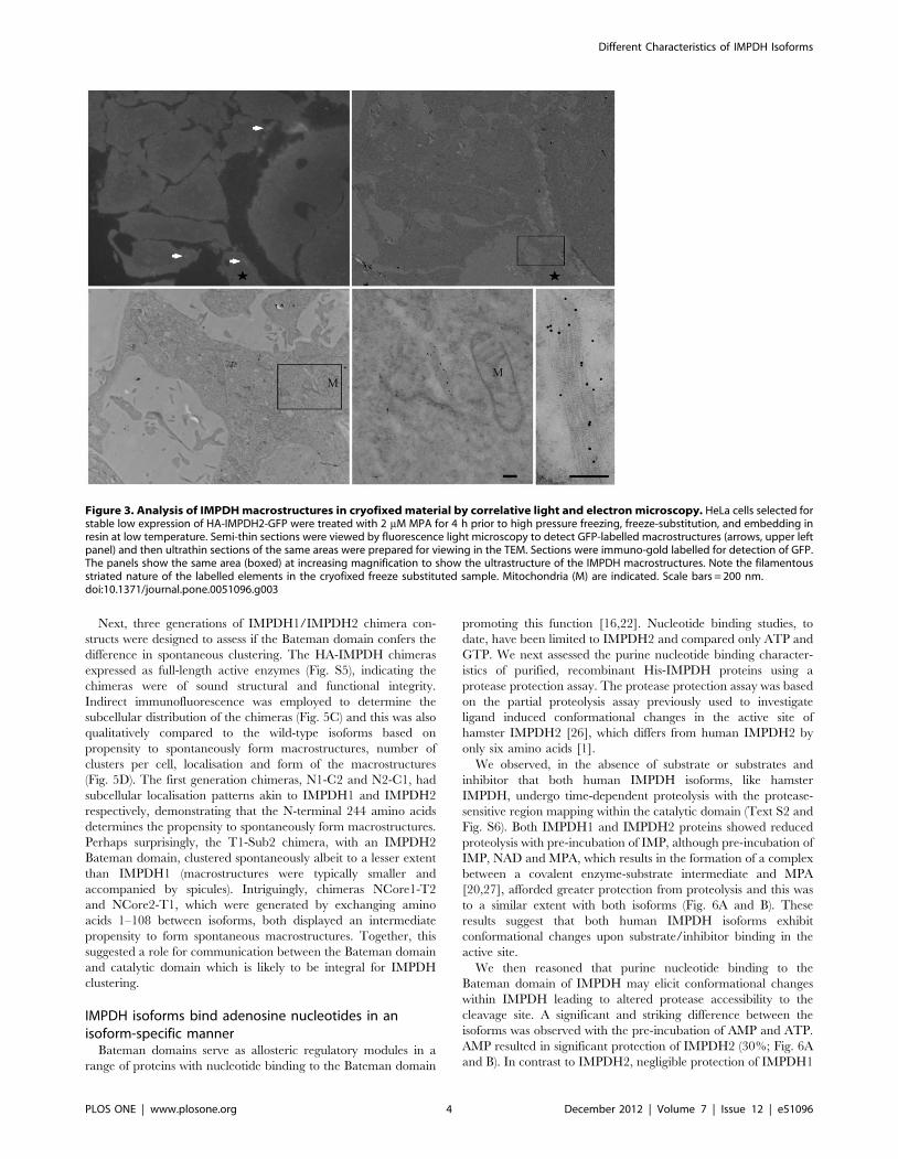
further investigate their ultrastructure in cryofixed cells in the
absence of primary fixation using a correlative light and electron
microscopic approach. HeLa cells selected for stable low
expression of a N-terminal HA-tagged, C-terminal GFP-tagged
IMPDH2, HA-IMPDH2-GFP (Text S1 and Fig. S1), were grown
on sapphire discs and then treated with MPA before rapid high
pressure freezing and processing (Fig. 3). GFP-labelled macro-
structures were visible in sections viewed by light microscopy
allowing us to identify the same structures in parallel ultrathin
sections viewed in the electron microscope and labelled with anti-
GFP antibodies and 10 nm protein A-gold. The macrostructures
were composed of linear elements up to a few microns in length
comprising filamentous elements with regular striations (approx-
imately 10 nm spacing).
IMPDH clustering in live cellsTo investigate the dynamics of IMPDH redistribution we
monitored clustering of GFP-tagged IMPDH2 fusion proteins
using 4D time-lapse videomicroscopy. HeLa cells stably expressing
low levels of HA-IMPDH2-GFP were treated with MPA (Fig. 4
and Video S1) or the GMP synthetase inhibitor, decoyinine (Fig.
S2, Video S3 and S4), to induce macrostructure formation. Live
cell imaging showed the time-dependent coalescence of HA-
IMPDH2-GFP from a diffuse distribution throughout the cyto-
plasm into spicules, increasing in both number and intensity, and
then into macrostructures. Moreover videomicroscopy revealed
the processive nature of macrostructure formation which typically
involves end-to-end fusion of spicules or less frequently lateral
merging of spicules (Video S2). The length of the HA-IMPDH2-
GFP macrostructures was typically shorter than those seen in cells
expressing only endogenous or recombinant non-GFP-tagged
IMPDH [9], although the temporal clustering of HA-IMPDH2-
GFP was similar to that seen for endogenous IMPDH (Fig. S3).
This probably reflects steric hindrance due to the large GFP
moiety.
The above observations raised the question whether the
cytoskeleton may play a role in coordinating the formation of
macrostructures. Discombobulation of microfilament or microtu-
bule networks, with cytochalasin D and nocodazole respectively,
had no obvious effect on the ability of IMPDH to cluster upon
decoyinine treatment (Fig. S4). While the cellular mechanisms
driving macrostructure formation remains to be determined; these
studies, combined with previous reports [9,23], demonstrate that
redistribution of IMPDH into macrostructures occurs in living
cells in response to treatments that perturb intracellular nucleotide
levels and importantly, do not represent an artefact of fixation or
processing.
Bateman domain alone does not confer IMPDH isoformclustering
We previously demonstrated that IMPDH1 has a higher
propensity than IMPDH2 to cluster spontaneously into macro-
structures [9]. Here we investigated whether a discrete region may
underpin the divergence in IMPDH distribution. Alignment of the
primary amino acid sequence of human IMPDH1 and IMPDH2
revealed 25% sequence variability within the Bateman domain
containing ‘‘sub’’ domain (amino acids 99–244), with CBS1
having 27% variability compared to 8% in CBS2. Whereas there
was only 12% sequence variation in the catalytic ‘‘core’’ domain
(amino acids 1–108+245–514). Superimposition of solved crystal
structures [24,25] further highlighted the conservation in tertiary
structure of the catalytic domain (Fig. 5A). Together, this
advocated the Bateman domain as the candidate region under-
pinning the intrinsic differences between the IMPDH isoforms.
To investigate the requirement of the Bateman domain in
clustering the localisation of HA-IMPDH2 sub and core
constructs, based on those of Nimmesgern et al., (1999) [17], were
examined by indirect immunofluorescence (Fig. 5B). Transiently
expressed full-length HA-IMPDH2 redistributed into macrostruc-
tures following MPA treatment [9]. In contrast, the sub and core
proteins displayed a diffuse cytoplasmic localisation in both control
and MPA treated cells. This suggests the intact enzyme is required
for redistribution, however, these results do not establish whether
the Bateman domain plays a direct structural role and/or a
regulatory role in clustering.
Figure 2. Immuno-EM of IMPDH localisation. Representativeelectron micrographs of CHO cells treated with (A) vehicle or (B and C)2 mM MPA for 4 h. Cells were fixed, processed and labelled with anti-panIMPDH antibody and gold-labelled anti-mouse secondary antibody,as outlined in methods. Plasma membrane (PM), Nucleus (N) andmitochondria (M) are indicated. Scale bars = 0.5 mm.doi:10.1371/journal.pone.0051096.g002
Different Characteristics of IMPDH Isoforms
PLOS ONE | www.plosone.org 3 December 2012 | Volume 7 | Issue 12 | e51096
Next, three generations of IMPDH1/IMPDH2 chimera con-
structs were designed to assess if the Bateman domain confers the
difference in spontaneous clustering. The HA-IMPDH chimeras
expressed as full-length active enzymes (Fig. S5), indicating the
chimeras were of sound structural and functional integrity.
Indirect immunofluorescence was employed to determine the
subcellular distribution of the chimeras (Fig. 5C) and this was also
qualitatively compared to the wild-type isoforms based on
propensity to spontaneously form macrostructures, number of
clusters per cell, localisation and form of the macrostructures
(Fig. 5D). The first generation chimeras, N1-C2 and N2-C1, had
subcellular localisation patterns akin to IMPDH1 and IMPDH2
respectively, demonstrating that the N-terminal 244 amino acids
determines the propensity to spontaneously form macrostructures.
Perhaps surprisingly, the T1-Sub2 chimera, with an IMPDH2
Bateman domain, clustered spontaneously albeit to a lesser extent
than IMPDH1 (macrostructures were typically smaller and
accompanied by spicules). Intriguingly, chimeras NCore1-T2
and NCore2-T1, which were generated by exchanging amino
acids 1–108 between isoforms, both displayed an intermediate
propensity to form spontaneous macrostructures. Together, this
suggested a role for communication between the Bateman domain
and catalytic domain which is likely to be integral for IMPDH
clustering.
IMPDH isoforms bind adenosine nucleotides in anisoform-specific manner
Bateman domains serve as allosteric regulatory modules in a
range of proteins with nucleotide binding to the Bateman domain
promoting this function [16,22]. Nucleotide binding studies, to
date, have been limited to IMPDH2 and compared only ATP and
GTP. We next assessed the purine nucleotide binding character-
istics of purified, recombinant His-IMPDH proteins using a
protease protection assay. The protease protection assay was based
on the partial proteolysis assay previously used to investigate
ligand induced conformational changes in the active site of
hamster IMPDH2 [26], which differs from human IMPDH2 by
only six amino acids [1].
We observed, in the absence of substrate or substrates and
inhibitor that both human IMPDH isoforms, like hamster
IMPDH, undergo time-dependent proteolysis with the protease-
sensitive region mapping within the catalytic domain (Text S2 and
Fig. S6). Both IMPDH1 and IMPDH2 proteins showed reduced
proteolysis with pre-incubation of IMP, although pre-incubation of
IMP, NAD and MPA, which results in the formation of a complex
between a covalent enzyme-substrate intermediate and MPA
[20,27], afforded greater protection from proteolysis and this was
to a similar extent with both isoforms (Fig. 6A and B). These
results suggest that both human IMPDH isoforms exhibit
conformational changes upon substrate/inhibitor binding in the
active site.
We then reasoned that purine nucleotide binding to the
Bateman domain of IMPDH may elicit conformational changes
within IMPDH leading to altered protease accessibility to the
cleavage site. A significant and striking difference between the
isoforms was observed with the pre-incubation of AMP and ATP.
AMP resulted in significant protection of IMPDH2 (30%; Fig. 6A
and B). In contrast to IMPDH2, negligible protection of IMPDH1
Figure 3. Analysis of IMPDH macrostructures in cryofixed material by correlative light and electron microscopy. HeLa cells selected forstable low expression of HA-IMPDH2-GFP were treated with 2 mM MPA for 4 h prior to high pressure freezing, freeze-substitution, and embedding inresin at low temperature. Semi-thin sections were viewed by fluorescence light microscopy to detect GFP-labelled macrostructures (arrows, upper leftpanel) and then ultrathin sections of the same areas were prepared for viewing in the TEM. Sections were immuno-gold labelled for detection of GFP.The panels show the same area (boxed) at increasing magnification to show the ultrastructure of the IMPDH macrostructures. Note the filamentousstriated nature of the labelled elements in the cryofixed freeze substituted sample. Mitochondria (M) are indicated. Scale bars = 200 nm.doi:10.1371/journal.pone.0051096.g003
Different Characteristics of IMPDH Isoforms
PLOS ONE | www.plosone.org 4 December 2012 | Volume 7 | Issue 12 | e51096

was afforded by AMP. However, IMPDH1 protein was signifi-
cantly protected from proteolysis by ATP (31%; Fig. 6B) whilst this
effect was not seen for IMPDH2. This observed protection of
IMPDH1 by ATP and IMPDH2 by AMP was also shown to be
dose-dependent (Fig. S7). No significant protection of either
IMPDH2 or IMPDH1 was observed with equimolar concentra-
tions of the other purine nucleotides. Specific ATP binding to
IMPDH1, but not IMPDH2, was supported by a direct filter
binding assay (Fig. 7A and B). These results suggest that IMPDH
exhibits isoform specificity for ATP and AMP, which may elicit
conformational changes within the enzyme, highlighting further
differences between the isoforms.
Bateman domain mediates nucleotide protectionGMP and, to a lesser extent, AMP have been reported to act as
competitive inhibitors of IMPDH [28,29]. Hence, to ascertain that
the protection afforded by AMP was modulated via the Bateman
domain, and not via binding to the active site, the protease
protection assay was performed with a purified, recombinant His-
IMPDH2 core protein (Fig. 6C and D). As expected, the core
protein was significantly protected by pre-incubation with IMP,
NAD and MPA. In contrast to the IMPDH2 protein, AMP did not
confer protection of the core protein. This data suggests that the
Bateman domain is integral for the protection afforded by
nucleotides and supports the hypothesis that nucleotides bind the
Bateman domain.
The R224P mutation disrupts ATP binding and IMPDH1subcellular distribution
The effects of the most-common RP-causing mutations on the
ATP-mediated protection of IMPDH1 were examined (Fig. 8A
and B). Consistent with wild-type protein, pre-incubation of the
purified, recombinant mutant proteins with IMP, NAD and MPA
significantly protected the enzymes from proteolysis, although
protection of the R224P mutant was significantly less than wild-
type. The R224P protein was not protected by ATP. Furthermore,
no ATP binding with the R224P mutant was confirmed with a
direct filter binding assay (Fig. 7A and B). In contrast, the D226N
Figure 4. MPA induced clustering of HA-IMPDH2-GFP in live cells. Images are maximum intensity projections from confocal z-series of liveHeLa HA-IMPDH2-GFP cells treated with 2 mM MPA. Selected frames, from Video S1, are presented with the time captured relative to the addition ofMPA recorded at the top right (h:mm:ss). Data is representative of three independent experiments. (A) Field of cells with low HA-IMPDH2-GFPexpression, which is initially diffuse throughout the cytosol and with time HA-IMPDH2-GFP clusters into spicules and macrostructures. Scalebar = 20 mm. (B) Selected frames within subset region shown in A, corresponds to Video S2, which highlight emergence of macrostructures,movement while undergoing a 180u clockwise rotation of in the X-Y plane (indicated by yellow arrow) and end-to-end fusion of spicules (indicated byyellow asterisk). Scale bar = 10 mm.doi:10.1371/journal.pone.0051096.g004
Different Characteristics of IMPDH Isoforms
PLOS ONE | www.plosone.org 5 December 2012 | Volume 7 | Issue 12 | e51096

mutant was significantly protected from protease digestion by
ATP, albeit qualitatively less than wild-type. Similar to IMPDH1,
neither mutant protein was protected by pre-incubation with
AMP.
This striking affect of the R224P, and not the D226N, mutation
on IMPDH1 properties correlated with an altered subcellular
distribution of this mutant protein (Fig. 8C and D). Unlike wild-
type, the R224P mutant had a very low tendency to spontaneously
cluster into macrostructures (,3%). HA-IMPDH1 R224P pre-
dominantly displayed a diffuse cytoplasmic pattern with spicules
being detected in 5–15% of cells. In contrast, the D226N mutant
had a high propensity (60–75% cells) to spontaneously cluster into
macrostructures. Treatment with MPA promoted clustering of
R224P and D226N mutant proteins, although to a lesser extent in
cells expressing the R224P mutant compared with either the wild-
type or D226N mutant proteins, which were similar (Fig. 7C and
D).
AMP and ATP are not allosteric activators of IMPDHFinally we investigated the catalytic activity of the purified,
recombinant His-IMPDH proteins in the presence of ATP, AMP
or XMP (Fig. 9). Compared with the inhibitory effects of XMP, no
significant effects on activity were observed with either ATP or
AMP. Importantly, in light of a previous report [22], there was no
evidence of an increase in activity of any of the IMPDH proteins
upon incubation with ATP. Intriguingly, the D226N mutant
showed a substantial 75–85% reduction in activity with XMP
(p,0.001 compared to wild-type) which suggests that this disease-
causing mutation may increase sensitivity to product inhibition.
Discussion
The current studies were prompted by the observation that
IMPDH1 and IMPDH2 may be distinguished by their propensity
to spontaneously cluster into macrostructures [9]. Here we report
Figure 5. Investigating a role for the Bateman domain in IMPDH clustering. (A) Superimposed structure of IMPDH2 (1B3O; yellow) withIMPDH1 (1JCN; red). Ligands have been removed for clarity. N labels the N-terminus. (B) Micrographs of CHO cells transiently expressing HA-IMPDHproteins (as labelled), treated with vehicle (control) or 1 mM MPA for 4 h. Cells were fixed and labelled for HA (HA-IMPDH; green) and nuclei werecounterstained with DAPI (blue). Representative of at least four independent experiments. (C) Representative micrographs of CHO cells transientlyexpressing IMPDH1/IMPDH2 chimera (as labelled). Cells were fixed and stained for HA (HA-IMPDH; green) and nuclei were counterstained with DAPI(blue). Scale bars = 10 mm. (D) Table shows qualitative scoring of chimera subcellular distribution pattern according to the similarity to IMPDH1(macrostructure formation) or IMPDH2 (diffuse) distribution. Assignments were based on three independent experiments. Shown by the schematicsare the IMPDH1 (red) and IMPDH2 (yellow) regions of chimera constructs, with internal numbering referring to IMPDH1 sequence boundary and thestriped boxes indicating the CBS dimer.doi:10.1371/journal.pone.0051096.g005
Different Characteristics of IMPDH Isoforms
PLOS ONE | www.plosone.org 6 December 2012 | Volume 7 | Issue 12 | e51096

isoform-specific differences in nucleotide binding, via a mechanism
involving the Bateman domain, which results in conformational
changes communicated between the catalytic domain and the
Bateman domain, consistent with a role for both domains in
determining the subcellular distribution of IMPDH.
Our microscopy analyses further defined the ultra-structure of
IMPDH macrostructures and provided insight into the dynamics
of clustering, but the functional significance of IMPDH clustering
remains an enigma. Our previous study showed the enzymes
upstream (aminoimidazolecarboxamide ribonucleotide formyl-
transferase/IMP cyclohydrolase (ATIC)) and downstream (GMP
synthetase) of IMPDH in the de novo synthesis pathway did not
redistribute with IMPDH, ruling out a role for substrate
channelling [9]. Interestingly, An and colleagues described co-
localisation of enzymes situated upstream of IMPDH in the de novo
pathway of purine biosynthesis, including ATIC, into rounded
punctate structures in response to changes in purine levels [30].
Assembly of these ‘‘purinosome’’ complexes appears dependent on
casein kinase II and, in contrast to IMPDH macrostructures, are
stabilised by microtubules [31]. Under conditions ascribed to
result in purinosome formation [30] we observed modest
redistribution of IMPDH into small macrostructures that did not
co-localise with a key component of the purinosomes, formylgly-
cinamidine-ribonucleotide synthetase (FGAMS)-OFP [30], or with
ATIC (ECT and JPW unpublished data). Collectively these
observations indicate that IMPDH macrostructures are distinct
from purinosomes. One possibility is that this alternate distribution
may facilitate preferential channelling of IMP into the biosynthetic
pathway of adenine nucleotides rather than guanine nucleotides.
Emerging evidence suggests protein clustering may be a general
response to metabolic challenge. Recently, 180 yeast proteins,
including an IMPDH homolog, were shown to cluster reversibly to
punctate ‘‘foci’’ upon induction of quiescence by nutrient
starvation [32]. A subsequent screen identified nine additional
GFP-tagged proteins which self-assembled into filamentous
structures, reminiscent of macrostructures, in response to distinct
Figure 6. Nucleotides protect IMPDH in an isoform-specificmanner via the Bateman domain. (A) Representative Coomassiestained SDS-PAGE gel of a protease protection assay experiment,performed as outline in methods, with His-IMPDH2. INM stands for IMP,NAD and MPA. Molecular weight marker sizes are indicated. (B)Quantitation of remaining full-length protein from protease protectionassay with His-IMPDH2 and His-IMPDH1 presented as percentprotection. Shown is the mean 6 SEM and the number of experiments(n) is indicated below the graph. (*p,0.05, **p,0.01, ***p,0.001 cf. thecontrol (digested), # p,0.01 IMPDH1 cf. IMPDH2). (C) Representativegel of a protease protection assay experiment with His-Core2 protein.(D) Quantitated results as per B. Shown is the mean 6 SEM from fiveindependent experiments. (***p,0.001 cf. the control (digested), ##p,0.001 IMP, NAD and MPA cf. adenosine nucleotides).doi:10.1371/journal.pone.0051096.g006
Figure 7. IMPDH1 directly binds ATP. (A) Representative ATPbinding experiment with His-IMPDH proteins, shows [32P] ATP bound toHis-IMPDH1 only in the presence of 5 mM cold ATP. Data representsmean counts 6 SD. (B) Specific binding was determined by expressingthe counts per reaction as a fold over the non-specific counts present inthe BSA sample. Shown is the mean specific binding 6 SEM (fold overnon-specific binding) from three to six independent experiments(***p,0.001 compared to the control).doi:10.1371/journal.pone.0051096.g007
Different Characteristics of IMPDH Isoforms
PLOS ONE | www.plosone.org 7 December 2012 | Volume 7 | Issue 12 | e51096

environmental conditions [33]. Although the yeast IMPDH
homolog IMD was not found to cluster it is possible that the
GFP-tag prevented this, since it was necessary for us to express
human IMPDH2-GFP at low levels in order to observe clustering.
Also of note was the assembly of the cytidine triphosphate synthase
(CTPS) homolog Ura7p into filaments following glucose depriva-
tion or treatment with sodium azide, suggesting Ura7p distribution
may also be sensitive to cellular energy status. Interestingly, a
Figure 8. R224P mutation affects ATP mediated protease protection and affects spontaneous clustering. (A) Representative Coomassiestained gel of a protease protection assay with His-IMPDH1 proteins. INM stands for IMP, NAD and MPA. (B) Quantitation of remaining full-lengthprotein from protease protection assay presented as percent protection. Shown is the mean 6 SEM and the number of experiments (n) is indicatedbelow the graph. (*p,0.05, **p,0.01, ***p,0.001 cf. the control (digested), # p,0.01 cf. IMPDH1. (C) Representative micrographs of CHO cellstransiently expressing HA-IMPDH1, HA-IMPDH1 R224P or HA-IMPDH1 D226N. Cells were fixed and stained for HA (HA-IMPDH; green) and nuclei werecounterstained with DAPI (blue). Representative of at least four independent experiments. Scale bar = 10 mm. (D) CHO cells transiently expressing HA-IMPDH proteins, as indicated, were treated with either vehicle, 2 mM MPA for 4 h or 2 mM MPA for 4 h and supplemented with 100 mM guanosine forthe final 2 h. Cells were fixed and stained with anti-HA antibody. From random fields, 50–80 labelled healthy cells were counted, in a blinded manner,and the classification of the subcellular distribution of the protein classified as diffuse (blue), in spicules (red) or macrostructures (green).Representative of two independent experiments.doi:10.1371/journal.pone.0051096.g008
Different Characteristics of IMPDH Isoforms
PLOS ONE | www.plosone.org 8 December 2012 | Volume 7 | Issue 12 | e51096

recent study by Carcamo and colleagues reported that CTPS1 was
present in macrostructures and that inhibition of CTPS was
sufficient to promote macrostructure formation [34]. These
observations are largely consistent with those from the Mitchell
lab [23] and our own ([9] and the current study) and suggest that
CTPS1 is present in macrostructures. However, it is noteworthy
that in vitro studies, using purified IMPDH, demonstrate that
IMPDH is able to form macrostructures in the absence of
additional proteins [23]. Moreover, biochemical analyses have
failed to identify interacting partners for IMPDH [23,35](ECT
and JPW unpublished) suggesting any interaction between
IMPDH2 and CTPS1 may be relatively labile. Nevertheless,
further investigations of macrostructures should include examina-
tion of the potential role and contribution of CTPS1.
Our studies suggested an unappreciated role for communication
between the Bateman and catalytic domains which are likely to be
integral to the specific regulatory properties of IMPDH isoforms.
The protease protection assay provided insight into the nucleotide
binding specificity of the isoforms under conditions that allowed us
to compare the effects of all purine nucleotides in parallel on both
isoforms. Interestingly, previous studies suggested IMPDH2 could
bind GTP [23] and ATP [22]. Whilst the latter study also reported
that ATP binding increased activity four-fold this finding has not
been recapitulated here or by others [12,21,23]. Although we
cannot exclude the possibility that IMPDH proteins may bind
GTP [23], unlike adenosine nucleotide binding to IMPDH, our
findings imply that it occurs without concurrent conformational
changes. Recent structural and biochemical studies of Bateman
domain containing proteins including MJ0100 [36], Cystathionine
b-Synthase [37] and AMPKc [15,38] suggest that such ligand-
induced conformational changes represent an important allosteric
regulatory mechanism.
The significance of the isoform specificity of IMPDH nucleotide
binding remains to be elucidated, however, an attractive possibility
is that it may reflect an adaptation to different demands of specific
cell types. Our attempt to characterise and compare the ‘energy
sensing ability’ of the human IMPDH isoforms in vivo, in guaB null
E.coli, proved unsuccessful since only human IMPDH2, but not
IMPDH1, was able to complement growth (ECT and JPW
unpublished data). It is intriguing that ADP was without significant
affect on the proteolysis profile of either isoform, particularly
IMPDH1, since it would be anticipated that the binding pocket
would be sufficiently large to accommodate ADP. Whilst our
observations of IMPDH binding adenosine nucleotides are in
accordance with biochemical and structural studies of other
Bateman domain containing proteins [22,39,40,41,42] recent
reports suggest that ADP binding to the regulatory c subunit of
AMPK may represent a key event in the allosteric regulation of
AMPK activity [15,38,43]. Further, detailed investigations into the
specificity of nucleotide binding of the IMPDH isoforms are
warranted. Moreover, detailed analysis of the kinetics of activation
of IMPDH1 and IMPDH2 by nucleotides, including AMP and
ATP, may provide additional and complementary insights into the
molecular mechanisms governing the allosteric regulation of the
IMPDH isoforms. However, such investigations were beyond the
scope of the current work.
The occurrence of disease-causing mutations in and around the
Bateman domain of IMPDH1, which have no effect on enzymatic
activity [10,11,12], together with our findings from the chimeras
suggests a physiologically important role of a regulatory region
outside the catalytic site of the enzyme. Currently, there is no
consensus as to the molecular effects yielded by RP-causing
mutations in IMPDH1 and intriguingly, the reported properties
are divergent between the R224P and D226N mutants
[10,11,12,44,45,46]. In the current study, the RP-causing muta-
tion, R224P, abolished ATP binding and this correlated with a
reduced propensity to cluster. In contrast the D226N mutant
protein had properties similar to wild-type, although there was a
trend towards reduced ATP binding. Interestingly, the D226N
mutant showed increased sensitivity to inhibition by XMP, which
raises the intriguing possibility that this mutation may elicit
pathogenicity by enhancing the product-inhibition of IMPDH. In
developmental terms, it is likely that any effects of dysregulation
are initially masked by IMPDH2 being the predominant isoform
in the developing retina with expression of IMPDH1 proteins
dominating only in the mature retina [9]. Future studies will be
required to examine the effect of these disease-causing mutations
in the context of the major retinal variants (type Ia and c).
Moreover, determination of the functional significance of wild-
Figure 9. IMPDH activity in the presence of nucleotides. Activity of purified His-IMPDH proteins following 20 min pre-incubation with 1 mM or5 mM nucleotides (ATP, AMP, XMP) and normalised to control, no addition, activity. Shown is the mean 6 SEM (n = 3, **p,0.01, ***p,0.001 cf. to thecontrol activity).doi:10.1371/journal.pone.0051096.g009
Different Characteristics of IMPDH Isoforms
PLOS ONE | www.plosone.org 9 December 2012 | Volume 7 | Issue 12 | e51096

type protein binding ATP will provide insight into the conse-
quence of the properties disrupted by the R224P mutation.
Recently, the Bateman domain of IMPDH has been revealed to
be a negative transregulator of adenosine nucleotide synthesis in
E.coli [21,47]. E.coli that were recombineered to express IMPDH
lacking a Bateman domain were sensitive to adenosine or inosine
induced growth arrest. This corresponded with an increase in the
adenosine nucleotide pool, resulting in allosteric inhibition of
PRPP synthase, reduced PRPP and subsequent pyrimidine
starvation [47]. While the molecular mechanism remains to be
elucidated, Pimkin et al., (2009) [47] suggested that this may occur
via an interaction with and inhibition of adenylosuccinate
synthetase. This raises the intriguing possibility that the nucleo-
tide-bound-state of IMPDH may contribute to the regulation of
adenosine nucleotide synthesis. Congruent to this, we propose that
IMPDH clustering may confer another level of regulation that
contributes to modulating a function of IMPDH outside of,
although perhaps linked to, IMP catalysis. Indeed, the observa-
tions of this study are complementary with the suggestions that
IMPDH has additional unappreciated ‘‘moonlighting’’ functions
which are distinct from its role in guanine nucleotide biosynthesis.
IMPDH has been implicated in lipid accumulation [35,48],
proposed to be involved in RNA metabolism, due to an association
of nucleic acids via the Bateman domain [12,44,45,46] and most
recently described as a DNA-binding transcriptional repressor
[49]. A common feature of all these proposed functions for the
Bateman domain of IMPDH is that it is likely to either regulate
IMPDH and/or be implicated in regulation afforded by IMPDH.
Furthermore, it is likely that additional levels of regulation, such as
phosphorylation [35,50,51], contribute to the complexity of
IMPDH modulation and allow for adaptation to the intracellular
environment.
In summary, in the present study we have demonstrated that the
nucleotide binding properties of the IMPDH isoforms differ.
Together with the striking differences between the propensities to
cluster into macrostructures these findings emphasise that
IMPDH1 and IMPDH2 have distinct properties. Moreover, a
disease causing mutation in IMPDH1, R224P, altered these
distinguishing properties. Collectively, these results raise the
possibility that the nucleotide sensing properties of the Bateman
domain in IMPDH serve to regulate IMPDH and co-ordinate
nucleotide homeostasis, thereby giving rise to cellular plasticity in
an isoform-specific manner to meet the requirements of the
cellular environment.
Materials and Methods
Reagents and MaterialsReagents were from Sigma–Aldrich (Castle Hill, NSW,
Australia) unless otherwise stated. Tissue culture reagents and
foetal bovine serum were from Invitrogen (Mount Waverley, VIC,
Australia) and Bovogen Biologicals (Essendon, VIC, Australia)
respectively. The anti-panIMPDH antibody was a kind gift from
Frank Collart [52], the anti-HA antibody was from Covance
(Berkley, CA, USA), the anti-tubulin antibody from Abcam
(Cambridge, UK), the anti-GFP antibody [53] and isoform-
specific IMPDH antibodies [9] were generated as previously
described. All secondary antibodies were from Molecular Probes
(Eugene, OR, USA).
Generation of IMPDH constructsTo yield pmEGFP-C1 HA-IMPDH2-GFP, HA-IMPDH2
cDNA [35] was amplified by PCR with forward 59-
GGTGGTGCTAGCGCCACCATGTACCCATACGATGTG-
CCAGATTACGCT-39 and reverse 59- GGTGGCGACCG-
GTCCACCAGAACCACCTGCACCAGATGCACCTGTTCC-
GAAAAGCCGCTTCTCATACG-39 primers, which was
inserted into pmEGFP-C1 (Clontech, Mountain View, CA,
USA) on NheI/AgeI sites. IMPDH chimeras and the truncated
core domain constructs were cloned with an N-terminal HA-tag
using a three-step PCR method (see Table S1 for primers)
employed by Nimmesgern et al., (1999) [17]. Firstly, two distinct
PCR products, A and B, were generated using template and
primer pairs detailed in Table S1. In the second round, PCR
products A and B became the template for amplification with the
forward primer of PCR A and reverse primer of PCR B resulting
in a chimeric full-length product. This amplicon was shuttled into
Blunt II TOPO (Invitrogen) prior to subcloning on HindIII/NotI
site into pcDNA5/FRT/TO (Invitrogen). A hexa-histidine (His)-
tag was inserted at the 59 end of human IMPDH1 or IMPDH2
cDNA [35] by PCR prior to cloning into pET20b (+) (Novagen,
Madison, WI, USA). QuikChange site-directed mutagenesis kit
(Stragene, La Jolla, CA, USA) was used to introduce point
mutations, CGC to CCC (R224P) and GAC to AAC (D226N).
Cell CultureChinese Hamster Ovary (CHO) cells and HeLa cells were
cultured in complete F12 HAMs and Dulbecco’s Modified Eagle’s
medium respectively, supplemented with 10% FBS and 2 mM L-
glutamine. Cells were transfected and treated as previously
described [9]. HeLa cells stably expressing HA-IMPDH2-GFP
were initially selected, and subsequently maintained, with genet-
icin (600 mg/ml) added to the media 24 h post transfection with
the pmEGFP-C1 HA-IMPDH2-GFP plasmid. A population of
cells with a low fluorescence, due to low expression of HA-
IMPDH2-GFP, were further selected by fluorescence activated
cell sorted analysis. The resulting heterogeneous stable population
of cells expressed HA-IMPDH2-GFP at approximately 10% of the
level of endogenous IMPDH. Cells were treated with either
vehicle, 2 mM MPA for either 4 h or as indicated or 2 mM MPA
for 4 h and supplemented with 100 mM guanosine for the final
2 h.
Immunofluorescence and time-lapse videomicroscopyIndirect immunofluorescence microscopy was performed as
previously described [9] with cells being imaged on a LSM510
META confocal microscope at 1006 magnification (Carl Zeiss
MicroImaging, Jena, Germany). For 3D time-lapse (4D) videomi-
croscopy, HeLa HA-IMPDH2-GFP cells were cultured on 24 mm
glass bottomed dishes (Proscitech, Qld, Australia). Cells were
washed with PBS and complete F12 HAMS prior to replacing
with complete F12 HAMS containing the inhibitors. Cells were
then imaged, on a pre-heated (37uC) stage top insert with 5%
humidified CO2 circulating, through a C-Apochromat 406/1.20
W Korr UV-VIS-IR M27 objective using 4–6% 488 nm laser
intensity on a LSM510 META confocal microscope (Zeiss).
Confocal Z-series (0.9–1.1 mm increments) were acquired over
time using the LSM software (Zeiss) and collected images were
processed, analysed and movies constructed using Image J v1.41
software (NIH). All images and movies are maximum intensity
projections of the 3D image.
Electron microscopy and correlative light and electronmicroscopy
CHO cells were fixed with 0.1% glutaraldehyde/4% PFA and
processed for EM as previously described [54]. Sections were
labelled with antibodies to IMPDH followed by 10 nm protein A-
Different Characteristics of IMPDH Isoforms
PLOS ONE | www.plosone.org 10 December 2012 | Volume 7 | Issue 12 | e51096
gold. For cryofixation and correlative light and electron micros-
copy, HeLa cells selected for stable low expression of HA-
IMPDH2-GFP were treated with MPA and then high pressure
frozen, freeze substituted and embedded in HM20 resin at low
temperature as described previously [55] with modifications to the
Lowicryl HM20 infiltration which was shortened to one day (50%,
75% and 100% for 1 h each followed by two 12 h 100%
infiltrations all at 250uC). Sections were labelled with antibodies
to GFP followed by 10 nm protein A-gold.
In silico analysisUCSF Chimera (version 1.3; [56]) was used to coordinate
superimposition of protein data bank files for human IMPDH2
(1B3O; [24]) and IMPDH1 (1JCN; [25]), utilising the default
parameters of the matchmaker function, and for the structure
visualisation.
Protein purificationEscherichia coli BL21 (DE3) transformed with pET20b constructs
were induced at room temperature (RT) for 12–14 h by addition
of isopropyl-beta-D-thiogalactopyranoside (1 mM). Cell pellets
were resuspended in either binding buffer 1 (50 mM Tris pH 8.0,
100 mM KCl, 30 mM imidazole, 1.5 M urea, 10 mM 2-
mercaptoethanol) for IMPDH1 proteins and the core protein or
binding buffer 2 (50 mM Tris pH 6.8, 500 mM KCl, 30 mM
imidazole) for IMPDH2, containing protease inhibitors (1 mg/ml
leupeptin, 1 mg/ml pepstatin, 1 mg/ml antipain, 250 mM benza-
midine and 3 mM AEBSF). Lysates were sonicated, centrifuged at
170006g for 30 min at 4uC and His-IMPDH proteins purified on
a talon affinity resin (Clontech) or nickel-nitriloacetic acid column
(Invitrogen) according to the manufacturer’s instructions. Protein
was eluted with appropriate binding buffer containing 250 mM
imidazole and dialysed into activity assay buffer (100 mM KCl,
100 mM Tris-HCl pH 8.0, 1 mM DTT) with glycerol added to a
final concentration of 20% for storage.
Protease protection assayPurified His-IMPDH protein (0.9 mM) was incubated for
20 min at RT with 1 mM nucleotides or 1 mM MPA with
1 mM IMP and NAD in activity assay buffer prior to addition of
20 mg/ml elastase for a further 20 min. Reactions were ceased by
addition of Laemmli SDS-PAGE sample buffer and heat
denaturation, then analysed by SDS-PAGE. Protein bands were
visualised with coomassie staining and the full-length (intact)
protein quantitated using the LI-COR Odyssey Infrared Imaging
System. Percentage protection was calculated using the following
formula: % protection = ((full-length protein remaining after
digestion in sample/undigested protein) – (full-length protein
remaining with elastase only digestion/undigested pro-
tein))6100%.
[32P] ATP filter binding assayThe ATP binding assay was based on those previously described
[22,23,57]. In brief, purified His-IMPDH protein (0.9 mM), or
BSA (used as a negative control), was mixed with 1 mM [a-32P]
ATP (800 Ci/mmol; Perkin Elmer, Waltham, MA, USA) and cold
ATP (5 mM or 100 mM) for 20 min at RT in binding buffer (50 ml
total reaction 2100 mM KCl, 100 mM Tris-HCl pH 8.0, 1 mM
DTT and 2.5 mM BSA) in duplicate. Reactions were terminated
by rapid filtration, loading 15 ml onto 36 pre-equilibrated (in
binding buffer) MF filter membrane discs (Millipore, Billerica,
MA, USA) under vacuum. Filters were washed rapidly (,20 s)
with 46150 ml of ice-cold binding buffer, dried and radioactivity
bound to filters measured by liquid scintillation counting.
Measurement of IMPDH activityThe IMPDH activity assay of purified protein was based on that
described for crude lysates [9] and the protease protection assay.
In duplicate wells of a 384-well plate, purified His-IMPDH
proteins (0.9 mM) were incubated in activity assay buffer
containing 3 mM EDTA to a final volume of 38 ml with 1 mM
or 5 mM nucleotides (ATP, AMP, XMP) and 0.5 mM NAD for
20 min at RT. IMP (0.5 mM) was added and the enzymatic
activity was measured at 37uC by monitoring NADH production
(A340 nm). Background measurements were determined with a
parallel sample without IMP. IMPDH activity reflects the rate of
change during a linear 15 min period.
StatisticsStatistical analysis was performed using GraphPad Prism
version 5.00 for Windows (GraphPad Software, San Diego, CA,
USA). Data were analysed using ANOVA and a Bonferroni post-
hoc test to compare the means between different treatments.
Supporting Information
Text S1 Supporting information for HA-IMPDH2-GFPfusion protein.(DOCX)
Text S2 Supporting information mapping the elastasecleavage site of human IMPDH.(DOCX)
Figure S1 Characterisation of HA-IMPDH2-GFP fusionprotein. (A) Cartoon of HA-IMPDH2-GFP fusion protein with
the linker sequence indicated between IMPDH and GFP (green
box). Yellow box represents the HA-tag, maroon boxes indicates
the CBS domains and the catalytic domain is shown in blue. (B)
Lysates (20 mg) of HeLa cells transiently expressing either GFP
(lane 1), HA-IMPDH type II (lane 2) or HA-IMPDH2-GFP (lane
3) or the stable HeLa HA-IMPDH2-GFP cells (lane 4) were
analysed by SDS-PAGE and western blotting. Membrane was
probed with anti-GFP antibody (red) and anti-panIMPDH
antibody (green). Asterisks indicate non-specific bands. (C)
IMPDH activity was determined in cell lysates of HeLa cells
transiently expressing HA-IMPDH2-GFP or HA-IMPDH2 or
GFP alone, to control for endogenous IMPDH activity, as
previously described [9]. Activity was normalised to exogenous
protein expression, as determined by western blot, and the values
expressed in terms of HA-IMPDH2 activity. Data represents the
mean 6 SD of two independent experiments. (D) Representative
epifluorescence micrographs of HeLa cells transiently expressing
HA-IMPDH2-GFP (green). Cells were incubated with vehicle
(control) or with 2 mM MPA for 4 h. Cells were fixed,
permeabilised and labelled with the anti-panIMPDH antibody
and Alexa-594 secondary antibody (red) and nuclei were
counterstained with DAPI (blue). Images are representative of at
least three independent experiments. Scale bar = 20 mm.
(TIF)
Figure S2 Decoyinine induced clustering of HA-IMPDH2-GFP in live cells. Images are maximum intensity
projections from confocal z-series of live HeLa HA-IMPDH2-GFP
cells treated with 2 mM decoyinine. Selected frames, from Video
S3, are presented with the time captured relative to the addition of
decoyinine recorded at the top right (h:mm:ss). Data is
representative of three independent experiments. Scale
Different Characteristics of IMPDH Isoforms
PLOS ONE | www.plosone.org 11 December 2012 | Volume 7 | Issue 12 | e51096

bar = 10 mm. (A) Field of cells with low HA-IMPDH2-GFP
expression, which is initially diffuse throughout the cytosol and
with increasing time HA-IMPDH2-GFP clusters into spicules and
macrostructures. (B) Selected frames within subset regions shown
in A, corresponds to Video S4, which highlight the emergence and
lateral fusion (indicated by yellow asterisk) of a HA-IMPDH2-GFP
macrostructure.
(TIF)
Figure S3 Time course of endogenous IMPDH cluster-ing. Micrographs of CHO cells treated with vehicle (control) or
2 mM MPA for the times indicated. Cells were fixed and labelled
for endogenous IMPDH (green) and nuclei were counterstained
with DAPI (blue). Representative of at least three independent
experiments.
(TIF)
Figure S4 Microfilament or Microtubules alone are notrequired for formation of IMPDH macrostructures.Representative confocal micrographs of HeLa HA-IMPDH2-
GFP cells. (A) Cells were treated with either vehicle or 4 mM
cytochalasin D (CD), to discombobulate microfilaments, for 1 h
15 min prior to either fixation (top panel) or addition of 2 mM
decoyinine (DCN) for an additional 3 h 30 min (bottom panel).
Cells were fixed, permeabilised and labelled with the anti-
panIMPDH antibody (green), filamentous actin was stained with
TRITC-phalloidin (red) and nuclei were counterstained with
DAPI (blue). Images are representative of at least three
independent experiments. Scale bar = 20 mm. (B) Cells were
treated with either vehicle or 20 mM nocodazole (Noc), to
destabilise microtubules, for 1 h 15 min prior to either fixation
or addition of 2 mM decoyinine (DCN) for an additional 3 h
50 min. Cells were fixed, permeablised and labelled with the anti-
tubulin antibody (left hand panel; red) or anti-panIMPDH
antibody (right hand panel; red) and nuclei were counterstained
with DAPI (blue). In green is the low fluorescence of the HA-
IMPDH2-GFP. Images are representative of at least two
independent experiments. Scale bar = 20 mm.
(TIF)
Figure S5 Expression and activity of IMPDH1/IMPDH2chimeras. HeLa cells were transiently transfected with HA-
tagged IMPDH chimera or wild-type constructs (as labelled) and
harvested in activity assay buffer. (A) Lysates (20 mg) were
processed by SDS-PAGE and analysed by western blot using
anti-HA antibody. Data is representative of three independent
experiments. (B) IMPDH activity was determined in cell lysates at
37uC by measuring the production of NADH as previously
described [9]. Values were normalised to exogenous HA-IMPDH
protein expression, as determined by western blot, and represent
the mean 6 SD of two independent experiments.
(TIF)
Figure S6 Sensitivity of human IMPDH to elastase.Representative Coomassie stained SDS-PAGE gels of (A) His-
tagged IMPDH1 protein or (B) His-IMPDH2 protein incubated in
in vitro activity assay buffer at RT with elastase (E) for the times
indicated. Note: elastase is a 26 kDa protein and was not included
in the zero time point. Molecular weight (MW) marker sizes are as
indicated. (C) Purified His-tagged IMPDH1 or (D) IMPDH2
protein was pre-incubated with 1 mM of ligands, as indicated, and
subjected to a protease protection assay. Protease protection assay
reactions were resolved by SDS-PAGE and gels were either
stained by coomassie or transferred onto membrane for Western
blotting with the anti-panIMPDH antibody or isoform-specific
antibodies (a6Core1; raised against peptide in IMPDH1 core
domain and a3Sub2 raised against peptide in IMPDH2 sub
domain [9]), as indicated, which were scanned in two fluorescent
channels using the LI-COR Odyssey system. Panels show the
individual blots and merged image (pan-IMPDH - green; isoform-
specific antibodies - red).
(TIF)
Figure S7 Dose dependent protection of IMPDH toelastase. Representative Coomassie stained gels of a protease
protection assay with His-IMPDH1 or His-IMPDH2 proteins
showing the dose-dependent increase in protection from proteol-
ysis of IMPDH2 by AMP and IMPDH1 by ATP. Concentrations
are in mM.
(TIF)
Video S1 Time-lapse of MPA induced clustering of HA-IMPDH2-GFP in live cells. Time-lapse corresponding to
Fig. 3A. After MPA addition (15:30), Z-series (total 100) were
captured at a rate of 1/80 s. The playback parameters were set at
a rate of 20 frames/s condensing the 2:13:15 movie into 5 s.
(AVI)
Video S2 Subset of MPA induced clustering of HA-IMPDH2-GFP in live cells. Time-lapse corresponding to
Fig. 3B and subset of Video S1.
(AVI)
Video S3 Time-lapse of decoyinine induced clusteringof HA-IMPDH2-GFP in live cells. Time-lapse corresponding
to Fig. S2A. After decoyinine addition (19:30), Z-series (total 151)
were captured at a rate of 1/90 s. The playback parameters match
the real-time rate of Video S1 and Video S2, a rate of 17.9
frames/s, condensing the 4:03:57 movie into 8.4 s.
(AVI)
Video S4 Time-lapse of decoyinine induced clusteringof HA-IMPDH2-GFP in live cells. Time-lapse corresponding
to Fig. S2B and subset of Video S3.
(AVI)
Table S1 Sequences of the oligonucleotides used togenerate chimeras.(DOC)
Acknowledgments
We thank Professor Beverly Mitchell (Stanford Cancer Center, Stanford
University, USA) for generously hosting ECT to perform the nucleotide
filter binding assays and Dr Fiona Simpson (University of Queensland
Diamantina Institute) for assistance with 4D microscopy. Electron
microscopy was performed at the Australian Microscopy and Microanal-
ysis Research Facility, Center for Microscopy and Microanalysis,
University of Queensland.
Author Contributions
Conceived and designed the experiments: ECT JHG JPW RGP.
Performed the experiments: ECT JAW VO NLS. Analyzed the data:
ECT JHG JPW RGP VO NLS. Contributed reagents/materials/analysis
tools: RGP JPW. Wrote the paper: ECT JPW. Edited the paper: ECT JPW
RGP.
Different Characteristics of IMPDH Isoforms
PLOS ONE | www.plosone.org 12 December 2012 | Volume 7 | Issue 12 | e51096

References
1. Collart FR, Huberman E (1988) Cloning and sequence analysis of the humanand Chinese hamster inosine-59-monophosphate dehydrogenase cDNAs. J Biol
Chem 263: 15769–15772.
2. Natsumeda Y, Ohno S, Kawasaki H, Konno Y, Weber G, et al. (1990) Two
distinct cDNAs for human IMP dehydrogenase. J Biol Chem 265: 5292–5295.
3. Carr SF, Papp E, Wu JC, Natsumeda Y (1993) Characterization of human type I
and type II IMP dehydrogenases. J Biol Chem 268: 27286–27290.
4. Hager PW, Collart FR, Huberman E, Mitchell BS (1995) Recombinant humaninosine monophosphate dehydrogenase type I and type II proteins. Purification
and characterization of inhibitor binding. Biochem Pharmacol 49: 1323–1329.
5. Jackson RC, Weber G, Morris HP (1975) IMP dehydrogenase, an enzyme
linked with proliferation and malignancy. Nature 256: 331–333.
6. Senda M, Natsumeda Y (1994) Tissue-differential expression of two distinct
genes for human IMP dehydrogenase (E.C.1.1.1.205). Life Sci 54: 1917–1926.
7. Bowne SJ, Sullivan LS, Blanton SH, Cepko CL, Blackshaw S, et al. (2002)Mutations in the inosine monophosphate dehydrogenase 1 gene (IMPDH1)
cause the RP10 form of autosomal dominant retinitis pigmentosa. Hum Mol
Genet 11: 559–568.
8. Kennan A, Aherne A, Palfi A, Humphries M, McKee A, et al. (2002)Identification of an IMPDH1 mutation in autosomal dominant retinitis
pigmentosa (RP10) revealed following comparative microarray analysis of
transcripts derived from retinas of wild-type and Rho(-/-) mice. Hum Mol Genet11: 547–557.
9. Gunter JH, Thomas EC, Lengefeld N, Kruger SJ, Worton L, et al. (2008)
Characterisation of inosine monophosphate dehydrogenase expression during
retinal development: Differences between variants and isoforms. Int J BiochemCell Biol 40: 1716–1728.
10. Bowne SJ, Sullivan LS, Mortimer SE, Hedstrom L, Zhu J, et al. (2006) Spectrum
and Frequency of Mutations in IMPDH1 Associated with Autosomal Dominant
Retinitis Pigmentosa and Leber Congenital Amaurosis. Invest Ophthalmol VisSci 47: 34–42.
11. Aherne A, Kennan A, Kenna PF, McNally N, Lloyd DG, et al. (2004) On the
molecular pathology of neurodegeneration in IMPDH1-based retinitis pigmen-tosa. Hum Mol Genet 13: 641–650.
12. Mortimer SE, Hedstrom L (2005) Autosomal dominant retinitis pigmentosamutations in inosine monophosphate dehydrogenase type I disrupt nucleic acid
binding. Biochem J 390: 41–47.
13. Bateman A (1997) The structure of a domain common to archaebacteria and the
homocystinuria disease protein. Trends in Biochemical Sciences 22: 12–13.
14. Kemp BE (2004) Bateman domains and adenosine derivatives form a bindingcontract. J Clin Invest 113: 182–184.
15. Oakhill JS, Scott JW, Kemp BE (2012) AMPK functions as an adenylate charge-regulated protein kinase. Trends Endocrinol Metab 23: 125–132.
16. Ignoul S, Eggermont J (2005) CBS domains: structure, function, and pathology
in human proteins. Am J Physiol Cell Physiol 289: C1369–1378.
17. Nimmesgern E, Black J, Futer O, Fulghum JR, Chambers SP, et al. (1999)
Biochemical analysis of the modular enzyme inosine 59-monophosphatedehydrogenase. Protein Expr Purif 17: 282–289.
18. Zhou X, Cahoon M, Rosa P, Hedstrom L (1997) Expression, purification, andcharacterization of inosine 59-monophosphate dehydrogenase from Borrelia
burgdorferi. J Biol Chem 272: 21977–21981.
19. Zhang R, Evans G, Rotella FJ, Westbrook EM, Beno D, et al. (1999)
Characteristics and crystal structure of bacterial inosine-59-monophosphatedehydrogenase. Biochemistry 38: 4691–4700.
20. Sintchak MD, Fleming MA, Futer O, Raybuck SA, Chambers SP, et al. (1996)
Structure and mechanism of inosine monophosphate dehydrogenase in complex
with the immunosuppressant mycophenolic acid. Cell 85: 921–930.
21. Pimkin M, Markham GD (2008) The CBS subdomain of inosine 59-monophosphate dehydrogenase regulates purine nucleotide turnover. Mol
Microbiol 68: 342–359.
22. Scott JW, Hawley SA, Green KA, Anis M, Stewart G, et al. (2004) CBS domains
form energy-sensing modules whose binding of adenosine ligands is disrupted bydisease mutations. J Clin Invest 113: 274–284.
23. Ji Y, Gu J, Makhov AM, Griffith JD, Mitchell BS (2006) Regulation of theinteraction of inosine monophosphate dehydrogenase with mycophenolic acid
by GTP. J Biol Chem 281: 206–212.
24. Colby TD, Vanderveen K, Strickler MD, Markham GD, Goldstein BM (1999)
Crystal structure of human type II inosine monophosphate dehydrogenase:implications for ligand binding and drug design. Proc Natl Acad Sci U S A 96:
3531–3536.
25. Risal D, Strickler MD, Goldstein BM (2003) Crystal Structure of the Human
Type I Inosine Monophosphate Dehydrogenase and Implications for IsoformSpecificity To be published.
26. Nimmesgern E, Fox T, Fleming MA, Thomson JA (1996) Conformational
changes and stabilization of inosine 59-monophosphate dehydrogenase associ-
ated with ligand binding and inhibition by mycophenolic acid. J Biol Chem 271:19421–19427.
27. Link JO, Straub K (1996) Trapping of an IMP Dehydrogenase - Substrate
Covalent Intermediate by Mycophenolic Acid. Journal of the AmericanChemical Society 118: 2091–2092.
28. Okada M, Shimura K, Shiraki H, Nakagawa H (1983) IMP dehydrogenase. II.
Purification and properties of the enzyme from Yoshida sarcoma ascites tumorcells. J Biochem (Tokyo) 94: 1605–1613.
29. Holmes EW, Pehlke DM, Kelley WN (1974) Human IMP dehydrogenase.
Kinetics and regulatory properties. Biochim Biophys Acta 364: 209–217.
30. An S, Kumar R, Sheets ED, Benkovic SJ (2008) Reversible compartmentali-zation of de novo purine biosynthetic complexes in living cells. Science 320:
103–106.
31. An S, Deng Y, Tomsho JW, Kyoung M, Benkovic SJ (2010) Microtubule-assisted mechanism for functional metabolic macromolecular complex forma-
tion. Proc Natl Acad Sci U S A 107: 12872–12876.
32. Narayanaswamy R, Levy M, Tsechansky M, Stovall GM, O’Connell JD, et al.
(2009) Widespread reorganization of metabolic enzymes into reversibleassemblies upon nutrient starvation. Proc Natl Acad Sci U S A 106: 10147–
10152.
33. Noree C, Sato BK, Broyer RM, Wilhelm JE (2010) Identification of novelfilament-forming proteins in Saccharomyces cerevisiae and Drosophila melano-
gaster. J Cell Biol 190: 541–551.
34. Carcamo WC, Satoh M, Kasahara H, Terada N, Hamazaki T, et al. (2011)Induction of cytoplasmic rods and rings structures by inhibition of the CTP and
GTP synthetic pathway in mammalian cells. PLoS One 6: e29690.
35. Whitehead JP, Simpson F, Hill MM, Thomas EC, Connolly LM, et al. (2004)Insulin and oleate promote translocation of inosine-59 monophosphate
dehydrogenase to lipid bodies. Traffic 5: 739–749.
36. Lucas M, Encinar JA, Arribas EA, Oyenarte I, Garcia IG, et al. (2010) Bindingof S-methyl-59-thioadenosine and S-adenosyl-L-methionine to protein MJ0100
triggers an open-to-closed conformational change in its CBS motif pair. J Mol
Biol 396: 800–820.37. Hnizda A, Spiwok V, Jurga V, Kozich V, Kodicek M, et al. (2010) Cross-talk
between the catalytic core and the regulatory domain in cystathionine beta-
synthase: study by differential covalent labeling and computational modeling.Biochemistry 49: 10526–10534.
38. Oakhill JS, Steel R, Chen ZP, Scott JW, Ling N, et al. (2011) AMPK is a direct
adenylate charge-regulated protein kinase. Science 332: 1433–1435.
39. Xiao B, Heath R, Saiu P, Leiper FC, Leone P, et al. (2007) Structural basis forAMP binding to mammalian AMP-activated protein kinase. Nature 449: 496–
500.
40. Day P, Sharff A, Parra L, Cleasby A, Williams M, et al. (2007) Structure of aCBS-domain pair from the regulatory gamma1 subunit of human AMPK in
complex with AMP and ZMP. Acta Crystallogr D Biol Crystallogr 63: 587–596.
41. Meyer S, Savaresi S, Forster IC, Dutzler R (2007) Nucleotide recognition by thecytoplasmic domain of the human chloride transporter ClC-5. Nat Struct Mol
Biol 14: 60–67.
42. Bennetts B, Rychkov GY, Ng HL, Morton CJ, Stapleton D, et al. (2005)Cytoplasmic ATP-sensing domains regulate gating of skeletal muscle ClC-1
chloride channels. J Biol Chem 280: 32452–32458.
43. Xiao B, Sanders MJ, Underwood E, Heath R, Mayer FV, et al. (2011) Structure
of mammalian AMPK and its regulation by ADP. Nature 472: 230–233.44. McLean JE, Hamaguchi N, Belenky P, Mortimer SE, Stanton M, et al. (2004)
Inosine 59-monophosphate dehydrogenase binds nucleic acids in vitro and in
vivo. Biochem J 379: 243–251.
45. Mortimer SE, Xu D, McGrew D, Hamaguchi N, Lim HC, et al. (2008) IMPdehydrogenase type 1 associates with polyribosomes translating rhodopsin
mRNA. J Biol Chem 283: 36354–36360.
46. Xu D, Cobb GC, Spellicy CJ, Bowne SJ, Daiger SP, et al. (2008) Retinalisoforms of inosine 59-monophosphate dehydrogenase type 1 are poor nucleic
acid binding proteins. Arch Biochem Biophys 472: 100–104.
47. Pimkin M, Pimkina J, Markham GD (2009) A regulatory role of the Batemandomain of IMP dehydrogenase in adenylate nucleotide biosynthesis. J Biol
Chem 284: 7960–7969.
48. Su H, Gunter JH, de Vries M, Connor T, Wanyonyi S, et al. (2009) Inhibition ofinosine monophosphate dehydrogenase reduces adipogenesis and diet-induced
obesity. Biochem Biophys Res Commun 386: 351–355.
49. Kozhevnikova EN, van der Knaap JA, Pindyurin AV, Ozgur Z, van Ijcken WF,et al. (2012) Metabolic Enzyme IMPDH Is Also a Transcription Factor
Regulated by Cellular State. Mol Cell 47: 133–139.
50. Olsen JV, Blagoev B, Gnad F, Macek B, Kumar C, et al. (2006) Global, In Vivo,and Site-Specific Phosphorylation Dynamics in Signaling Networks. Cell 127:
635–648.
51. Dephoure N, Zhou C, Villen J, Beausoleil SA, Bakalarski CE, et al. (2008) A
quantitative atlas of mitotic phosphorylation. Proc Natl Acad Sci U S A 105:10762–10767.
52. Glesne DA, Collart FR, Huberman E (1991) Regulation of IMP dehydrogenase
gene expression by its end products, guanine nucleotides. Mol Cell Biol 11:5417–5425.
53. Nixon SJ, Webb RI, Floetenmeyer M, Schieber N, Lo HP, et al. (2009) A single
method for cryofixation and correlative light, electron microscopy andtomography of zebrafish embryos. Traffic 10: 131–136.
54. Martin S, Driessen K, Nixon SJ, Zerial M, Parton RG (2005) Regulated
localization of Rab18 to lipid droplets: effects of lipolytic stimulation andinhibition of lipid droplet catabolism. J Biol Chem 280: 42325–42335.
Different Characteristics of IMPDH Isoforms
PLOS ONE | www.plosone.org 13 December 2012 | Volume 7 | Issue 12 | e51096

55. Schieber NL, Nixon SJ, Webb RI, Oorschot VM, Parton RG (2010) Modern
approaches for ultrastructural analysis of the zebrafish embryo. Methods CellBiol 96: 425–442.
56. Pettersen EF, Goddard TD, Huang CC, Couch GS, Greenblatt DM, et al.
(2004) UCSF Chimera–a visualization system for exploratory research andanalysis. J Comput Chem 25: 1605–1612.
57. Janosik M, Kery V, Gaustadnes M, Maclean KN, Kraus JP (2001) Regulation of
human cystathionine beta-synthase by S-adenosyl-L-methionine: evidence for
two catalytically active conformations involving an autoinhibitory domain in the
C-terminal region. Biochemistry 40: 10625–10633.
Different Characteristics of IMPDH Isoforms
PLOS ONE | www.plosone.org 14 December 2012 | Volume 7 | Issue 12 | e51096


