Download - Digestive System Review
Digestive System(Review)

Digestive (GI) Tract

Actions of Digestive (GI) Tract• Ingestion
– Occurs when material enters via the mouth
• Mechanical Processing– Crushing / Shearing –
makes material easier to move through the tract
• Digestion– Chemical breakdown of
food into small organic compounds for absorption
• Secretion– Release of water acids,
buffers, enzymes & salts by epithelium of GI tract and glandular organs
• Absorption– Movement of organic
substrates, electrolytes, vitamins & water across digestive epithelium
• Excretion– Removal of waste products
from body fluids

Histological Structure of the Digestive (GI) Tract
From esophagus to anus have same structure that consists of:
• Mucosa : epithelial lining, lamina propria and muscularis mucosa
• Submucosa : arteriole,venule, submucosal (Meissner’s) plexus
• Muscularis : circular and longitudinal muscle layers, myenteric (Auerbach’s) plexus


Coordination and Control of Digestive System Activity
• Submucosal (Meissner’s) plexus• Myenteric (Auerbach’s) plexus • Parasymphatetic (vagus) nerve

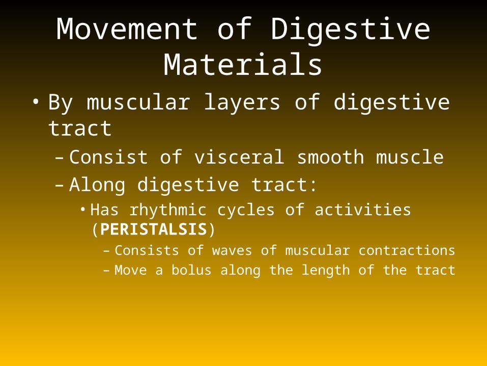
Movement of Digestive Materials
• By muscular layers of digestive tract– Consist of visceral smooth muscle– Along digestive tract:• Has rhythmic cycles of activities (PERISTALSIS)
– Consists of waves of muscular contractions– Move a bolus along the length of the tract

Peristalsis

Oral Cavity
• Mechanical processing– Through actions of teeth,
tongue, and palatal surfaces
• Lubrication– Mixing with mucus and
salivary gland secretions
• Limited digestion– Of carbohydrates and lipids

Major Salivary Glands
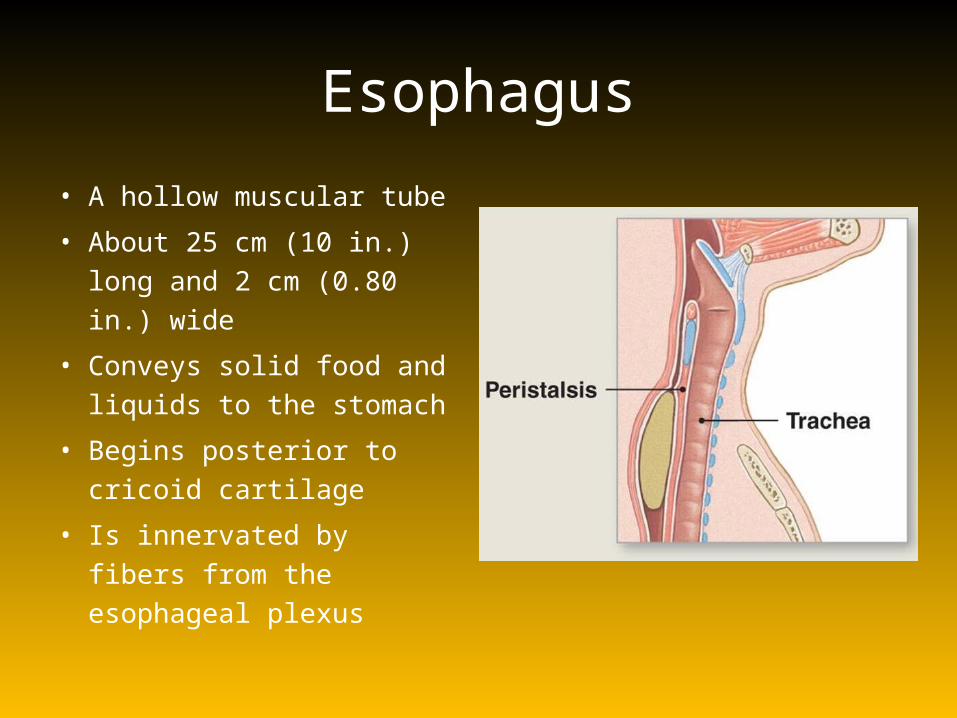
Esophagus
• A hollow muscular tube
• About 25 cm (10 in.) long and 2 cm (0.80 in.) wide
• Conveys solid food and liquids to the stomach
• Begins posterior to cricoid cartilage
• Is innervated by fibers from the esophageal plexus

Stomach
• Major Functions of the Stomach– Storage of ingested food
– Mechanical breakdown of ingested food
– Disruption of chemical bonds in food material by acid and enzymes
– Production of intrinsic factor, a glycoprotein required for absorption of vitamin B12 in small intestine

Gastric Anatomy

Digestion in the Stomach• Stomach performs:– preliminary digestion of proteins (by pepsin)– Some digestion of carbohydrates (by salivary amylase)– Lipids (by lingual lipase)
• Stomach contents:– Become more fluid– pH approaches 2.0– Pepsin activity increases– Protein disassembly begins
• Although digestion occurs in the stomach, nutrients are not absorbed

Gastric juice
1. Pepsinogen (chief cell)2. HCl (parietal cell)3. Intrinsik factor (parietal cell)4. Mucous (Goblet’s cell)
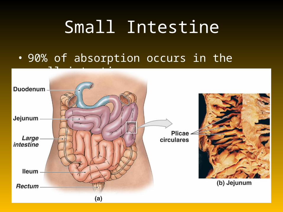
Small Intestine
• 90% of absorption occurs in the small intestine

Small Intestine
• The Duodenum – The segment of small intestine closest to stomach
– 25 cm (10 in.) long
– “Mixing bowl” that receives chyme from stomach and digestive secretions from pancreas and liver
– Functions of the duodenum • To receive chyme from stomach
• To neutralize acids before they can damage the absorptive surfaces of the small intestine

Small Intestine
• The Jejunum – Is the middle segment of small intestine– 2.5 meters (8.2 ft) long– Is the location of most• Chemical digestion• Nutrient absorption

Small Intestine
• The Ileum– The final segment of small intestine– 3.5 meters (11.48 ft) long – Ends at the ileocecal valve, a sphincter that
controls flow of material from the ileum into the large intestine

Small Intestine
• Intestinal Secretions– Watery intestinal juice
– 1.8 liters per day enter intestinal lumen
– Moisten chyme
– Assist in buffering acids
– Keep digestive enzymes and products of digestion in solution
• Intestinal Movements– Chyme arrives in duodenum
– Weak peristaltic contractions move it slowly toward jejunum• Myenteric reflexes
• Not under CNS control
• Parasympathetic stimulation accelerates local peristalsis and segmentation

Pancreas• Lies posterior to stomach
– From duodenum toward spleen
• Is bound to posterior wall of abdominal cavity
• Functions of the Pancreas1. Endocrine cells of the
pancreatic islets:• Secrete insulin and glucagon
into bloodstream2. Exocrine cells:
• Acinar cells and epithelial cells of duct system secrete pancreatic juice

Pancreas• Pancreatic Enzymes
– Pancreatic alpha-amylase• A carbohydrase• Breaks down starches• Similar to salivary amylase
– Pancreatic lipase• Breaks down complex lipids• Releases products (e.g., fatty
acids) that are easily absorbed
• Pancreatic Enzymes – Nucleases
• Break down nucleic acids
– Proteolytic enzymes• Break certain proteins apart• Proteases break large protein
complexes• Peptidases break small peptides
into amino acids• 70% of all pancreatic enzyme
production• Secreted as inactive proenzymes• Activated after reaching small
intestine

Liver
Liver Function
The Physiology of the Liver1. Metabolic regulation2. Hematological regulation3. Bile production

Liver Function• The Functions of Bile– Dietary lipids are not water soluble– Mechanical processing in stomach creates large drops
containing lipids – Pancreatic lipase is not lipid soluble
• Interacts only at surface of lipid droplet
– Bile salts break droplets apart (emulsification)• Increases surface area exposed to enzymatic attack • Creates tiny emulsion droplets coated with bile salts
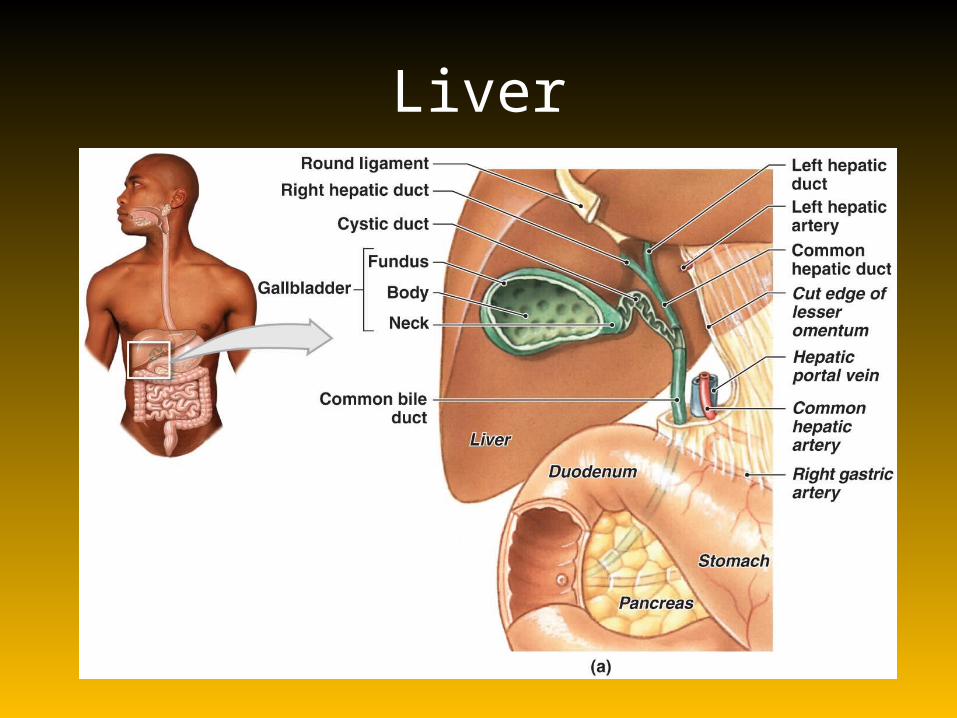
Liver

Gallbladder
• Is a pear-shaped, muscular sac• Stores and concentrates bile prior to excretion
into small intestine• Is located in the fossa on the posterior surface
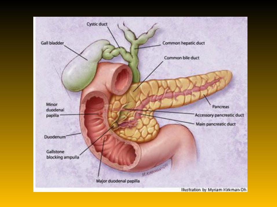
of the liver’s right lobe• The Cystic Duct– Extends from gallbladder– Union with common hepatic duct forms common
bile duct
Gallbladder
• Functions of the Gallbladder– Stores bile
– Releases bile into duodenum, but only under stimulation of hormone cholecystokinin (CCK)
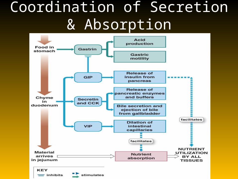
Coordination of Secretion & Absorption
Coordination of Secretion & Absorption
• Intestinal Absorption – It takes about 5 hours for materials
to pass from duodenum to end of ileum
– Movements of the mucosa increases absorptive effectiveness

Large Intestine
• Is horseshoe shaped
• Extends from end of ileum to anus
• Lies inferior to stomach and liver
• Frames the small intestine
• Also called large bowel
• Is about 1.5 meters (4.9 ft) long and 7.5 cm (3 in.) wide

Parts of Large Intestine• The Cecum
– Is an expanded pouch – Receives material arriving
from the ileum– Stores materials and begins
compaction
• Appendix– Also called vermiform appendix
– Is a slender, hollow appendage about 9 cm (3.6 in.) long
– Is dominated by lymphoid nodules (a lymphoid organ)
• Ascending Colon – Begins at superior border of cecum – Ascends along right lateral and posterior wall of peritoneal
cavity to inferior surface of the liver and bends at right colic flexure (hepatic flexure)
• Transverse Colon– Crosses abdomen from right to left; turns at left colic flexure
(splenic flexure)– Is supported by transverse mesocolon– Is separated from anterior abdominal wall by greater omentum
• The Descending Colon – Proceeds inferiorly along left side to the iliac fossa (inner
surface of left ilium)– Is retroperitoneal, firmly attached to abdominal wall
• The Sigmoid Colon – Is an S-shaped segment, about 15 cm (6 in.) long– Starts at sigmoid flexure– Lies posterior to urinary bladder– Is suspended from sigmoid mesocolon– Empties into rectum

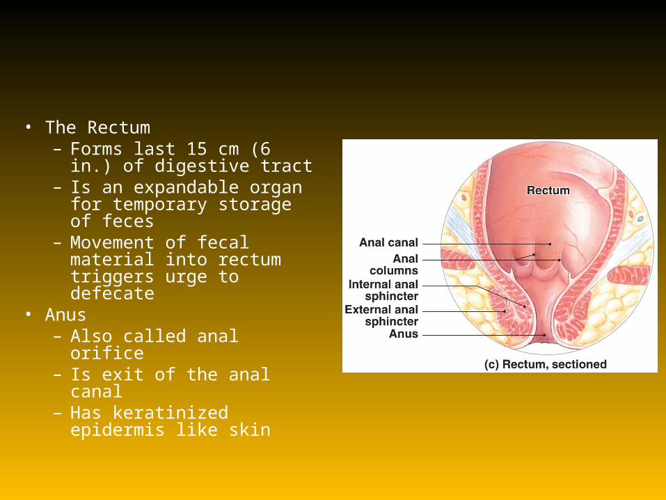
• The Rectum– Forms last 15 cm (6 in.) of
digestive tract– Is an expandable organ for
temporary storage of feces– Movement of fecal material
into rectum triggers urge to defecate
• Anus– Also called anal orifice– Is exit of the anal canal– Has keratinized epidermis
like skin

Parasymphatetic mechanism for enhancing the defecation reflex
Physiology of the Large Intestine
• Absorption in the Large Intestine– Reabsorption of water
– Reabsorption of bile salts• In the cecum
• Transported in blood to liver
– Absorption of vitamins produced by bacteria
– Absorption of organic wastes

Physiology of the Large Intestine
Three Vitamins Produced in the Large Intestine 1. Vitamin K (fat soluble):
• Required by liver for synthesizing four clotting factors, including prothrombin
2. Biotin (water soluble):• Important in glucose metabolism
3. Pantothenic acid: B5 (water soluble):
• Required in manufacture of steroid hormones and some neurotransmitters
Physiology of the Large Intestine
• Organic Wastes
– Bacteria convert bilirubin to urobilinogens and
stercobilinogens
– Bacteria break down peptides in feces and generate• Ammonia, Indole & skatole, hydrogen sulfide
– Bacteria feed on indigestible carbohydrates (complex polysaccharides)• Produce flatus, or intestinal gas, in large intestine

Absorptive and storage functions of the large intestine

Movements of the Large Intestine
• Gastroileal & gastroenteric reflexes– Move materials into cecum– Movement from cecum to transverse colon is very slow,
allowing hours for water absorption – Peristaltic waves move material along length of colon– Segmentation movements (haustral churning) mix
contents of adjacent haustra

Movements of the Large Intestine
• Movements from transverse colon through rest of large intestine results from powerful peristaltic contractions (mass movements)
• Stimulus is distension of stomach and duodenum, relayed over intestinal nerve plexuses
• Distension of the rectal wall triggers defecation reflex

THANK YOU


