
Dynamic Amphiphile Libraries To Screen for the “Fragrant” Deliveryof siRNA into HeLa Cells and Human Primary FibroblastsCharlotte Gehin,†,§ Javier Montenegro,†,§,∥ Eun-Kyoung Bang,† Ana Cajaraville,†,∥ Shota Takayama,‡,⊥
Hisaaki Hirose,‡,# Shiroh Futaki,‡ Stefan Matile,*,† and Howard Riezman*,†
†School of Chemistry and Biochemistry, National Centre of Competence in Research (NCCR) Chemical Biology, University ofGeneva, Geneva, Switzerland‡Institute for Chemical Research, Kyoto University, Uji, Kyoto 611-0011, Japan
*S Supporting Information
ABSTRACT: Dynamic amphiphiles are amphiphiles withdynamic covalent bridges between their hydrophilic headsand their hydrophobic tails. Their usefulness to activateion transporters, for odorant release, and for differentialsensing of odorants and perfumes, has been demonstratedrecently. Here, we report that the same “fragrant” dynamicamphiphiles are ideal to screen for new siRNA transfectionagents. The advantages of this approach include rapid accessto fairly large libraries of complex structures, and possibletransformation en route to assist uptake and minimizetoxicity. We report single-component systems that exceedthe best commercially available multicomponent cocktailswith regard to both efficiency and velocity of EGFPknockdown in HeLa cells. In human primary fibroblasts,siRNA-mediated enzyme knockdown nearly doubled from>30% for Lipofectamine to >60% for our best hit. Theidentified structures were predictable neither from literaturenor from results in fluorogenic vesicles and thus support theimportance of conceptually innovative screening approaches.
RNA interference (RNAi) has emerged as a powerful methodto inhibit the biosynthesis of specific proteins with their
complementary small interfering RNA (siRNA).1 Because of thegreat potential for applications in biology and medicine, thequestion of how to deliver siRNA to the cytosol has attractedsignificant scientific attention.1 Extensive experience with genetransfection could be adapted partially to yield a high number ofnonviral siRNA transporters, including cationic amphiphiles, peptides,polymers, dendrimers, nucleobase-lipid hybrids, and more complexarchitectures.1,2 Triggerable amphiphiles3a that transform in responseto the pH drop in the endosome include Thompson’s beautifulvinyl ethers,3b acetals,3c activated esters,3d−f or very elegant ortho-esters3g and phosphotriesters.3h Pioneering examples for genetransfection with acid-labile hydrazone bridges have suggestedthat degradation en route into cells could be advantageous tominimize cytotoxicity and maximize oligonucleotide release.4a
However, other studies have suggested that hydrazone-bridgedamphiphiles4 are inferior to disulfide-bridged amphiphiles.4b Theunique power of cytosolic disulfide reduction to minimizetoxicity and release the substrate is also attracting much attentionwith cell-penetrating poly(disulfide)s.5
Despite these impressive efforts and the enormous potentialfor applications in biology and medicine, problems concerning
their delivery into cells remain, particularly with regard to in vivoapplications. They include the frequent need of multicomponentcocktails in practice, variable efficiencies with different cell linesdepending on the specific properties of their cellular barriers,and, most importantly, the scarcity of general structure−activityguidelines for rational design. This situation calls for screeningapproaches.1d Dynamic amphiphiles appeared most attractive forrapid access to fairly large libraries. They are amphiphiles withdynamic covalent bonds between their hydrophilic heads andtheir hydrophobic tails. Dynamic covalent bonds are interestingtools because they can combine the advantages of strong covalentbonds and weak non-covalent bonds, depending on con-ditions.6−11 Dynamic amphiphiles have been introduced recentlyas activators of ion transporters,7 for slow release of odorants,8
dynamic micelles, vesicles and gels,9 and as biosensors7 or dif-ferential sensors10 that function in lipid bilayers. Here, we showthat the same odorant libraries used in “artificial noses” 10 can beused to screen for the “fragrant” delivery of siRNA. Facile accessto expanded libraries with hydrazone, oxime and disulfidebridges11 is used to screen up to 900 amphiphiles, either in modelvesicles,10,11 cells, or both. The screening results for GFP knock-down in HeLa cells disclosed in the following include hits withfairly complex, totally unpredictable structures that can competewith multicomponent systems on the market. They operate assingle-component systems, with ability to deliver siRNA intohard-to-transfect human primary fibroblasts, high reproducibilityand low toxicity.Our formal library of 900 dynamic amphiphiles was con-
structed from 18 heads and 50 tails (Figures 1 and 2). The headscontain one to two ammoniums (A) or guanidinium (G) cationsplus one to six reactive groups to form hydrazone (H) or oxime(O) bridges with aldehyde or ketone containing tails (T1−T50).For doubly bridged amphiphiles, preformed disulfide bridges (S)were placed in the head part before amphiphile formation.The synthesis of all peptide dendrons used as scaffolds in the
head groups has been reported.10,11Most tails were commerciallyavailable, some had to be prepared following or adapting straight-forward procedures (Scheme S1).12 Dynamic amphiphiles wereprepared by incubation of heads (e.g., G2H4) and tails (e.g.,T20) in DMSO, usually for 1 h at 60 °C (Figure 1). The for-mation of hydrazone-, oxime-, or disulfide-bridged amphiphiles
Received: April 26, 2013
Communication
pubs.acs.org/JACS
© XXXX American Chemical Society A dx.doi.org/10.1021/ja404153m | J. Am. Chem. Soc. XXXX, XXX, XXX−XXX
(e.g., G2H4T20) was confirmed by ESI-MS as describedpreviously.10,11 Some amphiphiles were eliminated early onbecause of poor physical properties. The characterization of mostamphiphiles as activators of DNA as cation transporters in fluo-rogenic vesicles has been reported.10,11 Several underperformingamphiphiles were also eliminated at this level. The remainingfocused library of 160 amphiphiles was tested for cellular uptake.For robotic library screening, a GFP silencing assay was
performed in HeLa GPI-EGFP cells (genetically engineeredHeLa cells that stably express GPI-anchored green fluorescentproteins at their plasma membrane). A liquid-handler (BiomeckFX) automatically mixed amphiphiles in serum-containingmedium,with a custom siRNA sequence that when properly delivered, hasthe ability to knockdown EGFP expression (siEGFP). The HeLaGPI-EGFP cells were incubated with this mixture for 72 h andEGFP expression was quantified with a fluorimeter. All experimentswere carried out at constant concentrations of siRNA (33.8 nM)and DMSO (0.25% (v/v)) and increasing concentrations ofdynamic amphiphiles (Figure 2). No pre-incubation step of siRNAand dynamic amphiphiles in low serummedia (i.e., Opti-MEM)wasrequired before transfection.For comparison, parallel experiments were performed with
scrambled siRNA as a negative control. G2H4T20 was used asthe reference in positive control experiments due to its similartransfection efficiency compared with Lipofectamine RNAiMax(Figure S1). EGFP knockdown efficiency was calculated as thepercentage of fluorescence decrease observed in cells transfectedwith siEGFP compared to transfection with scrambled siRNA.To facilitate high-throughput screening, cell viability was first
evaluated as the percentage of fluorescence decrease in samplestransfected with complexes made of scrambled siRNA andamphiphiles compared to untreated cells in medium supple-mented with 0.25% (v/v) DMSO (see SI for experimentaldetails).12 Only amphiphiles with cell viability up to 70% wereretained for further validation and optimization. In furtherexperiments, nontoxicity of selected amphiphiles was confirmedusing a commercially available kit (cytotoxicity detection kit, Roche)that detects cell death by measuring activity of lactate dehydrogenase(LDH) released from cells whose plasma membrane was damaged.The results from robotic high-throughput screening of our
library confirmed that heads and tails alone (33.8 μM finalconcentration) were neither toxic nor able to transfect siRNA(Figure 2g, ø). The best results were generally obtained forhydrazones with one or two guanidinium heads linked to three orfour aliphatic tails, either long unsaturated (T19, T20, T21, T22,and T24) or shorter saturated alkyl chains (T12, Figures 2, S2, andS3). The structures of most of the hits were predictable neitherfrom literature nor from activities in vesicles.10,11 G2H4T31 andA2H4T31 have been previously described to be very active to
transport DNA across fluorogenic vesicles10 but this propertywas weaker in the HeLa GPI-EGFP assay (Figure 2). Whereasthe oleyl tails inG2H4T20 are well precedented,1 lauryl tails havebeen implied as less suitable for cellular uptake.11,13 Althoughparticularly promising in vesicles,10,11 fragrant amphi-philes with tails from jasmine and cyclamen T44 and T45 did notperformwell.We also noticed thatminor structural changes down tosingle carbon homologues could cause large differences in activity.This overall poor predictability confirmed the importance of facileaccess to large amphiphile libraries as well as methods developmentfor high-throughput robotic screening of siRNA delivery.The enhanced detergent activity observed in vesicles for the
lengthened oxime and disulfide amphiphiles11 was translated intoan increase of the toxicity in cells (Figures 2, S2, andS3).Replacementof the guanidinium head by an ammoniumdecreased the transfectionefficiency of our cationic amphiphiles (Figures 2, S2, and S3).Screening a panel of increasing concentrations for each amphiphilesalso allowed overcoming the issue of false negative results due tocytotoxicity. Indeed, some amphiphiles (e.g.,G2H4T24) were evenmore active when lowering their concentration (Figures 2 and S2).After validation and optimization of siRNA transfection con-
ditions in HeLa GPI-EGFP (Figures S4−S6), a time-course assaywas performed in order to compare kinetics between active dynamicamphiphiles (i.e., G2H4T12 and G2H4T20) and LipofectamineRNAiMax. In cells whose siEGFP was transfected with dynamicamphiphiles, a strong fluorescence decrease was already observed24 h after transfection, well before Lipofectamine RNAiMax whoseaction started 48 h post transfection. These different kineticssuggested that dynamic amphiphiles take a different or more rapidpathway than Lipofectamine RNAiMax to enter cells (Figure 3a).siRNA transfection in hard-to-transfect primary human skin
fibroblasts was then evaluated using a custom siRNA sequencetargeting glyceraldehyde 3-phosphate dehydrogenase expression(siGAPDH).12 Comparison of a pair of our hits (G1H3T12 andG2H4T20) with Lipofectamine RNAiMax showed a clearlyimproved knockdown of GAPDH activity, particularly for thedynamic amphiphile G2H4T20 (Figure 3b). Cell viability offibroblasts transfected with amphiphile/siGAPDH complexeswas confirmed in the same time by measuring LDH activity incell supernatant (Figure 3b).The formation of complexes between siRNA and the best
dynamic amphiphiles was monitored by routine gel shift assays(Figure S9). According to dynamic light scattering, they have adiameter around 100 nm, and inversion of their ζ potentialsoccurs at a molar ratio of ∼200:1 (Figure S8).12 The lability ofthe hydrazone bridges at pH 5.5 was consistent with expectationsfrom the literature.11,14 In preliminary mechanistic studies, HeLacells were incubated for 1 h with 40 μM G1H3T12 and 2 μM ofan FITC-labeled DNA oligomer in serum at 37 °C. Afterwashing, mostly punctate fluorescence was observed (Figure S7).This finding was characteristic for the accumulation of DNA inthe endosome, although contributions from small aggregates oncell surfaces could not be fully excluded. Weaker diffuse emissionfrom cytosolic areas might suggest that a minor fraction of FITC-DNA can escape from the endosomes or enter cells directly bypassive diffusion. Clearly reduced uptake at 4 °C further supportedendocytosis as main pathway (Figure S7).In summary we have developed an easy and straightforward
methodology toward the preparation of controlled libraries ofdynamic amphiphiles for the screening of single componentsiRNA transfecting agents. The reported methodology allows therapid identification of unpredictable hits with similar perform-ance than the actual commercially available cocktails in different
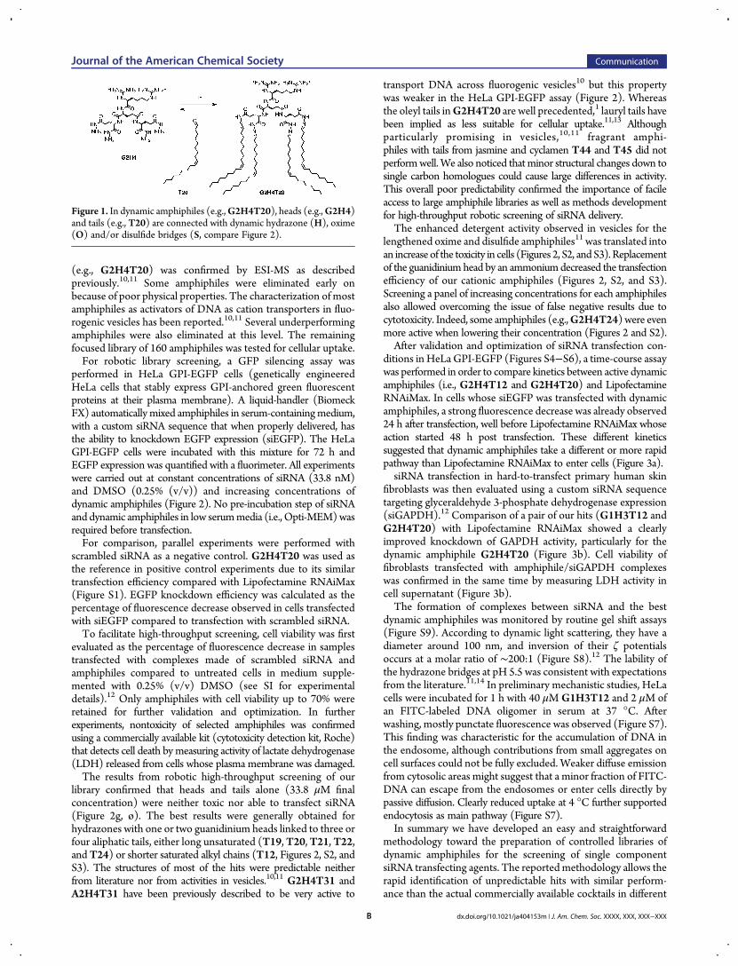
Figure 1. In dynamic amphiphiles (e.g.,G2H4T20), heads (e.g.,G2H4)and tails (e.g., T20) are connected with dynamic hydrazone (H), oxime(O) and/or disulfide bridges (S, compare Figure 2).
Journal of the American Chemical Society Communication
dx.doi.org/10.1021/ja404153m | J. Am. Chem. Soc. XXXX, XXX, XXX−XXXB

cells lines. siRNA-mediated enzyme knockdown in humanprimary fibroblasts almost doubled to >60% (compared to >30%
for Lipofectamine RNAiMax). Endocytosis of the siRNA seemsto be a major pathway of internalization, but kinetics experiments
Figure 2. (a) Structure of 18 heads and 50 tails used to build a formal library of 900 amphiphiles. (b−g) In vitro screening of 160 dynamic amphiphiles forsiRNA delivery. HeLa cells expressing GPI-EGFP were treated with EGFP-targeting siRNA mixed with dynamic amphiphiles. The average percentreduction in EGFP expression is shown after treatment with siRNA (33.8 nM) in the presence of (b) 3.4, (c) 6.8, (d) 13.5, (e) 20.3, (f) 27.0, and(g) 33.8 μM dynamic amphiphiles in the medium (in triplicate). Gray: >50% of cytotoxicity; from yellow to red: 0−20, 20−40, 40−60, 60−80, and80−100% GFP knockdown.
Journal of the American Chemical Society Communication
dx.doi.org/10.1021/ja404153m | J. Am. Chem. Soc. XXXX, XXX, XXX−XXXC

indicate that siRNA uptake is much faster than with Lipofect-amine RNAiMax. Therefore, it is possible that the effectiveamphiphile population enters the cell by a more direct route thanendocytosis. The effortless library preparation and the simplicityof transfection screening certainly certify the potential ofdynamic amphiphiles as controlled delivery vehicles.
■ ASSOCIATED CONTENT*S Supporting InformationDetailed experimental procedures. This material is available freeof charge via the Internet at http://pubs.acs.org.
■ AUTHOR INFORMATIONCorresponding [email protected]; [email protected] Addresses∥J.M., A.C.: Departamento de Quimica Organica y CentroSingular de Investigacion en Quimica Biologica y MaterialesMoleculares, University of Santiago de Compostela, Santiago deCompostela, Spain⊥S.T.: Department of Medicinal Chemistry, Tokyo University ofPharmacy and Life Sciences, Hachioji, Tokyo 192-0392, Japan#H.H.: Institut de Pharmacologie Moleculaire et Cellulaire,CNRS, Valbonne, FranceAuthor Contributions§C.G. and J.M. contributed equally.NotesThe authors declare no competing financial interest.
■ ACKNOWLEDGMENTSWe thank A. Fin, D.-H. Tran, Q. Verolet, S. Soleimanpour, and O.Kucherak for assistance with synthesis and sample preparation;NMR and MS Platforms and the Biomolecular Screening Facility(EPFL) for services; and the University of Geneva, the EuropeanResearch Council (ERC Advanced Investigator), the NCCRChemical Biology, and the Swiss NSF for financial support. H.H.is grateful for a JSPS Research Fellowship for Young Scientists.
■ REFERENCES(1) (a) Semple, S. C.; Akinc, A.; Chen, J.; Sandhu, A. P.; Mui, B. L.;Cho, C. K.; Sah, D. W. Y.; Stebbing, D.; Crosley, E. J.; Yaworski, E.;Hafez, I. M.; Dorkin, J. R.; Qin, J.; Lam, K.; Rajeev, K. G.; Wong, K. F.;
Jeffs, L. B.; Nechev, L.; Eisenhardt, M. L.; Jayaraman, M.; Kazem, M.;Maier, M. A.; Srinivasulu, M.; Weinstein, M. J.; Chen, Q.; Alvarez, R.;Barros, S. A.; De, S.; Klimuk, S. K.; Borland, T.; Kosovrasti, V.; Cantley,W. L.; Tam, Y. K.; Manoharan, M.; Ciufolini, M. A.; Tracy, M. A.; deFougerolles, A.; MacLachlan, I.; Cullis, P. R.; Madden, T. D.; Hope, M. J.Nat. Biotechnol. 2010, 28, 172. (b) Mevel, M.; Kamaly, N.; Carmona, S.;Oliver, M. H.; Jorgensen, M. R.; Crowther, C.; Salazar, F. H.; Marion, P.L.; Fujino, M.; Natori, Y.; Thanou, M.; Arbuthnot, P.; Yaouanc, J.-J.;Jaffres, P. A.; Miller, A. D. J. Controlled Release 2010, 143, 222. (c) Davis,M. E.; Zuckerman, J. E.; Choi, C. H. J.; Seligson, D.; Tolcher, A.; Alabi,C. A.; Yen, Y.; Heidel, J. D.; Ribas, A.Nature 2010, 464, 1067. (d) Akinc,A.; Zumbuehl, A.; Goldberg, M.; Leshchiner, E. S.; Busini, V.; Bacallado,S. A.; Hossain, N.; Alvarez, R.; Borodovsky, A.; Borland, T.; Constien,R.; de Fougerolles, A.; Dorkin, J. R.; Jayaprakash, K. N.; Jayaraman, M.;John, M.; Koteliansky, V.; Manoharan, M.; Nechev, L.; Qin, J.; Racie, T.;Raitcheva, D.; Rajeev, K. G.; Sah, D. W.; Soutschek, J.; Toudjarska, I.;Vornlocher, H.-P.; Zimmermann, T. S.; Langer, R.; Anderson, D. G.Nat.Biotechnol. 2008, 26, 561. (e) Zhang, X.-X.; LaManna, C. M.; Kohman,R. E.; McIntosh, T. J.; Han, X.; Grinstaff, M. W. Soft Matter 2013, 9,4472. (f)McCarthy, J.; O’Neill, M. J.; Bourre, L.;Walsh, D.; Quinlan, A.;Hurley, G.; Ogier, J.; Shanahan, F.; Melgar, S.; Darcy, R.; O’Driscoll, C.M. J. Controlled Release 2013, 168, 28. (g) Chen, D.; Love, K. T.; Chen,Y.; Eltoukhy, A. A.; Kastrup, C.; Sahay, G.; Jeon, A.; Dong, Y.;Whitehead, K. A.; Anderson, D. G. J. Am. Chem. Soc. 2012, 134, 6948.(2) Bhattacharya, S.; Bajaj, A. Chem. Commun. 2009, 45, 4632.(3) (a) Martin, B.; Sainlos, M.; Aissaoui, A.; Oudrhiri, N.;Hauchecorne, M.; Vigneron, J.-P.; Lehn, J.-M.; Lehn, P. Curr. Pharm.Design 2005, 11, 375. (b) Boomer, J. A.; Qualls, M.M.; Inerowicz, H. D.;Haynes, R. H.; Patri, V. S.; Kim, J. M.; Thompson, D. H. BioconjugateChem. 2009, 20, 47. (c) Cui, L.; Cohen, J. L.; Chu, C. K.; Wich, P. R.;Kierstead, P. H.; Frechet, J. M. J. J. Am. Chem. Soc. 2012, 134, 15840.(d) Pijper, D.; Bulten, E.; Smisterova, J.; Wagenaar, A.; Hoekstra, D.;Engberts, J. B. F. N.; Hulst, R. Eur. J. Org. Chem. 2003, 4406. (e) Prata, C.A. H.; Zhao, Y. X.; Barthelemy, P.; Li, Y. G.; Luo, D.; McIntosh, T. J.;Lee, S. J.; Grinstaff, M. W. J. Am. Chem. Soc. 2004, 126, 12196.(f) Barnard, A.; Posocco, P.; Pricl, S.; Calderon, M.; Haag, R.; Hwang,M. E.; Shum, V. W. T.; Pack, D. W.; Smith, D. K. J. Am. Chem. Soc. 2011,133, 20288. (g) Zhu, J.; Munn, R. J.; Nantz, M. H. J. Am. Chem. Soc.2000, 122, 2645. (h) Pierrat, P.; Kereselidze, D.; Wehrung, P.; Zuber,G.; Pons, F.; Lebeau, L. Pharm. Res. 2013, 30, 1362. (i) Aytar, B. S.;Muller, J. P. E.; Kondo, Y.; Talmon, Y.; Abbott, N. L.; Lynn, D. M. J. Am.Chem. Soc. 2013, DOI: 10.1021/ja403546b. (j) Tang, F.; Hughes, J. A.Biochem. Biophys. Res. Commun. 1998, 242, 141.(4) (a) Aissaoui, A.; Martin, B.; Kan, E.; Oudrhiri, N.; Hauchecorne,M.; Vigneron, J.-P.; Lehn, J.-M.; Lehn, P. J. Med. Chem. 2004, 47, 5210.(b) Xia, W.; Low, P. S. J. Med. Chem. 2010, 53, 6811. (c) Zhang, Y.;Satterlee, A.; Huang, L. Mol. Ther. 2012, 20, 1298. (d) Rehman, Z.;Zuhorn, I. S.; Hoekstra, D. J. Controlled Release 2013, 166, 46. (e) Chan,C.-L.; Majzoub, R. N.; Shirazi, R. S.; Ewert, K. K.; Chen, Y.-J.; Liang, K.S.; Safinya, C. R. Biomaterials 2012, 33, 4928.(5) (a) Bang, E.-K.; Gasparini, G.; Molinard, G.; Roux, A.; Sakai, N.;Matile, S. J. Am. Chem. Soc. 2013, 135, 2088. (b) Bang, E.-K.; Lista, M.;Sforazzini, G.; Sakai, N.; Matile, S. Chem. Sci. 2012, 3, 1752.(6) Otto, S. Acc. Chem. Res. 2012, 45, 2200.(7) Butterfield, S. M.; Miyatake, T.; Matile, S. Angew. Chem., Int. Ed.2009, 48, 325.(8) Herrmann, A. Chem.Eur. J. 2012, 18, 8568.(9) Minkenberg, C. B.; Li, F.; van Rijn, P.; Florusse, L.; Boekhoven, J.;Stuart, M. C. A.; Koper, G. J. M.; Eelkema, R.; van Esch, J. H. Angew.Chem., Int. Ed. 2011, 50, 3421.(10) Takeuchi, T.; Montenegro, J.; Hennig, A.; Matile, S. Chem. Sci.2011, 2, 303.(11) Montenegro, J.; Bang, E.-K.; Sakai, N.; Matile, S. Chem.Eur. J.2012, 18, 10436.(12) See SI.(13) (a) Nishihara, M.; Perret, F.; Takeuchi, T.; Futaki, S.; Lazar, A. N.;Coleman, A. W.; Sakai, N.; Matile, S. Org. Biomol. Chem. 2005, 3, 1659.(b) Som, A.; Reuter, A.; Tew, G. N. Angew. Chem., Int. Ed. 2012, 51, 980.(14) Kalia, J.; Raines, R. T. Angew. Chem., Int. Ed. 2008, 47, 7523.
Figure 3. (a) GFP knockdown (top) and cell viability (bottom) 24, 48,and 72 h after forward transfection of EGFP-targeting siRNA in HeLaGPI-EGFP with RNAiMax (blue, bar 1), G2H4T12 (green, bar 2),G2H4T20 (light green, bar 3) and DMSO (gray, bar 4). (b) Decrease inGAPDH activity (bars) and cell viability (■) 72 h after forwardtransfection of GAPDH-targeting siRNA in human primary skin fibroblastswith RNAiMax, G1H3T12, G2H4T20, and DMSO (left to right).
Journal of the American Chemical Society Communication
dx.doi.org/10.1021/ja404153m | J. Am. Chem. Soc. XXXX, XXX, XXX−XXXD


