Electric Field Detection in Sawfish and Shovelnose RaysBarbara E. Wueringer1,2*, Lyle Squire Jnr3, Stephen M. Kajiura4, Ian R. Tibbetts5, Nathan S. Hart4,
Shaun P. Collin1,4,6
1 The University of Queensland, School of Biomedical Sciences, Brisbane, Queensland, Australia, 2 The University of Western Australia, School of Animal Biology and the
UWA Oceans Institute, Crawley WA, Australia, 3 Cairns Marine, Stratford, Queensland, Australia, 4 Biological Sciences, Florida Atlantic University, Boca Raton, Florida,
United States of America, 5 The University of Queensland, School of Biological Sciences, Brisbane, Queensland, Australia, 6 The University of Queensland, Queensland
Brain Institute, Brisbane, Queensland, Australia
Abstract
In the aquatic environment, living organisms emit weak dipole electric fields, which spread in the surrounding water.Elasmobranchs detect these dipole electric fields with their highly sensitive electroreceptors, the ampullae of Lorenzini.Freshwater sawfish, Pristis microdon, and two species of shovelnose rays, Glaucostegus typus and Aptychotrema rostrata weretested for their reactions towards weak artificial electric dipole fields. The comparison of sawfishes and shovelnose rayssheds light on the evolution and function of the elongated rostrum (‘saw’) of sawfish, as both groups evolved from ashovelnose ray-like ancestor. Electric stimuli were presented both on the substrate (to mimic benthic prey) and suspendedin the water column (to mimic free-swimming prey). Analysis of around 480 behavioural sequences shows that all threespecies are highly sensitive towards weak electric dipole fields, and initiate behavioural responses at median field strengthsbetween 5.15 and 79.6 nVcm21. The response behaviours used by sawfish and shovelnose rays depended on the location ofthe dipoles. The elongation of the sawfish’s rostrum clearly expanded their electroreceptive search area into the watercolumn and enables them to target free-swimming prey.
Citation: Wueringer BE, Squire L Jnr, Kajiura SM, Tibbetts IR, Hart NS, et al. (2012) Electric Field Detection in Sawfish and Shovelnose Rays. PLoS ONE 7(7): e41605.doi:10.1371/journal.pone.0041605
Editor: Vincent Laudet, Ecole Normale Superieure de Lyon, France
Received April 11, 2012; Accepted June 22, 2012; Published July 25, 2012
Copyright: � 2012 Wueringer et al. This is an open-access article distributed under the terms of the Creative Commons Attribution License, which permitsunrestricted use, distribution, and reproduction in any medium, provided the original author and source are credited.
Funding: This study was funded by the Endeavour Europe Award to BEW, MBRS Student Funding to BEW, Sea World Research and Rescue Foundation Inc.funding to BEW and SPC, ARC Linkage grant no. LP0989676 to SPC. The funding agencies had no role in study design, data collection and analysis, decision topublish or preparation of the manuscript.
Competing Interests: One author is affiliated with Cairns Marine, but this does not alter the authors’ adherence to PLoS ONE policies on sharing data andmaterials.
* E-mail: [email protected]
Introduction
Elasmobranchs use electroreception to navigate in the earth’s
magnetic field and to detect inanimate objects and living
organisms such as predators, prey and conspecifics [1]. Scalloped
hammerhead sharks Sphyrna lewini are thought to follow the
geomagnetic field lines during their diurnal migrations to and from
seamounts in the Pacific Ocean [2]. Johnson et al. [3] conditioned
juvenile nurse sharks Ginglymostoma cirratum to successfully detect
and retrieve metallic spheres in a tank in the presence of a
background electric field. Round stingrays, Urolophus halleri use the
electric field produced by buried conspecifics to orient themselves
in order to optimize social interactions in the mating season [4].
Moreover, embryos of clearnose skate Raja eglanteria display a
freeze response while still encased in their egg purse, and cease all
ventilatory movement during the approach of an external electric
field resembling a predator [5].
In the context of prey capture, electroreception provides
elasmobranchs with the ability to precisely locate prey and hunt
in both the dark and/or in turbid waters, opening up a rich
ecological predatory niche [6]. Naturally occurring localized
dipole sources in the aquatic environment only originate from
living animals and therefore their presence equates to the presence
of another organism [7]. Various species of sharks and rays have
been shown to readily attack weak electric fields both in captivity
and under natural conditions [1,8–12]. The electro-location task
can be divided into three components, i.e. detection, character-
ization and localization [6]. These processes can be described
separately although they are tightly coupled and synchronous in
the nervous system [6].
Electroreceptive cues differ from stimuli passively perceived
with other sensory organs, as they do not provide the receptor with
a temporal component or propagation velocity vectors. The
frequencies of biologically important electrosensory cues range
from almost DC to a few 100 Hz, resulting in a wavelength of
several kilometres [13]. As a result, electric fields propagate with
nearly infinite speed, and are present throughout their full extent
almost instantaneously [13,14]. The biologically important char-
acteristics encoded in electric stimuli are the local intensity,
orientation and the polarity of a field [14]. Electric flux lines
describe a curved path along the direction of the current and do
not point straight to their source. Behavioural experiments
indicate that the stimulus frequency ranging from DC up to
8 Hz has little, if any significance for behaviour, as electrorecep-
tive predators attack artificial dipoles provided as long as their
frequencies are within the detectable range [14,15].
Physiologically, the ampullae of Lorenzini, which are the
electroreceptors of elasmobranchs, are not true DC receptors,
and this characteristic is important for their normal mode of
operation within the animal’s own DC background field [1,14,15].
In order to sense the DC field produced by prey, elasmobranchs
must move with respect to their prey [1,14,15].
PLoS ONE | www.plosone.org 1 July 2012 | Volume 7 | Issue 7 | e41605
Here, we compare the electroreceptive abilities and behaviours
of a sawfish (Pristis microdon) and two shovelnose rays (Aptychotrema
rostrata and Glaucostegus typus), which all evolved from a rhinobatid-
like ancestor [16–19]. The morphology of the ampullary systems
of all three species is known [20,21]. P. microdon possesses one of
the highest numbers of electroreceptors of any elasmobranch (and
twice as many pores as A. rostrata and G. typus [20]) and may thus
be considered an electroreception specialist [21]. They possess
more ampullary pores ventrally than dorsally, but dorsally pores
are concentrated along the rostral cartilage [21]. In both species of
shovelnose rays, ampullary pores on the ventral side of the head
are about five times more numerous than on the dorsal side, with
the highest concentrations on the ventral rostrum [20].
Even though commonly referred to as freshwater sawfish,
juvenile P. microdon occupy oligohaline to mesohaline low visibility
environments, while adults may move into saltwater [18,22].
Consequently electroreception may be especially important for the
detection and manipulation of prey by juvenile sawfish. Their diet
is dominated by benthopelagic teleosts and prawns of the genus
Macrobrachium spp. [22]. We thus hypothesise that freshwater
sawfish will orient towards artificial electric fields and that they will
be able to detect and respond to dipoles presented in the water
column, whereas rhinobatid shovelnose rays will not. Rhinobatid
shovelnose rays tend to inhabit clearer water and forage on
benthic invertebrates. The diet of G. typus is dominated by
brachyuran crabs and penaeid shrimp, which make up more than
50% of the relative importance of food items in animals below and
above 150 cm TL [23]. The diet of A. rostrata is dominated by
penaid prawns and carid shrimps, which make up more than 50%
of the index of relative importance [24].
Methods
Study SpeciesFreshwater sawfish Pristis microdon. Nineteen juvenile
freshwater sawfish Pristis microdon (12 males and 7 females, total
length between 96.0 and 208.0 cm) were captured from their
natural habitat in far North Queensland (Norman River, S 17u389,
E 141u09, Wenlock River, S 12u169, E 141u589) and transported to
the holding facility of Cairns Marine (Cairns, Queensland,
Australia) according to the company’s protocols and approval by
the UQ Animal Ethics Committee. In general, sawfish began to
feed after three days in captivity and were given at least another
three days of acclimation before experiments commenced.
The holding tanks, which also served as experimental tanks,
were made of fibreglass (diameter of 4 m, water depth of 80 cm).
The tanks rested on timber, which insulated them from the
ground. The water pump was earthed. Three to five sawfish were
kept together in each tank depending on their size. To minimize
stress in animals and also to minimize handling of potentially
dangerous animals, sawfish were not separated for behavioural
trials.
Rhinobatid shovelnose rays. Shovelnose rays belonged to
two species, namely the giant shovelnose ray Glaucostegus typus (20
juveniles, total length between 38.0 and 180.0 cm) and the eastern
shovelnose ray Aptychotrema rostrata (7 juveniles and adults, total
length between 44.0 and 76.5 cm). They were caught in marine
environments off Heron Island, Shark Bay (S 23u319, E 152u19),
off Adams Beach, North Stradbroke Island (S 27u299, E 153u249)
and off Double Island (S 16u439, E 145u419), Queensland,
Australia. All specimens were fed to satiation after capture and
then starved for 3 to 5 days before trials commenced to render
them in the same nutritional state. Animals were tested in the
holding tanks of the Heron Island Research Station (dimensions
464 m, water depth 50 cm), the Moreton Bay Research Station
(diameter 4 m, water depth 120 cm) and Cairns Marine (diameter
4 m, water depth 80 cm). All specimens caught on Heron and
North Stradbroke islands were released after trials ended (up to
28 days after capture).
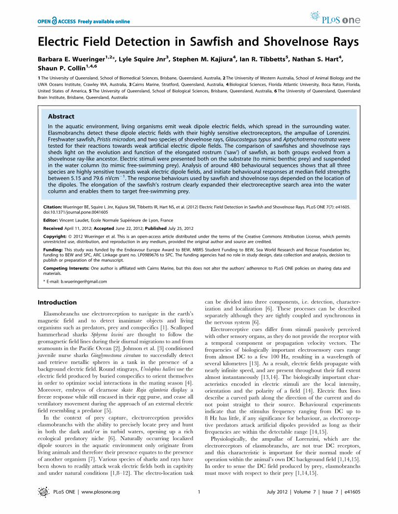
Stimulus DeliveryThe current-delivering device was modified after Kajiura and
Holland [10]. An electric circuit (figure 1a) delivered a constant
electric current. The strength of the current was varied with two
variable resistors. A multimeter (Fluke Electronics, 116 HVAC
Multimeter) was connected in series to measure the generated
electric current. Each electrode was made of a silver pin soldered
to a shielded coaxial cable (RG58C/U, Dick Smith Electronics)
that was plugged into the stimulus generator. The cable/silver
interface was shielded with marine silicone and shrink tubes so that
only the 5 mm long tip of the silver pin was in contact with the
water. Each silver pin extended into a transparent plastic tube
(length 50 cm, inner diameter 3 mm) filled with seawater, which
acted as a salt bridge. Both the shield of the cable and the electric
circuit were earthed.
Ten specimens of Glaucostegus typus were tested with a slightly
different experimental set up that used 5 mm long electrode tips
made of silver coated with chlorox solution (Ag/AgCl) to minimize
polarisation currents, but not salt-bridges. Electrodes were re-
coated between trials, and the two electrodes of a dipole were
spaced 1 cm apart.
Electric fields presented on the substrate. The experi-
mental set up was modified after Kajiura and Holland [10]. Four
dipoles made of eight electrodes were mounted on the underside of
a rubber mat, with only their ends protruding through the rubber
(Clark Rubber, 138 cm6138 cm, 1 mm thick, see figure 1b). The
distance between the two electrodes of a dipole was 2 cm and the
distance between dipoles was 50 cm. The distance of 2 cm
between electrodes was chosen as it resembles the distance
between the gills of potential teleost prey [25]. A black circle
(radius 10 cm) was drawn around each dipole and the dipole axis
was marked with a black line, to visualise the dipole centre on
video footage. A plastic tube (inner diameter 3 mm) was mounted
in the centre of the rubber mat, equidistant to all dipoles. It
delivered an olfactory cue made of blended and filtered mullet
chum via a syringe, which induced food searching behaviour and
also attracted the rays to the rubber mat. The rubber mat also
extended onto the tank wall, covering the coaxial cables and the
rubber tube to prevent entanglement of the animals.
Presentation of electric fields in the water column. The
dipole centres were suspended in the water column approximately
20 to 30 cm above the substrate. The same stimulus device was
used but dipoles were hung from a wooden pole that was laid
horizontally across the tank above the water line. To ensure even
spacing of the two plastic tubes of a salt-bridge, a plastic tube with
an outer diameter of 2 cm was placed between them. The three
tubes were held in place, protected and surrounded by a fourth
plastic tube, on which a black line was drawn 10 cm from the
dipole. The two dipoles (one active and one control) were
separated by 130 cm and a plastic tube (inner diameter 3 mm,
length 5 m) extended to the same depth into the water half way
between them. This tube delivered chum via a syringe.
Experimental ProcedureThe experimental set up was lowered into the tank at random
times to get animals accustomed to its presence and to avoid
pseudo-conditioning and associations of the set up with either food
or electric fields. Animals were tested twice a day before feeding,
Electroreception in Sawfish and Shovelnose Rays
PLoS ONE | www.plosone.org 2 July 2012 | Volume 7 | Issue 7 | e41605
and each experimental session lasted 40 mins. During behavioural
trials only one dipole was active, while the three/one other dipoles
served as controls. Trials with dipoles on the bottom were filmed
with one remotely controlled video camera (Sony, DCR HC96E
or DCR HC5) in a water resistant case (Sony, SPKHCB) mounted
above the tank. Trials in which dipoles were suspended in the
water column were filmed with two cameras, one mounted above
the tank and the other one behind a camouflaged window in the
tank wall. A trial lasted for 5 mins and the active dipole was
switched randomly between trials.
The electric current used was also randomized and noted for
each trial. Sawfish were tested with currents of 18.9–50.0 mA and
21.6–80.3 mA for electric fields presented on the bottom and in the
water column, respectively. Shovelnose rays were tested with
currents of 11.7–28.4 mA for electric fields on the bottom.
Currents that tested the response of G. typus towards electric fields
in the water measured 24.3–90.7 mA. Shovelnose rays were tested
in saltwater (36.4–38.6 ppt), while sawfish were tested in brackish
and saltwater (15.5–35.2 ppt). After each experimental session all
animals were fed to satiation. Before and after each experimental
session, water temperature, salinity and conductivity were
measured with a TPS WP-84 conductivity meter.
Shovelnose rays were only tested in natural light conditions,
while sawfish were also tested in infared light, to test the possibility
that visual stimuli were responsible for any orientation responses.
As inactive dipoles also served as visual controls, we hypothesized
that the infrared light condition would not influence the results.
For infrared trials, the tank was covered with two layers of black
plastic sheeting (thickness of one layer 200mm). Four infrared lights
(Jaycar Electronics QC3652, each with 56 units of IR LEDs
emitting light of 850 nm wavelength which is near infrared light)
were mounted 5 cm above the water surface. Trials were filmed
with an infrared sensitive security camera (Jaycar Electronics
digital CCD Camera) mounted above the tank. Illuminance was
not measured directly but as visible on video clips, the camera
switches from normal to infrared mode automatically at light levels
between 0.24 and 0 LUX. These light levels correspond to natural
light levels measured in air during deep twilight (1 LUX) and full
moon (0.1 LUX).
Analysis of ResultsVideos of experiments were digitized using iMovie HD 6.0.3.
When viewing these clips, behavioural units that the animals
displayed during each response towards a dipole were noted and
characterized. Behavioural units were defined after Barlow [26]: a
modal action pattern is a natural, stereotypic unit of behaviour
that contains a variable, taxic component. Therefore, the modal
action pattern describes a spatio-temporal pattern of movement
that cannot be further subdivided [26]. The descriptive ethogram
that was established for each species is described in detail
elsewhere [27].
To estimate the detection threshold, the strength of the electric
field at the point of initiation of a behavioural orientation response
( = PIR) was estimated after the method of Kajiura and Holland
[10]. PIR is defined as the point where an animal initiates a turn of
more than 20u towards the dipole. The respective frame was saved
from each video clip. In ImageJ 1.39u (http://rsb.info.nih.gov/ij/)
the following measurements were taken: the minimal distance r
[cm] from the centre of the dipole to the side of the head, taking
into account the distribution of the ampullae of Lorenzini [20,21],
and the angle a [u] between that point and the dipole axis. The
electric field strength E [nVcm21] at PIR was calculated using
Maxwell’s [28] electric field equation, E = (r.I.d. cosa)/(p.r3), in
which r is the resistivity of the water [V cm], d is the distance
between the two electrodes of a dipole [cm] and I is the electric
current [mA]. The equation was altered for the calculation of the
field strength at PIRs towards dipoles located in the water column,
where the field resembles a sphere. The distribution of data points
for each treatment was tested with a Kolmogorof-Smirnov test for
normality with Lilefors correction or a Shapiro-Wilk test for
normality. As field strengths at PIR for all treatments were skewed
to the right, non-parametric methods were used for analyses.
Orientation responses during which multiple animals were
visible in the frame were excluded from analysis, to avoid
misjudgement of unrelated behaviours as orientation responses.
Only the first orientation response was analysed if an animal
repeatedly returned to a dipole after an initial bite without leaving
the frame. Moreover, only turns that resulted in further reactions
towards the dipole were analysed, to exclude randomly initiated
turns from analyses.
How often animals passed within 10 cm of a dipole on the
bottom (categorized as active, left of active, right of active and
opposite of active dipole) or touched a dipole in the water column
(categorized as active and inactive) without displaying any reaction
was noted together with the trial duration and current. Frequen-
cies were analysed with Chi2 and binomial tests to determine if
animals searched all electrodes equally or if they passed over the
active dipole more often than predicted by chance alone. All
Figure 1. The experimental set up. (a) The electric circuit that delivered a low electric current into the experimental tank via electrodes. Thestrength of the current was varied with the two variable resistors (b) Schematic drawing of the transparent rubber mat on which dipoles weremounted that delivered electric fields on the bottom of the tank. Double layers of rubber are indicated with dashed lines. Cables were protected bythese layers to prevent entanglement of the animals. The two plastic tubes of one dipole were oriented approx. perpendicular to the centre of thedipole to avoid the build up of electric fields along the tubes.doi:10.1371/journal.pone.0041605.g001
Electroreception in Sawfish and Shovelnose Rays
PLoS ONE | www.plosone.org 3 July 2012 | Volume 7 | Issue 7 | e41605
statistical procedures were completed using Microsoft Excel 12.1.3
and PAWS 18.0 and follow Sokal and Rohlf [29].
Results
Reactive sequences are distinguished based on the location of
the dipoles, which can be on the bottom (P. microdon n = 146, A.
rostrata n = 60, G. typus n = 26), or in the water column (P. microdon
n = 57, G. typus n = 60). An additional 134 reactions were recorded
for G. typus reacting towards dipoles made of Ag/AgCl electrodes
presented on the bottom. Inactive dipoles presented on the
substrate were bitten once by P. microdon and once by G. typus.
Three times, sawfish bit the end of the bottom chum tube after
passing it when the mullet chum was released into the water. P.
microdon did not react to inactive dipoles suspended in the water
column. The reactions of G. typus towards dipoles in the water
column are discussed separately.
Behavioural AnalysisThe modal action patterns that sawfish and shovelnose rays
displayed are described in detail elsewhere [27], and are only
mentioned briefly here with their occurrence probabilities (po).
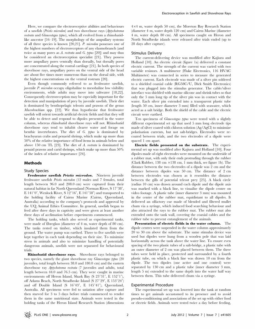
The behaviours that sawfish displayed when attacking the active
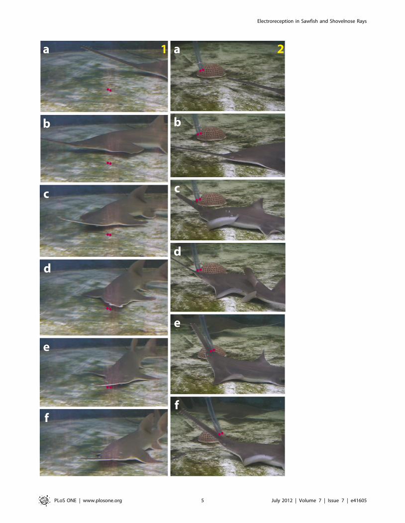
dipole depended on the location of the dipole (figure 2). Sawfish bit
dipoles on the bottom (po = 0.90, figure 2 section 1) but produced
fast lateral swipes aimed at dipoles located in the water column
(po = 0.81, figure 2 section 2). The only modal action pattern to
occur in both treatments is wiggle (on bottom po = 0.10, in water
column po = 0.19). During wiggle a sawfish produces a slight lateral
movement of the head.
When reacting to an active dipole on the bottom, shovelnose
rays always bit its centre (po = 1.00). Glaucostegus typus displayed two
modal action patterns towards dipoles suspended in the water
column that were neither displayed towards bottom dipoles nor by
sawfish, namely repeated bumps (po = 0.29, the dipole is repeatedly
bumped into with the rostrum) and spiral (a shovelnose ray swims
in a circle while maintaining contact between the side of the head
and the dipole). Spiral was displayed both towards the active
(po = 0.59) and inactive dipole (po = 0.12).
Detection Threshold of the Electric FieldThe median electric field strength (6 std. dev.) at PIR of P.
microdon for electric fields located on the bottom was
13.46700.1 nVcm21 (n = 118). The median electric field strength
at PIR for dipoles suspended in the water equalled
10.06166.2 nVcm21 (n = 18). The median electric field strengths
at PIR did not differ significantly between the treatments (dipoles
on bottom vs. dipoles in water column; Mann-Whitney U-test,
n = 118, z = 20.76 p = 0.45) and so the two subsamples were
pooled for further analysis. The median field strength at PIR of the
pooled data was 13.06640.0 nVcm21. There was also no
significant difference between the electric field strength at PIR in
daylight or infrared light trials (Mann-Whitney-U test, n = 118,
z = 21.74, p = 0.082).
Juvenile freshwater sawfish were tested for their reactions
towards weak electric fields in salinities between 15.5 and 35.2 ppt,
with a mean salinity of 28.364.8 ppt. To examine a possible
correlation between the field strength at PIR and salinity, data
were sorted by the following groups: 15–20 ppt, 20–25 ppt, 25–
30 ppt, 30–35 ppt. Field strength medians were compared
between groups via a Kruskal-Wallis test, which was corrected
for tied ranks. The results indicate that there is no significant
difference between the medians (X2 (3, N = 118) = 3.079, p = 0.38)
of different salinity treatments and therefore data were pooled.
The median electric field strength at PIR for electric fields
located on the bottom is 79.626126.51 nVcm21 for A. rostrata and
25.336424.78 nVcm21 for G. typus, respectively. The median
electric field strength at the PIR for ten specimens of G. typus
reacting towards electric fields on the bottom produced by Ag/
AgCl electrodes is 5.156202.9 nVcm21, which is significantly
lower than the median field strength at PIR for reactions towards
dipoles made of salt bridges (Mann-Whitney U-test, p,0.01). No
electric field strength at PIR could be calculated for responses by
G. typus towards electric fields suspended in the water, since the
reactions were quite erratic and a point of initiation of the response
could not be determined.
The median electric field strength at the PIR was compared
between test species for electric fields located on the bottom
produced with salt bridge electrodes. The median field strength
(median test, median = 17 nVcm21, df = 2, n = 141, p = 0.051)
does not differ across species.
When sawfish grow, the length of ampullary canals increases
[21]. As longer canals create a larger potential difference between
the inside and outside of the animal, longer ampullary canals are
predicted to increase the animal’s sensitivity to any electric field
[30]. To test this prediction, animals in analysis videos were
assigned to one of three different size classes (small, TL 96–
132 cm, medium TL 142–159 cm, large TL 178–208 cm).
However, neither the distribution of the detection threshold
electrical field strengths nor the median detection threshold
electric field strengths differed significantly among the groups
(df = 2, n = 112, Kruskal-Wallis test p = 0.080, median test
p = 0.401).
Correlation of the Distance between the PIR and theAngle to Dipole Axis
The electric field around a dipole is not uniform, as the electric
flux originates in the positive electrode and ends in the negative
one. Equipotential surfaces are perpendicular to the lines of
electric flux. As a result the electric field around a dipole at a given
distance from the dipole is strongest at a small angle to the dipole
axis and theoretically reaches zero perpendicular to the dipole
axis. A linear regression was performed to test if the approach
angle could predict the distance at PIR. However, the distance
between the dipole and the PIR was less than 3% predictable by
the angle of approach when all treatments (bottom and water
column, four salinity groups) were pooled (R2 = 0.03). Therefore
the approach angle does not significantly influence the distance at
which an orientation turn is initiated (ANOVA, F(1,116) = 2.2,
p = 0.14). Data separated by the treatments did not show
correlations either (figure 3a and 3b).
No strong correlation was found between the distance at PIR
and the angle between the PIR and the dipole axis, as indicated by
the Kendall t correlation coefficient (A. rostrata t= 20.06, not
significant at p = 0.63; G. typus t= 20.21 not significant at
p = 0.33). However, a small but slight trend is visible for all
species, in that the distance of PIR is larger at smaller angles (see
Figure 3).
Passes Under DipolesThere was no significant difference in how often sawfish passed
over active and inactive dipoles on the bottom (Chi2 Test,
p = 0.20, df = 3, n = 118), or how often they touched the touched
active and inactive dipoles in the water column (Binomial test,
n = 45, p = 0.37). Glaucostegus typus did not pass over any dipole on
the bottom more frequently than predicted by chance (binomial
test, p = 0.62, df = 36), and neither did A. rostrata (binomial test,
p = 0.1, df = 75). Accordingly, there was no difference between the
Electroreception in Sawfish and Shovelnose Rays
PLoS ONE | www.plosone.org 4 July 2012 | Volume 7 | Issue 7 | e41605
Electroreception in Sawfish and Shovelnose Rays
PLoS ONE | www.plosone.org 5 July 2012 | Volume 7 | Issue 7 | e41605

frequencies of G. typus passing under or touching the active or the
inactive dipole suspended in the water column (pass under dipoles:
binomial test, p = 0.24, df = 58; touch dipoles: binomial test,
p = 0.24, df = 18).
Discussion
The specific uses of the different sensory modalities of
elasmobranchs are better understood when classifying them
according to their detection ranges, which are determined by
the sensory capabilities, the stimulus strength and the physical
characteristics of the environment [31]. Both olfactory and
acoustic stimuli are detectable over long ranges and can attract
sharks from large distances. Gustation and touch are close range
senses that need physical contact for stimulus detection [31].
Electroreception, vision and mechanoreception function at medi-
um range, and are thus useful in prey capture. Within the medium
range senses, the effective detection distance of vision in the
underwater environment is the furthest, as it can be up to 100 m
depending on the habitat visibility. Mechanoreceptive stimuli are
detected by fish within a distance of about one body length [32].
The detection ranges of electroreceptive stimuli depend on the
stimulus strength. Sharks and rays respond to prey simulating
electric fields from distances of around 40 cm, which corresponds
to stimulus strengths of the order of 1 nVcm21 to 1 mVcm21
[10,28].
In this study, an olfactory stimulus was introduced into the tank
to elicit food-searching behaviour. As the chum delivering tube
was located some distance from the centre of the dipoles and was
rarely attacked, we do not consider that olfaction was used to
locate the source of the electric field. This is emphasised by the fact
that sharks (Carcharhinus menisorrah = falciformis) need an additional
stimulus to induce feeding, after the location of the source of an
olfactory cue has been confirmed [33].
A turn of more than 20u was assigned to be a ‘single turn’,
following the criteria of Kajiura and Holland [10] for comparative
purposes. The angle of 20u is clearly discernible to the observer
and is larger than the turns that sawfish and shovelnose rays make
during normal swimming movements. The location from where an
animal approaches the active dipole is guided by chance, but also
depends on the prevailing water currents in the tank that distribute
the olfactory cue. Moreover, if multiple animals are present in the
tank, they tend to synchronize their swimming direction.
Shovelnose rays and sawfish both responded equally to bottom
dipoles with a bite. The bite indicates the importance of
electroreception in guiding an elasmobranch to its prey. To date,
all elasmobranchs tested for their response towards electric dipoles
displayed either a biting response or produced suction feeding
movements [1,8–11,15,28,34–36], but freshwater sawfish also
produce lateral swipes of their rostrum towards dipoles suspended
in the water column [27]. Benthic skates are highly electro-
receptive [37–40], but Raja erinacea failed to react to the vertical
component of an electric field: when presented with a dipole above
the pectoral fins, the animals instead attacked a location on the
substrate under the pectorals fin directly below the centre of the
suspended dipole, apparently unable to interpret a signal with a
vertical component [7]. Thus sawfish, which are generally
considered to be sluggish benthic dwellers [41], may actually be
agile, demersal predators.
The occurrence of lateral swipes of the rostrum aimed at the
active dipole, and of wiggle in sawfish and not in shovelnose rays
indicates that these behaviours may have evolved in relation to the
evolution of the elongated rostrum [27]. The displays of spiral and
repeated bumps by shovelnose rays may indicate reactions to electric
dipoles in the water column, but the lack of skills to manipulate
them, as these skills evolved in sawfish together with the elongation
of the rostrum [27]. Thus, repeated bumps displayed by G. typus
towards dipoles in the water column may be an uncoordinated
predecessor of wiggle, a behaviour from which wiggle could have
evolved when sawfish evolved from shovelnose ray-like ancestors
[16–18,27]. The reactions by G. typus to dipoles suspended in the
Figure 2. Reaction sequence of juvenile sawfish towards electric dipoles. Dipoles (indicated with two red dots) are presented (1) on thesubstrate and (2) in the water column. The sawfish swims past the dipole and turns towards it (1a–b), wiggles the rostrum over the dipole centre (1c–d), approaches further (1e) and bites the dipole centre (1f). When reacting to the dipole suspended in the water, the sawfish almost passes the dipole(2a–b), turns towards it (2c) and produces ‘saw in water, (2d–f).doi:10.1371/journal.pone.0041605.g002
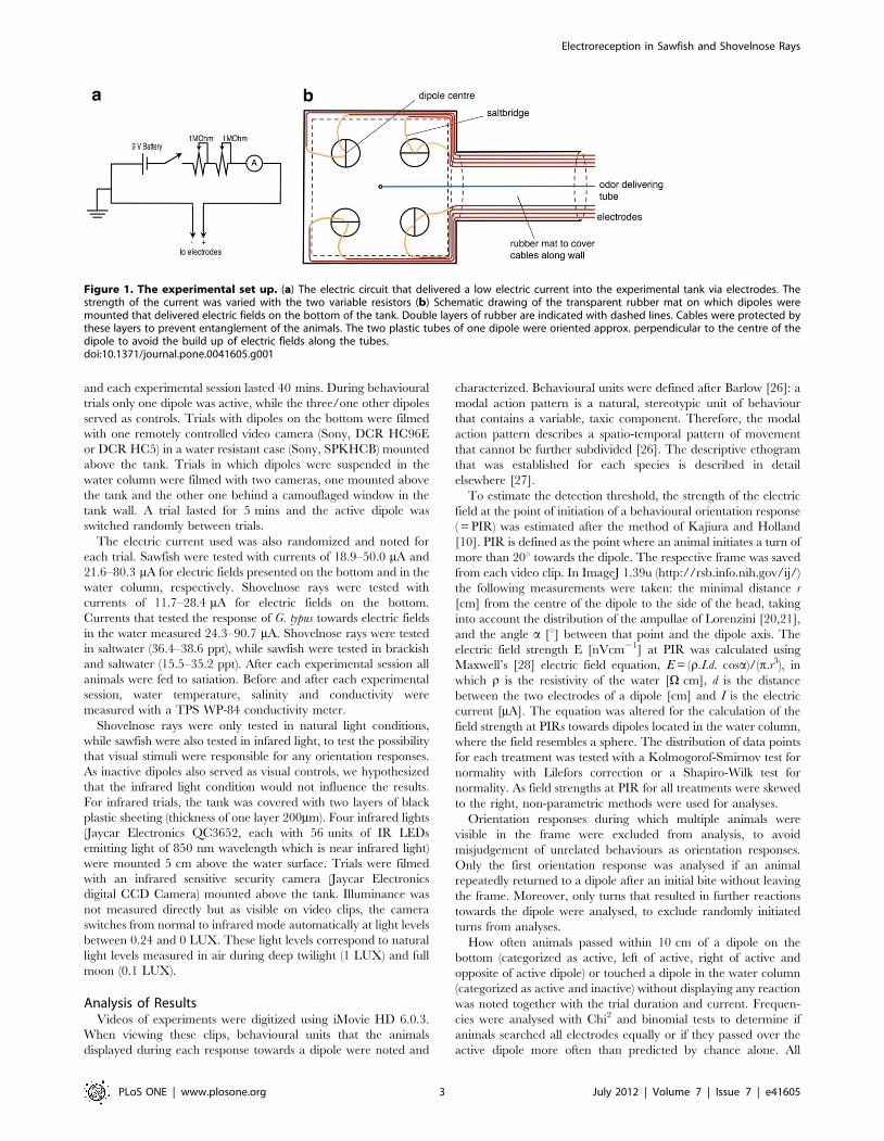
Figure 3. Scatterplot of the orientation distance [cm] plotted against the orientation angle [6] at PIR towards dipole electric fields.(a) Values measured for reactions by sawfish Pristis microdon and shovelnose rays Aptychotrema rostrata and Glaucostegus typus towards electricdipoles presented on the bottom, and (b) for P. microdon towards dipoles located on the bottom and in the water column. The orientation distancedecreases slightly at higher angles, but this relationship is not significant (see text for statistical analysis).doi:10.1371/journal.pone.0041605.g003
Electroreception in Sawfish and Shovelnose Rays
PLoS ONE | www.plosone.org 6 July 2012 | Volume 7 | Issue 7 | e41605

water column leave open the question about whether these
animals could attack prey in the water column. However, this
might depend on the type of prey and its mobility.
The display of spiral towards the inactive dipole presented in the
water column by G. typus is clearly distinguished from the display of
spiral towards the active dipole. When spiralling around the active
dipole, the head of the ray remains in contact with the end of the
dipole, where the centre of the electric field is located. When
spiralling around the inactive dipole, the shovelnose rays only ever
kept in contact with the middle of the plastic tube. This difference
may be random, but it may also indicate that shovelnose rays will
react to an obstruction in the water and investigate it, however, if
the obstruction is coupled with an electric field, this will be the
centre of investigation. Nevertheless, both repeated bumps and spiral
were quite erratic displays and so no further conclusions can be
drawn.
There are two possible explanations for shovelnose rays not
clearly reacting to electric dipoles presented in the water column:
Firstly, shovelnose rays may have lacked the necessary motivation.
As the field strengths were comparable to those that were readily
attacked on the substrate, this would indicate that the distance to
the substrate was unnaturally large for shovelnose rays. Secondly,
the stimulus in the water column may be not as easily detectable as
a stimulus on the substrate, as shovelnose rays possess fewer
ampullary pores dorsally than ventrally [20]. Both explanations
are strengthened by the fact that giant shovelnose rays feed mainly
on brachyuran crabs and penaeid shrimp [23].
The ampullary pore counts of P. microdon are twice as high as in
any other species of sawfish, and are also some of the highest in
elasmobranchs [21]. Pristis microdon are thus considered electro-
reception specialists [21]. As the visibility in the natural habitat of
juvenile freshwater sawfish in Australia can fall below 10 cm [42],
and sawfish can retract their eyes both during feeding and in
response to electric dipoles [27], we assume that electroreception
also guides the final strike during prey capture in wild freshwater
sawfish.
Behavioural Detection Threshold of Electric FieldThe median electric field strength PIR of freshwater sawfish lies
well within values reported for other elasmobranchs (Spyrna lewini
25.2 nVcm21, Carcharhinus plumbeus 30.0 nVcm21 [10], Sphyrna
tiburo 47.0 nVcm21 [11], Dasyatis sabina 5 nVcm21 [43], reviewed
in [12]). However, the present study finds that the field strength at
PIR does not differ between different size classes of P. microdon,
although it is assumed that the ampullary sensitivity increases with
the canal length [30]. A possible explanation is that the field
strength at PIR only indicates the detection threshold and not the
actual sensitivity of ampullary canals.
The ‘enhanced electroreception hypothesis’ by Kajiura and
Holland [10] predicts that the lateral expansion of the hammer-
head cephalofoil provides sphyrnid sharks with both an expanded
search area and increased sensitivity towards electric fields
compared to similar sized carcharhinid sharks. This hypothesis
can be modified to fit the present study in that the elongated
rostrum of sawfish has expanded the search area into the water
column and increased the sensitivity of the electroreceptive system
with an increase in canal length, compared to the closely-related
shovelnose rays. Moreover the high density of electroreceptors that
are evenly spread out along the whole length of the rostrum (both
ventrally and dorsally) [21] provides freshwater sawfish with high
electroreceptive resolution.
In keeping with the findings of McGowan and Kajiura [43],
there were no significant differences in electric field strength at
PIR in saltwater and brackish water treatments for juvenile
freshwater sawfish. The electroreceptive sensitivity of Dasyatis
sabina is reduced in freshwater compared to saltwater and brackish
water, although the strength of the electric field in freshwater is
increased [43]. This may be due to the increased electric
background noise levels in fresh water [14]. As freshwater sawfish
use freshwater and brackish waters as nurseries [18,44,45], while
adults prefer marine habitats, it would be interesting to repeat our
experiments in freshwater. This was not possible in the present
study due to husbandry constraints.
The strength of the electric field at PIR differed significantly
between the two species of shovelnose rays. A. rostrata reacted at
field strengths that are four times higher than the ones that G. typus
reacted to. There is no morphological explanation for the different
sensitivities of the two species, as their ampullae of Lorenzini are
quite comparable in morphology, distribution and number [20].
Therefore, a difference in motivation is suggested, which may be
related to the natural electric fields of the species’ prey items, but
this remains to be tested. The median electric field strength at PIR
for reactions by G. typus towards Ag/AgCl electrodes was even
more than twenty times higher than for reactions towards salt
bridge electrodes. The direct contact between the coated silver
pins and seawater of Ag/AgCl electrodes may have caused
polarisation currents that shovelnose rays could have detected.
This finding underlines the importance of using the same methods
as other authors when making comparisons between species.
This paper adds to our knowledge on the behavioural thresholds
of elasmobranchs in response to weak electric fields by expanding
the species and families tested. If applied in the captive
environment, the results of our study could provide an environ-
mental enrichment to rays that are normally fed with dead prey,
which lacks any electrical stimulus. The electroreceptive stimulus
presented in the water column should be of particular interest to
aquaria keeping sawfish, as the behavioural responses of juvenile
freshwater sawfish towards these prey-simulating stimuli differ
significantly from the behavioural responses elicited by dipoles
located on the bottom of the tank. Moreover, our findings show
that sawfish may not be benthic but demersal predators, based on
their response towards weak electric fields. As all species of sawfish
are Critically Endangered [46] understanding of these species’
predatory niches are essential for population recovery attempts.
Acknowledgments
This study was conducted under the following permits: UQ Animal Ethics
Committee permit Nos. VTHRC/717/06, VTHRC/835/07, VTHRC/
021/09, VTHRC/974/08/(NF); Department of Primary Industries and
Fisheries permit No. 87591. Great Barrier Reef Marine Parks Authorities
limited impact accreditation No.UQ001/2007. We thank the staff of
Cairns Marine, of the Moreton Bay Research Station and the Heron Island
Research Station. My volunteers were Vera Schluessel, Guy Ely, Steffen
Pauck, Sarah Stieb and Kurt. Thanks to John Page and Joe Stead for
providing shovelnose rays.
Author Contributions
Performed the experiments: BEW. Analyzed the data: BEW. Contributed
reagents/materials/analysis tools: LS SMK NSH IRT SPC. Wrote the
paper: BEW with corrections by all other authors. Provided animals, their
care and experimental space: LS. Project supervisors: SMK IRT SPC
NSH. Designed experiments: BEW SMK. Performed experiments and
analysed data: BEW. Provided animals their care and experimental space:
LS. Project supervisors: SMK IRT SPC NSH. Wrote the paper: BEW with
input from SPC SMK NSH.
Electroreception in Sawfish and Shovelnose Rays
PLoS ONE | www.plosone.org 7 July 2012 | Volume 7 | Issue 7 | e41605
References
1. Kalmijn AJ (1974) The detection of electric fields from inanimate and animate
sources other than electric organs. In: Fessard A, editor. Electroreceptors and
other specialized receptors in lower vertebrates. Berlin Heidelberg: Springer.147–200.
2. Klimley PA (1993) Highly directional swimming by scalloped hammerhead
sharks, Sphyrna lewini, and subsurface irradiance, temperature, bathymetry andgeomagnetic field. Mar Biol 117: 1–22.
3. Johnson CS, Scronce BL, McManus MW (1984) Detection of DC electric
dipoles in background fields by the nurse shark. J Comp Physiol A Sens Neur
Behav Physiol 155: 681–687.
4. Tricas TC, Michael SW, Sisneros JA (1995) Electrosensory optimization toconspecific phasing signals for mating. Neurosci Lett 202: 129–132.
5. Sisneros JA, Tricas TC, Luer CA (1998) Response properties and biological
function of the skate electrosensory system during ontogeny. J Comp
Physiol A Sens Neur Behav Physiol 183: 87–99.
6. Nelson M (2005) Target Detection, Image Analysis, and Modeling. In: BullockTH, Hopkins C, Popper AN, Fay RR, editors. Electroreception. New York:
Springer. 290–317.
7. Bodznick DA, Montgomery JC, Tricas TC (2003) Electroreception: Extracting
behaviourally important signals from noise. In: Collin SP, Marshall NJ, editors.Sensory processing in aquatic environments. New York: Springer Verlag. 389–
403.
8. Blonder BI, Alvezion WS (1988) Prey discrimination and electroreception in the
stingray Dasyatis sabina. Copeia 1: 33–36.
9. Haine OS, Ridd PV, Rowe RJ (2001) Range of electrosensory detection of preyby Carcharhinus melanopterus and Himantura granulata. Mar Freshw Res 52: 291–296.
10. Kajiura SM, Holland KN (2002) Electroreception in juvenile scalloped
hammerhead and sandbar sharks. J Exp Biol 205: 3609–3621.
11. Kajiura SM (2003) Electroreception in neonatal bonnethead sharks, Sphyrna
tiburo. Mar Biol 143: 603–611.
12. Peters RC, Eeuwes LB, Bretschneider F (2007) On the electrodetectionthreshold of aquatic vertebrates with ampullary or mucous gland electroreceptor
organs. Biol Rev Camb Philos Soc 82: 361–373.
13. Hopkins C (2005) Passive Electrolocation and the Sensory Guidance of Oriented
Behavior. In: Bullock TH, Hopkins C, Popper AN, Fay RR, editors.Electroreception. New York: Springer. 264–289.
14. Bodznick DA, Montgomery JC (2005) The Physiology of Low-Frequency
Electrosensory Systems. In: Bullock TH, Hopkins C, Popper AN, Fay RR,editors. Electroreception. New York: Springer. 132–153.
15. Kalmijn AJ (1978) Electric and magnetic sensory world of sharks, skates andrays. In: Hodgson ES, Mathewson RF, editors. Sensory biology of sharks, skates
and rays. Arlington, Va.: Department of the Navy, Office of Naval Research.507–528.
16. Schaeffer B (1963) Cretaceaous fishes from Bolovia, with comments on pristid
evolution. Am Mus Novit 2159: 1–20.
17. Cappetta H (1974) Sclerorhynchidae nov. fam., pristidae et pristiophoridae: un
exemple de parallelisme chez les selachiens. Compt Rend Acad Scie ParisSerie D 278: 225–228.
18. Wueringer BE, Squire LJ, Collin SP (2009) The biology of extinct and extant
sawfish (Batoidea: Sclerorhynchidae and Pristidae). Rev Fish Biol Fish 19: 445–
464.
19. Aschliman NC, Nishida M, Miya M, Inoue JG, Rosana KM, et al. (2012) Bodyplan convergence in the evolution of skates and rays (Chondrichthyes: Batoidea).
Mol Phylogenet Evol 63: 28–42.
20. Wueringer BE, Tibbetts IR (2008) Comparison of the lateral line and ampullary
system of two species of shovelnose ray. Rev Fish Biol Fish 18: 47–64.
21. Wueringer BE, Peverell SC, Seymour JE, Squire LJ, Kajiura SM, et al. (2011)Sensory systems in sawfishes: Part 1 the ampullae of Lorenzini. Brain Behav Evol
78: 139–149.
22. Peverell SC (2009) Sawfish (Pristidae) of the Gulf of Carpentaria, Queensland,
Australia. James Cook University, MSc Thesis:.
23. Vaudo JJ, Heithaus MR (2011) Dietary niche overlap in a nearshore
elasmobranch mesopredator community. Mar Ecol Prog Ser 425: 247–260.24. Kyne PM, Bennett MB (2002) Diet of the eastern shovelnose ray, Aptychotrema
rostrata (Shaw & Nodder, 1794), from Moreton Bay, Queensland, Australia. MarFreshwater Res 53: 679–686.
25. Kalmijn AJ (1972) Bioelectric fields in sea water and the function of the ampullae
of Lorenzini in elasmobranch fishes. Scripps Inst Oceanogr Ref Ser 72–83: 1–21.
26. Barlow GW (1977) Modal action patterns. In: Seboek TA, editor. How animalscommunicate. Bloomington: Indiana University Press. 98–134.
27. Wueringer BE, Squire LJ, Kajiura SM, Hart NS, Collin SP (2012) The function
of the sawfish’s saw. Curr Biol 22(5): R150–R151.28. Kalmijn AJ (1982) Electric and magnetic field detection in elasmobranch fishes.
Science 218: 916–918.29. Sokal RE, Rohlf FJ (1995) Biometry. The principles and practice of statistics in
biological research. New York: W. H. Freeman and Company. 887 p.30. Rivera-Vicente AC, Sewell J, Tricas TC (2011) Electrosensitive spatial vectors in
elasmobranch fishes: implications for source localization. PLoS One 6: e16008.
31. Hueter RE, Mann DA, Maruska KP, Sisneros JA, Demski LS (2004) Sensorybiology of elasmobranchs. In: Carrier CC, Musick JA, Heithaus MR, editors.
Biology of sharks and their relatives. CRC Press. 325–268.32. Kasumyan AO (2003) The lateral line in fish: Structure, function and role in
behaviour. J Ichtyol Res 43: S175–S203.
33. Kleerekoper H (1978) Chemoreception and the role of its interaction with flowand light perception in the locomotion and orientation of some elasmobranchs.
In: Hodgson ES, Mathewson RF, editors. Sensory biology of sharks, skates andrays. Arlington, Va.: Department of the Navy, Office of Naval Research. 269–
330.34. Kalmijn AJ (1971) The electric sense of sharks and rays. J Exp Biol 55: 371–383.
35. Tricas TC (1982) Bioelectric-mediated predation by swell sharks, Cephaloscyllium
ventriosum. Copeia 1982: 948–952.36. Whitehead DL (2002) Ampullary organs and electroreception in freshwater C.
leucas. J Physiol Paris 96: 391–395.37. Raschi WG (1984) Anatomical observations on the ampullae of Lorenzini from
selected skates and galeoid sharks of the western north Atlantic. The College of
William and Mary, PhD thesis: 78 p.38. Raschi WG (1986) A morphological analysis of the ampullae of Lorenzini in
selected skates (Pisces, Rajoidei). Journal of Morphology 189: 225–247.39. Raschi WG (1978) Notes on the gross functional morphology of the ampullary
system in two similar species of skates, Raja erinacea and R. ocellata. Copeia 1: 48–53.
40. Raschi WG, Adams WH (1988) Depth-related modifications in the electro-
receptive system of the euryhaline skate, Raja radiata (Chondrichtyes: Rajidae).Copeia 1: 116–123.
41. Niehm VH, Carpenter KE (1998) FAO species identification guide for fisheriespurposes. The living marine resources of the Western Central Pacific. Volume 2.
Cephalopods, crustaceans, holothurians and sharks. Rome: Food and Agricul-
tural Organization of the United Nations.42. Wueringer BE (2011) The sensory biology and feeding behaviour of sawfish.
University of Queensland, PhD thesis: 277.43. McGowan DW, Kajiura SM (2009) Electroreception in the euryhaline stingray,
Dasyatis sabina. J Exp Biol 212: 1544–1552.44. Mizue K, Hara M (1991) The rectal gland of freshwater sawfish, Pristis microdon,
and the bull shark, Carcharhinus leucas, collected from the Daly and Sepik River.
In: Shimizu M, Taniuchi T, editors. Studies on elasmobranchs collected fromseven river systems in Northern Australia and Papua New Guinea. Tokyo: Univ
Mus Univ Tokyo, Nature and Culture No. 3. 63–69.45. Thorburn DC, Morgan DL, Rowland AJ, Gill HS (2007) Freshwater sawfish
Pristis microdon Latham, 1794 (Chondrichthyes: Pristidae) in the Kimberley
region of Western Australia. Zootaxa 1471: 27–41.46. IUCN (2006) 2006 IUCN Red List of Threatened Species. Available: http://
www.iucnredlist.org. Accessed 2006 Aug 9.
Electroreception in Sawfish and Shovelnose Rays
PLoS ONE | www.plosone.org 8 July 2012 | Volume 7 | Issue 7 | e41605


