Electromagnetic Navigation in Total Knee Arthroplasty—A Single Center, Randomized, Single-Blind
Study Comparing the Results With Conventional Techniques
Mark J.G. Blyth, MBCHBa,
Julie R. Smith, PhDb,
Iain C. Anthony, PhDa, ,
Neville E. Strict, MBBChc,
Philip J. Rowe, PhDb,
Bryn G. Jones, MBCHBa
a Orthopaedic Research Unit, Glasgow Royal Infirmary, Gatehouse Building, Glasgow
b Bioengineering Unit, University of Strathclyde, Wolfson Building, Glasgow
c Orthopaedic Department, Southern Cross Hospital, Hamilton, New Zealand
Abstract
We report on the results of a randomized study (n = 200) to compare total knee arthroplasty
performed using conventional instrumentation or electromagnetic computer assisted surgical
technique. 92% of navigated and 85% of conventional knees were implanted within ± 3° from
neutral mechanical alignment; there was no statistically significant difference between these
proportions. There was also no difference in femoral or tibial rotation assessed by CT scan. At 1 year
follow up there was no statistical difference between the two groups in American Knee Society
Score, Oxford Knee Scores, patient satisfaction, quality of life, hospital length of stay, complication
rates or other adverse events. Tourniquet time in the navigated group was longer. Proving value for
navigation in total knee arthroplasty surgery remains a challenge.
Introduction
There remains considerable debate over the acceptable range of mechanical alignment for
successful total knee arthroplasty surgery. Most authors favor placing the mechanical axis within 3°
of a neutral mechanical axis 1., 2., 3. and 4. to improve implant survivorship although other studies
have challenged this assumption 5., 6. and 7..
While improved implant survivorship has been linked to improved mechanical alignment, improved
patient outcomes have been harder to demonstrate 8., 9., 10., 11. and 12.. Even randomized studies
using imageless, optical, infra-red navigation, while demonstrating improved mechanical alignment
9., 11., 13., 14., 15., 16., 17., 18. and 19., have still been unable show improved clinical outcomes. A
meta-analysis by Bauwens in 2007 suggested that there were few benefits with computer assisted
navigation in knee arthroplasty surgery and that the advantages remained unclear 14. and 20..
Electromagnetic (EM) navigation systems were developed to avoid the line of site problems seen at
the time of surgery with infra-red systems and the recurring contamination of reflector balls used on
the reference arrays from blood and saw aerosols. The EM system under study utilizes small
reference frames attached to the femur and tibia which are incorporated within the primary surgical
incision, which avoids the need for additional pin sites in the tibia and femur required for infra-red
trackers. A number of studies have highlighted the complications of infection and periprosthetic
fracture related to the use of these bone pins 21., 22., 23. and 24.. The development of EM systems
for use in Orthopaedic surgery however has had to overcome the interference of the
electromagnetic field used in referencing by the presence of ferrous materials commonly seen in the
theater environment including the operating table and surgical instruments [25].
The aim of the study was to assess the accuracy of implantation of components and the clinical
outcome and complications with the iNAV electromagnetic navigation system compared with
conventional techniques. We believe that this is the first published study to make this comparison in
a randomized controlled trial.
Methods
All patients were scheduled for primary TKA at Glasgow Royal Infirmary. The study was approved by
the Glasgow Royal Infirmary Local Ethics Committee and the University of Strathclyde Ethics
Committee. Overall 272 patients were approached for recruitment into the trial giving a recruitment
rate of 74% (Fig. 1, CONSORT flowchart). 200 patients were recruited and randomized between July
2007 and August 2010 at Glasgow Royal Infirmary. A computer generated random number table was
used to randomize patients based on the order of their recruitment. Inclusion criteria included the
presence of osteoarthritis suitable for total knee arthroplasty in patients capable of giving informed
consent. There were no specific limits on age or the severity of disease preoperatively. Due to
medical reasons one patient in the navigated and one in the conventional group had their surgery
postponed. The analysis was therefore completed on 101 navigated and 97 conventional patients.
Similar numbers of patients in both groups had their surgery performed by each surgeon.
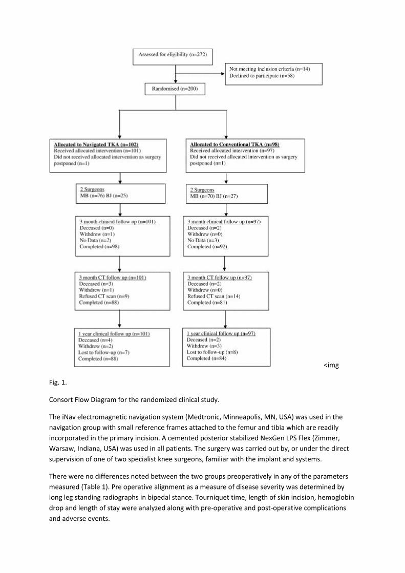
<img
Fig. 1.
Consort Flow Diagram for the randomized clinical study.
The iNav electromagnetic navigation system (Medtronic, Minneapolis, MN, USA) was used in the
navigation group with small reference frames attached to the femur and tibia which are readily
incorporated in the primary incision. A cemented posterior stabilized NexGen LPS Flex (Zimmer,
Warsaw, Indiana, USA) was used in all patients. The surgery was carried out by, or under the direct
supervision of one of two specialist knee surgeons, familiar with the implant and systems.
There were no differences noted between the two groups preoperatively in any of the parameters
measured (Table 1). Pre operative alignment as a measure of disease severity was determined by
long leg standing radiographs in bipedal stance. Tourniquet time, length of skin incision, hemoglobin
drop and length of stay were analyzed along with pre-operative and post-operative complications
and adverse events.
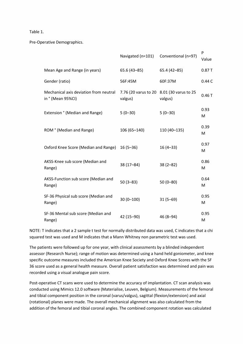
Table 1.
Pre-Operative Demographics.
Navigated (n=101) Conventional (n=97)
P
Value
Mean Age and Range (in years) 65.6 (43–85) 65.4 (42–85) 0.87 T
Gender (ratio) 56F:45M 60F:37M 0.44 C
Mechanical axis deviation from neutral
in ° (Mean 95%CI)
7.76 (20 varus to 20
valgus)
8.01 (30 varus to 25
valgus) 0.46 T
Extension ° (Median and Range) 5 (0–30) 5 (0–30) 0.93
M
ROM ° (Median and Range) 106 (65–140) 110 (40–135) 0.39
M
Oxford Knee Score (Median and Range) 16 (5–36) 16 (4–33) 0.97
M
AKSS-Knee sub score (Median and
Range) 38 (17–84) 38 (2–82)
0.86
M
AKSS-Function sub score (Median and
Range) 50 (3–83) 50 (0–80)
0.64
M
SF-36 Physical sub score (Median and
Range) 30 (0–100) 31 (5–69)
0.95
M
SF-36 Mental sub score (Median and
Range) 42 (15–90) 46 (8–94)
0.95
M
NOTE: T indicates that a 2 sample t test for normally distributed data was used, C indicates that a chi
squared test was used and M indicates that a Mann Whitney non parametric test was used.
The patients were followed up for one year, with clinical assessments by a blinded independent
assessor (Research Nurse); range of motion was determined using a hand held goniometer, and knee
specific outcome measures included the American Knee Society and Oxford Knee Scores with the SF
36 score used as a general health measure. Overall patient satisfaction was determined and pain was
recorded using a visual analogue pain score.
Post-operative CT scans were used to determine the accuracy of implantation. CT scan analysis was
conducted using Mimics 12.0 software (Materialise, Leuven, Belgium). Measurements of the femoral
and tibial component position in the coronal (varus/valgus), sagittal (flexion/extension) and axial
(rotational) planes were made. The overall mechanical alignment was also calculated from the
addition of the femoral and tibial coronal angles. The combined component rotation was calculated
from the addition of the femoral and tibial rotation angles. The rotations were measured using the
methods detailed in Berger et al (1998) [26]. In the coronal plane we aimed to position both femoral
and tibial implants at 90° to the mechanical axis. In the sagittal plane we aimed to position the
femoral component with a 5° slope relative to the mechanical axis, in line with the anterior cortex of
the distal femur. The tibial component was aimed to be positioned at a 7° slope, as per the
manufacturer’s guidelines. For femoral rotation we aimed to implant the femoral component in line
with the surgical trans-epicondylar axis of the femur. The reference for tibial rotation was a line from
the geometric center of the tibia to the center of the tuberosity. Rotational measurements were
calculated from a perpendicular line drawn from the posterior surface of the implant. As the
tuberosity is 18° externally rotated, we considered an 18° internal rotation of the implant to be a
neutral position [27]. (No obvious deformities of the tibia or previous fractures were noted in the
study cohort that could have influenced this value.) We considered the desired mechanical axis
alignment to be 0° with a range of ± 3°.
Statistics
A power calculation was performed based in data provided by a randomized controlled trial using
infra red optical tracking systems. Bathis et al reported 96% of patients with mechanical leg
alignment within 3° of neutral using navigation compared to just 78% with conventional
instrumentation [16]. In order to detect a difference of this magnitude with a power of 90% at alpha
= 0.05, we would require 82 patients per group, 164 in total. As the primary outcome measure was
based on post-operative CT scan we anticipated a higher than average loss to follow-up for the
primary outcome measure. We therefore allowed an additional 25% for loss to follow-up, giving a
total of 103 patients in each group.
Statistical analysis was performed using SigmaPlot 11.0 (Systat Software Inc). To evaluate differences
between the surgical groups either a two sample t test (normally distributed data) or a Mann
Whitney test (non parametric data) was performed. A Chi Squared test was used to analysis the
male: female ratio. A P value of less that 0.05 was considered significant.
Results
Surgical Differences
There was a small but statistically significant difference in skin incision length of 1 cm noted between
the 2 groups with longer incisions reported in the navigated group. Tourniquet times were also
statistically significantly longer in the navigated group; median 80 min for the navigated group
compared to a median of 65 min for the conventional group (P = 0.001).There were no differences in
mean drop in hemoglobin or length of hospital stay between the two groups ( Table 2).
Table 2.
Surgical Data for the Navigated and Conventional Groups.
Navigated (n = 101) Conventional (n = 97) P Value
Mean length of skin incision 18.2cms 17.0cms 0.0021T
Navigated (n = 101) Conventional (n = 97) P Value
Median tourniquet time (range) 80 min (45–130) 65 min (40–120) 0.001M
Median drop in Hb (range) 3.2 g/dl (0.5–6.9) 3.1 g/dl (1.5–10.0) 0.599 M
Median length of stay (range) 5 days (2–30) 5 days (2–15) 0.567 M
NOTE: T indicates that a 2 sample t test for normally distributed data was used, M indicates that
Mann Whitney Test used.
Clinical Outcome Scores
Although the navigated group had statistically significantly better absolute Oxford scores compared
to the conventional group at 3 months and a showed a trend for better AKSS scores at 3 months
(Table 3) the change in score from pre-operative values was not significantly different (P = 0.088). At
1 year post-operatively both groups had further improved their OKS and AKSS scores, with no
significant difference detected between the groups at this time point ( Table 4). There was also no
significant difference in range of motion, pain VAS or SF-36 scores ( Table 4). Table 5 shows that
there was no difference in the patient satisfaction ratings between the two groups. Overall there
was a 12% incidence of patients who were unsure, unsatisfied or very unsatisfied at 1 year post
surgery.
Table 3.
Clinical Scores for the Navigated and Conventional Surgical Groups 3 Months Post-Surgery.
3 Months Clinical Scores Navigated (n = 98) Conventional (n = 92) P Value
Median ROM (Range) 105 (68–130) 100 (43–133) 0.24
Median Oxford (Range) 32 (7–46) 29 (6–46) 0.031
Median AKSS-Knee (Range) 78.5 (38–95) 76 (15–94) 0.067
Median AKSS-Function (Range) 60 (10–100) 55 (0–55) 0.098
NOTE: Mann Whitney test used for P value.
Table 4.
Clinical Scores at 12 Months Post-Surgery.
1 Year Clinical Scores Navigated (n = 88) Conventional (n = 84) P Value
Median ROM (Range) 110 (80–135) 110 (75–135) 0.309
Median Oxford (Range) 34 (12–48) 36 (5–47) 0.682
1 Year Clinical Scores Navigated (n = 88) Conventional (n = 84) P Value
Median AKSS-Knee (Range) 85 (33–95) 86 (32–100) 0.910
Median AKSS-Function (Range) 70 (15–100) 65 (0–100) 0.274
Median SF-36 Physical (Range) 53 (3–99) 46 (9–96) 0.611
Median SF-36 Mental (Range) 69 (18–100) 70 (15–97) 0.529
Median Pain VAS (Range) 20 (0–90) 16 (0–98) 0.916
NOTE: Mann Whitney test used for P value.
Table 5.
Satisfaction Ratings at 12 Months Post-Surgery.
1 Year Satisfaction Rates Navigated (n = 88) Conventional (n = 84) P Value
% Very Satisfied 64 56
0.34
% Satisfied 26 30
% Don't Know 7 6
% Unsatisfied 1 5
% Very Unsatisfied 1 3
Accuracy Study
There was no statistically significant difference between the two groups in accuracy of placement of
either the femoral or tibial component in the coronal plane (Fig. 2 and Fig. 3). Nor was any significant
difference observed between the accuracy of placement in the sagittal plane (Fig. 4 and Fig. 5) or the
axial plane (Fig. 6 and Fig. 7).
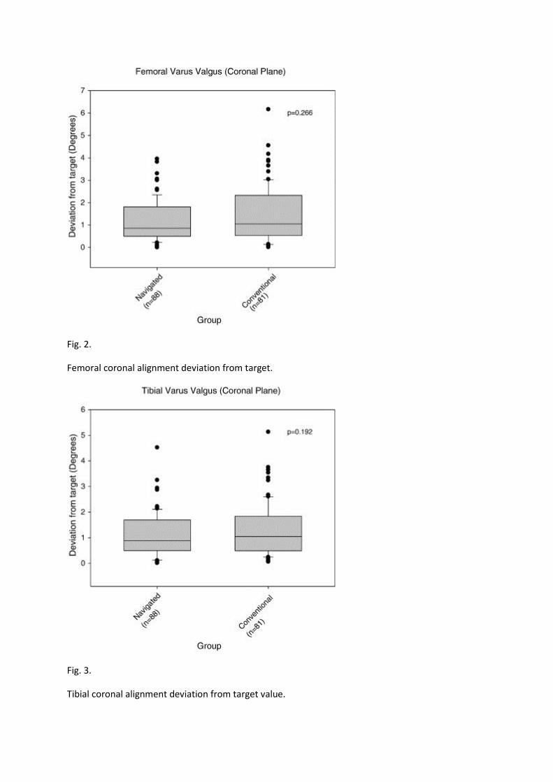
Fig. 2.
Femoral coronal alignment deviation from target.
Fig. 3.
Tibial coronal alignment deviation from target value.
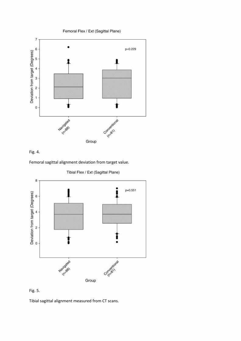
Fig. 4.
Femoral sagittal alignment deviation from target value.
Fig. 5.
Tibial sagittal alignment measured from CT scans.
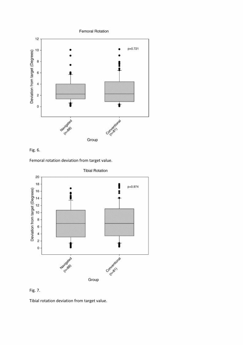
Fig. 6.
Femoral rotation deviation from target value.
Fig. 7.
Tibial rotation deviation from target value.
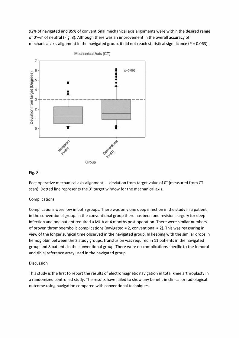
92% of navigated and 85% of conventional mechanical axis alignments were within the desired range
of 0°–3° of neutral (Fig. 8). Although there was an improvement in the overall accuracy of
mechanical axis alignment in the navigated group, it did not reach statistical significance (P = 0.063).
Fig. 8.
Post operative mechanical axis alignment — deviation from target value of 0° (measured from CT
scan). Dotted line represents the 3° target window for the mechanical axis.
Complications
Complications were low in both groups. There was only one deep infection in the study in a patient
in the conventional group. In the conventional group there has been one revision surgery for deep
infection and one patient required a MUA at 4 months post operation. There were similar numbers
of proven thromboembolic complications (navigated = 2, conventional = 2). This was reassuring in
view of the longer surgical time observed in the navigated group. In keeping with the similar drops in
hemoglobin between the 2 study groups, transfusion was required in 11 patients in the navigated
group and 8 patients in the conventional group. There were no complications specific to the femoral
and tibial reference array used in the navigated group.
Discussion
This study is the first to report the results of electromagnetic navigation in total knee arthroplasty in
a randomized controlled study. The results have failed to show any benefit in clinical or radiological
outcome using navigation compared with conventional techniques.
A slightly longer incision was employed in navigated surgery. This is likely to have occurred as a distal
extension of the wound was required to insert the tibial array. The 1 cm of additional length is
unlikely to be of any clinical significance however.
There was no reduction in hemoglobin loss with navigated surgery. The avoidance of intra-medullary
instrumentation has been suggested as a mechanism to reduce blood loss [17] and cerebral emboli
[28]. In the conventional group we used intramedullary instrumentation in the femur only and used
a bone plug to occlude the end of the femoral canal which might explain the failure to observe any
difference in change in post-operative hemoglobin.
Complications remained low in both groups however, with no increased rate of infection from the
increased tourniquet time in the navigated group. A previous meta-analysis [29] suggests that
increased operative times for navigated surgery in total knee arthroplasty are 20 min on average;
slightly longer than our increase of 15 min. Some of our increased time was associated with data
collection for the study to assist in post-operative analysis but the majority results from array
insertion, landmark registration and inevitable, although uncommon software glitches. This increase
in surgical time allied to the additional costs of hardware, software and disposables required to carry
out the surgery all put pressure on navigation techniques to deliver better surgical outcomes 11.,
14., 15., 16., 17., 19., 30., 31., 32., 33., 34., 35. and 36..
Although a slight reduction in mechanical axis outliers was seen in patients post-operatively at
3 months, this failed to reach statistical significance. This in part may be due to the good results seen
with conventional techniques with 85% of cases within the 3° desired range. Our power calculation
had been based on conventional techniques achieving implantation within the 3° window in only
78% of cases. Based on our actual study results we would have required 463 patients per group to
detect a statistically significant 7% difference, with a power of 90%. Other potential reasons for the
failure of the navigation system to achieve significantly better post-operative alignment might lie
with the single point landmark registration system employed, with errors in landmark registration
creating errors in the software algorithms. Our assessment of overall accuracy of implantation of
both femoral and tibial components in the sagittal, coronal and axial planes revealed no significant
increase in accuracy achieved using navigation compared to conventional instrumentation in any of
these individual parameters. Again this may in part be due to single point registration system and it
is possible that other registration techniques may provide more accurate navigation and
consequently greater precision in implant placement.
A potential weakness of our study is the methodology used to measure tibial rotational errors. There
is no universally accepted methodology for measuring tibial rotation. We have used as a reference a
line drawn from the geometric center of the tibia to the center of the tuberosity as previously
described by a number of authors 26., 27., 37. and 38.. However, other authors have argued that use
of the tibial tuberosity may give rise to errors as the position of the tuberosity is variable [39].
Deflection artifact with electromagnetic systems in an in vitro study has been suggested in the
presence of ferrous materials within 10 cm of the localizer and is another potential source of system
error [25]. Our experience was that in the presence of ferrous materials the system would go ‘blind’
and prior to loss of signal, no unexplained change in the system readout values was noted. Other in
vitro and in vivo studies looking at the results of electromagnetic navigation have confirmed that
system accuracy is not problematic, with good results comparable to infra-red systems 35.,
40. and 41.. Two clinical studies have compared the electromagnetic technique directly with
traditional infra-red navigation systems and found similar, high degrees of accuracy 42. and 43.,
although the numbers in these studies were small. Our much larger study has demonstrated good
results with EM navigation, comparable with other optical systems. Dutton et al have previously
reported 92% of patients achieving post-operative mechanical axis alignment within 3° of neutral,
Johnson et al 96% and in a meta-analysis of 29 studies Mason et al reported 91% 44., 45. and 46..
Although improvements in alignment have been demonstrated to be correlated with improvements
in implant survivorship in some studies 47., 48. and 49., improvements in function with navigation
have been harder to prove. This is possibly because the differences in alignment seen over
conventional surgery are not great enough. Our study reinforces others in the literature with no
difference seen in any of the patient centered, knee specific or general health measures used.
Improvements in the outcomes of knee arthroplasty surgery are needed however as our study
results mirror the majority of others, with some 12% of patients either unsure or dissatisfied with
their surgery. Dissatisfaction following knee arthroplasty surgery is multifactorial however and it is
perhaps simplistic to think that poor outcomes can be eliminated by surgical technique alone.
The advantages of navigation at the current time therefore remain unclear. A potential reduction in
revision burden 10 years following arthroplasty surgery from a reduction in outliers is a difficult
argument with which to engage health providers to justify increased costs of surgery, particularly in
the current financial climate. The difficulty that navigation faces is that total knee joint survival in
modern knee arthroplasties exceeds 97% at 10 years with aseptic loosening as the end point using
conventional techniques 50. and 51. which is a difficult benchmark to surpass. Indeed a recent study
by Kim in 520 patients comparing navigated and conventional techniques in simultaneous bilateral
total knee arthroplasties in the same patient revealed greater than 98% survivorship for both groups
at a mean of 10 years [52].
Work has been done to promote the teaching and training benefits of navigation 53. and 54., but its
value in routine total knee arthroplasty surgery remains under scrutiny.
1.
R.S. Jeffery, R.W. Morris, R.A. Denham
Coronal alignment after total knee replacement
J Bone Joint Surg (Br), 73 (5) (1991), p. 709
2.
I. Hvid, S. Nielsen
Total condylar knee arthroplasty. Prosthetic component positioning and radiolucent lines
Acta Orthop Scand, 55 (2) (1984), p. 160
3.
M.E. Berend, M.A. Ritter, J.B. Meding, et al.

Tibial component failure mechanisms in total knee arthroplasty
Clin Orthop Relat Res, 428 (2004), p. 26
4.
J.A. Rand, M.B. Coventry
Ten-year evaluation of geometric total knee arthroplasty
Clin Orthop Relat Res, 232 (1988), p. 168
5.
J.L. Smith Jr., H.S. Tullos, J.P. Davidson
Alignment of total knee arthroplasty
J Arthroplasty, 4 (1989), p. S55 [Suppl.]
6.
M.J. Bankes, D.L. Back, S.R. Cannon, et al.
The effect of component malalignment on the clinical and radiological outcome of the Kinemax total
knee replacement
Knee, 10 (1) (2003), p. 55
7.
M. Tew, W. Waugh
Tibiofemoral alignment and the results of knee replacement
J Bone Joint Surg (Br), 67 (4) (1985), p. 551
8.
J.M. Spencer, S.K. Chauhan, K. Sloan, et al.
Computer navigation versus conventional total knee replacement: no difference in functional results
at two years
J Bone Joint Surg (Br), 89 (4) (2007), p. 477
9.
A. Ensini, F. Catani, A. Leardini, et al.
Alignments and clinical results in conventional and navigated total knee arthroplasty
Clin Orthop Relat Res, 457 (2007), p. 156

10.
K.C. Anderson, K.C. Buehler, D.C. Markel
Computer assisted navigation in total knee arthroplasty: comparison with conventional methods
J Arthroplasty, 20 (7 Suppl 3) (2005), p. 132
11.
R. Decking, Y. Markmann, J. Fuchs, et al.
Leg axis after computer-navigated total knee arthroplasty: a prospective randomized trial comparing
computer-navigated and manual implantation
J Arthroplasty, 20 (3) (2005), p. 282
12.
D.M. Fang, M.A. Ritter, K.E. Davis
Coronal alignment in total knee arthroplasty: just how important is it?
J Arthroplasty, 24 (6 Suppl.) (2009), p. 39
13.
S.J. Kim, M. MacDonald, J. Hernandez, et al.
Computer assisted navigation in total knee arthroplasty: improved coronal alignment
J Arthroplasty, 20 (7 Suppl. 3) (2005), p. 123
14.
G. Matziolis, D. Krocker, U. Weiss, et al.
A prospective, randomized study of computer-assisted and conventional total knee arthroplasty.
Three-dimensional evaluation of implant alignment and rotation
J Bone Joint Surg Am, 89 (2) (2007), p. 236
15.
A. Martin, O. Wohlgenannt, M. Prenn, et al.
Imageless navigation for TKA increases implantation accuracy
Clin Orthop Relat Res, 460 (2007), p. 178
16.
H. Bathis, L. Perlick, M. Tingart, et al.

Alignment in total knee arthroplasty. A comparison of computer-assisted surgery with the
conventional technique
J Bone Joint Surg (Br), 86 (5) (2004), p. 682
17.
S.K. Chauhan, R.G. Scott, W. Breidahl, et al.
Computer-assisted knee arthroplasty versus a conventional jig-based technique. A randomised,
prospective trial
J Bone Joint Surg (Br), 86 (3) (2004), p. 372
18.
A. Mullaji, R. Kanna, S. Marawar, et al.
Comparison of limb and component alignment using computer-assisted navigation versus image
intensifier-guided conventional total knee arthroplasty: a prospective, randomized, single-surgeon
study of 467 knees
J Arthroplasty, 22 (7) (2007), p. 953
19.
F. Macule-Beneyto, D. Hernandez-Vaquero, J.M. Segur-Vilalta, et al.
Navigation in total knee arthroplasty. A multicenter study
Int Orthop, 30 (6) (2006), p. 536
20.
K. Bauwens, G. Matthes, M. Wich, et al.
Navigated total knee replacement. A meta-analysis
J Bone Joint Surg Am, 89 (2) (2007), p. 261
21.
P. Bonutti, D. Dethmers, J.B. Stiehl
Case report: femoral shaft fracture resulting from femoral tracker placement in navigated TKA
Clin Orthop Relat Res, 466 (6) (2008), p. 1499
22.
D. Hoke, S.M. Jafari, F. Orozco, et al.
Tibial shaft stress fractures resulting from placement of navigation tracker pins

J Arthroplasty, 26 (3) (2011), p. 504 e5
23.
F. Massai, F. Conteduca, A. Vadala, et al.
Tibial stress fracture after computer-navigated total knee arthroplasty
J Orthop Traumatol, 11 (2) (2010), p. 123
24.
J. Schmitt, C. Hauk, H. Kienapfel, et al.
Navigation of total knee arthroplasty: rotation of components and clinical results in a prospectively
randomized study
BMC Musculoskelet Disord, 12 (2011), p. 16
25.
F. Stevens, M.A. Conditt, N. Kulkarni, et al.
Minimizing electromagnetic interference from surgical instruments on electromagnetic surgical
navigation
26.
R.A. Berger, L.S. Crossett, J.J. Jacobs, et al.
Malrotation causing patellofemoral complications after total knee arthroplasty
Clin Orthop Relat Res, 356 (1998), p. 144
27.
D. Nicoll, D.I. Rowley
Internal rotational error of the tibial component is a major cause of pain after total knee
replacement
J Bone Joint Surg (Br), 92 (9) (2010), p. 1238
28.
Y. Kalairajah, A.J. Cossey, G.M. Verrall, et al.
Are systemic emboli reduced in computer-assisted knee surgery? A prospective, randomised, clinical
trial
J Bone Joint Surg (Br), 88 (2) (2006), p. 198
29.

Y.S. Brin, V.S. Nikolaou, L. Joseph, et al.
Imageless computer assisted versus conventional total knee replacement. A Bayesian meta-analysis
of 23 comparative studies
Int Orthop, 35 (3) (2011), p. 331
30.
Y.H. Kim, J.S. Kim, S.H. Yoon
Alignment and orientation of the components in total knee replacement with and without
navigation support: a prospective, randomised study
J Bone Joint Surg (Br), 89 (4) (2007), p. 471
31.
R.G. Haaker, M. Stockheim, M. Kamp, et al.
Computer-assisted navigation increases precision of component placement in total knee
arthroplasty
Clin Orthop Relat Res, 433 (2005), p. 152
32.
J.Y. Jenny, U. Clemens, S. Kohler, et al.
Consistency of implantation of a total knee arthroplasty with a non-image-based navigation system:
a case–control study of 235 cases compared with 235 conventionally implanted prostheses
J Arthroplasty, 20 (7) (2005), p. 832
33.
M. Tingart, C. Luring, H. Bathis, et al.
Computer-assisted total knee arthroplasty versus the conventional technique: how precise is
navigation in clinical routine?
Knee Surg Sports Traumatol Arthrosc, 16 (1) (2008), p. 44
View Record in Scopus
34.
J.Y. Jenny, C. Boeri
Computer-assisted implantation of total knee prostheses: a case–control comparative study with
classical instrumentation
Comput Aided Surg, 6 (4) (2001), p. 217

35.
A.J. Graydon, S. Malak, I.A. Anderson, et al.
Evaluation of accuracy of an electromagnetic computer-assisted navigation system in total knee
arthroplasty
Int Orthop, 33 (4) (2009), p. 975
36.
J.K. Seon, S.J. Park, K.B. Lee, et al.
Functional comparison of total knee arthroplasty performed with and without a navigation system
Int Orthop, 33 (4) (2009), p. 987
37.
S.W. Bell, P. Young, C. Drury, et al.
Component rotational alignment in unexplained painful primary total knee arthroplasty
Knee, 21 (1) (2014), p. 272
38.
M.K. Harman, S.A. Banks, S. Kirschner, et al.
Prosthesis alignment affects axial rotation motion after total knee replacement: a prospective in vivo
study combining computed tomography and fluoroscopic evaluations
BMC Musculoskelet Disord, 13 (2012), p. 206
39.
S.M. Howell, J. Chen, M.L. Hull
Variability of the location of the tibial tubercle affects the rotational alignment of the tibial
component in kinematically aligned total knee arthroplasty
Knee Surg Sports Traumatol Arthrosc, 21 (10) (2013), p. 2288
40.
T.W. Kim, S.H. Park, J.T. Suh
Comparison of mobile-bearing and fixed-bearing designs in high flexion total knee arthroplasty:
using a navigation system
Knee Surg Relat Res, 24 (1) (2012), p. 25
41.

D. Tigani, M. Busacca, A. Moio, et al.
Preliminary experience with electromagnetic navigation system in TKA
Knee, 16 (1) (2009), p. 33
42.
E.K. Song, J.K. Seon, S.J. Park, et al.
Accuracy of navigation: a comparative study of infrared optical and electromagnetic navigation
Orthopedics, 31 (10 Suppl 1) (2008)
43.
D.R. Lionberger, J. Weise, D.M. Ho, et al.
How does electromagnetic navigation stack up against infrared navigation in minimally invasive total
knee arthroplasties?
J Arthroplasty, 23 (4) (2008), p. 573
44.
A.Q. Dutton, S.J. Yeo
Computer-assisted minimally invasive total knee arthroplasty compared with standard total knee
arthroplasty. Surgical technique
J Bone Joint Surg Am, 91 (Suppl. 2 Pt 1) (2009), p. 116
45.
D.R. Johnson, D.A. Dennis, K.A. Kindsfater, et al.
Evaluation of total knee arthroplasty performed with and without computer navigation: a bilateral
total knee arthroplasty study
J Arthroplasty, 28 (3) (2013), p. 455
46.
J.B. Mason, T.K. Fehring, R. Estok, et al.
Meta-analysis of alignment outcomes in computer-assisted total knee arthroplasty surgery
J Arthroplasty, 22 (8) (2007), p. 1097
47.

P.A. Lotke, M.L. Ecker
Influence of positioning of prosthesis in total knee replacement
J Bone Joint Surg Am, 59 (1) (1977), p. 77
48.
M.A. Ritter, P.M. Faris, E.M. Keating, et al.
Postoperative alignment of total knee replacement. Its effect on survival
Clin Orthop Relat Res, 299 (1994), p. 153
49.
M.A. Ritter, K.E. Davis, P. Davis, et al.
Preoperative malalignment increases risk of failure after total knee arthroplasty
J Bone Joint Surg Am, 95 (2) (2013), p. 126
50.
J.N. Argenson, S. Parratte, A. Ashour, et al.
The outcome of rotating-platform total knee arthroplasty with cement at a minimum of ten years of
follow-up
J Bone Joint Surg Am, 94 (7) (2012), p. 638
51.
M. Meftah, A.S. Ranawat, C.S. Ranawat
Ten-year follow-up of a rotating-platform, posterior-stabilized total knee arthroplasty
J Bone Joint Surg Am, 94 (5) (2012), p. 426
52.
Y.H. Kim, J.W. Park, J.S. Kim
Computer-navigated versus conventional total knee arthroplasty: a prospective randomized trial
J Bone Joint Surg Am, 94 (22) (2012), pp. 2017–2024
53.
G.J. Love, A.W. Kinninmonth
Training benefits of computer navigated total knee arthroplasty
Knee, 20 (4) (2013), pp. 236–241

54.
S.D. Stulberg
Computer navigation as a teaching instrument in knee reconstruction surgery
J Knee Surg, 20 (2) (2007), p. 165


