
Engineered Nanomaterials: Investigating substitution and modification options to reduce potential hazards
August 2010
2
Engineered Nanomaterials: Investigating substitution and modification options to reduce potential hazards
Acknowledgement
This review was commissioned by Safe Work Australia, through funding provided under the National Nanotechnology Strategy and prepared by the RMIT OHS Research and Education Centre at RMIT University, Melbourne, Australia. The review was undertaken by Dr Neale Jackson (Project Manager), Associate Professor Susanne Tepe, and Associate Professor Paul Wright (Project Coordinator). This report has been reviewed by Safe Work Australia’s Nanotechnology OHS Advisory Group, and Dr Howard Morris, Nanotechnology Work Health and Safety Program Manager, Safe Work Australia. Additional helpful comments were provided by Professor Terry Turney, Centre for Green Chemistry, Monash University, Melbourne, Australia. Neale Jackson, Paul Wright and Terry Turney are members of the Nanosafe Australia research network (www.rmit.edu.au/nanosafe). Safe Work Australia gratefully acknowledges the ANU College of Science for the photograph used on the front cover.
Disclaimer
The information provided in this document can only assist you in the most general way. This document does not replace any statutory requirements under any relevant State and Territory legislation. Safe Work Australia is not liable for any loss resulting from any action taken or reliance made by you on the information or material contained on this document. Before relying on the material, users should carefully make their own assessment as to its accuracy, currency, completeness and relevance for their purposes, and should obtain any appropriate professional advice relevant to their particular circumstances. To the extent that the material on this document includes views or recommendations of third parties, such views or recommendations do not necessarily reflect the views of Safe Work Australia or indicate its commitment to a particular course of action.

3
Copyright Notice © Commonwealth of Australia 2010 ISBN 978-0-642-33100-7 (Online PDF) ISBN 978-0-642-33101-4 (Online RTF) This work is copyright. You may download, display, print and reproduce this material for your personal, non-commercial use or use within your organisation, provided that an appropriate acknowledgement is made (retaining this notice), and the material is not altered or subjected to derogatory treatment. Apart from any use as permitted under the Copyright Act 1968, all other rights are reserved. Requests and inquiries concerning reproduction and rights should be addressed to: Commonwealth Copyright Administration Attorney-General’s Department 3 - 5 National Circuit Barton ACT 2600 Email: [email protected] Web: http://www.ag.gov.au/cca
4
Table of Contents Acknowledgement........................................................................................................................2 Disclaimer ....................................................................................................................................2 List of abbreviations .....................................................................................................................6 Executive Summary .....................................................................................................................7
Summary from the survey ........................................................................................................7 Summary of the literature review..............................................................................................8
1 Background and scope of this report ......................................................................................10 1.1 Background ......................................................................................................................10 1.2 Scope of the review..........................................................................................................10
2 Substitution/modification of nanomaterials survey..................................................................12 2.1 Results of previous surveys on nanomaterials used in Australia .....................................12 2.2 Substitution/modification survey method..........................................................................14 2.3 Survey results...................................................................................................................14
2.3.1 Work Sector ...............................................................................................................14 2.3.2 Types of engineered nanomaterials ..........................................................................15 2.3.3 Types of activities using nanomaterials .....................................................................17 2.3.4 Sources of engineered nanomaterials .......................................................................18 2.3.5 Health and safety considerations...............................................................................19 2.3.6 Use of modification and substitution ..........................................................................20
2.4 Summary from the survey ................................................................................................23 3 Literature review for substitution/modification options for engineered nanomaterials.............25
3.1 Mechanisms of nanoparticle toxicity in biological systems...............................................25 3.1.1 Importance of nanoparticle size for cell uptake .........................................................29 3.1.2 Importance of surface charge for cell uptake.............................................................31 3.1.3 Importance of cell specific effects for nanoparticle uptake ........................................32 3.1.4 Importance of surface modification for nanoparticle uptake ......................................33 3.1.5 Biocompatibility and surface coatings........................................................................34
4 Substitution/modification options for specific engineered nanomaterials................................35 4.1 Carbon nanotubes............................................................................................................35
4.1.1 Background................................................................................................................35 4.1.2 Toxicology of carbon nanotubes................................................................................37 4.1.3 Potential substitution/modification of carbon nanotubes............................................38 4.1.4 Impact of modification on potential exposure levels ..................................................42 4.1.5 Conclusions ...............................................................................................................42
4.2 Fullerenes.........................................................................................................................43 4.2.1 Background................................................................................................................43 4.2.2 Toxicology of fullerenes .............................................................................................44 4.2.3 Potential substitution/modification of fullerenes.........................................................45 4.2.4 Conclusions ...............................................................................................................45
4.3 Nano titanium dioxide (TiO2) ............................................................................................46 4.3.1 Background................................................................................................................46 4.3.2 Toxicology of nano titanium dioxide...........................................................................46 4.3.3 Potential substitution/modification of nano titanium dioxide ......................................47 4.3.4 Conclusions ...............................................................................................................48
4.4 Nano cerium dioxide (CeO2).............................................................................................49 4.4.1 Background................................................................................................................49 4.4.2 Toxicology of nano cerium dioxide ............................................................................49 4.4.3 Conclusions ...............................................................................................................51
4.5 Nano zinc oxide (ZnO) .....................................................................................................51 4.5.1 Background................................................................................................................51
5
4.5.2 Toxicology of nano zinc oxide....................................................................................52 4.5.3 Potential substitution/modification of nano zinc oxide ...............................................52 4.5.4 Conclusions ...............................................................................................................54
4.6 Nano gold (Au) .................................................................................................................54 4.6.1 Background................................................................................................................54 4.6.2 Toxicology of nano gold.............................................................................................54 4.6.3 Potential substitution/modification of nano gold.........................................................57 4.6.4 Conclusions ...............................................................................................................58
4.7 Nano silver (Ag)................................................................................................................58 4.7.1 Background................................................................................................................58 4.7.2 Toxicology of nano silver ...........................................................................................59 4.7.3 Potential substitution/modification of nano silver .......................................................59 4.7.4 Conclusions ...............................................................................................................60
4.8 Nano silica (SiO2) .............................................................................................................60 4.8.1 Introduction ................................................................................................................60 4.8.2 Toxicology of nano silica............................................................................................61 4.8.3 Potential substitution/modification of nano silica .......................................................61 4.8.4 Conclusions ...............................................................................................................62
4.9 Quantum dots...................................................................................................................63 4.9.1 Background................................................................................................................63 4.9.2 Toxicology of quantum dots.......................................................................................63 4.9.3 Methods of modification of quantum dots ..................................................................64 4.9.4 Conclusions ...............................................................................................................65
4.10 Conclusions on substitution/modification options for engineered nanomaterials ...........66 References.................................................................................................................................69 Appendix 1 - Substitution/modification of nanomaterials survey questionnaire .........................79
6
List of abbreviations ANA Australian Nanotechnology Alliance ANBF Australian Nano Business Forum APTES Aminopropyltriethoxysilane ARCNN Australian Research Council Nanotechnology Network BSA Bovine Serum Albumin CME Clathrin-Mediated Endocytosis CNTs Carbon Nanotubes CPC Condensation Particle Counter CTAB Cetyltrimethyl Ammonium Bromide CVD Chemical Vapour Deposition DNA Deoxyribonucleic Acid EFTEM Energy Filtering Transmission Electron Microscopy FITC Fluorescein Isothiocyanate FR+ Folate Receptor Positive FTIR Fourier Transform Infrared Her2 Human Epidermal Growth Factor Type 2 HHPC Hand Held Particle Counter HIPCO High Pressure Carbon Monoxide hMSC Human Mesenchymal Stem Cell HSA Human Serum Albumin IARC International Agency for Research on Cancer IR Infrared LDH Lactate Dehydrogenase LDL Low Density Lipoprotein LMCS Low Molecular Weight Chitosan NICNAS National Industrial Chemicals Notification and Assessment Scheme for Australia NMs Nanomaterials NMR Nuclear Magnetic Resonance NP Nanoparticle MSDS Material Safety Data Sheet MSN Mesoporous Silica Nanoparticle MTT 3-(4,5-Dimethylthiazol-2-yl)-2,5-Diphenyltetrazolium Bromide MWCNT Multi-Walled Carbon Nanotubes OHS Occupational Health and Safety PEG Polyethylene glycol PLGA Poly(D,L-lactic-co-glycolic acid) PPE Personal Protective Equipment PVA Polyvinyl Alcohol QD Quantum Dot RBC Red Blood Cell RCEC Rabbit Conjunctival Epithelial Cells ROI Reactive Oxygen Intermediates ROS Reactive Oxygen Species SEM Scanning Electron Microscopy SWCNT Single-Walled Carbon Nanotubes TEM Transmission Electron Microscopy TPGS d-Alpha-Tocopheryl Polyethylene Glycol 1000 Succinate TSDC Thermally Stable Depolarisation Currents UFP Ultra Fine Particles XPS X-ray Photo Electron Spectroscopy XRD X-Ray Diffraction

7
Executive Summary
In a review of the evidence on the effectiveness of workplace controls to prevent exposure to engineered nanomaterials it was found that little focus has to date been placed on use of substitution or modification for nanotechnology work health and safety purposes. Therefore, Safe Work Australia commissioned RMIT to undertake a survey of the current substitution/modification practices used in Australian nanotechnology-related activities and a literature review in order to determine the potential substitution/modification options that may reduce the toxicity of engineered nanomaterials used in Australia.
Summary from the survey a) There were 38 respondents to the survey, who reported working on a range of different types of nanomaterials. The respondents’ organisations were primarily universities, commercial/industry and government research groups. The most common nanomaterials handled are metal oxides, metals and carbon nanotubes and the most common areas of application are into energy, medical, surface coating and textile uses. b) Many organisations (27/35), and notably universities (20/21), manufacture their own engineered nanomaterials, and a significant number also purchase them from overseas or from within Australia (see Figure 3). c) A number of respondents obtained work health and safety information about the nanomaterials that they are using from an MSDS. The main work health and safety issues examined for engineered nanomaterials are handling and storage, physical and chemical properties, toxicological data and exposure controls/personal protective equipment (PPE) (see Table D). The available information on these topics is limited. d) Most respondents indicated that substitution/modification is used to change the functional properties of the product (see Figure 4). A work sector analysis indicates that substitution/modification occurs more in university research and less in commercial/industry research which is as expected in product development. e) The five properties that are manipulated by modifying or substituting engineered nanomaterials by the highest number of organisations are particle size, physical properties, agglomeration properties, chemical properties and conductive properties. A small number of respondents indicated that they use substitution/modification to change the health or toxicological properties (see Figure 5). f) Adding functional groups (17 responses) and modifying surface characteristics (16 responses) are the two most popular methods for the substitution/modification of engineered nanomaterials. Others include changing the form of the material, the particle size and shape, and the crystalline structure (see Figure 6). g) Australia’s nanotechnology activities are generally at the early stage of nanomaterial development, i.e. more focussed on de novo research than later stages of product development/production. However substitution/modification methodologies are well known and used in Australia and thus there is an existing capability that might be applied more broadly to work health and safety related purposes.
8
Summary of the literature review a) The mechanisms by which nanoparticles enter biological systems and subsequently cause toxicity are dependent on factors such as nanoparticle or aggregate size, physicochemical characteristics of particle surfaces (e.g. surface charge), biocompatibility and cell-specific effects on nanoparticle uptake. Various substitution and modification strategies for a range of nanomaterials have been described in the scientific literature. b) Carbon nanotubes (CNTs) can be functionalised and surface-modified to increase their solubility and biocompatibility. It is also possible to reduce their chronic toxicity potential by using short CNTs and keeping their length to less than 5µm. Further investigation of the toxicity of these modified CNTs needs to be made to assess the extent of the reduction in potential workplace hazard. c) When formulating a new product or use, the toxicity of fullerenes can be controlled by attaching functional groups to the fullerene moiety. Specifically, attaching water solubilising groups such as carboxyl or alcohol groups, will increase the solubility and lead to reduced toxicity of the prepared fullerene. This modification will also alter particle aggregation behaviour in water and its potential bioavailability and reactivity in aquatic systems, and this area requires further investigation. d) It can be concluded that when formulating a new nano titanium dioxide (TiO2) product or use, its potential toxicity can be controlled by varying the crystalline form used, i.e. use the less reactive rutile form rather than the more reactive and photocatalyitc anatase form where functionally possible. e) It can be ascertained that nano ceria under specific conditions exhibits antioxidant and biocompatible properties. However, outside this range of conditions antioxidant behaviour is not exhibited, and its redox cycling ability may be pro-oxidant. In an aquatic system, nano ceria has been found to be more toxic than the micron sized particles. It is not possible at this stage to suggest modifications that can be made to nano ceria until more data are obtained. f) It can be concluded that nano zinc oxide (ZnO) used in sunscreen type products and for other similar applications exhibits a low level of toxicity and dermal penetration into the human body. There are surface modification options available for ZnO which have the potential to reduce toxicity further, in addition to structural modifications that help retain functionality, such as doping the ZnO crystalline lattice. g) Nano gold particles can be surface-coated, e.g. with phosphatidylcholine, or encapsulated with biocompatible biopolymers, e.g. chitosan or polyethylene glycol, to reduce toxicity, whilst retaining functionality and useability. Alkanethiol-capping may be used to increase biocompatibility and also functionalise the nano gold for a range of biomedical applications. h) Nano silver can be surface modified with hydrophilic groups, such as phosphorylcholine or phosphorylethanolamine, to increase biocompatibility. Such modifications would also decrease its antibacterial activity and potential usefulness in many current applications. However, further functionalisation of biocompatible forms of nano silver may provide potential new applications, such as in biomedical diagnostics and biosensors.

9
i) It is possible to modify the surface of nano silica with alkylsilylation, polymers or proteins to increase its hydrophobic character, causing increased particle aggregation and reduced direct membrane effects, and thereby improving its biocompatibility. Due to potential toxicity of silica nanomaterials with high aspect ratios, consideration should also be made as to whether nanowires may be substituted with nanospheres, while retaining functionality for a particular application. j) It is possible to encapsulate quantum dot cores with stable shell coatings made from biocompatible polymers, e.g. chitosan or polyethylene glycol, to significantly reduce their cellular uptake and degradation, and consequently their cytotoxicity, whilst retaining functionality and useability. Implications for work health and safety There are known methods that can be used to substitute/modify engineered nanomaterials that are used, or researched, in Australia. The methods of surface modification, encapsulation, particle size control, functional group addition and crystalline phase type control can each be employed for different engineered nanomaterials to decrease their potential toxicity. However in some cases, such modifications may affect the functionality of nanomaterials in relation to intended end-uses. If the researchers, developers and manufacturers of engineered nanomaterials adopt these methods then it is possible to re-engineer nanomaterials in the early stages of development to reduce the potential toxicity of manufactured nanomaterials. The downstream effect of this will be to reduce the risk posed by the use of these nanomaterials not only in the workplace but also in the general community.
10
1 Background and scope of this report
1.1 Background There has been an exponential growth in the development of nanomaterials and nanotechnology applications. This has been accompanied by an increased awareness of nanosafety issues in government, academia, industry and public groups.
In 2008, nanosafety experts at RMIT University were commissioned by Safe Work Australia to examine the evidence on the effectiveness of the workplace controls that are used to prevent or minimise exposure to engineered nanomaterials during their life-cycle of manufacture, handling, use and disposal (Jackson et al. 2009). This report indicated that there are a range of control methods that can be used effectively to protect workers from exposure to engineered nanomaterials. These are mainly based around the lower levels of the “hierarchy of controls”, i.e. engineering controls (enclosure, ventilation/extraction), administrative controls and PPE.
In order to move up the hierarchy of controls, it is necessary to consider options for the elimination, substitution and/or modification of the chemical and physical properties of engineered nanomaterials. The report (Jackson et al. 2009) found that a more detailed investigation of the substitution or modification control options for reducing the intrinsic hazard and toxic potential of nanomaterials was warranted. Consequently, Safe Work Australia commissioned nanosafety experts at RMIT University to undertake a further review to investigate substitution and modification options available to reduce potential hazards associated with different types of engineered nanomaterials. This report covers findings available in the open literature up to the last quarter of 2009, with a small amount of additional material included from early 2010 literature during the report review process. 1.2 Scope of the review The review was commissioned by Safe Work Australia to address the following matters:
identify Australian and overseas businesses, research institutions and organisations that are engaged in the examination of the potential substitution of engineered nanomaterials, and the topics being examined
evaluate research results relating to substitution/modification, including consideration of whether the modified materials maintain required functionality
identify potential substitution/modification opportunities, and compare the hazardous properties of currently used engineered nanomaterials with their substitutes where possible
evaluate potential opportunities for the protection of health and safety in Australian workplaces, and
identify issues for further consideration.
11
Input from relevant sources of nanotechnology, occupational hygiene, toxicology, particle characterisation and other scientific expertise was sought to ensure the accuracy of the assessment and relevance for nanotechnology applications in Australia. Two strategies were used in this study:
a) a survey of individuals employed in Australian nanotechnology-related activities in order to identify Australian businesses, research institutions and organisations that are engaged in the examination of potential substitution/modification of engineered nanomaterials, and to identify the engineered nanomaterials being examined
b) a literature review of the possible substitution and modification options for the range of engineered nanomaterials that are used in Australia which may reduce potential health and safety risks. In addition, the literature review covers relevant background material, e.g. toxicology, that is important in understanding substitution/modification options for different engineered nanomaterials.
12
2 Substitution/modification of nanomaterials survey
2.1 Results of previous surveys on nanomaterials used in Australia
The National Industrial Chemicals Notification and Assessment Scheme (NICNAS) for Australia issued a voluntary call for information on nanomaterials in February 2006. The notice appeared in the Chemical Gazette which is a monthly publication containing information relevant to NICNAS, such as changes to NICNAS legislation, newly assessed chemicals and the register of industrial chemical introducers. The call for information was directed to all persons who manufactured or imported nanomaterials or products (mixtures) containing nanomaterials for industrial uses during 2005 and 2006. Companies were asked to provide information on the types of nanomaterials, their volume of introduction and uses. Nanomaterials used exclusively as therapeutic goods (such as sunscreens), food or food additives and agricultural or veterinary chemicals, do not fall within the scope of NICNAS and were consequently outside the call for information (NICNAS 2007).
To ensure confidentiality of the information reported, the data were aggregated and presented as generic chemical names and ranges of materials used (NICNAS 2007). In this survey, 17 types of nanomaterials were used in a number of applications at various volume (tonnage) levels (Table A). Inorganic (e.g. metals) and organic (e.g. polymer) nanomaterials were reported as being used by four organisations for research and development purposes and by seventeen organisations for commercial purposes. Commercial applications were classified into cosmetics, domestic products, catalysts, water treatment, surface coatings and printing. Most materials were used in quantities of less than 1 tonne/year, however acrylic latex used in surface coatings at 10,000-50,000 tonnes/year is a significantly larger volume material. Metal oxides were the largest group, being used for domestic products, printing, cosmetics, water treatment, catalysts and surface coatings. All nanomaterial types were reported as being imported, but some, such as silicon dioxide, cerium oxide, zinc oxide and acrylic latex, were also manufactured in Australia (NICNAS 2007).
It should be noted that some of the total volume usage information appeared to be underestimated, which may have been due to the voluntary nature of the NICNAS survey. Notably, the usage of carbon black in vehicle tyres and photocopier cartridges was likely to have not been included in the quantity shown in Table A for carbon black pigment in surface coating applications. Similarly, the total volume usage for iron oxide in surface coatings appears to be an underestimate, considering its wide usage as a brown pigment in paint and staining products. Also the call for information targeted industrial use, and thus research organisations using for example carbon nanotubes (CNTs) would not show up in the survey results. NICNAS undertook a further voluntary survey during 2008/9, which also appeared in the Chemical Gazette, but received very few responses. This survey was open to all persons who had manufactured or imported nanomaterials, or products (mixtures) containing nanomaterials, for commercial or research and development purposes. NICNAS sought specific information about each nanomaterial above a 100g/year quantity threshold, including: chemical identity and volume; holdings of existing physicochemical data, environmental fate and ecotoxicological data, and human or modelled toxicological data; and usage and life cycle information.

13
Table A: Usage of nanomaterials from commercial sectors in Australia (taken from NICNAS 2007)
Chemical Name Applications Total volume
(Tonnes per year)
Acrylic latex Surface coatings 10000-50000
Aluminium oxide Printing 0.05-0.1
Aluminosilicates Water treatment 10-50
Carbon black pigment Surface coatings 10-50
Cerium oxide Catalysts 1-5
Iron oxide Surface coatings 1-5
Cosmetics <0.01
Pearl powder Cosmetics 0.01-0.05
Phthalocyanine Surface coatings 10-50
Polyurethane resin Surface coatings <0.01
Silica dimethyl silyate Cosmetics <0.01
Silicon dioxide Surface coatings 10-50
Water treatment 0.05-0.1
Sodium silicates Water treatment 0.1-0.5
Surface treated silicon dioxide Printing 1-5
Surface treated aluminium oxide Printing 0.1-0.5
Surface treated titanium oxide Printing 0.5-1
Titanium dioxide Water treatment 5-10
Domestic products 1-5
Cosmetics 1-5
Zinc oxide Surface coatings 5-10
Cosmetics 1-5

14
2.2 Substitution/modification survey method For the purposes of understanding what research was being done to reduce the hazard of nanomaterials through modification and substitution, a survey of organisations known to use engineered nanomaterials was undertaken in the form of an online questionnaire with specific focus on the use of substitution/modification options for engineered nanomaterials. Method: The Australian Nano Business Forum (ANBF), the Australian Nanotechnology Alliance (ANA) and the Australian Research Council Nanotechnology Network (ARCNN) were contacted to determine their willingness to assist in the distribution of the questionnaire. Each organisation distributed an email requesting participation in the survey to named individual representatives on the mailing list of their member organisations. The request described the purpose of the survey and provided a weblink to the online questionnaire. The survey was developed in SurveyMonkey (available at www.surveymonkey.com). See Appendix 1 for a full copy of the survey. It consisted of 12 questions, which either; (a) provided drop down lists of possible responses, or (b) asked for a free-text response. Specifically the following information was requested in the survey:
a) demographic information about the organisation b) the types of engineered nanomaterials being used c) the types of activities that are being undertaken with these engineered nanomaterials d) where and how the engineered nanomaterials are obtained e) the health and safety issues that are considered when deciding which engineered nanomaterials to purchase f) the health and safety issues that are considered in the design of a new engineered nanomaterial g) if substitution/modification options are considered as a means to change the attributes/properties of engineered nanomaterials h) the reasons why modification or substitution is undertaken for engineered nanomaterials i) the approaches that are considered in order to modify the engineered nanomaterial j) the willingness of participants to provide further information through interview. Thirty eight (38) survey responses were received and collated. The demographic information was used to further categorise the responses provided by individual respondents, using three research sector categories.
2.3 Survey results The following responses were received from respondents in answer to the survey questions.
2.3.1 Work Sector
Participants were asked in Question 1: Which of the following best describes the industry sector you work in?” Participants were provided with four response options (university research, commercial/industry research, government research and other) and multiple response options were allowed.
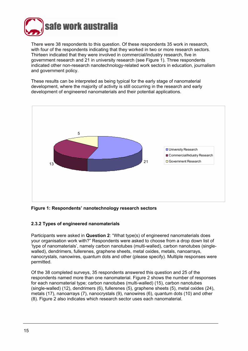
15
There were 38 respondents to this question. Of these respondents 35 work in research, with four of the respondents indicating that they worked in two or more research sectors. Thirteen indicated that they were involved in commercial/industry research, five in government research and 21 in university research (see Figure 1). Three respondents indicated other non-research nanotechnology-related work sectors in education, journalism and government policy. These results can be interpreted as being typical for the early stage of nanomaterial development, where the majority of activity is still occurring in the research and early development of engineered nanomaterials and their potential applications.
21
5
13
University Research
Commercial/Industry Research
Government Research
Figure 1: Respondents’ nanotechnology research sectors
2.3.2 Types of engineered nanomaterials
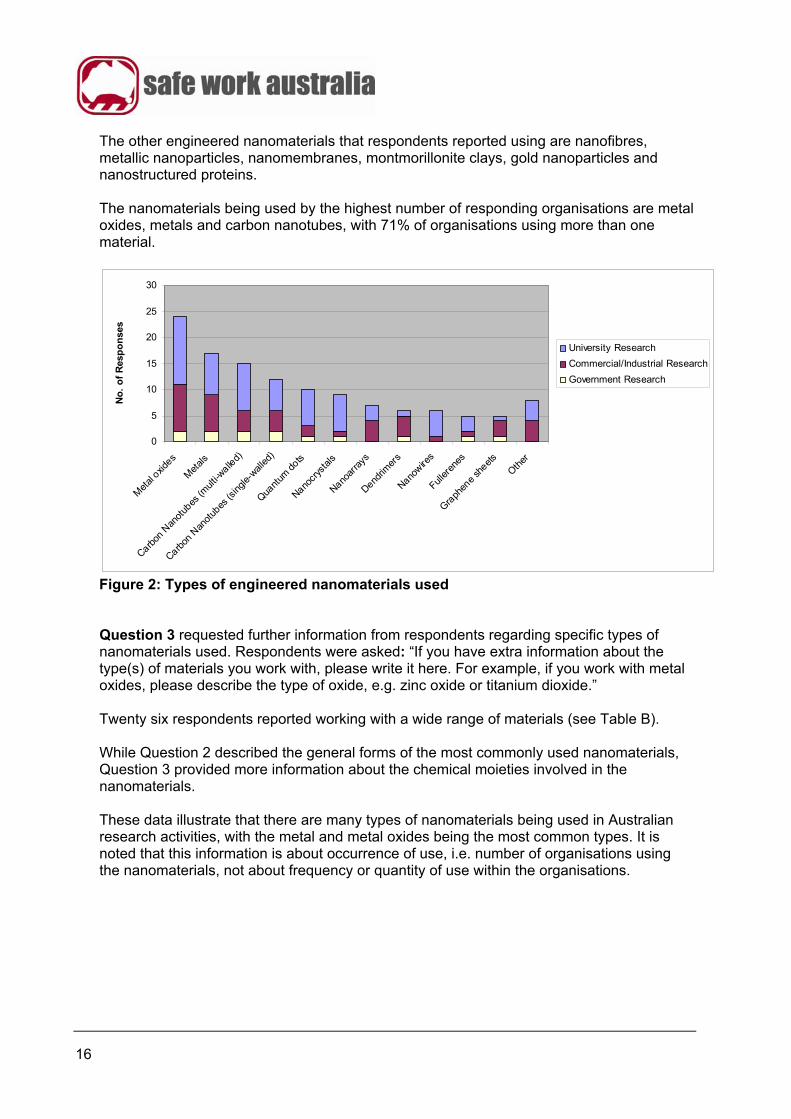
Participants were asked in Question 2: “What type(s) of engineered nanomaterials does your organisation work with?” Respondents were asked to choose from a drop down list of ‘type of nanomaterials’, namely carbon nanotubes (multi-walled), carbon nanotubes (single-walled), dendrimers, fullerenes, graphene sheets, metal oxides, metals, nanoarrays, nanocrystals, nanowires, quantum dots and other (please specify). Multiple responses were permitted. Of the 38 completed surveys, 35 respondents answered this question and 25 of the respondents named more than one nanomaterial. Figure 2 shows the number of responses for each nanomaterial type; carbon nanotubes (multi-walled) (15), carbon nanotubes (single-walled) (12), dendrimers (6), fullerenes (5), graphene sheets (5), metal oxides (24), metals (17), nanoarrays (7), nanocrystals (9), nanowires (6), quantum dots (10) and other (8). Figure 2 also indicates which research sector uses each nanomaterial.
16
The other engineered nanomaterials that respondents reported using are nanofibres, metallic nanoparticles, nanomembranes, montmorillonite clays, gold nanoparticles and nanostructured proteins. The nanomaterials being used by the highest number of responding organisations are metal oxides, metals and carbon nanotubes, with 71% of organisations using more than one material.
0
5
10
15
20
25
30
Met
al oxid
es
Met
als
Carbon
Nan
otub
es (m
ulti-w
alled)
Carbon
Nan
otub
es (s
ingle-
walled
)
Quant
um do
ts
Nanocr
ysta
ls
Nanoar
rays
Dendrim
ers
Nanowire
s
Fuller
enes
Graph
ene
sheets
Other
No
. o
f R
esp
on
ses
University Research
Commercial/Industrial Research
Government Research
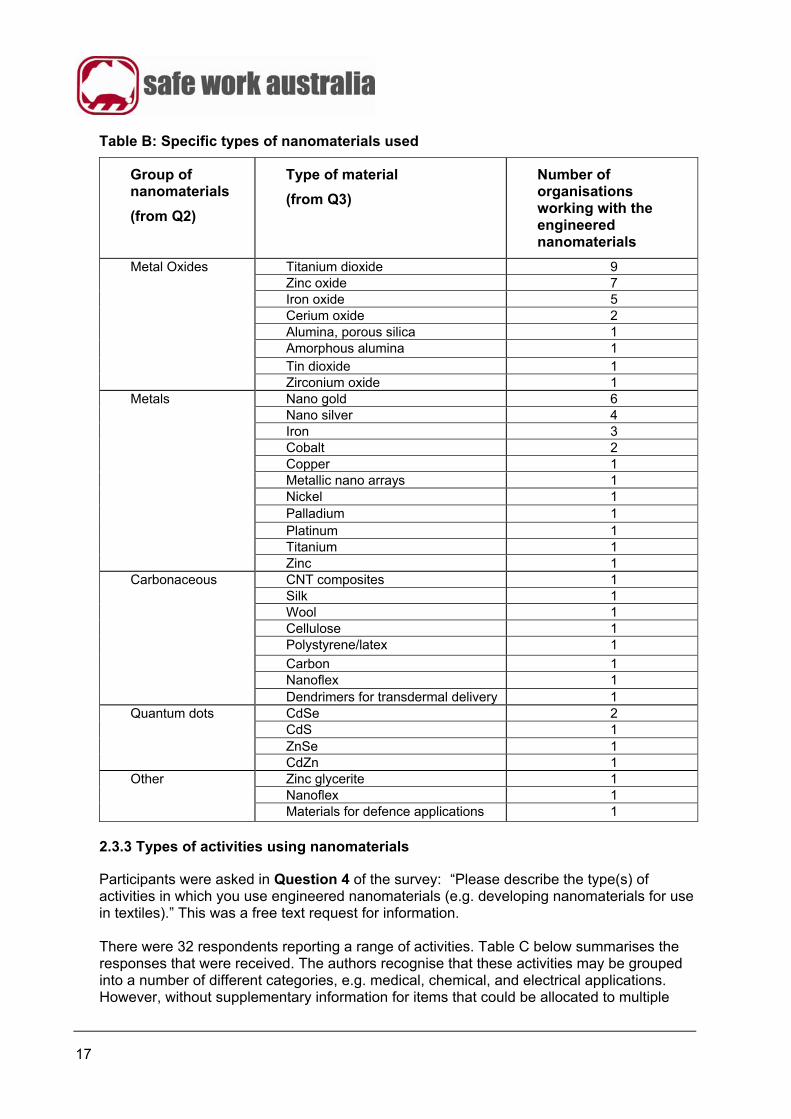
Figure 2: Types of engineered nanomaterials used Question 3 requested further information from respondents regarding specific types of nanomaterials used. Respondents were asked: “If you have extra information about the type(s) of materials you work with, please write it here. For example, if you work with metal oxides, please describe the type of oxide, e.g. zinc oxide or titanium dioxide.” Twenty six respondents reported working with a wide range of materials (see Table B). While Question 2 described the general forms of the most commonly used nanomaterials, Question 3 provided more information about the chemical moieties involved in the nanomaterials. These data illustrate that there are many types of nanomaterials being used in Australian research activities, with the metal and metal oxides being the most common types. It is noted that this information is about occurrence of use, i.e. number of organisations using the nanomaterials, not about frequency or quantity of use within the organisations.
17
Table B: Specific types of nanomaterials used
Group of nanomaterials
(from Q2)
Type of material
(from Q3)
Number of organisations working with the engineered nanomaterials
Titanium dioxide 9 Zinc oxide 7 Iron oxide 5 Cerium oxide 2 Alumina, porous silica 1 Amorphous alumina 1 Tin dioxide 1
Metal Oxides
Zirconium oxide 1 Nano gold 6 Nano silver 4 Iron 3 Cobalt 2 Copper 1 Metallic nano arrays 1 Nickel 1 Palladium 1 Platinum 1 Titanium 1
Metals
Zinc 1 CNT composites 1 Silk 1 Wool 1 Cellulose 1 Polystyrene/latex 1
Carbon 1 Nanoflex 1
Carbonaceous
Dendrimers for transdermal delivery 1 CdSe 2 CdS 1 ZnSe 1
Quantum dots
CdZn 1 Zinc glycerite 1 Nanoflex 1
Other
Materials for defence applications 1
2.3.3 Types of activities using nanomaterials
Participants were asked in Question 4 of the survey: “Please describe the type(s) of activities in which you use engineered nanomaterials (e.g. developing nanomaterials for use in textiles).” This was a free text request for information. There were 32 respondents reporting a range of activities. Table C below summarises the responses that were received. The authors recognise that these activities may be grouped into a number of different categories, e.g. medical, chemical, and electrical applications. However, without supplementary information for items that could be allocated to multiple
18
categories, such as ‘separation membranes’ and ‘size characterisation’, further classification of the activities has not been done for this report. Table C: Types of activities using nanomaterials as described by respondents
Description Number of respondents involved in this activity.
Solar cells 6
Medical 5 Composites 5 Better metallic properties 4 Textiles 4 Plastic modification 3 Sensors 2 1 response for each of the following activities
Adsorption of waste Ionic liquids Batteries Journalism Catalysis Nano scaffolds CVD growth Opto-electronic devices Dispersion of CNTs Paints Drug delivery Personal care products Electric conductive devices Pharmaceuticals Electro optical studies Pro-drugs using dendrimers Electrochemical processes Separation membranes Electronics Size characterisation Energy Surface coatings Environmental Thermal processes Fluorescence Toxicity testing Functional devices UV-shielding Functionalization Water treatment Hydrogen storage
2.3.4 Sources of engineered nanomaterials
Respondents were asked in Question 5 of the survey: “How do you obtain the engineered nanomaterials? Please tick all options that apply.” There were 35 respondents who answered this question, with 18 (51%) of the respondents indicating that they obtained nanomaterials from more than one source. Twenty seven (27) reported that they manufactured their own engineered nanomaterials, 19 reported they obtained them from overseas and 16 reported they obtained them from within Australia (see Figure 3). Twenty two out of 35 respondents (63%) indicated that they purchased at least some or all of their engineered nanomaterials. Figure 3 includes a work sector analysis which indicates that of the respondents, a higher proportion of researchers working in universities manufacture their own nanomaterials (20/21) than researchers working either in commercial/industrial (8/13) or government (3/5) sectors. These data indicate that with 77% of respondents manufacturing nanomaterials, there may be opportunities in Australia to examine substitution and modification of nanomaterials for
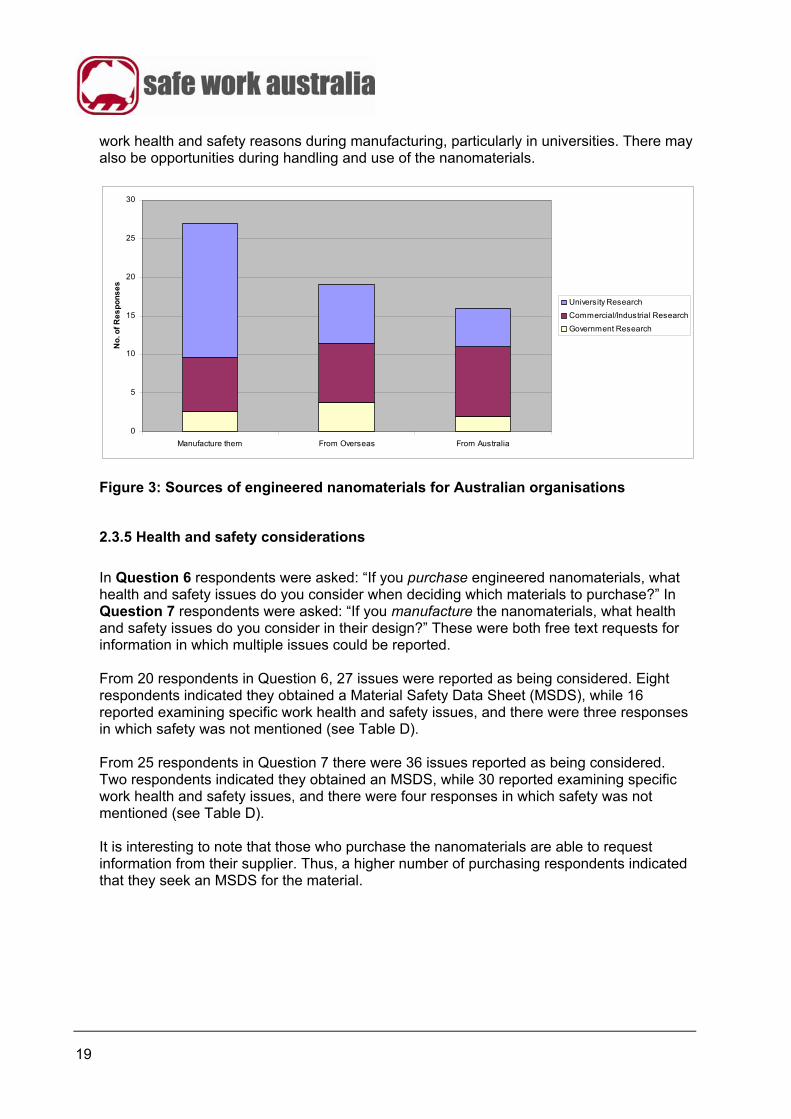
19
work health and safety reasons during manufacturing, particularly in universities. There may also be opportunities during handling and use of the nanomaterials.
0
5
10
15
20
25
30
Manufacture them From Overseas From Australia
No
. of
Re
sp
on
se
s
University Research
Commercial/Industrial Research
Government Research
Figure 3: Sources of engineered nanomaterials for Australian organisations
2.3.5 Health and safety considerations
In Question 6 respondents were asked: “If you purchase engineered nanomaterials, what health and safety issues do you consider when deciding which materials to purchase?” In Question 7 respondents were asked: “If you manufacture the nanomaterials, what health and safety issues do you consider in their design?” These were both free text requests for information in which multiple issues could be reported. From 20 respondents in Question 6, 27 issues were reported as being considered. Eight respondents indicated they obtained a Material Safety Data Sheet (MSDS), while 16 reported examining specific work health and safety issues, and there were three responses in which safety was not mentioned (see Table D). From 25 respondents in Question 7 there were 36 issues reported as being considered. Two respondents indicated they obtained an MSDS, while 30 reported examining specific work health and safety issues, and there were four responses in which safety was not mentioned (see Table D). It is interesting to note that those who purchase the nanomaterials are able to request information from their supplier. Thus, a higher number of purchasing respondents indicated that they seek an MSDS for the material.
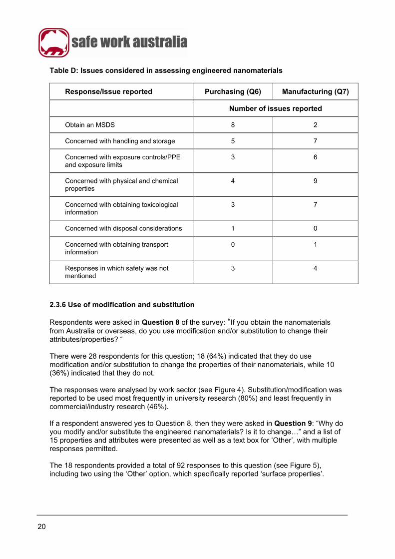
20
Table D: Issues considered in assessing engineered nanomaterials
Response/Issue reported Purchasing (Q6) Manufacturing (Q7)
Number of issues reported
Obtain an MSDS 8 2
Concerned with handling and storage 5 7
Concerned with exposure controls/PPE and exposure limits
3 6
Concerned with physical and chemical properties
4 9
Concerned with obtaining toxicological information
3 7
Concerned with disposal considerations 1 0
Concerned with obtaining transport information
0 1
Responses in which safety was not mentioned
3 4
2.3.6 Use of modification and substitution
Respondents were asked in Question 8 of the survey: “If you obtain the nanomaterials from Australia or overseas, do you use modification and/or substitution to change their attributes/properties? “ There were 28 respondents for this question; 18 (64%) indicated that they do use modification and/or substitution to change the properties of their nanomaterials, while 10 (36%) indicated that they do not. The responses were analysed by work sector (see Figure 4). Substitution/modification was reported to be used most frequently in university research (80%) and least frequently in commercial/industry research (46%). If a respondent answered yes to Question 8, then they were asked in Question 9: “Why do you modify and/or substitute the engineered nanomaterials? Is it to change…” and a list of 15 properties and attributes were presented as well as a text box for ‘Other’, with multiple responses permitted. The 18 respondents provided a total of 92 responses to this question (see Figure 5), including two using the ‘Other’ option, which specifically reported ‘surface properties’.
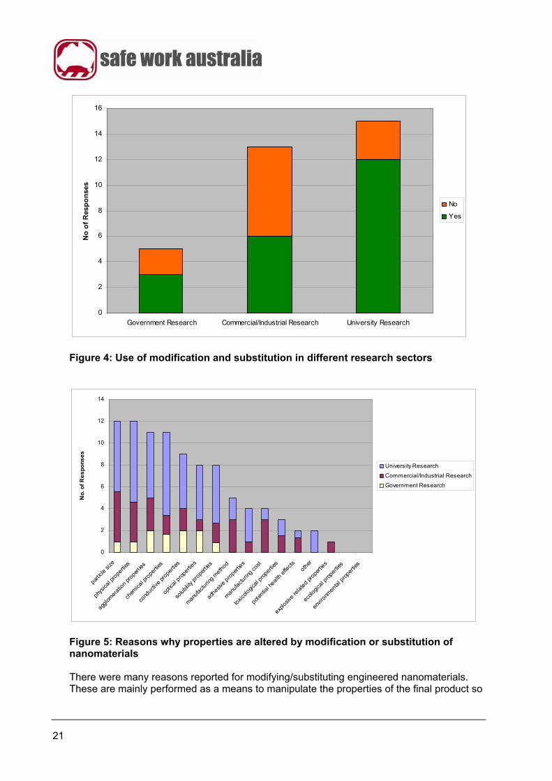
21
0
2
4
6
8
10
12
14
16
Government Research Commercial/Industrial Research University Research
No
of
Res
po
nse
s
No
Yes
Figure 4: Use of modification and substitution in different research sectors
0
2
4
6
8
10
12
14
parti
cle si
ze
phys
ical p
rope
rties
aggl
omer
atio
n pr
oper
ties
chem
ical p
rope
rties
cond
uctiv
e pr
operti
es
optic
al p
rope
rties
solub
ility p
rope
rties
man
ufac
turin
g m
ethod
adhes
ive p
roper
ties
man
ufac
turin
g co
st
toxic
ologic
al pro
perti
es
pote
ntia
l hea
lth e
ffects
othe
r
explo
sive
rela
ted
prop
ertie
s
ecolo
gical
pro
perti
es
envir
onm
enta
l pro
perti
es
No
. of
Re
sp
on
se
s
University Research
Commercial/Industrial Research
Government Research
Figure 5: Reasons why properties are altered by modification or substitution of nanomaterials There were many reasons reported for modifying/substituting engineered nanomaterials. These are mainly performed as a means to manipulate the properties of the final product so

22
that it exhibits the required functional properties, e.g. the particle size of nano titania is manipulated in order to optimise its optical properties when used as a sun screening agent. The five optional answers relating directly to work health and safety, hazardous, toxicological or environmental properties were either the least often selected or not selected at all. In Question 10 respondents were asked: “What approaches do you use to modify/substitute the nanomaterials? Please tick all options that apply.” Respondents were offered a list of six choices plus ‘other’. There were 77 responses given from 22 respondents (see Figure 6), including two responses using the ‘other’ option which specifically indicated ‘security in confidence’.
0
2
4
6
8
10
12
14
16
18
Add fu
nctio
nal g
roup
s
Mod
ify su
rface
char
acte
ristic
s
Chang
e fo
rm o
f the
mat
eria
l
Chang
e pa
rticle
size
Chang
e pa
rticle
shap
e
Mod
ify cry
stall
ine st
ructur
eOth
er
No
of
Re
sp
on
se
s
University Research
Commercial/Industrial Research
Government Research
Figure 6: Approaches used to modify or substitute nanomaterials The responses to this question show the different techniques used in the practice of substitution and/or modification of nanomaterials. As in Question 9, the techniques are indicative of those used to achieve the primary research goal of functional optimisation of the material. The most common forms of substitution/modification are functional group addition and modification of surface characteristics, next most common are to change the form of the material and to change particle size, and less common are to change particle shape and crystalline structure. Reflecting the overall use of substitution/modification (Figure 4), a work sector analysis indicated that the university research sector was the main cohort in all of the approaches used for substituting/modifying engineered nanomaterials.

23
As information progressively becomes available in the scientific and public domain about the potential use of a substitution and/or modification technique for specific engineered nanomaterials, such techniques are more likely to also be used as a means of reducing the hazards of nanomaterials. Additional comments were requested in Question 11. Respondents were asked; “If there is anything you would like to add about nanomaterial modification/substitution that you would like us to specifically consider, please write it here....”. There were five responses to this question, with respondents 1-4 working in research; these are given in Table E. Table E: Additional comments about the substitution/modification of engineered nanomaterials
Respondent Response
1 As a fundamental research institute, it is important that we have access to all types of nanomaterials to determine their uses and their safety/toxicity. I would like to see some guidelines for the researchers to protect their health from potentially toxic nanomaterials.
2 Most modifications performed in our laboratory are considered standard and in fact important avenues of research. Any dangers involved in the modification of nanomaterials seem parallel to chemical research of any other kind.
3 The nanoparticles are dispersed in molten polymer using shear to disperse them.
4 In general the use, manufacture and substitution/modification of nanomaterials is no different to that of ordinary chemistry based materials.
5 Yes, a lot more research into the OHS effects of working with nanomaterials
Of the responses to this question (see Table E), answers 2 and 4 indicate that nanomaterial research is similar to other chemical research, presumably indicating that they have other chemical research practices under appropriate control. Answers 1 and 5 indicate they would appreciate more specifically targeted work health and safety information. 2.4 Summary from the survey a) There were 38 respondents to the survey, who reported working on a range of different types of nanomaterials. The respondents’ organisations were primarily from universities, commercial/industry and government research groups. The most common nanomaterials handled are metal oxides, metals and carbon nanotubes and the most common areas of application are into energy, medical, surface coating and textile uses. b) Many organisations (27/35), and notably universities (20/21), manufacture their own engineered nanomaterials. A significant number also purchase them from overseas or from within Australia (see Figure 3). c) A number of respondents obtained work health and safety information about the nanomaterials that they are using from an MSDS. The main work health and safety issues examined for engineered nanomaterials are handling and storage, physical and chemical
24
properties, toxicological data and exposure controls/PPE (see Table D). The available information on these topics is limited. d) Most respondents indicated that substitution/modification is used to change the functional properties of the product (see Figure 4). A work sector analysis indicates that substitution/modification occurs more in university research and less in commercial/industry research which is as expected in product development. e) The five properties that are manipulated by modifying or substituting engineered nanomaterials by the highest number of organisations are particle size, physical properties, agglomeration properties, chemical properties and conductive properties. A small number of respondents indicated that they use substitution/modification to change the health hazard or toxicological properties (see Figure 5). f) Adding functional groups (17 responses) and modifying surface characteristics (16 responses) are the two most popular methods of substitution/modification of engineered nanomaterials. Others include changing the form of the material, the particle size and shape and the crystalline structure (see Figure 6). g) Australia’s nanotechnology activities are generally at the early stage of nanomaterial development, i.e. more focussed on de novo research than later stages of product development/production. However substitution/modification methodologies are well known and used in Australia and thus there is an existing capability that might be applied more broadly to work health and safety related purposes.
25
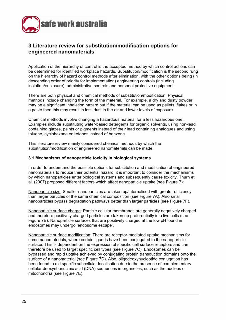
3 Literature review for substitution/modification options for engineered nanomaterials
Application of the hierarchy of control is the accepted method by which control actions can be determined for identified workplace hazards. Substitution/modification is the second rung on the hierarchy of hazard control methods after elimination, with the other options being (in descending order of priority for implementation) engineering controls (including isolation/enclosure), administrative controls and personal protective equipment. There are both physical and chemical methods of substitution/modification. Physical methods include changing the form of the material. For example, a dry and dusty powder may be a significant inhalation hazard but if the material can be used as pellets, flakes or in a paste then this may result in less dust in the air and lower levels of exposure. Chemical methods involve changing a hazardous material for a less hazardous one. Examples include substituting water-based detergents for organic solvents, using non-lead containing glazes, paints or pigments instead of their lead containing analogues and using toluene, cyclohexane or ketones instead of benzene. This literature review mainly considered chemical methods by which the substitution/modification of engineered nanomaterials can be made. 3.1 Mechanisms of nanoparticle toxicity in biological systems In order to understand the possible options for substitution and modification of engineered nanomaterials to reduce their potential hazard, it is important to consider the mechanisms by which nanoparticles enter biological systems and subsequently cause toxicity. Thurn et al. (2007) proposed different factors which affect nanoparticle uptake (see Figure 7): Nanoparticle size: Smaller nanoparticles are taken up/internalised with greater efficiency than larger particles of the same chemical composition (see Figure 7A). Also small nanoparticles bypass degradation pathways better than larger particles (see Figure 7F). Nanoparticle surface charge: Particle cellular membranes are generally negatively charged and therefore positively charged particles are taken up preferentially into live cells (see Figure 7B). Nanoparticle surfaces that are positively charged at the low pH found in endosomes may undergo ‘endosome escape’. Nanoparticle surface modification: There are receptor-mediated uptake mechanisms for some nanomaterials, where certain ligands have been conjugated to the nanoparticle surface. This is dependent on the expression of specific cell surface receptors and can therefore be used to target specific cell types (see Figure 7C). Endosomes can be bypassed and rapid uptake achieved by conjugating protein transduction domains onto the surface of a nanomaterial (see Figure 7D). Also, oligodeoxynucleotide conjugation has been found to aid specific subcellular localisation due to the presence of complementary cellular deoxyribonucleic acid (DNA) sequences in organelles, such as the nucleus or mitochondria (see Figure 7E).
26
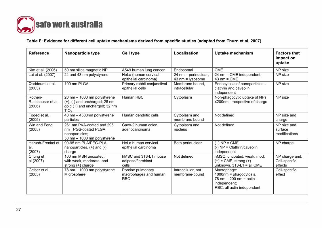
Figure 7: Mechanisms of cell entry and uptake for engineered nanomaterials (Thurn et al. 2007) The evidence of these mechanisms for the uptake of engineered nanomaterials into cells are summarised in Table F (Thurn et al. 2007). This information is key to understanding why certain substitution/modification options are more effective than others and will be highlighted throughout this review for specific processes. In summary, the most critical characteristics that affect nanoparticle uptake by cells are the particle size, surface charge or modification and the specific cell type involved. Studies have indicated that changes in one (or more) of these parameters can cause major differences in the efficiency and type of cellular uptake (Thurn et al. 2007).
27
Table F: Evidence for different cell uptake mechanisms derived from specific studies (adapted from Thurn et al. 2007)
Reference Nanoparticle type Cell type Localisation Uptake mechanism Factors that impact on uptake
Kim et al. (2006) 50 nm silica magnetic NP A549 human lung cancer Endosomal CME NP size Lai et al. (2007) 24 and 43 nm polystyrene HeLa (human cervical
epithelial carcinoma) 24 nm = perinuclear, 43 nm = lysosome
24 nm = CME independent, 43 nm = CME
NP size
Qaddoumi et al. (2003)
100 nm PLGA Primary rabbit conjunctival epithelial cells
Membrane bound, intracellular
Endocytosis of nanoparticles - clathrin and caveolin independent
NP size
Rothen-Rutishauser et al. (2006)
20 nm – 1000 nm polystyrene (+), (-) and uncharged; 25 nm gold (+) and uncharged; 32 nm TiO2
Human RBC Cytoplasm Non-phagocytic uptake of NPs ≤200nm, irrespective of charge
NP size
Foged et al. (2005)
40 nm – 4500nm polystyrene particles
Human dendritic cells Cytoplasm and membrane bound
Not defined NP size and charge
Win and Feng (2005)
261 nm PVA-coated and 295 nm TPGS-coated PLGA nanoparticles; 50 nm – 1000 nm polystyrene
Caco-2 human colon adenocarcinoma
Cytoplasm and nucleus
Not defined NP size and surface modifications
Harush-Frenkel et al. (2007)
90-95 nm PLA/PEG-PLA nanoparticles, (+) and (-) charge
HeLa human cervical epithelial carcinoma
Both perinuclear (+) NP = CME (-) NP = Clathrin/caveolin independent
NP charge
Chung et al.(2007)
100 nm MSN uncoated; with weak, moderate, and strong (+) charge
hMSC and 3T3-L1 mouse adipose/fibroblast cells
Not defined hMSC: uncoated, weak, mod. (+) = CME, strong (+) unknown. 3T3-L1 = all CME
NP charge and, Cell-specific effects
Geiser et al. (2005)
78 nm – 1000 nm polystyrene Microsphere
Porcine pulmonary macrophages and human RBC
Intracellular, not membrane-bound
Macrophage: 1000nm = phagocytosis, 78 nm – 200 nm = actin-independent; RBC: all actin-independent
Cell-specific effect
28
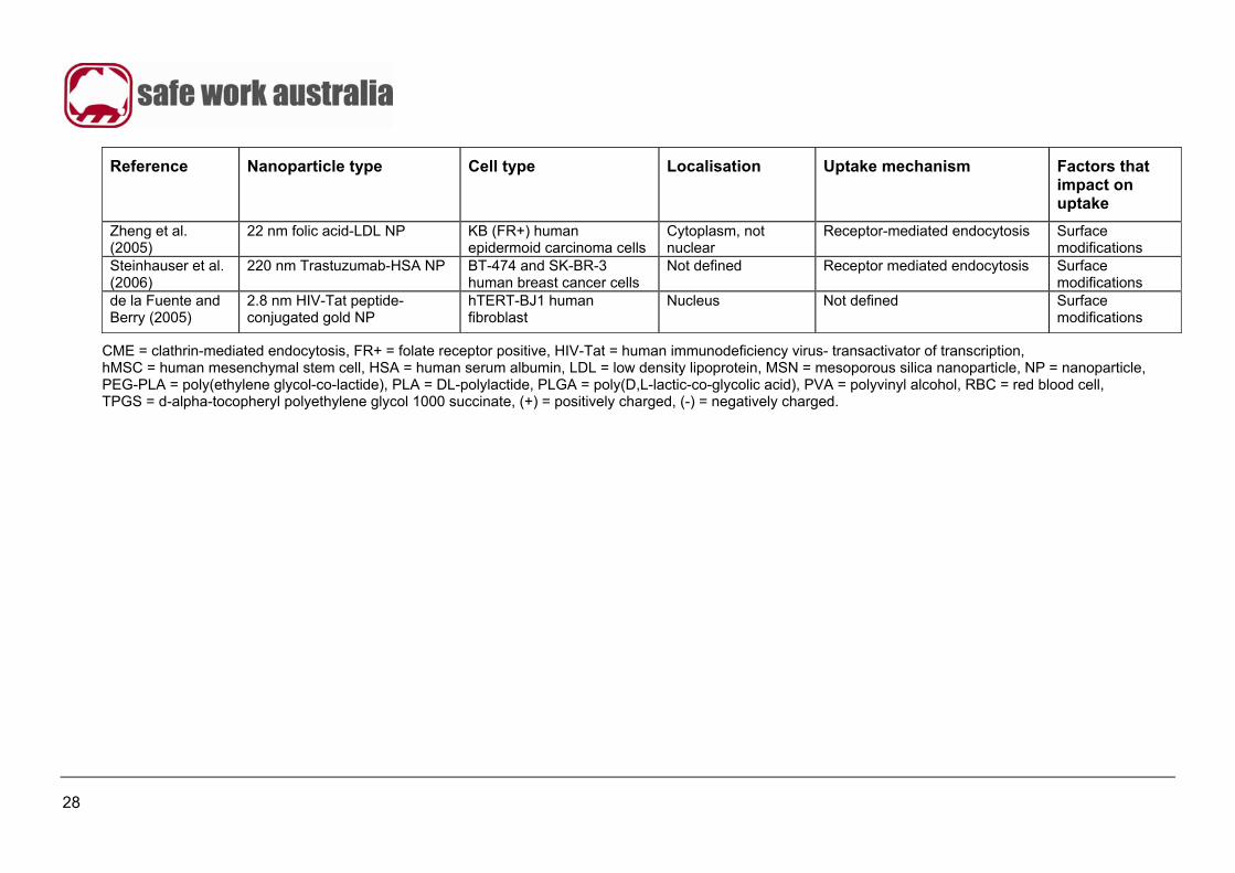
Reference Nanoparticle type Cell type Localisation Uptake mechanism Factors that impact on uptake
Zheng et al. (2005)
22 nm folic acid-LDL NP KB (FR+) human epidermoid carcinoma cells
Cytoplasm, not nuclear
Receptor-mediated endocytosis Surface modifications
Steinhauser et al. (2006)
220 nm Trastuzumab-HSA NP BT-474 and SK-BR-3 human breast cancer cells
Not defined Receptor mediated endocytosis Surface modifications
de la Fuente and Berry (2005)
2.8 nm HIV-Tat peptide-conjugated gold NP
hTERT-BJ1 human fibroblast
Nucleus Not defined Surface modifications
CME = clathrin-mediated endocytosis, FR+ = folate receptor positive, HIV-Tat = human immunodeficiency virus- transactivator of transcription, hMSC = human mesenchymal stem cell, HSA = human serum albumin, LDL = low density lipoprotein, MSN = mesoporous silica nanoparticle, NP = nanoparticle, PEG-PLA = poly(ethylene glycol-co-lactide), PLA = DL-polylactide, PLGA = poly(D,L-lactic-co-glycolic acid), PVA = polyvinyl alcohol, RBC = red blood cell, TPGS = d-alpha-tocopheryl polyethylene glycol 1000 succinate, (+) = positively charged, (-) = negatively charged.
29
3.1.1 Importance of nanoparticle size for cell uptake
Due to their small size, nanoparticles are easily taken up into cells, i.e. individual nanoparticles by receptor-mediated endocytosis, and their larger aggregates via phagocytosis and the receptor-independent endocytic pathway of macropinocytosis (see Figure 8), whereupon the nanoparticles have essentially free access to all cellular compartments (Xiang et al. 2006). Receptor-mediated endocytic mechanisms involve receptors on the cell membrane that capture particles or substances via binding to specific ligands. These include:
• clathrin mediated-endocytosis, the classical and well-described mechanism involving clathrin-coated pits in the membranes of most nucleated cells;
• caveolin-mediated endocytosis, the clathrin-independent uptake mechanism via flask-shaped cholesterol-rich invaginations of cell membranes;
• caveolin- and clathrin-independent, yet lipid raft-associated endocytosis, which is specific for the internalisation of certain cytokine receptors and proteins; and
• phagocytosis performed predominantly by macrophages and Langerhans cells (immature dendritic cells in the skin), which are antigen-presenting cells that act as sentinels of the immune system. Langerhans cells, and stimulated macrophages and endothelial cells, also perform the receptor-independent uptake process of macropinocytosis (Xiang et al. 2006).
The individual endocytic pathways also have a defined specific size range of engulfed particulate or soluble material (see Figure 8).
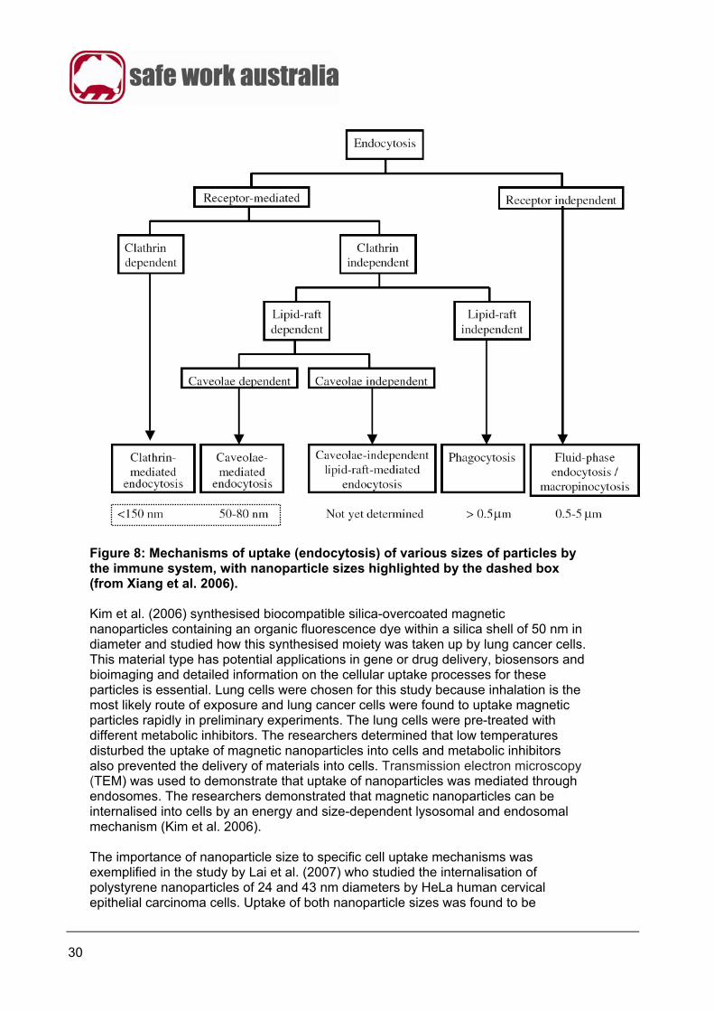
30
Figure 8: Mechanisms of uptake (endocytosis) of various sizes of particles by the immune system, with nanoparticle sizes highlighted by the dashed box (from Xiang et al. 2006). Kim et al. (2006) synthesised biocompatible silica-overcoated magnetic nanoparticles containing an organic fluorescence dye within a silica shell of 50 nm in diameter and studied how this synthesised moiety was taken up by lung cancer cells. This material type has potential applications in gene or drug delivery, biosensors and bioimaging and detailed information on the cellular uptake processes for these particles is essential. Lung cells were chosen for this study because inhalation is the most likely route of exposure and lung cancer cells were found to uptake magnetic particles rapidly in preliminary experiments. The lung cells were pre-treated with different metabolic inhibitors. The researchers determined that low temperatures disturbed the uptake of magnetic nanoparticles into cells and metabolic inhibitors also prevented the delivery of materials into cells. Transmission electron microscopy (TEM) was used to demonstrate that uptake of nanoparticles was mediated through endosomes. The researchers demonstrated that magnetic nanoparticles can be internalised into cells by an energy and size-dependent lysosomal and endosomal mechanism (Kim et al. 2006). The importance of nanoparticle size to specific cell uptake mechanisms was exemplified in the study by Lai et al. (2007) who studied the internalisation of polystyrene nanoparticles of 24 and 43 nm diameters by HeLa human cervical epithelial carcinoma cells. Uptake of both nanoparticle sizes was found to be
31
temperature dependent and caveolin independent. The larger nanoparticles entered cells through a degradative/clathrin dependent pathway that was not used by the smaller nanoparticles, with the latter exhibiting perinuclear localisation and avoiding endosomal/lysosomal entrapment. Therefore, the two particle sizes entered this cell type by two different mechanisms based purely on size (Lai et al. 2007). Qaddoumi et al. (2003) examined the expression of caveolin and clathrin in rabbit conjunctival epithelial cells (RCEC) to determine whether they play a role in endocytosis of 100 nm polylactic polyglycolic acid co-polymer (PLGA) nanoparticles. The effect of disrupting the formation of clathrin-coated vesicles by using pharmacological treatments (intracellular K+ depletion and hypertonic challenge) and caveolae (filipin and nystatin) on apical uptake of nanoparticles in primary cultured RCEC was investigated. The researchers concluded that endocytosis of the biodegradeable nanoparticles in RCECs occurred independently of caveolin and clathrin mediated pathways and that protein and gene expression of clathrin but not caveolin was identified in RCEC (Qaddoumi et al. 2003). Rothen-Rutishauser et al. (2006) combined different microscopic techniques to visualise the uptake of fine particles and nanoparticles in red blood cells (RBCs). Fluorescent polystyrene particles were analysed by laser scanning microscopy (LSM) combined with digital image restoration. Gold particles were analysed by TEM and titanium dioxide particles were analysed by energy filtering transmission electron microscopy (EFTEM). By using these microscopic techniques the researchers were able to visualise and detect fine particles and nanoparticles in red blood cells. The researchers found that only the particle size (less than 200 nm) but not surface charge influenced their uptake by RBCs. Win and Feng (2005) studied the cellular uptake of polymeric nanoparticles and fine particles by the Caco-2 human adenocarcinoma cell line using an in vitro model to examine the potential use of biodegradable polymeric nanoparticles for oral chemotherapy. Feasibility of this potential application was demonstrated by showing quantification and localisation of cell uptake of 50nm – 1000nm fluorescent polystyrene particles, 261 nm PLGA particles coated with polyvinyl alcohol (PVA) and 295 nm PLGA particles coated with a vitamin E derivative (tocopheryl polyethylene glycol succinate, TPGS). The effects of particle surface coating and particle size on the cellular uptake of the particles were quantified by spectrofluorometric measurement. Cellular uptake of vitamin E TPGS-coated PLGA particles was 1.4 folds higher than that of PVA-coated PLGA particles and 4-6 folds higher than that of polystyrene particles, demonstrating that surface and size modification of particles can modify cellular uptake (Win and Feng 2005).
3.1.2 Importance of surface charge for cell uptake
Cell membranes are usually negatively-charged and have a high affinity for molecules that are positively-charged. Using confocal microscopy, Harush-Frenkel et al. (2007) demonstrated that there was a higher uptake of fluorescent positively-charged polyactide/polyethylene glycol-co-lactide co-polymer (PLA/PEG-PLA) nanoparticles than negatively-charged nanoparticles of the same material in HeLa cells. The authors also determined that the lower rate of uptake of the negatively-charged nanoparticles is via a caveolin and clathrin-independent mechanism whilst the higher rate of uptake of positively charged particles is dependent on a clathrin mechanism.
32
Surface charge also affects the interaction of nanomaterials with serum proteins which may alter the nature of the particle’s surface that is presented to the cell membrane prior to uptake, but can also ultimately influence their clearance from the body. Recent studies involving intravenously-administered quantum dots (QDs) in rodents have indicated some nanoparticle requirements for rapid renal filtration and urinary excretion, i.e. Zwitterionic or neutral organic coatings of QDs prevented adsorption of serum proteins that would otherwise increase the QD hydrodynamic diameter by >15 nm and prevent renal excretion. Those QDs with a final hydrodynamic diameter <5.5 nm exhibited rapid and efficient urinary excretion and elimination from the body (Choi et al. 2007). Such findings show that the hydrodynamic size of a positively charged nanoparticle within a biological system may be much larger than the size of a pristine nanoparticle, due to protein-nanoparticle interactions. This would result in significantly slower renal clearance resulting in blood half-life values of hours rather than minutes, with major implications for the toxicokinetics of such nanoparticles that reach the circulatory system (Choi et al. 2007).
3.1.3 Importance of cell specific effects for nanoparticle uptake
Overall, when employing the weight of evidence approach concerning cell-specific effects of nanomaterials, a greater weight should be placed on data derived from in vivo rather than in vitro studies. In regard to in vitro studies, the best evidence will come from studies using human primary cells whereas a lower level of evidence is provided by studies using immortalised/tumour cell lines and animal cells. The premise is that the closer a study mimics a human system the more useful is the data that it provides. Different mechanisms and rates of uptake are exhibited by different cell types for the same nanomaterial. For example, when Chung et al. (2007) exposed mouse adipose/fibroblast 3T3-L1 and human mesenchymal stem cells (hMSC) to silica mesoporous nanoparticles with strong positive surface charges, it was found that the uptake mechanisms were cell type specific. The 3T3-L1 cells were found to utilise clathrin-mediated endocytosis to take up the nanoparticles, whereas hMSC cells used an undefined alternate mechanism. When the silica mesoporous nanoparticles were uncoated, or had weak or moderate positive charges, the uptake for both cell-types was by clathrin-mediated endocytosis. Geiser et al. (2005) performed an in vitro study of the uptake of fine (200 -1000 nm) and ultra fine particles (UFP) (<100 nm) by human red blood cells (RBCs) and porcine pulmonary macrophages. Using confocal laser scanning microscopy it was found that 77% of macrophages contained UFPs and 56% contained fine particles. However it was also found that only 40% of RBCs contained UFPs and none contained no fine particles. This further demonstrates that particle uptake is cell-specific.
33
3.1.4 Importance of surface modification for nanoparticle uptake
There have been several investigations aimed at increasing the efficacy of uptake of potentially therapeutic nanoparticles in to target cell types, in order to bypass intracellular obstacles such as endosomes. The general strategy has been to conjugate molecules to the surface of nanoparticles in order to ‘target’ their affinity for specific cell types and subcellular organelles. Short amphipathic peptides are one type of conjugate that enables nanoparticles to translocate rapidly across cell membranes (de la Fuente and Berry 2005; Koch et al. 2003; Gupta et al. 2005; Vives et al. 1997). The exact mechanism by which translocation occurs across the cell membrane is still a matter of scientific investigation. However, it is known to occur rapidly and efficiently. An example of the peptide conjugation technique has been reported for 2.8 nm gold nanoparticles that were conjugated with a cell-penetrating peptide, i.e. the human immunodeficiency virus transactivator of transcription (HIV-Tat) peptide fragment. Transmission electron microscopy (TEM) showed that they had translocated across the cell membrane and were sufficiently small (<30 nm) to pass through nuclear membrane pores to become localised within the nucleus of immortalised human fibroblast cells. In contrast gold nanoparticles that lacked the Tat peptide were found in cytoplasmic vacuoles or surrounding the mitochondria, but not in the nucleus (de la Fuente and Berry 2005). However, Tkachenko et al. (2003) conjugated 20 nm gold nanoparticles to HIV-Tat peptide. These were found to have localised in the cytoplasm rather than the nucleus. This indicates that even when the nanomaterial is conjugated with an organelle targeting peptide, its size is an important factor affecting the type of cellular uptake involved. It should be noted that nanoparticles <100 nm may also cross the nuclear membrane via receptor-mediated endocytosis. These researchers later reported that these Tat-conjugated gold nanoparticles also did not target the nucleus of 3T3/NIH murine fibroblastoma cells or HepG2 human hepatocarcinoma cells, but that conjugation with the adenovirus nuclear localisation signal or the integrin binding domain was successful in triggering nuclear uptake (Tkachenko et al. 2004). In another study which demonstrated the time-dependent nature of nanoparticle localisation within specific organelles, iron oxide nanoparticles labelled with the red fluorophore Cy3.5 and conjugated to a green fluorophore (fluorescein isothiocyanate, FITC)-Tat peptide were incubated with HeLa cells. The flow cytometry study indicated that the nanoparticles were rapidly internalised into the HeLa cells. Confocal microscopy 24 hours post-treatment showed co-localisation of Cy3.5 and FITC in the cytoplasm and the nucleus, which was lost after 72 hours (Koch et al. 2003). Other researchers have taken the different approach of conjugating monoclonal antibodies or receptor ligands to the surface of nanoparticles in order to specifically target tumour cells that over-express certain cell surface receptors. Steinhauser et al. (2006) were able to selectively target only those aggressive breast cancer cells that over-expressed human epidermal growth factor receptor type 2 (Her2) using nanoparticles conjugated with Trastuzumab, a specific antibody directed against Her2. A similar approach has used nanoparticles conjugated with folate in order to target the folate receptor on over-expressing nasopharyngeal and prostate cancer cells (Zheng et al. 2005; Hattori and Maitani 2005; Dixit et al. 2006). Furthermore, if nuclear or mitochondrial oligodeoxynucleotides are conjugated to a nanoparticle, then there is a marked increase in either the nuclear or mitochondrial subcellular
34
localisation within a targeted cell following the binding of the conjugated nanoparticle to complementary DNA sequences at these sites (Paunesku et al. 2003, 2007; Qin and Yung 2006).
3.1.5 Biocompatibility and surface coatings
When undertaking surface modification to change the efficiency and uptake of an engineered nanomaterial there is also the consideration of biocompatibility, which is important in negating or reducing potential toxic effects (Wang et al. 2004). Biocompatibility has been defined as ‘the property of being biologically compatible by not producing a toxic, injurious, or immunological response in living tissue”. A major goal in substituting or modifying an engineered nanomaterial should be to make it biocompatible. Therefore, any surface coating used with a nanomaterial should confer this property. An example of the application of the biocompatible principle is case of ‘synthetic’ parts (e.g. prostheses, artificial organs, contact lenses, artificial limbs, biosensors or encapsulating membranes), which are now being used as replacements for ‘defective’ parts in human hosts (Mathieu 2001; Sabbatini and Zambonin 1996; Good 1993; Aleyamma and Sharma 1991; Makohliso 1999; Wang and Ruckenstein 1993). The structural materials used in these replacement parts are usually in themselves bio-incompatible. However, in order for them to be accepted in their host they need to be made compatible. This has been done in many cases by grafting a biocompatible surface material onto the replacement part, which has the effect of preventing protein absorption and reduces or negates toxic effects of the bio-incompatible material (Marieb 1998). Such biocompatible materials include hydroxyapatite, chitosan, chitin coating, peptides, polysaccharides and other polymeric materials. There are a number of factors which determine the interaction of cells and biomaterials. These include surface energy; the balance between surface hydrophilicity and hydrophobicity; chemical structure and functional groups; type and the density of surface charges; molecular weight and conformational flexibility of the polymer; and surface topography and roughness (Wang et al. 2004). There has been much time and effort invested in the health care industry to develop and improve materials that are suitably biocompatible with the living environment (Aleyamma and Sharma 1991). Synthetic polymers are a broad range of materials that can easily be utilised as surface coatings. However, the issue of non-specific protein adsorption needs to be addressed to prevent inflammation in order that these materials can successfully become biocompatible. Therefore, research is directed towards polymers which have minimal protein adsorption by using strategies that mimic the external cell membrane or use biologically active materials such as proteins, peptides or polysaccharides. This type of procedure has the advantage of functionalising the surface of the material without modifying its bulk properties. Immobilisation of biomolecules on the surface of materials may be achieved by several mechanisms including covalent binding or physical adsorption (Mathieu 2001).
35
4 Substitution/modification options for specific engineered nanomaterials
This section addresses specific classes of engineered nanomaterials with particular reference to their background, toxicology and potential substitution/modification options to reduce workplace hazards.
4.1 Carbon nanotubes
4.1.1 Background
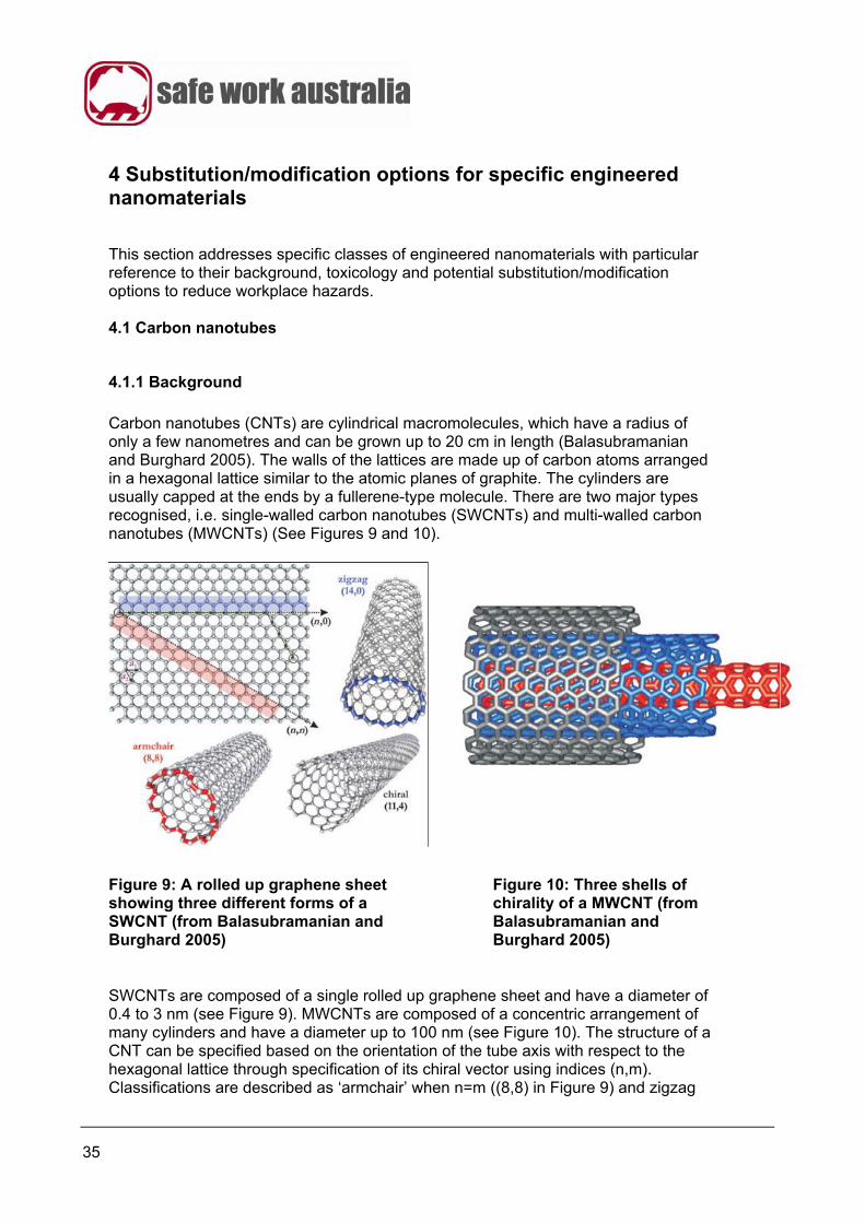
Carbon nanotubes (CNTs) are cylindrical macromolecules, which have a radius of only a few nanometres and can be grown up to 20 cm in length (Balasubramanian and Burghard 2005). The walls of the lattices are made up of carbon atoms arranged in a hexagonal lattice similar to the atomic planes of graphite. The cylinders are usually capped at the ends by a fullerene-type molecule. There are two major types recognised, i.e. single-walled carbon nanotubes (SWCNTs) and multi-walled carbon nanotubes (MWCNTs) (See Figures 9 and 10).
Figure 9: A rolled up graphene sheet showing three different forms of a SWCNT (from Balasubramanian and Burghard 2005)
Figure 10: Three shells of chirality of a MWCNT (from Balasubramanian and Burghard 2005)
SWCNTs are composed of a single rolled up graphene sheet and have a diameter of 0.4 to 3 nm (see Figure 9). MWCNTs are composed of a concentric arrangement of many cylinders and have a diameter up to 100 nm (see Figure 10). The structure of a CNT can be specified based on the orientation of the tube axis with respect to the hexagonal lattice through specification of its chiral vector using indices (n,m). Classifications are described as ‘armchair’ when n=m ((8,8) in Figure 9) and zigzag
36
when m=0 ((14,0) in Figure 9). There are three methods that have been established for the production of SWCNTs and MWCNTs. See Table G for details of these. Table G: Overview of the important synthesis procedures for CNTs (Adapted from Table 1 of Balasubramanian and Burghard 2005)
Synthetic method
Reaction principle Average diameter of the tubes
Maximum production rate
Electric arc discharge
Carbon atoms are generated through an electric arc discharge at T>3000˚C between two graphite rods. Nanotubes are formed in the presence of suitable catalyst metal particles (Fe, Co, or Ni).
1.3–1.4 nm
120 g per day
Laser ablation Generation of atomic carbon at T>3000˚C through laser irradiation of graphite, which contains appropriate catalyst particles (Fe, Co, or Ni), is followed by formation of nanotubes.
1.4 nm
50 g per day
Catalytic decomposition of gaseous hydrocarbons
Decomposition of a gaseous hydrocarbon source (e.g. an alkane or CO) is catalysed by metal nanoparticles (Co or Fe). Particles are prepared by pyrolysis of suitable precursors (e.g. [Fe(CO)5]) at 1000–1100˚C under high pressure.
1 nm
50 kg per day
For each of the three production methods, there are issues with the purity of the CNTs, with there being impurities such as metals (e.g. Fe or Co), fullerenes or amorphous carbon. Consequently, purification of CNTs is required prior to further processing (Balasubramanian and Burghard 2005) and typically involves the following steps:
a) thermal oxidation step in air
b) acid reflux step (using HCl)
c) filtration, and
d) thermal annealing. The synthesis of large quantities of CNTs usually results in bundles of hundreds of CNTs being produced. These bundles can easily be dispersed by ultrasonic treatment in aqueous surfactant solution, which leads to individual CNTs being enclosed in a detergent shell (Balasubramanian and Burghard 2005). An alternative method of preparing relatively pure isolated CNTs is by vapour deposition onto a solid support (see Figure 11).

37
Figure 11: Chemical vapour deposition used for the synthesis of individual CNTs by surface deposited catalyst particles (adapted from Balasubramanian and Burghard 2005) CNTs have attracted much interest because their specific strength, lightness, electrical and related properties may potentially be exploited in new products e.g. coloured textiles, conductors, semiconductors, composites etc. Like fullerenes and quantum dots, these are a new class of compound which are attractive because of their potential commercial applications.
4.1.2 Toxicology of carbon nanotubes
There is now a growing body of information about the potential hazards of CNTs. Further data is needed and it is noted that there are a number of difficulties associated with toxicity testing for CNTs, which include:
a) fibre agglomeration and difficulty involved in isolating single fibres
b) ensuring CNTs come into contact with cells for in vitro tests
c) difficulties involved in using standard toxicity tests, e.g. inhalation exposure studies in rats and mice failing because fibres did not get to the pleural cavity in the lungs, due to difficulties in aerosolising the CNTs
d) problems of interpretation of test data, e.g. when in vivo intratracheal instillation is used, excessive loads of CNTs can easily be deposited, which can lead to artificial biopersistence in the lung and questionable results (Drew 2009). Lam et al. (2006) in a review of the available literature on CNT toxicity concluded that “the results of rodent studies collectively showed that regardless of the process by which CNTs were synthesised and the types of metal impurities that they contained, CNTs were capable of producing inflammation, epithelioid granulomas, fibrosis and biochemical/toxicological changes in the lung”. They also indicated that SWCNTs were more toxic than quartz, a known chronic workplace inhalation hazard, and carbon black which exhibited minimal lung responses in comparative toxicological inhalation studies in mice. Lam et al. (2006) stated that SWCNTs present in the lungs of mice produce respiratory impairment, damage DNA in the aorta, increase aortic plaques, induce atherosclerotic lesions in arteries of the heart and retard bacterial clearance. There are two important in vivo studies that both studied the carcinogenic and fibrogenic behaviour of MWCNTs in the intraperitoneal cavity of mice (Poland et al. 2008, Takagi et al. 2008). Whilst there were experimental difficulties with both studies, it was clearly shown by Poland et al. (2008) that inflammatory and granuloma responses were associated with long
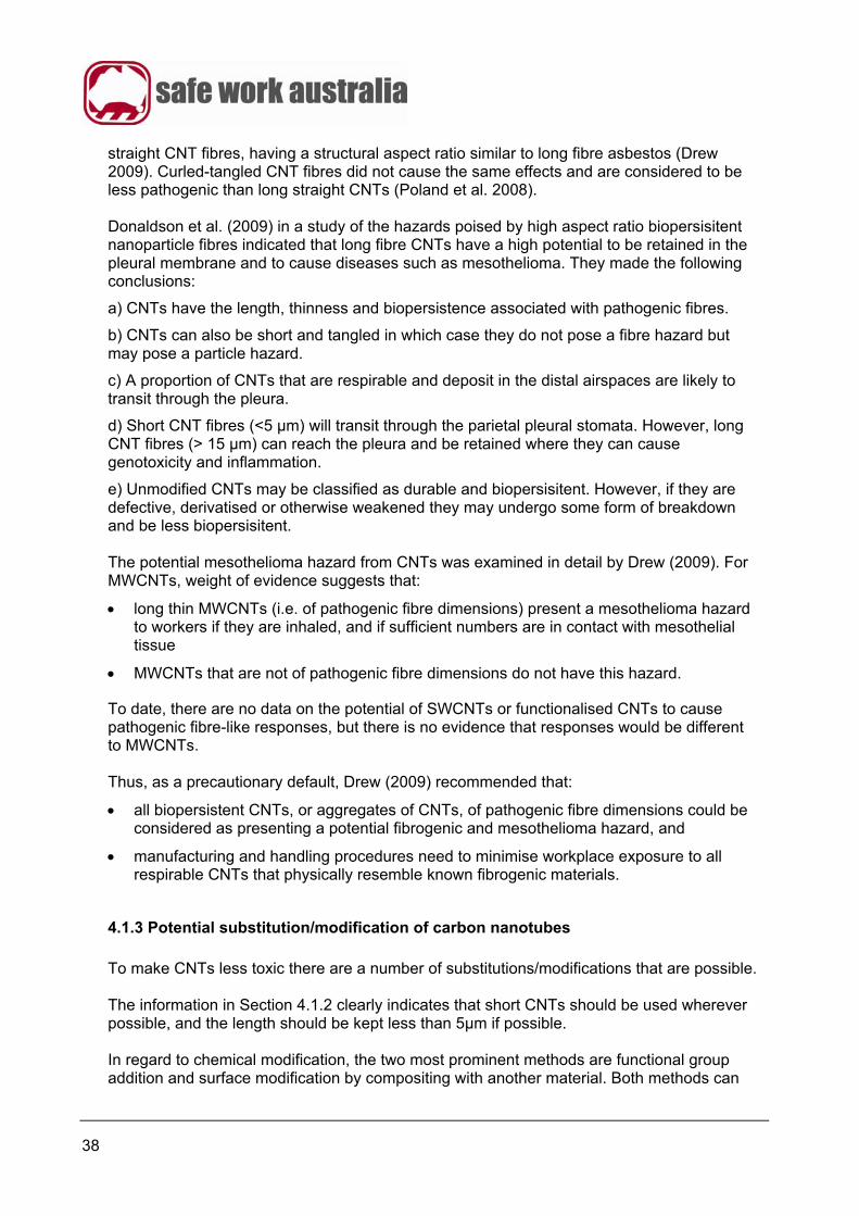
38
straight CNT fibres, having a structural aspect ratio similar to long fibre asbestos (Drew 2009). Curled-tangled CNT fibres did not cause the same effects and are considered to be less pathogenic than long straight CNTs (Poland et al. 2008). Donaldson et al. (2009) in a study of the hazards poised by high aspect ratio biopersisitent nanoparticle fibres indicated that long fibre CNTs have a high potential to be retained in the pleural membrane and to cause diseases such as mesothelioma. They made the following conclusions:
a) CNTs have the length, thinness and biopersistence associated with pathogenic fibres.
b) CNTs can also be short and tangled in which case they do not pose a fibre hazard but may pose a particle hazard.
c) A proportion of CNTs that are respirable and deposit in the distal airspaces are likely to transit through the pleura.
d) Short CNT fibres (<5 μm) will transit through the parietal pleural stomata. However, long CNT fibres (> 15 μm) can reach the pleura and be retained where they can cause genotoxicity and inflammation.
e) Unmodified CNTs may be classified as durable and biopersisitent. However, if they are defective, derivatised or otherwise weakened they may undergo some form of breakdown and be less biopersisitent. The potential mesothelioma hazard from CNTs was examined in detail by Drew (2009). For MWCNTs, weight of evidence suggests that:
long thin MWCNTs (i.e. of pathogenic fibre dimensions) present a mesothelioma hazard to workers if they are inhaled, and if sufficient numbers are in contact with mesothelial tissue
MWCNTs that are not of pathogenic fibre dimensions do not have this hazard.
To date, there are no data on the potential of SWCNTs or functionalised CNTs to cause pathogenic fibre-like responses, but there is no evidence that responses would be different to MWCNTs. Thus, as a precautionary default, Drew (2009) recommended that:
all biopersistent CNTs, or aggregates of CNTs, of pathogenic fibre dimensions could be considered as presenting a potential fibrogenic and mesothelioma hazard, and
manufacturing and handling procedures need to minimise workplace exposure to all respirable CNTs that physically resemble known fibrogenic materials.
4.1.3 Potential substitution/modification of carbon nanotubes
To make CNTs less toxic there are a number of substitutions/modifications that are possible. The information in Section 4.1.2 clearly indicates that short CNTs should be used wherever possible, and the length should be kept less than 5μm if possible. In regard to chemical modification, the two most prominent methods are functional group addition and surface modification by compositing with another material. Both methods can
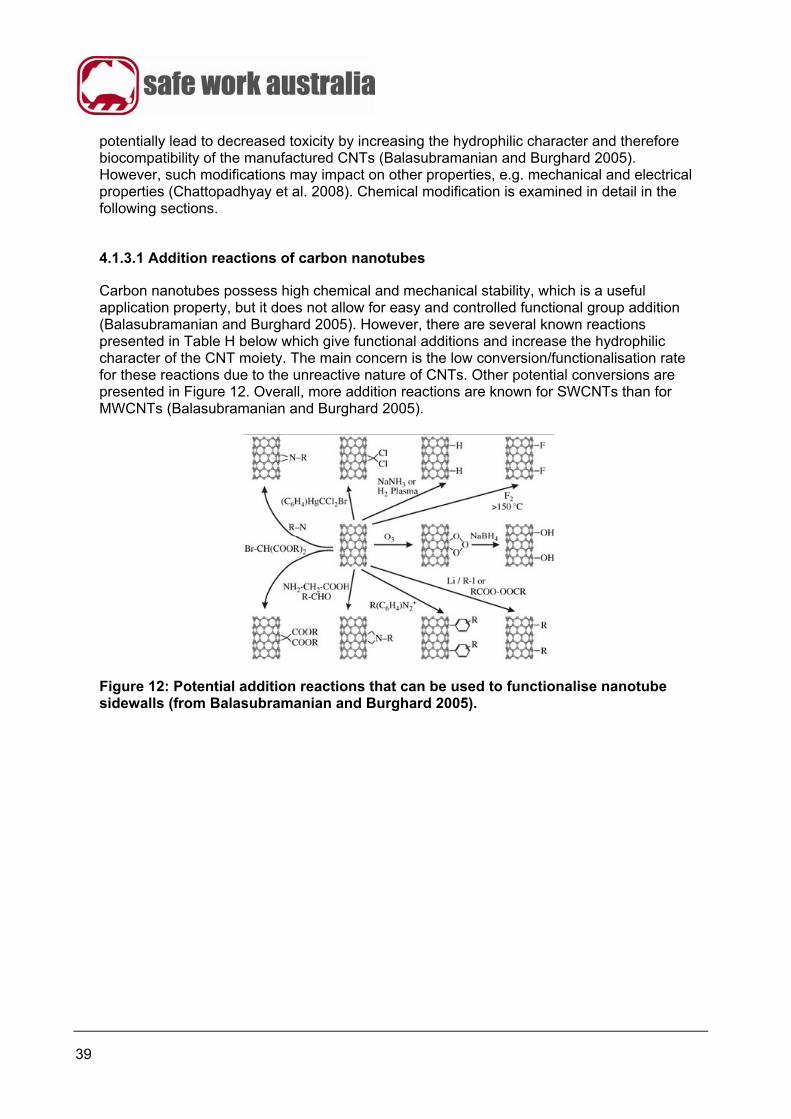
39
potentially lead to decreased toxicity by increasing the hydrophilic character and therefore biocompatibility of the manufactured CNTs (Balasubramanian and Burghard 2005). However, such modifications may impact on other properties, e.g. mechanical and electrical properties (Chattopadhyay et al. 2008). Chemical modification is examined in detail in the following sections.
4.1.3.1 Addition reactions of carbon nanotubes
Carbon nanotubes possess high chemical and mechanical stability, which is a useful application property, but it does not allow for easy and controlled functional group addition (Balasubramanian and Burghard 2005). However, there are several known reactions presented in Table H below which give functional additions and increase the hydrophilic character of the CNT moiety. The main concern is the low conversion/functionalisation rate for these reactions due to the unreactive nature of CNTs. Other potential conversions are presented in Figure 12. Overall, more addition reactions are known for SWCNTs than for MWCNTs (Balasubramanian and Burghard 2005).
Figure 12: Potential addition reactions that can be used to functionalise nanotube sidewalls (from Balasubramanian and Burghard 2005).

40
Table H: Known addition reactions for CNTs (summarised from Balasubramanian and Burghard 2005).
Type of CNT
Modification Product Reagents/Process Conditions/Comments References
SWCNTs Carboxylation Carboxyl SWCNT (5-10% conversion)
a) Nitric and sulfuric acids/ultrasonification b) Heat
a) End tubes open and holes form in side walls followed by oxidative etching of sidewalls
b) SWCNTs produced are100-300 nm in length with sidewalls containing carboxyl groups
c) Further reactions of amide and ester formation possible
d) Reaction occurs at wall defects
Zhang et al. 2003
Carboxyl-SWCNTs
Anhydride formation
SWCNT anhydride (1-5% conversion)
Acid treatment a) Ring closure reaction is possible b) A mixture of products can be
obtained c) Reaction occurs at wall defects
Sano et al. 2001
Nitrogen doped MWCNTs
a) Carboxylation b) Amidization
MWCNT ferritin or BSA amide
Amine groups of ferritin or Bovine Serum Albumin (BSA),
a) Reaction is a covalent binding of protein to MWCNT
b) Reaction occurs at wall defects
Katz and Willner 2004
SWCNTs Fluorination Fluorinated SWCNTs (10-20% conversion)
Solid SWCNTs with fluorine gas in a Monel flow reactor
a) SWCNTs used can be either from laser ablation method or from the high pressure carbon monoxide (HIPCO) process
b) Helium gas is required for dilution at elevated temperatures
c) Increased fluorination is observed at higher temperatures
Bettinger 2003
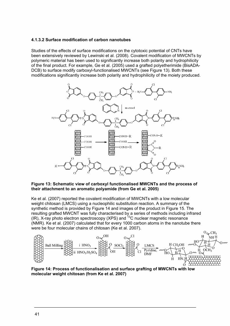
41
4.1.3.2 Surface modification of carbon nanotubes
Studies of the effects of surface modifications on the cytotoxic potential of CNTs have been extensively reviewed by Lewinski et al. (2008). Covalent modification of MWCNTs by polymeric material has been used to significantly increase both polarity and hydrophilicity of the final product. For example, Ge et al. (2005) used a grafted polyetherimide (BisADA-DCB) to surface modify carboxyl-functionalised MWCNTs (see Figure 13). Both these modifications significantly increase both polarity and hydrophilicity of the moiety produced.
Figure 13: Schematic view of carboxyl functionalised MWCNTs and the process of their attachment to an aromatic polyamide (from Ge et al. 2005) Ke et al. (2007) reported the covalent modification of MWCNTs with a low molecular weight chitosan (LMCS) using a nucleophilic substitution reaction. A summary of the synthetic method is provided by Figure 14 and images of the product in Figure 15. The resulting grafted MWCNT was fully characterised by a series of methods including infrared (IR), X-ray photo electron spectroscopy (XPS) and 13C nuclear magnetic resonance (NMR). Ke et al. (2007) calculated that for every 1000 carbon atoms in the nanotube there were be four molecular chains of chitosan (Ke et al. 2007).
Figure 14: Process of functionalisation and surface grafting of MWCNTs with low molecular weight chitosan (from Ke et al. 2007)
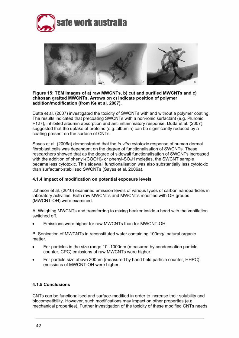
42
Figure 15: TEM images of a) raw MWCNTs, b) cut and purified MWCNTs and c) chitosan grafted MWCNTs. Arrows on c) indicate position of polymer addition/modification (from Ke et al. 2007). Dutta et al. (2007) investigated the toxicity of SWCNTs with and without a polymer coating. The results indicated that precoating SWCNTs with a non-ionic surfactant (e.g. Pluronic F127), inhibited albumin absorption and anti inflammatory response. Dutta et al. (2007) suggested that the uptake of proteins (e.g. albumin) can be significantly reduced by a coating present on the surface of CNTs. Sayes et al. (2006a) demonstrated that the in vitro cytotoxic response of human dermal fibroblast cells was dependent on the degree of functionalisation of SWCNTs. These researchers showed that as the degree of sidewall functionalisation of SWCNTs increased with the addition of phenyl-(COOH)2 or phenyl-SO3H moieties, the SWCNT sample became less cytotoxic. This sidewall functionalisation was also substantially less cytotoxic than surfactant-stabilised SWCNTs (Sayes et al. 2006a).
4.1.4 Impact of modification on potential exposure levels
Johnson et al. (2010) examined emission levels of various types of carbon nanoparticles in laboratory activities. Both raw MWCNTs and MWCNTs modified with OH groups (MWCNT-OH) were examined. A. Weighing MWCNTs and transferring to mixing beaker inside a hood with the ventilation switched off.
Emissions were higher for raw MWCNTs than for MWCNT-OH. B. Sonication of MWCNTs in reconstituted water containing 100mg/l natural organic matter.
For particles in the size range 10 -1000nm (measured by condensation particle counter, CPC) emissions of raw MWCNTs were higher.
For particle size above 300nm (measured by hand held particle counter, HHPC), emissions of MWCNT-OH were higher.
4.1.5 Conclusions
CNTs can be functionalised and surface-modified in order to increase their solubility and biocompatibility. However, such modifications may impact on other properties (e.g. mechanical properties). Further investigation of the toxicity of these modified CNTs needs
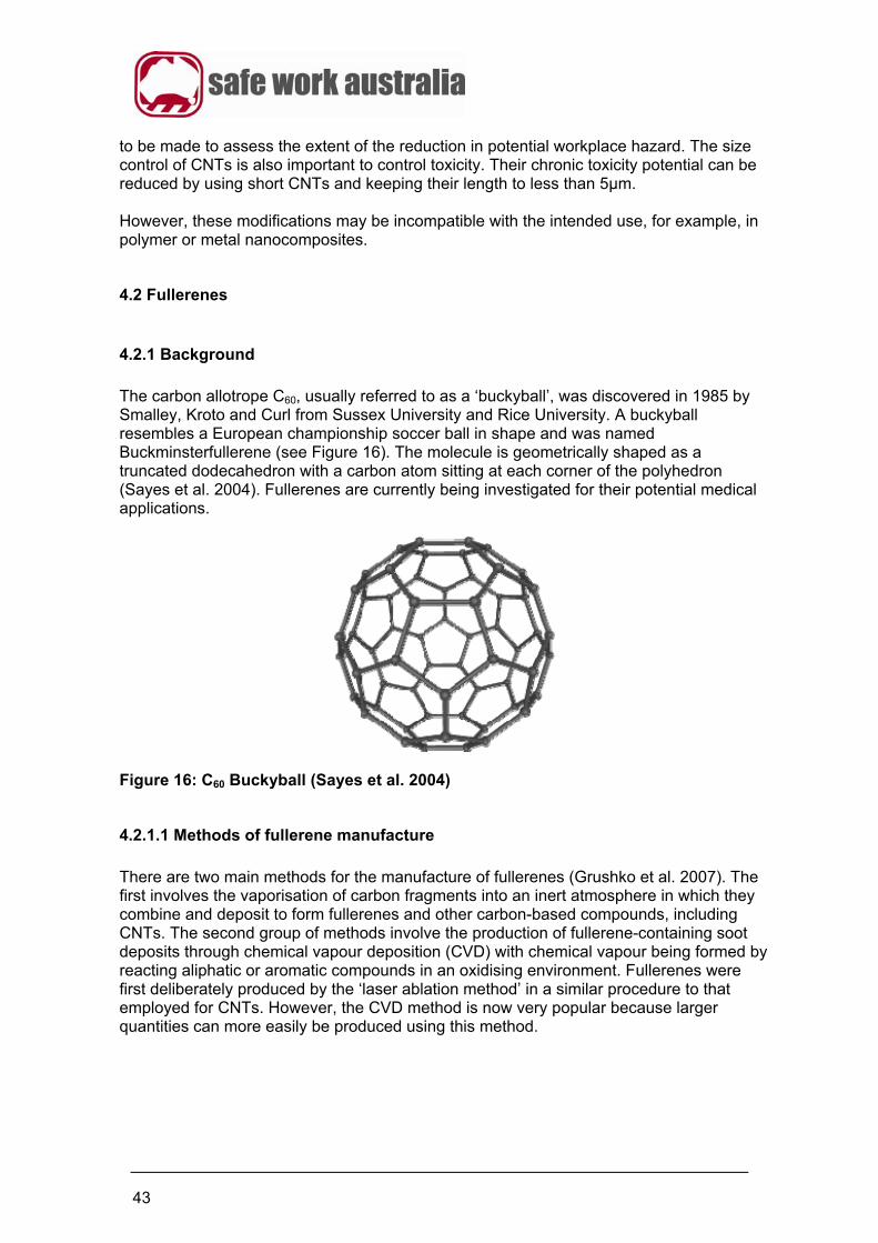
43
to be made to assess the extent of the reduction in potential workplace hazard. The size control of CNTs is also important to control toxicity. Their chronic toxicity potential can be reduced by using short CNTs and keeping their length to less than 5µm. However, these modifications may be incompatible with the intended use, for example, in polymer or metal nanocomposites.
4.2 Fullerenes
4.2.1 Background
The carbon allotrope C60, usually referred to as a ‘buckyball’, was discovered in 1985 by Smalley, Kroto and Curl from Sussex University and Rice University. A buckyball resembles a European championship soccer ball in shape and was named Buckminsterfullerene (see Figure 16). The molecule is geometrically shaped as a truncated dodecahedron with a carbon atom sitting at each corner of the polyhedron (Sayes et al. 2004). Fullerenes are currently being investigated for their potential medical applications.
Figure 16: C60 Buckyball (Sayes et al. 2004)
4.2.1.1 Methods of fullerene manufacture
There are two main methods for the manufacture of fullerenes (Grushko et al. 2007). The first involves the vaporisation of carbon fragments into an inert atmosphere in which they combine and deposit to form fullerenes and other carbon-based compounds, including CNTs. The second group of methods involve the production of fullerene-containing soot deposits through chemical vapour deposition (CVD) with chemical vapour being formed by reacting aliphatic or aromatic compounds in an oxidising environment. Fullerenes were first deliberately produced by the ‘laser ablation method’ in a similar procedure to that employed for CNTs. However, the CVD method is now very popular because larger quantities can more easily be produced using this method.
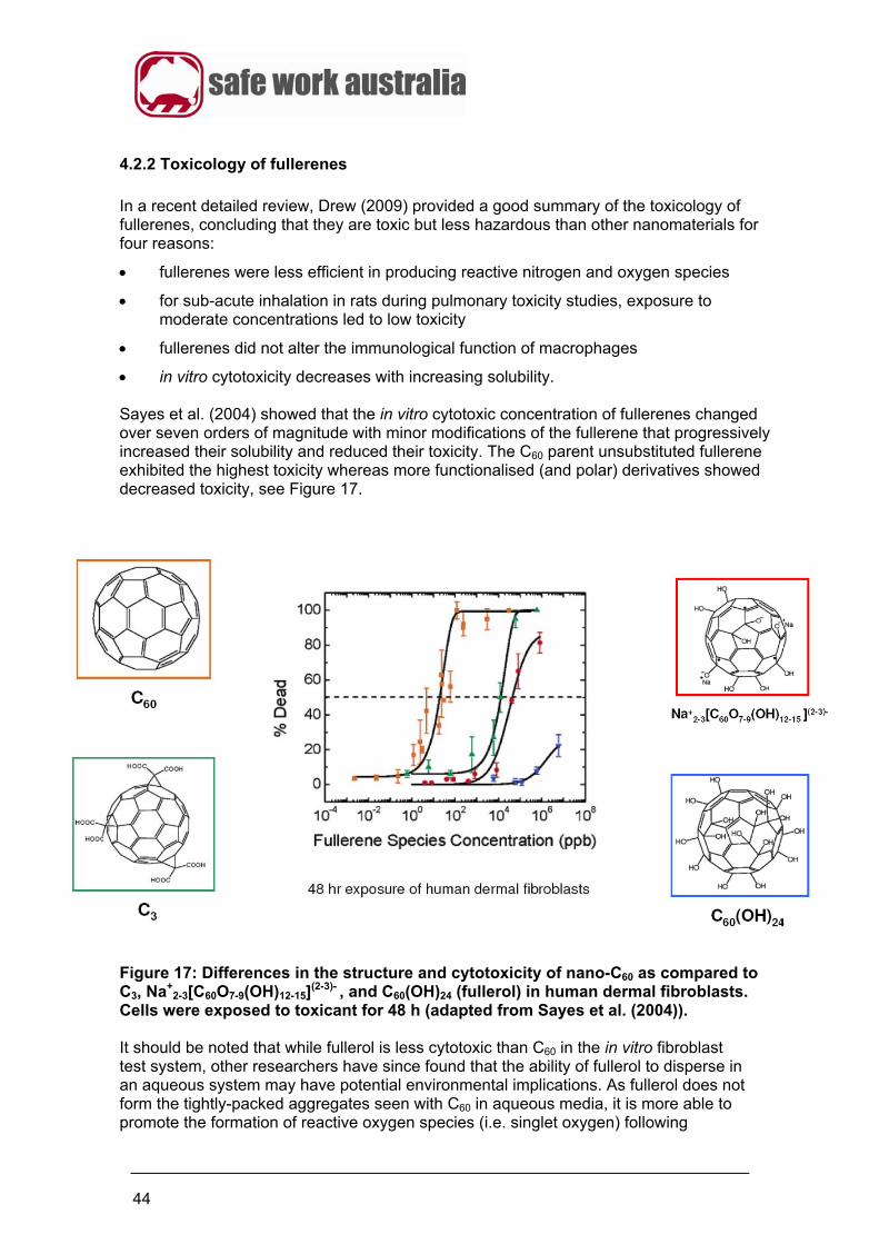
44
4.2.2 Toxicology of fullerenes
In a recent detailed review, Drew (2009) provided a good summary of the toxicology of fullerenes, concluding that they are toxic but less hazardous than other nanomaterials for four reasons:
fullerenes were less efficient in producing reactive nitrogen and oxygen species
for sub-acute inhalation in rats during pulmonary toxicity studies, exposure to moderate concentrations led to low toxicity
fullerenes did not alter the immunological function of macrophages
in vitro cytotoxicity decreases with increasing solubility. Sayes et al. (2004) showed that the in vitro cytotoxic concentration of fullerenes changed over seven orders of magnitude with minor modifications of the fullerene that progressively increased their solubility and reduced their toxicity. The C60 parent unsubstituted fullerene exhibited the highest toxicity whereas more functionalised (and polar) derivatives showed decreased toxicity, see Figure 17.
Figure 17: Differences in the structure and cytotoxicity of nano-C60 as compared to C3, Na+
2-3[C60O7-9(OH)12-15](2-3)- , and C60(OH)24 (fullerol) in human dermal fibroblasts.
Cells were exposed to toxicant for 48 h (adapted from Sayes et al. (2004)). It should be noted that while fullerol is less cytotoxic than C60 in the in vitro fibroblast test system, other researchers have since found that the ability of fullerol to disperse in an aqueous system may have potential environmental implications. As fullerol does not form the tightly-packed aggregates seen with C60 in aqueous media, it is more able to promote the formation of reactive oxygen species (i.e. singlet oxygen) following
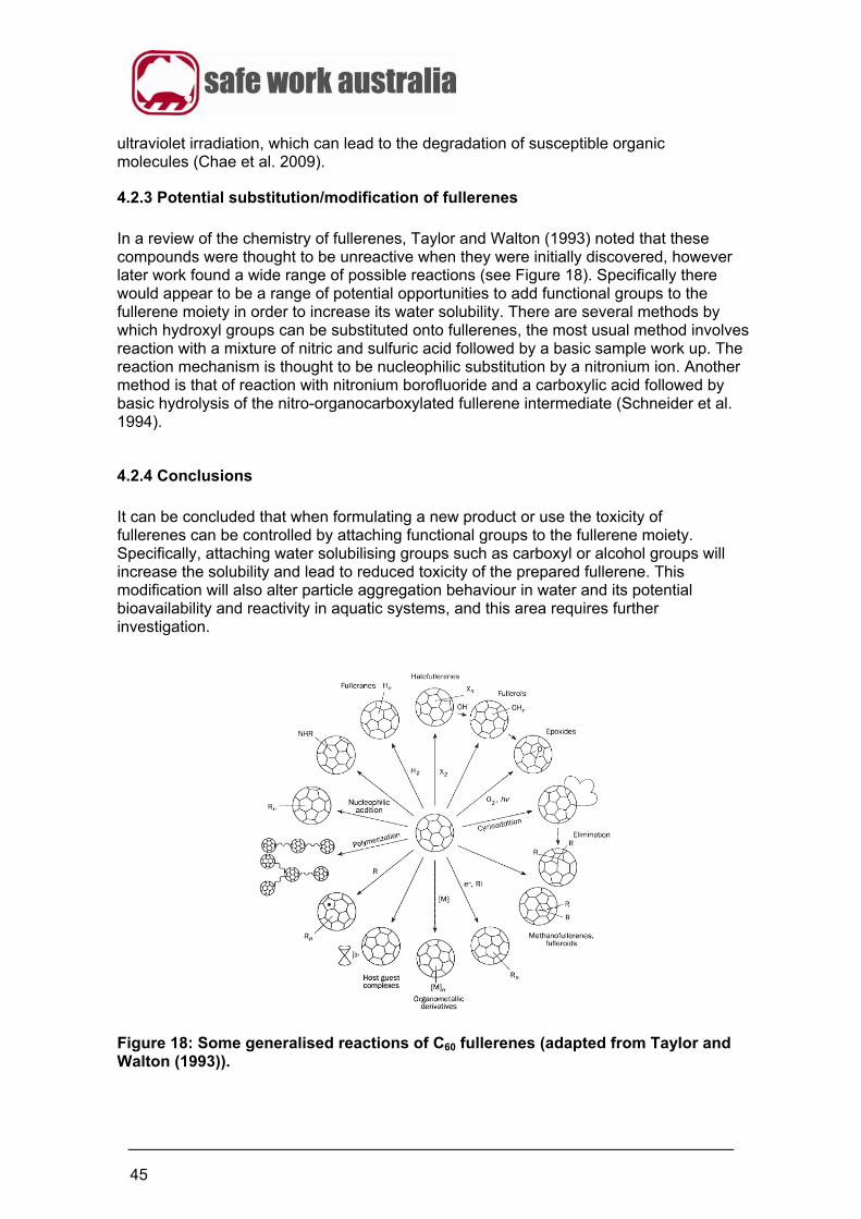
45
ultraviolet irradiation, which can lead to the degradation of susceptible organic molecules (Chae et al. 2009).
4.2.3 Potential substitution/modification of fullerenes
In a review of the chemistry of fullerenes, Taylor and Walton (1993) noted that these compounds were thought to be unreactive when they were initially discovered, however later work found a wide range of possible reactions (see Figure 18). Specifically there would appear to be a range of potential opportunities to add functional groups to the fullerene moiety in order to increase its water solubility. There are several methods by which hydroxyl groups can be substituted onto fullerenes, the most usual method involves reaction with a mixture of nitric and sulfuric acid followed by a basic sample work up. The reaction mechanism is thought to be nucleophilic substitution by a nitronium ion. Another method is that of reaction with nitronium borofluoride and a carboxylic acid followed by basic hydrolysis of the nitro-organocarboxylated fullerene intermediate (Schneider et al. 1994).
4.2.4 Conclusions
It can be concluded that when formulating a new product or use the toxicity of fullerenes can be controlled by attaching functional groups to the fullerene moiety. Specifically, attaching water solubilising groups such as carboxyl or alcohol groups will increase the solubility and lead to reduced toxicity of the prepared fullerene. This modification will also alter particle aggregation behaviour in water and its potential bioavailability and reactivity in aquatic systems, and this area requires further investigation.
Figure 18: Some generalised reactions of C60 fullerenes (adapted from Taylor and Walton (1993)).
46
4.3 Nano titanium dioxide (TiO2)
4.3.1 Background
Unlike CNTs and fullerenes which are synthesised, titanium dioxide (TiO2, titania) is a naturally occurring material which is normally produced from ores such as ilmenite or leucoxene. There are several different crystalline forms of the same material, of which the best known are rutile (the most common), brookite and anatase. Both brookite and anatase convert to rutile on heating. There are also three well-known high pressure forms: the orthorhombic α-PbO2 like form; a monoclinic baddeleyite-like form; and, a cotunnite-like form. Titanium dioxide has been used for a range of applications for several years, e.g. sunscreens, food-colouring and waste water remediation (Cotton and Wilkinson 1999). Major problems with the use of titanium dioxide have been the purity of the material and the optimisation of its properties for its applications, e.g. optimisation of ultraviolet (UV) light absorbing ability. The development of nano forms of TiO2 has become important as a method of optimising such properties, e.g. there is an increased UV-absorbing ability with the increased surface area of nano TiO2 compared to bulk TiO2 (Cotton and Wilkinson 1999). Nano TiO2 may be synthesised by a number of routes including the ‘solvothermal process’ in which the precursor solution is usually non-aqueous rather than the aqueous solutions used for the production of bulk TiO2. The reaction is carried out in a stainless steel autoclave, and experimental conditions/parameters such as reaction type, solvent type, temperature and surfactant type can be manipulated so that properties of TiO2 such as shape, size, size distribution, crystallinity and shape distribution can easily be controlled (Cotton and Wilkinson 1999).
4.3.2 Toxicology of nano titanium dioxide
In a general review of the toxicology of engineered nanomaterials, including nano TiO2, it was concluded by Drew (2009) that:
a) Titanium dioxide (TiO2) is regarded as a poorly soluble particle exhibiting low toxicity.
b) If experimental animals are exposed to high concentrations of TiO2 through inhalation, then they may become impaired of normal alveolar clearance mechanisms in the lung. This may result in particle build up leading to excessive lung burdens resulting in chronic alveolar inflammation.
c) The International Agency for Research on Cancer (IARC) has classified TiO2 as a Group 2B carcinogen – possibly carcinogenic to humans. The most important data for this classification was that from lung tumours in rats after high chronic exposures that would have caused the ‘particle overload phenomenon’, whereas mice and hamsters did not develop tumours. IARC considered the biological mechanisms occurring in the rat were still relevant to workers in situations of high exposures. However, with the mode of action involving a sustained inflammation, this implies that there is a threshold dose below which exposures will not be harmful. Some acute and intratracheal inhalation studies did not give the expected results of increased pulmonary toxicity due to aggregation and agglomeration of TiO2.

47
d) Dermal absorption studies for nano TiO2 conclude consistently that TiO2 is not absorbed through the skin.
e) Intravenous and oral toxicity studies have been used to investigate the systemic distribution of TiO2. The oral studies indicated that nano TiO2 is absorbed from the gastrointestinal tract and is distributed throughout the body. Intravenous and oral studies resulted in TiO2 retention in the lung, kidney, liver and spleen but not in the lymph nodes, brain, plasma or blood cells.
f) There is sufficient data to indicate that anatase nano TiO2 is more toxic/phototoxic than the rutile nano material. However, both forms can induce a pulmonary inflammation response if the concentration of exposure is high enough, with evidence for this being provided by rat inhalation studies.
g) A variety of traditional hazard/safety tests using nano-TiO2 have shown the material to be of low acute toxicity.
Relating to concerns about potential toxicity from the use of nano-TiO2 in sunscreens, Barker and Branch (2008) reported on the increased incidence of unsightly appearance defects of building materials due to nanoparticle-containing sunscreens. These defects were on recently installed prepainted steel roofs and other durable surface coatings, and were due to the aggressive activity of photocatalytic grades of nano TiO2 (especially anatase) in sunscreens that had been transferred to the building materials during construction handling. The photocatalytic particles had accelerated the weathering of the steel surface by >100 fold. However, the relevance of these findings to human sunscreen usage depends on long-term persistence of insoluble nano TiO2 on the skin following sunscreen application, which is unlikely due to the constant shedding of dead skin cells.
4.3.3 Potential substitution/modification of nano titanium dioxide
Sayes et al. (2006b) characterised the in vitro cytotoxicity of nanoscale anatase and rutile forms of TiO2 under ambient light conditions in cell culture. The authors found that cytotoxicity and pro-inflammatory responses were observed for anatase only, and only at relatively high concentrations (100 μg/ml) and these were found to increase with dose and the time of exposure. The phase composition of the crystalline structure, namely the anatase or rutile form, was an important factor with anatase being up to 100 times more cytotoxic than the rutile form (see Table I). The most cytotoxic form of nano TiO2 was found to be the most efficient at generating reactive oxygen species (ROS) in cell cultures. These results are important because they indicate that different crystalline forms of the same nanomaterial can exhibit dissimilar toxicological properties (Sayes et al. 2006b).
48
Table I: Summary of results for correlating nanoscale TiO2 structure with toxicity in cells (Adapted from Table 3 of Sayes et al. 2006b)
Particle sample
Photoactive Generates ROS
Produces cytotoxic response (LC50)
Induces membrane leakage
Decreases mitochondrial activity
Produces pro-inflammatory response
Nano-TiO2
Anatase particles
Yes. Up to 100 times greater than rutile.
Yes 1500 μg/ml
1500 μg/ml (w/o light) 30 μg/ml (w/light)
1500 μg/ml
300 μg/ml
Nano-TiO2 rutile particles
Yes. Much less than anatase.
No N/A N/A N/A N/A
N/A= not applicable - does not generate ROS (reactive oxygen species) w/o= without w/ = with The results of Sayes et al. (2006b) experimentally validated other findings for the nano TiO2 anatase and rutile phases. Selloni et al. (1998) and Vittadini et al. (1998) both showed that anatase and rutile particles differ significantly in their surface chemistry, specifically in relation to generating ROS. First principle calculations by these authors indicated that water molecules dissociatively absorb onto anatase surfaces, but not onto rutile surfaces. When water absorbed on to an anatase surface is illuminated with UV light, reactive photocarriers are trapped on the surface and come into contact with Ti-OH species, which then react to form hydroxyl radicals that are the primary oxidising species in oxidative photocatalytic processes. These findings are supported by the study of Jiang et al. (2008a), which demonstrated the oxidant generating capacity for different crystal phases of TiO2 nanoparticles was highest for amorphous, followed by anatase, and then anatase/rutile mixtures, and lowest for rutile samples. The paint industry has developed many methods for coating macroscopic TiO2 in order to decrease its photoactivity. However, these are generally not compatible for use with nanoparticulate TiO2. At this stage there are relatively few methods to introduce robust surface coatings onto nano TiO2.
4.3.4 Conclusions
It can be concluded that when formulating a new nano TiO2 product or use, its potential toxicity can be controlled by varying the crystalline form used. For example, the UV-photocatalytic activity of nano TiO2 can be reduced by partial or full replacement of the anatase form with the less reactive rutile form, especially in sunscreen applications and personal care products. In applications involving the use of an industrial-grade photocatalyst, such as the coatings on ‘self-cleaning’ glass surfaces, then the potential for shedding of anatase nanoparticles into the environment needs to be assessed and minimised to reduce environmental impact.
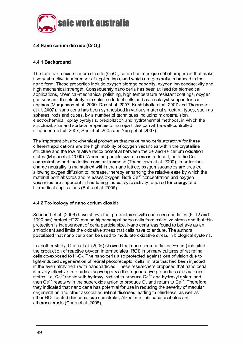
49
4.4 Nano cerium dioxide (CeO2)
4.4.1 Background
The rare-earth oxide cerium dioxide (CeO2, ceria) has a unique set of properties that make it very attractive in a number of applications, and which are generally enhanced in the nano form. These properties include oxygen storage capacity, oxygen ion conductivity and high mechanical strength. Consequently nano ceria has been utilised for biomedical applications, chemical-mechanical polishing, high temperature resistant coatings, oxygen gas sensors, the electrolyte in solid oxide fuel cells and as a catalyst support for car engines (Morgenson et al. 2000; Das et al. 2007; Kuchibhatla et al. 2007 and Thanneeru et al. 2007). Nano ceria has been synthesised in various material structural types, such as spheres, rods and cubes, by a number of techniques including microemulsion, electrochemical, spray pyrolysis, precipitation and hydrothermal methods, in which the structural, size and surface properties of nanoparticles can all be well-controlled (Thanneeru et al. 2007; Sun et al. 2005 and Yang et al. 2007). The important physico-chemical properties that make nano ceria attractive for these different applications are the high mobility of oxygen vacancies within the crystalline structure and the low relative redox potential between the 3+ and 4+ cerium oxidation states (Masui et al. 2000). When the particle size of ceria is reduced, both the Ce3+
concentration and the lattice constant increase (Tsunekawa et al. 2000). In order that charge neutrality is maintained within the nano lattice, oxygen vacancies are created, allowing oxygen diffusion to increase, thereby enhancing the relative ease by which the material both absorbs and releases oxygen. Both Ce3+
concentration and oxygen vacancies are important in fine tuning the catalytic activity required for energy and biomedical applications (Babu et al. 2009).
4.4.2 Toxicology of nano cerium dioxide
Schubert et al. (2006) have shown that pretreatment with nano ceria particles (6, 12 and 1000 nm) protect HT22 mouse hippocampal nerve cells from oxidative stress and that this protection is independent of ceria particle size. Nano ceria was found to behave as an antioxidant and limits the oxidative stress that cells have to endure. The authors postulated that nano ceria can be used to modulate oxidative stress in biological systems. In another study, Chen et al. (2006) showed that nano ceria particles (~5 nm) inhibited the production of reactive oxygen intermediates (ROI) in primary cultures of rat retina cells co-exposed to H2O2. The nano ceria also protected against loss of vision due to light-induced degeneration of retinal photoreceptor cells, in rats that had been injected in the eye (intravitreal) with nanoparticles. These researchers proposed that nano ceria is a very effective free radical scavenger via the regenerative properties of its valence states, i.e. Ce3+
reacts with hydroxyl radical to produce Ce4+ and hydroxyl anion, and then Ce4+ reacts with the superoxide anion to produce O2 and return to Ce3+. Therefore they indicated that nano ceria has potential for use in reducing the severity of macular degeneration and other associated retinal diseases leading to blindness, as well as other ROI-related diseases, such as stroke, Alzheimer’s disease, diabetes and atherosclerosis (Chen et al. 2006).
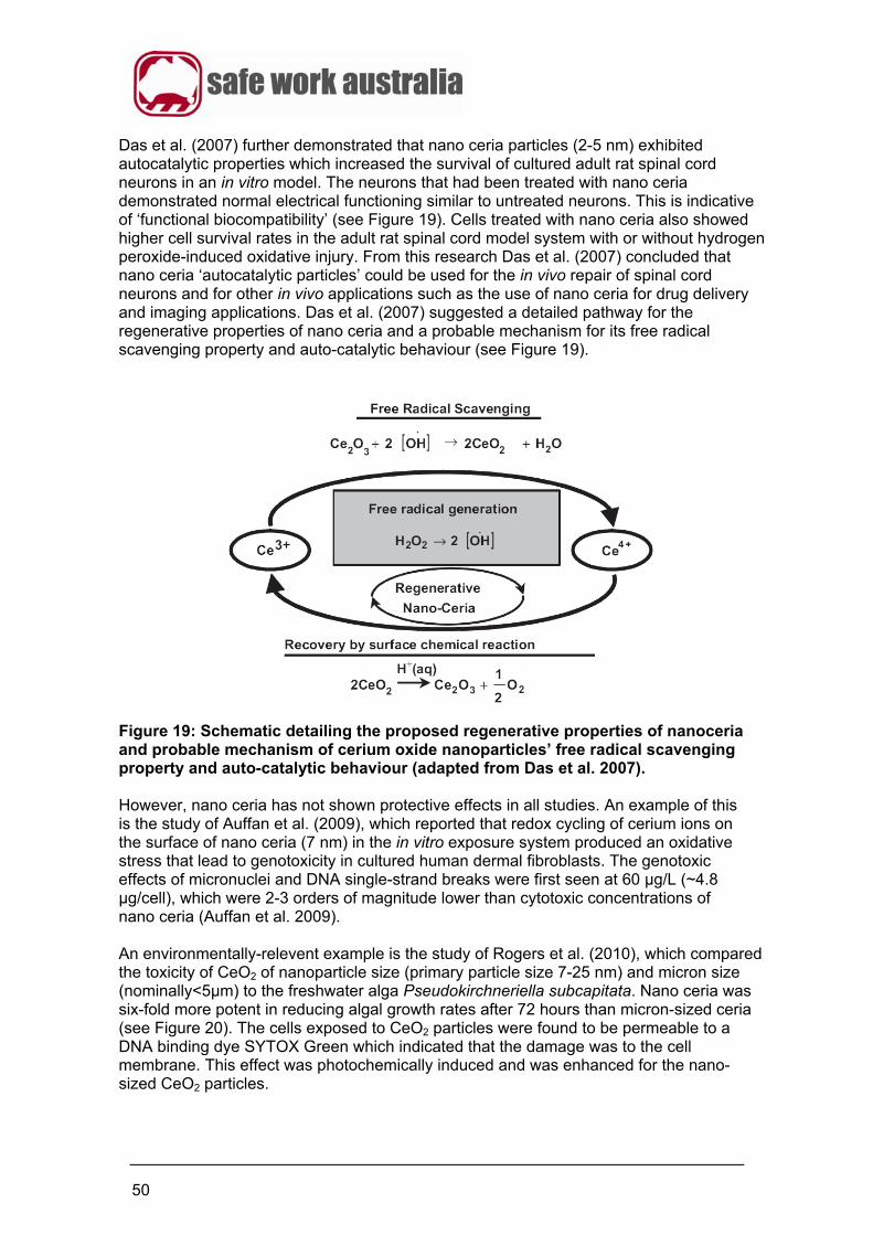
50
Das et al. (2007) further demonstrated that nano ceria particles (2-5 nm) exhibited autocatalytic properties which increased the survival of cultured adult rat spinal cord neurons in an in vitro model. The neurons that had been treated with nano ceria demonstrated normal electrical functioning similar to untreated neurons. This is indicative of ‘functional biocompatibility’ (see Figure 19). Cells treated with nano ceria also showed higher cell survival rates in the adult rat spinal cord model system with or without hydrogen peroxide-induced oxidative injury. From this research Das et al. (2007) concluded that nano ceria ‘autocatalytic particles’ could be used for the in vivo repair of spinal cord neurons and for other in vivo applications such as the use of nano ceria for drug delivery and imaging applications. Das et al. (2007) suggested a detailed pathway for the regenerative properties of nano ceria and a probable mechanism for its free radical scavenging property and auto-catalytic behaviour (see Figure 19).
Figure 19: Schematic detailing the proposed regenerative properties of nanoceria and probable mechanism of cerium oxide nanoparticles’ free radical scavenging property and auto-catalytic behaviour (adapted from Das et al. 2007). However, nano ceria has not shown protective effects in all studies. An example of this is the study of Auffan et al. (2009), which reported that redox cycling of cerium ions on the surface of nano ceria (7 nm) in the in vitro exposure system produced an oxidative stress that lead to genotoxicity in cultured human dermal fibroblasts. The genotoxic effects of micronuclei and DNA single-strand breaks were first seen at 60 μg/L (~4.8 μg/cell), which were 2-3 orders of magnitude lower than cytotoxic concentrations of nano ceria (Auffan et al. 2009). An environmentally-relevent example is the study of Rogers et al. (2010), which compared the toxicity of CeO2 of nanoparticle size (primary particle size 7-25 nm) and micron size (nominally<5μm) to the freshwater alga Pseudokirchneriella subcapitata. Nano ceria was six-fold more potent in reducing algal growth rates after 72 hours than micron-sized ceria (see Figure 20). The cells exposed to CeO2 particles were found to be permeable to a DNA binding dye SYTOX Green which indicated that the damage was to the cell membrane. This effect was photochemically induced and was enhanced for the nano-sized CeO2 particles.
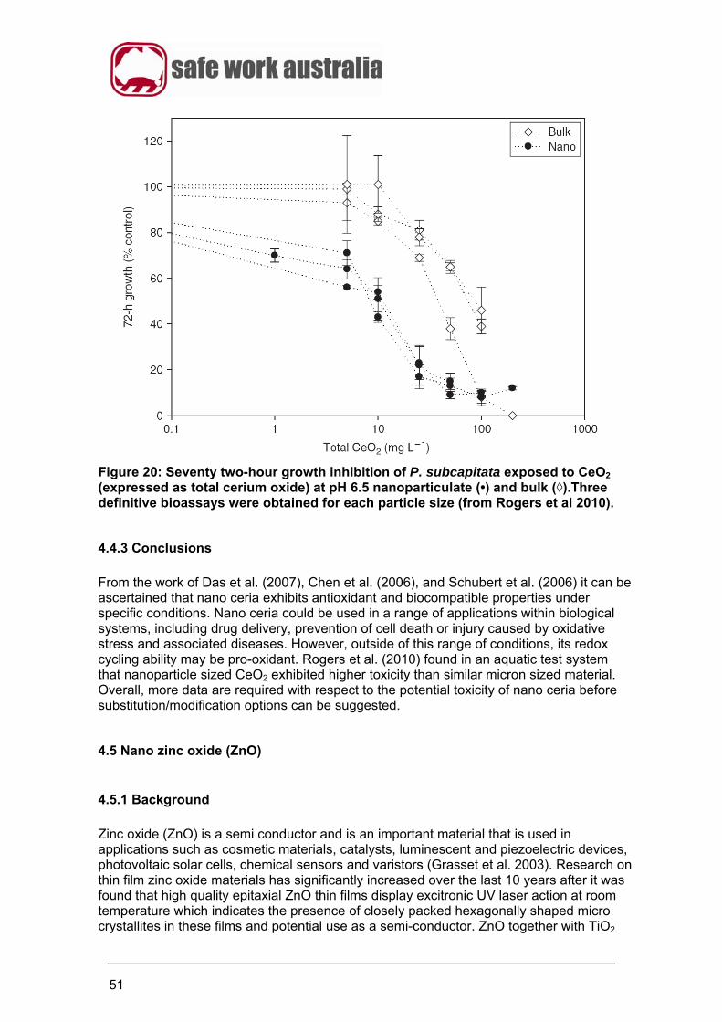
51
Figure 20: Seventy two-hour growth inhibition of P. subcapitata exposed to CeO2 (expressed as total cerium oxide) at pH 6.5 nanoparticulate (•) and bulk (◊).Three definitive bioassays were obtained for each particle size (from Rogers et al 2010).
4.4.3 Conclusions
From the work of Das et al. (2007), Chen et al. (2006), and Schubert et al. (2006) it can be ascertained that nano ceria exhibits antioxidant and biocompatible properties under specific conditions. Nano ceria could be used in a range of applications within biological systems, including drug delivery, prevention of cell death or injury caused by oxidative stress and associated diseases. However, outside of this range of conditions, its redox cycling ability may be pro-oxidant. Rogers et al. (2010) found in an aquatic test system that nanoparticle sized CeO2 exhibited higher toxicity than similar micron sized material. Overall, more data are required with respect to the potential toxicity of nano ceria before substitution/modification options can be suggested.
4.5 Nano zinc oxide (ZnO)
4.5.1 Background
Zinc oxide (ZnO) is a semi conductor and is an important material that is used in applications such as cosmetic materials, catalysts, luminescent and piezoelectric devices, photovoltaic solar cells, chemical sensors and varistors (Grasset et al. 2003). Research on thin film zinc oxide materials has significantly increased over the last 10 years after it was found that high quality epitaxial ZnO thin films display excitronic UV laser action at room temperature which indicates the presence of closely packed hexagonally shaped micro crystallites in these films and potential use as a semi-conductor. ZnO together with TiO2
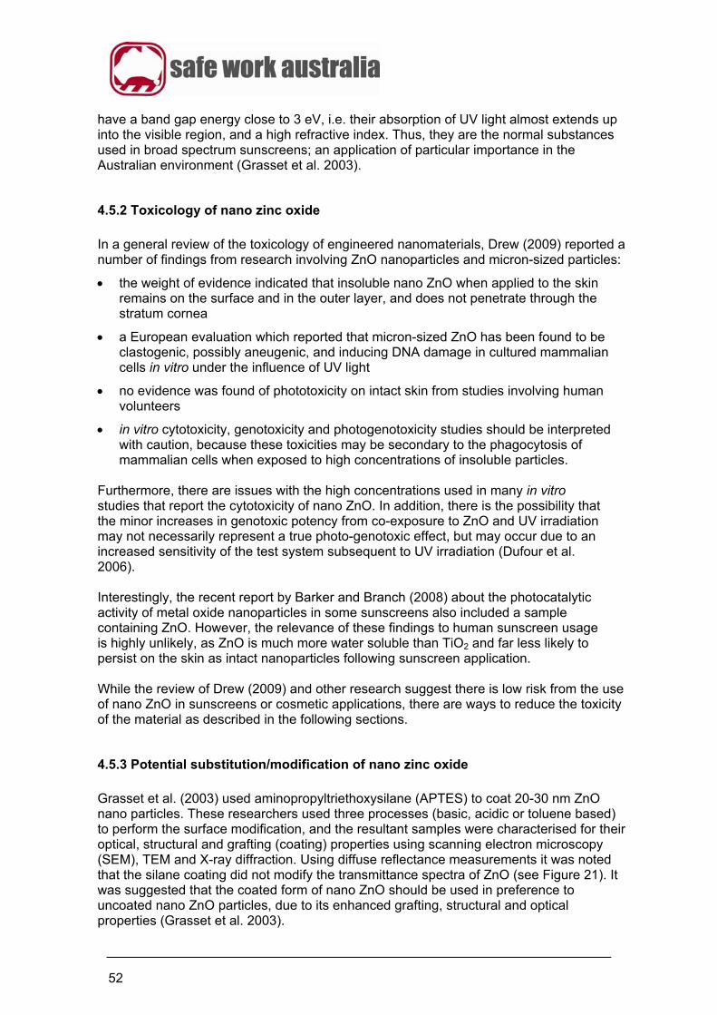
52
have a band gap energy close to 3 eV, i.e. their absorption of UV light almost extends up into the visible region, and a high refractive index. Thus, they are the normal substances used in broad spectrum sunscreens; an application of particular importance in the Australian environment (Grasset et al. 2003).
4.5.2 Toxicology of nano zinc oxide
In a general review of the toxicology of engineered nanomaterials, Drew (2009) reported a number of findings from research involving ZnO nanoparticles and micron-sized particles:
the weight of evidence indicated that insoluble nano ZnO when applied to the skin remains on the surface and in the outer layer, and does not penetrate through the stratum cornea
a European evaluation which reported that micron-sized ZnO has been found to be clastogenic, possibly aneugenic, and inducing DNA damage in cultured mammalian cells in vitro under the influence of UV light
no evidence was found of phototoxicity on intact skin from studies involving human volunteers
in vitro cytotoxicity, genotoxicity and photogenotoxicity studies should be interpreted with caution, because these toxicities may be secondary to the phagocytosis of mammalian cells when exposed to high concentrations of insoluble particles.
Furthermore, there are issues with the high concentrations used in many in vitro studies that report the cytotoxicity of nano ZnO. In addition, there is the possibility that the minor increases in genotoxic potency from co-exposure to ZnO and UV irradiation may not necessarily represent a true photo-genotoxic effect, but may occur due to an increased sensitivity of the test system subsequent to UV irradiation (Dufour et al. 2006). Interestingly, the recent report by Barker and Branch (2008) about the photocatalytic activity of metal oxide nanoparticles in some sunscreens also included a sample containing ZnO. However, the relevance of these findings to human sunscreen usage is highly unlikely, as ZnO is much more water soluble than TiO2 and far less likely to persist on the skin as intact nanoparticles following sunscreen application. While the review of Drew (2009) and other research suggest there is low risk from the use of nano ZnO in sunscreens or cosmetic applications, there are ways to reduce the toxicity of the material as described in the following sections.
4.5.3 Potential substitution/modification of nano zinc oxide
Grasset et al. (2003) used aminopropyltriethoxysilane (APTES) to coat 20-30 nm ZnO nano particles. These researchers used three processes (basic, acidic or toluene based) to perform the surface modification, and the resultant samples were characterised for their optical, structural and grafting (coating) properties using scanning electron microscopy (SEM), TEM and X-ray diffraction. Using diffuse reflectance measurements it was noted that the silane coating did not modify the transmittance spectra of ZnO (see Figure 21). It was suggested that the coated form of nano ZnO should be used in preference to uncoated nano ZnO particles, due to its enhanced grafting, structural and optical properties (Grasset et al. 2003).
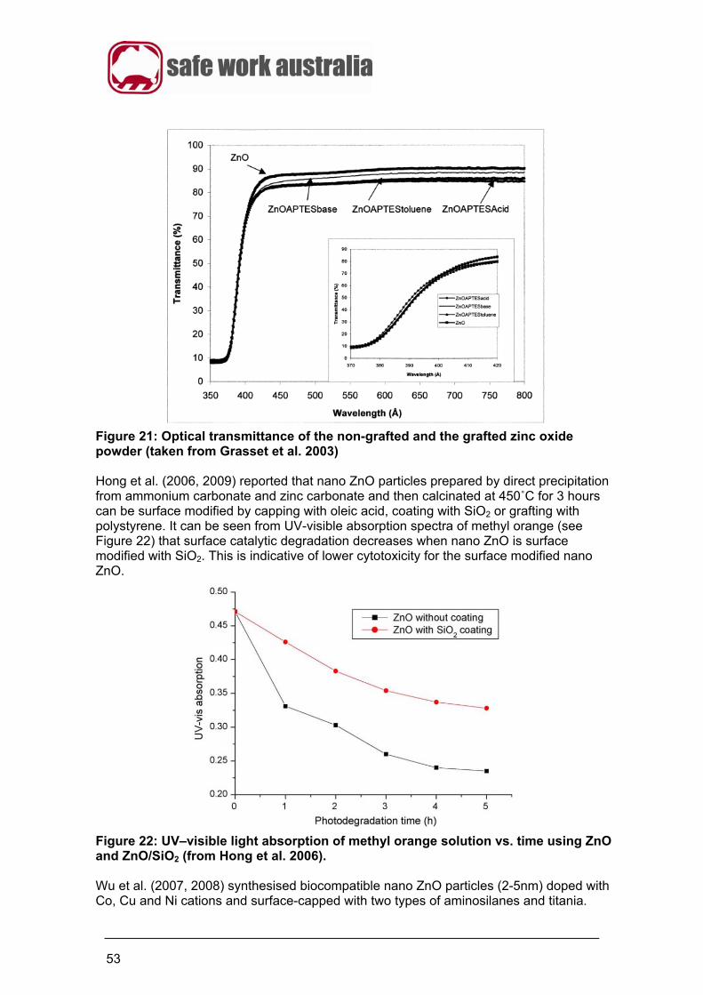
53
Figure 21: Optical transmittance of the non-grafted and the grafted zinc oxide powder (taken from Grasset et al. 2003) Hong et al. (2006, 2009) reported that nano ZnO particles prepared by direct precipitation from ammonium carbonate and zinc carbonate and then calcinated at 450˚C for 3 hours can be surface modified by capping with oleic acid, coating with SiO2 or grafting with polystyrene. It can be seen from UV-visible absorption spectra of methyl orange (see Figure 22) that surface catalytic degradation decreases when nano ZnO is surface modified with SiO2. This is indicative of lower cytotoxicity for the surface modified nano ZnO.
Figure 22: UV–visible light absorption of methyl orange solution vs. time using ZnO and ZnO/SiO2 (from Hong et al. 2006). Wu et al. (2007, 2008) synthesised biocompatible nano ZnO particles (2-5nm) doped with Co, Cu and Ni cations and surface-capped with two types of aminosilanes and titania.
54
Synthesis was by a colloidal ‘soft chemical’ process. The electrical, optical and magnetic properties of ZnO can be adjusted by doping of the ZnO lattice structure. Dual colour bioimaging on human cells (U-937 histiocytic lymphoma and MG-63 osteosarcoma cells) showed turquoise emission at the cytoplasm and blue emission at the nucleus simultaneously, with high photoluminescence emissions at the blue-violet and UV wavelength ranges. Mung bean seedlings labelled by synthesised ZnO nanocrystals gave a bright green emission. Cytotoxicity tests showed that the aminosilane-capped nanoparticles have little toxicity or are non-toxic. Quantum yields of 80-95% were typical for these nanocrystals. These results show the potential for live imaging of both plant systems and human cells using ZnO and Co-doped ZnO nanocrystals.
4.5.4 Conclusions
It can be concluded from the review of Drew (2009) that ZnO used in sunscreen type products and for other similar applications exhibits a low level of toxicity and penetration in to the human body. From the work of Grasset et al. (2003) and Hong et al. (2006, 2009), there are potential surface modification options available for ZnO which have the potential to reduce toxicity further. The work of Wu et al. (2007, 2008) indicates the potential live cell imaging opportunities for ZnO particles with a doped crystalline lattice, that have also been made more biocompatible by surface modification. 4.6 Nano gold (Au)
4.6.1 Background
Gold (Au) nanoparticles are used for a variety of applications including bioimaging, biosensing and drug delivery (West et al. 2003; Mann et al. 2000 and Hermanson 1996). Hybridisation assays, flow cytometry and immunobloting are three of the applications that rely on the excellent detection capabilities derived from nano Au’s optical and contrast properties (Demers et al. 2000; Thanh and Rosenzweig 2002). Nano Au is also receiving attention for potential medical radiation applications e.g. as a contrast agent for tumour detection and destruction, due to its radio-thermal properties. Two major issues involved in using nano Au are insuring biocompatibility and aqueous solubility because biological processes usually occur on a hydrophilic surface in an aqueous environment. Colloidal Au is a commercially available product but is unstable and aggregates when electrolytic solutions are introduced. In order to stabilise nano Au it may be coated with surfactant molecules (Eychmuller and Rogach 2000; Tan and Zhang 2005).
4.6.2 Toxicology of nano gold
Nano Au has been used for nuclear transfection and targeting in non-viral gene delivery applications because they easily enter cells, but their in vitro cytotoxicity in certain cell types at high concentrations has limited the use of uncoated nano Au and prompted the search for more biocompatible coatings that also aid in cell uptake (Lewinski et al. 2008). There is also evidence of other adverse effects, such as the potential for affecting immunological responses (Shukla et al. 2005). Studies of the cytotoxic potential of nano Au particles, shells and rods have been reviewed by Lewinski et al. (2008).
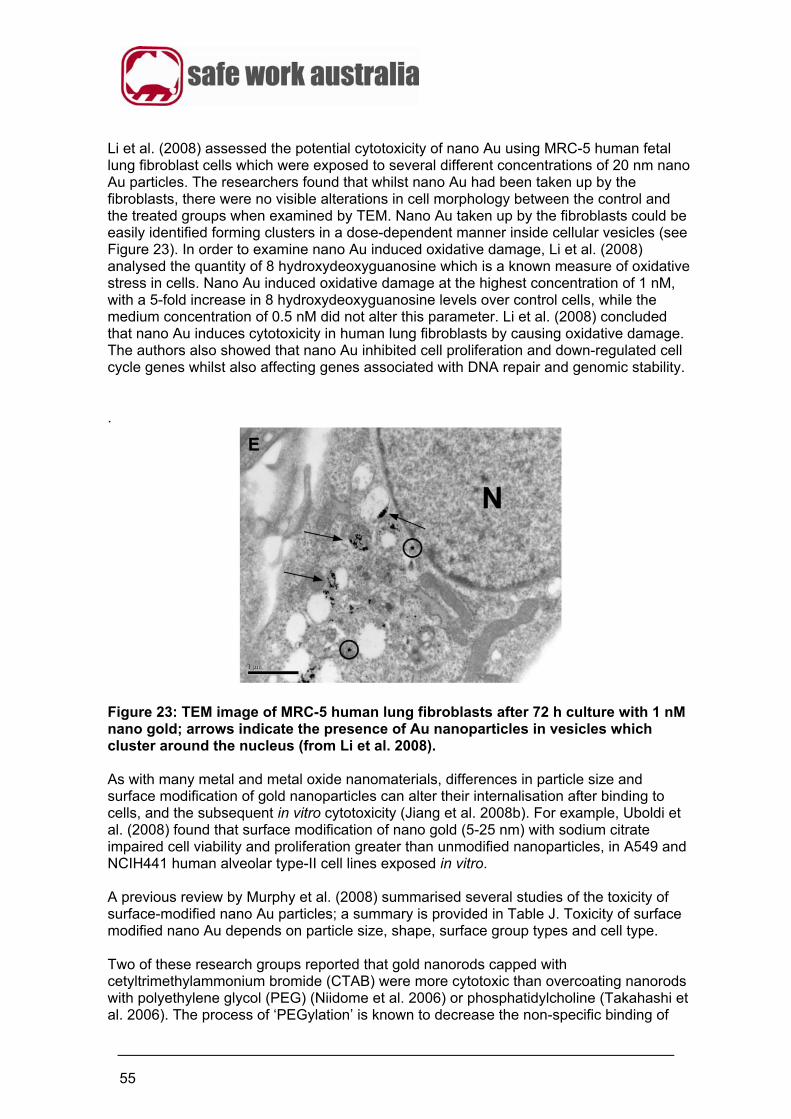
55
Li et al. (2008) assessed the potential cytotoxicity of nano Au using MRC-5 human fetal lung fibroblast cells which were exposed to several different concentrations of 20 nm nano Au particles. The researchers found that whilst nano Au had been taken up by the fibroblasts, there were no visible alterations in cell morphology between the control and the treated groups when examined by TEM. Nano Au taken up by the fibroblasts could be easily identified forming clusters in a dose-dependent manner inside cellular vesicles (see Figure 23). In order to examine nano Au induced oxidative damage, Li et al. (2008) analysed the quantity of 8 hydroxydeoxyguanosine which is a known measure of oxidative stress in cells. Nano Au induced oxidative damage at the highest concentration of 1 nM, with a 5-fold increase in 8 hydroxydeoxyguanosine levels over control cells, while the medium concentration of 0.5 nM did not alter this parameter. Li et al. (2008) concluded that nano Au induces cytotoxicity in human lung fibroblasts by causing oxidative damage. The authors also showed that nano Au inhibited cell proliferation and down-regulated cell cycle genes whilst also affecting genes associated with DNA repair and genomic stability. .
Figure 23: TEM image of MRC-5 human lung fibroblasts after 72 h culture with 1 nM nano gold; arrows indicate the presence of Au nanoparticles in vesicles which cluster around the nucleus (from Li et al. 2008). As with many metal and metal oxide nanomaterials, differences in particle size and surface modification of gold nanoparticles can alter their internalisation after binding to cells, and the subsequent in vitro cytotoxicity (Jiang et al. 2008b). For example, Uboldi et al. (2008) found that surface modification of nano gold (5-25 nm) with sodium citrate impaired cell viability and proliferation greater than unmodified nanoparticles, in A549 and NCIH441 human alveolar type-II cell lines exposed in vitro. A previous review by Murphy et al. (2008) summarised several studies of the toxicity of surface-modified nano Au particles; a summary is provided in Table J. Toxicity of surface modified nano Au depends on particle size, shape, surface group types and cell type. Two of these research groups reported that gold nanorods capped with cetyltrimethylammonium bromide (CTAB) were more cytotoxic than overcoating nanorods with polyethylene glycol (PEG) (Niidome et al. 2006) or phosphatidylcholine (Takahashi et al. 2006). The process of ‘PEGylation’ is known to decrease the non-specific binding of
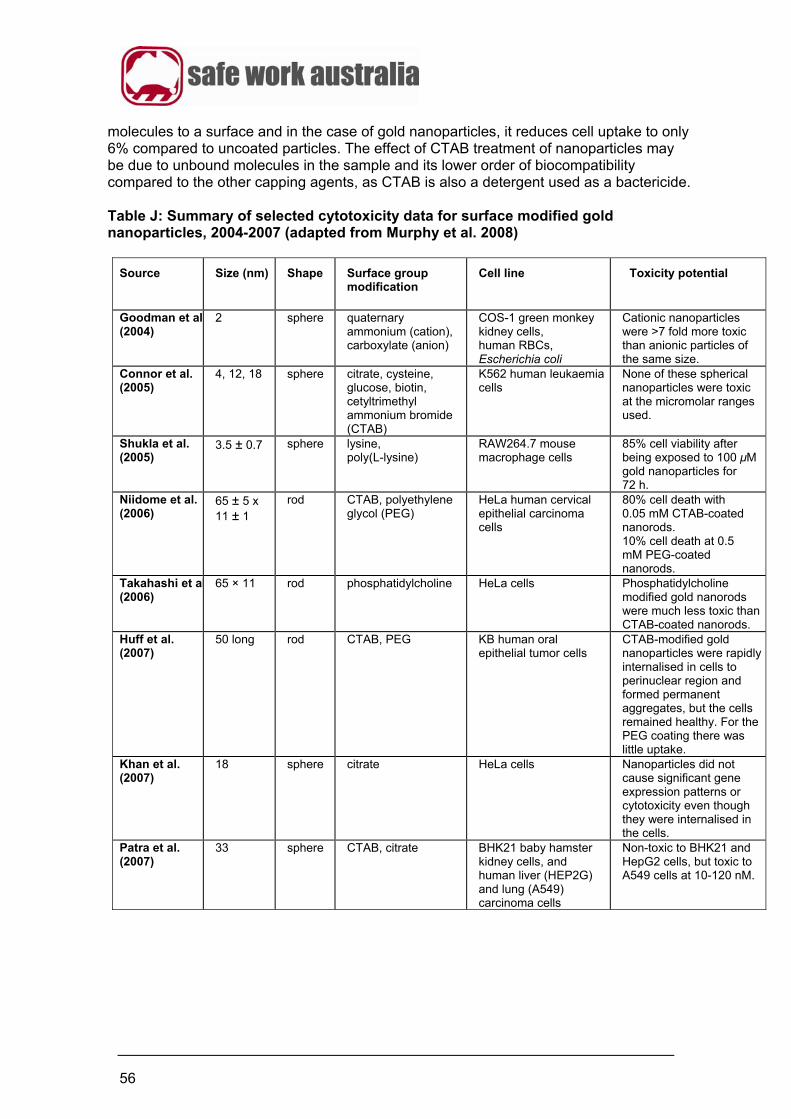
56
molecules to a surface and in the case of gold nanoparticles, it reduces cell uptake to only 6% compared to uncoated particles. The effect of CTAB treatment of nanoparticles may be due to unbound molecules in the sample and its lower order of biocompatibility compared to the other capping agents, as CTAB is also a detergent used as a bactericide. Table J: Summary of selected cytotoxicity data for surface modified gold nanoparticles, 2004-2007 (adapted from Murphy et al. 2008)
Source
Size (nm) Shape
Surface group modification
Cell line
Toxicity potential
Goodman et al(2004)
2
sphere
quaternary ammonium (cation), carboxylate (anion)
COS-1 green monkey kidney cells, human RBCs, Escherichia coli
Cationic nanoparticles were >7 fold more toxic than anionic particles of the same size.
Connor et al. (2005)
4, 12, 18
sphere
citrate, cysteine, glucose, biotin, cetyltrimethyl ammonium bromide (CTAB)
K562 human leukaemia cells
None of these spherical nanoparticles were toxic at the micromolar ranges used.
Shukla et al. (2005)
3.5 ± 0.7
sphere
lysine, poly(L-lysine)
RAW264.7 mouse macrophage cells
85% cell viability after being exposed to 100 μM gold nanoparticles for 72 h.
Niidome et al. (2006)
65 ± 5 x 11 ± 1
rod
CTAB, polyethylene glycol (PEG)
HeLa human cervical epithelial carcinoma cells
80% cell death with 0.05 mM CTAB-coated nanorods. 10% cell death at 0.5 mM PEG-coated nanorods.
Takahashi et al(2006)
65 × 11
rod
phosphatidylcholine
HeLa cells
Phosphatidylcholine modified gold nanorods were much less toxic than CTAB-coated nanorods.
Huff et al. (2007)
50 long rod
CTAB, PEG
KB human oral epithelial tumor cells
CTAB-modified gold nanoparticles were rapidly internalised in cells to perinuclear region and formed permanent aggregates, but the cells remained healthy. For the PEG coating there was little uptake.
Khan et al. (2007)
18
sphere
citrate
HeLa cells
Nanoparticles did not cause significant gene expression patterns or cytotoxicity even though they were internalised in the cells.
Patra et al. (2007)
33
sphere
CTAB, citrate
BHK21 baby hamster kidney cells, and human liver (HEP2G) and lung (A549) carcinoma cells
Non-toxic to BHK21 and HepG2 cells, but toxic to A549 cells at 10-120 nM.
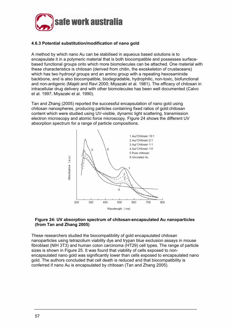
57
4.6.3 Potential substitution/modification of nano gold
A method by which nano Au can be stabilised in aqueous based solutions is to encapsulate it in a polymeric material that is both biocompatible and possesses surface-based functional groups onto which more biomolecules can be attached. One material with these characteristics is chitosan (derived from chitin, the exoskeleton of crustaceans) which has two hydroxyl groups and an amino group with a repeating hexosaminide backbone, and is also biocompatible, biodegradable, hydrophilic, non-toxic, biofunctional and non-antigenic (Majeti and Ravi 2000; Miyazaki et al. 1981). The efficacy of chitosan in intracellular drug delivery and with other biomolecules has been well documented (Calvo et al. 1997; Miyazaki et al. 1990). Tan and Zhang (2005) reported the successful encapsulation of nano gold using chitosan nanospheres, producing particles containing fixed ratios of gold:chitosan content which were studied using UV-visible, dynamic light scattering, transmission electron microscopy and atomic force microscopy. Figure 24 shows the different UV absorption spectrum for a range of particle compositions.
Figure 24: UV absorption spectrum of chitosan-encapsulated Au nanoparticles (from Tan and Zhang 2005)
These researchers studied the biocompatibility of gold encapsulated chitosan nanoparticles using tetrazolium viability dye and trypan blue exclusion assays in mouse fibroblast (NIH 3T3) and human colon carcinoma (HT29) cell types. The range of particle sizes is shown in Figure 25. It was found that viability of cells exposed to non-encapsulated nano gold was significantly lower than cells exposed to encapsulated nano gold. The authors concluded that cell death is reduced and that biocompatibility is conferred if nano Au is encapsulated by chitosan (Tan and Zhang 2005).
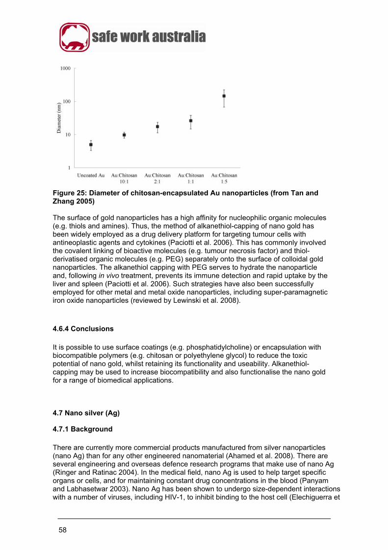
58
Figure 25: Diameter of chitosan-encapsulated Au nanoparticles (from Tan and Zhang 2005) The surface of gold nanoparticles has a high affinity for nucleophilic organic molecules (e.g. thiols and amines). Thus, the method of alkanethiol-capping of nano gold has been widely employed as a drug delivery platform for targeting tumour cells with antineoplastic agents and cytokines (Paciotti et al. 2006). This has commonly involved the covalent linking of bioactive molecules (e.g. tumour necrosis factor) and thiol-derivatised organic molecules (e.g. PEG) separately onto the surface of colloidal gold nanoparticles. The alkanethiol capping with PEG serves to hydrate the nanoparticle and, following in vivo treatment, prevents its immune detection and rapid uptake by the liver and spleen (Paciotti et al. 2006). Such strategies have also been successfully employed for other metal and metal oxide nanoparticles, including super-paramagnetic iron oxide nanoparticles (reviewed by Lewinski et al. 2008).
4.6.4 Conclusions
It is possible to use surface coatings (e.g. phosphatidylcholine) or encapsulation with biocompatible polymers (e.g. chitosan or polyethylene glycol) to reduce the toxic potential of nano gold, whilst retaining its functionality and useability. Alkanethiol-capping may be used to increase biocompatibility and also functionalise the nano gold for a range of biomedical applications.
4.7 Nano silver (Ag)
4.7.1 Background
There are currently more commercial products manufactured from silver nanoparticles (nano Ag) than for any other engineered nanomaterial (Ahamed et al. 2008). There are several engineering and overseas defence research programs that make use of nano Ag (Ringer and Ratinac 2004). In the medical field, nano Ag is used to help target specific organs or cells, and for maintaining constant drug concentrations in the blood (Panyam and Labhasetwar 2003). Nano Ag has been shown to undergo size-dependent interactions with a number of viruses, including HIV-1, to inhibit binding to the host cell (Elechiguerra et
59
al. 2005). Nano Ag coated with silica, functionalised with aldehyde groups and bio-conjugated with oligonucleotides, has been used to colorimetrically detect DNA (Liu et al. 2005). Previously referred to as colloidal silver, nano Ag possesses antimicrobial activity, and has been used for a long time to reduce the number and severity of infections in the treatment of burns, sterilise surfaces such as medical implements (e.g. catheters), and more recently to control bacterial growth on appliance and tool surfaces in the food industry (Ulkur et al. 2005; Samuel and Guggenbichler 2004; Ahamed et al. 2008).
4.7.2 Toxicology of nano silver
Wijnhoven et al. (2009) note that one of the main functions of nano Ag is its antimicrobial toxicity and therefore it should be regarded as a toxic agent. Wijnhoven et al. (2009) reviewed the available data on the toxicity of nano Ag, and indicated that long term high dose studies using particles of different sizes are required to ascertain its degree of toxicity, because its antimicrobial activity is due to the release of Ag+ ions, which is dependent on both the size and dispersal of nano Ag. The subchronic inhalation toxicity of nano Ag (18-19 nm) was studied in Sprague-Dawley rats by Sung et al. (2009). The animals were exposed to nano Ag (average diameter 18–19 nm) for six hours/day, five days/week, for 13 weeks in a whole-body inhalation chamber. A dose-dependent increase in bile-duct hyperplasia in the liver was seen in both the male and female rats, while mixed inflammatory cell infiltrate, chronic alveolar inflammation, and small granulomatous lesions were seen in the lungs. Target organs for silver nanoparticles were considered to be the lungs and liver in the male and female rats. A no observable adverse effect level (NOAEL) of 100 µg/m3 is suggested from the experiments. The oral toxicity of nano Ag (60 nm) was studied by Kim et al. (2008) over a period of 28 days (at 30, 300 and 1000 mg/kg/day) in Sprague-Dawley rats following the Organization for Economic Cooperation and Development (OECD) Test Guideline 407. The results indicated that nano Ag do not induce genetic toxicity in male and female rat bone marrow in vivo, but nonetheless, the tissue distribution of nano Ag did show a dose-dependent accumulation of silver content in all the tissues examined. In particular, a gender-related difference in the accumulation of silver was noted in the kidneys, with a two-fold increase in the female kidneys when compared with the male kidneys.
4.7.3 Potential substitution/modification of nano silver
Chung et al. (2008) surface modified nano Ag using phospholipids containing disulfide groups. Biocompatible ‘nanoclusters’ were formed when sodium borohydride was used to reduce both silver ions and sulfide bonds (see Figure 26). TEM and optical absorption spectra determined that well-dispersed ‘nanoclusters’ of 3.8 nm diameter were formed with a phospholipid/Ag ratio of 0.4. The elemental and molecular structure of the nanoparticles were characterised by XPS and Fourier Transform Infrared (FTIR) with biocompatibility being assessed using cell culture tests. ‘Nanoclusters’ became internalised into fibroblast or platelet cells after a short period of incubation and cells remained unharmed. As with nano gold, alkanethiol-capping may also be used to increase the biocompatibility of nano Ag, but such surface modifications would also decrease its antibacterial activity, and potentially reduce the bactericidal usefulness of the material.
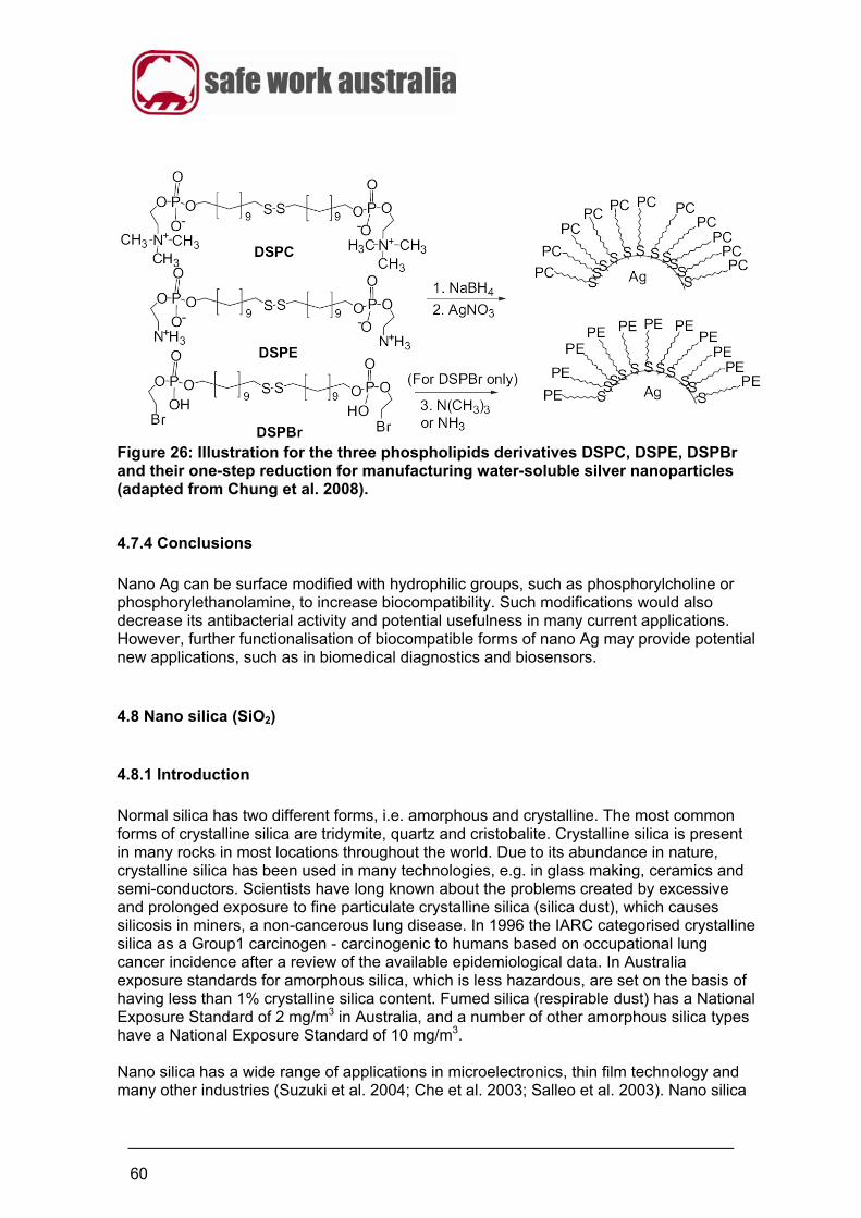
60
Figure 26: Illustration for the three phospholipids derivatives DSPC, DSPE, DSPBr and their one-step reduction for manufacturing water-soluble silver nanoparticles (adapted from Chung et al. 2008).
4.7.4 Conclusions
Nano Ag can be surface modified with hydrophilic groups, such as phosphorylcholine or phosphorylethanolamine, to increase biocompatibility. Such modifications would also decrease its antibacterial activity and potential usefulness in many current applications. However, further functionalisation of biocompatible forms of nano Ag may provide potential new applications, such as in biomedical diagnostics and biosensors.
4.8 Nano silica (SiO2)
4.8.1 Introduction
Normal silica has two different forms, i.e. amorphous and crystalline. The most common forms of crystalline silica are tridymite, quartz and cristobalite. Crystalline silica is present in many rocks in most locations throughout the world. Due to its abundance in nature, crystalline silica has been used in many technologies, e.g. in glass making, ceramics and semi-conductors. Scientists have long known about the problems created by excessive and prolonged exposure to fine particulate crystalline silica (silica dust), which causes silicosis in miners, a non-cancerous lung disease. In 1996 the IARC categorised crystalline silica as a Group1 carcinogen - carcinogenic to humans based on occupational lung cancer incidence after a review of the available epidemiological data. In Australia exposure standards for amorphous silica, which is less hazardous, are set on the basis of having less than 1% crystalline silica content. Fumed silica (respirable dust) has a National Exposure Standard of 2 mg/m3 in Australia, and a number of other amorphous silica types have a National Exposure Standard of 10 mg/m3. Nano silica has a wide range of applications in microelectronics, thin film technology and many other industries (Suzuki et al. 2004; Che et al. 2003; Salleo et al. 2003). Nano silica
61
also has applications as an enterosorbant and is a boosting agent used in pharmaceuticals (Alyushin and Astakova 1971; Chuiko 2003). Nano silica has also been used for medical and drug development applications such as the entrapment of biomolecules into ‘nanoshells’ which have a core of silica and a metallic outer layer. These particles can be injected into the subject and preferentially concentrate at the site of required drug release. Nanoshells have also been used to carry molecular conjugates to the antigen sites expressed on cancer cells and to malignant tumors (Hirsch et al. 2003a, b; Brongersma 2003).
4.8.2 Toxicology of nano silica
Nano silica exhibits concentration dependent toxicity towards different cell types, e.g. red blood cells (RBCs), micro-organisms, gametes, and white blood cells. The mechanism of toxicity of nano silica is thought to be due to its highly adhesive properties to cellular surfaces which can lead to changed membrane structures and cellular surfaces (Chuiko 2003; Gun’ko et al. 2007; Nishimori et al. 2009). Gun’ko et al. (2007) found the cytotoxicity of nano silica towards RBCs to be both concentration and size dependent. The interaction of RBCs with partially silylated (50%) and unmodified A-300 nano silica was studied by thermally stimulated depolarisation currents (TSDC), X-Ray Diffraction (XRD) and microscopy. Unmodified nano silica at a low concentration (<0.01 wt%) in a buffered aqueous suspension had a weak haemolytic effect on RBCs, but at a high concentration of 1 wt % unmodified nano silica haemolysed 100% of RBCs to shadow corpuscles. In the same experiments, partially (50%) silylated nano silica was less haemolytic in comparison with unmodified silica at the same concentration (Gun’ko et al. 2007). Nishimori et al. (2009) found that mice injected intravenously (twice weekly for four weeks) with 70 nm nano silica particles exhibited chronic toxicity in the spleen and liver while the kidney, lung, heart and brain remained unaffected. Histological analysis of these mice revealed splenic megakaryocyte accumulation of nano silica particles and hepatic microgranulation. The particle shape of nano silica is also an important factor in its toxic potential. For example, Nelson et al. (2010) microinjected silica nanospheres (50 and 200 nm) or nanowires (55 nm × 2.1 μm) into the yolk sack of embryonic zebrafish. They showed that silica nanomaterials with aspect ratios >1 were highly toxic (LD50 = 110 pg/g embryo) and caused embryo deformities, whereas silica nanomaterials with an aspect ratio of 1 were neither toxic nor teratogenic at the same concentrations. Silica nanowires also interfered with nerve development and function, prompting the researchers to emphasise the need for further testing of such nanomaterials before they can be used as platforms for drug delivery.
4.8.3 Potential substitution/modification of nano silica
Gun’ko et al. (2006) reviewed the properties of fumed nano silica that make it useful as a medicinal preparation with high sorption capacity for the treatment of different diseases. Six properties are highlighted:
the passive and unreactive nature of nano silica in redox and acid-base reactions together with low surface charge density (pH<8)
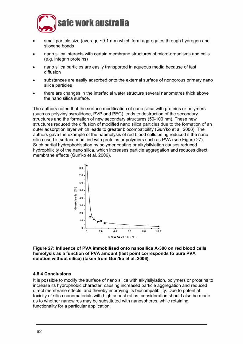
62
small particle size (average ~9.1 nm) which form aggregates through hydrogen and siloxane bonds
nano silica interacts with certain membrane structures of micro-organisms and cells (e.g. integrin proteins)
nano silica particles are easily transported in aqueous media because of fast diffusion
substances are easily adsorbed onto the external surface of nonporous primary nano silica particles
there are changes in the interfacial water structure several nanometres thick above the nano silica surface.
The authors noted that the surface modification of nano silica with proteins or polymers (such as polyvinylpyrrolidone, PVP and PEG) leads to destruction of the secondary structures and the formation of new secondary structures (50-100 nm). These new structures reduced the diffusion of modified nano silica particles due to the formation of an outer adsorption layer which leads to greater biocompatibility (Gun’ko et al. 2006). The authors gave the example of the haemolysis of red blood cells being reduced if the nano silica used is surface modified with proteins or polymers such as PVA (see Figure 27). Such partial hydrophobisation by polymer coating or alkylsilylation causes reduced hydrophilicity of the nano silica, which increases particle aggregation and reduces direct membrane effects (Gun’ko et al. 2006).
Figure 27: Influence of PVA immobilised onto nanosilica A-300 on red blood cells hemolysis as a function of PVA amount (last point corresponds to pure PVA solution without silica) (taken from Gun’ko et al. 2006).
4.8.4 Conclusions
It is possible to modify the surface of nano silica with alkylsilylation, polymers or proteins to increase its hydrophobic character, causing increased particle aggregation and reduced direct membrane effects, and thereby improving its biocompatibility. Due to potential toxicity of silica nanomaterials with high aspect ratios, consideration should also be made as to whether nanowires may be substituted with nanospheres, while retaining functionality for a particular application.
63
4.9 Quantum dots
4.9.1 Background
Quantum dots (QDs) are semi-conducting nano crystalline structures, ranging from 2 to 100 nm depending on the types of surface coating or functional group added (Lewinski et al. 2008), made from materials such as cadmium selenide or cadmium telluride (Tan and Zhang 2005). For biological applications, QDs typically have a core/shell conjugate structure, with the core composed of atoms from groups II–VI (e.g. CdSe, CdTe, CdS, PbSe, ZnS, and ZnSe) and groups III–V (e.g. GaAs, GaN, InP, and InAs) on the periodic table (Lewinski et al. 2008). QDs absorb white light which is then re-emitted as a specific colour after a couple of nanoseconds. QDs range from 10 to 250 atoms in diameter and contain anywhere from 100 to 20000 electrons. Energy levels of QDs can be controlled by changing their shape and size in order to control the depth of the potential. The science of QDs started to develop during the 1980s when their potential was realised to build nano-scale computer applications in which light is used to process information. Since this time more recent medical applications have included ‘molecular tagging’ in order that proteins, viruses, antibodies or DNA can be tracked in the human body. In these applications, QDs behave as a ‘fluorophore’ absorbing energy at a particular wavelength and re-emitting energy at a different longer wavelength (Tan and Zhang 2005).
4.9.2 Toxicology of quantum dots
Many of the core elements in QDs are known to be toxic at low concentrations, e.g. cadmium, selenium, lead and arsenic. Therefore, stability is a critical factor in the cytotoxic potential of QDs, as toxicity will eventuate from the release of toxic metal ions under conditions that promote QD degradation, such as an oxidative environment (Lewinski et al. 2008). Bare QD cores, and some QDs with biodegradable shells, are readily endocytosed by cells into endosomes, which are then trafficked to various cellular compartments. These include the acidic (~pH5) and oxidative environments of lysosomes and peroxisomes, which can degrade QDs and result in the leaching of toxic metals from the core (Chang et al. 2006). Studies of the cytotoxic potential of QDs have mainly concerned CdSe/ZnS cores (recently reviewed by Lewinski et al. 2008), as these are considered the most versatile for biological applications. In a general review of the toxicology of QDs by Drew (2009), a number of conclusions were made including:
the metal core of QDs can easily be exposed and can be a means by which toxicity can occur if the body is exposed to this material
internalisation of uncoated QDs led to leaching of both cadmium and selenium from the QD core in the low pH of endosomes
applying a surface coating such as chitosan or polyethylene glycol to a QD can make it biocompatible and confer specific functionality
QDs are recognised in many instances as being foreign to the body and may therefore be sequestered by the reticuloendothelial systems in the major organs
64
QDs do not appear to cause acute toxicity after being injected into the venous system, however this does depend on whether QDs are internalised by cells
there is no data available on the toxicity due to inhalation of QDs
the core metal cadmium along with other similar heavy metals are known toxic agents in most biological systems.
4.9.3 Methods of modification of quantum dots
For biomedical applications, QDs require a stable biocompatible shell coating to prevent core degradation, such as chitosan, polyethylene glycol or silica (Gerion et al. 2001; Ryman-Rasmussen et al. 2006). Additional functionalities or bioconjugates can be added to the surface to improve bioavailability or introduce bioactivity. The cytotoxicity of QDs containing Cd/Se cores can be progressively reduced by PEG-polymer coatings of increasing thickness, which prevent QDs from being internalised into the cell and subsequent stages of intracellular degradation (Chang et al. 2006). Surface chemistry and charge also affects the interaction of QDs with serum proteins, which can bind to the surface to increase the overall hydrodynamic diameter of the QD >5.5 nm, and result in markedly delayed clearance from the body (as detailed in section 3.1.2) (Choi et al. 2007). The successful surface modification with chitosan of nano Au has been employed to increase biocompatibility (Tan and Zhang 2005), see Section 4.6.3, and can also be used for modifying QDs. In vitro exposure studies using NIH 3T3 mouse fibroblast and HT29 human colon carcinoma cells and Trypan blue and MTT exclusion assays revealed that chitosan-encapsulated QDs exhibited improved biocompatibility over their non-encapsulated analogues. A number of different QD/chitosan complexes with varying light absorbance properties can be formed in this process depending on the ratio of components used (Tan and Zhang 2005). Figure 28 shows the different UV absorption spectra of the different chitosan-encapsulated complexes.
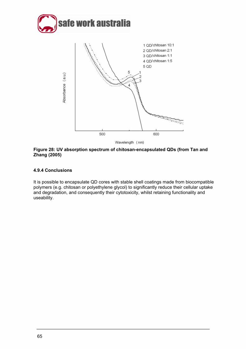
65
Figure 28: UV absorption spectrum of chitosan-encapsulated QDs (from Tan and Zhang (2005)
4.9.4 Conclusions
It is possible to encapsulate QD cores with stable shell coatings made from biocompatible polymers (e.g. chitosan or polyethylene glycol) to significantly reduce their cellular uptake and degradation, and consequently their cytotoxicity, whilst retaining functionality and useability.
66
4.10 Conclusions on substitution/modification options for engineered nanomaterials The potential modifications for the different engineered nanomaterials are listed in Table K. For most of the engineered nanomaterials examined in this review there are substitution/modification options that have the potential to reduce the toxicity for specific engineered nanomaterials. Surface modification is the most common substitution/modification method used. Summary There are known methods that can be used to substitute/modify engineered nanomaterials that are used (or researched) in Australia. The methods of surface modification, encapsulation, particle size control, functional group addition and crystalline phase type control can each be employed for different engineered nanomaterials in order to decrease their potential toxicity. However in some cases, such modifications may affect the functionality of nanomaterials in relation to intended end-uses. If the researchers, developers and manufacturers of engineered nanomaterials adopt these methods then it is possible at this early stage of nanomaterial development to decrease toxicity of materials that are to be manufactured. The downstream effect of this will be to reduce the risk posed by the use of these nanomaterials not only in the workplace but also in the general community.
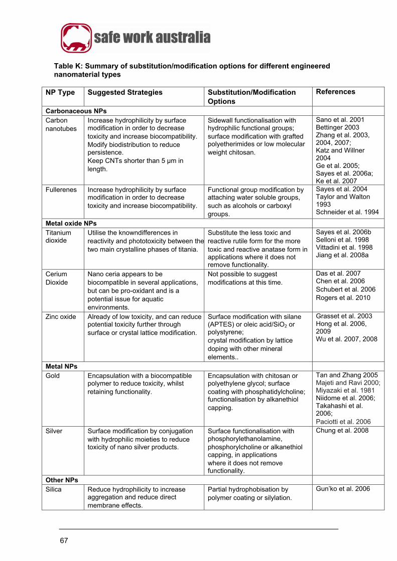
67
Table K: Summary of substitution/modification options for different engineered nanomaterial types
NP Type Suggested Strategies Substitution/Modification Options
References
Carbonaceous NPs Carbon nanotubes
Increase hydrophilicity by surface modification in order to decrease toxicity and increase biocompatibility. Modify biodistribution to reduce persistence. Keep CNTs shorter than 5 µm in length.
Sidewall functionalisation with hydrophilic functional groups; surface modification with grafted polyetherimides or low molecular weight chitosan.
Sano et al. 2001 Bettinger 2003 Zhang et al. 2003, 2004, 2007; Katz and Willner 2004 Ge et al. 2005; Sayes et al. 2006a; Ke et al. 2007
Fullerenes Increase hydrophilicity by surface modification in order to decrease toxicity and increase biocompatibility.
Functional group modification by attaching water soluble groups, such as alcohols or carboxyl groups.
Sayes et al. 2004 Taylor and Walton 1993 Schneider et al. 1994
Metal oxide NPs
Titanium dioxide
Utilise the knowndifferences in reactivity and phototoxicity between thetwo main crystalline phases of titania.
Substitute the less toxic and reactive rutile form for the more toxic and reactive anatase form in applications where it does not remove functionality.
Sayes et al. 2006b Selloni et al. 1998 Vittadini et al. 1998 Jiang et al. 2008a
Cerium Dioxide
Nano ceria appears to be biocompatible in several applications, but can be pro-oxidant and is a potential issue for aquatic environments.
Not possible to suggest modifications at this time.
Das et al. 2007 Chen et al. 2006 Schubert et al. 2006 Rogers et al. 2010
Zinc oxide Already of low toxicity, and can reduce potential toxicity further through surface or crystal lattice modification.
Surface modification with silane (APTES) or oleic acid/SiO2 or polystyrene; crystal modification by lattice doping with other mineral elements..
Grasset et al. 2003 Hong et al. 2006, 2009 Wu et al. 2007, 2008
Metal NPs Gold Encapsulation with a biocompatible
polymer to reduce toxicity, whilst retaining functionality.
Encapsulation with chitosan or polyethylene glycol; surface coating with phosphatidylcholine; functionalisation by alkanethiol capping.
Tan and Zhang 2005 Majeti and Ravi 2000; Miyazaki et al. 1981 Niidome et al. 2006; Takahashi et al. 2006; Paciotti et al. 2006
Silver Surface modification by conjugation with hydrophilic moieties to reduce toxicity of nano silver products.
Surface functionalisation with phosphorylethanolamine, phosphorylcholine or alkanethiol capping, in applications where it does not remove functionality.
Chung et al. 2008
Other NPs Silica Reduce hydrophilicity to increase
aggregation and reduce direct membrane effects.
Partial hydrophobisation by polymer coating or silylation.
Gun’ko et al. 2006

68
Quantum dots
Decrease cellular uptake and intracellular degradation to reduce toxicity from leaching metals, whilst retaining functionality in most cases.
Encapsulation of core with stable biocompatible shell, e.g with chitosan, silica or PEG, that decreases endocytosis and endosome degradation.
Tan and Zhang 2005 Gerion et al. 2001
69
References
Ahamed M, Karns M, Goodson M, Rowe J, Hussain SM, Schlager JJ and Hong Y (2008). DNA damage response to different surface chemistry of silver nanoparticles in mammalian cells. Toxicol and Appl Pharm 233:404–410. Aleyamma AJ and Sharma CP (1991). Blood Compatible Materials and Devices, Sharma CP, Szycher M (eds). Technomic Publishing: Lancaster, 123–130. Alyushin MT and Astakhova MN (1971). Aerosil and its application in pharmaceutical practice. Pharmacy 6:73–77. Auffan JR, Rose J, Orsiere T, De Meo M, Thill A, Zeyons O, Proux O, Masion A, Chaurand P, Spalla O, Botta A, Wiesner, MR and Bottero JY (2009). CeO2 nanoparticles induce DNA damage towards human dermal fibroblasts in vitro. Nanotoxicology 3(2):161–171. Babu S, Thanneeru R, Inerbaev T, Day R, Masunov AE, Schulte A and Seal S (2009) Dopant-mediated oxygen vacancy tuning in ceria nanoparticles. Nanotechnology 20: 085713 (5pp) doi:10.1088/0957-4484/20/8/085713. Balasubramanian K and Burghard M (2005). Chemically functionalized carbon nanotubes. Small 1(2):180–192. Barker PJ and Branch A (2008). The interaction of modern sunscreen formulations with surface coatings. Progress in Organic Coatings 62:313–320. Bettinger HF (2003). Experimental and Computational Investigations of the Properties of Fluorinated Single-Walled Carbon Nanotubes. Chem. Phys. Chem. 4:1283–1289. Brongersma ML (2003). Nanoscale photonics: nanoshells: gifts in a gold wrapper. Nat Mater 2:296–297. Calvo P, Reuman-Lopez C, Vila-Jato JL and Alonso MJ. (1997). Novel hydrophilic chitosan-polyethylene oxide nanoparticles as protein carriers. J. Appl. Polym. Sci. 63:125–132.
Chae SR, Hotze EM and Wiesner MR (2009). Evaluation of the Oxidation of Organic Compounds by Aqueous Suspensions of Photosensitized Hydroxylated-C60 Fullerene Aggregates. Environ. Sci. Technol. 43:6208–6213. Chang E, Thekkek N, Yu WW, Colvin VL and Drezek R (2006). Evaluation of Quantum Dot Cytotoxicity Based on Intracellular Uptake. Small 2(12):1412–1417. Chattopadhyay CN, Billups WE and Bandaru PR (2008). Modification of the electrical characteristics of single wall carbon nanotubes through selective functionalization. App. Phys. Lett. 93:243113–243116. Che S, Bennett AEG, Yokoi T, Sakamoto K, Kunieda H, Terasaki O and Tatsumi T (2003). A novel anionic surfactant templating route for synthesizing mesoporous silica with unique structure. Nat. Mater. 2:801–805.
70
Chen J, Swanang P, Seal S and McGinnis JF (2006). Rare earth nanoparticles prevent retinal degeneration induced by intracellular peroxides. Nature Nanotechnology 1:142–150. Choi HS, Liu W, Misra P, Tanaka E, Zimmer JP, Itty Ipe B, Bawendi MG and Frangioni JV (2007). Renal clearance of quantum dots. Nature Biotechnology 25(10):1165–1170. Chuiko AA (ed) (2003). In: Medical chemistry and clinical application of silicon dioxide. Nukova Dumka, Kiev, Russia. pp 143–157.
Chung TH, Wu SH, Yao M, Lu CW, Lin YS, Hung Y, Mou CY, Chen YC and Huang DM (2007). The effect of surface charge on the uptake and biological function of mesoporous silica nanoparticles in 3T3-L1 cells and human mesenchymal stem cells. Biomaterials 28 (19):2959–2966. Chung YC, Chen IH and Chen CJ (2008). The surface modification of silver nanoparticles by phosphoryl disulfides for improved biocompatibility and intracellular uptake. Biomaterials 29: 1807–1816. Connor EE, Mwamuka J, Gole A, Murphy CJ and Wyatt MD (2005). Gold Nanoparticles Are Taken Up by Human Cells but Do Not Cause Acute Cytotoxicity. Small 1:325–327. Cotton FA and Wilkinson G (1999). Advanced Inorganic Chemistry (6th Edn) Wiley. Das M, Patil S, Bhargava N, Kang JF, M L. Seal RS and Hickman JJ (2007). Auto-catalytic ceria nanoparticles offer neuroprotection to adult rat spinal cord neurons. Biomaterials 28: 1918–1925. Demers LM, Mirkin CA, Mucic RC, Reynolds RA, Letsinger RL, Elghanian R and Viswanadham G (2000). Fluorescence-Based Method for Determining the Surface Coverage and Hybridization Efficiency of Thiol-Capped Oligonucleotides Bound to Gold Thin Films and Nanoparticles. Anal. Chem. 72:5535–5541. Dixit V, Van De Bossche J, Sherman DM, Thompson DH and Andres RP (2006). Synthesis and Grafting of Thioctic Acid−PEG−Folate Conjugates onto Au Nanoparticles for Selective Targeting of Folate Receptor-Positive Tumor Cells. Bioconjug. Chem. 17(3):603-609. Donaldson K, Murphy F, Poland C, Duffin R, Osmond M, McCall M and Hawkins S (2009). High aspect ratio nanoparticles: the hazard from long biopersistent fibres. Presented at 4th International conference on Nanotechnology-Occupational and Environmental Health. 26-29 August 2009. Drew R (2009). Engineered nanomaterials: A review of the toxicology and health hazards. Available at: http://www.safeworkaustralia.gov.au/NR/rdonlyres/47D5968D-4A11-45A0-8121-8C992E111447/0/ToxicologyReview_Nov09.pdf. Accessed 4th Dec 2009.
Dufour EK, Kumaravel T, Nohynek GJ, Kirkland D and Toutain H (2006). Clastogenicity, photo-clastogenicity or pseudo-photo-clastogenicity: Genotoxic effects of zinc oxide in the dark, in pre-irradiated or simultaneously irradiated Chinese Hamster Ovary cells. Toxicology Letters 164 (Suppl. 1):S290–S291.
71
Dutta D, Sundaram SK, Teeguarden JG, Riley BJ, Fifield LS and Weber TJ (2007). Adsorbed Proteins Influence the Biological Activity and Molecular: Targeting of Nanomaterials. Toxicological Sciences 100(1):303–315. Elechiguerra JL, Burt JL, Morones JR, Camacho-Bragado, Gao X, Lara HH and Yacaman MJ (2005). Interaction of silver nanoparticles with HIV-1. Journal of Nanobiotechnology 3:6 doi:10.1186/1477-3155-3-6. Eychmuller A and Rogach AL (2000). Spectroscopic and other physical properties–chemistry and photophysics of thiol-stabilized II-VI semiconductor nanocrystals. Pure Appl. Chem. 72:179–188. Foged C, Brodin B, Frokjaer S and Sundblad A (2005). Particle size and surface charge affect particle uptake by human dendritic cells in an in vitro model. International Journal of Pharmaceutics 298:315–322. de la Fuente JM and Berry CC (2005). Tat Peptide as an efficient molecule to translocate gold nanoparticles into the cell nucleus. Bioconjugate Chem. 16(5):1176–1180. Ge JJ, Zhang D, Li Q, Hou H, Graham MJ, Dai L, Harris FW and Cheng SZD (2005). Multiwalled carbon nanotubes with chemically grafted polyetherimides. J. Am. Chem. Soc. 127:9984–9985. Geiser M, Rothen-Rutishauser B, Kapp N, Schurch S, Kreyling W, Semmler M, Hof VI, Heyder J and Peter G (2005). Ultrafine particles cross cellular membranes by nonphagocytic mechanisms in lungs and in cultured cells. Environmental Health Perspectives 113(11):535–542. Gerion D, Pinaud F, Williams SC, ParakWJ, Zanchet D, Weiss S and Alivisatos AP (2001). Synthesis and Properties of Biocompatible Water-Soluble Silica-Coated CdSe/ZnS Semiconductor Quantum Dots. J. Phys. Chem. B. 105:8861–8871. Good RJ (1993). Contact Angle, Wettability and Adhesion. Mittal KL (ed.). VSP, Wiley: Weinheim, pp3–36. Goodman CM, McCusker CD, Yilmaz T and Rotello VM (2004). Toxicity of Gold Nanoparticles Functionalized with Cationic and Anionic Side Chains. Bioconjugate Chem. 15:897–900. Grasset F, Saito ND, Lia D, Park D, Sakaguchi I, Ohashia N, Haneda H, Roisnel T, Mornet S and Duguet E (2003). Surface modification of zinc oxide nanoparticles by aminopropyltriethoxysilane. Journal of Alloys and Compounds 360:298–311. Grushko YS, Sedov VP, and Shilin VA (2007). Technology for Manufacture of Pure Fullerenes C60, C70 and a Concentrate of Higher Fullerenes. Russian Journal of Applied Chemistry 80(3):448–455. Gun’ko VM, Turov VV and Chuiko AA (2006). Fundamentals of nanosilica applications for human protection, in ‘Surface chemistry in biomedical and environmental science’ in Proceedings of the NATO Advanced Research Workshop on Pure and Applied Surface Chemistry and Nanomaterials for Human Life and Environmental Protection, Kyiv, Ukraine, 14-17 September 2005 Series: NATO Science Series II: Mathematics, Physics and Chemistry , Vol. 228, XVII, 444 p., Hardcover ISBN: 978-1-4020-4739-8.
72
Gun’ko VM, Galagan NP, Grytsenko IV, Zarko VI, Oranska OI, Osaulenko VL, Bogatyrev VM and Turov VV (2007). Interaction of unmodified and partially silylated nanosilica with red blood cells. Central European Journal of Chemistry 5(4):951–969. Gupta B, Levchenko TS and Torchilin VP (2005). Intracellular delivery of large molecules and small particles by cell-penetrating proteins and peptides Adv. Drug Deliv. Rev. 57(4):637–651. Harush-Frenkel O, Debotton N, Benita S and Altschuler Y (2007). Targeting of nanoparticles to the clathrin-mediate endocytic pathway Biochemical and Biophysical Research Communications 353:26–32. Hattori Y and Maitani Y (2005). Folate-linked nanoparticle-mediated suicide gene therapy in human prostate cancer and nasopharyngeal cancer with herpes simplex virus thymidine kinase. Cancer Gene Ther. 12(10):796–809. Hermanson GT (1996). Bioconjugate techniques. London: Academic Press; 1996. Chapter 8. Hirsch LR, Jackson JB, Lee A, Halas NJ and West JL (2003a). A whole blood immunoassay using gold nanoshells. Anal. Chem. 75(10):2377–2381. Hirsch LR, Stafford RJ, Bankson JA, Sershen SR, Rivera B, Price RE, Hazle JD, Halas NJ, West JL (2003b). Nanoshell-mediated near-infrared thermal therapy of tumors under magnetic resonance guidance. Proc. Natl Acad. Sci. USA 100:3549–13554. Hong R, Pan T, Qian J and Li H (2006). Synthesis and surface modification of ZnO nanoparticles. Chemical Engineering Journal 119:71–81. Hong RY, Li JH, Chen LL, Liu DQ, Li HZ, Zheng Y and Ding J (2009). Synthesis, surface modification and photocatalytic property of ZnO nanoparticles. Powder Technology 189:426–432. Huff TB, Hansen MN, Zhao Y, Cheng JX and Wei A (2007). Controlling the Cellular Uptake of Gold Nanorods. Langmuir 23:1596–1599. Iler RK (1979). The chemistry of silica. Wiley, New York Jackson N, Elms T, Lopatas A and Wright P (2009). Engineered Nanomaterials: Evidence on the Effectiveness of Workplace Controls to Prevent Exposure. Available at: http://www.safeworkaustralia.gov.au/NR/rdonlyres/E3C113AC-4363-4533-A128-6D682FDE99E0/0/EffectivenessReport.pdf Accessed 4th Dec 2009 Jiang J, Oberdörster G, Elder A, Gelein R, Mercer P and Biswas P (2008a). Does nanoparticle activity depend upon size and crystal phase? Nanotoxicology 2(1):33–42. Jiang W, Kim BYS, Rutka JT and Chan WCW (2008b). Nanoparticle-mediated cellular response is size-dependent. Nature Nanotechnology 3:145–150.
Johnson DR, Methner MM, Kennedy AJ and Steevens JA (2010). Potential for Occupational Exposure to Engineered Carbon-Based Nanomaterials in Environmental Laboratory Studies. Environmental Health Perspectives 118:(1)49–54.
73
Katz E and Willner I (2004). Biomolecule-Functionalized Carbon Nanotubes: Applications in Nanobioelectronics. Chem. Phys. Chem. 5:1085–1104. Ke G, Guan WC, Tang CY, Hu Z, Guan WJ, Zeng DL and Deng F (2007). Covalent modification of multiwalled carbon nanotubes with low molecular weight chitosan. Chinese Chemical Letters 18:361–364. Khan JA, Pillai B, Das TK, Singh Y and Maiti S (2007). Molecular Effects of Gold Nanoparticles in HeLa Cells. Chem. Bio. Chem. 8:1237–1240. Kim JS, Yoon TJ, Yu KN, Noh MS, Woo M, Kim BG, Lee KH, Sohn, BH, Park SB, Lee JK, and Cho MH (2006). Cellular uptake of magnetic nanoparticle is mediated through energydependent endocytosis in A549 cells. J. Vet. Sci. 7(4):321–326. Kim YS, Kim JS, Cho HS, Rha DS, Kim JM, Park JD, Choi BS, Lim R, Chang HK, Chung YH, Kwon IH, Jeong J, Han BS and Yu IJ (2008). Twenty-eight-day oral toxicity, genotoxicity, and gender-related tissue distribution of silver nanoparticles in Sprague-Dawley rats. Inhal Toxicol. 20(6):575–583. Koch AM, Reynolds F, Kircher MF, Merkle HP, Weissleder R and Josephson L (2003). Uptake and Metabolism of a Dual Fluorochrome Tat-nanoparticle in HeLa Cells. Bioconjug. Chem. 14(6):1115–1121. Kuchibhatla SVNT, Karakoti AS, Debasis B and Seal S (2007). One dimensional nanostructured materials. Progr. Mater. Sci. 52:699–913. Lai SK, Hida K, Man ST, Chen C, Machamer C, Schroer T and Hanes J (2007). Privileged delivery of polymer nanoparticles to the perinuclear region of live cells via a non-clathrin, non-degradative pathway. Biomaterials 28(18):2876–2884. Lam, CW, James JT, McCluskey R, Arepalli S and Hunter RL (2006). A review of carbon nanotube toxicity and assessment of potential occupational and environmental health risks. Critical Reviews in Toxicology 36(3):189–217. Lewinski N, Colvin V and Drezek R (2008). Cytotoxicity of Nanoparticles. Small 4(1):26–49. Li JJ, Zou L, Hartono D, Ong CN, Bay BH, and Yung LYL (2008). Gold nanoparticles induce oxidative damage in lung fibroblasts in vitro. Adv. Mater. 20:138–142. Liu S, Zhang Z and Han M (2005). Gram-Scale Synthesis and Biofunctionalization of Silica-Coated Silver Nanoparticles for Fast Colorimetric DNA Detection. Anal. Chem. 77:2595–2600. Majeti NV and Ravi K (2000). A review of chitin and chitosan applications. React. Funct. Polym. 46:1–27. Makohliso SA (1999). Thesis: a Surface Engineering Approach Towards the Development of Cell-based Biochip Sensors. Ecole Polytechnique Federale de Lausanne, Lausanne. Mann S, Shenton W, Li M, Connoly S and Fitzmaurice D (2000). Biologically programmed nanoparticle assembly. Adv. Mater. 12:147–150.
74
Marieb EN (1998). Human Anatomy and Physiology (4th edn). Addison- Wesley: Reading, MA. Masui T, Ozaki T, Machida K and Adachi G (2000). Preparation of ceria-zirconia sub-catalysts for automotive exhaust cleaning. Journal of Alloys and Compounds 303–304:49–55. Mathieu HJ (2001). Bioengineered material surfaces for medical applications. Surf. Interface Anal. 32:3–9. Miyazaki S, Ishii K and Nadai T (1981). The Use of Chitin and Chitosan as Drug Carriers. Chem. Pharm. Bull. 29(10):3067–3069. Miyazaki S, Yamaguchi H, Takada M, Hou WM, Takeichi Y and Yasubuchi H (1990). Pharmaceutical application of biomedical polymers: preliminary study on film dosage form prepared from chitosan for oral drug delivery. Acta Pharm. Nord. 2:401–406. Morgenson M, Sammes SN, and Tompsett GA (2000). Physical, chemical and electrochemical properties of pure and doped ceria. Solid State Ionics 129:63–94. Murphy CJ, Gole AM, Stone JW, Sisco PN, Alkilany AM, Goldsmith EC, and Baxter SC (2008). Gold nanoparticles in biology: beyond toxicity to cellular imaging. Acc. Chem. Res. 41(12):1721–1730. Nelson SM, Mahmoud T, Beaux M, Shapiro P, McIlroy DN, and Stenkamp DL (2010). Toxic and teratogenic silica nanowires in developing vertebrate embryos. Nanomedicine 6:93–102. NICNAS (2007). Summary of call for information on the use of Nanomaterials. Available at: http://www.nicnas.gov.au/Publications/Information_Sheets/General_Information_Sheets/NIS_Call_for_info_Nanomaterials.pdf. Accessed 24/09/09. Niidome T, Yamagata M, Okamoto Y, Akiyama Y, Takahashi H, Kawano T, Katayama Y and Niidome Y (2006). PEG-Modified Gold Nanorods with a stealth character for in vivo applications. J. Controlled Release 114(3):343–347. Nishimori H, Kondoh M, Isoda K, Tsunoda S, Tsutsumi Y and Yagi K (2009). Histological analysis of 70-nm silica particles-induced chronic toxicity in mice. European Journal of Pharmaceutics and Biopharmaceutics 72:626-631. Paciotti GF, Kingston DGI and Tamarkin L (2006). Colloidal Gold Nanoparticles: A Novel Nanoparticle Platform for Developing Multifunctional Tumor-Targeted Drug Delivery Vectors. Drug Development Research 67:47–54. Panyam J. and Labhasetwar V. (2003). Dynamics of endocytosis and exocytosis of poly (DL-lactide-co-glycolide) nanoparticles in vascular smooth muscle cells. Pharm. Res. 20(2):110–118. Patra HK, Banerjee S, Chaudhuri U, Lahiri P and Dasgupta KA (2007). Cell selective response to gold nanoparticles. Nanomedicine: Nanotechnology, Biology, and Medicine 3:111–119.
75
Paunesku T, Rajh T, Wiederrecht G, Maser J, Vogt S, Stojicevic N, Protic M, Lai B, Oryhon J, Thurnauer M and Woloschak G (2003). Biology of TiO2–oligonucleotide nanocomposites. Nat. Mater. 2(5):343–346. Paunesku T, Vogt S, Lai B, Maser J, Stojicevic N, Thurn KT, Osipo C, Liu H, Legnini D, Wang Z, Lee C and Woloschak GE (2007). Intracellular Distribution of TiO2−DNA Oligonucleotide Nanoconjugates Directed to Nucleolus and Mitochondria Indicates Sequence Specificity. Nano Lett. 7(3):596–601.
Poland CA, Duffin R, Kinloch I, Maynard A, Wallace WAH, Seaton A, Stone V, Brown S, MacNee W and Donaldson K (2008). Carbon nanotubes introduced into the abdominal cavity of mice show asbestos-like pathogenicity in a pilot study. Nature Nanotechnology 3(7):423–428. Qaddoumi MG, Gukasyan HJ, Davda J, Labhasetwar V, Kim KJ and Lee VHL (2003). Clathrin and caveolin-1 expression in primary pigmented rabbit conjunctival epithelial cells: Role in PLGA nanoparticle endocytosis. Mol. Vis. 9:559–568. Qin WJ and Yung LY (2006). Efficient Manipulation of Nanoparticle-Bound DNA via Restriction Endonuclease Biomacromolecules 7(11):3047–3051. Ringer SP and Ratinac KR (2004). On the role of characterization in the design of interfaces in nanoscale materials technology. Microscopy and Microanalysis 10 (3):324–335. Rogers NJ, Franklin NM, Apte SC, Batley GE, Angel BM, Lead JR and Baalousha M (2010). Physico-chemical behaviour and algal toxicity of nanoparticle CeO2 in freshwater. Environ. Chem. 7:50–60. Rothen-Rutishauser BM, Schurch S, Haenni B, Kapp N and Gehr P (2006). Interaction of fine particles and nanoparticles with red blood cells visualised with advanced microscopic techniques. Environ. Sci. Technol. 40:4353–4359. Ryman-Rasmussen JP, Rivere JE and Monteiro-Rivere NA (2006). Surface coatings determine cytotoxicity and irritation potential of quantum dot nanoparticles in epidermal keratinocytes. Journal of Investigative Dermatology 127:143–153. Sabbatini L and Zambonin PG (1996). XPS and SIMS surface chemical analysis of some important classes of polymeric biomaterials. J. Electron Spectrosc. Relat. Phenom. 81:285–301. Salleo A, Taylor ST, Martin MC, Panero W, Jeanloz R, Sands T and Génin FY (2003). Laser driven phase transformations in amorphous silica. Nat. Mat. 2(12):796–798. Samuel U and Guggenbichler JP (2004). Prevention of catheter-related infections: the potential of a new nano-silver impregnated catheter. Int. J. Antimicrob. Agents 23(S1):75–78. Sano M, Kamino A, Okamura J and Shinkai S (2001). Ring Closure of Carbon Nanotubes. Science 293:1299–1301. Sayes CM, Fortner JD, Guo W, Lyon D, Boyd AM, Ausman KD, Tao YJ, Sitharaman B, Wilson LJ. Hughes JB, West JL, and Colvin VL (2004). The differential cytotoxicity of water-soluble fullerenes. Nano Letters 4(10):1881–1887.
76
Sayes CM, Liang F, Hudson JL, Mendez J, Guo W, Beach JM, Moore VC, Doyle CD, West JL, Billups WE, Ausman KD and Colvin VL (2006a). Functionalization density dependence of single-walled carbon nanotubes cytoxicity in vitro. Toxicology Letters 161:135–142. Sayes CM, Wahi R, Kurian PA, Liu Y, West JL, Ausman KD, Warheit DB and Colvin VL (2006b). Correlating Nanoscale Titania Structure with Toxicity: A Cytotoxicity and Inflammatory Response Study with Human Dermal Fibroblasts and Human Lung Epithelial Cells. Toxicological Sciences 92(1):174–185. Schneider NS, Darwish AD, Kroto HW, Taylor R and Walton DRM (1994). Formation of fullerols via hydroboration of fullerene-CGO. J. Chem. Soc. Chem. Commun. 4:463–464. Schubert D, Dargusch R, Raitano J and Chan SW (2006). Cerium and yttrium oxide nanoparticles are neuroprotective. Biochemical and Biophysical Research Communications 342:86–91. Selloni A and Vittadini A (1998). The adsorption of small molecules on the TiO2 anatase (101) surface by first-principles molecular dynamics. Surface Science 402-404:219–222. Shukla R, Bansal V, Chaudhary M, Basu A, Bhonde R and Sastry M (2005). Biocompatibility of Gold Nanoparticles and Their Endocytotic Fate Inside the Cellular Compartment: A Microscopic Overview. Langmuir 21:10644–10654. Steinhauser I, Spankuch B, Strebhardt K and Langer K (2006). Trastuzumab-modified nanoparticles: optimisation of preparation and uptake in cancer cells. Biomaterials 27:4975–4983. Sun C, Li H, Zhang H, Wang Z and Chen L (2005). Controlled synthesis of CeO2 nanorods by a solvothermal method. Nanotechnology 16(2005):1454–1463. Sung JH, Ji JH, Park JD, Yoon JU, Kim DS, Jeon KS, Song MY, Jeong J, Han BS, Han HJ, Chung H, Chang HK, Lee JH, Cho MH, Kelman BJ and Yu IJ (2009). Subchronic Inhalation Toxicity of Silver Nanoparticles. Toxicological Sciences 108(2):452–461. Suzuki T, Sugiura I, Sato S and Nakamura T (2004). Thermal expansion coefficients of nano-clustering silica (NCS) films measured by X-Ray reflectivity and substrate curvature methods. Jpn. J. Appl. Phys. 43:614–616. Takagi A, Hirose A, Nishimura T, Fukumori N, Ogato A, Ohashi N, Kitajima S and Kanno J (2008). Induction of mesothelioma in p53+/- mouse by intraperitoneal application of multi-wall carbon nanotube. Journal of Toxicological Sciences 33(1):105–116. Takahashi H, Niidome Y, Niidome T, Kaneko K, Kawasaki H and Yamada S (2006). Modification of Gold Nanorods Using Phosphatidylcholine to Reduce Cytotoxicity. Langmuir 22:2–5. Tan WB and Zhang Y (2005). Surface modification of gold and quantum dot nanoparticles with chitosan for bioapplications. J. Biomed. Mat. Res. Part B: Applied Biomaterials 75A(1):56–62. Taylor R and Walton DRM (1993). The chemistry of fullerenes. Nature 363:685–697.
77
Thanh NTK and Rosenzweig Z (2002). Development of an Aggregation-Based Immunoassay for Anti-Protein A Using Gold Nanoparticles. Anal. Chem. 74:1624–1628. Thanneeru R, Patil S, Deshpande S and Seal S (2007). Effect of trivalent rare earth dopants in nanocrystalline ceria coatings for high-temperature oxidation resistance. Acta Materialia 55:3457–3466. Thurn KD, Brown EMB, Wu A, Vogt S, Lai B, Maser J, Paunesku T and Woloschak GE (2007). Nanoparticles for Applications in Cellular Imaging. Nanoscale Res. Lett. 2:430–441. Tkachenko AG, Xie H, Coleman D, Glomm W, Ryan J, Anderson MF, Franzen S and Feldheim DL (2003). Multifunctional Gold Nanoparticle-Peptide Complexes for Nuclear Targeting. J. Am. Chem. Soc. 125:4700–4701.
Tkachenko AG, Xie H, Liu Y, Coleman D, Ryan J, Glomm WR, Shipton MK, Franzen S and Feldheim DL (2004). Cellular Trajectories of Peptide-Modified Gold Particle Complexes: Comparison of Nuclear Localization Signals and Peptide Transduction Domains. Bioconjugate Chem. 15:482–490. Tsunekawa S, Ishikawa K, Li ZQ, Kawazoe Y and Kasuya A (2000). Origin of anomalous lattice expansion in oxide nanoparticles. Phys. Rev. Lett. 85:3440–3443.
Uboldi C, Barth S, Unger RE and Kirkpatrick JC (2008). Surface modification influences the in vitro toxicity of gold nanoparticles in human alveolar type-II cell lines. Alternatives to Animal Experimentation (ALTEX); 25(Suppl. 1):74. Ulkur E, Oncul O, Karagoz H, Yeniz E and Celikoz B (2005). Comparison of silver coated dressing, chlorhexidine acetate and fusidic acid for topical antibacterial effect in methicillin- resistant staphylocci-contaminated, full skin thickness rat burn wound. Burns 31:874–879 Vittadini A, Selloni A, Rotzinger FP and Grätzel M (1998). Structure and Energetics of Water Adsorbed at TiO2 Anatase (101) and (001). Phys. Rev. Lett. 81(14):2954–2957. Vives E, Brodin P and Lebleu B (1997). A Truncated HIV-1 Tat Protein Basic Domain Rapidly Translocates through the Plasma Membrane and Accumulates in the Cell Nucleus. J. Biol. Chem. 272(25):18010–18017. Wang X and Ruckenstein E (1993). On the gel effect in the presence of a chain transfer agent in methyl methacrylate polymerization and its copolymerization with various acrylates J. Appl. Polym. Sci. 49:2179–2188. Wang YX, Robertson JL, Spillman WB and Claus RO (2004). Effects of the Chemical Structure and the Surface Properties of Polymeric Biomaterials on their Biocompatibility. Pharmaceutical Research 21(8):1362–1373. West JL and Halas NJ (2003). Engineered nanomaterials for biophotonics applications: improving sensing, imaging, and therapeutics. Ann. Rev. Biomed. Eng. 5:285–292. Wijnhoven SWP, Peijnenburg JGM, Herberts CA and Hagens WI (2009). Nano-silver - a review of available data and knowledge gaps in human and environmental risk assessment. Nanotoxicology 1:30–40.
78
Win KY and Feng SS (2005). Effect of particle size and surface coating on cellular uptake of polymeric nanoparticles for oral delivery of anticancer drugs. Biomaterials 26:2713–2722. Wu YL, Lim CS, Fu S, Tok AIY, Lau HM, Boey FYC and Zeng XT (2007). Surface modifications of ZnO quantum dots for bio-imaging. Nanotechnology 18:215604 (9pp). Wu YL, Fu S, Tok AIY, Zeng XT, Lim CS, Kwek LC and Boey FYC (2008). A dual-colored bio-marker made of doped ZnO nanocrystals. Nanotechnology 19:345605 (9pp). Xiang SD, Scholzen A, Minigo G, David C, Apostolopoulos V, Mottram PL and Plebanski M (2006). Pathogen recognition and development of particulate vaccines: Does size matter? Methods 40:1–9. Yang Z, Zhou K, Liu X, Tian Q, Lu D and Yang S (2007). Single-crystalline ceria nanocubes: size-controlled synthesis, characterization and redox property. Nanotechnology 18:185606 (5pp). Zhang J, Zou H, Qing Q, Yang Y, Li Q, Liu Z and Du J (2003) Effects of Chemical Oxidation on the structure of single walled carbon nanotubes. J. Phys. Chem. B 107:3712–3718. Zhang L, Kiny VU, Peng H, Zhu J, Lobo RFM, Margrave JL and Khabashesku VN (2004). Sidewall functionalization of single-walled carbon nanotubes with hydroxyl group-terminated moieties. Chem. Mater. 16(11):2055–2061. Zhang LW, Zeng L, Barron AR and Monteiro-Riviere NA (2007). Biological interactions of functionalized single-wall carbon nanotubes in human epidermal keratinocytes. International Journal of Toxicology 26(2):103–113. Zheng G, Chen J, Li H and Glickson G (2005). Rerouting lipoprotein nanoparticles to selected alternate receptors for the targeted delivery of cancer diagnostic and therapeutic agents. Proc. Natl Acad. Sci. USA 102(49):17757–17762.

79
Appendix 1 - Substitution/modification of nanomaterials survey questionnaire
(developed in SurveyMonkey and made available via www.surveymonkey.com) BACKGROUND RMIT University has been commissioned by the Office of the Australian Safety and Compensation Council (ASCC) to examine options for substitution/modification of engineered nanomaterials, to reduce the potential hazards associated with handling the materials in the workplace. The project is part of the Nanotechnology OHS Program, in support of the National Nanotechnology Strategy. Information about the Program can be found at: http://www.ascc.gov.au/ascc/HealthSafety/EmergingIssues/Nanotechnology/ This survey will provide RMIT University with information on the substitution/modification approaches that Australian nanotechnology organisations use. It will also inform about health and safety considerations when purchasing and manufacturing nanomaterials. The project also involves a literature review of nanomaterials modifications, to determine how hazard properties change as a result of modifying the materials. PARTICIPATION IN THE SURVEY We would like to invite you, as a member of the Australian Nano Business Forum (ANBF), or the Australian Nanotechnology Alliance (ANA) or the Australian Research Council Nanotechnology Network (ARCNN), to participate in this survey. Your participation is voluntary. The information you give us will be treated in the strictest confidence. No details about you or your organisation will be provided to anyone. All identifying information (such as names, email addresses and telephone numbers) will be destroyed at the end of the project. If there are any questions you cannot answer, please leave them blank. If you agree, we may ask if we can interview you to gain a more detailed understanding of specific issues. Survey findings will be documented in a report to be published on the Office of the ASCC’s website. 1. Which of the following best describes the sector you work in? □ Commercial/Industry Research □ Government Research □ University Research Other (please specify)

80
2. What type(s) of engineered nanomaterials does your organisation work with? Please indicate all those that apply: □ Carbon Nanotubes (Multi-walled) □ Carbon Nanotubes (Single-walled) □ Dendrimers □ Fullerenes □ Graphene sheets □ Metal oxides □ Metals □ Nanoarrays □ Nanocrystals □ Nanowires □ Quantum dots Other (please specify) 3. If you have extra information about the type(s) of materials you work with, please write it here. For example, if you work with metal oxides, please describe the type of oxide, e.g. zinc oxide or titanium dioxide. 4. Please describe the type(s) of activities in which you use engineered nanomaterials (e.g. developing nanomaterials for use in textiles). 5. How do you obtain the engineered nanomaterials? Please tick all options that apply. Do you.. □ Obtain them from within Australia □ Obtain them from overseas □ Manufacture them 6. If you purchase engineered nanomaterials, what health and safety issues do you consider when deciding which materials to purchase? 7. If you manufacture the nanomaterials, what health and safety issues do you consider in their design? 8. If you obtain the nanomaterials from Australia or overseas, do you use modification and/or substitution to change their attributes/properties? □ Yes □ No (If answer is no, please go to question 11)

81
9. If yes to Q8 above, why do you modify and/or substitute the engineered nanomaterials? Is it to change.. □ adhesive properties □ agglomeration properties □ chemical properties □ conductive properties □ ecological properties □ environmental properties □ explosive related properties □ manufacturing cost □ manufacturing method □ optical properties □ particle size □ physical properties □ potential health effects □ solubility properties □ toxicological properties Other (please specify) 10. What approaches do you use to modify/substitute the nanomaterials? Please tick all options that apply. □ Add functional groups □ Change form of the material, e.g. change from powder to dispersion in liquid □ Change particle shape □ Change particle size □ Modify crystalline structure, e.g. inserting dopants □ Modify surface characteristics Other (please specify) 11. If there is anything you would like to add about nanomaterial modification/substitution that you would like us to specifically consider, please write it here... 12. Would you be willing for us to contact you directly to discuss your work involving engineered nanomaterials? If yes, please provide us with your contact details:
Name:
Email Address:
Phone Number:
Done


