
Original Research
Clinical Medicine & ResearchVolume 5, Number 3: 155-162©2007 Marshfield Clinic http://www.clinmedres.org
Fixation of Winged Scapula in Facioscapulohumeral Muscular Dystrophy
Sandro Giannini, MD; Cesare Faldini, MD; Stavroula Pagkrati, MD; Gianluca Grandi, MD;Vitantonio Digennaro, MD; Deianira Luciani, MD; and Luciano Merlini, MD
Objective:To verify if stabilizing the scapulothoracic joint without arthrodesis could lead to functionalimprovement of shoulder range of motion and clinical improvement of winged scapula, weincorporated four additional patients into our previous analysis to determine if the results obtainedwere long lasting, and to compare this fixation with the other techniques described in the literature,balancing the benefits with the complications.
Design: A retrospective study.
Participants: Thirteen patients with bilateral winged scapula affected by facioscapulohumeralmuscular dystrophy. Nine of these patients had been analyzed in our previous study.
Methods: Patients were operated on by bilateral fixing of the scapula to the rib cage using metal wireswithout arthrodesis (scapulopexy).
Results: All patients experienced improvement in active range of motion of the shoulder and all ofthem had clinical improvement with complete resolution of the winged scapula. In all twenty-sixsurgical interventions of scapulopexy, a stable and long-lasting fixation of the scapula to the rib cagewas achieved.The complications strictly associated to the surgical technique encountered were onepneumothorax, which was resolved spontaneously, and one wire breakage without trauma. Averagefollow-up was 10 years (range, 3 to 18 years).
Conclusion: The scapulopexy used in this extended series of patients consisted of repositioning thescapula and fixing it to four ribs by using metal wires without performing arthrodesis.This techniquehas a low rate of complications, is reproducible, safe and effective, resulting in clinical and functionalimprovement.
Reprint Requests: Prof. Sandro Giannini, Department of OrthopaedicSurgery, University of Bologna, Istituto Ortopedico RizzoliVia G. Pupilli 1, 40136 Bologna Italy, Tel: +39 051583217,Email: [email protected]
Received: November 10, 2006Revised: February 25, 2007Accepted: April 10, 2007
doi:10.3121/cmr.2007.736
Keywords: Muscular dystrophy; Scapulopexy; Winged scapula
155
Facioscapulohumeral (FSH) muscular dystrophy, initiallydescribed by Landouzy and Dejerine, represents one of themost frequent congenital myopathies.1-7 It is an autosomaldominant myopathy, although 10% to 30% of the cases arisefrom de novo mutation. The onset can be at any age, butsymptoms commonly begin in the second decade of life. FSHmuscular dystrophy has a complete penetrance but variableexpressivity, so the spectrum of clinical presentation can vary
from mild to severe cases.1,6,8 FSH muscular dystrophy ischaracterized by prevalent weakness of facial and proximalupper limb muscles that progresses very slowly. Facialweakness causes an amimic, smooth face with inability topurse the lips and incomplete eye closure. Shoulder girdleweakness is the most constant feature of this disease andleads to scapular winging. Other muscles can also beinvolved by the myopathy, such as neck muscles, hip muscleswith a compensatory lumbar lordosis, and muscles of theanterior portion of the leg leading to foot drop.1,4,6,8

Fixation of Winged Scapula in Muscular Dystrophy
During the physiological movements of the shoulder, thescapulothoracic joint muscles stabilize the scapula to the ribcage. While the contraction of the glenohumeral joint musclesis mainly responsible for the first 90˚ of abduction, theremaining 90˚ of shoulder abduction is produced by asynergic movement of the muscles of both joints. Shouldergirdle instability in FSH muscular dystrophy is caused byweakness of the muscles, which normally stabilize thescapula to the rib cage. When a patient affected by FSHmuscular dystrophy tends to abduct or forward flex theshoulder, the scapula cannot be held to the rib cage becauseof weakness of the scapulothoracic joint muscles.Consequently, the contraction of the spared glenohumeraljoint muscles produces an initial abduction of the shoulderand at the same time an abnormal scapular internal rotation.9-11
The clinical and functional consequences of thesebiomechanical changes of the shoulder motion are scapularwinging, and limitation of shoulder flexion and abduction(figure 1). At rest position, scapular asymmetry is noted,while scapular winging is a dynamic deformity with cosmeticdissatisfaction of the patients. Patients get easily tired and areunable to hold heavy objects or perform overhead activities,such as combing hair or shaving, but they do not complain ofpain.12-14
Patients affected by FSH muscular dystrophy who presentwith scapular winging can be treated by scapularstabilization. The stabilization can be carried out bynonoperative or surgical techniques. Conservative treatmentconsists of application of an orthotic device, which pressesthe scapula to the rib cage, stabilizing it temporally.15 Theseorthoses can be cumbersome, difficult for patients to accept,and improvement in shoulder movement is present onlyduring their use. For these reasons, conservative treatment isnot suggested. Surgical correction of winged scapula is thetreatment of choice because it fixes the scapula permanentlyto the rib cage.16 This fixation can be obtained by differenttechniques, which are grouped into scapulothoracic fusion orarthrodesis (scapulodesis)12,13,17-27 and scapulothoracicfixation without arthrodesis (scapulopexy).14,28-31
An alternative surgical technique of scapulopexy wasdescribed by Giannini et al in 1992.29 In the present paper,this technique was used in a series of thirteen patients withbilateral winged scapula affected by FSH muscular dystrophy.Nine of these patients had been analyzed previously.14 Theaim of the study was to verify if fixing the scapula to the ribcage using metal wires without arthrodesis could lead tofunctional and clinical improvement, if the results obtainedwere long-lasting, and to compare this fixation with the othertechniques described in the literature, balancing the benefitswith the complications.
MethodsBetween 1983 and 2003, 26 shoulders in thirteen patientswith winged scapula, secondary to FSH muscular dystrophy,had fixation of the scapula by scapulopexy. Nine of thesepatients had been analyzed previously.14
The diagnosis of FSH muscular dystrophy was based onfamily history, age of first symptoms onset, estimation ofcreatine kinase activity and muscular biopsy. In ten patients,genetic analysis was performed in order to confirm thediagnosis of FSH muscular dystrophy. Clinically, all patientspresented bilateral shoulder anteposition and scapularwinging, and they were all ambulatory.
As the severity and presentation of FSH muscular dystrophycan vary from patient to patient, surgical selection wasperformed regardless of the patient’s age, and the correcttiming for surgery was considered when a patient haddeveloped a severe scapulothoracic joint weakness associated
CM&R 2007 : 3 (October)156
Figure 1. An 18-year-old patient affected byfacioscapulohumeral muscular dystrophy. (A) During activearm abduction, the scapula abnormally internally rotates,becoming winging, and the range of motion is limited. (B) Theupper border of the scapula produces an abnormal profile ofthe neck. Note the characteristic aspect of the mouth causedby weakness of the face muscles and the excessivecontraction of the auxiliary muscles, such as thesternocleidomastoid.
A
B
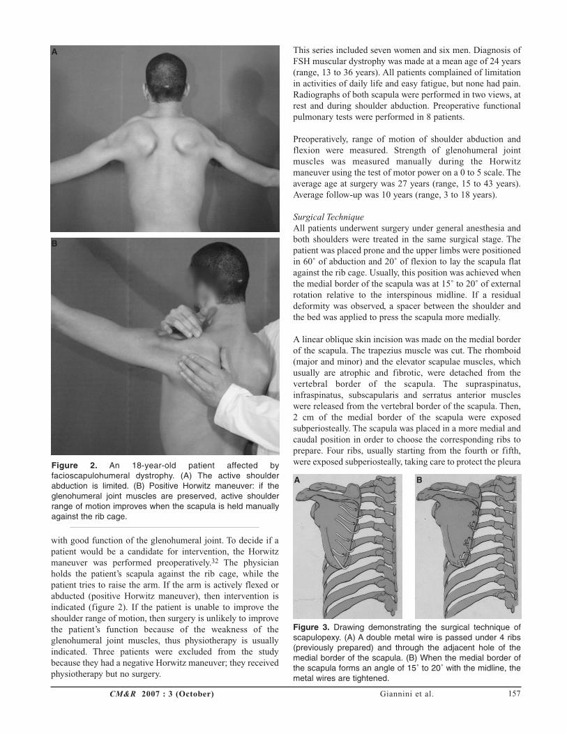
with good function of the glenohumeral joint. To decide if apatient would be a candidate for intervention, the Horwitzmaneuver was performed preoperatively.32 The physicianholds the patient’s scapula against the rib cage, while thepatient tries to raise the arm. If the arm is actively flexed orabducted (positive Horwitz maneuver), then intervention isindicated (figure 2). If the patient is unable to improve theshoulder range of motion, then surgery is unlikely to improvethe patient’s function because of the weakness of theglenohumeral joint muscles, thus physiotherapy is usuallyindicated. Three patients were excluded from the studybecause they had a negative Horwitz maneuver; they receivedphysiotherapy but no surgery.
This series included seven women and six men. Diagnosis ofFSH muscular dystrophy was made at a mean age of 24 years(range, 13 to 36 years). All patients complained of limitationin activities of daily life and easy fatigue, but none had pain.Radiographs of both scapula were performed in two views, atrest and during shoulder abduction. Preoperative functionalpulmonary tests were performed in 8 patients.
Preoperatively, range of motion of shoulder abduction andflexion were measured. Strength of glenohumeral jointmuscles was measured manually during the Horwitzmaneuver using the test of motor power on a 0 to 5 scale. Theaverage age at surgery was 27 years (range, 15 to 43 years).Average follow-up was 10 years (range, 3 to 18 years).
Surgical TechniqueAll patients underwent surgery under general anesthesia andboth shoulders were treated in the same surgical stage. Thepatient was placed prone and the upper limbs were positionedin 60˚ of abduction and 20˚ of flexion to lay the scapula flatagainst the rib cage. Usually, this position was achieved whenthe medial border of the scapula was at 15˚ to 20˚ of externalrotation relative to the interspinous midline. If a residualdeformity was observed, a spacer between the shoulder andthe bed was applied to press the scapula more medially.
A linear oblique skin incision was made on the medial borderof the scapula. The trapezius muscle was cut. The rhomboid(major and minor) and the elevator scapulae muscles, whichusually are atrophic and fibrotic, were detached from thevertebral border of the scapula. The supraspinatus,infraspinatus, subscapularis and serratus anterior muscleswere released from the vertebral border of the scapula. Then,2 cm of the medial border of the scapula were exposedsubperiosteally. The scapula was placed in a more medial andcaudal position in order to choose the corresponding ribs toprepare. Four ribs, usually starting from the fourth or fifth,were exposed subperiosteally, taking care to protect the pleura
Giannini et al.CM&R 2007 : 3 (October) 157
Figure 2. An 18-year-old patient affected byfacioscapulohumeral dystrophy. (A) The active shoulderabduction is limited. (B) Positive Horwitz maneuver: if theglenohumeral joint muscles are preserved, active shoulderrange of motion improves when the scapula is held manuallyagainst the rib cage.
A
B
Figure 3. Drawing demonstrating the surgical technique ofscapulopexy. (A) A double metal wire is passed under 4 ribs(previously prepared) and through the adjacent hole of themedial border of the scapula. (B) When the medial border ofthe scapula forms an angle of 15˚ to 20˚ with the midline, themetal wires are tightened.
A B

Fixation of Winged Scapula in Muscular Dystrophy
and the subcostal neurovascular bundles. A double metal wire(1 mm diameter) was passed under each rib. The scapula wasplaced over the rib cage to its definitive position and four 2.8 mm holes were drilled approximately 1.5 cm from themedial border of the scapula opposite the prepared ribs. Oneend of each wire was passed through the adjacent hole of themedial border of the scapula. With the scapula held in its finalposition, the metal wires were twisted and tightened untilbone-to-bone contact between the scapula and the ribs wasobtained. The wires were cut (figure 3). The muscles wereclosed over the posterior surface of the scapula and the woundclosed routinely. A postoperative radiograph was taken toassess the position of the metal wires and to assess forpneumothorax.
The postoperative management consisted of the application ofa “figure eight” dressing immediately after surgery (figure 4).This immobilization was maintained for 6 weeks.
Physiotherapy of the shoulder joint was initiated as soon astolerated and consisted of isometric deltoid exercises andactive exercises in order to strengthen the glenohumeral jointmuscles and regain as great a range of motion as possible.Patients were discharged from the hospital 4 to 7 dayspostoperatively.
All patients were followed monthly for 3 months and thenyearly. At the preoperative control, one year after surgery andat last available follow-up, shoulder range of motion inabduction and flexion was evaluated, and a test of motorpower was performed. At every follow-up appointment,radiographs of both scapulae were taken to compare theposition of the scapula on the rib cage and to check theposition and the condition of the metal wires. All patientsstated their subjective satisfaction, including functionalimprovement and cosmesis, according to a visual analogicalscale as very satisfied, satisfied, fairly satisfied or unsatisfied.
ResultsAll patients experienced improvement in active range ofmotion of the shoulder. Shoulder abduction improved from anaverage preoperative value of 70˚ (SD = 20) to 98˚ (SD = 18)at 1-year follow-up (figure 5A and 5B). Shoulder flexionimproved from an average preoperative value of 55˚ (SD =16) to 120˚ (SD = 33) at 1-year follow-up (figure 5C and 5D).At an average follow-up time of 10 years (range, 3 to 18years), shoulder abduction decreased to 90˚ (SD = 17) andshoulder flexion decreased to 105˚ (SD = 22) (table 1). Thetest of motor power showed that at 1-year follow-up musclestrength during shoulder abduction increased from 2.5preoperatively to 4.5. Muscle strength during shoulder flexionincreased from 2.4 preoperatively to 4.5.The test of motorpower performed at an average follow-up of 10 years (range,3 to 18 years) showed muscle strength decreased. The meanvalue of muscle strength was 4 during shoulder abduction and4.1 during shoulder flexion.
CM&R 2007 : 3 (October)158
Figure 4. A “figure-eight” dressing is applied postoperativelyand it is maintained for 6 weeks.
Table 1. Shoulder range of motion.
Flexion (degrees) Abduction (degrees)Patient Age at
Number/ Operation 1-year Last 1-year Last Follow-upGender (years) Preoperative follow-up follow-up Preoperative follow-up follow-up (years)
1/F 17 70 100 80 70 100 85 152/F 35 45 90 90 60 95 90 153/M 26 90 120 105 90 105 100 184/F 15 45 150 100 75 90 80 165/M 26 70 120 95 50 90 80 186/F 31 40 80 75 45 60 60 87/F 27 50 100 100 90 110 110 78/M 18 45 105 105 45 95 90 69/F 32 60 180 180 90 120 115 5
10/F* 30 45 130 120 60 110 100 511/M* 32 70 175 160 70 120 105 412/M* 43 40 120 90 65 85 70 413/M* 18 60 150 140 75 100 90 3
All data are shown in degrees.*Indicates patients not included in the previous analysis.

All patients were very satisfied with the cosmetic outcome.All patients had clinical improvement with completeresolution of the winged scapula and the shoulders regained anormal shape. Twelve patients stated they were very satisfiedwith the functional results and one patient was satisfied.
In all 26 cases of scapulopexy, a stable and long-lastingfixation of the scapula to the rib cage was achieved. Theposition of the scapula was maintained at the last availablefollow-up, as demonstrated in the radiographs (figure 6). Theeight patients who had lung function testing preoperatively,were retested at 1-year follow-up and showed no significantdifference of vital capacity.
There were no major surgical complications, except for onepatient with unilateral pneumothorax 1-day postoperativelythat resolved spontaneously. One patient presented one metalwire breakage at the radiographic routine control seven yearsafter surgery. The scapula remained well fixed to the thoraxand the patient did not complain of any symptoms, thus nointervention was performed. One patient fell accidentally
three years after surgery because of bilateral drop foot and shehad breakage of two metal wires. Functional and clinicalimprovement were maintained, but pleural pain led to metalwires removal. One patient had a high-impact car accidentseven months after surgery with breakage of all wires of theright scapula, representing winged scapula. Revision surgerywas performed with the same technique of scapulopexyresulting in very satisfactory clinical and functionalimprovement at 3-years follow-up.
DiscussionWinged scapula is the most common clinical presentation ofinstability of the scapulothoracic joint. Pathologicalconditions that can lead to winged scapula include FSHmuscular dystrophy,1-7 congenital elevation of the scapula(Sprengel’s disease), traumatic paralysis of the serratusanterior muscle, and polio.26,33
Patients affected by FSH muscular dystrophy have a normallife expectancy, so surgical treatment to fix the scapula to therib cage, improving function, is advisable.1,2,6,7
Giannini et al.CM&R 2007 : 3 (October) 159
Figure 5. A 30-year-old patient affected by facioscapulomuscular dystrophy. (A) Preoperative active abduction of the shoulders.(B) The patient had good cosmetic and functional results after bilateral scapulopexy. (C) The patient tries to forward flex thearms, but the movement is limited. (D) Improvement of active flexion after bilateral scapulopexy.
A B
C D

Fixation of Winged Scapula in Muscular Dystrophy
Scapulothoracic fixation can be useful only if theglenohumeral joint muscles are preserved. When the scapulais stabilized to the rib cage, the deltoid contraction is able toabduct and flex the shoulder. However, full shoulder range ofmotion is not possible because the fixed scapulothoracic jointcannot contribute to the movement.
Several methods of scapulothoracic fixation in FSH musculardystrophy have been described in the literature. Thesemethods include scapulothoracic fusion or arthrodesis(scapulodesis)12,13,17-27 and scapulothoracic fixation withoutarthrodesis (scapulopexy).14,28-31
Different techniques of scapulodesis have been reported usingtibial cortical and cancellous bone graft and screws,12 metalplates,26,27 screws and cancellous bone graft,25 and metalwires and bone graft,22 with good clinical and functionalresults which have been maintained over a long period oftime. However, being that ribs and scapula are flat bones, theyrender scapulodesis a demanding technique, causing manycomplications. In fact, rib fractures, scapula fractures, stressfractures, nonunions, mobilization of the fixation devices,wire breakings and neurologic complications such as brachialplexus palsy, pneumothoraces, hemothoraces, and atelectaseshave been very often reported.
Scapulopexy is a less rigid fixation than scapulodesis becauseit leads to scapulothoracic fixation without using bone graftor exposing cancellous bone of the ribs or scapula. There arefew reports concerning scapulopexy in FSH musculardystrophy in the literature. Rinaldi30 used strips of fascia lata,Ketenjian31 used fascia lata grafts, Mersilene or Dacronstrips, and Giannini et al14,29 used metal wires tied around theribs through holes drilled in the medial border of the scapula.Clinical and functional results of these studies weresatisfactory. Few complications were reported and includeddeterioration of fascial slings and wire breakings.
The scapulopexy we used by stabilizing the scapula to the ribcage allowed extensive bone-to-bone contact between the ribsand the scapula that was obtained by firm tightening of fourmetal wires. Between the scapula and the ribs, a fibrotic scarwas formed. This dense fibrotic tissue stabilizes thescapulothoracic joint, but allows micromotion that isresponsible for fatigue breakage of the metal wires.
All patients had considerable improvement of shoulder rangeof motion in abduction and flexion. The improvement inactive range of motion depends on the deltoid strength at thetime of surgery, which should be at least 4 or 5. They all hadan improvement in doing activities of daily life, did notcomplain of easy fatigue and were very satisfied with thecosmetic result. The scapulothoracic fixation persisted withtime, so the cosmetic results, and the minimized fatigue anddiscomfort were long-lasting. However, being that FSHmuscular dystrophy is a progressive myopathy, it can causevery slow deltoid weakening that may lead to a diminishingshoulder range of motion.
CM&R 2007 : 3 (October)160
Figure 6. (A) Preoperative radiograph, at rest, of the scapulaeof a patient affected by facioscapulohumeral dystrophy. Thescapula is internally rotated and its superior border tends tobecome vertical. (B) Preoperative radiograph of the scapulaeduring active shoulder abduction. The internal rotation of thescapula and the verticalization of its superior border becomemore evident. (C) The scapulae, fixed to the ribs by 4 doublemetal wires, remained in the correct position until last availablefollow-up.
A B
C

The complications strictly associated to the surgical techniqueused in this series were one pneumothorax, which resolvedspontaneously, and one wire breakage without trauma.Unfortunately, two patients experienced a trauma that, in onecase, was of low entity and led to wire breakage without lossof the scapulothoracic fixation. In the second case, a highimpact car accident caused monolateral breakage of all wiresand winged scapula, and therefore needed revision.
In order to obtain good functional results by scapulothoracicfixation, the position of the scapula to the rib cage is veryimportant. If the scapula is fixed too caudally, neurologicalcomplications such as stretching of the brachial plexus canoccur.34 A scapula fixed in an adducted position limits thefunction of the glenohumeral joint, while if it is fixed in anabducted position, the patient is able to move theglenohumeral joint through more functional range of motion.An excessively abducted or rotated scapula causes anabnormally high shoulder. Diab et al27 fixed the scapula withits vertebral border externally rotated at an angle of 25˚ to themidline. Krishnan et al26 reported that ideal positioning of thescapula is in the 20˚ to 25˚ external rotation relative to themidline. We obtained good functional results withoutneurological complications by fixing the scapula 15˚ to 20˚between its medial border and the interspinous line.
ConclusionWinging of the scapula in FSH muscular dystrophy is causedby weakness of the muscles that stabilize the scapula to the ribcage. Surgical operations aim to fix permanently the scapulato the ribs using different techniques and devices. Thebenefits from surgery have to be balanced against thepotential postoperative complications. The scapulopexy usedin this extended series of patients consisted of repositioningthe scapula and fixing it to four ribs by using metal wireswithout performing arthrodesis. All patients obtained a verysatisfactory cosmetic result, functional improvement inshoulder abduction and flexion, and stabilization of thescapulothoracic fixation over time. This technique has a smallrate of complications. It is reproducible, safe and effective,resulting in clinical and functional improvement.
AcknowledgmentsWe would like to thank the physiotherapist, Olivia Faldini, forher invaluable work in evaluating the patients at follow-upand assistance in writing the manuscript.
References1. Dubowitz V. The muscular dystrophies In: Muscle disorders in
childhood, 2nd ed. London: WB Saunders Company LTD;1995. 111-119.
2. Fitzsimons RB. Facioscapulohumeral muscular dystrophy. CurrOpin Neurol 1999;12:501-511.
3. Hughes MI, Hicks EM, Nevin NC, Patterson VH. Theprevalence of inherited neuromuscular disease in NorthernIreland. Neuromuscul Disord 1996;6:69-73.
4. Merlini L, Forst J. Surgical management of muscular dystrophy.In: Emery AE, ed. The muscular dystrophies. New York;Oxford University: 2001. 284-296.
5. Tawil R, Figlewicz DA, Griggs RC, Weiffenbach B.Facioscapulohumeral dystrophy: a distinct regional myopathywith a novel molecular pathogenesis. FSH Consortium. AnnNeurol 1998;43:279-282.
6. Kissel JT. Facioscapulohumeral dystrophy. Semin Neurol1999;19:35-43.
7. Tonini MM, Passos-Bueno MR, Cerqueira A, Pavanello R,Vainzof M, Dubowitz V, Zatz M.. Facioscapulohumeral(FSHD1) and other forms of muscular dystrophy in the samefamily: is there more in muscular dystrophy than meets theeye? Neuromuscul Disord 2002;12:554-557.
8. Nelson MR. Rehabilitation concerns in myopathies. In: BraddomRL, ed. Physical medicine and rehabilitation. Philadelphia,PA: W B Saunders Co; 1996. 1012-1013.
9. Bagg SD, Forrest WJ. A biomechanical analysis of scapularrotation during arm abduction in the scapular plane. Am JPhys Med Rehabil 1988;67:238-245.
10. Inman VT, Ralston HJ, Todd F. Human walking. Baltimore,MD: Williams & Wilkins; 1981. 96-97.
11. Siegel IM. Muscle and its desease: an outline primer of basicscience and clinical method. Chicago, IL: Year BookMedical; 1986. 252-254.
12. Copeland SA, Howard RC. Thoracoscapular fusion forfacioscapulohumeral dystrophy. J Bone Joint Surg Br1978;60-B:547-551.
13. Copeland SA, Levy O, Warner GC, Dodenhoff RM. Theshoulder in patients with muscular dystrophy. Clin OrthopRelat Res 1999;(368):80-91.
14. Giannini S, Ceccarelli F, Faldini C, Pagkrati S, Merlini L.Scapulopexy of winged scapula secondary tofacioscapulohumeral muscular dystrophy. Clin Orthop RelatRes 2006;449:288-294.
15. Barnett ND, Mander M, Peacock JC, Bushby K, Gardner-Medwin D, Johnson GR. Winging of the scapula: theunderlying biomechanics and an orthotic solution. Proc InstMech Eng [H] 1995;209:215-223.
16. Mummery CJ, Copeland SA, Rose MR. Scapular fixation inmuscular dystrophy. Cochrane Database Syst Rev2003;(3):CD003278.
17. Toni A, Merlini L, Sudanese A, Baldini N, Granata C. Thoraco-scapular arthrodesis in facioscapulohumeraldystrophy. Chir Organi Mov 1986;71:127-131.
18. Twyman RS, Harper GD, Edgar MA. Thoracoscapular fusion infacioscapulohumeral dystrophy: clinical review of a newsurgical method. J Shoulder Elbow Surg 1996;5:201-205.
19. Andrews CT, Taylor TC, Patterson VH. Scapulothoracicarthrodesis for patients with facioscapulohumeral musculardystrophy. Neuromuscul Disord 1998;8:580-584.
20. Berne D, Laude F, Laporte C, Fardeau M, Saillant G.Scapulothoracic arthrodesis in facioscapulohumeral musculardystrophy. Clin Orthop Relat Res 2003;(409):106-113.
21. Bunch WH. Scapulo-thoracic fusion. Minn Med 1973;56:391-394.
22. Bunch WH, Siegel IM. Scapulothoracic arthrodesis infacioscapulohumeral muscular dystrophy. Review ofseventeen procedures with three to twenty-one-year follow-up. J Bone Joint Surg Am 1993;75:372-376.
23. Howard RC. Thoraco-scapular arthrodesis. J Bone Joint Surg1961;43-B:175.
24. Jakab E, Gledhill RB. Simplified technique for scapulocostalfusion in facioscapulohumeral dystrophy. J Pediatr Orthop1993;13:749-751.
25. Letournel E, Fardeau M, Lytle JO, Serrault M, Gosselin RA.Scapulothoracic arthrodesis for patients who havefascioscapulohumeral muscular dystrophy. J Bone Joint SurgAm 1990;72:78-84.
Giannini et al.CM&R 2007 : 3 (October) 161

Fixation of Winged Scapula in Muscular Dystrophy
26. Krishnan SG, Hawkins RJ, Michelotti JD, Litchfield R, WillisRB, Kim YK. Scapulothoracic arthrodesis: indications,technique, and results. Clin Orthop Relat Res2005;(435):126-133.
27. Diab M, Darras BT, Shapiro F. Scapulothoracic fusion forfacioscapulohumeral muscular dystrophy. J Bone Joint SurgAm 2005;87:2267-2275.
28. Putti V. L’osteodesi interscapolare in un caso di miopatiaatrofica progressiva. Arch Ortop 1906;23:319-331.
29. Giannini S, Ceccarelli F, Coppola G. Fissazione chirurgicadella scapola nella distrofia facio-scapolo-omerale. Progressiin patologia vertebrale 1992;XIII:283-288.
30. Rinaldi F: Terapia chirurgica nella forma “facio-scapolo-omerale” della distrofia muscolare primitiva. ClinicaOrtopedica 1964;16:233-243.
31. Ketenjian AY. Scapulocostal stabilization for scapular wingingin facioscapulohumeral muscular dystrophy. J Bone JointSurg Am 1978;60:476-480.
32. Horwitz MT, Tocantins LM. Isolated paralysis of the serratusanterior (magnus) muscle. J Bone Joint Surg Am1938;20:720-725.
33. Whitman A. Congenital elevation of the scapula and paralysisof serratus magnus muscle. JAMA 1932;99:1332-1334.
34. Mackenzie WG, Riddle EC, Earley JL, Sawatzky BJ. Aneurovascular complication after scapulothoracic arthrodesis.Clin Orthop Relat Res 2003;(408):157-161.
Author AffiliationsSandro Giannini, MDDepartment of Orthopaedic SurgeryUniversity of Bologna, Istituto Ortopedico Rizzoli, ItalyVia G. Pupilli 1, 40136 Bologna, Italy
Cesare Faldini, MDDepartment of Orthopaedic SurgeryUniversity of Bologna, Istituto Ortopedico Rizzoli, ItalyVia G. Pupilli 1, 40136 Bologna, Italy
Stavroula Pagkrati, MDDepartment of Orthopaedic SurgeryUniversity of Bologna, Istituto Ortopedico Rizzoli, ItalyVia G. Pupilli 1, 40136 Bologna, Italy
Gianluca Grandi, MDDepartment of Orthopaedic SurgeryUniversity of Bologna, Istituto Ortopedico Rizzoli, ItalyVia G. Pupilli 1, 40136 Bologna, Italy
Vitantonio Digennaro, MDDepartment of Orthopaedic SurgeryUniversity of Bologna, Istituto Ortopedico Rizzoli, ItalyVia G. Pupilli 1, 40136 Bologna, Italy
Deianira Luciani, MDDepartment of Orthopaedic SurgeryUniversity of Bologna, Istituto Ortopedico Rizzoli, ItalyVia G. Pupilli 1, 40136 Bologna, Italy
Luciano Merlini, MDMuscle Unit, Division of Medical GeneticsDepartment of Experimental and Diagnostic MedicineUniversity of Ferrara, Italy
CM&R 2007 : 3 (October)162


