
Foundations in
Microbiology Seventh Edition
Chapter 17
Diagnosing Infections
Lecture PowerPoint to accompany
Talaro
Copyright © The McGraw-Hill Companies, Inc. Permission required for reproduction or display.

17.1 Survey of Microbial Disease
Methods of identifying unknown microbes
fall into three categories:
1. Phenotypic – observable microscopic
and macroscopic characteristics
2. Genotypic – genetic make up
3. Immunological – serology; antibody-
antigen reactions

Phenotypic Methods
• Microscopic morphology – fresh or stained microorganisms from specimen; shape, size, stain reaction, cell structures
• Macroscopic morphology – colony appearance; texture, size, shape, pigment, growth requirements
• Physiological/biochemical characteristics – detection of presence or absence of particular enzymes or metabolic pathways
• Chemical analysis – analyze specific chemical composition; cell wall peptides, cell membrane lipids

Genotypic Methods
• Assess genetic make-up
• Culture is not necessary
• Precise, automated methods, quick results

Immunological Methods
• Specific antibodies are used to detect
antigens
– Easier than testing for the microbe itself

17.2 Specimen Collection and
Laboratory Methods
• Sampling body sites or fluids for
suspected infectious agent
• Results depend on specimen collection,
handling, transport, and storage
• Aseptic procedures should be used

Figure 17.1 Sampling sites


Overview of
Laboratory Techniques • Routes taken in specimen analysis
– Direct tests (microscopic, immunologic, or
genetic)
– Cultivation, isolation, and identification
(general and specific tests)
• Two categories of results
– Presumptive
– Confirmatory

Figure 17.2 Scheme of specimen isolation and
identification

17.3 Phenotypic Methods
• Immediate direct examination • Microscopic – differential and special stains – Gram,
DFA, direct antigen testing

Phenotypic Methods
• Cultivation of Specimen
– Colony appearance, growth requirements,
appropriate media
• Biochemical testing
– Physiological reactions to nutrients as
evidence of the absence or presence of
enzymes
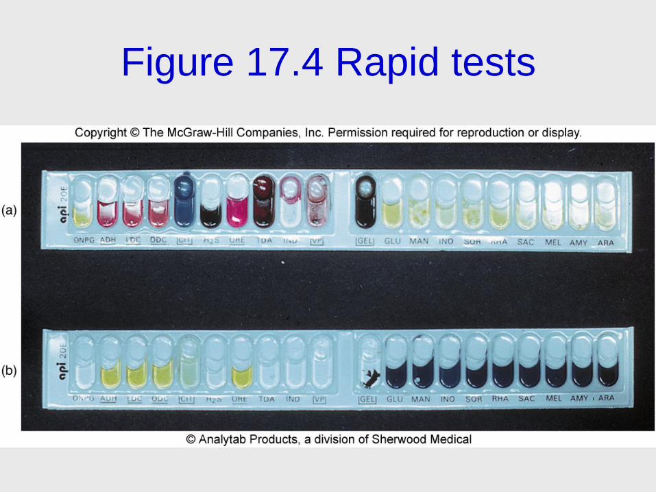
Figure 17.4 Rapid tests

Figure 17.5

Phenotypic Methods
• Miscellaneous tests
– Phage typing
– Animal inoculation
– Antimicrobial sensitivity

• Important to consider whether microbe
recovered and identified is actually
causing the disease or simply normal flora
17.4 Genotypic Methods
• DNA analysis
– Hybridization
• Probes complementary to the specific sequences
of a particular microbe
– PCR
• DNA and RNA analysis
• Ribosomal RNA
– Comparison of the sequence of nitrogen bases in
ribosomal RNA

17.5 Immunological Methods
• Serology – in vitro diagnostic testing of
serum
– Antibodies have extreme specificity for
antigens
• Visible reactions include precipitates, color
changes, or the release of radioactivity
• Tests can be used to identify and to
determine the amount of antibody in
serum – titer

Figure 17.7 Basic principles of serological testing using antibodies and antigens

Figure 17.8 Specificity and sensitivity in
immune testing

Agglutination and Precipitation
Reactions
• Agglutination testing – antibody cross links whole-cell antigens, forming complexes that settle out and form visible clumps – Blood typing, some bacterial and viral diseases
• Precipitation tests – soluble antigen is made insoluble by an antibody – VDRL
– Most precipitation reactions are carried out in agar gels

Insert figure 17.10 Cellular\molecular view
Figure 17.9 Agglutination and precipitation reactions

Figure 17.10 Precipitation reactions

Figure 17.11 Immunoelectrophoresis of normal human serum

The Western Blot for
Detecting Proteins
• Electrophoretic separation of proteins, followed by an immunoassay to detect these proteins
• Highly specific and sensitive
• Sites of specific antibody binding will appear as a pattern of bands
• Second test used to verify HIV status
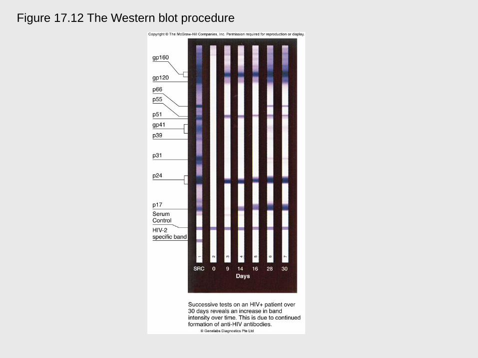
Figure 17.12 The Western blot procedure
Complement Fixation
• Lysin mediated hemolysis
• Test uses four components
– Antigen, antibody, complement, and sensitized
sheep RBCs
• Steps of test
1. Test antigen reacts with test antibody
2. Contents of tube from (1.) are mixed with
sheep RBCs
• Complement used up in first stage, no hemolysis
• Unfixed complement, hemolysis

Figure 17.13 Complement fixation test

Fluorescent Antibody and
Immunofluorescent Testing • Fluorescent antibody
– Monoclonal antibody labeled by a fluorescent
dye
• Two ways FABs are used
– Direct testing
– Indirect testing

Figure 17.14 (a, b)

Figure 17.14 (c)

Immunoassays • Extremely sensitive to detect trace antigens
and antibodies
• Radioimmunoassay (RIA) – antigens or
antibodies labeled with radioactive isotopes
• Enzyme-linked immunosorbent assay
(ELISA) – enzyme-antibody complex
produces a colored product when an
enzyme-substrate reaction occurs – Indirect
– Capture

Figure 17.15 Methods of ELISA testing

Tests that Differentiate
T Cells and B Cells • Rosette formation
– Mix T cells with sheep red blood cells
• Fluorescent techniques
– Differentiates T and B cells and subgroups
them

In vivo Testing
• Antigens are introduced directly into the
body to determine the presence or
absence of antibodies
– Tuberculin skin test, allergy testing

A Viral Example
• Viruses present special difficulties
because they are not cells
• Viruses are labor intensive to culture in the
laboratory

Figure 17.17 Diagnosing viral infections


