1 There is no universal agreement as to whether the separation of humans and their kin from
great apes and their kin should be recognised at Family level (Hominidae) or Tribe level
(Hominini) or Sub-Tribe level (Hominina). Contributors to this volume have agreed, while
choosing their own term, to recognize that disagreement by inserting this footnote.
Functional Anatomy, Biomechanical Performance Capabilities and Potential Niche of 1
StW 573: an Australopithecus Skeleton (circa 3.67 Ma) From Sterkfontein Member 2, 2
and its significance for The Last Common Ancestor of the African Apes and for Hominin 3
Origins 4
Authors: 5
Robin Huw Cromptona,b
*, Juliet McClymontc, Susannah Thorpe
d, William Sellers
e, Jason 6
Heatonf,g
, Travis Rayne Pickeringg,h
, Todd Patakyi , Dominic Stratford
j, Kristian Carlson
g,k, 7
Tea Jashashvilig, l, m
, Amélie Beaudetj,n
, Laurent Bruxelles,j,o,p
Colleen Gohq, Kathleen 8
Kumanj, Ronald Clarke
g 9
10
* corresponding author. 11
12
Affiliations: 13
a Department of Musculoskeletal Biology, Institute of Ageing and Chronic Disease, 14
University of Liverpool, The William Henry Duncan Building, 6 West Derby Street, Liverpool 15
L7 8TX, email: [email protected]* 16
b Department of Rheumatology, Aintree University NHS Trust, Liverpool L9 7AL, UK 17
c School of Health Sciences, Centre for Health Research, Aldro Building, University of 18
Brighton, Eastbourne, United Kingdom, BN207UR 19
d School of Biosciences, University of Birmingham, Edgbaston, Birmingham, United 20
Kingdom. 21
e School of Earth and Environmental Science, University of Manchester, Manchester, United 22
Kingdom. 23
f Department of Biology, Birmingham-Southern College, Birmingham, Alabama, 35254, 24
U.S.A. 25
g Evolutionary Studies Institute, University of the Witwatersrand, Johannesburg, South 26
Africa, WITS 2050. 27
was not certified by peer review) is the author/funder. All rights reserved. No reuse allowed without permission. The copyright holder for this preprint (whichthis version posted November 29, 2018. ; https://doi.org/10.1101/481556doi: bioRxiv preprint

2
h Department of Anthropology, University of Wisconsin Madison, 1180 Observatory Drive, 28
Madison, Wisconsin, 53706, U.S.A. 29
i Department of Human and Health Sciences, Kyoto University, Kyoto, Japan 30
j School of Geography, Archaeology and Environmental Studies, University of 31
Witwatersrand, WITS 2050, Johannesburg, South Africa 32
k Department of Integrative Anatomical Sciences,
Keck School of Medicine, University of 33
Southern California, Los Angeles, California 90033, U.S.A. 34
l Molecular Imaging Center, Department of Radiology, Keck School of Medicine, University 35
of Southern California, Los Angeles, California 90033, U.S.A. 36
m Department of Geology and Paleontology, Georgian National Museum, Tbilisi 0105, 37
Georgia 38
n Department of Anatomy, University of Pretoria, PO Box 2034, Pretoria 0001, South Africa 39
o French National Institute for Preventive Archaeological Research (INRAP), Nîmes, France 40
p French Institute of South Africa (IFAS), USR 3336 CNRS, Johannesburg 2001, South Africa 41
q Warwick Medical School, Coventry CV4 UK 42
43
*Corresponding author. 44
E-mail address: [email protected] 45
46
Keywords: Australopithecus; biomechanics; ecomorphology; ‘Little Foot’; locomotion; 47
plasticity; Sterkfontein 48
49
Abstract (300 words) 50
51
StW 573, from Sterkfontein Member 2, dated ca 3.67 Ma, is by far the most complete 52
skeleton of an australopith to date. Joint morphology is in many cases closely matched in 53
available elements of Australopithecus anamensis (eg. proximal and distal tibial and humeral 54
joint-surfaces) and there are also close similarities to features of the scapula, in particular , of 55
KSD-VP-1/1 A. afarensis from Woranso-Mille. The closest similarities are, however, to the 56
partial skeleton of StW 431 from Sterkfontein Member 4. When considered together, both 57
StW 573 and StW 431 express an hip joint morphology quite distinct from that of A. 58
africanus Sts14, and a proximal femur of a presumed A. africanus from Jacovec Cavern at 59
Sterkfontein, StW 598. This, and other evidence presented herein, suggests there are two 60
pelvic girdle morphs at Sterkfontein, supporting Clarke (2013) in his recognition of a second 61
was not certified by peer review) is the author/funder. All rights reserved. No reuse allowed without permission. The copyright holder for this preprint (whichthis version posted November 29, 2018. ; https://doi.org/10.1101/481556doi: bioRxiv preprint
3
species, A. prometheus, containing StW 573 and StW 431. StW 573 is the first hominid 62
skeleton where limb proportions are known unequivocally. It demonstrates that some early 63
hominins, at the time of formation of the Laetoli footprints (3.6 Ma), were large-bodied. with 64
hindlimbs longer than forelimbs. Modelling studies on extant primates indicate that the 65
intermembral index (IMI) of StW 573, low for a non-human great ape, would have 66
substantially enhanced economy of bipedal walking over medium-to-long distances, but that 67
it was still too high for effective walking while load-carrying. It would, however, have 68
somewhat reduced the economy of horizontal climbing, but made Gorilla-like embracing of 69
large tree-trunks less possible. Consideration of both ethnographic evidence from modern 70
indigenous arboreal foragers and modern degeneracy theory cautions against prescriptive 71
interpretations of hand- and foot-function, by confirming that both human-like upright 72
bipedalism and functional capabilities of the hand and foot can be effective in short-distance 73
arboreal locomotion. 74
75
1. Introduction 76
77
While it is now largely accepted that there was no phase of terrestrial knucklewalking in 78
hominin evolution (see eg., Dainton and Macho, 1999; Dainton, 2001; Clarke, 2002; Kivell 79
and Schmitt, 2009), and that australopiths show adaptations to both terrestrial bipedalism and 80
arboreal locomotion, there is still no firm consensus on whether the ‘arboreal’ features of 81
australopith postcrania would have been the subject of positive selection or were selectively 82
neutral anachronisms (Ward 2002, 2013). The view that the two activities must be 83
substantially mechanically incompatible is still current (see eg., Kappelman et al., 2016). The 84
two alternative paradigms date from extended debates (see eg., Latimer, 1991 versus Stern 85
and Susman, 1991) concerning the significance of the AL-288-1 ‘Lucy’ skeleton of 86
Australopithecus afarensis in 1974. Although some one-third complete, this partial skeleton, 87
and other more recently discovered partial australopith skeletons (eg., the Woranso-Mille 88
Australopithecus afarensis skeleton, KSD-VP-1/1 ca. 3.6 Ma [Haile-Selassie et al. 2010]) and 89
the Malapa A. sediba skeletons, MH-1 and MH-2, ca. 1.977 Ma [Berger, 2013]) are too 90
incomplete to provide reliable upper and lower limb lengths, a crucial variable in assessing 91
terrestrial and arboreal locomotor performance capabilities, and lack other auxiliary features 92
which might offer a clear signal of the existence of selection for arboreal performance 93
capabilities. 94
95
was not certified by peer review) is the author/funder. All rights reserved. No reuse allowed without permission. The copyright holder for this preprint (whichthis version posted November 29, 2018. ; https://doi.org/10.1101/481556doi: bioRxiv preprint

4
It has become increasingly clear, since the discovery of the foot bones of StW 573 96
(‘Little Foot’) some 23 years ago (Clarke and Tobias, 1995), that the continued painstaking 97
freeing of Little Foot’s fragile bones from their matrix of hard breccia might for the first time 98
provide unequivocal data on australopith postcranial anatomy. Some missing small skeletal 99
elements may be recovered with further excavation, and some (such as the distal foot) were 100
destroyed by lime mining a very long time ago, but at least 90% of the skeleton, complete 101
enough for unequivocal knowledge of limb lengths, and with remarkably good preservation 102
of joint surfaces and other vital detail has now been excavated and prepared (Clarke 2018, 103
Figure 26). This completeness is unmatched until Homo ergaster KNM-WT 15000, at 1.5 104
Ma. After much debate (see Bruxelles et al. 2018, submitted), the age of this specimen is now 105
confidently set at ca 3.67 Ma, very close to the age of the Laetoli footprint trails (Leakey and 106
Hay, 1979), which were previously our best source of information on the locomotor 107
capabilities of australopiths. Now, StW 573 for the first time offers unequivocal information 108
on limb proportions of forelimb and hindlimb. 109
110
Here we review aspects of contributions on StW 573 which bear on its potential niche 111
as an individual. These aspects include the geological and palaeoenvironmental context 112
(Bruxelles et al. 2018, submitted), its craniodental anatomy (Clarke and Kuman, 2018, 113
submitted), the endocast (Beaudet et al., 2018a, in press), the inner ear (Beaudet et al. 2018b, 114
submitted), the scapula and clavicle (Carlson et al., 2018, in prep.), the hand (Jashashvili et 115
al., 2018 in prep.) and the foot (Deloison, 2004). Heaton et al. (2018, submitted) focus on 116
statistical/morphometric descriptions of the longbones of StW 573 and comparisons with 117
other species, and this paper is referred to where appropriate to avoid duplication. However, 118
the present paper is quite distinct in its focus on: a) comparisons of those aspects of joint 119
shape which in the literature are generally regarded as informative concerning locomotor 120
function , and b) reconstruction of StW 573’s potential ecological niche by proxy 121
experiments and extant in-silico modelling. We adopt the hypothesis-based approach of 122
Wainwright (1991) to species ecomorphology, which focuses on individual performance (and 123
in this respect differs considerably from Bock and Von Wahlert’s (eg. 1965) previous 124
formulation which includes form, function and biological role but not performance, and is not 125
therefore suited to experimental testing). This paper, and Heaton et al. (2018, submitted) are 126
thus distinct in aims and methods, and while they refer to each other where appropriate and 127
may indeed to be read in tandem as complementary/companion papers, they can equally 128
was not certified by peer review) is the author/funder. All rights reserved. No reuse allowed without permission. The copyright holder for this preprint (whichthis version posted November 29, 2018. ; https://doi.org/10.1101/481556doi: bioRxiv preprint

5
stand, and can be assessed, quite independently. In addition, we pay close attention to the 129
significance of modern ecological dynamics theory, particularly plasticity (see eg. Neufuss et 130
al. 2014); and neurobiological degeneracy (see eg. Edelman and Gally, 2001), for the 131
cheiridia (hands and feet) in particular, which suggest that circumspection needs to be applied 132
to functional interpretation of foot (see eg. Deloison, 2004) and hand morphology. 133
134
Further, euhominoids (otherwise known as crown hominoids, so excluding eg. 135
Proconsulidae, Pliopithecidae etc) display high levels of plasticity in muscle architecture: we 136
have already noted that while DeSilva (2009) asserted that humans could not achieve the 137
required dorsiflexion for chimpanzee-like vertical climbing, Venkataraman et al. (2013) 138
showed that (presumably developmental) fibre-length plasticity enables some forest hunter-139
gatherers to do so, while neighbouring non-climbing populations cannot. Further, Neufuss et 140
al. (2014) showed that while lemurs, like all primarily pronograde mammals studied to date, 141
exhibit a dichotomy in axial musculature between deep slow contracting local stabilizer 142
muscles and superficial fast contracting global mobilizers and stabilizers, hominoids, as 143
previously shown for Homo, show no regionalization. Thus, it appears that hominoids have 144
been under selective pressure to develop and sustain high functional versatility of the axial 145
musculature, reflecting a wide range of mechanical demands on the trunk in orthogrady. 146
Neufuss et al. (2014). Using this analytical framework, focusing throughout on StW 573’s 147
individual locomotor performance capabilities, we hope to advance understanding of her 148
biomechanical interaction with her environment and potential niche: her ecomorphology 149
sensu Wainwright (1991). 150
151
152
153
Background 154
155
2.1. The Sterkfontein Formation 156
157
The Sterkfontein Formation is the world’s longest succession documenting the 158
evolution of hominins, fauna, landscape and ecology. Member 5 holds Homo habilis and 159
Homo ergaster, Paranthropus, and evidence for over half a million years’ evolution of early 160
stone tool technologies. Member 4, dating to between at least 2.8 Ma. and 2.15 Ma, is the 161
was not certified by peer review) is the author/funder. All rights reserved. No reuse allowed without permission. The copyright holder for this preprint (whichthis version posted November 29, 2018. ; https://doi.org/10.1101/481556doi: bioRxiv preprint

6
richest Australopithecus-bearing deposit in the world and contains two species of 162
Australopithecus, together with diverse and extensive faunal evidence. Four metres below, 163
under the yet unexplored and extensive Member 3, which potentially documents a long 164
period of evolution of Australopithecus, lies Member 2, which has yielded the world’s most 165
complete Australopithecus skeleton, StW 573, the only hominin found in this deposit. 166
Member 2 formed around 3.67 Ma (Granger et al. 2015) based on isochron burial dating 167
using cosmogenic aluminium-26 and beryllium-10. The detailed descriptions of the Member 168
2 sedimentary units by Bruxelles et al. (2014, 2018 submitted) confirm the 3.67 Ma age of 169
the skeleton published by Granger et al. (2015). Pickering and colleagues’ recent (2018) 170
repetition of a 2.8 Ma date fails to take into account, and fails to cite, Bruxelles and 171
colleagues’ (2014) demonstration that the flowstones are intrusive and dates for StW 573 172
based on them thus invalid. 173
174
Importantly, taphonomic evidence (Clarke 2018 submitted; Bruxelles et al. 2018 175
submitted) indicates that StW 573 died and was fossilized (below the ecological context in 176
which she lived), from a fall into a steep cave shaft leading to an underground cavern. The 177
skeleton is associated, in Member 2, with fauna dominated by cercopithecoids and carnivores 178
(see section 2.4). 179
180
181
2.2 The StW 573 partial skeleton 182
183
The skeletal elements found to date are shown in assembly in Figure 26 of Clarke 184
(2018, submitted). The skeleton offers, for the first time in one individual Australopithecus, 185
complete (if deformed) skull and mandible, many vertebrae and ribs, a crushed pelvis and 186
ischiopubic ramus, femora (broken but with overlapping morphology allowing confident 187
length reconstruction), one intact and one slightly damaged but measurable tibia, partial left 188
and right fibulae which overlap sufficiently to be sure of length and morphology, a partial 189
foot (representing primarily the medial column) foot, two scapulae (one articulated with the 190
upper limb), both claviculae (one partial and one complete), both humeri (one partially 191
crushed), both radii and ulnae (one side near-intact and the other crushed and deformed most 192
probably by a badly healed injury in-vivo), and finally one partial and one virtually complete 193
hand (missing only one distal phalanx). 194
195
was not certified by peer review) is the author/funder. All rights reserved. No reuse allowed without permission. The copyright holder for this preprint (whichthis version posted November 29, 2018. ; https://doi.org/10.1101/481556doi: bioRxiv preprint

7
Their taphonomy and condition are discussed in detail in Clarke (2018, submitted) 196
and the stratigraphic context in Bruxelles et al. (2018, submitted). StW 573’s pelvis was 197
substantially flattened post-mortem, but preservation of its margins is good enough to 198
identify an obtuse greater sciatic notch angle (Figure 1 top) and hence suggest female sex. In 199
contrast Figure 1 bottom shows the original reconstruction of the pelvis of StW 431 from 200
Member 4, which appears closely similar but is indicative of a male from the acuteness of the 201
greater sciatic notch. Lipping of the margins of the vertebral bodies of StW 573 (Figure 1, 202
top) and heavy toothwear (Clarke et al., 2018, submitted), indicate that she was an old 203
individual. StW 573 would have been some 130 cm in stature (RJC pers.comm. to RHC), 204
which is some 10 cm. less than the average for modern Bolivian women, the world’s shortest 205
female population. By contrast, the stature of AL-288-1 would have been some 107 cm 206
(Jungers, 1988). The considerable difference in stature is in accord with conclusions from the 207
dimensions of the penecontemporaneous Laetoli footprints (both Deloison [1993, pp. 624-208
629], for Laetoli G and Masao et al. [2016] for the more extensive Laetoli S) that there was a 209
large range in stature in early hominins. The slightly younger KSD-VP-1/1 partial skeleton 210
confirms this conclusion for A. afarensis, to which it is referred. 211
212
2.3. Environmental Context 213
214
While based on the bovids, Vrba (1975) suggested a medium density woodland with a 215
substantial open component, and based on the overall community of mammals, Reed (1997) 216
similarly suggested open woodland with bush, However, most of the Sterkfontein Member 2 217
fauna represent cercopithecoid or carnivore taxa which are today habitual climbers, and 218
ancient gravels indicate a large, slow flowing river in the base of the valley (Pickering et al., 219
2004). Similarly, Elton et al. (2016) indicate that the cercopithecoids which are found in 220
Member 2 were probably to some extent ecologically dependent upon trees for foraging, 221
predator avoidance, or both. Thus, Pickering et al., (2004).suggest a paleohabitat of rocky 222
hills covered in brush and scrub, but valley bottoms with riverine forest, swamp and standing 223
water. Such a paleoenvironment might resemble that in today’s Odzala-Koukoua National 224
Park, Congo, where grassland, standing water and forest are interspersed (see eg., 225
https://reefandrainforest.co.uk/news-item/trip-report-wildlife-republic-congo. Member 4 226
preserves a takin-like (and hence presumably also woodland) bovid Makapania, as well as 227
large cercopithecoids (Pickering et al. 2004), which are associated with forest vines requiring 228
large trees (Bamford, 1999), including one today known exclusively from central and 229
was not certified by peer review) is the author/funder. All rights reserved. No reuse allowed without permission. The copyright holder for this preprint (whichthis version posted November 29, 2018. ; https://doi.org/10.1101/481556doi: bioRxiv preprint

8
Western African tropical forest. This evidence suggests that little dessication occurred until 230
Member 5 times. A carbon isotope study of faunal teeth by Luyt and Lee Thorpe (2003) also 231
confirms that a drier, more open environment was only established by 1.7 Ma at Sterkfontein. 232
233
234
2.4. Species affinities 235
236
There has been a long debate concerning the number of species within 237
Australopithecus in South Africa, and particularly with relationship to the validity of 238
Australopithecus prometheus. Grine (2013) feels that craniodental and some ancillary 239
paleoenvironmental data are insufficient to justify splitting A. africanus, but Clarke (2013) 240
presents craniodental and also postcranial evidence for species diversity at Sterkfontein. Of 241
particular relevance here, he finds a distinction between A. prometheus, represented by, for 242
example, the partial skeleton of StW 431 and the near-complete StW 573 skeleton and A. 243
africanus , represented by Sts 14. Most of these discussions have focused on dental and 244
gnathocranial evidence, but recent postcranial discoveries of postcrania have broadened the 245
evidence base considerably, with particular attention now being paid to the pelvis and hip 246
joint, which are crucial to both obstetric and locomotor evolution. 247
248
Thus, Figure 2 shows that the ilia of the Member 4 StW 431 and the Member 2 StW 249
573 are closely similar in size and shape, even though the greater sciatic notches indicate that 250
StW 573, with an obtuse notch, is most likely a female, and StW 431, with an acute notch, is 251
apparently a male. By contrast, Figure 3 shows that the ilia of Sts14 and A. afarensis AL-288-252
1 -- from their obtuse greater sciatic notches both apparent females – are substantially 253
smaller. While Toussaint et al. (2003), who described StW 431, refer it to A. africanus, co-254
author Macho prefers attribution to A. prometheus (G.A.M. pers. comm. to RHC). 255
Importantly, both the unreconstructed pelvis described by Toussaint et al. (2003) and the 256
Kibii and Clarke (2003) reconstruction show that the acetabular margin is well preserved, and 257
acetabular size well defined. Toussaint et al. (2003) note: ‘The acetabulum is clearly large: to 258
judge by the preserved part, its vertical diameter would exceed 42 mm compared with 29.2 259
mm in Sts 14’ (page 219). 260
261
Acetabular size is also large in StW 573, probably over 36 mm (Figure 4), although 262
the acetabulum has been compressed by taphonomic events. Figures 5 and 6 show that not 263
was not certified by peer review) is the author/funder. All rights reserved. No reuse allowed without permission. The copyright holder for this preprint (whichthis version posted November 29, 2018. ; https://doi.org/10.1101/481556doi: bioRxiv preprint

9
only is the acetabulum large, but the StW 573 femoral head is a close match for the 264
acetabulum of StW 431, while the head of the A. africanus proximal femur from Jacovec 265
Cavern StW 598 is markedly smaller than the acetabulum of StW 431 or StW 573. The 266
Jacovec Cavern proximal femur (StW 598) is, however, a good match for the acetabulum of 267
Sts 14 (Figure 7 top left). That this reflects more than allometry is shown by the fact that the 268
femur of A. afarensis also has a small head but lacks an obviously long femoral neck. We 269
predict that other isolated material referable to A. africanus will similarly be found to have a 270
long femoral neck. The StW 367 femur from Member 4 Sterkfontein shows a remarkable 271
similarity to that of Jacovec StW 598 (Figure 7, top right). Thus both the small-bodied, long 272
femoral neck/small femoral head morph (eg. Jacovec, StW 367) and the large-bodied, short 273
femoral neck but large femoral head large hip joint morph (StW 431, StW 573) were present 274
in both Member 2 and Member 4 times. The StW 573 femur resembles those of both humans 275
and KNM WT 15000 (Figure 7, bottom) (and see Heaton et al. 2018, submitted, for 276
morphometric detail). A large femoral head is commonly, and reasonably, associated with 277
large forces operating across the hip joint and may be expected to correlate with body size. 278
Femoral neck length, however, is likely related to the moment arm of the hip abductors (see 279
eg. McHenry, 1975), but in what way, and with what iliac geometries, remains to be tested. It 280
should be noted that a long femoral neck can prima facie be assumed to increase the risk of 281
femoral neck fracture during instability events or falls, as it will increase the moment arm 282
about the femoral neck from the impact or instability site. Stern (2000) is cited by Toussaint 283
et al. (2003) in reference to the possibility that A. afarensis and A. africanus may have had a 284
less effective abduction capacity in gluteus medius ‘thus compromising stabilization during 285
walking’ (page 222). This could readily, and will be, tested in silico. 286
287
Fornai et al. (2018) recently reported that StW 431 also differs markedly from Sts 14 288
in sacral shape, and, like ourselves, they suggest that functional morphs exist in South 289
African Australopithecus. In this respect, it is interesting that Toussaint et al. (2003) noted 290
very different body mass estimates for StW 431-- 42.5 kg using a hominoid RMA regression 291
line, and 41.1 kg using a human regression line, but only 33.4 kg and 22.6 kg respectively on 292
the basis of the lumbosacral region. There are clearly major biomechanical distinctions in the 293
lumbosacral and hip regions, key for the effectiveness of upright walking, between 294
universally recognised A. africanus (eg., Sts 14) and both StW 573 and StW 431. Not all of 295
them can be put down to a simple relationship to body size – although body size itself is a 296
major difference. Since a palaeospecies is identified by morphological (and one would hope, 297
was not certified by peer review) is the author/funder. All rights reserved. No reuse allowed without permission. The copyright holder for this preprint (whichthis version posted November 29, 2018. ; https://doi.org/10.1101/481556doi: bioRxiv preprint
10
functional) distance, we argue that the balance of evidence is now strongly in favor of broad 298
recognition of A. prometheus as a species distinct from A. africanus. 299
300
301
302
2.5 Cranial and Dental Anatomy and Diet 303
304
An overall cranial shape similarity is evident with the Bouri Hata hominin, ca. 2.5 305
Ma, (Asfaw et al. 1999), but some aspects of cranial morphology suggest to Clarke and 306
Kuman (2018, submitted) that an ancestral relationship of StW 573 to Paranthropus may be 307
possible. Beaudet et al. (2018a, in press) conclude from the remarkably well preserved 308
endocast that the brain was small (perhaps surprisingly so) and undistinguished from that of 309
other non-human great apes (NHGAs). 310
Although it will take some time for the mandible to be detached safely from the 311
cranium, microCT scanning has revealed that wear distribution as well as dental arcade shape 312
resemble those of Kanapoi A. anamensis , 4.17-4.12 Ma., where Ward et al. (2001, p. 351) 313
found that the ‘teeth exhibit a distinctive pattern of wear. Evident in older individuals, the 314
anterior teeth are worn very heavily, much more so than the molars and premolars.’ Ward et 315
al. (2001) cite evidence that A. anamensis was taking a tough C4 diet, which might suggest 316
open environments. But faunal analysis suggested to Reed (1997) that Kanapoi 317
paleoenvironments at the time of A. anamensis were closed woodland. However, 318
Behrensmeyer and Reed (2013) note that other evidence, including stable isotopes, possibly 319
non-arboreal monkeys and micromammals, and characteristics of paleosols, suggest that open 320
habitats also existed. Similarly, Cerling et al. (2013) found that (δ13
C) stable isotopes in 321
dental enamel of A. anamensis suggest a C3-dominated diet (leaves and fruits from trees and 322
shrubs, etc.). Further, comparative evidence from extant colobines (Koyabu and Endo, 2010) 323
indicates that similar wear distribution may result from consumption of tough-skinned 324
arboreal fruit. Of course consumption of tough-coated arboreal fruit and consumption of 325
tough-coated terrestrial resources (such as corms and tubers) are not mutually exclusive. The 326
Woranso-Mille hominin KSD-VP-1/1, now dated to some 3.6 Ma (Haile-Selassie, 2016) and 327
attributed to A. afarensis, also appears from faunal evidence to have occupied a primarily 328
wooded environment (Su, 2016), with browsers dominant and grazers a relatively small 329
component, but with some aquatic species, such as crocodiles and an otter, Torolutra, 330
was not certified by peer review) is the author/funder. All rights reserved. No reuse allowed without permission. The copyright holder for this preprint (whichthis version posted November 29, 2018. ; https://doi.org/10.1101/481556doi: bioRxiv preprint

11
suggesting that the locality samples a riverbank community. δ13C determinations from A. 331
afarensis at Woranso-Mille suggest a balance of C3 and C4 items in diet (Wynn et al., 2013; 332
Levin et al., 2015), but microwear (Ungar et al. 2010) closely resembles that in A. 333
anamensis. 334
335
336
3. Functional Interpretation 337
338
As noted above, morphometric and general anatomical descriptions of StW 573 long bones 339
are provided by Heaton et al. (2018, submitted); of the scapula by Carlson et al. (2018, in 340
prep.), of the hand by Jashashvili et al. (2018, in prep.), and the foot by Deloison (2004). 341
Detailed descriptions and morphometrics should be sought therein, as here we restrict our 342
attention to the significance of the postcranial anatomy of StW 573 for locomotor ecology of 343
early hominins. We focus our comparative attention primarily on A. anamensis from Kanapoi 344
and KSD-VP-1/1, as they bracket StW 573 in time and are of similar size. StW 431 is 345
younger than StW 573, but we regard this specimen as conspecific. Some comparisons will 346
be made to Sts 14 and AL-288-1, but these specimens are considerably smaller and they are 347
more likely to be adaptively different, quite possibly being (in the case of Sts 14 at least) 348
more arboreal. We do not refer extensively to the considerably later A. sediba. As noted by 349
Lovejoy et al. (2016), its forelimb seems curiously derived towards some kind of suspensory 350
locomotion and/or feeding, and its hindlimbs are not reliably reconstructed, the lower limb 351
length having been assumed by Berger (eg. 2013) to equal that of proximal and distal 352
fragments plus the length of the empty matrix between them in situ, despite the likelihood 353
that taphonomic events would affect such an indirect length estimate. We make broad 354
comparisons to the well preserved skeleton of Homo ergaster, KNM WT 15000 (Walker and 355
Leakey, 1993), ca 1.5 Mya., as appropriate. 356
357
3.1. Thorax and Pectoral Girdle 358
The ribs and vertebral column are currently under study by our team but it appears 359
that the thoracic inlet is narrow, unlike the penecontemporaneous KSD-VP-l/1. This does not 360
support the generalization of Lovejoy et al. (2016) from KSD-VP-l/1 that early hominins had 361
abandoned the superiorly narrow ribcage typical of NHGAs. On the other hand, the clavicles 362
(Figure 9 top) are broadly humanlike in form, and indeed remarkably long, very similar to 363
was not certified by peer review) is the author/funder. All rights reserved. No reuse allowed without permission. The copyright holder for this preprint (whichthis version posted November 29, 2018. ; https://doi.org/10.1101/481556doi: bioRxiv preprint

12
those in the much taller KNM WT 15000 Homo ergaster. The right clavicle is complete 364
(Figure 9 top). Like that of KNM WT 15000 (Figure 9 bottom), it is delicate, with a clear S 365
shape very similar to that exhibited by humans. The strong sigmoid curvatures would 366
increase moment arm for potential stabilizers of the shoulder girdle against the humerus, such 367
as the clavicular head of the pectoralis major, the deltoid, and pectoralis minor. The most 368
remarkable feature, however, is the length: 14 cm, in an early human ancestor estimated to be 369
ca. 130 cm in stature (RJC pers. comm. to RHC). This clavicular length equals typical means 370
for adult humans worldwide (Trinkaus et al., 2014) and is in striking contrast with the short 371
clavicle of A. sediba MH2 as reported by Schmid et al. (2013). 372
Given the likely close relationship of the male StW 431 to StW 573, we virtually 373
bisected the reconstructed articulated os innominatum and sacrum through the sacral midline 374
and mirrored it. The bi-iliac width of the reconstructed StW 431 pelvis (Fig. 1, bottom) was 375
thus estimated at 30 cm. Given the dimensional similarities between the StW 573a and StW 376
431 pelves, the bi-iliac breadth of StW 573 cannot have been much less than the 30 cm. bi-377
iliac distance in StW 431, which compares to mean values in modern human females of 378
around 28 cm. (see eg. Simpson et al., 2008). Since StW 573’s clavicle was 14 cm long, and 379
assuming some 3 cm inter-clavicular distance (we lack a sternum), her bi-acromial distance 380
would have been some 28-30 cm, very similar to the likely bi-iliac breadth, suggesting that 381
the trunk was more or less of equal width superiorly and inferiorly, unlike the ribcage. This 382
mismatch between a narrow thoracic inlet and broad shoulders suggests the latter was the 383
subject of active selection for large moments at the glenohumeral joint, and hence powerful 384
climbing. A preliminary canonical variates plot of scapular geometry by Carlson et al. (2018, 385
in prep.) based on MicroCT and virtual reconstruction of the shattered scapular blade shows 386
that StW 573 occupies a position very close to MH2 A. sediba , but also close to KSD-VP-387
1/1, Gorilla and Pongo. However, plots for Pan and Homo (particularly KNM-WT 15000) 388
lie quite distant from StW 573, at the left and right extremes of the plot. The glenoid fossa is 389
certainly more cranially oriented than in Homo. Either way, the geometry of the pectoral 390
girdle of A. prometheus does not seem to resemble the ‘shrugged’ girdle proposed by 391
Churchill et al. (2013) for A. sediba. Weak expression of the mastoid process on the skull of 392
StW 573 (Figure 10) indicates that the sternocleidomastoid was by no means as powerful as 393
would be expected with such a ‘shrugged’ posture. Indeed, the distinction between the short 394
clavicle of A. sediba (1.97 Ma) and the long clavicle of the much earlier A. prometheus (3.67 395
Ma) suggests that any elevated pectoral girdle posture in A. sediba is derived, not ancestral as 396
was not certified by peer review) is the author/funder. All rights reserved. No reuse allowed without permission. The copyright holder for this preprint (whichthis version posted November 29, 2018. ; https://doi.org/10.1101/481556doi: bioRxiv preprint

13
claimed by Churchill et al. (2013). Following Rein et al. (2017), we must consider whether 397
suspensory performance was selected for in A. sediba, possibly in connection with postural 398
feeding adaptations. 399
400
3.2 Arm 401
402
The right humerus is crushed but intact. It is articulated proximally with the scapula 403
and distally with the radius and ulna. The head of the detached left humerus is crushed and so 404
the size of the deltoid tuberosity cannot be assessed. Muscle markings are moderately strong, 405
particularly the intact brachioradialis crest (Figure 10 top), which appears substantially larger 406
than the damaged crest in KSD-VP-1/1b figured by Lovejoy et al. (2016). This implies more 407
power in pronation in StW 573 (which hypothesis again can be tested in silico). The distal 408
humeral condyles (Figure 10 bottom) appear very similar in form to those of the Kanapoi A. 409
anamensis KNM-KP 271, figured by Hill and Ward (2018) in having, for example, a more 410
salient lateral margin for the trochlear articulation than KNM WT 15000 (Figure 11). This 411
feature might imply less axial ‘rocking’ of the ulna than occurs in our genus, but as Lovejoy 412
and colleagues (2016) note, these distinctions are not so major as to necessarily imply active 413
selection. And, as Hill and Ward (1988) note, distal humeral morphology is very variable in 414
humans. Further, Hill and Ward (1988) comment that the Kanapoi distal humerus shows a 415
clear fracture and needs to be considered with caution. 416
417
With regard to the ulna, the shape of the StW 573 trochlear notch agrees more closely 418
with the human ulna figured in Lovejoy et al. (2016) than with either KSD-VP-1/1 or AL-419
288-1 in its somewhat less anterior orientation. Following those authors, we refrain from 420
functional interpretation at this moment. Shaft curvature appears more marked than that 421
figured by Lovejoy et al. (2016) for KSD-VP-1/1, but it appears from Drapeau et al. (2005) 422
that curvature is variable in early hominins. There is no radius for KSD-VP-1/1, but that of 423
the Kanapoi A. anamensis, as figured by Ward et al. (2001), is both similar in morphology 424
and near-identical in length to that of StW 573. Retention of such a long radius (see section 425
3.5, Limb proportions), especially in combination with a relatively powerful brachioradialis, 426
implies power in flexed/pronated elbow postures, most likely employed during climbing. 427
428
3.3. Pelvic girdle 429
was not certified by peer review) is the author/funder. All rights reserved. No reuse allowed without permission. The copyright holder for this preprint (whichthis version posted November 29, 2018. ; https://doi.org/10.1101/481556doi: bioRxiv preprint

14
The os innominatum of both StW 573 and StW 431 corresponds broadly with the 430
form shown by Lovejoy et al. (2016, Fig 8.21) for A. afarensis, with both a greater sciatic 431
notch and anterior inferior iliac spine evident, although the latter has sheared off in StW 573. 432
We need not refer further to the pelvic girdle until the crushed pelvis of StW 573 has been 433
restored by retrodeformation. Hence most information is drawn from StW 431 (see Toussaint 434
et al., 2002 and Kibii and Clarke, 2003). However, we should note that as Kozma et al. 435
(2018, p. 1) pithily conclude from a study of hip extensor mechanicsm, ‘Ardipithecus was 436
capable of nearly human-like hip extension during bipedal walking, but retained the capacity 437
for powerful, ape-like hip extension during vertical climbing. Hip extension capability was 438
essentially human-like in Australopithecus afarensis and Australopithecus africanus, 439
suggesting an economical walking gait but reduced mechanical advantage for powered hip 440
extension during climbing.’ Contra Lovejoy et al. (2016) who unequivocally attribute a short 441
ischium in Homo to running, Kozma et al. (2018) demonstrate that a short ischium greatly 442
enhances distance travelled for energy consumed in walking. But it is worth noting that 443
musculoskeletal modelling by some of us (Goh et al., 2017) showed that in terms of joint 444
moments and torques exerted by all major lower limb extrinsic muscles, the ability of gorillas 445
to walk bipedally is not limited by their adaptations for quadrupedalism and vertical 446
climbing. 447
448
449
3.4 Femur, Tibia, Hip, Knee and Ankle 450
451
We have noted that the femoral head of StW 573 is large, and the femoral neck is 452
short compared to A. africanus sensu stricto (e.g., the proximal femur from Jacovec Cavern 453
StW 598 [Partridge et al., 2003]) and A. afarensis AL-288-1. In that respect it resembles 454
KNM-WT 15000 more closely. Unfortunately, there is as yet no proximal femur for the more 455
size and age-matched A. afarensis KSD-VP-1/1. The left distal femur of A. afarensis KSD-456
VP-1/1 is poorly preserved, especially the medial condyle, but Lovejoy et al. (2016) report 457
that the (restored) lateral condyle is ‘elliptical’, and like StW 573, the patellar groove is deep 458
and shows a high lateral wall for patellar retention, as noted by Heaton et al. (2018, 459
submitted). 460
461
However, the lateral femoral condyle of StW 573 (Figure 12) is not only posteriorly 462
‘elliptical’ (to use Lovejoy and colleagues’ [2016] term), but more specifically like humans, 463
was not certified by peer review) is the author/funder. All rights reserved. No reuse allowed without permission. The copyright holder for this preprint (whichthis version posted November 29, 2018. ; https://doi.org/10.1101/481556doi: bioRxiv preprint

15
has a relatively rounded posterior/dorsal section and flat anterior/ventral section. Again like 464
humans, the medial femoral condyle is more evenly rounded dorsoventrally (Figure 13). The 465
knee of KSD-VP-1/1 does, as Lovejoy et al. (2016) state, appear to show a valgus angle (see 466
their Figures 8.6 and 8.6). But it is, like that of StW 573 (Figure 14) more weakly marked 467
than in KNM-WT 15000 and particularly than in AL-288-1 (Stern and Susman 1983), where, 468
taken at face value, the angle probably reaches an extreme among hominins. 469
470
There is detailed evidence of the morphology of the proximal surface of the A. 471
anamensis tibia from Kanapoi KNM-KP 29285A, 4.16 Ma (reviewed in Ward et al., 2001), 472
which is shown in Figure 15 as a visualization of an stl file (open source, from: 473
africanfossils.org. XYZ dimensions 68.00; 103.30; 60.66 mm.). Ligamentous and muscular 474
attachments are detailed, but although preservation of StW 573 and KNM WT 15000 is 475
excellent, these are not identifiable with any confidence in either specimen (Figure 16 top and 476
bottom). The tibia of KSD-VP-1/1 is heavily damaged throughout and the proximal surface 477
carries little information. From Figures 17, 18 and 19, it is clear that KNM-KP 29285A, StW 478
573 and KNM WT 15000 all have long, concave condyles on the medial side, and short, less 479
concave condyles on the lateral side, which in KNM-WT 15000 and StW 573 are matched by 480
a long rounded section on the medial femoral condyle but an anteriorly flatter lateral condyle. 481
This is the bony basis of the ‘locking’ or ‘screw-home’mechanism of the knee (see eg. Dye, 482
1987 and Lovejoy, 2007). The condyles and cruciate ligaments form a four-bar linkage. In 483
knee extension, because of the flatter condylar morphology of the ventral part of the lateral 484
condyles, they cease sagittal rotation motion before the medial condyle, and rollback occurs, 485
compressing the lateral meniscus and further immobilizing the lateral condyle so that a 486
passive coronal rotation results, spiralizing fibres in the cruciate ligaments and stabilizing the 487
knee. This allows standing with minimal expenditure of muscular energy for balance and 488
signifies that early hominins from 4.16 Ma onwards (including both A. anamensis and A . 489
prometheus) were able to stand upright with enhanced efficiency. ARA VP1/701 490
Ardipithecus ramidus lacks most of the femur, and curiously, the nearly complete tibia is 491
largely unreported (see eg. White et al., 2009), so we cannot assess whether Ar. ramidus had 492
this important mechanism, despite Lovejoy’s reference to his own (2007) paper discussing 493
the so-called ‘screw-home’ mechanism reviewed above. But as might be expected the 494
associated distal femoral condyle asymmetry is evident in the morphology of the AL-288-1 495
distal femur, 3.4 Ma (Figure 20) (and see Stern and Susman, 1983 and Lovejoy, 2007). 496
was not certified by peer review) is the author/funder. All rights reserved. No reuse allowed without permission. The copyright holder for this preprint (whichthis version posted November 29, 2018. ; https://doi.org/10.1101/481556doi: bioRxiv preprint

16
In upright arboreal bipedalism, some of us have shown experimentally (Johannsen et 497
al., 2017) that ‘light touch’ with the fingers on supports between shoulder and waist height 498
significantly enhances balance on unstable supports and reduces thigh muscle activity 499
required to counteract perturbation by some 30%. Ar. ramidus could have used this 500
mechanism in upright bipedal walking in the trees, and thus it could have been an effective 501
upright arboreal biped. The same applies to Pierolapithecus (12.5-13 Ma, Moyà-Solà et al., 502
2004). 503
504
Lovejoy et al. (2016) draw attention to the short radius of curvature in the talar joint 505
surface of the distal tibia KSD-VP-1/1, , versus the flatter talar joint surface in NHGAs 506
(DeSilva, 2009). Figure 21 shows that the radius of curvature is as short in StW 573 as it is 507
in KNM-WT 15000, and a similarly short radius of curvature can be seen in Figure 22 (an stl 508
model of Kanapoi distal right tibia KNMKP 29285, downloaded from [open source] www. 509
africanfossils.org, Dimensions: x=40.16; y=97.82; z=40.50 mm). Ward et al. (2001) note 510
that the maximum concavity of that Kanapoi talar joint surface/plafond is 5 mm. In StW 573 511
it is ca 4.5 mm, and in KNM-WT 15000 (depending on side) it is also ca 4.5 mm. In each 512
case the shape of the talar joint surface is square, rather than rectangular as tends to be the 513
case in NHGAs. 514
515
DeSilva (2009) claimed that the human ankle joint was incapable of dorsiflexion to 516
the extent required for ‘chimpanzee-like’ vertical climbing, and this view has been widely 517
taken on board, particularly by Lovejoy et al. (e.g., 2016). However, Venkataraman et al. 518
(2013a) showed that Twa hunter-gatherers can indeed achieve high ankle dorsiflexion, and 519
engage in vertical climbing since they tend to have longer fibers in the gastrocnemius muscle 520
than neighbouring, non-climbing agricultural communities. The latter is an excellent example 521
of the importance of plasticity - the ability to adapt musculoskeletal anatomy during 522
development to enhance function in the realized niche - to all great apes, including humans, 523
to which we shall return later. 524
StW 573, KNM-WT15000 and Kanapoi A. anamensis thus appear to have very 525
similar proximal and distal tibial morphology, which strongly suggests similarity in function. 526
However, isolated and species-unidentified specimens from Sterkfontein Member 4 often 527
show rather variable morphology. The Member 4 specimen StW 514 assumed to be A. 528
africanus by Berger and Tobias (1996), however, combines an A. anamensis-like distal tibial 529
was not certified by peer review) is the author/funder. All rights reserved. No reuse allowed without permission. The copyright holder for this preprint (whichthis version posted November 29, 2018. ; https://doi.org/10.1101/481556doi: bioRxiv preprint

17
condyle (StW 514b) with a proximal condyle (StW 514a), which Berger and Tobias (1996) 530
claimed had distinctly more convex condyles than A. afarensis. Organ and Ward (2006), 531
however, found no difference in lateral tibial condyle geometry between StW 514a and A. 532
afarensis, and the debate concerning whether, and to what extent, Member 4 australopiths 533
developed a wider range of locomotor adaptations continues. The case of the peculiar 534
pectoral girdle adaptations of A. sediba from Malapa, for example (Churchill et al., 2013) is 535
strong evidence that some South African species may have adopted unique modes of postural 536
feeding. 537
538
539
3.5. Limb proportions 540
541
Figure 23 shows the long bones of the upper and lower limbs of StW 573 compared. 542
At a likely 130 cm tall (RJC pers. comm. to RHC), she was some 10 cm shorter than the 543
average for modern Bolivian women, but some 23 cm taller than AL-288-1 Lucy (106.68 cm 544
according to Jungers, 1988). She was a little shorter than A. afarensis KSD-VP-1/1, by the 545
margin which might be expected in a female. Her left humerus’ maximum length is 29 cm; 546
her radius is 24.4 cm long (RJC pers. comm. to RHC), almost identical to the length of the A. 547
anamensis radius from Allia Bay, East Turkana, KNM-ER 20419, which it also resembles 548
closely in its (conservative) morphology. Her ulna was 26.3 cm. long. Her total arm length 549
(humerus plus radius) was 53.4 cm. Her femora would have been 33 cm in length, 28.5 for 550
the tibia, giving a total leg length of 61.5 cm (RJC, pers. comm. to RHC and see Heaton et 551
al., 2018 submitted). Thus it is no longer a subject for debate whether some early hominids, 552
living at about the time the Laetoli G and S trails were laid down, had hindlimbs that were as 553
long or longer than their forelimbs. StW 573 is the first hominin fossil in which this is 554
unequivocal. 555
556
These values give an intermembral index of 86.8 (ratio of h + r L to f + t). Other 557
indices are discussed in Heaton et al. (2018 submitted). This is outside and above the human 558
range as reported by Schultz (1937) at 64.5-78, but below that of Gorilla at 110-125, that of 559
Pongo at 135-150.9, and that of Pan at 100.4-100.5, but clearly much closer to the human 560
range than that of the other great apes. The range in Pan is so narrow compared to all other 561
great apes as to suggest it is under strong selective control, most likely very tight tuning for 562
was not certified by peer review) is the author/funder. All rights reserved. No reuse allowed without permission. The copyright holder for this preprint (whichthis version posted November 29, 2018. ; https://doi.org/10.1101/481556doi: bioRxiv preprint

18
effectiveness in quadrupedalism (see Isler et al. 2006). Indeed Drapeau and Ward (2007) 563
note that the proportions of the forelimb in Pan are highly derived. 564
565
566
567
4. Discussion 568
569
4.1 Ecomorphology: Testable Hypotheses on Potential Niche 570
571
Bock and von Wahlert’s (1965) classic paper, ‘Adaptation and the Form-Function Complex,’ 572
stressed form, function and biological role. It inspired a generation, some to explore 573
biological role by field studies in the natural environment, and others to pursue analyses of 574
the biomechanics of living primates held in captivity. Despite this, it may fairly be said to 575
have had relatively little influence in changing methodology in hominin paleontology, where 576
morphometrics – now most often geometric morphometrics -- continues to dominate research 577
activity, although the introduction of biomechanical modelling techniques such as Finite 578
Elements Stress Analysis and Dynamic Modelling, has been pursued by a (growing) 579
minority. In our view, a newer ecological formulation that is hypothesis- and experiment-580
driven would be greatly beneficial. This was provided by Wainwright’s (1991) 581
‘Ecomorphology: Experimental Functional Anatomy for Ecological Problems’. It updates 582
Bock and Von Wahlert (1965) in its focus on performance, and specifically performance of 583
the individual, which is vital because it is the reproductive success of the individual which 584
drives adaptation at population and species levels. Wainwright (1991, p. 680) says: 585
‘morphology influences ecology by limiting the ability of the individual to perform key tasks 586
in its daily life. In this scheme the effect of morphological variation on behavioral 587
performance is first tested in laboratory experiments. As the behavioral capability of an 588
individual defines the range of ecological resources that it can potentially make use of (the 589
potential niche), the second step in the scheme involves comparing the potential niche of an 590
individual to actual patterns of resource use (the realized niche)’. Those of us who study 591
fossils can rarely carry out ‘laboratory experiments on the effect of morphological variation 592
on behavioural performance’ (p. 680), but increasingly, we can do so in silico, using custom-593
designed-and-written software. This is usually open-source, such as OpenSim 594
(http://opensim.stanford.edu/work/index.html) and co-author Sellers’ GaitSym 595
(www.animalsimulation.org). The latter has been specifically written for comparative, not 596
was not certified by peer review) is the author/funder. All rights reserved. No reuse allowed without permission. The copyright holder for this preprint (whichthis version posted November 29, 2018. ; https://doi.org/10.1101/481556doi: bioRxiv preprint

19
human biomechanics, and for palaeontology. Another approach is experimentation using 597
human proxies: we have cited one such study, Johanssen et al. (2017), which tested the effect 598
of light touch on stabilization of the body on unstable supports in a visual simulation of 599
rainforest environments. Similarly, we have just reported that upper limb lengths were short 600
in StW 573 compared to the NHGAs. This suggests less ability to embrace large supports, 601
and particularly, shorter reach, which we hypothesize to reduce the energetic efficiency of 602
arboreal locomotion. Halsey et al. (2017) measured the impact of variation in morphology 603
and locomotor behaviour on the rate of oxygen consumption of 19 elite male parkour athletes 604
as they repeatedly traversed an arboreal-like assault course of 103 m horizontal length. The 605
course consisted of a range of generic gymnasium apparatus such as vaulting horses, raised 606
blocks, high bars, wall bars, and areas filled with loose foam blocks to emulate the range of 607
mechanical conditions present in an arboreal pathway, rather than the exact structure of the 608
forest canopy. Thus, parts of the course incorporated support compliance, irregularity and 609
discontinuity to reflect the conditions experienced during gap crossing between tree crowns, 610
while others were rigid and predicated to reflect the phases between bouts of gap crossing 611
when even large-bodied apes may walk into and out of the core of a tree along thick boughs. 612
They found familiarity with the course had a substantial effect on reducing energetic costs, 613
but there was no evidence to suggest that the locomotor behavior profile of each individual 614
(or the combination of locomotor behaviors that they selected between first and last trials) 615
influenced their ability to attenuate costs. We must therefore, presume more subtle 616
mechanical adjustments are being made to attenuate locomotor challenges. Importantly, 617
athletes with longer arm spans and shorter legs were particularly able to find energetic 618
economies. Thus, our hypothesis that shorter reach would reduce the efficiency of arboreal 619
locomotion is confirmed for one hominin at least, namely Homo sapiens. Therefore based on 620
this analogy we conclude that the limb proportions of StW 573 would have reduced her 621
energetic efficiency in arboreal climbing. 622
623
A second hypothesis would then be that her long legs and shorter arms would have 624
increased her distance-specific effectiveness in bipedalism. While we have commenced in-625
silico modelling of StW 573 using sophisticated forwards dynamics modelling under 626
GaitSym, successful fully 3D modelling inevitably takes a great deal of iterative computation, 627
and thus time. However, previous studies of other hominins and of the biomechanical 628
consequences of their body and limb proportions provide strong indications of likely 629
findings. Wang and Crompton (2003, Figure 3), using mass and stature estimates from the 630
was not certified by peer review) is the author/funder. All rights reserved. No reuse allowed without permission. The copyright holder for this preprint (whichthis version posted November 29, 2018. ; https://doi.org/10.1101/481556doi: bioRxiv preprint

20
literature, found that dimensionless power, mass and stature are closely related, and that 631
humans have arrived at a better combination of these parameters for long distance bipedalism 632
than KNM WT 15000, AL-333 and SK 82. However, as shown by Wang and Crompton 633
(2003, Figure 3) all these fossils occupy a considerably more optimal place on a 3D plot of 634
dimensionless power, mass and stature than for example AL-288-1 , OH 62 (a supposed 635
Homo habilis—Johanson et al., 1987, but see Clarke, 2017 for a view contra) and Sts 14. 636
Given her estimated stature (130 cm), StW 573 would occupy a position closer to KNM-WT 637
15000 and AL-333 than to Sts 14, OH 62 and AL-288-1. Thus, our second hypothesis is 638
confirmed by analogy: that StW 573’s distance-specific effectiveness in bipedalism would be 639
enhanced by her longer legs. 640
641
On the other hand, following the calculations of Wang et al. (2003), StW 573’s intermembral 642
index of 86.8, outside the human range and larger than that of KNM-WT15000, would not 643
have allowed her to hand-carry loads more than the weight of the upper limb without losing 644
swing symmetry. This contrasts with their estimate that KNM-WT 15000 could effectively 645
carry loads of three times the weight of the upper limb while maintaining swing symmetry. 646
Interestingly, chimpanzees proved unable to hand-carry loads at all without losing swing 647
symmetry, which is interesting in the light of data showing manuports used by chimpanzees 648
in cracking Panda oleosa nuts in the Taï forest are carried no more than 10-15 m (Profitt et 649
al., 2018). Similarly, but using inverse dynamics and shoulder-borne loads, Wang and 650
Crompton (2004) showed that, for the given body proportions, KNM-WT 15000 could carry 651
loads of 10-15% body mass for no greater mechanical cost than AL-288-1 would incur 652
walking upright but unloaded. StW 573 would, we predict, function better in this regard than 653
AL-288-1, but by no means as well as KNM-WT 15000. This strongly suggests that her 654
performance capabilities balanced distance-specific terrestrial effectiveness against retention 655
of efficiency in arboreal climbing. 656
657
These hypotheses need to be tested, and currently are being tested for StW 573, using 658
forwards dynamic modelling. Again, we can use this technique to discover what advantage 659
would have been delivered to StW 573 by her short femoral neck, given her substantial pelvic 660
flare. Further, as suggested above, given her more cranially oriented glenoid fossa and 661
scapular form (similar to that of Gorilla as well as A. sediba and KSD-VP-1/1), but her long 662
clavicles (very unlike A. sediba) we can use dynamic modelling of her own unique pectoral 663
girdle and pectoral limb architecture to explore the power that she could exert in moderately 664
was not certified by peer review) is the author/funder. All rights reserved. No reuse allowed without permission. The copyright holder for this preprint (whichthis version posted November 29, 2018. ; https://doi.org/10.1101/481556doi: bioRxiv preprint

21
elevated glenohumeral postures. Thus we will assess the hypothesis that her large 665
brachioradialis flange (suggesting a semiflexed/semipronated elbow posture) would 666
maximize flexor power. This would facilitate climbing on narrow diameter treetrunks and 667
vines with similar kinematics to that recorded for modern human indigenous arboreal 668
foragers, particularly when using hallucal grasping. 669
4.1. Implications of ecological dynamics for functional capabilities of hands and feet 670
671
Ecological dynamics seeks to explain coordination and control processes in 672
movement systems during performance of complex multi-articular tasks. This is ner more 673
obvious than during grasping and stepping where multiple rows of bones each form multiple 674
joints controlled by many ligaments and tendons (see Seifert et al., 2016). Here, the hands 675
and feet are interacting directly with the environment and with technology. 676
The ability of anatomically and morphologically complex organs to adapt efficiently 677
to changes in the environment is driven by the evolutionary mechanism of neurobiological 678
degeneracy. This is the ability of biological elements that are structurally different to perform 679
similar functional outputs (Edelman, 1987). It is quite different to the common engineering 680
concept of redundancy, which refers to the duplication or repetition of similar shaped 681
elements to provide alternative functional outputs in times of mechanical failure (Bernstein, 682
1967). Therefore multiple means of achieving the same or different functions (according to 683
ecological context) exist by recruitment of structurally different elements. Neurobiological 684
degeneracy ‘is a prominent property of gene networks, neural networks, and evolution itself’ 685
(Edelman and Gally, 2001, p. 13763). 686
687
Further, euhominoids/crown hominoids (i.e., Hominidea excluding eg. Proconsulidae, 688
Pliopithecidae etc.) display high levels of plasticity in muscle architecture. Venkataraman et 689
al. (2013) showed that (presumably developmental) fibre-length plasticity enables some 690
human forest hunter-gatherers to dorsiflex the ankle to the extent required for chimpanzee-691
like vertical climbing, while neighbouring non-climbing populations cannot. Further, 692
Neufuss et al. (2014) showed that while lemurs, like all primarily pronograde mammals 693
studied to date, exhibit a dichotomy in axial musculature between deep slow contracting 694
local stabilizer muscles and superficial fast contracting global mobilizers and stabilizers, 695
hominoids, as previously shown for Homo, show no regionalization. Thus, it appears that 696
hominoids have been under selective pressure to develop and sustain high functional 697
versatility of the axial musculature, reflecting a wide range of mechanical demands on the 698
was not certified by peer review) is the author/funder. All rights reserved. No reuse allowed without permission. The copyright holder for this preprint (whichthis version posted November 29, 2018. ; https://doi.org/10.1101/481556doi: bioRxiv preprint

22
trunk in orthogrady. Neufuss et al. (2014) suggest that this is a derived characteristic acquired 699
by early euhominoids. Most likely, this characteristic was acquired by euhominoids such as 700
Morotopithecus, or at least Pierolapithecus. 701
702
Thus, locomotor flexibility is a characteristic of the euhominoid/crown hominoid 703
clade. But in individuals, degeneracy not only stabilizes under perturbation as in light touch 704
(see eg. Johanssen et al. 2017), but helps individuals exhibit adaptability. Multiple 705
alternative recruitment patterns exist in the motor control system, and are variously selected 706
by the CNS (central nervous system) in each grasp or step, as the CNS seeks to optimize 707
performance. It results in functional intra-individual movement variability (Seifert et al., 708
2014). Thus, it is unsurprising that high intra- and inter-subject variability in human foot 709
pressure cannot be characterized reliably by less than 400 step trials (McClymont 2016; 710
McClymont and Crompton, submitted ms.). Such variability is a natural product of a 711
degenerate system so that, for example, even in small samples, peak midfoot pressures 712
overlap in human, bonobo and orang-utan populations (Bates et al., 2013). Further, 713
prehensive capabilities of the human foot need to be assessed in the context of the greater 714
abduction of the hallux known for many years to exist in habitual barefoot walkers such as 715
Hoffman’s (1905) indigenous forest foragers (and see D'Août et al., 2009). Indigenous human 716
arboreal gatherers such as the Ba’aka, Twa and Batek have the ability to climb small vines 717
using a hallucal grasp (see eg. Figure 25), as observed by Kraft et al. (2014), and equally that 718
of Western adults with reduced pollical capabilities or no pollex to substitute skilled hallucal 719
grasping. Figure 26 illustrates the refined grasp that can be performed by the hallux of some 720
such individuals. The latter, in particular, is an excellent demonstration of how 721
neurobiological degeneracy allows the foot to perform the many fine locomotor skills we 722
tend to associate with the hand. Figure 27 demonstrates that parkour athletes can perform 723
brachiation on an I-beam (here demonstrating the range of plasticity which exists in human 724
finger capabilities, in performing behaviors we normally associate with gibbons and NHGAs, 725
The relative proportions of the thumb and fingers of StW 573 (Figure 28) are modern-726
human-like (Clarke, 1999), as is the case with the A. afarensis hand from AL 333 and AL 727
333w, according to Alba et al. (2003). This suggests that modern human-like hand 728
proportions , as well as grasping capacities (Clarke, 1999, 2002) had their origins in arboreal 729
behaviour before they were exploited in more terrestrial hominins for tool-use. Clarke (2002) 730
notes that no stone tools have been found in Member 2, and there is no suggestion that StW 731
was not certified by peer review) is the author/funder. All rights reserved. No reuse allowed without permission. The copyright holder for this preprint (whichthis version posted November 29, 2018. ; https://doi.org/10.1101/481556doi: bioRxiv preprint

23
573 made stone tools. On the contrary, Little Foot’s hand bears a salient apical ridge on the 732
trapezium, a feature commonly present, and marked, in living gorillas (Figure 29). This might 733
have reduced effectiveness of deep, soft opposition (for discussions of prehension see eg. 734
Marzke et al., 1997, and Tocheri et al., 2008). 735
Trapezium morphology is highly variable in primates (Napier and Davis, 1959; 736
Hellier and Jeffery, 2006), so care must be taken in interpretation, but it is likely that this 737
structure, absent in humans, might help brace the thumb and its ulnar and radial 738
carpometacarpal and metacarpophalangeal collateral ligaments against forced abduction, 739
similar to ‘gamekeeper’s thumb’ which tends to affect skiers who fall on their hand while still 740
grasping their poles, or football (soccer) goalkeepers who fall while holding a football 741
(Glickel, Barron and Eaton, 1999). In gorillas, the apical ridge might therefore stabilize the 742
pollex in abducted pinch grips during climbing, and we suggest that the case would be the 743
same in StW 573. 744
745
Available footbones of StW 573 have been discussed in detail by Clarke (1998, 746
2002), Clarke and Tobias (1995) and Deloison (2004). Proportions and general morphology 747
broadly resemble those from Woranso-Mille (Haile-Selassie et al., 2012) and Dikika 748
(DeSilva et al., 2018), and the high functional plasticity of the human hallux discussed above 749
must be taken into account in any discussion of hallucal function. Human feet as a whole are 750
highly plastic and functionally degenerate, and as shown by Venkataraman et al. (2013b) 751
and Kraft et al. (2014), they are perfectly capable of functioning efficiently in climbing as 752
well as terrestrial bipedal walking and running, having unquestionably retained a prehensile 753
(if relatively adducted) hallux (see e.g. Figures 25 and 26), contra Holowka and Lieberman 754
(2018). The high human death rates from falls from trees of less than 20 m. quoted by 755
Venkataraman et al. (2013b) are a clear indication that, even were plasticity and degeneracy 756
insufficient, selection would certainly favour retention of hallucal prehension in any human 757
population engaging in barefoot climbing (common in human childhood). It is also highly 758
pertinent to this discussion that analyses of the Laetoli G1 and G2 footprint trails, both of 759
which were formed by hominins penecontemporaneous with StW 573 and KSD-VP-1/1, 760
show that only for very small areas of the foot can external function be statistically 761
distinguished from those made by Holocene human pastoralists and Western humans 762
(McClymont, 2016; McClymont and Crompton submitted ms.). This indicates that the 763
external function of the foot during terrestrial bipedal walking has changed very little since 764
was not certified by peer review) is the author/funder. All rights reserved. No reuse allowed without permission. The copyright holder for this preprint (whichthis version posted November 29, 2018. ; https://doi.org/10.1101/481556doi: bioRxiv preprint

24
the time of StW 573. Preliminary studies by Raichlen and Gordon (2017) for the new Laetoli 765
S trails are in agreement with this conclusion. 766
767
768
4.2 Significance of StW 573 for Hominin origins and the Last Common Ancestor of 769
African Apes 770
In summaries of the findings in the 2009 special issue of Science on Ardipithecus 771
ramidus, Lovejoy (p. 74e1) claims ‘Ar. ramidus was already well-adapted to bipedality, even 772
though it retained arboreal capabilities. Its postcranial anatomy reveals that locomotion in the 773
chimpanzee/human last common ancestor (hereafter the CLCA) must have retained 774
generalized above-branch quadrupedality, never relying sufficiently on suspension, vertical 775
climbing, or knuckle walking to have elicited any musculoskeletal adaptations to these 776
behaviors.’ While we agree strongly with Lovejoy’s (2009) view, expressed elsewhere in the 777
same paper, that the human/chimpanzee ancestor was not chimpanzee-like, at least in 778
postcranial morphology, we differ with his conclusion that the Pan/Homo LCA must have 779
retained ‘generalized above-branch quadrupedality’. Pan’s forelimb morphology is highly 780
derived (Drapeau and Ward, 2007) and its intermembral index optimized for quadrupedalism 781
(Isler et al., 2006). Why should the LCA not have been a ‘well-adapted’ arboreal biped as 782
some of us (eg. Crompton et al. 2010) have suggested from field data on Pongo and Gorilla 783
locomotion? The work of Johannsen et al. (2017) demonstrates clearly that humans retain 784
neural mechanisms for fast response to perturbation in bipedalism on narrow, unstable 785
supports via light touch with the fingers. These would be completely incompatible with a 786
hand loaded in quadrupedal posture. 787
The skeletal similarity of StW 573 to KSD-VP 1/1 and particularly A. anamensis, and 788
evidence for a similar diet to the latter -- substantially C3 foods -- suggests that these 789
hominins had a similar potential niche. It further suggests that the contemporaneous Laetoli G 790
and S trails were made by a very similar hominin which combined continued, if uniquely 791
hominin, modes of arboreal foraging -- in mesic environments -- with effective terrestrial 792
bipedalism. While Ward et al. (2001) concluded that A. anamensis was very largely 793
terrestrial, they made a point of not ruling out a substantial arboreal component in its ecology. 794
The postcranial evidence shows that selection was operating on A. prometheus to retain 795
considerable arboreal competence: from limb proportions, through the long radius shared 796
with A. anamensis, (as indicated by the KNM-ER 20419 Sibilot radius from Allia Bay [see 797
was not certified by peer review) is the author/funder. All rights reserved. No reuse allowed without permission. The copyright holder for this preprint (whichthis version posted November 29, 2018. ; https://doi.org/10.1101/481556doi: bioRxiv preprint

25
Ward et al. 2001]) to the apical ridge on the trapezium. Indeed the retention of an inner-ear 798
mechanism suited for motion in a complex, 3D environment demonstrated by Beaudet et al. 799
(2018b, submitted) is clear endorsement of the interpretation of a substantially arboreal 800
habitus for Au. prometheus. We are thus now able to confirm that the apparent ‘arboreal’ 801
features of early hominins were indeed the subject of positive selection, not selectively 802
neutral anachronisms (see Ward 2002, 2013). 803
Frequent skeletal similarities of the StW 573 postcranium (e.g., the scapula) to 804
Gorilla gorilla, lacking in Pan, suggest availability of a similar potential niche, but with 805
reduced use, compared to Gorilla, of large tree trunks and increased use of vines and small 806
treetrunks, as noted by Venkataraman et al. (2013b) for living human arboreal foragers. Thus 807
we differ also with White et al. (2009, p. 64) in their scenario, which places Gorilla on an 808
‘adaptive pedestal’ separated from australopiths by the chimpanzees, which suggests 809
unidirectional evolution of hominin locomotion. Pan is biomechanically highly derived. It is 810
clear that effective arboreal, as well as terrestrial, foraging, albeit less effective than in 811
NHGAs due to adaptations for increased terrestrial effectiveness, were part of the australopith 812
niche and, given locomotor plasticity and degeneracy, remain part of the potential niche of 813
Homo sapiens (Kraft et al., 2014). 814
815
5. Conclusions 816
Following Wainwright’s (1991) formulation of ecomorphology, we predict that StW 817
573’s potential niche was exploitation of both arboreal and terrestrial resources, facilitated by 818
plasticity and degeneracy. Toothwear and postcranial similarities to A. anamensis suggest a 819
similar primarily C3 diet in mesic mixed forest/grassland. This might include fibrous tubers 820
on the ground and at water margins, as well as tough-skinned arboreal fruit. StW 573 was an 821
effective arboreal biped and climber which had, however, sacrificed some arboreal 822
effectiveness in favour of enhanced energetic efficiency in walking medium to long distances 823
on the ground. She would not have been as effective when load carrying, unlike Homo 824
ergaster. Her locomotor posture was upright bipedalism, whether on the ground or on 825
branches, and she was able to stand upright without much muscular activity because of a 826
‘locking’ or ‘screw-home’ mechanism in the knee which does not seem to have been present 827
in Ar. ramidus. A. anamensis and KSD-VP-1/1 probably shared a similar niche. However, we 828
require new fieldwork on lowland gorilla arboreality to establish how the realized niche of A. 829
was not certified by peer review) is the author/funder. All rights reserved. No reuse allowed without permission. The copyright holder for this preprint (whichthis version posted November 29, 2018. ; https://doi.org/10.1101/481556doi: bioRxiv preprint

26
prometheus, A. anamensis and Ar. ramidus in arboreal foraging might have differed from that 830
of Gorilla, accompanied by in-silico testing of locomotor hypotheses concerning early 831
hominin performance capabilities. 832
833
Acknowledgements 834
835
This paper was written under an Emeritus Fellowship EM-2017-010 from The 836
Leverhulme Trust to RHC, whose wider research in hominin biology has been primarily 837
funded by The UK Natural Environment Research Foundation and The Leverhulme Trust. 838
Major funding for the Sterkfontein excavations and MicroCT scanning work have 839
been provided by National Research Foundation grants to KK (#82591 and 82611) and to DS 840
(#98808) and by the Palaeontological Scientific Trust (PAST), without whose support this 841
research would not have been able to continue. We particularly thank Andrea Leenen and 842
Rob Blumenschine for their help in securing major corporate funding, including sustained 843
support from Standard Bank and JP Morgan. 844
RHC thanks Matt Lotter, Matt Caruana and Kristiaan D’Aout for photographic 845
assistance, and Sarah Elton for her kind advice and support of this special issue. Scans of 846
Australopithecus anamensis come from open source: www.africanfossils.org. The authors 847
have no competing interests to declare. 848
RHC and JMC dedicate their contribution to the memory of their close friend 849
and colleague, Russ Savage. 850
851
852
853
References 854
855
Alba, D.M., Moyà-Solà, S., Köhler, M., 2003. Morphological affinities of the 856
Australopithecus afarensis hand on the basis of manual proportions and relative 857
thumb length. J. Hum. Evol. 44, 225-254 858
Asfaw, B., White, T., Lovejoy, O., Latimer, B., Simpson, S. and Suwa, G., 1999. 859
Australopithecus garhi: a new species of early hominid from 860
Ethiopia. Science, 284.629-635. 861
Bamford, M. 1999 Pliocene fossil woods from an early hominid cave deposit, Sterkfontein, 862
South Africa. South African Journal of Science 95, 231-237 863
was not certified by peer review) is the author/funder. All rights reserved. No reuse allowed without permission. The copyright holder for this preprint (whichthis version posted November 29, 2018. ; https://doi.org/10.1101/481556doi: bioRxiv preprint

27
Bates, K.T., Collins, D., Savage, R., McClymont, J., Webster, E., Pataky, T.C., D'Août, K., 864
Sellers, W.I., Bennett, M.R. and Crompton, R.H., 2013. The evolution of compliance 865
in the human lateral mid-foot. Proceedings of the Royal Society B, 280 (1769), p. 866
20131818. 867
Berger, L., 2013 The Mosaic Nature of Australopithecus sediba. Science 340, 163. 868
Beaudet, A., Clarke, R.J.,.de Jager, E.J.,.Bruxelles, L., Carlson, K.J., Crompton, R., de Beer, 869
F., Dhaene, J., Heaton, J.L., Jakata, K., Jashashvili, T., Kuman, K., McClymont, J.,870
Pickering,T.R., Stratford, D. 2018 a The endocast of StW 573 (“Little Foot”) and 871
hominin brain evolution Journal of Human Evolution, in press. 872
Beaudet, A., Clarke, R.J.,. Bruxelles, L., Carlson, K.J., Crompton, R., de Beer, F., Dhaene, 873
J., Heaton, J.L., Jakata, K., Jashashvili, T., Kuman, K., McClymont, J., Pickering, 874
T.R., Stratford, D. 2018b. The bony labyrinth of StW 573: implications 875
Behrensmeyer A. K., Reed, K.E. 2013 Reconstructing the Habitats of Australopithecus: 876
Paleoenvironments, Site Taphonomy, and Faunas In Reed, K.E., Fleagle, J.G. and 877
Leakey, R,E.F. (Eds.) The Paleobiology of Australopithecus. 878
Dordrecht: Springer pp. 41-60 879
Bernstein, N. 1967 The co-ordination and regulation of movements. Oxford: Pergamon. 880
Bock, W.J. and Wahlert, G., 1965. Adaptation and the form function complex. 881
Evolution, 19(3), 269-299 882
Berger, L.R. and Tobias, P.V., 1996. A chimpanzee-like tibia from Sterkfontein, South Africa 883
and its implications for the interpretation of bipedalism in 884
Australopithecus africanus. Journal of Human Evolution, 30(4), 343-348. 885
Bruxelles, L., Clarke, R.J., Maire, R., Ortega, R. and Stratford, D., 2014. 886
Stratigraphic analysis of the Sterkfontein StW 573 Australopithecus skeleton and 887
implications for its age. Journal of Human Evolution, 70, 36-48. 888
Carlson, K.J., Green, D., Jashashvili, T., Pickering, T.R. Heaton, J.L., Beaudet, A. 889
Crompton, R., Kuman, K. Bruxelles, L.. McClymont, J. Stratford, D. Clarke R.J..2018 890
The pectoral girdle of StW 573 (Little Foot) and its implications for hominin 891
evolution. Journal of Human Evolution, submitted, 892
Cerling, T.E., Manthi, F.K., Mbua, E.N., Leakey, L.N., Leakey, M.G., Leakey, R.E., Brown, 893
F.H., Grine, F.E., Hart, J.A., Kaleme, P. and Roche, H., 2013. Stable isotope-based 894
diet reconstructions of Turkana Basin hominins. Proceedings of the National 895
Academy of Sciences, 110(26), 10501-10506. 896
Churchill, S.E., Holliday, T.W., Carlson, K.J., Jashashvili, T., Macias, M.E., Mathews, S., 897
was not certified by peer review) is the author/funder. All rights reserved. No reuse allowed without permission. The copyright holder for this preprint (whichthis version posted November 29, 2018. ; https://doi.org/10.1101/481556doi: bioRxiv preprint

28
Sparling, T.L., Schmid, P., de Ruiter, D.J. and Berger, L.R., 2013. The upper limb898
.of Australopithecus sediba. Science, 340(6129), 1233477-1.-5 899
Clarke, R.J. and Tobias, P.V., 1995. Sterkfontein Member 2 foot bones of the 900
oldest South African hominid. Science, 269 (5223), 521-524. 901
Clarke, R.J., 1998. Discovery of complete arm and hand of the 3.3 million- 902
year-old Australopithecus skeleton from Sterkfontein. 903
South African Journal of Science, 95(11/12), 477-480. 904
Clarke, R.J., 2002. Newly revealed information on the Sterkfontein Member 2 905
Australopithecus skeleton: news & views. 906
South African Journal of Science, 98(11-12), 523-526. 907
Clarke, R.J. 2013 Australopithecus from Sterkfontein Caves, South Africa. 908
In: Reed, K.E., Fleagle, J.G., Leakey, R.E.F. (Eds.) 909
The Paleobiology of Australopithecus, Dordrecht: 910
Springer pp. 105-123. 911
Clarke, R.J. 2017. Homo habilis: the inside story. In Sahnouni M., Semaw, S., Garaizar, J.R. 912
(Eds.), Proceedings of the IIth. Meeting of African Prehistory. Centro Nacional de 913
Investigación sobre la Evolución Humana, Burgos (Spain), pp. 25-51. 914
Clarke, R.J. 2018, submitted. Excavation, reconstruction and taphonomy of the StW 573 915
Australopithecus skeleton from Sterkfontein Caves, South Africa. Journal of Human 916
Evolution, submitted. 917
Clarke, R.J., Kuman, K. 2018, submitted. The skull of StW 573, a ca 3.67 Ma 918
Australopithecus skeleton from Sterkfontein Caves, South Africa. Journal of Human 919
Evolution. 920
Crompton, R.H., Sellers, W.I., Thorpe, S.K.S. 2010. Arboreality, terrestriality and 921
bipedalism. Philosophical Transactions of the Royal Society, Series B 365, 3301-922
3315. 923
Dainton, M., Macho, G.A. 1999 Did knuckle walking evolve twice? 924
Journal of Human Evolution 36, 171–194. 925 926 Dainton, M., 2001. Did our ancestors knuckle-walk? Nature 410, 324–325. 927
928
Deloison Y., 2004. A New Hypothesis on the Origin of Hominoid Locomotion. 929
In: Meldrum D.J., Hilton C.E. (Eds.) From Biped to Strider. 930
Boston, MA: Springer, pp. 35-48. 931
Deloison, Y., 1993. Etude des restes fossils des pieds des premiers hominides: 932
Australopithecus et Homo habilis. Essai d’interpretation de leur mode de 933
was not certified by peer review) is the author/funder. All rights reserved. No reuse allowed without permission. The copyright holder for this preprint (whichthis version posted November 29, 2018. ; https://doi.org/10.1101/481556doi: bioRxiv preprint

29
locomotion. These de Doctorat D’Etat es Sciences Naturelles, L’Universite Rene 934
Descates, Paris V. 935
D'Août, K., Pataky, T.C, De Clercq, D., and Aerts, P., 2009, The effects of habitual 936
footwear use: foot shape and function in native barefoot walkers. 937
Footwear Science Vol. 1, No. 2, 81–94. 938
Deloison, Y. 2004 A New Hypothesis on the Origin of Hominoid Locomotion. 939
In: Meldrum D.J., Hilton, C.E.(Eds.) From Biped to Strider 940
Hominidés), Kluwer Academic/Plenum Press, New York pp. 35-47. 941
DeSilva, J.M., 2009. Functional morphology of the ankle and the likelihood of climbing 942
in early hominins. Proceedings of the National Academy of Sciences of the USA 943
106(16): 6567–6572. 944
DeSilva, J.M. , Gill, G.M, Prang,T.C. , Bredella, M.A. , Alemseged, Z. 2018 A nearly 945
complete foot from Dikika, Ethiopia and its implications for the ontogeny and 946
function of Australopithecus afarensis. Science Advances: 4 (7) eaar7723, 1-8. 947
Drapeau, M.S.M., Ward, C.V. 2007 Forelimb segment length proportions 948
in extant hominoids and Australopithecus afarensis. 949
American Journal of Physical Anthropology 132, 327–343. 950
Dye, S.F. 1987 An evolutionary perspective of the knee. The Journal of Bone and Joint 951
Surgery (American Volume) 69 (7), 976-983. 952
Edelman, G.M. and Gally, J.A., 2001. Degeneracy and complexity in biological 953
systems. Proceedings of the National Academy of Sciences, 98(24), pp.13763-13768. 954
Elton, S., Jansson, A-U., Meloro, C., Louys,J. Plummers, T.,. Bishop, L.C. 2016 955
Exploring morphological generality in the Old World monkey postcranium using an 956
Ecomorphological framework. Journal of Anatomy. 228, 534--560 957
Fornai, C., Krenn, V.A., Nueesch, M. Haeusler, M., (2018). Unexpectedly high 958
morphological variability in the Australopithecus sacrum. Implications for sexual 959
and taxonomic diversity. Proceedings of the European Society for the study of 960
Human Evolution, 67. 961
Glickel, S.Z. Baron, O.A. Eaton, R.G. (1999) Dislocations and ligament injuries in the 962
digits. In: Green, D.P., Hotchkiss, M,., Pederson, W.C. (Eds.) Green’s Operative 963
Hand Surgery, Fourth edition, Volume 1, p.p 772-808. 964
Philadelphia: Churchill Livingstone. 965
Goh, C., Blanchard, M.L., Crompton, R.H., Gunther, M.M., Macaulay, S., Bates, K.T (2017) 966
A 3D musculoskeletal model of the western lowland gorilla hind limb: moment arms 967
was not certified by peer review) is the author/funder. All rights reserved. No reuse allowed without permission. The copyright holder for this preprint (whichthis version posted November 29, 2018. ; https://doi.org/10.1101/481556doi: bioRxiv preprint

30
and torque of the hip, knee and ankle. Journal of Anatomy 231, 568--584 968
Granger, D.E., Gibbon, R.J., Kuman, K., Clarke, R.J., Bruxelles, L. and Caffee, M.W., 969
2015. New cosmogenic burial ages for Sterkfontein Member 2 Australopithecus and 970
Member 5 Oldowan. Nature, 522 (7554), 85-88. 971
Grine F.E. 2013 The Alpha Taxonomy of Australopithecus africanus. In: Reed, K.E., Fleagle, 972
J.G., Leakey, R.E.F. (Eds.) The Paleobiology of Australopithecus, Dordrecht: 973
Springer pp. 73-104. 974
Haile-Selassie, Y., Latimer, B.M., Alene, M., Deino, A.L., Gibert, L., Melillo, S.M., 975
Saylor, B.Z., Scott, G.R. and Lovejoy, C.O., 2010. An early Australopithecus 976
afarensis postcranium from Woranso-Mille, Ethiopia. Proceedings of the National 977
Academy of Sciences, 107(27), 12121-12126. 978
Haile-Selassie, Y., Saylor, B.Z., Deino, A., Levin, N.E., Alene, M., Latimer, B.M. 979
2012 A new hominin foot from Ethiopia shows multiple Pliocene bipedal 980
adaptations. Nature 483, 565–569. 981
Halsey L. G., Coward,, S. R. L., Crompton R. H., Thorpe S. K. S., 2017 982
Practice makes perfect: Performance optimisation in ‘arboreal’ parkour athletes 983
illuminates the evolutionary ecology of great ape anatomy 984
Journal of Human Evolution 103, 45-52. 985
Heaton, J.L. Pickering T.R., Beaudet, A., Clarke, R.J., Bruxelles, L., Carlson, K.J., 986
Crompton, R., Stratford, D.T., Kuman, K., McClymont, J., Jashashvili, T., 2018 The 987
Long Limb Bones of the StW 573 Australopithecus Skeleton from Sterkfontein 988
Member 2: Descriptions and Proportions. Journal of Human Evolution, submitted. 989
Hellier, C. A. , Jeffery, N. 2006 Morphological plasticity in the juvenile talus. 990
Foot and Ankle Surgery 12, 139-147. 991
Hill, A., Ward, S., (1988). Origin of the Hominidae: The Record of African Large 992
Hominoid Evolution Between 14 My and 4 My. Yearbook of Physical Anthropology 993
31, 49-83. 994
Hoffmann, P. 1905 Conclusions drawn from a comparative study of the feet of barefooted995
and shoe-wearing peoples. The Journal of Bone and Joint Surgery s2-3, 105-136. 996
Holowka, N.B., Leiberman, D.L. (2018) Rethinking the evolution of the huamn foot: insights 997
From experimental research. Journal of experimental biology 221, jeb174425.1-12 998
Isler, K., Payne, R.C., Günther, M.M., Thorpe, S.K., Li, Y., Savage, R. and Crompton, R.H., 999
2006. Inertial properties of hominoid limb segments. Journal of Anatomy, 209(2), 1000
201-218. 1001
was not certified by peer review) is the author/funder. All rights reserved. No reuse allowed without permission. The copyright holder for this preprint (whichthis version posted November 29, 2018. ; https://doi.org/10.1101/481556doi: bioRxiv preprint

31
Jashashvili, T. , Carlson, K.J., Heaton, J.L. Pickering T.R., Beaudet, A., Clarke, 1002
R.J.,.,.Bruxelles, L., McClymont, J., ., Crompton, R., Stratford, D.T., Kuman, K., 1003
2018 Hand of StW 573 (“Little Foot”) Journal of Human Evolution, submitted 1004
Johannsen, L. Coward, S. R. L., Martin, G. R., Wing, A. M., van Casteren, A., Sellers, W. I. 1005
Ennos, A. R., Crompton, R. H., Thorpe, S. K. S., 2017 Human bipedal instability in 1006
tree canopy environments is reduced by “light touch” fingertip support 1007
Nature Scientific Reports 7:1135, 1- 12. 1008
Johanson, D.C., Masao, F.T., Eck, G.G., White, T.D., Walter, R.C., Kimbel, W.H., Asfaw, B., 1009
Manega, P., Ndessokia, P. Sua, G., 1987. New partial skeleton of Homo habilis from 1010
Olduvai Gorge, Tanzania. Nature 327, 205-209. 1011
Jungers, W., 1988 Lucy’s Length : Stature Reconstruction in Australopithecus 1012
afarensis (A.L.288-1) With Implications for Other Small-Bodied Hominids 1013
American Journal of Physical Anthropology 76, 227-231 1014
Kappelman, J., Ketcham, R.A,. Pearce, S. Todd, L., Atkins, W., Colbert, M.W., 1015
Feseha, M., Maisano, J.A., Witzel, A. 2016. Perimortem fractures in Lucy 1016
Suggest mortality from fall out of tall tree. Nature 537, 504- 507. 1017
Kibii, J.M. and Clarke, R.J., 2003. A reconstruction of the Stw 431 Australopithecus 1018
pelvis based on newly discovered fragments: news and views. South African Journal 1019
of Science, 99(5-6), 225-226. 1020
Kivell, T.L. and Schmitt, D., 2009. Independent evolution of knuckle-walking in 1021
African apes shows that humans did not evolve from a knuckle-walking ancestor. 1022
Proceedings of the National Academy of Sciences, 106(34), 14241-14246. 1023
Kozma E.E., Webb, N.M., Harcourt-Smith, W.E.H.,, Raichlen, D.R., D’Aout, K., Brown, 1024
M.H., Finestone, E.M. , Ross, S.R., Aerts, P., Pontzer, H. 2018 Hip extensor 1025
mechanics and the evolution of walking and climbing capabilities in humans, apes, 1026
and fossil hominins DOI:10.1073/pnas.1715120115 1027
Koyabu, D.B. and Endo, H., 2010. Craniodental mechanics and diet in Asian colobines: 1028
morphological evidence of mature seed predation and sclerocarpy. 1029
American Journal of Physical Anthropology, 142(1), 137-148. 1030
Kraft, T.S., Venkataraman, V.V. and Dominy, N.J., 2014. A natural history of human 1031
Tree climbing. Journal of Human Evolution, 71, 105-118. 1032
Latimer, B. 1991 Locomotor adaptations in Australopithecus afarensis: the issue of 1033
arboreality. In: Senut B, Coppens, Y. (Eds.) Origine(s) de la bipédie chez les 1034
Hominidés), CNRS, Paris pp. 169–176. 1035
was not certified by peer review) is the author/funder. All rights reserved. No reuse allowed without permission. The copyright holder for this preprint (whichthis version posted November 29, 2018. ; https://doi.org/10.1101/481556doi: bioRxiv preprint

32
Leakey, M.G., Feibel, C. S., McDougall, I., Walker,A. 1995. 1036
New four-million-year-old hominid species from Kanapoi and Allia Bay, Kenya. 1037
Nature 376, 565–571. 1038
Leakey, M.G., Feibel, C. S., McDougall, I., Ward, C. V., Walker, A. 1998 1039
New specimens and confirmation of an early age for 1040
Australopithecus anamensis. Nature 363, 62–66. 1041
Leakey, M.G., Hay, R.L., 1979. Pliocene footprints in the Laeotolil Beds at Laetoli, northern 1042
Tanzania. Nature 278, 317-323. 1043
Levin, N.E., Haile-Selassie, Y., Frost, S.R., & Saylor, B.Z. 2015 1044
Dietary change among hominins and cercopithecids in Ethiopia 1045
during the early Pliocene. Proceedings of the National Academy of Sciences, 1046
112, 12304–12309. 1047
Lovejoy, C.O., Latimer, B.M., Spurlock, L, Haile-Selaissie, Y. 2016 1048
The Pelvic Girdle and Limb Bones of KSD/VP/1-1. Ethiopia In: Haile-Selasssie Y, 1049
Su, D.F. (Eds.) The Postcranial Anatomy of Australopithecus afarensis: 1050
New Insights from KSD-VP-1/1. Dordrecht: Springer pp. 155-177. 1051
Lovejoy, C.O. Simpson, S.W., White, T.D., Asfaw, B. Suwa G.(2009) Careful Climbing 1052
in the Miocene: The Forelimbs of Ardipithecus ramidus and Humans Are Primitive 1053
Science 326 (5949),70, 1-8. 1054
Luyt, C.J., Lee-Thorp, J.A., 2003. Carbon isotope ratios of Sterkfontein fossils indicate a 1055
marked shift to open environments c. 1.7 Myr ago. South African Journal of 1056
Science 99, 271-273. 1057
Marzke, M.W., 1997. Precision grips, hand morphology, and tools. 1058
American Journal of Physical Anthropology, 102(1),.91-110. 1059
Masao, F.T., Ichumbaki E.B., Cherin, M., Barili, A., Boschian, G., Iurino, D.A., 1060
Menconero, S., Moggi-Cecchi, J., Manzi, G. 2016 New footprints from Laetoli 1061
(Tanzania) provide evidence for marked body size variation in early hominins 1062
eLife 2016;5:e19568. DOI: 10.7554/eLife.19568 1063
McClymont, J. 2017. Foot Pressure Variability and Locomotor Plasticity in Hominins 1064
Thesis submitted in accordance with the requirements of the University of Liverpool 1065
for the degree of Doctor of Philosophy 1066
McClymont, J., Crompton, R.H. Locomotor plasticity in hominins: 1067
From the ground, up? Submitted Manuscript 1068
McHenry, H.M., 1975. Biomechanical interpretation of the early hominid hip. 1069
was not certified by peer review) is the author/funder. All rights reserved. No reuse allowed without permission. The copyright holder for this preprint (whichthis version posted November 29, 2018. ; https://doi.org/10.1101/481556doi: bioRxiv preprint

33
Journal of Human Evolution 4(5) 343-355. 1070
Moyà-Solà,S.; Köhler, M.; Alba, D. M.; Casanovas-Vilar, I.; Galindo, J. 2004 1071
Pierolapithecus catalaunicus, a New Middle Miocene Great Ape from 1072
Spain. Science 306 (5700): 1339–1344. 1073
Napier, J. R. Davis, P. R. 1959 The fore-limb skeleton and associated remains of Proconsul 1074
africanus. London: British Museum (Natural History), 1075
Neufuss, J., Hesse, B., Thorpe, S.K.S., Vereecke, E.E., D'Août, K., Fischer, M.S 1076
and Schilling, N., 2014. Fibre type composition in the lumbar perivertebral 1077
muscles of primates: implications for the evolution of orthogrady in hominoids. 1078
Journal of Anatomy, 224(2), 113-131. 1079
Organ, J.M., Ward CV. 2006 Contours of the lateral tibial condyle with implications 1080
for Australopithecus. Journal of Human Evolution 51: 113-127. 1081
Partridge, T.C., Granger, D.E., Caffee, M.W., Clarke, R.J., 2003. Lower Pliocene hominid 1082
remains from Sterkfontein. Science 300, 607-612. Pickering, T.R., Clarke, R.J., 1083
Heaton, J.L., 2004. The context of Stw 573, an early hominid 1084
skull and skeleton from Sterkfontein Member 2: taphonomy and paleoenvironment. 1085
Journal of Human Evolution 46, 277-295. 1086
Pickering, R., Herries, A.I.R., Woodhead, J.D., Hellstrom, J.C., 1087
Green, H.E., Paul, B.,Ritzman, T., Strait, D.S., Schoville, 1088
B.J., Hancox, P.J. 2018 U–Pb-dated flowstones restrict South 1089
African early hominin record to dry climate phases. 1090
Nature DOI: 10.1038/s41586-018-0711-0. 1091
Profitt, T., Haslam, M,, Merader, J., Boesch. C. Luncz, L. 2018 Revisiting Panda 100: 1092
Reanalysis of the first archaeological chimpanze e nut cracking lithic assemblage and 1093
its relevance to understanding the emergence of hominin technology. Proceedings of 1094
the 8th
. Annual Meeting of the European Society for the study of Human Evolution, 1095
Faro, p. 157. 1096
Raichlen, D.A. & Gordon, A.D. (2017) Interpretation of Footprints from Site S Confirms 1097
Human-like Bipedal Biomechanics in Laetoli Hominins. Journal of Human 1098
Evolution 107:134-138. 1099
Reed, K. E. 1997. Early hominid evolution and ecological change through the 1100
African Plio-Pleistocene. Journal of Human Evolution 32, 289–322. 1101
Rein, T.R., Harrison, T., Carlson, K.J. and Harvati, K., 2017. Adaptation to suspensory 1102
locomotion in Australopithecus sediba. Journal of Human Evolution, 104,.1-12. 1103
was not certified by peer review) is the author/funder. All rights reserved. No reuse allowed without permission. The copyright holder for this preprint (whichthis version posted November 29, 2018. ; https://doi.org/10.1101/481556doi: bioRxiv preprint

34
Robinson, J.T. 1972 Early Hominid Posture and Locomotion. 1104
Chicago: The University of Chicago Press. 1105
Schultz, A.H. 1937 Proportions, Variability and Asymmetries of the Long Bones of the 1106
Limbs and Clavicles in Man and Apes. Human Biology 9 (3), 281-328. 1107
Seifert, L., Komar, J., Araújo, D. and Davids, K., 2016. Neurobiological degeneracy: a key 1108
property for functional adaptations of perception and action to constraints. 1109
Neuroscience & Biobehavioral Reviews, 69, pp.159-165. 1110
Simpson, S.W., Quade, J., Levin, N.E., Butler, R., Dupont-Nivet, G., Everett, M. and 1111
Semaw, S. 2008. A female Homo erectus pelvis from Gona, 1112
Ethiopia. Science, 322(5904), 1089-1092. 1113
Stern, J.T. 2000. Climbing to the top: a personal memoir of Australopithecus afarensis. 1114
Evolutionary Anthropology 9, 113–133. 1115
Stern, J. T., Susman, R. L. 1983. The locomotor anatomy of Australopithecus afarensis. 1116
American Journal of Physical Anthropology, 60, 279-317. 1117
Stern, J.T,, Susman, R.L. 1991 ‘Total morphological pattern’ versus the ‘magic trait:’ 1118
conflicting approaches to the study of early hominid bipedalism. In: Senut B, 1119
Coppens, Y. (Eds.) Origine(s) de la bipédie chez les 1120
Hominidés), CNRS, Paris pp. 99–112: 1121
Su, D.F. 2016 The Taphonomy and Paleoecology of Korsi Dora Vertebrate Locality 1, 1122
Woranso-Mille Study Area, Ethiopia In: Haile-Selasssie Y, Su, D.F. (Eds.) The 1123
Postcranial Anatomy of Australopithecus afarensis: 1124
New Insights from KSD-VP-1/1. Dordrecht: Springer pp. 25-37 1125
Tocheri, M.W., Marzke, M.W., Liu, D., Bae, M., Jones, G.P., Williams, R.C. and 1126
Razdan, A., 2003. Functional capabilities of modern and fossil hominid hands: 1127
Three‐dimensional analysis of trapezia. American Journal of Physical 1128
Anthropology, 122(2), 101-112. 1129
Tocheri, M.W., Orr, C.M,, Jacofsky, M.C., Marzke, M.W. 2008 The evolutionary history of 1130
the hominin hand since the last common ancestor of Pan and Homo. 1131
Journal of Anatomy 212(4):544-562. 1132
Toussaint, M., Macho, G.A., Tobias, P.V., Partridge, T.C, Hughes, A.R (2003) The third 1133
partial skeleton of a late Pliocene hominin (StW 431) from Sterkfontein, South Africa 1134
South African Journal of Science 99, 215-223. 1135
was not certified by peer review) is the author/funder. All rights reserved. No reuse allowed without permission. The copyright holder for this preprint (whichthis version posted November 29, 2018. ; https://doi.org/10.1101/481556doi: bioRxiv preprint

35
Trinkaus, E., Holliday, T.W. and Auerbach, B.M., 2014. Neandertal clavicle length. 1136
Proceedings of the National Academy of Sciences, 111(12), 4438-4442. 1137
Ungar, P. S., Scott, R. S., Grine, F. E., & Teaford, M. F. (2010). Molar 1138
microwear textures and the diets of Australopithecus anamensis and 1139
Australopithecus afarensis. Philosophical Transactions of the Royal 1140
Society of London, B, Biological Sciences, 365, 3345–3354. 1141
Venkataraman, V., Kraft, T., Desilva, J., Dominy, N., 2013a. Phenotypic plasticity of 1142
climbing-related traits in the ankle joint of great apes and rainforest hunter-gatherers. 1143
Human Biology 85, 309-328. 1144
Venkataraman, V.V., Kraft, T.S. and Dominy, N.J., 2013b. 1145
Tree climbing and human evolution. Proceedings of the National Academy of 1146
Sciences, 110(4),1237-1242. 1147
Vrba, E. S. 1975. Some evidence of chronology and palaeoecology of Sterkfontein, 1148
Swartkrans, and Kromdraai from the fossil Bovidae. Nature, 254, 301–304. 1149
Wagner, A. 2008 Robustness and evolvability: a paradox resolved. 1150
Proceedings of the Royal Society of London 275, 91-100. 1151
Wainwright, P. 1991 Ecomorphlogy: Experimental Functional Anatomy for Ecological 1152
Problems. American Zoologist 31, 680-693. 1153
Walker, A., Leakey, R.E.F. 1993 The postcranial bones. In: Walker, A., Leakey, R.E.F. 1154
(Eds.) The Nariokotome Homo erectus skeleton. Cambridge, Mass, 1155
Harvard University Press pp. 95-160. 1156
Wang, W.J. and Crompton, R.H., 2003. Size and power required for motion with 1157
implication for the evolution of early hominids. 1158
Journal of Biomechanics, 36(9), pp.1237-1246. 1159
Wang, W.J. and Crompton, R.H., 2004. The role of load‐carrying in the evolution 1160
of modern body proportions. Journal of Anatomy, 204(5),417-430. 1161
Ward, C.V., 2002. Interpreting the posture and locomotion of Australopithecus afarensis: 1162
where do we stand? Yearbook of Physical Anthropology 35, 185–215. 1163
Ward, C.V., 2013. Postural and Locomotor Adaptations of Australopithecus Species. In: 1164
K.E., Reed, Fleagle, J.G., Leakey, R.E. (Eds.), The Paleobiology of Australopithecus. 1165
Springer, Dordrecht, pp. 235–245. 1166
Ward, C.V., Leakey, M.G. and Walker, A., 2001. Morphology of Australopithecus anamensis1167
from Kanapoi and Allia Bay, Kenya. Journal of Human Evolution, 41(4), 255-368. 1168
Whitacre, J. M. 2010. Degeneracy: a link between evolvability, robustness and complexity in 1169
was not certified by peer review) is the author/funder. All rights reserved. No reuse allowed without permission. The copyright holder for this preprint (whichthis version posted November 29, 2018. ; https://doi.org/10.1101/481556doi: bioRxiv preprint

36
biological system. BMC Journal of Theoretical Biology and Medical Modelling, 1170
7, 1-0. 1171
White, TD, Asfaw B, Beyene, Y, Haile-Selassie, Y, Lovejoy CO, Suwa, G WoldeGabriel G 1172
Ardipithecus ramidus and the Paleobiology of Early Hominids. Science 326 (5949) 1173
64-86. DOI:10.11256/Sience.1175802. 1174
Wynn, J. G., Sponheimer, M., Kimbel, W. H., Alemseged, Z., 1175
Reed, K., Bedaso, Z. K., Wilson, J.N. 2013. Diet of Australopithecus 1176
afarensis from the Pliocene Hadar Formation, Ethiopia. 1177
Proceedings of the National Academy of Sciences USA, 110, 10495–10500. 1178
1179
Figures 1180
1181
All images from casts unless otherwise stated 1182
1183
1 Top: Pelvis of StW 573, Bottom: the Robinson (1972) reconstruction of the pelvis of StW 1184 431 (original photograph) 1185 1186
2 Innominates of (Left) StW 431 (Right) StW 573 1187
1188
3 Innominates of (Left to Right) StW 431, Sts14 and AL-288-1 1189
1190
4 Innominate of StW 573 showing acetabulum 1191
1192 1193
5 Top, proximal femur of StW 573 mounted in its acetabulum; bottom, mounted in 1194
acetabulum of StW 431 innominate 1195
1196
6 Top, the proximal femur from Jacovec Cavern StW598 mounted in acetabulum of StW 1197
573; bottom, the proximal femur of StW 573 mounted in acetabulum of the Sts 14 innominate 1198
1199
7 Figure 7 Top left, the proximal femur from Jacovec Cavern StW598 mounted in the 1200
acetabulum of Sts 14; Top right, the StW367 proximal femur from Member 4; Bottom; 1201
Proximal femora of (left to right) Jacovec StW598, AL-288-1, StW 573 and KNM WT 15000 1202
1203
8 Top, the right clavicle of StW 573, Bottom, the right clavicle of KNM WT 15000 1204
was not certified by peer review) is the author/funder. All rights reserved. No reuse allowed without permission. The copyright holder for this preprint (whichthis version posted November 29, 2018. ; https://doi.org/10.1101/481556doi: bioRxiv preprint

37
1205
9 The mastoid process (arrow) on the skull of StW 573 1206
1207
10 Distal humerus of StW 573. Top, ventral view showing brachioradialis crest; bottom, 1208
dorsal view showing shape of distal condyles 1209
1210
11 Distal humerus of KNM WT 15000, dorsal view 1211
1212
12 Lateral distal femoral condyle of StW 573 1213
1214
13 Medial distal femoral condyle of StW 573 1215
1216
14 Valgus angles of the knee in, left to right: KNM WT 15000, AL-288-1 and StW 573 1217
1218
15 Proximal tibial surface of Kanapoi KNM KP 29285A 1219
(downloaded from open source: www.africanfossils.org, 1220
XYZ dimensions 68.00; 103.30; 60.66 mm) 1221
1222
16 Proximal tibial surface of (Top) StW 573 and (Bottom) KNM WT 15000 1223
1224
17 (Top) Frontal, (Bottom left) Lateral, and (Bottom) Medial perspectives of Kanapoi 1225
KNM KP 29285A (downloaded from open source: www.africanfossils.org, XYZ dimensions 1226
68.00; 103.30; 60.66 mm) 1227
1228
18 (Top) Frontal, (Middle) Lateral and (Bottom) Medial perspectives of proximal tibial 1229
condyles of StW 573 1230
1231
19 (Top) Frontal, (Middle) Lateral and (Bottom) Medial perspectives of proximal tibial 1232
condyles of KNM WT 15000 1233
1234
20 (Top) Axial, (Middle) Lateral and (Bottom) Medial perspectives of distal femoral 1235
condyles of AL-288-1 1236
1237
was not certified by peer review) is the author/funder. All rights reserved. No reuse allowed without permission. The copyright holder for this preprint (whichthis version posted November 29, 2018. ; https://doi.org/10.1101/481556doi: bioRxiv preprint

38
21 Radius of curvature of the distal tibial condyle/plafond 1238
Top: StW 573 Bottom: KNM WT 15000 1239
1240
22 Kanapoi distal right tibia KNM KP 29285 from lateral side, ((downloaded from open 1241
source: www.africanfossils.org, 1242
Dimensions: x=40.16; y=97.82; z=40.50 mm.) 1243
1244
23 Long bones of the upper and lower limbs of StW 573 1245
1246
24 Figure 24 Archival image of an indigenous arboreal forager climbing a thin vine using 1247
flexed elbow postures and hallucal grasp (courtesy of Kirk Endicott) 1248
1249
1250
25 An indigenous Batek arboreal forager demonstrating his hallucal grasp for climbing a 1251
small vine (video frame, courtesy of Vivek Venkataraman) 1252
1253
26 Frames from the official video ('Drawin' on Darwin' - Live drawing created at the Meeting 1254
of the Primate Society of Great Britain | Saranjit Birdi | Axisweb) of :Top, the artist Saranjit 1255
Birdi drawing with his foot Bottom: closeup to show precision of hallucal grasp (courtesy of 1256
Emily Saunders and the artist) 1257
1258
1259
27 Video frame of a parkour athlete brachiating on an I-beam, demonstrating the plasticity of 1260
human finger capabilities (see Halsey et al. 2017, permission of the subject and courtesy 1261
Susannah K.S. Thorpe) 1262
1263
28 Handbones of StW 573 1264
1265
28 Apical ridge on the trapezium of: left Gorilla gorilla beringei, right StW 573 (originals) 1266
was not certified by peer review) is the author/funder. All rights reserved. No reuse allowed without permission. The copyright holder for this preprint (whichthis version posted November 29, 2018. ; https://doi.org/10.1101/481556doi: bioRxiv preprint

39
Figure 1 Top: Pelvis of StW 573, Bottom: the Robinson (1972) reconstruction of the pelvis of 1267
StW 431 (archival photograph) 1268
1269
1270
1271
1272
1273
1274
1275
1276
1277
was not certified by peer review) is the author/funder. All rights reserved. No reuse allowed without permission. The copyright holder for this preprint (whichthis version posted November 29, 2018. ; https://doi.org/10.1101/481556doi: bioRxiv preprint

40
Figure 2 Innominates of (Left) StW 431 (Right) StW 573 1278
1279
1280
1281
1282
1283
1284
1285
1286
1287
1288
1289
1290
1291
1292
1293
was not certified by peer review) is the author/funder. All rights reserved. No reuse allowed without permission. The copyright holder for this preprint (whichthis version posted November 29, 2018. ; https://doi.org/10.1101/481556doi: bioRxiv preprint

41
Figure 3 Innominates of (Left to Right) StW 431, Sts 14 and AL-288-1 1294
1295
1296
1297
1298
1299
1300
1301
1302
1303
1304
1305
1306
1307
1308
1309
1310
1311
1312
was not certified by peer review) is the author/funder. All rights reserved. No reuse allowed without permission. The copyright holder for this preprint (whichthis version posted November 29, 2018. ; https://doi.org/10.1101/481556doi: bioRxiv preprint

42
Figure 4 Innominate of StW 573 showing acetabulum 1313
1314
1315
1316
was not certified by peer review) is the author/funder. All rights reserved. No reuse allowed without permission. The copyright holder for this preprint (whichthis version posted November 29, 2018. ; https://doi.org/10.1101/481556doi: bioRxiv preprint

43
Figure 5 Top, proximal femur of StW 573 mounted in its acetabulum; Bottom, mounted 1317
in acetabulum of StW 431 innominate 1318
1319
1320
1321
1322
1323
1324
was not certified by peer review) is the author/funder. All rights reserved. No reuse allowed without permission. The copyright holder for this preprint (whichthis version posted November 29, 2018. ; https://doi.org/10.1101/481556doi: bioRxiv preprint

44
Figure 6: Top, the proximal femur from Jacovec Cavern StW 598 mounted in acetabulum 1325
of StW 573; Bottom, the proximal femur of StW 573 mounted in acetabulum of the Sts 1326
14 innominate 1327
1328
1329
1330
1331
1332
1333
was not certified by peer review) is the author/funder. All rights reserved. No reuse allowed without permission. The copyright holder for this preprint (whichthis version posted November 29, 2018. ; https://doi.org/10.1101/481556doi: bioRxiv preprint

45
Figure 7: Top left, the proximal femur from Jacovec Cavern StW 598 mounted in the 1334
acetabulum of Sts 14; Top right, the StW 367 proximal femur from Member 4; Bottom; 1335
Proximal femora of (left to right) Jacovec StW 598, AL-288-1, StW 573 and KNM WT 1336
15000 1337
1338
1339
1340
1341
1342
1343
1344
was not certified by peer review) is the author/funder. All rights reserved. No reuse allowed without permission. The copyright holder for this preprint (whichthis version posted November 29, 2018. ; https://doi.org/10.1101/481556doi: bioRxiv preprint

46
Figure 8 Top: The right clavicle of StW 573 Bottom: The right clavicle of KNM WT 15000 1345
1346
1347
1348
1349
1350
1351
1352
1353
1354
1355
was not certified by peer review) is the author/funder. All rights reserved. No reuse allowed without permission. The copyright holder for this preprint (whichthis version posted November 29, 2018. ; https://doi.org/10.1101/481556doi: bioRxiv preprint

47
Figure 9: The mastoid process (arrow) on the skull of StW 573 1356
1357
1358
1359
1360
1361
1362
1363
1364
1365
was not certified by peer review) is the author/funder. All rights reserved. No reuse allowed without permission. The copyright holder for this preprint (whichthis version posted November 29, 2018. ; https://doi.org/10.1101/481556doi: bioRxiv preprint

48
Figure 10: Distal humerus of StW 573. Top, ventral view showing brachioradialis crest; 1366
Bottom, dorsal view showing shape of distal condyles 1367
1368
1369
1370
1371
1372
was not certified by peer review) is the author/funder. All rights reserved. No reuse allowed without permission. The copyright holder for this preprint (whichthis version posted November 29, 2018. ; https://doi.org/10.1101/481556doi: bioRxiv preprint

49
Figure 11: Distal humerus of KNM WT 15000, dorsal view 1373
1374
1375
1376
was not certified by peer review) is the author/funder. All rights reserved. No reuse allowed without permission. The copyright holder for this preprint (whichthis version posted November 29, 2018. ; https://doi.org/10.1101/481556doi: bioRxiv preprint

50
Figure 12: Lateral distal femoral condyle of StW 573 1377
1378
1379
1380
1381
1382
1383
1384
1385
1386
1387
was not certified by peer review) is the author/funder. All rights reserved. No reuse allowed without permission. The copyright holder for this preprint (whichthis version posted November 29, 2018. ; https://doi.org/10.1101/481556doi: bioRxiv preprint

51
Figure 13: Medial distal femoral condyle of StW 573 1388
1389
1390
1391
1392
was not certified by peer review) is the author/funder. All rights reserved. No reuse allowed without permission. The copyright holder for this preprint (whichthis version posted November 29, 2018. ; https://doi.org/10.1101/481556doi: bioRxiv preprint

52
Figure 14: Valgus angles of the knee in, left to right: KNM WT 15000, AL-288-1 and StW 1393
573 1394
1395
1396
1397
1398
1399
1400
1401
was not certified by peer review) is the author/funder. All rights reserved. No reuse allowed without permission. The copyright holder for this preprint (whichthis version posted November 29, 2018. ; https://doi.org/10.1101/481556doi: bioRxiv preprint

53
Figure 15: Proximal tibial surface of Kanapoi KNM-KP 29285A 1402
(downloaded from open source: www.africanfossils.org, 1403
XYZ dimensions 68.00; 103.30; 60.66 mm) 1404
1405
1406
1407
1408 1409
1410
1411
1412
1413
1414
was not certified by peer review) is the author/funder. All rights reserved. No reuse allowed without permission. The copyright holder for this preprint (whichthis version posted November 29, 2018. ; https://doi.org/10.1101/481556doi: bioRxiv preprint

54
Figure 16: Proximal tibial surface of (Top) StW 573 and (Bottom) KNM WT 15000 1415
1416
1417
1418
1419
1420
1421
1422
1423
was not certified by peer review) is the author/funder. All rights reserved. No reuse allowed without permission. The copyright holder for this preprint (whichthis version posted November 29, 2018. ; https://doi.org/10.1101/481556doi: bioRxiv preprint

55
Figure 17 (Top) Frontal, (Bottom left) Lateral, and (Bottom) Medial perspectives of 1424
Kanapoi KNM-KP 29285A (downloaded from open source: www.africanfossils.org, XYZ 1425
dimensions 68.00; 103.30; 60.66 mm) 1426
1427
1428
1429
1430
1431
1432
1433
1434
1435
was not certified by peer review) is the author/funder. All rights reserved. No reuse allowed without permission. The copyright holder for this preprint (whichthis version posted November 29, 2018. ; https://doi.org/10.1101/481556doi: bioRxiv preprint

56
Figure 18 (Top) Frontal, (Middle) Lateral and (Bottom) Medial perspectives of proximal 1436
tibial condyles of StW 573 1437
1438
1439
1440
1441
was not certified by peer review) is the author/funder. All rights reserved. No reuse allowed without permission. The copyright holder for this preprint (whichthis version posted November 29, 2018. ; https://doi.org/10.1101/481556doi: bioRxiv preprint

57
Figure 19 (Top) Frontal, (Middle) Lateral and (Bottom) Medial perspectives of proximal 1442
tibial condyles of KNM WT 15000 1443
1444
1445
1446
1447
1448
1449
was not certified by peer review) is the author/funder. All rights reserved. No reuse allowed without permission. The copyright holder for this preprint (whichthis version posted November 29, 2018. ; https://doi.org/10.1101/481556doi: bioRxiv preprint

58
Figure 20 (Top) Axial, (Middle) Lateral and (Bottom) Medial perspectives of distal femoral 1450
condyles of AL-288-1 1451
1452
1453
1454
1455
1456
1457
1458
1459
1460
was not certified by peer review) is the author/funder. All rights reserved. No reuse allowed without permission. The copyright holder for this preprint (whichthis version posted November 29, 2018. ; https://doi.org/10.1101/481556doi: bioRxiv preprint

59
Figure 21 Radius of curvature of the distal tibial condyle/plafond 1461
Top: StW 573 Bottom: KNM WT 15000 1462
1463
1464
1465
1466
1467
1468
was not certified by peer review) is the author/funder. All rights reserved. No reuse allowed without permission. The copyright holder for this preprint (whichthis version posted November 29, 2018. ; https://doi.org/10.1101/481556doi: bioRxiv preprint
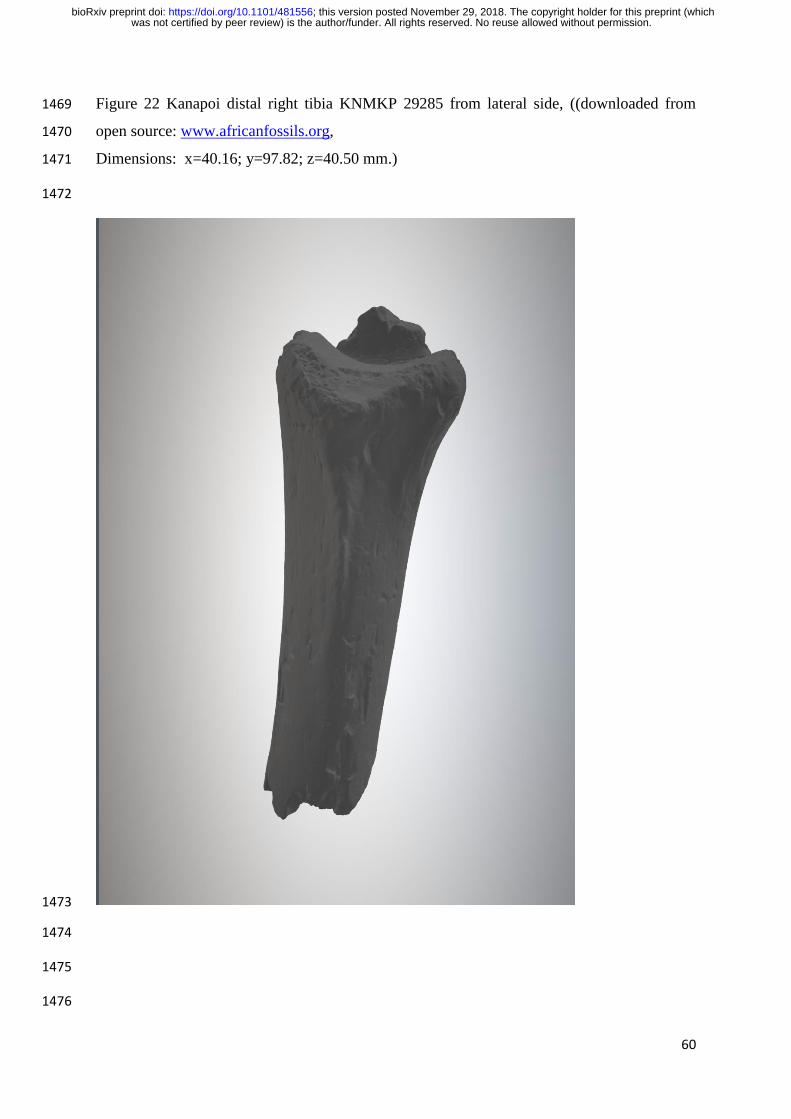
60
Figure 22 Kanapoi distal right tibia KNMKP 29285 from lateral side, ((downloaded from 1469
open source: www.africanfossils.org, 1470
Dimensions: x=40.16; y=97.82; z=40.50 mm.) 1471
1472
1473
1474
1475
1476
was not certified by peer review) is the author/funder. All rights reserved. No reuse allowed without permission. The copyright holder for this preprint (whichthis version posted November 29, 2018. ; https://doi.org/10.1101/481556doi: bioRxiv preprint

61
Figure 23 Long bones of the upper and lower limbs of StW 573 1477
1478
1479
1480
1481
was not certified by peer review) is the author/funder. All rights reserved. No reuse allowed without permission. The copyright holder for this preprint (whichthis version posted November 29, 2018. ; https://doi.org/10.1101/481556doi: bioRxiv preprint

62
Figure 24 Archival image of an indigenous arboreal forager climbing a thin vine using flexed 1482
elbow postures and hallucal grasp (courtesy of Kirk Endicott) 1483
1484
1485
1486
1487
1488
1489
was not certified by peer review) is the author/funder. All rights reserved. No reuse allowed without permission. The copyright holder for this preprint (whichthis version posted November 29, 2018. ; https://doi.org/10.1101/481556doi: bioRxiv preprint

63
Figure 25: An indigenous Batek arboreal forager demonstrating his hallucal grasp for 1490
climbing a small vine (video frame, courtesy of Vivek Venkataraman) 1491
1492
1493
1494
1495
1496
1497
was not certified by peer review) is the author/funder. All rights reserved. No reuse allowed without permission. The copyright holder for this preprint (whichthis version posted November 29, 2018. ; https://doi.org/10.1101/481556doi: bioRxiv preprint

64
Figure 26: Frames from the official video ('Drawin' on Darwin' - Live drawing created at the 1498
Meeting of the Primate Society of Great Britain | Saranjit Birdi | Axisweb) of: Top, the artist 1499
Saranjit Birdi drawing with his foot Bottom: closeup to show precision of hallucal grasp 1500
(courtesy of Emily Saunders and the artist); 1501
1502
1503
1504
1505
was not certified by peer review) is the author/funder. All rights reserved. No reuse allowed without permission. The copyright holder for this preprint (whichthis version posted November 29, 2018. ; https://doi.org/10.1101/481556doi: bioRxiv preprint

65
Figure 27: Video frame of a parkour athlete brachiating on an I-beam, demonstrating the 1506
plasticity of human finger capabilities (see Halsey et al. 2017, permission of the subject and 1507
courtesy Susannah K.S. Thorpe) 1508
1509
1510
1511
was not certified by peer review) is the author/funder. All rights reserved. No reuse allowed without permission. The copyright holder for this preprint (whichthis version posted November 29, 2018. ; https://doi.org/10.1101/481556doi: bioRxiv preprint

66
Figure 28: Handbones of StW 573 1512
1513
1514
1515
1516
1517
was not certified by peer review) is the author/funder. All rights reserved. No reuse allowed without permission. The copyright holder for this preprint (whichthis version posted November 29, 2018. ; https://doi.org/10.1101/481556doi: bioRxiv preprint

67
Figure 29: Apical ridge on the trapezium of: left Gorilla gorilla beringei, right StW 573 1518
(originals) 1519
1520
1521
was not certified by peer review) is the author/funder. All rights reserved. No reuse allowed without permission. The copyright holder for this preprint (whichthis version posted November 29, 2018. ; https://doi.org/10.1101/481556doi: bioRxiv preprint


