Download - Genetically predicted testosterone and

1
Type of manuscript: Research Article
Genetically predicted testosterone and electrocardiographic QT interval duration in Chinese: a
Mendelian randomization analysis in the Guangzhou Biobank Cohort Study
Jie Zhao,1 Chaoqiang Jiang,
2 Tai Hing Lam,
1 Bin Liu,
2 Kar Keung Cheng,
3 Lin Xu,
1 Mei Jing Long,
1 Weisen
Zhang,2 Gabriel M Leung,
1 C Mary Schooling
1,4
1School of Public Health, Li Ka Shing Faculty of Medicine, The University of Hong Kong, Hong Kong SAR,
China; 2Guangzhou Number 12 Hospital, Guangzhou, China;
3Department of Public Health and Epidemiology,
University of Birmingham, UK; 4 CUNY School of Public Health at Hunter College, New York, USA
Address for Correspondence:
Lam TH, 5/F William MW Mong Block, 21 Sasson Road, Hong Kong
Telephone: 852-28199287; Email: [email protected]; Fax: 852-28559528
Word Count: 2,999
2
Abstract
Background: QT interval prolongation, a predictor of cardiac arrhythmias, and elevated heart rate are
associated with higher risk of cardiovascular mortality. Observationally testosterone is associated with shorter
corrected QT interval and slower heart rate, however, the evidence is open to residual confounding and reverse
causality. We examined the association of testosterone with electrocardiogram (ECG) parameters using a
separate-sample instrumental variable (SSIV) estimator.
Methods: To minimize reverse causality, a genetic score predicting testosterone was developed in 289 young
Chinese men from Hong Kong based on a parsimonious set of single nuclear polymorphisms (rs10046,
rs1008805 and rs1256031). Linear regression was used to examine the association of genetically predicted
testosterone with QT interval, corrected QT interval (using the Framingham formula (QTf) and Bazett formula
(QTb)), and heart rate in 4212 older (50+ years) Chinese men from the Guangzhou Biobank Cohort Study.
Results: Predicted testosterone was not associated with QT interval (-0.08 milliseconds (ms) per nmol/L
testosterone, 95% confidence interval (CI) -0.81 to 0.65), QTf interval (0.40 ms per nmol/L testosterone, 95%
CI -0.12 to 0.93) or heart rate (0.26 beats per minute per nmol/L testosterone, 95% CI -0.04 to 0.56), but was
associated with longer QTb interval (0.66 ms per nmol/L testosterone, 95% CI 0.02 to 1.31).
Conclusions: Our findings do not corroborate observed protective associations of testosterone with QT interval
or heart rate among men, but potentially suggest effects in the other direction. Replication in a larger sample is
required.
Key words: Mendelian randomization, testosterone, QT interval
3
Introduction
Cardiovascular disease is the leading cause of mortality globally.1 Prolonged QT interval is associated with
higher risk of cardiac arrhythmias,2 and a longer QT interval, even within the normal physiological range, is
associated with higher risk of cardiovascular disease (CVD) mortality with a graded association.3 Men have a
shorter QT interval than women, with the difference emerging at puberty,4 potentially implicating sex hormones,
although this difference in QT interval may also be related to a slower heart rate (HR) among men.5 Heart rate is
another indicator of cardiovascular health, which is positively associated with CVD mortality.6
Observational studies generally show testosterone associated with shorter corrected QT interval5, 7
and slower
heart rate,5 suggesting a beneficial effect of androgens on cardiac arrhythmias and/or heart rate among men.
However, the observed association of endogenous testosterone with a shorter corrected QT interval and a lower
heart rate is not evident for a different androgen biomarker (3α-diol-G).8 Additionally, these observations are
somewhat inconsistent with the experimental evidence.9-15
In animal experiments, increased testosterone shows
varied effects on QT interval, with reports of both shortening16, 17
and prolongation,9, 10
and also varied effects on
HR, with reports of increases,16
no effect,10
and decreases.17
Small randomized controlled trials (RCTs) have
reported that testosterone has no effect on QT interval11-13
or shortens QT interval,18
and has no effect on HR11-13
or increases HR14
. One single-arm trial of an anti-androgen also reported no effect on QT interval or HR.15
These discrepancies within observational studies and by study design may have occurred for several reasons.
Trials examining the effect of exogenous androgens may be too small to detect minor differences.11-13, 15
Observations concerning endogenous testosterone,5, 7
although well-conducted, are open to reverse causality
arising from testosterone falling with age and being affected by ill-health.19, 20
Moreover, numerous factors may
affect CVD risk, such as lipids, blood pressure and diabetes. However, observations of serum testosterone

4
associated with a healthier CVD risk factor profile have not been substantiated by Mendelian randomization
(MR) studies.21, 22
While a large-scale RCT to examine the effects of testosterone has been suggested,23
the
Institute of Medicine recommended such RCTs only after the benefits of testosterone had been established;24
concerns about the cardiovascular safety of androgen replacement25
have also raised questions about such RCTs.
Given the widespread prescription of testosterone for men, particularly in North America,26
the cardiovascular
effect of testosterone is an important question relevant to clinical practice and policy, where evidence is limited.
Health Canada recently issued a warning about cardiovascular disease, including risk of irregular heart rate.27
The Food and Drug Administration (FDA) advisory committee also recommended restricting testosterone
prescription and investigation of cardiovascular risk.28
Using naturally occurring testosterone related genetic
variants to predict peak testosterone, unaffected by aging or ill-health, in an MR analysis with a separate-sample
instrumental variable (SSIV) enables examination of the causal effects of endogenous peak testosterone, without
any intervention. Here, we used a separate-sample two-stage MR analysis to assess the effects of endogenous
testosterone on QT interval, corrected QT interval and heart rate among older men, to minimize reverse
causality.
Methods
Study design
A separate-sample two-stage MR analysis was used. First, a genetic score predicting serum testosterone was
developed in young Chinese men from Hong Kong, as described previously.22
Second, the association of
predicted testosterone with QT interval, and corrected QT interval and HR was examined in older Chinese men
from the Guangzhou Biobank Cohort Study.

5
Sources of data
To generate a prediction rule for testosterone, a sample of students was recruited from the University of Hong
Kong, restricted to those with both parents and at least three grandparents born in Hong Kong or Guangdong
who were not taking medication affecting hormones. Morning blood samples were collected for testosterone
assessment and DNA extraction. Testosterone was assessed by radioimmunoassay (Roche Diagnostics GmbH,
Mannheim, Germany). DNA was extracted and analyzed at the Centre for Genomic Sciences of the University
of Hong Kong for selected SNPs including SNPs from ESR1 (rs722208 and rs2175898), CYP19A1 (rs10046 and
rs1008805) and ESR2 (rs1256030 and rs1256031) using a Mass ARRAY system (Sequenom, San Diego,
California). Stepwise linear regression with all candidate SNPs (excluding those deviating from
Hardy-Weinburg equilibrium or in linkage disequilibrium) was used to find a parsimonious set of SNPs which
best predicted log testosterone in the sample of young men (as shown for flow chart in Appendix Figure 1 and
for each selected SNP in Appendix Table 1), as described previously.22
A self-administered questionnaire was
used to collect socioeconomic position and health status.
To assess the effects of genetically predicted testosterone on electrocardiogram (ECG) parameters, we used a
large sample of older people (50+ years) from the Guangzhou Biobank Cohort Study (GBCS). GBCS is an
ongoing collaboration of Guangzhou Number 12 Hospital, and the Universities of Hong Kong and Birmingham,
UK.29
Recruitment of participants was in 3 phases. All participants were permanent residents of Guangzhou and
members of the “The Guangzhou Health and Happiness Association for the Respectable Elders” (GHHARE), a
community social and welfare association unofficially aligned with the municipal government. Membership is
open to older people for a monthly fee of 4 Yuan (50 US cents). About 7% of permanent Guangzhou residents
6
aged 50+ years are members of GHHARE, of whom 11% (about 10,000 participants) enrolled for each of phase
one, two and three. Inclusion criteria were that they were capable of consenting, ambulatory, and not receiving
treatment modalities which if omitted may result in immediate life threatening risk, such as chemotherapy or
radiotherapy for cancer, or dialysis for renal failure. Fasting blood samples were collected for storage at
recruitment in phase 3 and at follow-up for participants recruited in other phases. Samples were stored, as whole
blood or as buffy coat and sera, at -80°C for all apart from a subset of phase 3 participants whose DNA was
extracted from fresh blood and stored at -80°C. Selected SNPs were analyzed by a commercial company
(Beijing CapitalBio Corporation30
) in Beijing using a Mass ARRAY system (Sequenom, San Diego, California).
The University of Hong Kong-Hospital Authority Hong Kong West Cluster Joint Institutional Review Board
approved the study. The Guangzhou Medical Ethics Committee of the Chinese Medical Association approved
GBCS, including the use of genetic data, and all participants gave written, informed consent prior to
participation.
Electrocardiogram (ECG)
A standard ECG was performed in the supine position after resting for 5 minutes using a 3-channel, 12-lead
ECG (Marquette MAC-500) in phase 1 and at the start of phase 2 and a synchronous 12-lead ECG (Marquette
Cam-14 acquisition module) in the rest of phase 2 and in phase 3.31
The ECG tracings obtained by the Marquette
MAC-500 electrocardiograph were evenly distributed to two qualified physicians and measured independently,
blinded to other information.32
QT interval was examined from the earliest QRS onset to the end of T-wave. Any
uncertainties were resolved through discussion and consensus. In the rest of phase 2 and phase 3 the QT interval
and HR were measured automatically by the ECG machine.

7
Exposure
The primary exposure was predicted testosterone estimated as the anti-log of genetically predicted log
testosterone. Testosterone, instead of log testosterone, was used as the exposure for ease of interpretation as they
both gave the same pattern of results.
Outcomes
The outcomes were QT interval, HR corrected QT intervals using the Framingham formula (QTf), calculated as
QT+154×(1-60/HR),33
and the Bazett formula (QTb), calculated as QT{HR/60}1/2
,34
as well as HR. QT interval
is influenced by HR, so HR correction is required for the analysis of repolarization duration.35
The Bazett
formula is the most widely used method for HR correction, but tends to underestimate or overestimate the
duration of cardiac repolarization at extremely low or high HR.36
The Framingham correction performs better at
extreme values of HR.33
Statistical analysis
We used an SSIV estimate from two separate samples.37
Genetically predicted log testosterone among men was
calculated as -0.07×rs1008805+0.07×rs10046-0.07×rs1256031+3.0 (Appendix Table 1), as previously.22
The
F-statistic for the regression of log testosterone on genetically predicted log testosterone was obtained; an
F-statistic >10 suggests a reliable genetic instrument.38
ANOVA was used to compare genetically predicted testosterone by key characteristics. Linear regression was
used to assess the association of genetically predicted testosterone with QT, QTf, QTb intervals and HR. Model
1 had no covariates, because the genetic associations are unlikely to be confounded in a homogenous population.

8
Model 2 used bootstrapping with 1000 replications for internal validation. We assessed whether the association
of genetically predicted testosterone with QT intervals and HR varied with ECG measurement methods from the
relevant interaction term. As a sensitivity analysis, men with a major intraventricular conduction defect, as
indicated by QRS≥120 milliseconds (ms), were excluded because the repolarization abnormalities in these men
could be secondary to the conduction defects. To account for variations due to health status, a sensitivity
analysis was also performed among a subgroup of healthy men (without self-reported CVD or hypertension and
not taking medication to reduce blood pressure). All statistical analyses were conducted using Stata 10.1
(StataCorp LP, College Station, Texas).
Results
Among the 8,450 men in all 3 phases of GBCS, DNA for SNP testing was available for 4,262 men, with
availability depending on the phase of recruitment and other logistical concerns, but not on ECG parameters.
Among the 4,262 men, 4,212 (98.8%) had all relevant SNPs and of these, 3,864 (91.7%) had complete data on
QT interval and HR.
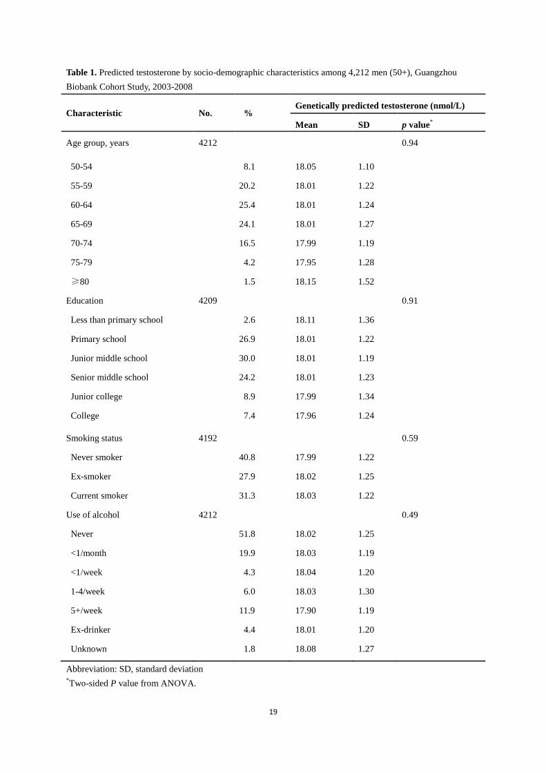
The proportion of variance in log testosterone explained by the genetic score was 4.1%. The F-statistics was
13.3, suggesting a reliable genetic instrument. As would be expected, age, socioeconomic position and lifestyle,
including smoking status and use of alcohol, were not associated with genetically predicted testosterone (Table
1).
Table 2 shows genetically predicted testosterone was not associated with QT interval among men. The estimates
were close to null. Predicted testosterone was associated with longer QTb and QTf intervals as well as faster HR,

9
but the confidence intervals for QTf and HR included the null. The results remained unchanged after
bootstrapping replication (Model 2). The association of genetically predicted testosterone with QT, QTb, QTf
intervals and HR did not vary with ECG measurement method (p-values for interaction 0.27, 0.73, 0.48 and 0.55
respectively). Sensitivity analysis excluding those with QRS≥120 ms and among healthy men both showed
similar results (Appendix Table 2), although the confidence interval for QTb interval among healthy men
included the null, whilst the confidence interval for HR among healthy men no longer included the null.
Discussion
Using an MR analysis with an SSIV estimator to minimize reverse causality, our study does not show any
negative association of endogenous testosterone with QT interval or HR in men. Instead, predicted testosterone
was associated with longer corrected QT interval. Our novel study provides no support for protective effects of
endogenous testosterone on QT interval or HR among men.
To our knowledge, this is the first MR analysis using an SSIV estimator to provide an unbiased estimation of the
effects of testosterone on QT interval or HR. SSIV is useful and cost-efficient when the phenotype of interest
was either not measured or was measured with substantial error in the sample with the outcome.37, 39
It is
infeasible to obtain lifetime sex hormones for older people; neither biomaterials nor peak sex hormones dating
back to the 1960s are available. The MR design makes it feasible to use testosterone in early adulthood as a
marker of lifetime exposure. Thus, we can minimize reverse causality and avoid the imprecision in MR
estimates that could arise from assessing the genetic association with testosterone at older ages, which may
reflect ill-health rather than life-long exposure, inducing an underestimation of the genetic association with
exposure inflating the MR estimates.40
SSIV also remediates weak-instruments bias, reducing concerns about

10
using multiple polymorphisms as IVs.37
Any correlation of the genetic variants with unmeasured confounders in
the sample with the phenotype is unlikely to be replicated in the sample with the outcome due to the different
data structures.37
We also restricted the sample of young people to those with both parents and at least 3
grandparents born in Hong Kong or Guangdong province, to ensure genetic homogeneity.
Although we used MR which can mimic the randomized treatment allocation in RCT,41
several limitations exist.
First, QT interval was measured using two methods. Assessing QT interval manually added some measurement
error and thereby reduced differences between groups. However, any measurement error is likely to be random,
which is compensated for by our large sample size, and the effect of genetically predicted testosterone did not
vary with measurement method. QT interval and HR had the expected associations with age and sex (data not
shown).42, 43
Second, the estimates for QTf and QTb are different, although they are in the same direction.
Different correction methods may provide discordant result, as observed previously.44
The wide range of HR in
our study may account for the differences in the two correction methods; as such the Framingham correction
may be more reliable. However, all our corrected estimates were in the same direction with only a difference in
the level of variability. The estimates in the sensitivity analysis were also in the same direction, but sometimes
had confidence intervals including the null, perhaps because of the smaller sample size. Third, this study is not
totally representative, although the QT interval is similar to that in a nationally representative study.45
The
results would only be invalidated if the relation of genetic variants with testosterone or with ECG parameters in
our sample differs from that in the general population, which is unlikely as the relevant genetic variants were not
associated with socioeconomic position or lifestyle. Fourth, only Chinese men were included which could
restrict generalizability. However, the association of genetic variants with testosterone or ECG parameters is
unlikely to vary by setting or ethnicity. Fifth, the selected SNPs may affect ECG parameters directly rather than
11
via testosterone. To our knowledge, no study indicates a direct association of these SNPs with QT interval or
HR. Sixth, our SNP selection is restricted by the limited availability of genome-wide association studies
(GWAS) of testosterone, which are also among older men, and so might reflect the factors causing testosterone
to fall with age rather than testosterone. Moreover, several SNPs identified in GWAS of testosterone among
Caucasian men have not replicated in Chinese men,46
whilst one SNP that did replicate (rs2075230) was
associated with both testosterone and SHBG,46
making it unsuitable as a genetic instrument for testosterone
because of pleiotropy. In addition, genetic structure might vary by ethnicity, however, only Chinese originating
from Guangdong province were included. We also tested for linkage disequilibrium in the young men, and
discarded any SNPs in linkage disequilibrium.22
To avoid these issues, we generated a genetic prediction rule
for testosterone from genes functionally relevant to testosterone in a sample of young men with the same genetic
origin as the sample of older men with ECG data. Using the same genetic prediction rule in the same sample of
older men, we have previously shown an association of genetically predicted testosterone associated with lower
HDL-cholesterol,22
as would be expected from meta-analysis of RCTs,47
which suggests some validity. Seventh,
genetic variants typically have small effects. MR estimates are less precise than conventional regression
estimates.48
However, MR avoids the confounding which occurs in traditional observational studies.49
We used
a weighted genetic score instead of one SNP as instrument, reducing variability in MR estimation.50
MR studies
require large sample sizes. The sample size of ~4,000 had 0.8 power to detect a relatively small effect size of
0.22. Meta-analysis combining our findings with previous MR estimates is required, however, no other MR
study exists. Eighth, correction for multiple testing might be required. Such correction is widely used in agnostic
or exploratory studies, such as GWAS,51
to control for type 1 error, while it may increase the risk of type 2
error.52, 53
However, our study aims at confirmation and given our negative findings, controlling for type 2 error
is important from a public health perspective. Ninth, we cannot determine the clinical significance of the small

12
effect size, because MR estimates should be interpreted as hypothesis testing for causation, rather than
indicating the size of causal effects.40
However, small effects that may not be clinically significant may still be
an important determinant of population health. Tenth, serum testosterone might not be a good indicator for
androgen activity. Successful use of anti-androgens at castrate levels of serum testosterone in prostate cancer
trials has also challenged the extent to which serum testosterone reflects all androgen activity.54
The glucuronide
derivatives of androgens, such as androsterone glucuronide or androstenediol glucuronide, are obligatory for
elimination of androgens, and provide a measure or correlate of total androgen activity.55, 56
Observationally,
androstenediol glucuronide is poorly correlated with serum testosterone.57
Replication using a measure of
androgen activity is required.
Our study did not corroborate observations of endogenous testosterone associated with shorter corrected QT
interval5, 7
and slower HR.5 Serum testosterone is associated with health status, and falls in response to obesity
or ill-health.20
Our study is more consistent with small RCTs which generally report no effect on QT interval or
HR.11-13
Notably, androgenic steroid users have also been reported to have prolonged QT interval and potentially
higher risk of cardiac arrhythmias,58
although the dose is much higher than that in RCTs. Previous MR studies
have similarly not corroborated the associations of endogenous testosterone with healthier values of CVD risk
factors,21, 22
but suggest adverse effects on lipids.22
A meta-analysis of RCTs among men suggests testosterone
replacement increases the risk of CVD-related events.25
.
From a public health perspective, the benefits and the CVD safety of androgens have important clinical
implications for the increasing numbers, of particularly older men, using testosterone replacement.26
Our novel

13
study adds to the limited evidence concerning testosterone and ECG parameters, with corresponding
implications for the role of testosterone in cardiovascular health.
Conclusion
The study did not corroborate observations of an association of higher endogenous testosterone with lower QT
interval or slower HR, and correspondingly a potentially protective effect of testosterone. Replication in a larger
sample is required.
Acknowledgements
The Guangzhou Biobank Cohort Study investigators include: Guangzhou No. 12 Hospital--Dr. Zhang WS, Dr.
Zhu T, Dr. Liu B, Prof. Jiang CQ (Co-Principal Investigator (PI)); University of Hong Kong-Dr. C. M.
Schooling, Prof. SM McGhee SM, Prof. R Fielding, Prof. GM Leung, Prof. TH Lam (Co-PI); University of
Birmingham--Dr. G. N. Thomas, Dr. P Adab, Prof. KK Cheng (Co-PI). The authors thank Drs SL Au Yeung and
Sushma Kavikondala for convoking the student sample, Drs Garcia-Barcelo MM, So Man, and Zhu T for
facilitating the DNA extraction.
This work was supported by the University of Hong Kong Foundation for Development and Research (Hong
Kong, China); the University of Hong Kong University Research Committee Strategic Research Theme of
Public Health (Hong Kong, China); Guangzhou Public Health Bureau (Guangzhou, China; Key technology
collaboration project, [grant number 2012J5100041]), Guangzhou Science and Technology Bureau (Guangzhou,
China) and the University of Birmingham (Birmingham, United Kingdom). This sub-study was funded by the

14
Research Grant Council General Research Fund [grant number 769710], Research Grant Council of Hong Kong,
Hong Kong SAR, People’s Republic of China.
The funders had no role in the study design, data collection and analysis, the decision to publish, or preparation
of the manuscript.
Conflict of interest: None declared.

15
KEY MESSAGES
The role of androgens in cardiovascular disease is controversial.
We used a Mendelian randomization analysis with a separate-sample instrumental
variable to examine the causal effects of endogenous testosterone on
electrocardiogram parameters.
Our findings do not corroborate observed protective associations of testosterone with
cardiac arrhythmias or heart rate among men, but instead suggests testosterone could
lengthen corrected QT interval.

16
References:
1. Lozano R, Naghavi M, Foreman K, et al. Global and regional mortality from 235 causes of death for 20
age groups in 1990 and 2010: a systematic analysis for the Global Burden of Disease Study 2010. Lancet
2013;380(9859): 2095-128.
2. Algra A, Tijssen JG, Roelandt JR, Pool J, Lubsen J. QTc prolongation measured by standard 12-lead
electrocardiography is an independent risk factor for sudden death due to cardiac arrest. Circulation 1991;83(6):
1888-94.
3. Zhang Y, Post WS, Blasco-Colmenares E, Dalal D, Tomaselli GF, Guallar E. Electrocardiographic QT
interval and mortality: a meta-analysis. Epidemiology 2011;22(5): 660-70.
4. Rautaharju PM, Zhou SH, Wong S, et al. Sex differences in the evolution of the electrocardiographic QT
interval with age. Can J Cardiol 1992;8(7): 690-5.
5. van Noord C, Dorr M, Sturkenboom MC, et al. The association of serum testosterone levels and ventricular
repolarization. Eur J Epidemiol 2010;25(1): 21-8.
6. Fox KM, Ferrari R. Heart rate: a forgotten link in coronary artery disease? Nat Rev Cardiol 2011;8(7):
369-79.
7. Zhang Y, Ouyang P, Post WS, et al. Sex-steroid hormones and electrocardiographic QT-interval duration:
findings from the third National Health and Nutrition Examination Survey and the Multi-Ethnic Study of
Atherosclerosis. Am J Epidemiol 2011;174(4): 403-11.
8. Schooling CM, Zhao J, Zhang Y. The association of androgens with QT interval and heart rate in US men.
Int J Cardiol 2014.
9. Drici MD, Burklow TR, Haridasse V, Glazer RI, Woosley RL. Sex hormones prolong the QT interval and
downregulate potassium channel expression in the rabbit heart. Circulation 1996;94(6): 1471-4.
10. Marques Neto SR, da HSA, dos Santos MC, Ferraz EF, Nascimento JH. The blockade of angiotensin AT1
and aldosterone receptors protects rats from synthetic androgen-induced cardiac autonomic dysfunction. Acta
physiologica 2013;208(2): 166-71.
11. White CM, Ferraro-Borgida MJ, Moyna NM, et al. The effect of pharmacokinetically guided acute
intravenous testosterone administration on electrocardiographic and blood pressure variables. J Clin Pharmacol
1999;39(10): 1038-43.
12. Malkin CJ, Morris PD, Pugh PJ, English KM, Channer KS. Effect of testosterone therapy on QT dispersion
in men with heart failure. Am J Cardiol 2003;92(10): 1241-3.
13. Chung T, Kelleher S, Liu PY, Conway AJ, Kritharides L, Handelsman DJ. Effects of testosterone and
nandrolone on cardiac function: a randomized, placebo-controlled study. Clin Endocrinol (Oxf) 2007;66(2):
235-45.
14. Rosano GM, Leonardo F, Pagnotta P, et al. Acute anti-ischemic effect of testosterone in men with coronary
artery disease. Circulation 1999;99(13): 1666-70.
15. Tolcher AW, Chi KN, Shore ND, et al. Effect of abiraterone acetate plus prednisone on the QT interval in
patients with metastatic castration-resistant prostate cancer. Cancer Chemother Pharmacol 2012;70(2): 305-13.
16. Fulop L, Banyasz T, Szabo G, et al. Effects of sex hormones on ECG parameters and expression of cardiac
ion channels in dogs. Acta physiologica 2006;188(3-4): 163-71.
17. Eleawa SM, Sakr HF, Hussein AM, Assiri AS, Bayoumy NM, Alkhateeb M. Effect of testosterone
replacement therapy on cardiac performance and oxidative stress in orchidectomized rats. Acta physiologica
2013;209(2): 136-47.
18. Schwartz JB, Volterrani M, Caminiti G, et al. Effects of testosterone on the Q-T interval in older men and
older women with chronic heart failure. Int J Androl 2011;34(5 Pt 2): e415-21.

17
19. Feldman HA, Longcope C, Derby CA, et al. Age trends in the level of serum testosterone and other
hormones in middle-aged men: longitudinal results from the Massachusetts male aging study. J Clin Endocrinol
Metab 2002;87(2): 589-98.
20. Shi Z, Araujo AB, Martin S, O'Loughlin P, Wittert GA. Longitudinal changes in testosterone over five
years in community-dwelling men. J Clin Endocrinol Metab 2013;98(8): 3289-97.
21. Haring R, Teumer A, Volker U, et al. Mendelian randomization suggests non-causal associations of
testosterone with cardiometabolic risk factors and mortality. Andrology 2013;1(1): 17-23.
22. Zhao J, Jiang C, Lam TH, et al. Genetically predicted testosterone and cardiovascular risk factors in men: a
Mendelian randomization analysis in the Guangzhou Biobank Cohort Study. Int J Epidemiol 2014;43(1): 140-8.
23. Schwartz LM, Woloshin S. Low "t" as in "template": how to sell disease. JAMA Intern Med 2013;173(15):
1460-2.
24. Committee on Assessing the Need for Clinical Trials of Testosterone Replacement Therapy.
TESTOSTERONE AND AGING Clinical Research Directions. Washington: THE NATIONAL ACADEMIES
PRESS; 2004.
25. Xu L, Freeman G, Cowling BJ, Schooling CM. Testosterone therapy and cardiovascular events among men:
a systematic review and meta-analysis of placebo-controlled randomized trials. BMC Med 2013;11: 108.
26. Handelsman DJ. Global trends in testosterone prescribing, 2000-2011: expanding the spectrum of
prescription drug misuse. Med J Aust 2013;199(8): 548-51.
27. Health Canada. Summary Safety Review-Testosterone Replacement Products-Cardiovascular Risk.
Available from:
http://www.hc-scgcca/dhp-mps/medeff/advisories-avis/review-examen/testosterone-engphp#fnb1.
28. F.D.A. Panel Backs Limits on Testosterone Drugs. Available from:
http://www.nytimes.com/2014/09/18/health/testosterone-drugs-fda.html?_r=0.
29. Jiang C, Thomas GN, Lam TH, et al. Cohort profile: The Guangzhou Biobank Cohort Study, a
Guangzhou-Hong Kong-Birmingham collaboration. Int J Epidemiol 2006;35(4): 844-52.
30. CapitalBio Corporation. Available from: http://science.capitalbio.com/khfbwz/smkxfwxg/index.shtml.
31. Long MJ, Jiang CQ, Lam TH, et al. Alcohol consumption and electrocardiographic left ventricular
hypertrophy and mediation by elevated blood pressure in older Chinese men: the Guangzhou Biobank Cohort
Study. Alcohol 2013;47(6): 473-80.
32. Long MJ, Jiang CQ, Lam TH, et al. Atrial fibrillation and obesity among older Chinese: the Guangzhou
Biobank Cohort Study. Int J Cardiol 2011;148(1): 48-52.
33. Sagie A, Larson MG, Goldberg RJ, Bengtson JR, Levy D. An improved method for adjusting the QT
interval for heart rate (the Framingham Heart Study). Am J Cardiol 1992;70(7): 797-801.
34. Bazett HC. An analysis of time relations of electrocardiograms. Heart 1920;7: 353-67.
35. Goldenberg I, Moss AJ, Zareba W. QT interval: how to measure it and what is "normal". J Cardiovasc
Electrophysiol 2006;17(3): 333-6.
36. Johnson JN, Ackerman MJ. QTc: how long is too long? Br J Sports Med 2009;43(9): 657-62.
37. Tchetgen Tchetgen EJ, Walter S, Glymour MM. Commentary: building an evidence base for mendelian
randomization studies: assessing the validity and strength of proposed genetic instrumental variables. Int J
Epidemiol 2013;42(1): 328-31.
38. Stock JH, Yogo M. Testing for Weak Instruments in Linear IV Regression. Cambridge, MA: National
Bureau of Economic Research; 2002.
39. Pierce BL, Burgess S. Efficient Design for Mendelian Randomization Studies: Subsample and 2-Sample
Instrumental Variable Estimators. Am J Epidemiol 2013;178(7):1177-84.
18
40. Schooling CM, Au Yeung SL, Freeman G. Mendelian randomization estimates may be inflated. J Am Coll
Cardiol 2013;61(18): 1931.
41. Bennett DA. An introduction to instrumental variables--part 2: Mendelian randomisation.
Neuroepidemiology 2010;35(4): 307-10.
42. Mangoni AA, Kinirons MT, Swift CG, Jackson SH. Impact of age on QT interval and QT dispersion in
healthy subjects: a regression analysis. Age Ageing 2003;32(3): 326-31.
43. Mason JW, Ramseth DJ, Chanter DO, Moon TE, Goodman DB, Mendzelevski B. Electrocardiographic
reference ranges derived from 79,743 ambulatory subjects. J Electrocardiol 2007;40(3): 228-34.
44. Anttonen O, Junttila MJ, Rissanen H, Reunanen A, Viitasalo M, Huikuri HV. Prevalence and prognostic
significance of short QT interval in a middle-aged Finnish population. Circulation 2007;116(7): 714-20.
45. Wu J, Kors JA, Rijnbeek PR, van Herpen G, Lu Z, Xu C. Normal limits of the electrocardiogram in
Chinese subjects. Int J Cardiol 2003;87(1): 37-51.
46. Chen Z, Tao S, Gao Y, et al. Genome-wide association study of sex hormones, gonadotropins and sex
hormone-binding protein in Chinese men. J Med Genet 2013;50(12): 794-801.
47. Fernandez-Balsells MM, Murad MH, Lane M, et al. Clinical review 1: Adverse effects of testosterone
therapy in adult men: a systematic review and meta-analysis. J Clin Endocrinol Metab 2010;95(6): 2560-75.
48. Kivimaki M, Magnussen CG, Juonala M, et al. Conventional and Mendelian randomization analyses
suggest no association between lipoprotein(a) and early atherosclerosis: the Young Finns Study. Int J Epidemiol
2011;40(2): 470-8.
49. Smith GD, Ebrahim S. Mendelian randomization: prospects, potentials, and limitations. Int J Epidemiol
2004;33(1): 30-42.
50. Burgess S, Thompson SG, Collaboration CCG. Avoiding bias from weak instruments in Mendelian
randomization studies. Int J Epidemiol 2011;40(3): 755-64.
51. Johnson RC, Nelson GW, Troyer JL, et al. Accounting for multiple comparisons in a genome-wide
association study (GWAS). BMC Genomics 2010;11: 724.
52. Feise RJ. Do multiple outcome measures require p-value adjustment? BMC Med Res Methodol 2002;2: 8.
53. Perneger TV. What's wrong with Bonferroni adjustments. BMJ 1998;316(7139): 1236-8.
54. Ryan CJ, Smith MR, de Bono JS, et al. Abiraterone in metastatic prostate cancer without previous
chemotherapy. N Engl J Med 2013;368(2): 138-48.
55. Labrie F, Belanger A, Belanger P, et al. Androgen glucuronides, instead of testosterone, as the new markers
of androgenic activity in women. J Steroid Biochem Mol Biol 2006;99(4-5): 182-8.
56. Labrie F, Cusan L, Gomez JL, et al. Comparable amounts of sex steroids are made outside the gonads in
men and women: strong lesson for hormone therapy of prostate and breast cancer. J Steroid Biochem Mol Biol
2009;113(1-2): 52-6.
57. Schooling CM. Androgen activity, ischaemic heart disease and risk factors among men in NHANES III.
Eur J Clin Invest 2013;43(12): 1273-81.
58. Maior AS, Menezes P, Pedrosa RC, Carvalho DP, Soares PP, Nascimento JH. Abnormal cardiac
repolarization in anabolic androgenic steroid users carrying out submaximal exercise testing. Clin Exp
Pharmacol Physiol 2010;37(12): 1129-33.
19
Table 1. Predicted testosterone by socio-demographic characteristics among 4,212 men (50+), Guangzhou
Biobank Cohort Study, 2003-2008
Characteristic No. % Genetically predicted testosterone (nmol/L)
Mean SD p value*
Age group, years 4212 0.94
50-54 8.1 18.05 1.10
55-59 20.2 18.01 1.22
60-64 25.4 18.01 1.24
65-69 24.1 18.01 1.27
70-74 16.5 17.99 1.19
75-79 4.2 17.95 1.28
≥80 1.5 18.15 1.52
Education 4209 0.91
Less than primary school 2.6 18.11 1.36
Primary school 26.9 18.01 1.22
Junior middle school 30.0 18.01 1.19
Senior middle school 24.2 18.01 1.23
Junior college 8.9 17.99 1.34
College 7.4 17.96 1.24
Smoking status 4192 0.59
Never smoker 40.8 17.99 1.22
Ex-smoker 27.9 18.02 1.25
Current smoker 31.3 18.03 1.22
Use of alcohol 4212 0.49
Never 51.8 18.02 1.25
<1/month 19.9 18.03 1.19
<1/week 4.3 18.04 1.20
1-4/week 6.0 18.03 1.30
5+/week 11.9 17.90 1.19
Ex-drinker 4.4 18.01 1.20
Unknown 1.8 18.08 1.27
Abbreviation: SD, standard deviation *Two-sided P value from ANOVA.

20
Table 2. Effect of genetically predicted testosterone on QT interval, corrected QT interval and heart rate among
men (50+ years), Guangzhou Biobank Cohort Study, 2003-2008
Outcome
(n=3864) Mean (SD) Model
* β coefficient
† 95% CI P value
QT interval
(milliseconds)
385.8 (28.2) 1 -0.08 -0.81 to 0.65 0.83
QTf interval
(milliseconds)
406.4 (20.4) 1 0.40 -0.12 to 0.93 0.13
2 0.40 -0.11 to 0.91 0.12
QTb interval
(milliseconds)
416.7 (24.9) 1 0.66 0.02 to 1.31 0.04
2 0.66 0.04 to 1.29 0.04
Heart rate
(beats per minute)
71.0 (11.5) 1 0.26 -0.04 to 0.56 0.09
2 0.26 -0.04 to 0.56 0.09
Abbreviations: SD, standard deviation; CI, confidence interval *Model 1 had no covariates; Model 2 used bootstrapping with 1000 replications for internal validation for model
1. †β coefficient refers to the average change in QT, QTf, QTb intervals and heart rate with each unit (nmol/L)
increase in genetically predicted testosterone.

21
Appendix Table 1. Stepwise linear regression model for prediction of log testosterone in the young men*
Predictors Beta-coefficient 95% CI P value
rs1008805 (CYP19A1) -0.07 -0.14, -0.01 0.03
rs10046 (CYP19A1) 0.07 0.01, 0.13 0.02
rs1256031 (ESR2) -0.07 -0.12, -0.02 0.01
Constant 3.0 -- --
*In the regression model, log testosterone was used as outcome, because the distribution of testosterone was
skewed. Two outliers (Cook’D value>0.05) were dropped when establishing the genetic prediction rule for log
testosterone, so 287 men were included in the prediction model.

22
Appendix Table 2. Sensitivity analysis on the effect of genetically predicted testosterone on QT interval, corrected QT interval and heart rate among older men (50+ years),
Guangzhou Biobank Cohort Study, 2003-2008, excluding those with QRS≥120 milliseconds and only including *healthy men
Outcome Sample n Mean (SD) β coefficient† 95% CI P value
QT interval
(milliseconds)
Excluding QRS≥120 milliseconds 3697 384.6 (27.5) 0.08 -0.64 to 0.80 0.83
Healthy men only 2680 385.7 (28.0) -0.40 -1.27 to 0.47 0.37
QTf interval
(milliseconds)
Excluding QRS≥120 milliseconds 3697 405.2 (19.4) 0.49 -0.02 to 1.00 0.06
Healthy men only 2680 405.6 (20.2) 0.27 -0.35 to 0.90 0.39
QTb interval
(milliseconds)
Excluding QRS≥120 milliseconds 3697 415.4 (23.9) 0.70 0.08 to 1.33 0.03
Healthy men only 2680 415.4 (24.6) 0.63 -0.13 to 1.39 0.11
Heart rate
(beats per minute)
Excluding QRS≥120 milliseconds 3697 71.0 (11.5) 0.22 -0.08 to 0.52 0.16
Healthy men only 2680 70.6 (11.2) 0.39 0.04 to 0.73 0.03
Abbreviations: SD, standard deviation; CI, confidence interval *Healthy men were men without self-reported CVD or hypertension and not taking medication to reduce blood pressure.
†β coefficient refers to the average change in QT, QTf, QTb intervals and heart rate with each unit (nmol/L) increase in genetically predicted testosterone.

23
Appendix Figure 1. Flow chart for establishing the genetic prediction rule. For references identified in the
figure, please see Appendix references.
Appendix References:
1. Olson SH, Bandera EV, Orlow I. Variants in estrogen biosynthesis genes, sex steroid hormone levels, and
endometrial cancer: a HuGE review. Am J Epidemiol 2007;165(3): 235-45.
2. Travis RC, Schumacher F, Hirschhorn JN, et al. CYP19A1 genetic variation in relation to prostate cancer
risk and circulating sex hormone concentrations in men from the Breast and Prostate Cancer Cohort Consortium.
Cancer Epidemiol Biomarkers Prev 2009;18(10): 2734-44.
3. Sowers MR, Wilson AL, Kardia SR, Chu J, Ferrell R. Aromatase gene (CYP 19) polymorphisms and
endogenous androgen concentrations in a multiracial/multiethnic, multisite study of women at midlife. Am J
Med 2006;119(9 Suppl 1): S23-30.
4. Ahn J, Schumacher FR, Berndt SI, et al. Quantitative trait loci predicting circulating sex steroid hormones
in men from the NCI-Breast and Prostate Cancer Cohort Consortium (BPC3). Hum Mol Genet 2009;18(19):
3749-57.
5. Chen YC, Kraft P, Bretsky P, et al. Sequence variants of estrogen receptor beta and risk of prostate cancer
in the National Cancer Institute Breast and Prostate Cancer Cohort Consortium. Cancer Epidemiol Biomarkers
Prev 2007;16(10): 1973-81.


