
Genome-Wide Analysis Reveals the Vacuolar pH-Stat ofSaccharomyces cerevisiaeChristopher L. Brett1*¤, Laura Kallay1, Zhaolin Hua2, Richard Green2, Anthony Chyou1, Yongqiang
Zhang1, Todd R. Graham2, Mark Donowitz1,3, Rajini Rao1*
1 Department of Physiology, Johns Hopkins University School of Medicine, Baltimore, Maryland, United States of America, 2 Department of Biological Sciences, Vanderbilt
University, Nashville, Tennessee, United States of America, 3 Department of Medicine, Johns Hopkins University School of Medicine, Baltimore, Maryland, United States of
America
Abstract
Protons, the smallest and most ubiquitous of ions, are central to physiological processes. Transmembrane proton gradientsdrive ATP synthesis, metabolite transport, receptor recycling and vesicle trafficking, while compartmental pH controlsenzyme function. Despite this fundamental importance, the mechanisms underlying pH homeostasis are not entirelyaccounted for in any organelle or organism. We undertook a genome-wide survey of vacuole pH (pHv) in 4,606 single-genedeletion mutants of Saccharomyces cerevisiae under control, acid and alkali stress conditions to reveal the vacuolar pH-stat.Median pHv (5.2760.13) was resistant to acid stress (5.2860.14) but shifted significantly in response to alkali stress(5.8360.13). Of 107 mutants that displayed aberrant pHv under more than one external pH condition, functional categoriesof transporters, membrane biogenesis and trafficking machinery were significantly enriched. Phospholipid flippases,encoded by the family of P4-type ATPases, emerged as pH regulators, as did the yeast ortholog of Niemann Pick Type Cprotein, implicated in sterol trafficking. An independent genetic screen revealed that correction of pHv dysregulation in aneo1ts mutant restored viability whereas cholesterol accumulation in human NPC12/2 fibroblasts diminished upontreatment with a proton ionophore. Furthermore, while it is established that lumenal pH affects trafficking, this studyrevealed a reciprocal link with many mutants defective in anterograde pathways being hyperacidic and retrograde pathwaymutants with alkaline vacuoles. In these and other examples, pH perturbations emerge as a hitherto unrecognizedphenotype that may contribute to the cellular basis of disease and offer potential therapeutic intervention through pHmodulation.
Citation: Brett CL, Kallay L, Hua Z, Green R, Chyou A, et al. (2011) Genome-Wide Analysis Reveals the Vacuolar pH-Stat of Saccharomyces cerevisiae. PLoS ONE 6(3):e17619. doi:10.1371/journal.pone.0017619
Editor: Matt Kaeberlein, University of Washington, United States of America
Received January 3, 2011; Accepted February 2, 2011; Published March 14, 2011
Copyright: � 2011 Brett et al. This is an open-access article distributed under the terms of the Creative Commons Attribution License, which permitsunrestricted use, distribution, and reproduction in any medium, provided the original author and source are credited.
Funding: This work was supported by National Institutes of Health grants R01 DK54214 to R.R., GM62367 to T.R.G. and DK26523 to M.D. C.L.B. was a predoctoralfellow of the American Heart Association. A.C. received support from a Provost’s Undergraduate Research Award of the Johns Hopkins University. The funders hadno role in study design, data collection and analysis, decision to publish, or preparation of the manuscript.
Competing Interests: The authors have declared that no competing interests exist.
* E-mail: [email protected] (CLB); [email protected] (RR)
¤ Current address: Department of Biology, Concordia University, Montreal, Quebec, Canada
Introduction
The ability to control compartmental pH is an essential cellular
function. Pioneering work by Metchnikoff and de Duve empha-
sized the importance of lumenal acidification for bacterial killing in
phagosomes, and within lysosomes for acid hydrolase maturation
and activity [1,2]. Additionally, a critical role for lumenal pH
within the endocytic pathway has been implicated in the
recruitment and assembly of trafficking machinery, reorganization
of membrane lipids and actin cytoskeleton in directional vesicle
trafficking and in organellar morphology [3,4,5,6]. These cellular
pathways contribute to normal cell development and to almost all
cell functions from plants to humans. Moreover, defects in pH
homeostasis at the organellar level lead to bacterial infection or
specific disorders including Dent’s disease and numerous lysosom-
al storage diseases that are linked to pH dysregulation of
endosomes and lysosomes, respectively [7,8,9,10].
The H+ electrochemical gradient in endosomes and lysosomes is
derived from the balance of H+ loading and leak across the
membrane [11,12,13,14]. The organellar V-type H+ ATPase is the
major source of lumenal protons [6,15,16,17]. Charge compen-
sation is also critical for the build up of a significant H+ chemical
gradient (DpHv), since the V-ATPase is electrogenic, and there is
evidence for both inward movement of Cl2 ions and outward
movement of cations [13]. H+ leak pathways modulate lumenal
pH and are unmasked when the V-ATPase is blocked. Faster leak
rates in endosomes when compared to lysosomes correlates with
organelle buffering capacity and surface area [15,18]. A family of
intracellular Na+(K+)/H+ exchangers, represented by mammalian
isoforms NHE6-9 with overlapping distribution throughout the
endocytic pathway and trans Golgi network, are emerging as
significant contributors to H+ leak [19,20,21]. Genetic variants in
human NHE6 and NHE9 have been associated with severe X-
linked mental retardation, autism, attention deficit hyperactivity
disorder and epilepsy [22,23,24]. We found that overexpression or
deletion of Nhx1, the orthologous Na+(K+)/H+ exchanger in the
late endosome of Saccharomyces cerevisiae, resulted in alkalinization or
acidification of vacuole pH, respectively [20,25]. However, Nhx1
PLoS ONE | www.plosone.org 1 March 2011 | Volume 6 | Issue 3 | e17619
does not fully account for the leak, and many other contributors to
H+ leak and lumenal buffering remain to be identified. To find
these and other mechanisms of pH regulation, we conducted a
genome-wide survey of vacuolar pH using the S. cerevisiae library of
viable, single gene deletions. We identified 107 gene deletions with
significant alterations in vacuole pH under more than one external
pH condition. Unexpectedly, mechanisms that control membrane
composition such as the P4-ATPase type lipid flippases, compo-
nents of ergosterol biogenesis and transport including the ortholog
of Neimann Pick disease Type C1 protein, were found to be
important contributors to vacuole pH homeostasis.
Results and Discussion
A genome-wide screen identifies contributors to vacuolepH homeostasis
To gain a comprehensive view of the vacuolar ‘pH-stat’, we
measured vacuole pH in a collection of 4,606 viable, single gene
deletion strains that span the S. cerevisiae genome (Figure 1a; see
Supplemental Materials Table S1 for the complete dataset). The
majority of these mutant strains (4,336) had vacuole pH values
within the range observed in wild type cells (4.81,pHv,5.41;
Figure 1a, bottom panel), most falling near the median of
5.2760.13. However, single gene mutants with abnormally acidic
(224) or alkaline (46) vacuoles were discovered (Figure 1a, middle
panel). Lumenal pH in these mutants reached as low as 4.17 and
high as 6.68, representing a sizeable range tolerated by the cell
without notable effects on mid-log growth or viability (see
Supplemental Materials Figure S1).
We repeated this survey under acidic (pH 2.7) or alkaline
(pH 7.0) conditions to unmask mechanisms important for
homeostasis in response to pH stress (Figure 1b; see Supplemental
Materials Table S1 for the complete dataset). When acid stressed,
the median pHv of wild type cells and the mutant population
remained the same (5.2860.14) and a similar number of mutants
with acidic vacuoles were observed (206, of which 74 were also
acidic under control conditions). However pHv of these mutants
strayed further from the median compared to control conditions,
consistent with a greater vacuolar accumulation of H+ in acidic
medium. This is most apparent in Figure 1c, which compares pHv
obtained under control conditions against those measured under
acid or alkali stress for each mutant strain. In contrast to acid
stress, the median pHv of wild type and mutant population jumped
from 5.2760.13 to 5.8360.13 upon alkali stress. Few mutants
with acidic vacuoles were observed under alkali stress (36), and
nearly all mutants (with the exception of 4 strains) with hyperacidic
vacuoles at external pH#4.0 were able to recover when external
pH was raised. As expected, we observed many more mutants with
alkaline vacuoles under this condition (115, of which 12 were also
alkaline at lower external pH), consistent with difficulty in
scavenging protons when the external H+ concentration is low.
Notably, the range of vacuolar pH values observed in the mutant
population was the same under each condition. Mutants that
displayed acidic vacuoles (77) and alkali vacuoles (29) in more than
one external pH condition (Figure 1d) are listed in Table S3. Of
these 107 genes, three functional categories were significantly
enriched: transporters, membrane organization and biogenesis,
and membrane trafficking machinery.
P4-ATPase phospholipid translocases regulate vacuolepH homeostasis
Gene disruption of 11 subunits or assembly factors of the
vacuolar H+-ATPase [6,15,16,17] resulted in prominent vacuole
alkalinization (Tables S2 and S3), validating our screen. Only one
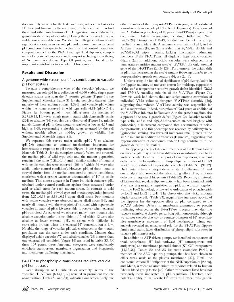
other member of the transport ATPase category, drs2D, exhibited
a vma-like shift in vacuole pH (Table S2; Figure 2a). Drs2 is one of
five ATP-driven phospholipid flippases (P4-ATPases) in yeast that
contribute to bilayer asymmetry, including Dnf1-3 and Neo1
[26,27,28]. Disruption of Dnf3, another member of this group,
resulted in an acidic shift. A systematic evaluation of pHv in P4-
ATPase mutants (Figure 2a) revealed that dnf1dnf2D double and
dnf1dnf2dnf3D triple mutants, lacking functionally redundant
members of the P4-ATPases, all displayed hyperacidic vacuoles
(Figure 2a). In addition, acidic vacuoles were observed in a
temperature-sensitive mutant (neo1-1) of NEO1, the only essential
gene of the P4-ATPase family [29]. Furthermore, the acidic shift
in pHv was increased in the neo1-1 mutant following transfer to the
non-permissive growth temperature (Figure 2a, d).
Underscoring the functional significance of pH dysregulation in
the flippase mutants, an unbiased screen for multicopy suppressors
of the neo1-ts temperature sensitive growth defect identified VMA5
and VMA11, encoding subunits of the V-ATPase (Figure 2b).
Previous work had shown that non-stoichiometric expression of
individual VMA subunits disrupted V-ATPase assembly [30],
suggesting that reduced V-ATPase activity was responsible for
neo1-ts suppression. Indeed, disruption of VMA genes or addition of
the V-ATPase inhibitor bafilomycin A to the growth medium also
suppressed the neo1-1 growth defect (Figure 2c). Relative to wild-
type cells, neo1-ts and dnf1,2,3D vacuoles stained brightly with
quinacrine, a fluorescent compound that accumulates in acidic
compartments, and this phenotype was reversed by bafilomycin A.
Quinacrine staining also revealed numerous small puncta in the
neo1-1 mutant in addition to vacuoles (Figure 2d), suggesting that
hyperacidification of endosomes and/or Golgi contributes to the
growth defect in this mutant.
The opposing effects of different members of the flippase family
on vacuole pH may arise from differences in substrate specificity
and/or cellular location. In support of this hypothesis, a mutant
defective in the biosynthesis of phospholipid substrates of Dnf1-3
muq1D, also exhibited hyperacidic vacuoles (Table S3) [31,32].
Drs2 mutants have a unique defect in sterol trafficking [33] and
our analysis also revealed the alkalinizing effect of erg mutants
defective in ergosterol biogenesis (Table S2). Recently, a network
of kinases that regulate flippase activity has been identified, with
Ypk1 exerting negative regulation on Fpk1, an activator (together
with the Fpk2 homolog), of inward translocation of phospholipids
by Dnf1 and Dnf2 [31,34]. The observation that ypk1D mutants
display alkaline pHv (Table S3), suggests that increased activity of
the flippases has the opposite effect on pHv compared to the
dnf1,2D deletion. Defects in membrane asymmetry or protein
trafficking observed in the P4-ATPase mutants may alter the
vacuole membrane thereby perturbing pHv homeostasis, although
we cannot exclude that co- or counter-transport of H+ accompa-
nies transbilayer movement of lipid headgroups. Thus, our
analysis revealed an unexpected role for the P4-ATPase flippase
family and transbilayer distribution of phospholipid substrates in
vacuole pH homeostasis.
In addition to ATP-driven pumps, we identified transporters of
weak acids/bases, H+ leak pathways (H+ cotransporters and
antiporters) and membrane potential shunts (K+, Cl2 transporters)
[13,35,36]. Tables S2 and S3 list some examples: Pdr12, a
member of the ABC type drug pumps, that has been shown to
efflux weak acids at the plasma membrane [37], Nhx1, the
endosomal cation/H+ antiporter of the NHE superfamily [20,25],
and Mep3, a vacuolar ammonium transporter related to human
Rhesus blood group factor [38]. Other transporters listed have not
previously been implicated in pH regulation. Therefore their
potential ability to translocate H+ warrants further investigation.
Genome Wide Analysis of Vacuole pH
PLoS ONE | www.plosone.org 2 March 2011 | Volume 6 | Issue 3 | e17619

However, in some cases, effects of a transported substrate, such as
glucose (e.g., Hxt10) [39] on downstream metabolic pathways
could indirectly influence vacuole pH.
Sterol biogenesis and transport is linked to vacuole pHIn addition to phospholipids, sterols are critical components of
the lipid bilayer that may impact electrochemical ion gradients by
influencing membrane permeability or by modulating the activity of
membrane transport proteins. We recently demonstrated a
requirement for ergosterol in H+ pumping and ATP hydrolysis
activity of the V-ATPase [40]. Consistent with this functional
requirement, we observed prominent vacuolar alkalization in
multiple erg mutants defective in sterol biogenesis, including erg2D,
erg3D, erg4D, erg6D and erg24D (Table S2 and Figure 3a). Conversely,
we found a reciprocal acidic pHv shift in ncr1D strain that
accumulates sterol in endosomal compartments, which we demon-
strate by intracellular filipin staining (Table S2; Figure 3a) [41,42].
Similarly, ste20D mutants, lacking a PAK family kinase that
negatively regulates sterol biogenesis, also exhibited increased sterol
levels [43] and corresponding acidic shift in pHv (Table S3).
Since Ncr1 is the yeast ortholog of Niemann-Pick type C protein,
we sought to evaluate whether the pH perturbations revealed by our
analysis represent a new phenotype that may contribute to the cellular
basis of disease. As previously reported [44], human NPC12/2
fibroblasts, carrying mutant alleles P237S/I1061T, showed excessive
intracellular accumulation of unesterified cholesterol, as revealed by
filipin staining, consistent with disruption of cholesterol trafficking in
patients with Niemann-Pick Type C disease (Figure 3d). We found
that lysosomes of mutant fibroblasts accumulated significantly more
acridine orange fluorescence than control, suggesting a shift to
hyperacidic pHv, as seen in the yeast ncr1D mutant (Figure 3c).
Inhibition of the V-ATPase with concanamycin A eliminated pH-
dependent acridine orange fluorescence in both mutant and wild type
lysosomes, as expected (Figure 3c). Although intracellular cholesterol
Figure 1. A genome-wide screen for mutations that disrupt vacuole pH in yeast. (a) Distribution of vacuole pH values at external pH 4.0.Top panel, a histogram showing the distribution of vacuole pH values observed in the ResGen collection of single gene knockout strains (n = 4606).Outliers (middle panel) fall beyond the distribution of vacuole pH values measured in wild type cells (bottom panel, n = 46). (b) Cumulative probabilityplots showing the distribution of vacuole pH values measured in the mutant collection at external pH 2.7 (acidic conditions, n = 4469; red), pH 4.0(control conditions, n = 4606; black) or pH 7.0 (alkaline conditions, n = 4593; blue). Median values are shown for each population (indicated by thehorizontal line). (c) Vacuole pH values from the mutant collection grown under acidic conditions (top) or alkaline conditions (bottom) were comparedto values measured under control conditions. Minimum (red lines) and maximum (blue lines) pH values observed in the wild type population (lightgrey points) are indicated for each condition. Mutants with abnormally acidic (red) or alkaline (blue) vacuole under both conditions are highlighted;black closed circles indicate mutants with normal vacuole pH values, black open circles are mutants with abnormal vacuole pH values observed undera single condition. (d) Venn diagram displaying the distribution of outliers identified under all three conditions. ,20% of the mutants displayed eitherhyperacidic vacuoles (red, n = 77) or alkaline vacuoles (blue; n = 27) in at least two growth conditions with different pH; these are listed in Table S3.doi:10.1371/journal.pone.0017619.g001
Genome Wide Analysis of Vacuole pH
PLoS ONE | www.plosone.org 3 March 2011 | Volume 6 | Issue 3 | e17619
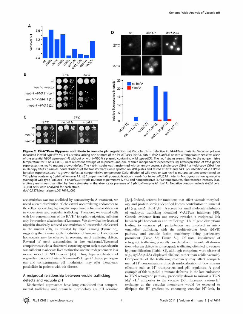
accumulation was not abolished by concanamycin A treatment, we
noted altered distribution of cholesterol accumulating endosomes to
the cell periphery, highlighting the importance of luminal acidification
in endocytosis and vesicular trafficking. Therefore, we treated cells
with low concentrations of the K+/H+ ionophore nigericin, sufficient
only for transient alkalization of lysosomes. We show that low levels of
nigericin drastically reduced accumulation of unesterified cholesterol
in the mutant cells, as revealed by filipin staining (Figure 3d),
suggesting that a more subtle modulation of lumenal pH and cation
homeostasis may be effective in reversing sterol trafficking defects.
Reversal of sterol accumulation in late endosomal/llysosomal
compartments with a cholesterol extracting agent such as cyclodextrin
was sufficient to alleviate liver dysfunction and neurodegeneration in a
mouse model of NPC disease [45]. Thus, hyperacidification of
organelles may contribute to Niemann-Pick type C disease pathogen-
esis and compartmental pH modulation may offer therapeutic
possibilities in patients with this disease.
A reciprocal relationship between vesicle traffickingdefects and vacuole pH
Biochemical approaches have long established that compart-
mental trafficking and organelle morphology are pH sensitive
[3,4]. Indeed, screens for mutations that affect vacuole morphol-
ogy and protein sorting identified known contributors to lumenal
pH (e.g. vmaD) [46,47,48]. A screen for small molecule inhibitors
of endocytic trafficking identified V-ATPase inhibitors [49].
Genetic evidence from our survey revealed a reciprocal link
between pH homeostasis and trafficking: 11% of gene disruptions
leading to vacuolar pH perturbation are involved in inter-
organellar trafficking, with the multivesicular body (MVB)
pathway and vacuole fusion machinery being particularly
prominent (Table S3; Figure S2). Of note, impairment of
retrograde trafficking generally correlated with vacuole alkaliniza-
tion, whereas defects in anterograde trafficking often led to vacuole
hyperacidification (Table S2), although exceptions were observed
(e.g., snf7D/vps32D displayed alkaline, rather than acidic vacuole).
Components of the trafficking machinery may affect compart-
mental H+ concentrations through mislocalization of downstream
effectors such as H+ transporters and pH regulators. A good
example of this is vps5D, a mutant defective in the late endosome
to TGN retrograde pathway, previously shown to missort a TGN
Mg2+/H+ antiporter to the vacuole [50]. Increased cation/H+
exchange at the vacuolar membrane would be expected to
dissipate the H+ gradient by enhancing vacuolar H+ leak. In
Figure 2. P4-ATPase flippases contribute to vacuole pH regulation. (a) Vacuolar pH is defective in P4-ATPase mutants. Vacuolar pH wasmeasured in wild type BY4742 cells, strains lacking one or more of the P4-ATPases (drs2D, dnf1D, dnf2D, dnf3D) or with a temperature sensitive alleleof the essential NEO1 gene (neo1-1) without or with (+NEO1) a plasmid containing wild type NEO1. The neo1 strains were shifted to the nonpermisivetemperature for 1 hour (34uC). Data represent average of duplicates and one of three independent experiments. (b) Overexpression of VMA genessuppresses the neo1-1 mutant growth defect. The neo1-1 strain was transformed with an empty vector, a single copy VMA11, a multi-copy VMA11, ormulti-copy VMA5 plasmids. Serial dilutions of the transformants were spotted on YPD plates and tested at 27uC and 34uC. (c) Inhibition of V-ATPasefunction suppresses neo1-ts growth defect at nonpermissive temperature. Serial dilution of wild type or two neo1-ts mutant cultures were tested onYPD plates containing 5 mM bafilomycin A1. (d) Compartmental hyperacidification in neo1-1 or triple dnf1,2,3D mutants. Micrographs show quinacrinestaining of wild type (wt), neo1-1 or dnf1,2,3D triple mutants at permissive (27uC) and nonpermissive (37uC) temperatures. Fluorescence intensity (a.u.,arbitrary units) was quantified by flow cytometry in the absence or presence of 5 mM bafilomycin A1 (baf A). Negative controls include drs2D cells.30,000 cells were analyzed for each strain.doi:10.1371/journal.pone.0017619.g002
Genome Wide Analysis of Vacuole pH
PLoS ONE | www.plosone.org 4 March 2011 | Volume 6 | Issue 3 | e17619

Figure 3. Lysosomal hyperacidification accompanies a cellular Niemann-Pick type C phenotype. (a) Vacuoles in ergosterol accumulatingncr1D cells are hyperacidic, whereas erg mutants lacking ergosterol have abnormally alkaline vacuoles. (b) Intracellular sterol accumulates in ncr1Dyeast. Yeast cells were stained with filipin to detect sterol in the presence or absence of NCR1. Bar, 2 mm. (c) Fibroblasts harboring pathogenic allelesof NPC1 (P237S/I1061T) have hyperacidified lysosomes. Wild type cells from a control subject (wt) or mutant cells from a patient with Niemann-Pick
Genome Wide Analysis of Vacuole pH
PLoS ONE | www.plosone.org 5 March 2011 | Volume 6 | Issue 3 | e17619
validation of this hypothesis, we observed prominent alkalinization
of pHv in vps5D as well as other mutants lacking components of the
retromer complex (vps5D, vps17D, vps29D; Table S3) [51].
Similarly, anterograde trafficking mutants might disrupt localiza-
tion and function of vacuolar/late endosomal transporters that
contribute to the H+ leak, with ensuing vacuolar hyperacidifica-
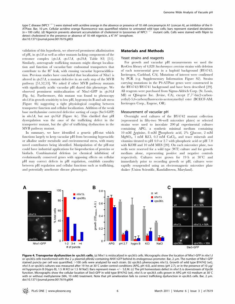
tion. Previous studies have concluded that localization of Nhx1 is
altered in vps27D, a mutant defective in an early step of the MVB
pathway [51,52,53]. We asked if other MVB pathway mutants
with significantly acidic vacuolar pH shared this phenotype. We
observed prominent mislocalization of Nhx1-GFP in vps36D(Fig. 4a). Furthermore, this mutant was found to phenocopy
nhx1D in growth sensitivity to low pH, hygromycin B and salt stress
(Figure 4b) suggesting a tight physiological coupling between
transporter function and cellular localization. Addition of the weak
base methylamine corrected defective sorting of cargo (Ste3-GFP)
in nhx1D, but not vps36D (Figure 4c). This clarified that pH
dysregulation was the cause of the trafficking defect in the
transporter mutant, but the effect of trafficking dysfunction in the
MVB pathway mutant.
In summary, we have identified a genetic pH-stat which
functions largely to keep vacuolar pH from becoming hyperacidic
or alkaline under metabolic and environmental stress, with many
novel contributors being identified. Manipulation of the pH-stat
could have industrial applications for bioproduction of proteins or
biofuels. Combinatorial deletion (or chemical inhibition) of
evolutionarily conserved genes with opposing effects on cellular
pH may correct defects in pH regulation, establish causality
between pH regulation and cellular functions such as trafficking,
and potentially ameliorate disease phenotypes.
Materials and Methods
Yeast strains and reagentsFor growth and vacuolar pH measurements we used the
ResGen library of 4,828 Saccharomyces cerevisiae strains with deletion
of each nonessential gene in a haploid background (BY4742;
Invitrogen, Carlsbad, CA). Mutations of interest were confirmed
by PCR (e.g. Supplementary Information Figure S2). Strains
carrying mutations in the P4-ATPase genes were constructed in
the BY4742/BY4741 background and have been described [29].
All reagents were purchased from Sigma-Aldrich Corp. (St. Louis,
MI) or Qbiogene Inc. (Irvine, CA), except 29,79-bis(2-carbox-
yethyl)-5,6-carboxyfluorescein-acetoxymethyl ester (BCECF-AM;
Invitrogen Corp., Eugene, OR).
Measurement of vacuolar pHOvernight seed cultures of the BY4742 mutant collection
(represented in fifty-two 96-well microtiter plates) or selected
strains were used to inoculate 200 ml experimental cultures
containing APG, a synthetic minimal medium containing
10 mM Arginine, 8 mM Phosphoric acid, 2% Glucose, 2 mM
MgSO4, 1 mM KCl, 0.2 mM CaCl2, and trace minerals and
vitamins titrated to pH 4.0 or 2.7 with phosphoric acid or pH 7.0
with KOH and 10 mM MES [20]. On each microtiter plate, two
wells were reserved for a wild type (WT) culture and for growth
medium alone, representing positive and negative controls
respectively. Cultures were grown for 19 h at 30uC and
immediately prior to recording growth or pH, cultures were
rapidly resuspended using an electromagnetic microtiter plate
shaker (Union Scientific, Randallstown, Maryland).
Figure 4. Transporter dysfunction in vps36D cells. (a) Nhx1 is mislocalized in vps36D cells. Micrographs show the location of Nhx1-GFP in nhx1Dor vps36D cells transformed with the 2 m plasmid pRin82 containing NHX1:GFP behind its endogenous promoter. Bar, 2 mm. The number of Nhx1-GFPstained puncta per cell was quantified; .100 cells were analyzed for each strain. (b) vps36D phenocopies nhx1D. Growth of wild type BY4742 (wt),nhx1D or vps36D cultures was measured after 19 hrs at 30uC under control conditions (APG, pH 4.0), acid stress (pH 2.7), or in the presence of 10 mg/ml hygromycin B (Hygro B), 1.5 M KCl or 1.5 M NaCl. Bars represent mean +/2 S.E.M. (c) The pH homeostasis defect in nhx1D is downstream of Vps36function. Micrographs show the cellular location of Ste3-GFP in wild type BY4742 (wt), nhx1D or vps36D cells grown in APG pH 4.0 medium at 30uCwith or without methylamine (MA, 10 mM) treatment. Note that pH amelioration fails to correct trafficking dysfunction in vps36D cells. Bar, 2 mm.doi:10.1371/journal.pone.0017619.g004
type C disease (NPC12/2) were stained with acridine orange in the absence or presence of 10 nM concanamycin A1 (concan A), an inhibitor of the V-ATPase. Bar, 10 mm. Cellular acridine orange fluorescence was quantified relative to untreated wild type cells; bars represent standard deviations(n = 100 cells). (d) Nigericin prevents aberrant accumulation of cholesterol in lysosomes of NPC12/2 mutant cells. Cells were stained with filipin todetect cholesterol in the presence or absence of 10 nM nigericin, a K+/H+ ionophore.doi:10.1371/journal.pone.0017619.g003
Genome Wide Analysis of Vacuole pH
PLoS ONE | www.plosone.org 6 March 2011 | Volume 6 | Issue 3 | e17619

Vacuolar pH measurements were performed using methods
previously described with modification to accommodate high-
throughput analysis [25]. Briefly, after growth was assessed,
experimental cultures were incubated with 50 mM 29,79-bis(2-
carboxyethyl)-5,6-carboxyfluorescein-acetoxymethyl ester (BCECF-
AM; Molecular Probes, Eugene, OR) at 30uC for 30 min., and then
washed and resuspended in APG medium at the appropriate pH.
Single fluorescence intensity (excitation at 485 nm, emission at
520 nm) and absorbance (at 600 nm) were then measured. Ninety-six
independent calibration experiments were performed and vacuolar
pH values were calculated as described previously. All fluorescence
and absorbance readings were taken at 30uC using a BMG
FLUOstar Optima multimode plate reader with accompanying
BMG FLUOstar Optima Version 1.20-0 software (BMG Lab-
technologies, Durham, NC). Accurate pH estimations could not be
made when strains showed severe growth defects (,20% of wild type
growth) or poor loading, poor expression or mislocalization of the
pH-sensitive probe. Vacuolar pH was not assessed for 6.7% of the
mutant collection; vacuolar pH measurements were recorded from
4469, 4606, or 4593 mutants under acid stress, control conditions or
alkali stress respectively.
Data analysisMutant strains showing ,20% of WT growth under control
conditions (pH 4.0) were omitted from analysis. Medians and
median absolute deviations (MADs) were calculated for each
dataset and descriptive statistical analysis was performed. Mutant
strains with vacuolar pH values above the maximum or below the
minimum pHv observed in the wild type population (n = 46) were
considered to have abnormally alkaline or acidic vacuoles,
respectively (Figure 1a; Supplementary Information Table S1).
Data were organized, plotted and analyzed using Excel X
(Microsoft Corp., Redmond, WA), JMP vs. 5.0.1a (SAS Institute,
Carey, NC), and KaliedaGraph vs. 4.0 (Synergy Software
Technologies Inc., Essex Junction, VT).
Isolation of the neo1-1 temperature sensitive allele andits suppressors
A Yep13 genomic library (ATCC) was used for the neo1-1 multi-
copy suppressor screen. Strain ZHY628-15B (BY4742 neo1Dcontaining p413-neo1-1) was transformed with library DNA and
incubated at 27uC. Transformants were then replicated and
incubated at 37uC. Plasmids rescued from suppressor colonies
were transformed again into ZHY628-15B and tested for growth
at 37uC. A SpeI/SnaBI fragment of one suppressor clone (named
pNS7) was inserted into pRS315 or pRS425 to generate the single
copy or multi-copy VMA11 plasmids. The plasmid containing
VMA5 (Yep352-VMA5) was a gift from Patricia Kane (SUNY
Health Sciences Center, Syracuse, NY).
Quinacrine stainingFor quinacrine staining, strains BY4741 (WT), ZHY124-15B1B
(neo1-ts), PFY3273A (dnf1,2,3D), ZHY615D1C (drs2D) were grown
to mid-logarithmic phase in YPD at 27uC and half of the neo1-ts
strain was shifted to 37uC for 2 hrs. Each culture was treated with
or without bafilomycin A (5 mM) for 25 minutes prior to
harvesting. Cells were resuspended in YPD medium containing
100 mM Hepes pH 7.6, 200 mM quinacrine, 25 mg/ml propidium
iodide with or without 5 mM bafilomycin A, and stained for 8 min
at 27 or 37uC as indicated in Figure 2d. Stained cells were washed
three times with 100 mM Hepes pH 7.6, 0.2% glucose and stored
on ice prior measuring the fluorescence intensity of 30,000 living
cells using the FITC channel of a fluorescence activated cell sorter.
The fluorescence intensity of mock-treated cells incubated without
quinacrine were used to subtract background fluorescence. Images
of stained cells were captured using a Zeiss Axioplan fluorescence
microscope, a cooled CCD camera and MetaMorph 4.5 software
(Molecular Devices, Sunnyvale, CA).
Mammalian cell culture and microscopyWild type (GM05659, designated wt) and Niemann-Pick Type
C1 (GM03123, designated NPC12/2, a gift from Dr. Laura
Liscum, Tufts University School of Medicine) [54] fibroblasts were
cultured according to Coriell Cell Repositories recommendations:
minimal essential media supplemented with 10% Fetal Bovine
Serum, nonessential amino acids and penicillin/streptomycin at
37uC, 5% CO2 in a humidified incubator. Cells were plated onto
glass coverslips 24 hours prior to treatment to allow adherence
then grown an additional 48 hours in the presence of either
DMSO, 10 nM Concanamycin A, 10 nM Nigericin or left
untreated. Cells were incubated with 1 mM acridine orange in
standard bath solution for 10 min at 37uC [54]. Images were
acquired with the same parameters (exposure time and fluores-
cence intensity) using a Zeiss Axiovert fluorescent microscope
equipped with a Photometrics CoolSnap CCD camera. Average
fluorescence of 4 independent fields of wild type and NPC1 cells
for each treatment was measured using MetaMorph image
analysis software (Molecular Devices Corperation, Downingtown,
PA). Filipin staining was performed by exposing paraformaldehyde
fixed cells (3% in PBS) to 25 mg/ml filipin for 30 minutes at room
temperature as described [55]. Samples were visualized as
described above.
Supporting Information
Figure S1 Growth of yeast mutant strains does notcorrelate with pHv. Defects in vacuole pH do not correlate with
poor growth. Vacuole pH values shown in Figure 1b n ($4469)
are plotted against yeast culture growth measured under acidic
(top), standard (middle) or alkaline (bottom panel) conditions. Resulting
log-log plots indicate that vacuole pH and growth are independent
variables.
(PDF)
Figure S2 Vesicle trafficking defects lead to pHv
dysregulation. Mutants with defective pHv also identified by
genome-wide screens for endocytic trafficking defects and/or
vacuole fusion defects are shown (numbers indicated in parenthe-
ses). Arrows indicate directional trafficking pathways between the
following organelles: vacuole (Vac), late endosome (LE), intralu-
menal vesicles of multivesicular bodies (ILV), trans-Golgi network
(TGN), Golgi apparatus (GA), endoplasmic reticulum (ER),
nucleus (Nu), cytoplasmic (Cyto), plasma membrane (PM), and
cell wall (CW); the V-ATPase, autophagic process and cytoskel-
eton are also shown. The mutants identified either showed basic
(blue) or acidic vacuole (red). The pHv of mutants shown are listed
in Supplementary Table S2.
(PDF)
Table S1 Complete dataset of yeast vacuole pH andgrowth measured at three external pH values. Vacuole
pH and culture growth (Absorbance at 600 nm) values for 4,606
yeast single gene deletion strains examined in this study. Prior to
recording, cultures were grown for 19 hours in APG media
adjusted to pH 2.7, 4.0 or 7.0. Vacuole pH datasets (also plotted in
Figure 1) were sorted and strains are shown in order from lowest to
highest vacuole pH. In addition to the sample size, median, and
Genome Wide Analysis of Vacuole pH
PLoS ONE | www.plosone.org 7 March 2011 | Volume 6 | Issue 3 | e17619

MAD, acidic and basic thresholds and lists of outliers are shown
for each condition. Growth datasets were grouped and sorted;
strains are shown in order from lowest to highest growth measured
under control conditions. Unsorted vacuole pH datasets are also
provided.
(XLS)
Table S2 Major functional groups of the vacuolar pH-stat. Genes identified in the vacuolar pH survey of the yeast single
gene deletion library were sorted into functional categories. Major
functional groups of membrane phospholipid and sterol distribu-
tion, endocytic trafficking and transporters are listed. Growth and
vacuole pH values obtained from strains with gene deletions are
shown and outlying vacuole pH values are indicated (acidic, red;
basic, blue).
(XLS)
Table S3 Summary of major contributors to vacuolarpH-stat. Summary of 107 genes identified as contributors to the
vacuole pH-stat under more than one external pH condition (see
Figure 1d). The genes are sorted by effect of deletion on vacuole
pH and by cellular function.
(PDF)
Acknowledgments
We thank Dr. Patricia Kane for the Yep-352-VMA5 plasmid and Dr.
Laura Liscum for providing the NPC1 null fibroblasts.
Author Contributions
Conceived and designed the experiments: CLB LK YZ RG ZH MD TRG
RR. Performed the experiments: CLB LK YZ RG ZH AC. Analyzed the
data: CLB LK YZ RG ZH MD TRG RR. Wrote the paper: CLB MD
TRG RR.
References
1. Kaufmann SH (2008) Immunology’s foundation: the 100-year anniversary of theNobel Prize to Paul Ehrlich and Elie Metchnikoff. Nat Immunol 9: 705–712.
2. de Duve C (2005) The lysosome turns fifty. Nat Cell Biol 7: 847–849.
3. Heuser J (1989) Changes in lysosome shape and distribution correlated withchanges in cytoplasmic pH. J Cell Biol 108: 855–864.
4. Mellman I (1992) The importance of being acid: the role of acidification inintracellular membrane traffic. J Exp Biol 172: 39–45.
5. Maranda B, Brown D, Bourgoin S, Casanova JE, Vinay P, et al. (2001) Intra-
endosomal pH-sensitive recruitment of the Arf-nucleotide exchange factor
ARNO and Arf6 from cytoplasm to proximal tubule endosomes. J Biol Chem276: 18540–18550.
6. Nishi T, Forgac M (2002) The vacuolar (H+)-ATPases–nature’s most versatile
proton pumps. Nat Rev Mol Cell Biol 3: 94–103.
7. Weisz OA (2003) Organelle acidification and disease. Traffic 4: 57–64.
8. Futerman AH, van Meer G (2004) The cell biology of lysosomal storage
disorders. Nat Rev Mol Cell Biol 5: 554–565.
9. Huynh KK, Grinstein S (2007) Regulation of vacuolar pH and its modulation by
some microbial species. Microbiol Mol Biol Rev 71: 452–462.
10. Walls KC, Ghosh AP, Franklin AV, Klocke BJ, Ballestas M, et al. Lysosome
dysfunction triggers Atg7-dependent neural apoptosis. J Biol Chem 285:10497–10507.
11. Grabe M, Oster G (2001) Regulation of organelle acidity. J Gen Physiol 117:
329–344.
12. Roos A, Boron WF (1981) Intracellular pH. Physiol Rev 61: 296–434.
13. Steinberg BE, Huynh KK, Brodovitch A, Jabs S, Stauber T, et al. (2010) A
cation counterflux supports lysosomal acidification. J Cell Biol 189: 1171–1186.
14. Steinberg BE, Touret N, Vargas-Caballero M, Grinstein S (2007) In situ
measurement of the electrical potential across the phagosomal membrane usingFRET and its contribution to the proton-motive force. Proc Natl Acad Sci U S A
104: 9523–9528.
15. Graham LA, Flannery AR, Stevens TH (2003) Structure and assembly of the
yeast V-ATPase. J Bioenerg Biomembr 35: 301–312.
16. Kane PM (2006) The where, when, and how of organelle acidification by the
yeast vacuolar H+-ATPase. Microbiol Mol Biol Rev 70: 177–191.
17. Forgac M (2007) Vacuolar ATPases: rotary proton pumps in physiology andpathophysiology. Nat Rev Mol Cell Biol 8: 917–929.
18. Brett CL, Donowitz M, Rao R (2006) Does the proteome encode organellar pH?FEBS Lett 580: 717–719.
19. Ohgaki R, Matsushita M, Kanazawa H, Ogihara S, Hoekstra D, et al. (2010)
The Na+/H+ exchanger NHE6 in the endosomal recycling system is involved in
the development of apical bile canalicular surface domains in HepG2 cells. MolBiol Cell 21: 1293–1304.
20. Brett CL, Tukaye DN, Mukherjee S, Rao R (2005) The yeast endosomal
Na+K+/H+ exchanger Nhx1 regulates cellular pH to control vesicle trafficking.
Mol Biol Cell 16: 1396–1405.
21. Nakamura N, Tanaka S, Teko Y, Mitsui K, Kanazawa H (2005) Four Na+/H+exchanger isoforms are distributed to Golgi and post-Golgi compartments and
are involved in organelle pH regulation. J Biol Chem 280: 1561–1572.
22. Gilfillan GD, Selmer KK, Roxrud I, Smith R, Kyllerman M, et al. (2008) SLC9A6
mutations cause X-linked mental retardation, microcephaly, epilepsy, and ataxia, aphenotype mimicking Angelman syndrome. Am J Hum Genet 82: 1003–1010.
23. Morrow EM, Yoo SY, Flavell SW, Kim TK, Lin Y, et al. (2008) Identifyingautism loci and genes by tracing recent shared ancestry. Science 321: 218–223.
24. Franke B, Neale BM, Faraone SV (2009) Genome-wide association studies in
ADHD. Hum Genet 126: 13–50.
25. Ali R, Brett CL, Mukherjee S, Rao R (2004) Inhibition of sodium/proton
exchange by a Rab-GTPase-activating protein regulates endosomal traffic inyeast. J Biol Chem 279: 4498–4506.
26. Pomorski T, Lombardi R, Riezman H, Devaux PF, van Meer G, et al. (2003)
Drs2p-related P-type ATPases Dnf1p and Dnf2p are required for phospholipid
translocation across the yeast plasma membrane and serve a role in endocytosis.
Mol Biol Cell 14: 1240–1254.
27. Natarajan P, Wang J, Hua Z, Graham TR (2004) Drs2p-coupled aminopho-
spholipid translocase activity in yeast Golgi membranes and relationship to in
vivo function. Proc Natl Acad Sci U S A 101: 10614–10619.
28. Zhou X, Graham TR (2009) Reconstitution of phospholipid translocase activity
with purified Drs2p, a type-IV P-type ATPase from budding yeast. Proc Natl
Acad Sci U S A 106: 16586–16591.
29. Hua Z, Fatheddin P, Graham TR (2002) An essential subfamily of Drs2p-related
P-type ATPases is required for protein trafficking between Golgi complex and
endosomal/vacuolar system. Mol Biol Cell 13: 3162–3177.
30. Keenan Curtis K, Kane PM (2002) Novel vacuolar H+-ATPase complexes
resulting from overproduction of Vma5p and Vma13p. J Biol Chem 277:
2716–2724.
31. Nikawa J, Yonemura K, Yamashita S (1983) Yeast mutant with thermolabile
CDP-choline synthesis. Isolation and characterization of a cholinephosphate
cytidyltransferase mutant. Eur J Biochem 131: 223–229.
32. Riekhof WR, Wu J, Gijon MA, Zarini S, Murphy RC, et al. (2007)
Lysophosphatidylcholine metabolism in Saccharomyces cerevisiae: the role of
P-type ATPases in transport and a broad specificity acyltransferase in acylation.
J Biol Chem 282: 36853–36861.
33. Muthusamy BP, Raychaudhuri S, Natarajan P, Abe F, Liu K, et al. (2009)
Control of protein and sterol trafficking by antagonistic activities of a type IV P-
type ATPase and oxysterol binding protein homologue. Mol Biol Cell 20:
2920–2931.
34. Roelants FM, Baltz AG, Trott AE, Fereres S, Thorner J (2010) A protein kinase
network regulates the function of aminophospholipid flippases. Proc Natl Acad
Sci U S A 107: 34–39.
35. Sakano K (1998) Revision of Biochemical pH-Stat: Involvement of Alternative
Pathway Metabolisms. Plant Cell Physiology 39: 467–473.
36. Felle HH (2005) pH regulation in anoxic plants. Ann Bot 96: 519–532.
37. Piper P, Mahe Y, Thompson S, Pandjaitan R, Holyoak C, et al. (1998) The
pdr12 ABC transporter is required for the development of weak organic acid
resistance in yeast. Embo J 17: 4257–4265.
38. Marini AM, Matassi G, Raynal V, Andre B, Cartron JP, et al. (2000) The
human Rhesus-associated RhAG protein and a kidney homologue promote
ammonium transport in yeast. Nat Genet 26: 341–344.
39. Palma M, Seret ML, Baret PV (2009) Combined phylogenetic and neighbour-
hood analysis of the hexose transporters and glucose sensors in yeasts. FEMS
Yeast Res 9: 526–534.
40. Zhang YQ, Gamarra S, Garcia-Effron G, Park S, Perlin DS, et al. (2010)
Requirement for ergosterol in V-ATPase function underlies antifungal activity of
azole drugs. PLoS Pathog 6: e1000939.
41. Loftus SK, Morris JA, Carstea ED, Gu JZ, Cummings C, et al. (1997) Murine
model of Niemann-Pick C disease: mutation in a cholesterol homeostasis gene.
Science 277: 232–235.
42. Berger AC, Hanson PK, Wylie Nichols J, Corbett AH (2005) A yeast model
system for functional analysis of the Niemann-Pick type C protein 1 homolog,
Ncr1p. Traffic 6: 907–917.
43. Lin M, Grillitsch K, Daum G, Just U, Hofken T (2009) Modulation of sterol
homeostasis by the Cdc42p effectors Cla4p and Ste20p in the yeast
Saccharomyces cerevisiae. Febs J 276: 7253–7264.
44. Battisti C, Tarugi P, Dotti MT, De Stefano N, Vattimo A, et al. (2003) Adult
onset Niemann-Pick type C disease: A clinical, neuroimaging and molecular
genetic study. Mov Disord 18: 1405–1409.
Genome Wide Analysis of Vacuole pH
PLoS ONE | www.plosone.org 8 March 2011 | Volume 6 | Issue 3 | e17619

45. Liu B, Turley SD, Burns DK, Miller AM, Repa JJ, et al. (2009) Reversal of
defective lysosomal transport in NPC disease ameliorates liver dysfunction and
neurodegeneration in the npc12/2 mouse. Proc Natl Acad Sci U S A 106:
2377–2382.
46. Bonangelino CJ, Chavez EM, Bonifacino JS (2002) Genomic screen for vacuolar
protein sorting genes in Saccharomyces cerevisiae. Mol Biol Cell 13: 2486–2501.
47. Preston RA, Reinagel PS, Jones EW (1992) Genes required for vacuolar acidity
in Saccharomyces cerevisiae. Genetics 131: 551–558.
48. Seeley ES, Kato M, Margolis N, Wickner W, Eitzen G (2002) Genomic analysis
of homotypic vacuole fusion. Mol Biol Cell 13: 782–794.
49. Nieland TJ, Feng Y, Brown JX, Chuang TD, Buckett PD, et al. (2004) Chemical
genetic screening identifies sulfonamides that raise organellar pH and interfere
with membrane traffic. Traffic 5: 478–492.
50. Borrelly G, Boyer JC, Touraine B, Szponarski W, Rambier M, et al. (2001) The
yeast mutant vps5Delta affected in the recycling of Golgi membrane proteins
displays an enhanced vacuolar Mg2+/H+ exchange activity. Proc Natl Acad
Sci U S A 98: 9660–9665.51. Schellmann S, Pimpl P (2009) Coats of endosomal protein sorting: retromer and
ESCRT. Curr Opin Plant Biol 12: 670–676.
52. Bowers K, Levi BP, Patel FI, Stevens TH (2000) The sodium/proton exchangerNhx1p is required for endosomal protein trafficking in the yeast Saccharomyces
cerevisiae. Mol Biol Cell 11: 4277–4294.53. Nickerson DP, Russell MR, Odorizzi G (2007) A concentric circle model of
multivesicular body cargo sorting. EMBO Rep 8: 644–650.
54. Jacobs NL, Andemariam B, Underwood KW, Panchalingam K, Sternberg D,et al. (1997) Analysis of a Chinese hamster ovary cell mutant with defective
mobilization of cholesterol from the plasma membrane to the endoplasmicreticulum. J Lipid Res 38: 1973–1987.
55. Kiselyov K, Chen J, Rbaibi Y, Oberdick D, Tjon-Kon-Sang S, et al. (2005)TRP-ML1 is a lysosomal monovalent cation channel that undergoes proteolytic
cleavage. J Biol Chem 280: 43218–43223.
Genome Wide Analysis of Vacuole pH
PLoS ONE | www.plosone.org 9 March 2011 | Volume 6 | Issue 3 | e17619


