
Glutamaie-evoked release of ATP from cortical astroglial cells
Gloria Queiroz Faculty of Pharmacy University of Porto, 1999

Faculdade de Farmácia
Universidade do Porto
Glutamate-evoked release of ATP from cortical astroglial cells
Dissertação de candidatura ao grau de Doutor
apresentada à Faculdade de Farmácia da
Universidade do Porto
Maria da Glória Correia da Silva Queiroz
Porto, 1999
Ao meu pai
À minha mãe

The experimental work was carried out in the Phamakologisches Institut, Albert-Lwdwigs-
Universitãt, Freiburg i.Br. and was supported by the Deutsche Forschungsgemeinschaft
(SFB 505) and by the European Commission (BMH4 CT96-0676).
The results presented in this thesis were published in:
Queiroz G, Gebicke-Haerter PJ, Schobert A, Starke K, von Kugelgen I (1997) Release of
ATP from cultured rat astrocytes elicited by glutamate receptor activation. Neuroscience 78,
1203-12088.
Queiroz G, Meyer DK, Meyer A, Starke K, von Kùgelgen I (1999) A study of the mechanism
of the release of ATP from rat cortical astroglial cells evoked by activation of glutamate
receptors. Neuroscience 91, 1171-1181.

Table of contents
Summary
Resumo
Résumé
Abbreviat ions
Glial Cells
The astrocyte Classification Astrocyte physiology
Communication between glial cells and neurones Glial cells as targets of substances released by Glial cells as modulators of neuronal activity
Control of neuronal microenvironment Release of neuroactive mediators
Study of glial cells Astrocytes in culture
Glutamate in the CNS
lonotropic glutamate receptors AM PA receptors Kainate receptors NMDA receptors
Metabotropic glutamate receptors Glutamate receptors on glial cells
Cellular communicat ion via ATP in CNS
Purinergic receptors
Role of ATP in the CNS
ATP sources and release mechanisms

Aims 31
Experimental Procedures 32
Cell cultures 32 Immunocytochemistry 33 ATP release 33 Lactate dehydrogenase release 35 Materials 36 Statistics 37
Results 38
ATP release: effect of glutamate receptor agonists 38
ATP release: fraction released 41
ATP release: possible contribution of neurones 43
ATP release: interaction with antagonists 46
Lactate dehydrogenase release 46 ATP release: effect of ionomycin 49 ATP release: omission of extracellular calcium 49 ATP release: effect of blockers of voltage-dependent Ca2+ channels 53 ATP release: effect of a-latrotoxin and botulinum toxin B 53 ATP release: interaction with atractyloside and DIDS 57 ATP release: interaction with glibenclamide and DPC 57 ATP release: interaction with lithium 62
Discussion 63
The role of Ca2+ in ATP release 67 On the mechanism of ATP release 68
Neurone-like exocytosis? 69 Membrane passage by means of channels or transporters? 70
The effect of lithium 72
Conclusions 7 5
References 7 6
Acknowledgements 9 1
1
Summary
Excitatory amino acids are known to release adenyl compounds in the brain. Both the
possibility that astroglial cells may release ATP in response to excitatory amino acids and the
mechanism of that release were studied in astrocyte cultures derived from the brain
hemispheres of newborn rats. ATP was measured with the luciferine-luciferase assay.
In primary cultures of astrocytes, glutamate receptor agonists did not increase the
release of lactate dehydrogenase but elicited the release of ATP. There was a basal efflux of
ATP, which was increased up to 19-fold by glutamate (100-1000 uM), N-methyl-D-aspartate
(NMDA; 20-500 uM), a-amino-3-hydroxy-5-methylisoxazole-4-propionate (AMPA; 30-100
uM), kainate (20 uM) and (1S,3R)-1-amino-cyclopentane-1,3-dicarboxylic acid (frans-ACPD;
100-1000 uM). The NMDA receptor-selective antagonist 2-amino-5-phosphonopentanoate
(AP5, 100 uM) blocked the effect of NMDA but not the effects of AMPA, kainate and
glutamate. The AMPA receptor-selective antagonist 2,3-dihydroxy-6-nitro-7-sulfamoyl-
benzo(f)quinoxaline (NBQX; 30 uM) blocked the effect of AMPA and also of glutamate and
NMDA but not the effect of kainate. The kainate receptor-selective antagonist y-D-glutamyl-
amino-methanesulfonate (GAMS; 30 uM) blocked the effect of kainate but not of glutamate.
The calcium ionophore ionomycin (5 uM) elicited release of ATP but only in the
presence of external calcium. The release of ATP elicited by NMDA and kainate was
abolished (or greatly reduced) by withdrawal of extracellular calcium as well as by cadmium
(1 mM) and nicardipine (10 uM). In contrast, the release of ATP elicited by AMPA was not
changed by these interventions; ATP release elicited by frans-ACPD was also not changed

2
by withdrawal of external calcium. a-Latrotoxin failed to elicit ATP release. Some putative
blockers of ATP transporters or channels were tested on the ATP release elicited by
glutamate receptor agonists. Only the two blockers of the cystic fibrosis transmembrane
conductance regulator (CFTR), glibenclamide (100 uM) and diphenylamine-2-carboxylate
(500 uM) changed the release of ATP. Both reduced the effect of AMPA without changing
the effects of NMDA and kainate (only glibenclamide tested). Furthermore, lithium (1 mM)
abolished the release of ATP evoked by glutamate and AMPA but only reduced the release
elicited by NMDA and kainate.
The present study shows that cultured astroglial cells respond to activation of NMDA,
AMPA, and kainate receptors with release of ATP. Two different mechanisms seem to be
involved in the release of ATP from astroglial cells upon activation of ionotropic glutamate
receptors: one involved in the NMDA- and kainate-induced release of ATP, the other in
AMPA-induced release. The NMDA- and kainate-induced release of ATP requires an influx
of calcium, is not due to neurone-like exocytosis, is not mediated by CFTR or a mechanism
regulated by CFTR, and is reduced but not abolished by lithium. The AMPA-induced release
does not require extracellular calcium may be mediated by CFTR or a mechanism regulated
by CFTR, and is abolished by lithium.
By showing that astrocytes may release a neuroactive substance, namely ATP, with
known modulatory and trophic actions in the central nervous system, this study supports the
view that glial cells are intimately involved in the active control of neuronal activity and should
be considered as integral modulatory elements of synapses.
3
Resumo
Vários estudos demonstraram que os aminoácidos excitatórios induzem a libertação
de purinas (nomeadamente adenosina) no cérebro. Utilizando como modelo culturas
primárias de astrócitos obtidas de cérebro de rato recém-nascido, estudou-se a
possibilidade das células gliais, em particular os astrócitos, poderem libertar ATP no cérebro
em resposta é estimulação com aminoácidos excitatórios bem como o mecanismo de
libertação envolvido. O ATP libertado foi quantificado pelo método da luciferina-luciferase.
Em culturas primárias de astrócitos, nenhum dos agonistas dos receptores do
glutamato testados aumentou a libertação da enzima lactato desidrogenase, embora o
efluxo basal de ATP tenha sido aumentado (até cerca de 19 vezes) pelo glutamato (100-
1000 uM), N-metil-D-aspartato (NMDA; 20-500 uM), ácido a-amino-3-hidroxi-5-metilisoxazol-
4-propiónico (AMPA; 30-100 uM), kainato (20 uM) e pelo ácido (1S,3R)-1-amino-
ciclopentano-1,3-dicarboxilico (trans-ACPD; 100-1000 uM). O antagonista selectivo dos
receptores do NMDA, o ácido 2-amino-5-fosfono pentanóico (AP5, 100 uM), bloqueou o
efeito do NMDA mas não modificou os efeitos do AMPA, kainato e glutamato. O antagonista
selectivo dos receptores do AMPA, 2,3-dihidroxi-6-nitro-7-sulfamoil-benzo(f)quinoxalina
(NBQX; 30 uM) antagonizou o efeito do AMPA e também os efeitos do glutamato e NMDA,
mas não modificou a libertação de ATP induzida pelo kainato. O antagonista selectivo dos
receptores do kainato, y-D-glutamil-amino-metanosulfonato (GAMS; 30 uM), bloqueou o
efeito do kainato mas não modificou significativamente o efeito do glutamato.

4
A ionomicina (5 |jM), um ionóforo do cálcio, induziu a libertação de ATP mas apenas
na presença de cálcio no meio extracelular. A libertação de ATP induzida pelo NMDA e pelo
kainato foi abolida ou significativamente reduzida com a omissão de cálcio do meio
extracelular, na presença de cádmio (1 mM) ou na presença de nicardipina (10 uM). Pelo
contrário, a libertação de ATP induzida pelo AMPA não sofreu qualquer alteração pelas
intervenções referidas e a libertação de ATP induzida pelo trans-ACPD também não sofreu
alteração quando o cálcio extracelular foi omitido do meio. A a-latrotoxina não teve qualquer
efeito sobre a libertação de ATP. Alguns compostos com potencialidade de bloquear
transportadores ou canais permeáveis ao ATP foram testados sobre a libertação de ATP
induzida pelos agonistas dos receptores do glutamato. Dos compostos testados apenas dois
bloqueadores do CFTR (cystic fibrosis transmembrane conductance regulator), a
glibenclamida (100 uM) e o ácido difenilamino-2-carboxílico (500 uM), modificaram a
libertação de ATP. Ambos os compostos reduziram o efeito do AMPA, mas não modificaram
o efeito do NMDA e kainato (apenas a glibenclamida foi testada). O lítio (1 mM) aboliu a
libertação de ATP induzida pelo glutamato e AMPA e reduziu significativamente a libertação
de ATP induzida pelo NMDA e kainato.
Os resultados obtidos demonstram que a activação dos receptores do NMDA, AMPA,
e kainato induz a libertação de ATP de astrócitos em cultura. Dois mecanismos distintos
parecem estar envolvidos na libertação de ATP: um envolvido na libertação de ATP induzida
pelo NMDA e kainato, e um outro mecanismo envolvido na libertação de ATP induzida pelo
AMPA. A libertação de ATP induzida pelo NMDA e kainato requer o influxo de cálcio, não
envolve um processo de exocitose semelhante ao que ocorre nos neurónios, não é mediada
pelo CFTR ou por um mecanismo regulado pelo CFTR e é reduzida pelo lítio. A libertação
de ATP induzida pelo AMPA não é dependente do cálcio extracelular, podendo ser mediada
pelo CFTR ou por um mecanismo regulado pelo CFTR e é abolida pelo lítio.
A demonstração de que os astrócitos libertam um composto neuroactivo, o ATP, com
conhecidas acções moduladoras e tróficas no sistema nervoso central, vem corroborar com
a tese de que as células gliais estão intimamente envolvidas no controlo da actividade
neuronal e devem ser consideradas como elementos integrantes das sinapses.

5
Résumé
Il est resté démontré, en divers études, que les aminoacides excitateurs induisent la
libération de dérivés de purines (nommément l'adénosine) dans le cerbère. En utilisant
comme modèle des cultures primaires d'astrocytes obtenues à partir de cerbère de rat
nouveau-né, on a cherché à déterminer la possibilité des cellules gliales, en particulier des
astrocytes, pouvoir libérer ATP dans le cerbère et son mécanisme de libération, en réponse
à l'estimulation avec des aminoacides excitateurs. L'ATP libéré a été quantifié par la
méthode de luciférine-luciférase.
Aucun des agonistes des récepteurs du glutamate testés, a augmenté la libération de
l'enzyme lacticodéshydrogénase, mais l'eflux basai de ATP a été augmenté (jusqu'à 19 fois,
moins au plus) par le glutamate (100-1000 uM), N-méthyle-D-aspartate (NMDA; 20-500 uM),
a-amino-3-hydroxy-5-méthylisoxasole-4-propionate (AMPA; 30-100 uM), kainate (20 uM) et
par l'acide (1S,3R)-1-amino-ciclopentane-1,3-dicarboxilique (trans-ACPD; 100-1000 uM).
L'antagoniste sélectif des récepteurs du NMDA, l'acide 2-amino-5-fosfonopentanoïque
(AP5;100 uM), a bloqué l'effet du NMDA, mais n'a eu aucune influence sur les effets du
AMPA, kainate et glutamate. L'antagoniste sélectif des récepteurs du AMPA, 2,3-dihydroxy-
6-nitro-7-sulfamoyl-benzo(f)quinoxaline (NBQX; 30 uM) a bloqué l'effet du AMPA bien que
les effets du glutamate et NMDA, mais il n'a eu aucun effet sur la libération de ATP induite
par le kainate. L'antagoniste sélectif des récepteurs du kainate, acide y-D-glutamyl-amino-
méthanesulfonique (GAMS; 30 uM), a bloqué l'effet du kainate, mais il n'a pas modifié
significativement l'effet du glutamate.
6
L'ionomycine (5 uM), un ionophore du calcium, a induit la libération de ATP
seulement en présence de calcium au milieu extracellulaire. La libération de ATP induite par
le NMDA et par le kainate a été anéantie ou significativement réduite dans l'absence de
calcium extracellulaire, en présence de cadmium (1 mM) ou en présence de nicardipine (10
uM). Au contraire, la libération de ATP induite par le AMPA n'a subi aucune altération par les
interventions déjà référées et la libération de ATP induite par le trans-ACPD n'a pas subi
aussi d'altération quand le calcium extracellulaire a été omis du milieu. La a-latrotoxine n'a
eu aucun effet sur la libération de ATP. Parmi les divers composés testés avec potentiel
d'empêché transporteurs ou canaux perméables à le ATP, seulement deux empêcheurs du
CFTR (cystic fibrosis transmembrane conductance regulator), la glibenclamide (100 uM) et
l'acide diphenylamine-2-carboxílique (500 uM) ont modifié la libération de ATP. Tous les
deux composés ont réduit l'effet du AMPA, mais n'ont pas modifié l'effet du NMDA et du
kainate (la glibenclamide, c'est la seule qui a été testée). Le lithium (1 mM) a anéanti la
libération de ATP induite par le glutamate et AMPA et a réduit la libération de ATP induite
par le NMDA et kainate.
Les résultats obtenus démontrent que l'activation des récepteurs du NMDA, AMPA
and kainate induit la libération de ATP de astrocytes en culture. Deux mécanismes différents
semblent être engagés dans la libération de ATP: l'un engagé dans la libération de ATP
induite par NMDA et kainate et, un autre mécanisme engagé dans la libération de ATP
induite par le AMPA. La libération de ATP induite par NMDA et kainate a besoin d'un influx
de calcium; n'engage pas un procès d'exocytose semblable à ce qui pourvoit dans les
neurones, n'est pas moyennée par le CFTR ou par un mécanisme régulé par le CFTR et est
réduite par le lítio. La libération de ATP induite par le AMPA n'a pas besoin du calcium
extracellulaire pouvant être moyennée par le CFTR ou par un mécanisme régulé par le
CFTR et elle est anéantie par le lítio.
La situation qui démontre que les astrocytes libèrent un composé neuroactif, le ATP,
avec des actions modulatrices et trophiques dans le système nerveux central, vient confirmer
la thèse qui défend que les cellules gliales son intimement engagés dans le contrôle de
l'activité neurale et doivent être considérés comme des éléments intégrants des synapses.

7
Abbreviations
ADP adenosine-5'-diphosphate
AMP adenosine-5'-monophosphate
AM PA a-amino-3-hydroxy-5-methylisoxazole-4-propionate
AP5 (±)-2-amino-5-phosphonopentanoicacid
ATP adenosine-5'-triphosphate
cAMP 3',5'-cyclic adenosine phosphate
CFTR cystic fibrosis transmembrane conductance regulator
cGMP 3',5'-cyclic guanosine phosphate
CNS central nervous system
DIDS 4,4'-diisothiocyanato-stilbene-2,2'-disulfonate
DPC diphenylamine-2-carboxylate
GABA y-aminobutyric acid
GAMS y-D-glutamyl-amino-methanesulfonicacid
GFAP glial fibrillary acidic protein
HEPES N-[2-hydroxyethyl]piperazine-N'-[2-ethanosulfonicacid]
INT iodonitrotetrazolium
IP3 inositol trisphosphate
LDH lactate dehydrogenase
LPS lipopolysaccharide
MAP2a,b microtubule associated protein-2a,b
NBQX 2,3-dihydroxy-6-nitro-7-sulfamoyl-benzo(f)-quinoxaline
NMDA N-methyl-D-aspartate
NO nitric oxide
frans-ACPD (1 S,3R)-1 -aminocyclopentane-1,3-dicarboxylic acid
VDCC voltage-dependent calcium channels

8
Glial Cells
The number of neurones in the central nervous system (CNS) is enormous but they
are exceeded in number 5-10 fold by cells collectively called neuroglia or glial cells. These
cells were described for the first time in 1846 by Rudolf Virchow, but only in the beginning of
this century the major glial populations: astrocytes, oligodendrocytes, and microglial cells
were identified.
Glial cells were defined for a long time as neurone supporting cells, representing the
CNS version of connective tissue cells. Over the last 30 years, the information obtained from
the study of glial cell physiology has completely changed this view. Nowadays, glial cells are
considered more than merely mechanical supportive cells. They are known to have an active
role in the CNS, assisting nerve cells in the performance of their integrative and
communicative functions. They are able to respond to extracellular messengers and to
release neuroactive mediators, the same mediators neurones use to communicate. The
present work demonstrates that astrocytes, like neurones, are the target of neurotransmitters
and the source of a neuroactive compound, namely ATP.

9
The astrocyte
Classification
Astrocytes are a heterogeneous population of cells and, based on anatomic and
morphologic criteria, it is possible to distinguish two major populations: the fibrous and the
protoplasmic astrocytes. Fibrous astrocytes are found mainly in the white matter of the CNS.
They have long thin processes that branch infrequently and contain abundant intermediate
filaments. Protoplasmic astrocytes are found mainly in the grey matter of the CNS. They
have a stellate form with multiple highly branched processes, which contain fewer
intermediate filaments than fibrous astrocytes, have abundant cytoplasm and a nucleus that
is larger than the nucleus of any the other type of glial cells.
Another approach to identify and classify astrocytes has emerged from in vitro
studies, namely astrocytes in culture (also called astroglia). It is based on astroglia
morphology and on antigenic phenotypes, using immunocytochemical methods.
Glial filaments are known to contain glial fibrillary acidic glycoprotein (GFAP) and the
detection of GFAP is used to identify these cells. Astroglia are, thus, GFAP positive. Based
on the morphology of GFAP positive cells and on the use of antibodies directed against
astroglia antigens it was possible to identify two subtypes of astroglial cells, named type 1
and type 2 astroglia (Raff et al., 1983a). The monoclonal antibody A2B5, which binds to
polysialogangliosides, was the first immunological cell marker used to distinguish between
these two subtypes of astroglia. Type 1 astroglia are polygonal, GFAP positive and A2B5
negative cells. Type 2 astroglia are process bearing, GFAP positive, and A2B5 positive cells.
Type 1 and type 2 astroglia were originally proposed to be the cell culture subtypes
equivalent to fibrous and protoplasmic astrocytes, respectively (Miller and Raff, 1984). In
spite of this putative equivalence, most investigators still prefer to use the terms protoplasmic

10
and fibrous to classify astrocytes in vivo and type 1 and type 2 to classify astrocytes in
culture (Miller et al., 1989).
Astrocyte physiology
Astrocytes have an important role in homeostasis in the CNS. They regulate the
composition of extracellular fluid and synaptic transmission; they remove the excess of K+
ions that accumulate in the extracellular space following neuronal activity and take up and
inactivate neurotransmitters.
Astrocytes also play an important role in neuronal development and neuronal
plasticity (Rakic, 1981; Muller and Best, 1989). In the developing nervous system, they
provide scaffoldings for the inward migration of young neurones to their final position and, in
the adult, they form an elaborate framework in which neurones are deployed.
The intimate relationship between astrocyte processes and blood vessels (astrocyte
end-feet are in contact with capillaries and arterioles) suggests that they may play important
roles in neuronal energy supply and reserve (astrocytes are the main glycogen storage cells
in the CNS). They also participate in the formation of the blood-brain barrier and in the
regulation of blood flow during regional increases of neuronal activity (Janzer and Raff, 1987;
Paulson and Newman, 1987).
Astrocytes and astroglia are extensively coupled through gap junctions resulting in
the formation of a cellular syncytium (Bennett et al., 1991). Communication between
astrocytes through gap junctions is though to underlie their ability to redistribute the excess
of extracellular K+ ions that occurs following neuronal activity. It has been suggested that gap
junctions connecting astrocytes are of primary importance for regulation of glial metabolism

11
and distribution of glucose (Tabernero et al., 1996); regulation of cell volume and proliferation
(Kimelberg and Kettenmann, 1990); and intracellular Ca2+ mediated communication between
astrocytes (Cornell-Bell et al., 1990) and between astrocytes and neurones (Nedergaard,
1994).
Like neurones, astrocytes and astroglia express voltage-dependent ion-channels
(Barres et al., 1990a), namely voltage-dependent K+, Na+ and Ca2+ channels (MacVicar,
1984; Barres et al., 1990a). In general, the density of K+ channels in glial cells and in
neurones is similar, while the densities of Na+ and Ca2+ channels in astroglia are
considerably lower than in neurones. Furthermore, the properties of astrocyte ion channels
seem to be different from those of neurones; Na+ channels in type 1 astroglia open at more
negative potentials and more slowly than neuronal Na+ channels in neurones (Barres et al.,
1989).
Glial cells, in spite of the presence of Na+ channels, are not able to generate action
potentials; thus, the role of these channels in glia is still a matter of debate. It has been
proposed that glial cells synthesise Na+ channels and donate them to adjacent neurones,
reducing the neuronal biosynthetic demands. Na+-channels may also act as sensors to
detect electric activity in neighbouring neurones or be coupled to Na+/K+-ATPase,
generating the Na+ gradient needed for the uptake of K+ from the extracellular space
(Sontheimeretal., 1996).
Astroglia responds to a variety of neurotransmitters (Bevan, 1990). Several studies
have demonstrated that glial cells express one or several types of glutamate- and GABA-
receptor gated ion channels (Bormann and Kettenmann, 1988). In addition to these
ionotropic receptors, astrocytes in culture also express many metabotropic receptors coupled
to adenylate cyclase, guanylate cyclase, phospholipases A2 and C and nitric oxide synthase.
Glial metabotropic receptors modulate the activity of ion channels and enzymes and,
consequently, mediate cellular responses by changing intracellular levels of intracellular
12
messengers diacylglycerol, inositol trisphosphate (IP3), Ca2+, cAMP and cGMP (Murphy and
Pearce, 1987; Teichberg, 1991).
Communication between glial cells and neurones
Glial cells as targets of substances released by neurones
Substances released from neurones including K+, neurotransmitters, and metabolites
(such as C02) may act on glial cells and influence glial metabolism. For instance, the K+
released during neuronal activity causes a rapid astrocyte depolarisation and generates K+
spatial-buffering currents within astroglia that lead to extracellular K+ redistribution (Newman,
1986). An increase in extracellular K+ also causes glial metabolic changes by stimulation of
glycogenosis (Reichenbach et al., 1993).
Astrocytes are also targets for neurotransmitters. For example, a rise in intracellular
Ca2+ can be detected in glial cells after glutamate release from synaptic nerve terminals
(Porter and McCarthy, 1996). This effect can be due to activation of astrocyte glutamate
receptors or may be secondary to glutamate-receptor induced release of a mediator, namely
NO. This possibility is supported by the observation that glutamate induces synthesis of NO
in neurones by activation of NO synthase (Garthwaite et al., 1989a,b). Since NO is
membrane soluble, it can diffuse into neurones and glia and activate their guanylate cyclase.
An increase in cGMP may decrease astrocyte coupling through gap-junctions or modulate
their resting membrane conductance (see de Vente et al., 1989).
13
Glial cells as modulators of neuronal activity
Control of neuronal microenvironment
Glial cells can modulate neuronal activity and synaptic transmission by controlling the
concentration of ions and neurotransmitters in the extracellular fluid. For example, they take
up the excess of K+ that accumulates during neuronal activity. A decrease in the efficacy of
K+ removal causes K+ accumulation and a consequent increase on neuronal excitability,
altering spontaneous and impulse-mediated neurotransmitter release.
Additionally astrocytes may modulate neuronal excitability through interference with
the extracellular Ca2+ concentration and pH. Astrocytes take up Ca2+ and changes in the
efficacy of this system may have important consequences for the Ca2+-dependent
transmitter release (Barres, 1991). Through the operation of a Na+-HC03" transport system
present in glia but not in neurones astrocytes regulate the extracellular pH with consequent
changes on neuronal excitability (Ransom and Orkand, 1996).
Astrocytes located close to synapses regulate neurotransmission by participating in
neurotransmitter inactivation, providing synaptic insulation, and preventing neurotransmitter
diffusion to the nearby synapses. One of the best examples of glial regulation of synaptic
transmission is glutamatergic transmission. Glial cells take up glutamate using high affinity
carriers (Rothstein et al., 1994). Glutamate is subsequently transformed to glutamine, by
glutamine synthetase; an enzyme synthesised by astrocytes but not by neurones (Derouiche
and Frotscher, 1991). Glial glutamine serves to maintain levels of extracellular glutamine,
which in turn can be used to replenish neuronal glutamate pools. Changes in the glial
glutamate-uptake system, namely a decrease in glutamate uptake, would increase baseline
glutamate concentration in the synaptic cleft causing either an enhancement of glutamatergic
transmission or a decrease in synaptic efficacy due to glutamate receptor desensitisation.

14
Release of neuroactive mediators
Glial cells may also synthesise and release neuroactive mediators such as glutamate
(Parpura et al., 1994) and this observation supports the existence of a glutamatergic bi
directional communication between neurones and astrocytes. Glutamate release may occur
either by Ca2+-dependent (Jeftinija et al., 1996) or Ca2+-independent mechanisms
(Szatkowski et al., 1990; Rutledge and Kimelberg, 1996). Glutamate released from glial cells
may be the mediator of a feedforward mechanism that regulates neuronal activity and
synaptic strength. For example, glutamate released from glial cells activates neuronal
glutamate receptors and evokes glutamate release from neuronal cells (Parpura et al., 1994;
Bezzietal., 1998).
Astroglia also synthesises and releases glycine (co-agonist of glutamate receptors),
quinolinic and homocysteic acids (agonists of glutamate receptors) and kynurenic acid
(antagonist of glutamate receptors) which may modulate responses to glutamate (see
Barres, 1991 for review). Glial cells also synthesise and release arachidonic acid (Stella et
al., 1994). Arachidonic acid causes a sustained inhibition of the electrogenic glutamate
uptake-system (Barbour et al, 1989) and may potentiate glutamate transmission (Williams et
al., 1989).
Study of glial cells
Astrocytes are more difficult to study in vivo than neurones. The methods used to
study neurones in vivo, such as electrophysiology, neurotransmitter histochemical tracking,
or selective lesioning, are not easily applied to astrocytes in vivo or slices. Thus, in spite of

15
the abundance of astrocytes in the CNS, relatively few studies have addressed the role of
these cells on normal brain functions.
In the early eighties some new techniques became available to be used in the study
of glial functions. Among these new techniques are the methods to obtain purified cell
cultures, the availability of specific cell markers to identify cell-types, the use of imaging
techniques to measure intracellular Ca2+ changes and the patch-clamp technique to study
ionic currents. Although some of these new techniques can be applied on intact systems
such as brain slices, the study of astrocytes in this type of preparations still raises some
problems. Slice preparations do not usually provide exposed membrane surfaces for patch-
clamp measurements, or accessibility for the antibodies used in cell identification. Moreover,
glial cells are connected through gap junctions, and the electrical properties of the syncytium
they form raises difficulties in the control of membrane potential and the measurement of
ionic currents. Thus, most of the knowledge of glial physiology came from in vitro studies,
namely on cell cultures and acutely dissociated cells. Acute dissociation of cells has a
serious disadvantage; it often shears off the astrocyte fine processes that may contain
certain channel types or receptors. In culture, these processes may regrow, but it is not
certain whether astroglial cells in culture have the same properties as astrocytes in vivo.
Astrocytes in culture
The study of astroglia has been simplified since it became possible to prepare
cultures of glial cells from immature brain tissue with a higher efficacy (McCarthy and de
Vellis, 1980). Another important step was the development of immunological markers to
identify the cell types present in culture (Antanitus et al., 1975; Bock et al., 1977).

16
Primary cultures of astroglial cells, especially of cortical astrocytes (referred as
cortical type 1 astrocytes), are used as standard preparations to study glial functions. The
reason for their wide application is the fact that they are easily prepared. Since contamination
with other cell types can be minimised by the use of appropriated conditions, almost pure
cultures can be obtained. The few oligodendrocytes or their precursors, which appear on the
top of the astroglial layer, can be removed by vigorous shaking (McCarthy and de Vellis,
1980). Contaminating microglial cells can either be removed by replating, or severely
reduced in their proliferation, by adding lipopolyssacharide (LPS; Gebicke-Haerter et al.,
1989).
The wide use of astroglia cell cultures allowed important advances in the knowledge
of glial physiology, namely:
Receptor expression - More than 25 different types of receptors are known to be
expressed by type 1 astrocytes, coupled to transducing systems that control ion channels to
gene expression (Arenander et al., 1989a,b).
Lineage studies and cell differentiation - For example, it was shown that the stem cell
0-2A progenitor cell has the potential to became a oligodendrocyte or a type 2 astrocyte
depending on the culture medium (Raff et al., 1983b).
Neuronal-glial interactions - Based on results obtained in cell cultures, it is now
accepted that interactions between neurones and glial cells occur in brain, particularly during
development, that may be critical to normal brain function. The nature of such interactions
was, for a long time, ignored or unknown due to the impossibility of their study in situ. Studies
carried out with co-cultures of astrocytes and neurones confirmed the existence of neuronal-
glia interactions, namely the ability of astroglia to support neurones in culture (Banker 1980;
Unsicker et al., 1987). Astroglial cells grown in the presence of neurones also show marked
morphological changes that appear to be a consequence of a direct contact between

17
astrocytes and neurones (Lerea and McCarthy, 1990), and also of the release of paracrine
factors.
Although being a very convenient method to study the glial cells properties, the
extrapolation of data obtained in vitro to in vivo has important limitations that must be
considered.
First, astroglial cultures are generally prepared from immature brain tissue and the
cells obtained do not mature to the same degree that occurs in vivo. It is possible to prepare
cultures from mature brains, but the cells obtained most likely represent stem (immature)
cells that still might be present in adult tissue. Since there are no specific markers of
astrocytic maturation, it is prudent to consider cultured astroglia as immature cells with
properties similar to the properties that astroblasts have in vivo.
A second limitation involves the existence of sub-populations of astroglia. Because
there are no biochemical criteria that allow distinction different populations of astroglia cells,
and because they have a homogeneous morphology, cultured astrocytes are thought to
represent a single population of cells. However, receptor expression by different sub-
populations of glial cells can vary considerably, depending on factors such as the culture
conditions, developmental stage, or duration of cell maintenance in culture. Even within the
same culture, different cells can express different sets of receptors (see Steinhãuser and
Gallo, 1996). These observations reflect a high flexibility of glial cells with respect to receptor
expression and suggest that, in pharmacological terms, astroglia in culture is a
heterogeneous population of cells.
A third limitation is the lack of a clear relationship between the subtypes of astrocytes
in vitro and astrocytes in vivo, in terms of homology. For instance, is not known if astrocytes
that survive in cultures are only protoplasmic astrocytes, or if fibrous astrocytes also survive.
18
It is also uncertain whether the protoplasmic and fibrous phenotypes reflect intrinsic or
environmental differences of astrocytes (see Barres, 1991).
Another limitation of these single cell-type cultures is the possibility that certain
properties of astroglial cells depend on interactions with specific neuronal populations. For
example, type 1 astrocytes in culture do not have processes while, in situ, they show many
processes. This difference may be due to the lack of some "neuronal influence" since the
presence of neurones induces the formation of processes by astroglia (Hatten, 1985).
The influence of neurones is also detectable in the membrane properties of astroglia.
Neurones influence the expression of ion channels (Barres et al., 1990b; Corvalan et al.,
1990), neurotransmitter receptors (Maderspach and Fajszi, 1983) and neurotransmitter
inactivation systems by astroglia (Westergaard et al., 1991).
In conclusion, the development of astroglial cultures allowed the characterisation of
these cells in terms of receptor expression, secretion of neurotrophic factors, ion fluxes,
enzyme induction, protein synthesis, and phosphorylation, uptake of neurotransmitters,
metabolic processes and lipid metabolism. However, the possibility of complex interactions
between neurones and glial cells suggests caution in the extrapolation of data obtained from
studies on astrocytes in vitro to the astrocyte in vivo.

19
Glutamate in the CNS
The amino acid glutamate is a ubiquitous transmitter in the CNS and the main
neurotransmitter involved on the fast excitatory transmission. Aspartate and homocysteate
may also mimic glutamate effects.
Glutamate does not cross the blood-brain barrier; it is synthesised within the brain.
The pathways for glutamate synthesis include transamination, reduction of 2-oxoglutarate by
glutamate dehydrogenase and deamination of glutamine by glutaminase (Nicholls, 1994).
In the neurones, glutamate is stored in synaptic vesicles and is mainly released by
Ca2+-dependent exocytosis (McMahon and Nicholls, 1991). Release of glutamate can also
occur by Ca2+-independent mechanisms: by reversal of the glutamate transporter (Attwell et
al., 1993) or by a swelling-induced mechanism (Kimelberg et al., 1990). Once released
glutamate is removed from the synaptic cleft by Na+-coupled transporters present on glia
cells and on neurones. Glial cells convert glutamate to glutamine by the enzyme glutamine
synthase. Glutamine then diffuses to the cerebrospinal fluid where it is present at high
concentrations (0.5 mM), and enters the neurone where it can be transformed to glutamate
after hydrolysis by the mitochondrial glutaminase.
Two distinct classes of receptors mediate the effects of glutamate: the ionotropic
and the metabotropic glutamate receptors. The ionotropic receptors are cationic channels,

20
permeable to Na+, K+ and (some of them) to Ca2+. The metabotropic receptors are coupled
to G-proteins and mediate changes on intracellular second messenger levels.
lonotropic glutamate receptors
lonotropic glutamate receptors have been classified into three subclasses named
according to their selective agonists, AM PA (a-amino-3-hydroxy-5-methyl-4-isoxazole-
propionate), kainate, and NMDA (N-methyl-D-aspartate).
AMPA receptors - AMPA receptors were initially named quisqualate receptors.
However, they were renamed AMPA since quisqualate was found to act also on
metabotropic glutamate receptors and AMPA was a more selective agonist.
AMPA receptors are either homomeric or heteromeric oligomers, composed of
multiple subunits. Up to now four different subunits have been cloned: GluR1, GluR2, GluR3,
and GluR4 (Keinànen et al., 1990).
Native AMPA receptors show remarkable differences in functional properties, as
consequence of differences on subunit composition. The GluR2 subunit determines the
channel conductance and Ca2+ permeability. For example, homomeric receptors made of
GluR1, GluR3 or GluR4 subunits show high Ca2+ permeability. In contrast, homomeric
receptors made of GluR2 subunits, as well as heteromeric receptors having the GluR2
subunit, show low permeability to Ca2+ (Burnashev et al., 1992; Jonas et al., 1994). AMPA,
glutamate and kainate are agonists of AMPA receptors and, with the exception of kainate,
these agonists evoke rapid and desensitising responses (Trussell et al., 1988; Tang et al.,
1989).

21
AMPA receptors have three separate binding sites at which agonists or antagonists
can act: the agonist binding site, the desensitisation, and the intra-ion channel binding sites.
The agonist binding site is also the binding site for the competitive antagonist 2,3-dihydroxy-
6-nitro-7-sulphamoyl-benzo(f)quinoxaline (NBQX; Sheardown et al., 1990) and 6-(1H-
imidazol-1-yl)-7-nitro-2,3(1H,4H)-quinoxalinedione (YM90K; Ohmori et al., 1994).
Some drugs such as cyclothiazide, aniracetam or diazoxide prevent the AMPA and
glutamate induced receptor-desensitisation (Yamada and Rothman, 1992; Patneau et al.,
1993). The joro spider toxin (JSTX) and analogues bind to a site located within the channel
and block ion flows (Blaschke et al., 1993; lino et al., 1996). Some recently developed 2,3-
benzodiazepines are also highly selective non-competitive antagonists at AMPA receptors
and seem to interact with the desensitising site (Donevan and Rogawski, 1993; Palmer and
Lodge, 1993). Among them are 1-(4-aminophenyl)-4-methyl-7, 8-methylenedioxy-5H-2, 3-
benzodiazepine (GYKI 52466) and 1-(4-aminophenyl)-4-methyl-7,8-methylenedioxy-5H-(3N-
methylcarbamate)-2,3-benzodiaze-pine (GYKI 53655).
AMPA receptors mediate fast excitatory transmission in most of the synapses in the
CNS. Several studies also suggest that Ca2+ entry through Ca2+-permeable AMPA
receptors plays a role in modulation of long-term synaptic function and participate in the
regulation of synaptic plasticity in the hippocampus (Gu et al., 1996; Jia et al., 1996).
Kainate receptors - Although kainate is a potent agonist at AMPA receptors, it also
activates a distinct class of ionotropic glutamate receptors, called kainate-preferring
receptors or simply kainate receptors (Egebjerg et al., 1991).
Kainate receptors are made up of the GluR5, GluR6, GluR7, KA1, and KA2 subunits
(Chittajallu et al., 1999). Kainate-evoked responses have been observed upon activation of
homomeric receptors made up of GluR5 or GluR6 subunits (Hollmann and Heinemann,
1994; Bettler and Mulle, 1995). Homomeric assembly of GLU7, KA1 or KA2 subunits does
22
not form functional kainate receptors (Herb et al., 1992). Homomeric GluR5 or GluR6
channels are highly permeable to Ca2+ and are desensitised after a long exposition to
kainate (Herb et al., 1992).
Pharmacological characterisation of kainate receptors has been difficult to establish
because of the lack of selectivity of kainate and also because the desensitising kainate-
receptor mediated response is easily masked by the non-desensitising AMPA-receptor
mediated responses to kainate.
Some recent pharmacological tools may contribute to the pharmacological
characterisation of kainate receptors, namely concanavalin A, a lectin that selectively
prevents desensitisation of kainate receptor-mediated responses without affecting AMPA
receptor-mediated responses (Partin et al., 1993); 5-nitro-6,7,8,9-tetrahydrobenzo(g)indole-
2,3-dione-3-oxine (NS-102), an antagonist that selectively and reversibly blocks kainate
responses with almost no effect on AMPA receptors (Johansen et al., 1993; Lerma et al.,
1993); and 2S,4R-4-methylglutamate (SYM2081), a glutamate analogue that selectively
desensitises kainate receptor-mediated currents (Zhou et al., 1997).
Kainate receptors have a widespread distribution throughout the CNS. Acting on
presynaptic kainate receptors, kainate seems to exert modulatory actions that may vary
according to the synapse; suppression vs. potentiation of synaptic transmission in CA1 and
CA3 regions of the hippocampus, respectively (Malva et al., 1995, 1996; Chittajallu et al.,
1996). It also has a potent convulsive action when applied in vivo probably due to the
presynaptic action on mossy fibre terminals, leading to a massive glutamate release
(Debonnel et al., 1989; Gaiarsa et al., 1994).
NMDA receptors - NMDA receptors are highly permeable to Ca2+ (Mayer and
Westbrook, 1987), with slow gating kinetics (Lester et al., 1990), and are gated by Mg2+ in a
voltage dependent manner (Nowak et al., 1984).

23
Molecular cloning studies have shown that the NMDA receptor is composed of two
types of subunits, NMDAR1, and NMDAR2, the latter consisting of four homologous
isoforms, NMDAR2A, NMDAR2B, NMDAR2C, and NMDAR2D (Hollmann and Heinemann,
1994). NMDAR1 is a fundamental subunit to form a functional NMDA receptor (Moriyoshi et
al., 1991). In contrast, NMDAR2 receptor subunits will not form functional, homologous
channels but rather functional heteromeric channels composed of NMDAR1 and one or more
NMDAR2 subunits (Laurie and Seeburg, 1994).
NMDA receptors have several recognition sites, where agonists, modulators and non
competitive antagonists bind: the agonist site, the glycine site, the channel, and modulatory
sites (such as the redox modulatory site, proton sensitive site, Zn2 + site, and the polyamine
site; Sucher et al., 1996). Glutamate, ibotenate, quisqualate, and homocysteate are all potent
NMDA receptor agonists and their effect is due to the binding at the glutamate/NMDA
recognition site of the NMDA receptor. The NMDA-receptor competitive antagonists D-(-)-2-
amino-5-phosphovalerate (AP5) and 3-[(±)-2-carboxypiperazin-4-yl]-propyl-1-phosphonate
(CGS-19755) also act on the glutamate/NMDA recognition site.
NMDA receptors are implicated in phenomena of synaptic plasticity, such as long-
term potentiation and long-term depression in the hippocampus and neocortex (Kaczmarek
et al., 1997). Under certain pathophysiological conditions such hypoglycaemia, ischemic
insults, head trauma, or epileptic seizures, glutamate levels increase in the brain and can
cause overactivation of NMDA receptors. Overactivation of NMDA receptors may lead to a
variety of neurological disorders (Javitt and Zukin, 1991), or can cause activation of a
cascade of cellular events that lead to neuronal cell death (Choi, 1988; Choi and Rothman,
1990).

24
Metabotropic glutamate receptors
Glutamate activates also G-protein coupled receptors, the metabotropic glutamate
receptors. The gene family of metabotropic glutamate receptors comprises eight members
identified as mGluR1-mGluR8. Although structurally related (some exhibiting more than 40 %
homology), mGluR subtypes are highly heterogeneous in what concerns their agonist
selectivity, signal transduction mechanisms and distribution in the CNS (Pin and Duvoisim,
1995).
Glutamate, quisqualate, and ibotenate activate metabotropic glutamate receptors.
Furthermore, some glutamate analogues, such as 1-aminocyclopentane-1,3-dicarboxylic
acid (ACPD) and s-2-amino-4-phosphonobutyrate (L-AP4), are specific for mGluRs, while
their potencies differ for each mGluR subtype. The eight mGluR subtypes have been
classified into three subgroups based on the extent of homology of their amino acid
sequences, agonist selectivity and associated signal transduction mechanisms (Nakanishi,
1992).
Group I receptors include mGluRi and mGluRs. They are coupled to a Gq/11 -
phospholipase C system and stimulate IP3 formation and intracellular Ca2+ mobilisation
(Pickering et al., 1993). Group II receptors include mGlu2 and mGlu3. They are negatively
coupled to adenylyl cyclase and inhibit the formation of cAMP (Thomsen et al., 1992; Kemp
et al., 1994). The response to mGlu stimulation is pertussis toxin-sensitive, thus suggesting
the involvement of a Gi protein. The group III receptors include mGlu4 6 7 and 8. Like the
mGluR of group II, they are also coupled to Gi but show a different pattern of agonist and
antagonist selectivity (Prézeau et al., 1994; Bedingfield et al., 1995). Some mGluRs seem to
be coupled to phospholipase D but the nature of the (novel?) receptor involved has not been
elucidated (Holler at al., 1993).
Metabotropic glutamate receptors modulate neuronal excitability in the CNS.

25
The mGluR-mediated modulation seems to involve both direct excitatory postsynaptic
effects (Charpak et al., 1990; Crépel at al., 1994) and inhibitory presynaptic effects at both
excitatory and inhibitory synapses (see Nakanishi, 1994 for review).
Glutamate receptors on glial cells
Glial cells express ionotropic and metabotropic glutamate receptors (see Steinhãuser
and Gallo, 1996). The existence of functional NMDA receptors is still questionable, but there
is some indirect evidence suggesting that astrocytes express some of the NMDA receptor
subunits (Aoki at al., 1994; Luque and Richards, 1995). NMDA-induced currents have been
observed in Bergmann glia (Mùller et al., 1993) and in hippocampal and cortical astrocytes
(Steinhãuser et al., 1994; Porter and McCarthy, 1995). However, in these studies it was not
possible to exclude an indirect effect of NMDA, via activation of neuronal NMDA-receptors
with subsequent glutamate release. The presence of functional NMDA channels in glia has
been more clearly demonstrated in a recent study on retinal Mùller cells (Puro et al., 1996).
Glutamate may cause glial cell depolarisation (Bowman and Kimelberg, 1984),
change of second messengers levels (Pin and Duvoisin, 1995; Porter and McCarthy, 1995),
and release of autocrine and paracrine messengers that mediate glial-glial and glial-neuronal
communication (Bezzi et al., 1998).
Glutamate also seems to cause long-term changes in glial-cell function or
development, through modulation of gene expression (Sanchez and Ortega, 1994; Condorelli
et al., 1993; Mack et al., 1994). For instance, glial glutamate receptors, activated by
glutamate released during neuronal activity, may regulate glial proliferation and differentiation
by modulating the expression of distinct sets of voltage-gated ionic channels (Gallo and
Armstrong, 1995; Gallo et al., 1996).

26
Cellular communication via ATP in the CNS
Drury and Szent-Gyórgyi (1929) published the first report about potent actions of
adenyl purines. Three decades later it was described that ATP was released from sensory
neurones supplying the rabbit ear artery when submitted to antidromic stimulation (Holton,
1959). This observation was the base to propose ATP as a neurotransmitter. The role of ATP
as neurotransmitter in the peripheral nervous system was supported in the coming years
(Burnstock, 1972, 1990a). In the peripheral nervous system ATP is co-released with
acetylcholine or noradrenaline, being a neurotransmitter involved in fast synaptic
transmission (Burnstock, 1990b; von Kugelgen and Starke, 1991a). Furthermore, ATP also
has excitatory effects on dorsal horn neurones (Evans et al., 1992; Gu and MacDermott,
1997) and in the CNS (see Inoue et al., 1996).
ATP can be rapidly metabolised in the synaptic cleft giving rise to adenosine which is
a potent neuromodulator in both the peripheral and CNS (Ribeiro, 1995).
In addition to the short-term transmitter or modulator effects, purines may also
participate in long-term effects (so called trophic effects). Purines seem to play important
roles in embryonic development, growth, and cell proliferation and apoptosis. Together with
growth factors, purines activate glial cells and neurones, and participate in modulation of
immune response mechanisms in the CNS (see Burnstock, 1996; 1997 for review).

27
Purinergic receptors
The responses to extracellular purines are mainly due to ATP or one of its metabolic
products, ADP, AMP, or adenosine. Receptors for ATP and other purines have been named
purinergic receptors. These receptors are subdivided into two major classes: Pi
purinoceptors (also called adenosine receptors) and P2 purinoceptors (also called receptors
for nucleotides; Bumstock, 1978).
Pi-purinoceptors have been subdivided in A-i, A2A- A2B and A3 subtypes (Fredholm et
al., 1994). All are G-protein coupled receptors. P2-purinoceptors receptors are further
subdivided in two main classes: the ionotropic receptors (P2X) and metabotropic receptors
(P2Y)- P2X-purinoceptors are ligand-gated ion channels, mediating Na+ and Ca2+ influx
(Bean, 1992). P2Y receptors are G protein-coupled receptors often coupled to activation of
phospholipase C and IP3 formation, to cAMP generation and arachidonic acid mobilisation
(see Windscheif, 1996 for review).
Recently, it has been demonstrated that some of the P2-purinoceptors can also be
activated by pyrimidine nucleotides and that there are P2-purinoceptors activated by UTP (a
pyrimidine nucleotide) but not by adenine nucleotides (Lazarowski and Harden, 1994). To
overcome the nomenclature problems of having P2 purinoceptors which are not activated by
adenine nucleotides, P2-purinoceptors are now called P2-receptors and include all receptors
responsive to purine and/or pyrimidine nucleotides or dinucleotides (see Fredholm et al.,
1997).

28
RoleofATPintheCNS
Contrasting with the strong evidence that ATP acts as an excitatory neurotransmitter
at synapses in the peripheral nervous system very little is known about ATP effects in the
CNS. In the hippocampus, ATP is released particularly during high frequency stimulation
(Wieraszko and Ehrlich, 1994). In some CNS synapses, ATP acts as a fast excitatory
neurotransmitter. In slices from medial habenula (Edwards et al., 1992; Spérlagh et al., 1995)
and locus coeruleus (Nies et al., 1996), and in cultured neurones from the hippocampus
(Inoue et al., 1992; 1995) ATP induces fast synaptic currents with pharmacological and
electrophysiological properties consistent with the activation of receptors of the P2X subtype.
ATP also regulates neurotransmitter release, an effect mediated by P2Y receptors. In
cortical brain slices, activation of P2Y receptors causes an inhibition of noradrenaline (von
Kùgelgen et al., 1994a) and serotonin (von Kùgelgen et al., 1997) and facilitation of
glutamate release (Motin and Bennett, 1995). A P2Y-receptor mediated inhibition of
noradrenaline release was also described in hippocampal slices (Koch et al., 1997).
ATP can induce a different type of responses not mediated by P2-receptors.
Extracellular ATP may be involved on phosphorylation of membrane proteins, an effect
catalysed by ecto-protein kinases. This type of phosphorylation that seems to occur in a
variety of cellular systems can cause, for example, stimulation of noradrenaline uptake into
synaptosomes (Hardwick et al., 1989) or inhibition of nerve growth factor-induced neurite
extension (Pawlowska et al., 1993).
ATP also acts on glial cells, stimulating Ca2+ influx, IP3 formation, and mobilisation of
intracellular Ca2+ (Kastritis et al., 1992; Pearce and Langley, 1994). Furthermore, a P2y-
receptor mediated arachidonic acid mobilisation and stimulation of prostaglandin synthesis in
astrocytes has been reported (Gebicke-Haerter et al., 1988; Bruner and Murphy, 1990).

29
Adenine nucleotides and nucleosides can cause long-term (trophic) effects both on
neurones and glial cells, by influencing cell proliferation, growth and cytotoxicity (Abbracchio
et al., 1996). ATP released during tissue injury is known to exert P2-receptor mediated
morphogenic and mitogenic effects on astrocytes (Abbrachio et al., 1994; Neary et al., 1994).
The fact neurones and glial cells both express P2-receptors and respond to ATP suggests
that it may be a messenger involved in communication between astrocytes and neurones.
ATP sources and release mechanisms
ATP is present in millimolar concentrations (-3-5 mM) in the cytosol of all cell types.
However, extracellular levels of the nucleotide are normally extremely low for two reasons.
First, membrane permeability to ATP or MgATP (the predominant cytosolic form) is minimal.
Second, ubiquitous ecto-ATPases and ectophosphatases rapidly and efficiently hydrolyse
extracellular ATP (Ziganshin et al., 1994; Zimmermann, 1994). Thus, significant increases of
extracellular ATP levels occur only transiently and in response to specific physiological
and/or pathological stimuli.
In the CNS, release of ATP from synaptosomal preparations can be evoked by high
KCI or veratridine (Potter and White, 1980). In slices of medial habenula (Sperlágh et al.,
1995) and Schaffer collateral-comissural afférents in hippocampal slices (Wiercaszko et al.,
1989) release of ATP may be elicited by electrical stimulation.
Release of adenine nucleotides and/or nucleosides evoked by glutamate is also a
frequent observation in the CNS. Jhamandas and Dumbrille (1980) have shown that
glutamate and aspartate can evoke release of [3H]-adenosine and [3H]-adenine nucleotides
from the cortical surface in vivo. This observation has been confirmed by Perkins and Stone
(1983) who studied the effect of glutamate and glutamate analogues on the release of pH]-

30
purines. In cortex and hippocampus slices, activation of any of the three ionotropic glutamate
receptors evokes release of adenosine (Hoehn and White, 1990a; Pedata et al., 1991; Craig
and White, 1992).
Furthermore, it has been suggested that the type of adenyl compound released may
be dependent on the type of ionotropic glutamate receptor involved. For example, in brain
cortex slices, activation of NMDA receptors primarily releases a nucleotide, whereas
activation of AMPA and kainate receptors releases adenosine (Craig and White, 1993).
ATP is released from neurones by exocytosis of secretory granules or vesicles that
store it. This mechanisms is also involved in the release of ATP from certain types of
secretory cells such as platelets, adrenal chromaffin cells, mast cells and basophils (see
Dubyak and El-Moatassim, 1993). Furthermore, ATP release is a common observation due
to membrane leakage of damaged cells after an ischemic insult and cell necrosis.
ATP can also be released from sites that apparently lack vesicular storage organelles
and in the absence of irreversible cytolysis. For example, smooth muscle cells and
endothelial cells release ATP upon activation of specific membrane receptors by a
mechanism that is neither due to cell damage nor exocytosis (Yang et al., 1994). Recently, it
has been shown that two members of the ABC (ATP-binding cassette) protein family, the P-
glycoprotein and the cystic fibrosis transmembrane conductance regulator (CFTR), are ATP-
conductive channels (Abraham et al., 1993; Reisin et al., 1994; Schwiebert at al., 1995). This
suggests that ATP release may be through a carrier or channel. Whether an intrinsic plasma
membrane channel or carrier is a generalised mechanism involved in ATP release,
particularly from non-neuronal cells, remains an open question.
The data available strongly support the view that in the brain, glutamate evokes
release of adenyl compounds; however, several points deserve some clarification. First,
which purine or purines is (are) released in first place upon activation of glutamate receptors.
In most of the studies, only tritiated compounds or endogenous adenosine were measured

31
and the possibility that activation of glutamate receptors releases primarily endogenous ATP
was never clearly demonstrated.
The second question raised concerns the source of these purines. Since ATP is a
central transmitter, neurones are a likely source. Astroglial cells are also another possibility;
they constitute most of the brain's mass, they express glutamate receptors and there is
evidence that electrical stimulation evokes the release of [3H]-adenyl compounds from glial
cells (Caciagli et al., 1988). However, the possibility of a glutamate receptor evoked release
of endogenous ATP from astroglial cells and the question whether there is a preferential
glutamate receptor involved on ATP release, has not been investigated.
Aims
In the present study the effect of glutamate and related agonists on the release of a
defined nucleotide, ATP, from defined brain cells, namely cultured astrocytes derived from
the cerebral hemispheres of the rat, was investigated. It was also examined whether any of
the release mechanisms previously discussed mediate the release of ATP from primary
cultures of astrocytes in response to glutamate receptor activation. The first approach was to
verify whether the release of ATP was a consequence of cell lysis. The two other
mechanisms discussed: the operation of a plasma membrane transporter or channel and the
possibility of a Ca^-dependent plasma membrane/vesicle fusion mechanism, similar to
neuronal regulated exocytosis, were also investigated.

32
Experimental Procedures
Cell cultures
Primary astroglial cultures were prepared from cerebral hemispheres of newborn
Wistar rats as described by Keller et al. (1985). The brains were placed in ice-cold
phosphate-buffered saline (PBS) containing 0.2% glucose and hemispheres were freed from
meninges and blood vessels. After washing twice with ice-cold PBS, the hemispheres were
cut into small pieces in "culture medium", i.e. Dulbecco's Modified Eagle Medium containing
1 g/l N-acetyl-alanyl-L-glutamine and supplemented with 10% foetal calf serum, 50 U/ml
penicillin, 50 ug/ml streptomycin and 10 ng/ml LPS (Gebicke-Haerter et al., 1989). Tissue
from 2 to 4 hemispheres was then dissociated by trituration in 10 ml culture medium. The cell
suspension was passed through a 50-um pore nylon mesh and centrifuged at 200g for 5 min.
The pellet was resuspended in medium and the suspension was centrifuged again at 200g
for 5 min. Resuspension and centrifugation were repeated and the final pellet suspended in 1
ml medium. The number of cells (with 3.4 urn diameter) was determined and adjusted to
approximately 200,000 cells/ml. One hundred microliter of this cell suspension was seeded
on 5-mm diameter Thermanox coverslips (Nunc, Wiesbaden, Germany) placed in the wells
of 96-well microtiter plates. The cultures were incubated at 37° C in a humidified atmosphere
of 95% air and 5% C02 .
The culture medium was replaced one day after the preparation and subsequently

33
twice a week unless stated otherwise. Confluent 13- to 16-day-old cultures were used for
immunostaining and for release experiments. In some cases, cultures were incubated with
botulinum toxin B for 16 h prior to the release experiment.
Immunocytochemistry
Immunocytochemistry was performed using antibodies against the glial fibrillary acidic
protein (GFAP) and against the microtubule associated protein-2a,b (MAP-2a,b). The
cultures were fixed in phosphate buffer (100 mM NaH2P04, 50 mM NaCI, pH adjusted to 7.3)
containing 4% formaldehyde and 4% sucrose. Non-specific binding was prevented by
incubation with normal goat serum. The cells were then incubated with the primary antibody
directed against the protein of interest.
GFAP was labelled with a rabbit polyclonnal anti-GFAP antiserum (1:400) at 4° C
overnight. For detection of the immune complex a secondary antibody (anti-rabbit, 1:50)
labelled with fluorescein isothiocyanate (FITC; Biozol, Eching, Germany) was used.
For immunostaining of MAP-2a,b a mouse monoclonal antibody (Sigma, Munchen,
Germany) was used at a concentration of 5 ug per ml. The resulting immune complex was
detected with a biotinylated secondary antibody. The colour reaction was enhanced with the
Vectastain ABC kit and 3,3'-diaminobenzidine as a substrate (Sigma).
ATP release
Two coverslips were transferred to each of twelve 0.3-ml superfusion chambers and
superfused at a rate of 0.6 ml/min with "superfusion buffer" at 25° C; preliminary experiments

34
showed that the basal efflux of ATP was lower, whereas the glutamate receptor agonist-
evoked overflow of ATP was similar at 25° C as compared to 37° C. Unless stated otherwise,
the buffer contained (mM) 110 NaCI, 5.4 KCI, 0.8 MgS04, 1.8 CaCI2, 1.0 NaH2P04, 25
NaHC03, 10 glucose, 1.0 sodium pyruvate and was gassed with a mixture of 95% 0 2 and
5% C02 . The pH was adjusted to 7.4 with NaOH (1 M). After 45 minutes of superfusion, 3-
min fractions of superfusate were collected for 24 minutes. Glutamate receptor agonists,
ionomycin and a-latrotoxin were added 54 minutes after onset of superfusion for the
remaining 15 min of the experiment. For Ca2+-free experiments, cells were superfused with
Ca2+-free buffer (the buffer had the same composition as described, except that CaCI2 was
replaced by NaCI) for 30 minutes before and during stimulation with the agonists or
ionomycin. In experiments performed in the presence of cadmium, the buffer had the
following composition (mM): 135 NaCI, 5.4 KCI, 0.8 MgS04, 1.8 CaCI2, 10 HEPES, 10
glucose, 1.0 sodium pyruvate, pH adjusted to 7.4 with NaOH (1 M). Glutamate receptor
antagonists, cadmium, nicardipine, 4,4'-diisothio-cyanato-stilbene-2, 2'-disulfonate (DIDS),
atractyloside, glibenclamide, diphenylamine-2-carboxylate (DPC) and lithium were present
from the beginning of superfusion.
In some experiments, cells were lysed at the end of superfusion by incubation for 20
min in 300 pi of ice-cold water. After taking a 90 pi sample for protein determination (see
below), the suspension was centrifuged at 4000g for 3 min, and a 100 pi aliquot was taken
from the supernatant.
ATP was measured in the superfusat.es, and in some experiments in the supernatant
of lysed cells, with the luciferin-luciferase technique using the ATP bioluminescence HS
assay kit (Sigma). The general chemical reactions of the ATP bioluminescence HS assay kit
are as follows:

35
ATP + Luciferin
Firefly Luciferase -z >
Mg 2+ Adenyl-luciferin + PPi (1)
Adenyl-luciferin + O2 ■ > Oxyluciferin + AMP + C0 2 + Light (2)
ATP is consumed and light is emitted when firefly luciferase catalyses the oxidation of
D-luciferin. Reaction (1) is reversible and the equilibrium lies far to the right. Reaction (2) is
essentially irreversible. When ATP is the limiting reagent, the light emitted is proportional to
the ATP present. Drugs present throughout superfusion were also included in blank and
calibration curve media. The protein content of the cells was determined at the end of
experiments. For protein determination, cells were dissolved in NaOH 0.1 N and protein
content was determined according to Lowry et al. (1951), using bovine albumin as standard.
The outflow of ATP was expressed as pmol per mg protein per minute, and cellular
ATP as nmol per mg protein. The overflow of ATP induced by glutamate receptor agonists
and ionomycin was calculated by subtraction of the basal outflow from the total outflow of
ATP measured in the 15 min after addition of stimulant; the basal outflow was taken to be the
average outflow in the 9 min before stimulant addition. Experiments with solvent instead of
stimulant were evaluated in the same manner. None of the drugs present throughout
superfusion changed the basal efflux of ATP.
Lactate dehydrogenase release
Cell damage was assessed quantitatively by measuring lactate dehydrogenase (LDH)
release into the medium in the presence of glutamate receptor agonists. Two coverslips were
36
incubated under gentle shaking in 100 pi of "superfusion buffer" at 37° C in an atmosphere
of 95% air and 5% CO2 for two consecutive periods of 15 min each, in the first 15-min period
without, in the second with glutamate receptor agonists. After 15 min, the medium was
removed for determination of LDH activity and a second 100 pi sample of either superfusion
buffer or buffer containing glutamate receptor agonists was added. The cells were incubated
for another 15 min, and a second sample was withdrawn. The cells where then disrupted by
addition of 10 pi of Triton X-100 at 9% (v/v), the mixture was centrifuged at 4000g for 10 min,
and a final 100 pi aliquot was taken from the supernatant. LDH released in each period and
after cell lysis was measured in aliquots of 50 pi with the CytoTox 96 Non-Radioactive
Cytotoxicity Assay (Promega, Madison, Wl, USA). The general chemical reactions of
CytoTox 96 Assay are as follows:
LDH NAD+ + Lactate > pyruvate + NAD H
Diaphorase NADH + INT > NAD+ + formazan (red)
The light absorbance at 490 nm is proportional to the amount of LDH released. Results were
expressed as percentage of the total LDH released after cell lysis.
Materials
The following materials were used: (1S,3R)-1-amino-cyclopentane-1,3-dicarboxylic
acid (frans-ACPD) and a-latrotoxin (a-LTX) (Alexis, Grunberg, Germany); (±)-oc-amino-3-

37
hydroxy-5-methylisoxazole-4-propionic acid hydrobromide (AMPA), 2,3-dihydroxy-6-nitro-7-
sulfamoyl-benzo(f)-quinoxaline (NBQX), glibenclamide, y-D-glutamyl-amino-methanesulfonic
acid (GAMS) (Research Biochemicals, Kóln, Germany); (±)-2-amino-5-phosphonopentanoic
acid (AP5), atractyloside sodium salt, botulinum toxin B, 4,4'-diisothiocyanato-stilbene-2,2'-
disulfonic acid dissodium salt (DIDS), ionomycin calcium salt, kainic acid, L-glutamic acid
monosodium salt, lithium chloride, nicardipine hydrochloride and N-methyl-D-aspartic acid
(NMDA; Sigma, Deisenhofen, Germany); cadmium chloride monohydrate (Merck, Darmstadt,
Germany); diphenyl-amine-2-carboxylate (DPC) (Fluka, Neu-Ulm, Germany); Dulbecco's
Modified Eagle Medium (Seromed, Biochrom, Berlin, Germany); foetal calf serum (Serva,
Heidelberg, Germany); lipopolysaccharide from Salmonella typhimurium (LPS; Sebak,
Aidenbach, Germany); penicillin G and streptomycin (Gibco, Eggenstein, Germany).
Glutamate receptor agonists, AP5, GAMS, cadmium, atractyloside and lithium were
dissolved in the superfusion medium; nicardipine in dimethyl sulfoxide (final concentration 7
uM); ionomycin, DIDS and glibenclamide and NBQX in dimethyl sulfoxide (final concentration
14 uM); DPC in ethanol (final concentration 0.4%); a-latrotoxin in water.
Statistics
Results are given as arithmetic means ± S.E.M. from n observations. Differences
between means were tested for significance by the Mann-Whitney test. A P-value less than
0.05 was taken to be statistically significant. For multiple comparisons with the same control,
P values were adjusted according to Bonferroni.

38
Results
ATP release: effect of glutamate receptor agonists
The basal outflow of ATP in the 9 min before stimulation by glutamate receptor
agonists averaged 0.60 ± 0.02 pmol per mg protein per min (n = 564).
Glutamate and the three receptor type-selective agonists NMDA, AMPA and kainate
all caused significant and, where several concentrations were tested, concentration-
dependent increases in the outflow of ATP (Fig. 1). Trans-ACPD, a selective agonist at
metabotropic glutamate receptors and devoid of activity at ionotropic glutamate receptors
(Palmer et al. 1989; Manzoni et al., 1990), also caused a concentration-dependent release of
ATP from astrocytes (Fig. 1). The order of potency was AMPA > NMDA > trans-ACPD >
glutamate. AMPA had the maximal effect.
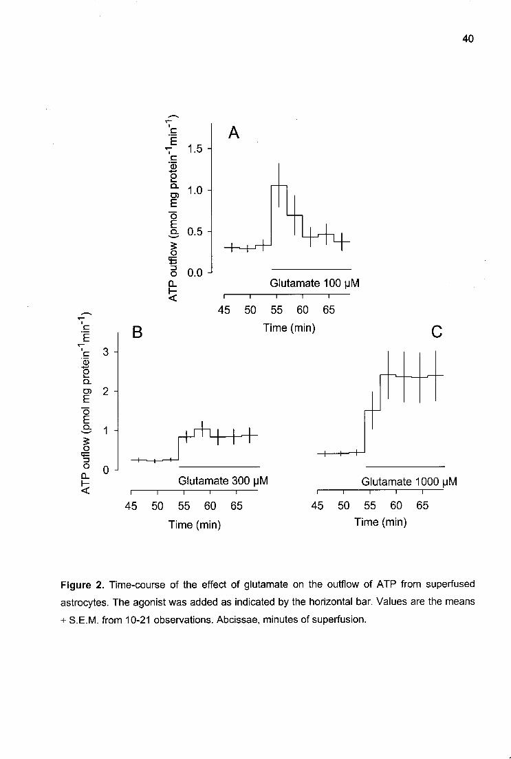
Glutamate itself elicited a transient overflow of ATP at the lowest concentration (100
uM; Fig. 2A) but a long-lasting overflow at higher concentrations (Fig. 2B and C). The
maximal acceleration of the outflow of ATP was 6-fold (at glutamate 1000 uM, Fig. 2C).

39
120 -
î 100
80 -
Q. O) E õ 3 60 I > O CL
40 -
20 -
0 J
Kainate
AMPA
trans-ACPD
Glutamate
■ ■ i l i i i i r 1 1 1 1—i—i—i i i |
10 100 1000 uM
Figure 1. ATP release evoked by glutamate receptor agonists from superfused primary cultures of astrocytes: concentration response curves. Values are the means ± S.E.M from 6-
21 observations.
40
c ^E 'c 'd)
2
E õ E Q .
O ( ^ -I—»
O Q.
5
1.5
1.0
0.5
0.0 Glutamate 100 u M
i i i i i
45 50 55 60 65 c E B T
c 3 -Q)
2 Q.
1 2" O
E S 1 - I41 1 + o 4—
O ,-»
n ° " LL I-<
Glutamate 300 |JM LL I-< i
45 I
50 55 60 65
Time (min)
H 1—+
Glutamate 1000 uM — i 1 1
Time (min)
45 50 55 60 65 Time (min)
Figure 2. Time-course of the effect of glutamate on the outflow of ATP from superfused astrocytes. The agonist was added as indicated by the horizontal bar. Values are the means ± S.E.M. from 10-21 observations. Abcissae, minutes of superfusion.

41
The effect of NMDA was transient at the lowest concentration examined as shown for
a concentration of 100 uM in Fig. 3A, but very variable at the highest concentration tested
(500 uM; not shown). The maximal acceleration of ATP outflow was 19-fold (at NMDA 500
uM). AMPA caused long-lasting increases at all concentrations examined as shown for 30
uM in Fig. 3B. The maximal acceleration was 15-fold (AMPA 50 uM). The time course of the
effect of kainate (20 uM) seemed to be intermediate between the transient increase elicited
by NMDA and the maintained increase elicited by AMPA (Fig. 3C). The maximal acceleration
was 5-fold. frans-ACPD, like AMPA, caused long-lasting increases at all concentrations
tested as shown for trans-ACPD 300 uM (Fig. 3D). The maximal acceleration of ATP outflow
was 7-fold (at 1000 uM frans-ACPD).
ATP release: fraction released
In some experiments with glutamate and NMDA, the ATP content of the cells was
determined at the end of superfusion. It averaged 3.5 ± 0.3 nmol per mg protein (n=18). The
outflow of ATP in the 9 min before agonist addition averaged 0.85 ±0.13 pmol per mg
protein per min (n=18), which was 0.03 ± 0.01 % of the cellular content per min. Glutamate
(1000 uM) released 13.4 ± 1.9 pmol ATP per mg protein, which was 0.55 ±0.14 % of the
cellular content (n=6). NMDA (500 uM) released 11.7 ± 3.0 pmol ATP per mg protein, which
was 0.32 ± 0.06 % of the cellular content (n=8).

42
I 'JB 0 J c
2 Q.
E õ E Q.
O
O CL
3 -
1
0 -
N M D A 1 0 0 | J M n 1 1 i 1 1 -
45 50 55 60 65 Time (min)
Kainate 20 |jM I 1 1 1 1
45 50 55 60 65 Time (min)
B
AMPA 30 MM I 1 1 1 1 —
45 50 55 60 65
D Time (min)
+-+
f-ACPD 300 MM -1 i 1 1 r-
45 50 55 60 65 Time (min)
Figure 3. Time-course of the effect of glutamate receptor type-selective agonists on the outflow of ATP from superfused astrocytes. The agonists were added as indicated by the horizontal bar. Values are the means ± S.E.M. from 6-18 observations. Abcissae, minutes of superfusion.

43
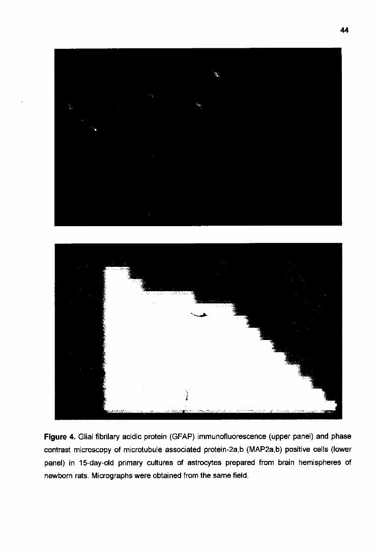
ATP release: possible contribution of neurones
Immunocytochemistry demonstrated that most cells in culture were immunopositive
for GFAP but some were positive for MAP2a,b, a specific marker for neurones (Fig. 4; cf.
Hildebrand et al., 1997). Since we did not succeed in preparing cultures completely devoid of
MAP2a,b positive cells, the release of ATP, evoked by glutamate receptor agonists, from
cultures with different contents of MAP2a,b positive cells was compared. Cultures containing
a lower number of MAP2a,b positive cells were prepared as described under Experimental
Procedures with the exception that the culture medium was changed every day during the
first 4 days of culturing. With this procedure, the number of MAP2a,b positive cells present in
the cultures was reduced from 33.2% to 10.6% on average, i.e. by about 2/3 (Table 1). The
release of ATP evoked by NMDA, AMPA and kainate did not differ between the two culture
types (Table 1).
44
*»*' tik -
Figure 4. Glial fibrilary acidic protein (GFAP) immunofluorescence (upper panel) and phase contrast microscopy of microtubule associated protein-2a,b (MAP2a,b) positive cells (lower panel) in 15-day-old primary cultures of astrocytes prepared from brain hemispheres of newborn rats. Micrographs were obtained from the same field.

Table 1. Glutamate receptor agonist-evoked release of ATP from primary cultures of
astrocytes containing different percentages of MAP2a,b positive cells.
Agonist-evoked release of ATP (pmol per mg protein)
Agonist (MM) 33.2% MAP2a,b cells 10.6% MAP2a,b cells
NMDA 500 38.0 ± 3.3 (3)
AM PA 30 18.3 ±2.2 (6)
Kainate 50 17.3 ±4.8 (3)
37.9 ±1.1 (3)
27.9 ± 3.6 (6)
17.6 ±4.7 (3)
Cultures containing 33.2 ± 4.3% MAP2a,b positive cells were obtained as described in
Experimental Procedures. Cultures with 10.6 ± 2.3% MAP2a,b positive cells were
obtained in the same manner, but the medium was changed every day during the first 4
days in culture. The two kinds of cultures were run strictly in parallel. Cells were exposed
to agonist from 54 min of superfusion onwards for 15 min. Overflow of ATP evoked by
glutamate receptor agonists was calculated by subtraction of basal outflow from total
outflow observed from 54 to 69 min of superfusion. Means ± S.E.M. from (n)
observations.
46
ATP release: interaction with antagonists
Three antagonists - AP5, NBQX, and GAMS - were tested against the agonists. They
did not change the basal efflux of ATP. The NMDA receptor-selective antagonist AP5 (see
Discussion for references), given at a concentration of 100 uM, abolished the effect of
NMDA, did not significantly change the effects of AMPA and kainate, and also did not
change the effect of glutamate (Table 2). NBQX is selective for AMPA receptors. At a
concentration of 30 uM it prevented the effect of AMPA and did not significantly change that
of kainate, although there was a tendency for a decrease. NBQX also abolished the increase
in ATP outflow elicited by glutamate and, unexpectedly, the increase caused by NMDA
(Table 2).
Finally, the kainate receptor-selective antagonist GAMS, given at a concentration of
30 uM, blocked the effect of kainate but did not change that of glutamate (Table 2).
Lactate dehydrogenase release
Release of the cytoplasmic enzyme LDH was examined in order to detect possible
cell damage caused by glutamate. LDH was not discovered in the medium in superfusion
experiments of the kind described so far. Therefore, a different protocol had to be used (see
Experimental Procedures). Glutamate receptor agonists, at concentrations releasing ATP,
failed to release LDH: when the agonists were added during the second of two 15-min
incubation periods, release of LDH from astrocytes slightly declined rather than increased, as
it did in controls without agonists (Table 3).

Table 2. Glutamate receptor agonist-evoked release of ATP from superfused astrocytes: interaction with
antagonists.
Agonist-evoked release of ATP (pmol per mg protein)
Solvent AP5 NBQX GAMS Agonist (MM) (100 uM) (30 uM) (30 uM)
0.2+ 0.4 0.6+ 0.8 -0.9 ± 0.4 -0.8 ± 0.9 (67) (9) (4) (7)
Glutamate 100 4.4+ 1.7 (17)
4.2+ 1.9 (6)
— —
300 9.6 ± 2.8** 22.4 ± 3.9** -3.1 + 1.7tt 30.8 ± 8.7** (18) (5) (4) (6)
1000 26.7+ 8.3* 21.9 ± 4 .8" — 49.3 ±13.3** (21) (7) (6)
NMDA 20 4.1 + 0.9* (13)
-1.1 + 1.4t (3)
— —
100 11.7 ± 2.7** -2.1 ± 1.0tt 0.6 ±1.4 — (18) (5) (5)
500 49.0 ± 11.8** 7.2 ± 3.8*tt 3.4 ± 0.4tt — (16) (5) (3)
AMPA 10 5.6 ± 3.6 (8)
— — —
30 9.1 ± 3.0** (10) "
50 103.6 + 26.4** (8)
57.2 + 23.5 (7) ~ "
100 63.5 ±17.3** (19)
-2.2 ± 0.3tt (4)
Kainate 20 25.1 ± 7.5** 25.1 ± 6.5** 12.1 ±4.8* -0.1 ± 0.7t (19) (8) (5) (5)
frans-ACPD 100 8.3+ 1.8* (5)
— — —
300 21.5 ± 1.8** (6) "
1000 56.3+ 5.9** (6)
The antagonists AP5 (100 uM), NBQX (30 uM) or GAMS (30 uM) were present throughout superfusion.
Overflow of ATP evoked by glutamate receptor agonists was calculated by subtraction of basal outflow.
Means ± S.E.M. from (n) observations. * and ** indicate significant differences from first line (no agonist;
P<0.05 and P<0.01, respectively), t and t t indicate significant differences from effect of agonist in the
absence of antagonist (P<0.05 and P<0.01, respectively).

Table 3. Release of lactate dehydrogenase (LDH) from astrocytes: effect of glutamate
receptor agonists.
LDH in incubation medium (% of total)
Drug present from 15-30 min (MM) 0-15 min 15-30 min n
— 6.4 ±1.2 4.3 ±1.6 13
Glutamate 1000 5.7 ±2.1 5.1 ±2.2 6
NMDA 500 9.4 ±2.3 6.6 ±1.5 6
AM PA 30 5.5 ±0.5 4.0 ±0.8 5
Kainate 50 12.1 ±2.4 5.1 ± 1.8 6
frans-ACPD 300 5.3 ±1.2 4.3 ±0.9 5
Cells were incubated in 100 pi buffer for two successive periods of 15 min (0-15 and
15-30 min). Glutamate receptor agonists were present during the second 15-min
period. LDH release is expressed as a percentage of the total LDH released after cell
lysis. Values are means ± S.E.M. from (n) observations.
49
ATP release: effect ofionomycin
The calcium ionophore ionomycin was tested to determine whether an increase in
intracellular calcium was sufficient to release ATP. Ionomycin (5 uM) progressively increased
the outflow of ATP from astrocytes over the 15 minutes of application in the presence of
external calcium but not in its absence (Fig. 5). The overflow of ATP evoked by ionomycin
was 21.9 ± 2.6 pmol per mg protein in the presence of calcium and 2.2 ± 0.9 pmol per mg
protein its absence (n=17 and n=8 respectively; P<0.01).
ATP release: omission of extracellular calcium
Withdrawal of calcium increased the basal outflow of ATP (see Fig. 6). From 21 to 30
min of superfusion with nominally Ca2+-free medium, i.e. in the 9 min before stimulation by
glutamate receptor agonists, ATP outflow was 1.94 ± 0.15 pmol per mg protein per min, as
compared to 0.72 ± 0.05 pmol per mg protein per min in the presence of calcium (n=64 and
n=57, respectively; P<0.01).
Superfusion with Ca2+- free buffer solution greatly reduced or abolished the overflow
of ATP evoked by glutamate (1000 uM), NMDA and kainate and did not significantly change
the overflow evoked by glutamate (300 uM), AMPA and trans-ACPD (Table 4). The loss of
effect of NMDA in the absence of external Ca2+ is also shown in Fig. 6.

50
I 4-c +-■
2 Q. D) E o E o
O Q_ h-<
.E 5 E
0
lonomycin (5 |JM)
lonomycin (5 |jM) -Ca 2+
■1-...|..-.-^->-
i 1 1 1 1 —
45 50 55 60 65
Time (min)
— 30 i
E "(D - I - '
2 Q.
g 20 õ E Q . O •E CD > O
CL h-<
10
0
* *
#
lonomycin lonomycin (5 \M) -Ca
2+
Figure 5. Release of ATP from superfused astrocytes elicited by ionomycin (5 pM).
lonomycin was added as indicated by the horizontal bar. In some experiments, calcium was
omitted from the superfusion buffer 30 min before stimulation by ionomycin. Abcissae of left-
hand panel, minutes of superfusion. Values are the means ± S.E.M. from 17 (ionomycin at
normal Ca2+) and 8 (ionomycin and Ca2+ withdrawal) observations.

51
f 5
-I—" O l _
Q. 03 3
o E 2 -
ë 1 -o & 0
45
NMDA (500 MM) NMDA (500 MM) -Ca 2+
— i —
60 50 55 60 65
Time (min)
^ 50 i c
"(D ■+-> o 1 _ Q. D1 E "õ E $ o •E CD > O Q_
<
40 -
30 -
20 -
10 -
0 J
* *
#
NMDA NMDA (500 |JM) -Ca 2+
Figure 6. NMDA-evoked release of ATP from superfused astrocytes: effect of calcium withdrawal. NMDA was added as indicated by the horizontal bar. Calcium was omitted from the superfusion buffer 30 min before stimulation with NMDA. Abcissae of left-hand panel, minutes of superfusion. Values are the means ± S.E.M. from 6 (NMDA at normal Ca
2+) and
11 (NMDA and Ca2+ withdrawal) observations.

Table 4. Glutamate receptor agonist-evoked release of ATP from superfused astrocytes:
effect of calcium withdrawal.
Agonist-evoked release of ATP (pmol per mg protein)
Agonist (MM) With
Calcium Without Calcium
Glutamate
NMDA
AM PA
Kainate
300
1000
500
10
30
50
irans-ACPD 300
-1.7 ±0.8 (7)
14.314.9* (6)
25.5 ± 3.9 * (9)
29.8 ± 6 . 0 ' (6)
23.5 + 5.3* (5)
91.1 ±3.2* 0)
19.4 ±3.4* (12)
26.9 + 1.9 (3)
2.7 ± 2.3 (10)
14.5 ± 5.9 (6)
10.9+ 2.9 t t (8)
4.2+ 6.1 t (11)
25.5 ± 4.3** (6)
75.0 + 10.4** (9)
1.5 ± 1.7 t t (11)
26.6+ 4.4* (3)
Calcium was omitted from the superfusion buffer 30 minutes before stimulation with
agonists. Overflow of ATP evoked by glutamate receptor agonists was calculated by
subtraction of basal outflow. Means ± S.E.M. from (n) observations. * and ** indicate
significant differences from first line (no agonist; P<0.05 and P<0.01, respectively), t and
t t indicate significant differences from effect of agonist in the presence of calcium
(P<0.05 and PO.01, respectively).

53
ATP release: effect of blockers of voltage-dependent Ca2+channels
Calcium can enter the cytoplasm of glial cells via ionotropic glutamate receptor
channels (Finkbeiner, 1993; Porter and McCarthy, 1995) as well as through voltage-
dependent Ca2+ channels (VDCCs; MacVicar, 1984).
In order to examine whether the release of ATP evoked by ionotropic glutamate
receptor agonists required influx of Ca2+ through VDCCs, cadmium and nicardipine were
tested againts the agonists. Like calcium withdrawal, cadmium, a non-selective blocker of
VDCCs, at a concentration of 1 mM greatly reduced or abolished the overflow of ATP evoked
by glutamate (1000 uM), NMDA and kainate and did not change the overflow evoked by
glutamate (300 uM) and AMPA (Table 5). A similar pattern was observed with the selective
L-type VDCC blocker nicardipine (10 uM, Table 6).
ATP release: effect of a-latrotoxin and botulinum toxin B
There is evidence that a-latrotoxin, an active compound of the black widow spider
venom, elicits, whereas botulinum toxin B inhibits, exocytotic release of neurotransmitters
from neurones and glutamate from astrocytes (see Discussion). However, addition of a-
latrotoxin (3 nM; for 15 min) did not cause a significant increase in the outflow of ATP from
astrocyte cultures (basal outflow over the 9 min before a-latrotoxin, 1.1 ± 0.2 pmol per mg
protein per min; outflow over the 15 min in the presence of a-latrotoxin, 1.2 ± 0.3 pmol per
mg protein per min; n=9).
Pre-treatment of astrocyte cultures with botulinum toxin B (100 nM) for 16 h did not
significantly change the overflow of ATP evoked by NMDA (500 uM) or kainate (50 uM; Fig.
7).

54
Table 5. Glutamate receptor agonist-evoked release of ATP from superfused astrocytes:
effect of cadmium.
Agonist-evoked release of ATP (pmol per mg protein)
Agonist (MM) Solvent Cadmium
(1 mM)
Glutamate 300
1000
NMDA
AM PA
Kainate
500
30
50
0.2 ± 0.5 (26)
10.3 ± 2.6* (14)
15.3 ± 3.7* (12)
41.1 ±10.0 ' (7)
50.9 ±10.4 ' (6)
19.1 ± 2.9* (8)
0.6 ± 0.5 (22)
10.3+ 5.3 (13)
5.9+ 2.5 t (11)
18.6 ± 3.2 " t (9)
37.2+ 8.1 ** (8)
9.5 ± 2.3 **t (8)
Cadmium (1 mM) was present throughout superfusion. Overflow of ATP evoked by
glutamate receptor agonists was calculated by subtraction of basal outflow. Means ±
S.E.M. from (n) observations. ** indicates a significant difference from first line (no
agonist; P<0.01). t indicates a significant difference from effect of agonist in the absence
of cadmium (solvent; P<0.05 and PO.01, respectively).

55
Table 6. Glutamate receptor agonist-evoked release of ATP from superfused astrocytes:
effect of nicardipine.
Agonist-evoked release of ATP (pmol per mg protein)
Agonist (MM) Solvent Nicardipine
(10 uM)
NMDA
AM PA
Kainate
500
30
50
0.2 ± 0.4 (6)
21.4 + 3.6 (11)
16.6 ±3.6 (6)
14.7 + 2.3 (9)
0.4 + 0.9 0)
8.9±1.9**t (10)
24.0 ± 6.7 ** (6)
4.2 ± 1.4 t t (12)
Nicardipine (10 uM) was present throughout superfusion. Overflow of ATP evoked by
glutamate receptor agonists was calculated by subtraction of basal outflow. Means ±
S.E.M. from (n) observations. ** indicates a significant difference from first line (no
agonist; P<0.01). t and t t indicate significant differences from effect of agonist in the
absence of nicardipine (solvent; P<0.05 and PO.01, respectively).

56
1 £ o c CD
-I—»
2 Q . O) E õ E Q. <ï O O
CL
4 n
3 -
2 -
1 -
0 J
NMDA 500 (JM + BoTxB(100nM)
i i i i i
45 50 55 60 65 Time (min)
Kainate 50 |jM + BoTxB(100nM)
i 1 1 1 1—
45 50 55 60 65 Time (min)
c -+—»
2 Q.
E õ E Q.
O <t O > o Q. H <
30
25
20
15
10
5
0
15
10 -
5 -
0
* * * *
NMDA NMDA (500 |JM) + BoTxB
* * * *
Kainate Kainate (50 M M) + BoTxB
Figure 7. Release of ATP from superfused astrocytes elicited by NMDA and kainate: effect of botulinum toxin B. Agonists were added as indicated by the horizontal bar. Cultures were incubated with botulinum toxin B 16 h before superfusion. Abcissae of left-hand panel, minutes of superfusion. Values are the means ± S.E.M. from 8 observations with NMDA and 5 with kainate (control cultures); 12 observations with NMDA and 6 with kainate (cultures treated with botulinum toxin B).

57
ATP release: interaction with atractyloside and DIDS
The mitochondrial inner membrane possesses an ADP/ATP transporter that is
blocked by atractyloside. Some other intracellular membranes, for example the endoplasmic
reticulum, possess a voltage-dependent anion channel that is ATP permeable and blocked
by DIDS but not by atractyloside (see Discussion). Atractyloside (10 uM) had no effect on
ATP overflow evoked by AMPA (30 uM) (not shown). DIDS (100 uM), when present
throughout superfusion, did not change the overflow of ATP evoked by NMDA (500 uM) or
AMPA (30 uM; Fig.8).
ATP release: interaction with glibenclamide and DPC
Glibenclamide was tested for two reasons: because, at low concentrations (1 uM), it
blocks the endothelial release of ATP in response to shear stress (Hasséssian et al., 1993);
and because, at high concentrations (100 uM), it inhibits the cystic fibrosis transmembrane
conductance regulator (CFTR; Sheppard and Welsh, 1993), which can also mediate, or
regulate, cellular ATP release. The CFTR-mediated release of ATP is also blocked by DPC
(Reisinetal., 1994).
Glibenclamide (1 uM) had no effect on the overflow of ATP from astrocytes evoked by
NMDA (500 uM) or AMPA (30 uM; Table 7). Glibenclamide (100 uM), in contrast, although it
did not change the overflow of ATP evoked by glutamate, NMDA and kainate, significantly
reduced the overflow evoked by AMPA (Table 8). Similarly, DPC (500 uM) had no effect on
the release of ATP evoked by NMDA but significantly reduced that evoked by AMPA (Table
9)-

58
c i c
O Q. O) E
o O
CL
<
4 -
0 J
8
0
NMDA 500 |JM + D I D S ( 1 0 0 | J M )
I 1 1 1 1—
45 50 55 60 65
Time (min)
■ AMPA 30 M M + D I D S ( 1 0 0 | J M )
I 1 1 1 1 —
45 50 55 60 65
60
40 -
Tc 2 0 -
O i— Q.
E o E
O «E > o
CL
0
**
**
NMDA NMDA (500 (JM) +DIDS
80
60
40
20
0
** **
Time (min)
AMPA AMPA (30 MM) +DIDS
Figure 8. Release of ATP from superfused astrocytes elicited by NMDA and AMPA: effect of DIDS (100 |JM). Agonists were added as indicated by the horizontal bar. DIDS was present throughout superfusion. Abcissae of left-hand panel, minutes of superfusion. Values are the means ± S.E.M. from 6 observations with NMDA and 11 with AMPA (normal buffer); 4 observations with NMDA and 8 with AMPA (in the presence of DIDS 100 uM).

59
Table 7. Glutamate receptor agonist-evoked release of ATP from superfused astrocytes:
effect of glibenclamide (1 uM).
Agonist-evoked release of ATP (pmol per mg protein)
Solvent Glibenclamide Agonist (uM) (1 uM)
NMDA 500 107.1 ±12.1 87.0 + 17.4 (3) (3)
AMPA 30 38.5 ± 8.9 43.3 ± 9.9 (3) (3)
Glibenclamide (1 uM) was present throughout superfusion. Overflow of ATP evoked by
glutamate receptor agonists was calculated by subtraction of basal outflow. Means ±
S.E.M. from (n) observations.
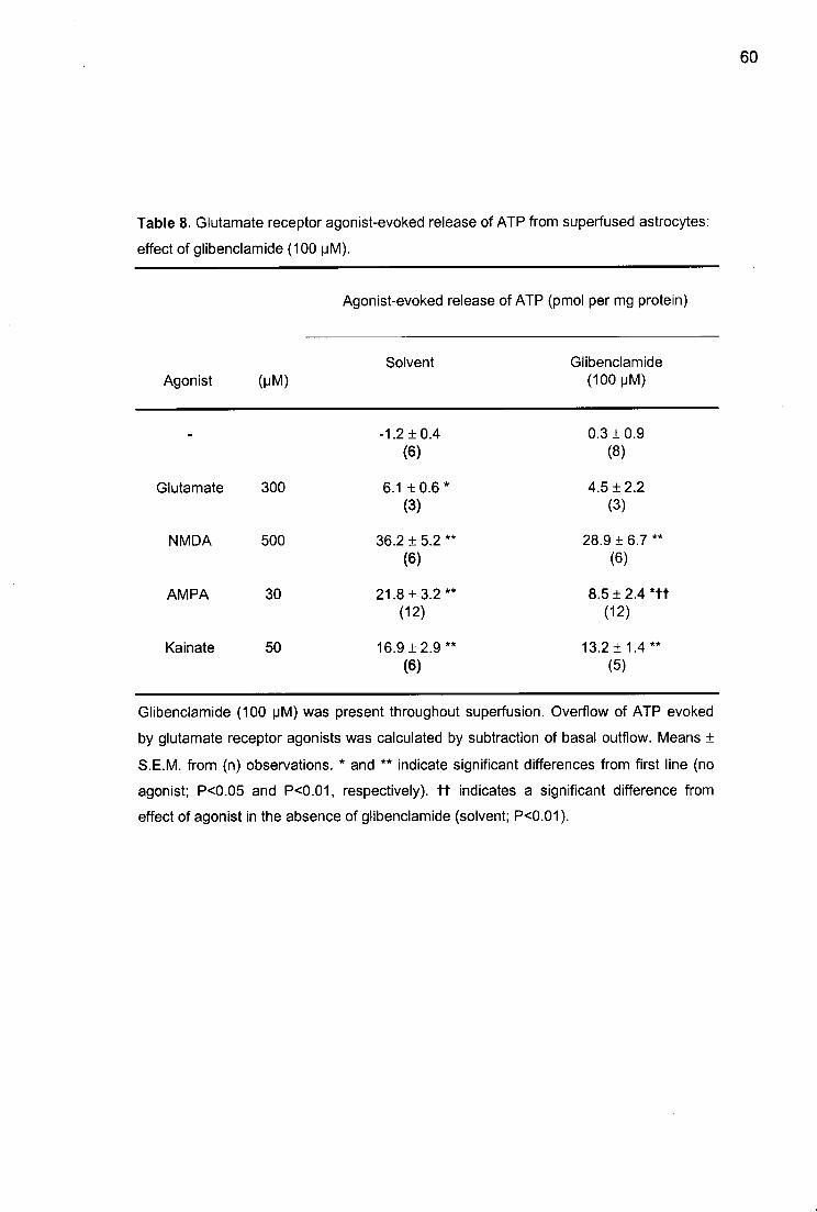
Table 8. Glutamate receptor agonist-evoked release of ATP from superfused astrocytes:
effect of glibenclamide (100 uM).
Agonist-evoked release of ATP (pmol per mg protein)
Agonist (MM) Solvent Glibenclamide
(100 pM)
Glutamate 300
NMDA
AM PA
Kainate
500
30
50
-1.2 + 0.4 (6)
6.1 ±0.6 ' (3)
36.2 ± 5.2 (6)
21.8 ±3.2 (12)
16.9 ±2.9 (6)
0.3 ±0.9 (8)
4.5 ±2.2 (3)
28.9 ±6.7** (6)
8.5±2.4* t t (12)
13.2 ±1.4** (5)
Glibenclamide (100 pM) was present throughout superfusion. Overflow of ATP evoked
by glutamate receptor agonists was calculated by subtraction of basal outflow. Means ±
S.E.M. from (n) observations. * and ** indicate significant differences from first line (no
agonist; P<0.05 and P<0.01, respectively), t t indicates a significant difference from
effect of agonist in the absence of glibenclamide (solvent; PO.01 ).

61
Table 9. Glutamate receptor agonist-evoked release of ATP from superfused astrocytes:
effect of DPC.
Agonist-evoked release of ATP (pmol per mg protein)
Agonist (MM) Solvent DPC
(500 uM)
NMDA 100
AM PA 30
-0.3 ± 0.2 (3)
28.9 + 4.7* (6)
26.9 ±4 .7* (6)
-0.4 ±0.5 (5)
27.8 ±8.8** (5)
13.0 ± 1.8 **t t (6)
DPC (500 uM) was present throughout superfusion. Overflow of ATP evoked by
glutamate receptor agonists was calculated by subtraction of basal outflow. Means ±
S.E.M. from (n) observations. * and ** indicate significant differences from first line (no
agonist; P<0.05 and PO.01, respectively), t t indicates a significant difference from
effect of agonist in the absence of DPC (solvent; P<0.01 ).

62
ATP release: interaction with lithium
Lithium has been reported to block the release of ATP from guinea-pig ileal segments
stimulated by a,p-methylene ATP or bethanechol (Katsuragi et al., 1996). At a concentration
of 1 mM, lithium abolished the overflow of ATP evoked by glutamate and AMPA and greatly
reduced the overflow evoked by NMDA and kainate (Table 10).
Table 10. Glutamate receptor agonist-evoked release of ATP from superfused
astrocytes: effect of lithium.
Agonist-evoked release of ATP (pmol per mg protein)
Agonist (MM) Solvent Lithium
(1 mM)
Glutamate
NMDA
AMPA
Kainate
300
500
30
50
1.0 ±1.2 (10)
7.4 ±1.1 ** (6)
72.2 + 9.7** (6)
31.0 ±4.7** (5)
42.1 ±7.9** (7)
-1.0 + 1.2 (8)
0 .9±1 .2 t t (6)
24.2 ±4.1 **tt (6)
-2.6 ± 3.2 t t (6)
16.2 ± 4.3 " t t (8)
Lithium (1 mM) was present throughout superfusion. Overflow of ATP evoked by
glutamate receptor agonists was calculated by subtraction of basal outflow. Means ±
S.E.M. from (n) observations. ** indicates a significant difference from first line (no
agonist; P<0.01). t t indicates a significant difference from effect of agonist in the
absence of lithium (solvent; P<0.01).

63
Discussion
The results obtained in the present work identify ATP as one adenyl compound that is
released by excitatory amino acids from brain cells, and identify astrocytes as cells from
which it may originate. The release of ATP from astroglia was elicited by glutamate and by
selective agonists of each ionotropic glutamate receptor known, NMDA, AMPA and kainate
as well frans-ACPD, a selective agonist of the metabotropic glutamate receptors devoid of
activity at ionotropic receptors (Palmer at al., 1989; Manzoni et al., 1990).
The response to glutamate receptor agonists was variable; for example release of
ATP elicited by NMDA was transient in some experiments (Fig. 3) but maintained in others
(Fig. 6). Another example of variability is the large difference in ATP release between
identically treated groups; for example, AMPA (30 uM) caused an averaged release of 18.3
pmol per mg protein in one group (Table 1) and 91.1 pmol per mg protein in another (Table
4). This variability can be explained by variations in the levels of receptor expression that
may occur between different astrocyte cultures and even in different astrocytes within the
same culture (Verkhratsky and Kettermann, 1996). To overcome the influences of such
variability on the effects of drugs, drug-treated cultures where always compared with a strictly
parallel control group.
NMDA, AMPA and kainate elicited the release of ATP from astroglial cells probably
by acting through the receptors named after them. This view is supported by the fact that
64
these compounds released ATP at concentrations known to exert effects through NMDA,
AMPA, and kainate receptors, respectively. Among those effects are the release of
adenosine from rat brain cortex and hippocampal brain slices (Craig and White, 1993;
Pedata et al., 1991) and the indirect adenosine-mediated inhibition of noradrenaline release
from the rabbit brain cortex (von Kugelgen et al., 1993).
The effects of selective ionotropic glutamate receptor antagonists confirmed the view
that NMDA, AMPA and kainate evoked the release of ATP through activation of the
respective receptors; i) the NMDA receptor-selective antagonist AP5 (Evans et al., 1982)
attenuated the effect of NMDA but not of AMPA and kainate; ii) the AMPA receptor-selective
antagonist NBQX (Sheardown et al., 1990) abolished the effect of AMPA but not of kainate;
iii) the kainate receptor-selective antagonist GAMS (Jones et al., 1984; Turski et al., 1985)
blocked the effect of kainate.
Two observations on the effects of NMDA require additional comment. First, NMDA
released ATP despite the presence of a physiological concentration of Mg2+, in contrast to
the classical NMDA receptor-mediated effects (Nowak et al., 1984). Resistance to Mg2+ has
also been observed for the NMDA-induced release of adenosine from rat brain slices (Hoehn
et al., 1990; Pedata et al., 1991) and for the NMDA-induced, adenosine-mediated inhibition
of the release of noradrenaline in the rabbit brain cortex (von Kùgelgen et al., 1993). A
possible reason is a low membrane potential of the glial cells under our culture and
superfusion conditions, alleviating the Mg2+ block of the NMDA receptor channel. An
alternative explanation is the occurrence of spare NMDA receptors as it has been suggested
for the NMDA-evoked adenosine release (Hoehn et al., 1990). According to this hypothesis,
the block of Mg2+ can be overcome by the high concentrations of NMDA used. Second, the
AMPA receptor antagonist NBQX prevented not only the release of ATP elicited by AMPA
but also the release elicited by NMDA. Possibly, NBQX blocked the glycine site of the

65
astrocyte NMDA receptor since other quinoxaline derivatives have been shown to block the
glycine site of NMDA receptors at low concentrations and thus block the NMDA-receptor
mediated responses (Kessler et al., 1989).
Glutamate receptor agonists exert additional effects on glial cells, such as
depolarisation and an increase in intracellular Ca2+. In glial cells in situ, the effects seem to
be mediated by NMDA, AMPA as well as by kainate receptors (Porter and McCarthy, 1995).
Immunocytochemical studies on the distribution of cortical NMDA receptor have shown that
cortical astrocytes express NMDA receptors (Conti et al., 1997). However, the presence of
NMDA receptors in astroglial cells is still a matter of debate. In most of the studies on
cultured glial cells, NMDA receptors mediating membrane depolarisation and an increase in
Ca2+ have not been detected (see Teichberg, 1991). Recent reports suggest that glial cells
can express functional NMDA receptors. For instance, in human cultured astrogial cells
NMDA has been shown to stimulate release of prostaglandin E2 (Mollace et al., 1995). The
retinal Muller glial cells express one of the essential components of the NMDA receptor-
channels, the NMDAR1 subunit, and are also responsive to NMDA even at resting
membrane potential (Puro et al., 1996).
In the present study, glutamate, one of the "endogenous" ligands of glutamate
receptors, also released ATP. However, it was not possible to identify clearly the receptor
involved. The selective antagonism by NBQX, but not AP5 and GAMS (Table 2), suggests
the involvement of AMPA receptor. However, NBQX also blocked the effect of NMDA and a
simultaneous activation of AMPA and NMDA receptors can not be ruled out. Glutamate may
also have acted at metabotropic receptors because frans-ACPD, a selective agonist of
metabotropic glutamate receptors, also evoked ATP release in the present study.
Variability in the glutamate receptor involved has also been observed on glutamate
evoked release of other adenyl compounds in the brain. For instance, glutamate evokes

66
release of adenosine from rat brain slices through NMDA as well as non-NMDA receptors,
(Craig and White, 1993; Hoehn and White, 1990a). In rabbit cortical slices glutamate evokes
adenosine release through non-NMDA receptors only (see von Kugelgen and Starke, 1995)
whereas and in cortical synaptosomes, uptake of glutamate seems to be a prerequisite for
the release (Hoehn and White, 1990b).
The occurrence of contaminating neurones in astroglial cultures has always been
regarded as negligible. Neurones could be found immediately after seeding, however these
neurones disappeared when the proliferating astroglial cells began to form a monolayer.
Recently it has been demonstrated that some neuronal percursors do not turn into
differentiated cells, but keep their ability to proliferate (Weiss et al., 1996; Eriksson et al.,
1998). Neuronal precursors seem to be capable of survival and to proliferate in culture, as it
has been demonstrated in primary cultures of astroglial cells prepared from neocortex of
new-born rats (Hildebrand et al., 1997). The first neuronal cells appear shortly before
confluence, when the glial monolayer is being formed. After confluence, these still
undifferentiated cells increase in number and later become immunohistochemically positive
for the neurone-specific marker microtubule associated protein 2a,b (MAP2a,b). In spite of
the known interest of this finding, i.e. providing a model for the investigation of neuronal-glial
interactions, it introduces another variable in the astroglial model.
In the present study, the observation that astroglial cultures prepared from the rat
cerebral cortex may contain neurones has also been confirmed. However, the release of
ATP elicited by glutamate agonists was not changed when the contamination by neurones
was reduced by about 2/3, suggesting that neurones contributed little, if at all, to ATP
release.

67
The role of Ca?+ in ATP release
Astrocytes in situ as well as in culture respond to glutamate receptor agonists with an
increase in intracellular Ca2+ (Cornell-Bell et al., 1990; Porter and McCarthy, 1995). Both
transmembrane influx and release of Ca2+ from intracellular stores can contribute (Glaum et
al., 1990). Transmembrane entry predominates upon activation of NMDA and kainate
receptors, whereas mobilisation from intracellular stores predominates upon activation of
metabotropic glutamate receptors (Ahmed et al., 1990; Porter and McCarthy, 1995). AMPA
receptors, although ionotropic receptors, can also trigger metabotropic transduction
mechanisms including possibly the mobilisation of intracellular Ca2+ (Wang et al., 1997;
Bezzi et al., 1998). From what is known about glial glutamate receptors and Ca2+, therefore,
a role of Ca2+ in the release of ATP from astrocytes would seem possible.
An increase in intracellular Ca2+ seems to be sufficient to evoke the release of ATP
from astrocytes: the Ca2+ ionophore ionomycin elicited such release, and it did so only in the
presence but not in the absence of external Ca2+.
Calcium may cross the plasma membrane either through the ionotropic glutamate
receptors themselves or through VDCCs that are opened by depolarisation (Barres et al.,
1990b; Barres, 1991). The release of ATP evoked by NMDA and kainate from astroglial cells,
was abolished or greatly reduced by withdrawal of extracellular Ca2+, by cadmium and
nicardipine. These findings support the following: i) Ca2+ is needed for the release of ATP
triggered by NMDA and kainate receptor activation; ii) the Ca2+ involved comes from the
extracellular space and; iii) Ca2+ influx occurs partly through VDCCs, mainly of the L-type.
In contrast, release of ATP evoked by AMPA was not reduced by withdrawal of
external Ca2+ and was not changed by cadmium and nicardipine. A similar pattern was
observed for the ATP release evoked by frans-ACPD. These findings exclude a major

68
contribution of Ca2+ entry to AMPA- and frans-ACPD-evoked ATP release. They do not, of
course, exclude a role of Ca2+ mobilised from intracellular stores (see above).
Agonists at any of the three ionotropic glutamate receptors also release adenosine
from brain tissue, and some of the adenosine may be formed from released ATP (Craig and
White, 1993). In accord with the present results on glial ATP release, in rat brain slices the
release of adenosine elicited by NMDA depended on extracellular Ca2+ whereas the release
elicited by AMPA did not (Craig and White, 1993).
On the mechanism of ATP release
Once the role of Ca2+ on the release of ATP evoked by the glutamate receptor
agonists from astroglial cells had been established, attempts were made to establish the
mechanism(s) involved in the release of ATP from astroglial cells.
The release of ATP was not accompanied by release of the cytoplasmic enzyme
LDH, indicating that there was no immediate major damage to the cells upon exposure to the
excitatory amino acids. The fraction of cellular ATP that is released upon exposure to
glutamate receptors agonists is also very low (less that 1%), lower that one's might expect to
be observed as a consequence of cell lysis.
In order to explain the mechanism involved in the release of ATP from astrocytes, two
other hypotheses have been considered and tested. One was the involvement of a plasma
membrane carrier or channel. The other was the occurrence of a vesicular release similar to
neuronal exocytosis. The possibility of a neurone-like exocytosis, the possible involvement of
carrier molecules, and the effect of lithium will be discussed in turn.

69
Neurone-like exocytosis?
Astrocytes express cellubrevin, synaptobrevin II and syntaxin, three of the proteins
involved in the pore formation during neural exocytosis (Parpura et al., 1995a).
A Ca2+-dependent glutamate release, possibly of exocyotic nature, has been
demonstrated to occur from astrocytes in primary culture, upon activation of bradykinin
receptors (Jeftinija et al., 1996; Parpura et al., 1995b). In accord with a neurone-like
exocytosis, botulinum toxin B and tetanus toxin, which proteolyses synaptobrevin II (Huttner,
1993), prevented the release of glutamate by bradykinin (Jeftinija et al., 1997).
The hypothesis of exocytotic glutamate release from astrocytes was also supported
by the effect of a-latrotoxin, a component of the black widow spider venom. a-Latrotoxin
binds to a receptor on nerve terminals and induces the release of neurotransmitter by directly
stimulating the secretory machinery (Petrenko, 1993). One of the receptors for this toxin is a
member of a protein family, the neurexins, which are transmembrane proteins that bind
intracellulary to the synaptic vesicle protein, synaptotagmin (Ushkaryov et al., 1992).
Interacting directly with synaptic proteins, a-latrotoxin bypasses the Ca2+-requirement of
transmitter release. a-Latrotoxin also evoked the release of glutamate from cultured
astrocytes suggesting that astrocytes have a vesicle release machinery similar to the
vesicular apparatus of the nerve terminal (Parpura et al., 1995a).
NMDA- and kainate-evoked release of ATP from astrocytes was Ca2+-dependent.
The possibility of an exocytotic mechanism was excluded since neither a-latrotoxin released
ATP, nor did botulinum toxin B reduce the release of ATP elicited by NMDA and kainate.

70
Membrane passage by means of channels or transporters?
Besides cell damage and exocytosis, a transport molecule or channel is a third
mechanism by which ATP might cross the astrocyte plasma membrane.
ATP is synthesised in mitochondria and is then transported across mitochondrial
membranes to the cytoplasm. ATP is also necessary in other compartments of the cell such
as the endoplasmic reticulum and Golgi apparatus. Furthermore, in nerve terminals ATP is
also concentrated in vesicles being stored with neurotransmitters. To reach these
compartments ATP must cross the membranes that surround them. This task is
accomplished by several intracellular transporters or channels; the ADP/ATP transporter of
the mitochondrial inner membrane, the ATP-permeable anion channel of the endoplasmic
reticulum and other intracellular compartments, and the vesicular transporter that
concentrates ATP within vesicles. Based on this knowledge the hypothesis that one of these
systems could operate in the plasma membrane, and be responsible for the permeation of
membrane to ATP, was considered.
Atractyloside blocks the ADP/ATP exchanger of the mitochondrial inner membrane
(Ziegler and Penefsky, 1993) and the vesicular transporter (Gualix et al., 1996). It did not
affect the release of ATP elicited by AMPA. DIDS is a chloride channel antagonist, which
also blocks a pore-forming protein expressed for example in the endoplasmic reticulum and
Golgi apparatus and involved in ATP transport (Clairmont et al., 1992; Guillen and
Hirschberg, 1995; Shoshan-Barmatz at al., 1996). DIDS had no effect on ATP release
evoked by NMDA and AMPA. These two observations exclude the possibility that these
intracellular ATP transporters occur in the plasma membrane and are responsible for the
release of ATP from astrocytes in response to glutamate receptor activation.

71
Furthermore, release of ATP from endothelial cells upon shear stress and from
epithelial cells has been shown to occur by a glibenclamide sensitive mechanism. In blood
vessel endothelia ATP release elicited by shear stress is blocked by a low concentration of
glibenclamide (1 uM; Hasséssian et al., 1993). However, at a concentration of 1 uM
glibenclamide did not influence ATP release evoked by NMDA and AMPA, from astroglial
cells, which excludes this mechanism as well.
In contrast to these negative findings, a significant effect was obtained when the role
of CFTR (which is involved in the release of ATP from epithelial cells) was examined. CFTR
is a member of the ATP-binding cassette protein family which has been reported to serve as
an ATP-conducting channel in the plasma membrane. CFTR has been shown to be
expressed not only in epithelial cells but also in several cell types that release ATP such as
muscle cells, erythrocytes and endothelial cells (Yamazaki and Hume, 1997; Sprague et al.,
1998; Tousson et al., 1998). CFTR-mediated release of ATP is blocked by higher
concentrations of glibenclamide (100 uM) as well as by DPC, but not by DIDS (Reisin et al.,
1994; Schwiebert et al., 1995).
Both glibenclamide (100 uM) and DPC, while having no significant effect on the
release of ATP evoked by NMDA and kainate significantly reduced the effect of AMPA. The
effect of glibenclamide on AMPA evoked ATP release was not due to blockade of ATP-
dependent K+-channels, because these (like the endothelial release of ATP caused by shear
stress) are also blocked by glibenclamide (1 uM; Quast and Cook, 1989), the concentration
that did not change AMPA-evoked ATP release (see above).
Whether astrocytes in fact express CFTR or a similar transporter is not known. The
present results suggest that this may be the case and that a molecule with a CFTR-like
pharmacology may mediate the AMPA-induced, but not the NMDA- and kainate-induced, exit
of ATP from astrocytes. Thus ATP release evoked by NMDA and kainate seems to be

72
mediated by a CFTR-independent mechanism. Release of ATP by other mechanisms
besides CFTR-mediated transport has also been described in other systems, suggesting that
ATP release can proceed through independent routes, even in the same cell type (Mitchell et
al., 1998). For example, ocular cilliary epithelial cells release ATP by several mechanisms;
hypotonic conditions induce the release of ATP by a mechanism that is not blocked by
glibenclamide and DPC, thus not mediated by CFTR, but is sensitive to chloride channel
blockers whereas ionomycin releases ATP through a mechanism that is neither mediated by
CFTR nor sensitive to chloride channel blockers (Mitchell et al., 1998). Other mechanisms of
ATP release, CFTR-independent, have also been shown to be physiological in cells either
expressing or not the CFTR (Grygorczyk and Hanrahan, 1997; Watt et al., 1998).
The effect of lithium
Lithium blocks the release of ATP from smoth muscle cells elicited by activation of
P2Y and muscarinic receptors (Katsuragi et al., 1996). In the present experiments, lithium
abolished the release elicited by AMPA and significantly reduced the release of ATP evoked
by NMDA and kainate. What could be the mechanism of action of lithium and in what extent
this result may help to clarify the mechanisms involved on AMPA, NMDA and kainate evoked
ATP release?
The best-studied primary effect of lithium is inhibition of inositol monophosphatase
and, hence, inhibition of the inositol phosphate signal transduction pathway because of
depletion of inositol (Atack et al., 1995). In astrocytes, inositol phosphates are formed upon
activation of metabotropic glutamate receptors (Steinhãuser and Gallo, 1996), but inositol
phosphate formation upon activation of ionotropic glutamate receptors was never shown. In
neurones, activation of all three ionotropic receptors leads to inositol phosphate formation
73
(Hartmann and Mùller, 1993; Reis et al., 1995), probably due to activation of phospholipase
C subsequent to an increase in intracellular Ca2+ (Baird and Naorski, 1990; Hartmann and
Mùller, 1993; Reis et al., 1995). On the basis of these findings one may speculate that
release of ATP evoked by NMDA, AMPA and kainate might involve inositol phosphates, and
that inhibition by lithium of inositol phosphate synthesis is the mechanism of blockade of the
glutamate receptor-mediated ATP release. However, lithium exerts many other effects on
cells (see Risby et al., 1991), and other mechanisms cannot be ruled out. Also, the
mechanisms by which lithium influenced the release of ATP elicited by AMPA on the one
hand and the release of ATP elicited by NMDA and kainate on the other hand might differ.
When one summarises the influence of various interventions on the release of ATP
by the three selective ionotropic glutamate receptor agonists, the latter fall into two groups
(Table 11). One comprises NMDA and kainate, the releasing effect of which depended on
extracellular Ca2+, was not changed by glibenclamide (100 uM) and DPC, and was reduced
but not abolished by lithium. The other comprises AMPA only, the effect of which did not
depend on extracellular Ca2+, was reduced by glibenclamide (100 uM) and DPC, and was
abolished by lithium. The effect of glutamate was intermediate.
Glutamate possibly elicits release of ATP from astrocytes through both NMDA and
AMPA receptors; such a dual action may be responsible for its intermediate behaviour in the
present study (Table 11).

74
Table 11. Glutamate receptor agonist-evoked release of ATP from superfused astrocytes:
effect of Ca2+ withdrawal and drugs.
Effect of:
ATP release evoked by
Glutamate NMDA AMPA Kainate /-ACPD
(MM) (MM) (MM) (MM) (MM)
Calcium withdrawal 300-> 500 i 10->
1000 1 30->
5 0 i 300
Cadmium (1000 MM) 300 -> 500 I 30 ->
1000 1
50 I
Nicardipine (10 pM) 500 i 30 -> 50 4
Botulinum toxin B (0.1 MM) 500 -> 50
Atractyloside (10 pM) 30->
DIDS(100pM)
Glibenclamide (1 pM)
Glibenclamide(100 pM)
500 -> 30 ->
500 -> 30
3 0 0 ^ 500-> 30 i 50->
DPC (500 pM)
Lithium (1000 pM)
100-> 30 4-
300 1 5001 30 i 501
I and -> indicate, respectively, a decrease and no change of the release of ATP evoked by
glutamate receptor agonists.

75
Conclusions
Cultured astroglial cells respond to activation of ionotropic glutamate receptors with
release of ATP. They thus resemble other non-neuronal cells such as smooth muscle
(Katsuragi et al., 1990; von Kúgelgen and Starke, 1991b; Kurz et al., 1994) and endothelial
cells (Yang et al., 1994) which release ATP upon activation of certain (although not
glutamate) receptors. Neurones release ATP by exocytosis (see von Kúgelgen et al., 1994b).
Two different mechanisms seem to be involved in the release of ATP from astroglial cells
upon activation of ionotropic glutamate receptors. The NMDA- and kainate-induced release
of ATP requires an influx of Ca2+, is not due to neurone-like exocytosis, is not mediated by
CFTR or a mechanism regulated by CFTR, and is reduced (by an unknown mechanism) but
not abolished by lithium. The AMPA-induced release does not require extracellular Ca2+,
may be mediated by CFTR or a mechanism regulated by CFTR, and is abolished (by an
unknown mechanism) by lithium.
Purines are important modulators of neuronal functions (Zimmermann, 1994;
Burnstock, 1997). They also may act as trophic factors for glial cells and neurones (Neary et
al., 1996). The ability of astrocytes to release ATP and respond to ATP (King et al., 1996)
suggests that ATP may act as an autocrine or paracrine messenger between these cells, and
possibly between astrocytes and neurones.

76
References
Abbracchio MP., Ceruti S., Bolego C, Puglisi L., Bumstock G. and Cattabeni F. (1996) Trophic roles of P2 purinoceptors in central nervous system astroglial eels. Ciba Found. Symp. 198, 142-147.
Abbracchio M.P., Saffrey M.J., Hõpker V. and Bumstock G. (1994) Modulation of astroglial cell proliferation by analogues of adenosine and ATP in primary cultures of rat striatum. Neuroscience 59, 67-76.
Abraham E.H., Prat A.G., Gerweck L, Seneveratne T., Arceci R.J., Kramer R., Guidotti G. and Cantiello H.F. (1993) The multidrug resistance (mdrl) gene product functions as an ATP channel. Proc. Natl. Acad. Sci. USA 90, 312-316.
Ahmed Z., Lewis C.A. and Faber D.S. (1990) Glutamate stimulates release of Ca2+ from internal stores in astroglia. Brain Res. 516, 165-169.
Antanitus D.S., Choi B.H. and Lapham L.W. (1975) Immunofluorescence staining in vitro using antiserum to glial fibrillary acidic protein. Brain Res. 89, 363-367
Aoki C, Venkatesan C, Go C-G., Mong J.A. and Dawson T.M. (1994) Cellular and subcellular localization of NMDA-R1 subunit immunoreactivity in the visual cortex of adult and neonatal rats. J. Neurosci. 14, 5202-5222.
Arenander AT., de Vellis J. and Herschman H.R. (1989a) Induction of c-fos and TIS genes in cultured rat astrocytes by neurotransmitters. J. Neurosci. Res. 24, 107-114
Arenander AT., Lim R.W., Varnum B.C., Cole R., de Vellis J. and Herschman H.R. (1989b) TIS gene expression in cultured rat astrocytes: multiple pathways of conduction by mitogens. J. Neurosci. Res.. 23, 257-265.
Atack J.R., Broughton H.B. and Pollack S.J. (1995) Inositol monophosphatase - a putative target for Li+ in the treatment of bipolar disorder. Trends Neurosci. 18, 343-349.
Attwell D., Barbour B. and Szatkowski M. (1993) Nonvesicular release of neurotransmitter. Neuron 11, 401-407.
77
Baird J.G. and Nahorski S.R. (1990) Increased intracellular calcium stimulates 3H-inositol polyphosphate accumulation in rat cerebral cortical slices. J. Neurochem. 54, 555-561.
Banker G.A. (1980) Trophic actions between astroglial cells and hippocampal neurones in culture.
Science 209, 809-810.
Barbour B., Szatkowski M., Ingledew N. and Attwell D. (1989) Arachidonic acid induces a prolonged inhibition of glutamate uptake into glial cells. Nature 342, 918-920.
Barres B.A. (1991) New roles for glia. J. Neurosci. 11, 3685-3694.
Barres B.A., Chun L.L. and Corey DP. (1989) Glial and neuronal forms of the voltage-dependent sodium channel: characteristics and cell-type distribuition. Neuron 2, 1375-1388.
Barres B.A., Chun L.L.Y. and Corey DP. (1990a) Ion channels in vertebrate glia. Annu. Rev.
Neurosci. 13, 441-474.
Barres B.A., Koroshetz W.J., Chun L.L.Y. and Corey DP. (1990b) Ion channel expression by white matter glia: the type-1 astrocyte. Neuron 5, 527-544.
Bean BP. (1992) Pharmacology and electrophysiology of ATP-activated ion channels. Trends Pharmacol. Sci. 13, 87-90.
Bedingfield J.S., Kemp M.C., Jane D.E., Tse H.W., Roberts P.J. and Watkins J.C. (1995) Structure-activity relationships for a series of phenylglycine derivatives acting at metabotropic glutamate receptors (mGluRs). Br. J. Pharmacol. 116, 3323-3329.
Bennett M.V.L., Barrio T.A., Bargiello T.A., Spray D.C., Hertzberg E. and Sáez J.C. (1991) Gap junctions: new tools, new answers, new questions. Neuron 6, 305-320.
Bettler B. and Mulle C. (1995) Review: neurotransmitter receptors. II. AMPA and kainate receptors. Neuropharmacology 34, 123-139.
Bevan S. (1990) Ion channels and neurotransmitter receptors in glia. Semin. Neurosci. 2, 467-481.
Bezzi P., Carmignoto G., Pasti L., Vesce S., Rossi D., Rizzini B.L., Pozzan T. and Volterra A. (1998) Prostaglandins stimulate calcium-dependent glutamate release in astrocytes. Nature 391, 281-285.
Blaschke M., Keller B.U., Rivosecchi R., Hollmann M., Heinemann S. and Konnerth A. (1993) A single amino acid determines the subunit-specific spider toxin block of a-amino-3-hydroxy-5-methylisoxazole-4-propionate/kainate receptor channels. Proc. Natl. Acad. Sci. USA 90, 6528-6532.
Bock E., Moller M., Nissen C. and Sensenbrenner M. (1977) Glial fibrillary acidic protein in primary astroglial cell cultures derived from newborn rat brain. FEBS Lett. 83, 207-211.
Bormann J. and Kettenmann H. (1988) Patch-clamp study of GABA receptor Ch channels in cultured astrocytes. Proc. Natl. Acad. Sci. USA 85, 9336-9340.
Bowman C.L. and Kimelberg H.K. (1984) Excitatory amino acids directly depolarize in rat brain astrocytes in primary culture. Nature 311, 656-659.

78
Bruner G. and Murphy S. (1990) ATP-evoked arachidonic acid mobilization in astrocytes is via a P2y-
purinergic receptor. J. Neurochem. 55, 1569-1575.
Burnashev N., Khodorova A., Jonas P., Helm P.J., Wisden W., Monyer H., Seeburg P.H. and
Sakmann B. (1992) Calcium-permeable AMPA-kainate receptors in fusiform cerebellar glial cells.
Science 256,1566-1570.
Burnstock G. (1972) Purinergic nerves. Pharmacol. Rev. 24, 509-581
Burnstock G. (1978) A basis for distinguishing two types of purinergic receptor. In: Straub RW, Bolis L (eds) Cell membrane receptors for drugs and hormones: a multidisciplinar/ approach. Raven Press,
New York, pp 107-118.
Burnstock G. (1990a) Noradrenaline and ATP as co-transmitters in sympathetic nerves. Neurochem.
Int. 17, 357-368.
Burnstock G. (1990b) Co-transmission. Arch. Int. Pharmacodyn. 304, 7-33.
Burnstock G. (1996) P2 purinoceptors: historical perspective and classification. P2 purinoceptors:
localization, function and transaction mechanisms. Ciba Found. Symp. 198. 1-28.
Burnstock G. (1997) The past, present and future of purine nucleotides as signalling molecules.
Neuropharmacology 36, 1127-1139.
Caciagli F., Ciccarelli R., Di lorio P., Ballerini P. and Tacconelli L. (1988) Cultures of glial cells release
purines under field electrical stimulation: the possible ionic mechanisms. Pharmacol. Res. Commun.
20, 935-947.
Charpak S., Gahwiler B.H., Do K.Q. and Knõpfel T. (1990) Potassium conductances in hippocampal neurons blocked by excitatory amino-acid transmitters. Nature 347, 765-767.
Chittajallu R., Braithwaite S.P., Clarke V.R.J, and Henley J.M. (1999) Kainate receptors: subunits, synaptic localization and function. Trends Pharmacol. Sci. 20, 26-35.
Chittajallu R., Vignes M., Dev K.K., Barnes J.M., Collingridge G.L. and Henley J.M. (1996) Regulation of glutamate release by presynaptic kainate receptors in the hippocampus. Nature 379, 78-81.
Choi D.W. (1988) Calcium-mediated neurotoxicity: relationship to specific channel types and role in ischemic damage. Trends Neurosci. 11, 465-469.
Choi D.W. and Rothman S.M. (1990) The role of glutamate neurotoxicity in hypoxic-ischemic neuronal death. Annu. Rev. Neurosci. 13, 171-182.
Clairmont CA., de Maio A. and Hirschberg C.B. (1992) Translocation of ATP into the lumen of rough endoplasmatic reticulum-derived vesicles and ist binding to luminal proteins including bip (GRP 78) and GRP 94. J. Biol. Chem. 267, 3983-3990.
Condorelli D.F., Dell'Albani P., Amico C, Kaczmarek L, Nicoletti F., Lukasiuk K. and Stella A.M. (1993) Induction of primary response genes by excitatory amino acid receptor agonists in primary astroglial cultures. J. Neurochem. 60, 877-885.

79
Conti F., Minelli A., DeBiasi S. and Melone M. (1997) Neuronal and glial localization of NMDA
receptors in the cerebral cortex. Mol. Neurobiol. 14, 1-18.
Cornell-Bell A.H., Finkbeiner S.M., Cooper M.S. and Smith S.J. (1990) Glutamate induces calcium waves in cultured astrocytes: long-range glial signaling. Science 247, 470-473.
Corvalan V., Cole R., de Vellis J. and Hagiiwara S. (1990) Neuronal modulation of calcium channel
activity in cultured rat astrocytes. Proc. Natl. Acad. Sci. USA 87, 4345-4348
Craig C.G. and White T.D. (1992) Low-level N-methyl-D-aspartate receptor activation provides a purinergic inhibitory threshold against further N-methyl-D-aspartate-mediated neurotransmission in the cortex. J. Pharmacol. Exp. Ther. 260, 1278-1284.
Craig C.G. and White T.D. (1993) N-methyl-D-aspartate- and non-N-methyl-D-aspartate-evoked adenosine release from rat cortical slices: distinct purinergic sources and mechanisms of release. J.
Neurochem. 60, 1073-1080.
Crépel V., Aniksztejn L, Ben-Ari Y. and Hammond C. (1994) Glutamate metabotropic receptors increase a Ca2+-activated nonspecific cationic current in CA1 hippocampal neurons. J. Neurophysiol.
72, 1561-1569.
de Vente J., Bol J.G. and Steinbusch H.W. (1989) Localisation of cGMP in the cerebellum of the adult rat: an immunohistochemical study. Brain Res. 504, 332-337.
Debonnel G., Weiss M. and de Montigny C. (1989) Reduced neuroexcitatory effect of damoic acid following mossy fiber denervation of the rat dorsal hippocampus: further evidence that toxicity of damoic acid involves kainate receptor activation. Can. J. Physiol. Pharmacol. 67, 904-908.
Derouiche A. and Frotscher M. (1991) Astroglial processes around identified glutamatergic synapses contain glutamine synthetase: evidence for transmitter degradation. Brain Res. 552, 346-350.
Donevan S.D. and Rogawski M.A. (1993) GYKI 52466, a 2,3-benzodiazepine, is a highly selective, noncompetitive antagonist of AMPA/kainate receptor responses. Neuron 10, 51-59.
Dairy A.N. and Szent-Gyórgyi A. (1929) The physiological activity of adenine compounds with special reference to their action upon the mammalian heart. J. Physiol. 68, 213-237.
Dubyak G.R. and El-Moatassim C. (1993) Signal transdution via P2-purinergic receptors for extracellular ATP and other nucleotides. Am. J. Physiol. 265, C577-C606.
Edwards F.A., Gibb A.J. and Colquhoun D. (1992) ATP receptor-mediated synaptic currents in the
central nervous system. Nature 359, 144-147.
Egebjerg J., Bettler B., Hermans-Borgmeyer I. and Heinemann S. (1991) Cloning of cDNA for a glutamate receptor subunit activated by kainate but not AMPA. Nature 351, 745-748.
Eriksson P.S., Perfilieva E., Bjõrk-Eriksson T., Alborn A.M., Nordborg C, Peterson D.A., Gage F.H. (1998) Neurogenesis in the adult human hippocampus. Nat. Med. 4, 1313-1317.

80
Evans J.R., Derckach V. and Suprenant A. (1992) ATP mediates fast synaptic transmisssion in
mammalian neurons. Nature 357, 503-505.
Evans R.H., Francis A.A., Jones A.W., Smith D.A.S. and Watkins J.C. (1982) The effects of a series of
co-phosphonic a-carboxylic amino acids on electrically evoked and excitant amino acid-induced
responses in isolated spinal cord preparations. Br. J. Pharmacol. 75, 65-75.
Finkbeiner S.M. (1993) Glial calcium. Glia 9, 83-104.
Fredholm B.B., Abbrachio M.P., Burnstock G., Daly J.W., Harden T.K., Jacobson K.A., Left P. and Williams M. (1994) Nomenclature and classification of purinoceptors. Pharmacol. Rev. 46, 143-156.
Fredholm F., Abbracchio M.P., Burnstock G., Dubyak G.R., Harden T.K., Jacobson K.A., Schwabe U. and Williams M. (1997) Towards a revised nomemclature of P1 and P2 receptors. Trends Pharmacol. Sci. 18,79-82.
Gaiarsa J.L., Zagrean L. and Ben-Ari Y. (1994) Neonatal irradiation prevents the foration of hippocampal mossy fibers and the epileptic action of kainate on rat CA3 pyramidal neurons. J. Neurophysiol. 71, 204-215.
Gallo V. and Armstrong R.C. (1995) Development and growth factor-induced regulation of nestin in oligodendrocytes lineage cells. J. Neurosci. 15, 394-406.
Gallo V., Zhou J.M., McBain C.J., Wright P., Knutson P.L. and Armstrong R.C. (1996) Oligodendrocyte progenitor cell proliferation and lineage progression are regulated by glutamate receptor-mediated K+
channel block. J. Neurosci. 16, 2659-2670.
Garthwaite J., Garthwaite G., Palmer R.M. and Moncada S. (1989a) NMDA receptor activation induces nitric oxide synthesis from arginine in rat brain slices. Eur. J. Pharmacol. 172, 413-416.
Garthwaite J., Southam E. and Anderton M. (1989b) A kainate receptor linked to nitric oxide synthesis from arginine. J. Neurochem. 53,1952-1954.
Gebicke-Haerter P.J., Bauer J., Schobert A. and Northoff H. (1989) Lipopolysaccharide-free conditions in primary astrocyte cultures allow growth and isolation of microgial cells. J. Neurosci. 9, 183-194.
Gebicke-Haerter P.J., Wurster S., Schobert A. and Hertting G. (1988) P2-purinoceptor induced prostaglandin synthesis in primary rat astrocyte cultures. Naunyn-Schmiedberg's Arch. Pharmacol. 338, 704-707.
Glaum S.R., Holzwarth J.A. and Miller R.J. (1990) Glutamate receptors activate Ca2+ mobilization and Ca2+ influx into astrocytes. Proc. Natl. Acad. Sci. USA 87, 3454-3458.
Grygorczyk R. and Hanrahan J.W. (1997) CFTR-independent ATP release from epithelial cells triggered by mechanical stimuli. Am. J. Physiol. 272, C1058-C1066.
Gu J.G. and MacDermott A.B. (1997) Activation of ATP P2X receptors elicits glutamate release from sensory synapses. Nature 389, 749-753.

*
81
Gu J.G., Albuquerque C, Lee C.J. and Macdermott A.B. (1996) Synaptic strengthening through
activation of Ca2+-permeable AMPA receptors. Nature 381, 793-796.
Gualix J., Abal M., Pintor J., Garcia-Carmona F. and Miras-Portugal M.T. (1996) Nucleotide vesicular transporter of bovine chromaffin granules. Evidence for a mnemonic regulation. J. Biol. Chem. 271,
1957-1965.
Guillen E. and Hirschberg C.B. (1995) Transport of adenosine triphosphate into endoplasmic reticulum
proteoliposomes. Biochemistry 34, 5472-5476.
Hardwick J.C., Ehrlich Y.H. and Hendley E.D. (1989) Extracellular ATP stimulates norepinephrine
uptake in PC12 cells. J. Neurochem. 53, 1512-1518.
Hartmann H. and Muller W.E. (1993) Age-related changes in receptor-mediated and depolarization-induced phosphatidylinositol turnover in mouse brain. Brain Res. 622, 86-92.
Hasséssian H., Bodin P. and Burnstock G. (1993) Blockade by glibenclamide of the flow-evoked
endothelial release of ATP that contributes to vasodilatation in the pulmunary vascular bed of the rat.
Br. J. Pharmacol. 109, 466-472.
Hatten M.E. (1985) Neuronal regulation of astroglial morphology and proliferation in vitro. J. Cell Biol.
100,384-396.
Herb A., Burnashev N., Werner P., Sakmann B., Wisden W. and Seeburg P.H. (1992) The KA-2 subunit of excitatory aminoacid receptors shows widspread expression in brain and forms channels with distantly related subunits. Neuron 8, 775-785.
Hildebrand B., Olenik C and Meyer D.K. (1997) Neurons are generated in confluent astroglial cultures of rat neonatal neocortex. Neuroscience 78, 957-966.
Hoehn K. and White T.D. (1990a) N-methyl-D-aspartate, kainate and quisqualate release endogenous
adenosine from rat cortical slices. Neuroscience 39, 441-450.
Hoehn K. and White T.D. (1990b) Glutamate-evoked release of endogenous adenosine from rat cortical synaptosomes is mediated by glutamate uptake and not by receptors. J. Neurochem. 54, 1716-1724.
Hoehn K., Craig C.G. and White T.D. (1990) A comparison of N-methyl-D-aspartate-evoked release of adenosine and [3H]norepinephrine from rat cortical slices. J. Pharmacol. Exp. Ther. 255, 174-181.
Holler T., Cappel E., Klein J. and Lõffelholz K. (1993) Glutamate activates phospholipase D in hippocampal slices of newborn and adult rats. J. Neurochem. 61, 1569-1572.
Hollmann M. and Heinemann. S (1994) Cloned glutamate receptors. Annu. Rev. Neurosci. 17, 31-108.
Holton P. (1959) The liberation of adenosine triphosphate on antidromic stimulation of sensory nerves.
J. Physiol. (Lond) 145, 494-504.
Huttner W.B. (1993) Snappy exocytoxins. Nature 365, 104-105.

82
lino M., Koike M., Isa T. and Ozawa S. (1996) Voltage-dependent blockage of Ca2+- permeable AMPA receptors by joro spider toxin in cultured rat hippocampal neurones. J. Physiol. (Lond) 496, 431-437.
Hies P., Nieber K., Fróhlich R. and Nórenberg. W. (1996) P2 purinoceptors and pyrimidinoceptors of catecholamine-producing cells and immunocytes. Ciba Found. Symp. 198, 110-125.
Inoue K., Koizumi S. and Nakazawa K. (1995) Glutamate-evoked release of adenosine 5'-triphosphate causing an increase in intracellular calcium in hippocampal neurons. Neuroreport 6, 437-440.
Inoue K., Koizumi S. and Ueno S. (1996) Implications of ATP receptors in brain functions. Prog.
Neurobiol. 50, 483-492.
Inoue K., Nakazawa K., Fujimori K., Watano T. and Takanaka A. (1992) Extracellular adenosine 5'-
triphosphate-evoked glutamate release in cultured hippocampal neurones. Neurosci. Lett. 134, 215-
218.
Janzer R.C. and Raff M.C. (1987) Astrocytes induce blood-brain barrier properties in endothelial cells.
Nature 325, 253-257.
Javitt D.C. and Zukin S.R. (1991) Recent advances in the phencydine model of schizophrenia. Am. J.
Psychiatry 148,1301-1308.
Jeftinija S.D., Jeftinija K.V. and Stefanovic G. (1997) Cultured astrocytes express proteins involved in
vesicular glutamate release. Brain Res. 750, 41-47.
Jeftinija S.D., Jeftinija K.V., Stefanovic G. and Liu F. (1996) Neuroligand-evoked calcium-dependent release of excitatory amino acids from cultured astrocytes. J. Neurochem. 66, 676-684.
Jhamandas K. and Dumbrille A. (1980) Regional release of pHjadenosine derivatives from rat brain in vivo: effect of excitatory amino acids, opiate agonists, and benzodiazepines. Can. J. Physiol.
Pharmacol. 58, 1262-1278.
Jia Z., Agoyan N., Miu P., Xiong Z., Henderson J., Gertai R., Taverna F.A., Velumian A., MacDonald J., Carlen P., Abramow-Newerly W. and Roder J. (1996) Enhanced LTP in mice deficient in the AMPA receptor GluR2. Neuron 17, 945-956.
Johansen T.H., Drejer J., Watjen F. and Nielsen E.O. (1993) A novel non-NMDA receptor antagonist shows selective displacement of low-affinity [3H]kainate binding. Eur. J. Pharmacol. 246, 195-204.
Jonas P., Racca C, Sakmann B., Seeburg P.H. and Monyer H. (1994) Differences in Ca2+
permeability of AMPA-type glutamate receptor channels in neocortical neurones caused by differential GluR-B subunit expression. Neuron 12, 1281-1289.
Jones A.W., Smith D.A.S. and Watkins J.C. (1984) Structure-activity relations of dipeptide antagonists of excitatory amino acids. Neuroscience 13, 573-581.
Kaczmarek L, Kossut M. and Skangiel-Kramska J. (1997) Glutamate receptors in cortical plasticity: molecular and cellular biology. Physiol. Rev. 77, 217-255.

83
Kastritsis C.H., Salm A.K. and McCarthy K. (1992) Stimulation of P2Y purinergic receptor on type 1 astroglia results in inositol phosphate formation and calcium mobilization. J. Neurochem. 58, 1277-1284.
Katsuragi T., Matsuo K., Sato C, Honda K., Kamiya H-O. and Furukawa T. (1996) Non-neuronal release of ATP and inositol-1,4,5-trisphosphate accumulation evoked by P2- and M-receptor stimulation in guinea pig ileal segments. J. Pharmacol. Exp. Ther. 227, 747-752.
Katsuragi T., Tokunaga T., Usune S and Furukawa T. (1990) A possible coupling of postjunctional ATP release and transmitters' receptor stimulation in smooth muscles. Life Sci. 46, 1301-1307.
Keinanen K., Wisden W., Sommer B., Werner P., Herb A., Verdoom T.A., Sakmann B. and Seeburg P.H. (1990) A family of AMPA-selective glutamate receptors. Science 249, 556-560.
Keller M., Jackisch R., Seregi A. and Hertting G. (1985) Comparison of prostanoid forming capacity of neuronal and astrogial cells in primary cultures. Neurochem. Int. 7, 655-665.
Kemp M., Roberts P., Pook P., Jane D., Jones A., Jones P., Sunter D., Udvarhelyi P. and Watkins J. (1994) Antagonism of presynaptically mediated depressant responses and cyclic AMP-coupled metabotropic glutamate receptors. Eur. J. Pharmacol. 266, 187-192.
Kessler M., Baudry M. and Lynch G. (1989) Qinoxaline derivatives are high-affinity antagonists of the NMDA receptor-associated glycine sites. Brain Res. 489, 377-382.
Kimelberg H.K. and Kettenmann H. (1990) Swelling-induced changes in electrophysiological properties of cultured astrocytes and oligodendrocytes. I. Effects on membrane potentials, input impedance and cell-cell coupling. Brain Res. 529, 255-261.
Kimelberg H.K., Goderie S.K., Higman S., Pang S. and Waniewski R.A. (1990) Swelling-induced release of glutamate, aspartate and taurine from astrocyte cultures. J. Neurosci. 10, 1583-1591.
King B.F., Neary J.T., Zhu Q., Wang S., Norenberg M.D. and Burnstock G. (1996) P2 purinoceptors in rat cortical astrocytes: expression, calcium-imaging and signalling studies. Neuroscience 74, 1187-1196.
Koch H., von Kugelgen I. and Starke K. (1997) P2-Receptor mediated inhibition of noradrenaline release in the rat hippocampus. Naunyn-Schmiedeberg's Arch. Pharmacol. 355, 707-715.
Kurz A.K., Bùltmann R., Driessen B., von Kûgelgen I. and Starke K. (1994) Release of ATP in rat vas deferens: origin and role of calcium. Naunyn-Schmiedeberg's Arch. Pharmacol. 350, 491-498.
Laurie D.J. and Seeburg P.H. (1994) Ligand affinities at recombinant N-methyl-D-aspartate receptors depend on subunit composition. Eur. J. Pharmacol. 268, 335-345.
Lazarowski E.R. and Harden T.K. (1994) Identification of a uridine nucleotide-selective G-protein-linked receptor that activates phospholipase C. J. Biol. Chem. 269, 11830-11836.
Lerea L.S. and McCarthy K.D. (1990) Neuron-associated astroglial cells express beta- and alfal-adrenergic receptors in vitro. Brain Res. 521, 14-27.
84
Lerma J., Patermain A.V., Naranjo J.R. and Mellstrõm B. (1993) Functional kainate-selective glutamate receptors in cultured hippocampal neurons. Proc. Natl. Acad. Sci. USA 90, 11688-11692.
Lester R.A., Clements J.D., westbrook G.L. and Jahr CE. (1990) Channel kinetics determine the time course of NMDA receptor-mediated synaptic currents. Nature 346, 565-567.
Lowry O.H., Rosebrough N.J., Farr A.L. and Randall R.J. (1951) Protein measurement with the Folin phenol reagent. J. Biol. Chem. 193, 265-275.
Luque J.M. and Richards J.G. (1995) Expression of NMDA2B receptor subunit mRNA in Bergmann
glia. Glia 13, 228-232.
Mack K.J., Kriegler S., Chang S. and Chiu S.Y. (1994) Transcription factor expression is induced by axonal stimulation and glutamate in the glia of the developing optic nerve. Brain Res. Mol. Brain. Res.
23, 73-78.
MacVicar B.A. (1984) Voltage-dependent calcium channels in glial cells. Science 226, 1345-1347.
Maderspach K. and Fajszi C. (1983) Development of beta-adrenergic receptors and their function in glia-neuron communication in cultured chick brain. Brain Res. 282, 251-257
Malva J.O., Ambrósio A.F., Cunha R.A., Ribeiro J.A., Carvalho A.P. and Carvalho A.M. (1995) A functionally active presynaptic high-affinity receptor in the rat hippocampal CA3 subregion. Neurosci. Lett. 185, 83-86.
Malva J.O., Carvalho A.P. and Carvalho CM. (1996) Damoic acid induces the release of glutamate in the rat hippocampal CA3 subregion. Neuroreport7, 1330-1334.
Manzoni O., Fagni L, Pin J.P., Rassendren F., Poulat F., Sladeczek F. and Bockaert J. (1990) {trans)-1-Amino-cyclopentil-1,3-dicarboxylate stimulates quisqualate phosphoinositide-coupled receptors but not ionotropic glutamate receptors in striatal neurons and xenopous oocytes. Mol. Pharmacol. 38,1-6.
Mayer M.L. and Westbrook G.L. (1987) Permeation and block of N-methyl-D-aspartic acid receptor channels by divalent cations in mouse cultured central neurones. J. Physiol. (Lond) 394, 501-527.
McCarthy K.D. and de Vellis J. (1980) Preparation of separate astroglial and oligodendroglial cell cultures from rat cerebral tissue. J. Cell Biol. 85, 890-902.
McMahon H.T. and Nicholls D.G. (1991) Transmitter glutamate release from isolated nerve terminals: evidence for biphasic release and triggering by localised Ca2+. J. Neurochem. 56, 86-94.
Miller R.H. and Raff M.C (1984) Fibrous and protoplasmic astrocytes are biochemically and developmentally distinct. J. Neurosci. 4, 585-592.
Miller R.H., Ffrench-Constance C. and Raff M.C. (1989) The macroglial cells of the rat optic nerve.
Ann. Rev. Neurosci. 12, 517-534.
Mitchell C.H., Carré D.A., McGlinn A.M., Stone R.A. and Civan M.M. (1998) A release mechanism for stored ATP in ocular ciliary epithelial cells. Proc. Natl. Acad. Sci. USA 95, 7174-7178.
85
Mollace V., Colasanti M., Rodino P., Lauro G.M., Rotiroti D. and Nistico G. (1995) NMDA-dependent
prostaglandin E2 release by human cultured astroglial cells is driven by nitric oxide. Biochem. Biophys.
Res. Commun. 251, 793-799.
Moriyoshi K., Masu M., Ishii T., Shigemoto R., Mizuno N. and Nakanishi S. (1991) Molecular cloning and characterization of the rat NMDA receptor. Nature 354, 31-37.
Motin L. and Bennett M.R. (1995) Effect of P2-purinoceptor antagonists on glutamatergic transmission
in the rat hippocampus. Br. J. Pharmacol. 115, 1276-1280.
Muller CM. and Best J. (1989) Ocular dominance plasticity in adult cat visual cortex after
transplantation of cultured astrocytes. Nature 342, 427-430.
Muller T., Grosche J., Ohlemeyer C. and Kettenmann H. (1993) NMDA-activated currents in Bergmann glial cells. Neuroreport 4, 671-674.
Murphy S. and Pearce B. (1987) Functional receptors for neurotransmitters on astroglial cells.
Neuroscience 22, 381-394.
Nakanishi S. (1992) Molecular diversity of glutamate receptors and implications for brain function.
Science 258, 597-603.
Nakanishi S. (1994) Metabotropic glutamate receptors: synaptic transmission, modulation, and
plasticity. Neuron 13, 1031-1037.
Neary J.T., Baker L, Jorgensen S.L. and Norenberg M.D. (1994) Extracellular ATP induces stellation and increases glial fibrillary acidic protein content and DNA synthesis in primary astrocyte cultures. Acta Neuropathol. (Bert) 87, 8-13.
Neary J.T., Rathbone M.P., Cattabeni F., Abbracchio M.P. and Burnstock G. (1996) Trophic actions of extracellular nucleotides and nucleosides on glial and neuronal cells. Trends Neurosci. 19,13-18.
Nedergaard M. (1994) Direct signalling from astrocytes to neurones in cultures of mammalian brain cells. Science 263, 1768-1771.
Newman E.A. (1986) High potassium conductance in astrocyte endfeet. Science 233, 453-454.
Nicholls D.G. (1994) Aminoacids as transmitters. In: Proteins, transmitters and synapses. Blackwell Scientific Publications, pp 155-168.
Nowak L, Bregestovski P., Ascher P., Herbet A. and Prochiantz A. (1984) Magnesium gates glutamate-activated channels in mouse central neurones. Nature 307, 462-465.
Ohmori J., Sekamoto S., Kubota H., Shimizu-Sasamata M., Okada M., Kawasaki S., Hidaka K., Togami J., Furuya T. and Murase K. (1994) 6-(1H-imidazol-1-yl)-7-nitro-2,3(1H,4H)-quinoxalinedione hydrocloride (YM90K) and related compounds: structure-activity relationships for the AMPA type nonselective NMDA receptor. J. Med. Chem. 37, 467-475.
Palmer A.J. and Lodge D. (1993) Cyclothiazide reverses AMPA receptor antagonism of the 2,3-benzodiazepine, GYKI 53655. Eur. J. Pharmacol. 244, 193-194.

86
Palmer E., Monaghan D.T. and Cotman C.W. (1989) Trans-ACPD, a selective agonist of the phosphoinositide-coupled excitatory amino acid receptor. Eur. J. Pharmacol. 166, 585-587.
Parpura V., Basarsky T.A., Liu F., Jeftinija K., Jeftinija S. and Haydon P.G. (1994) Glutamate-
mediated astrocyte-neuron signalling. Nature 369, 744-747.
Parpura V., Fang Y., Basarsky T., Jahn R. and Haydon P.G. (1995a) Expression of synaptobrevin II, cellubrevin and syntaxin but not SNAP-25 in cultured astrocytes. FEBS Lett. 377, 489-492.
Parpura V., Liu F., Brethorst S., Jeftinija K., Jeftinija S. and Haydon P.G. (1995b) a-Latrotoxin stimulates glutamate release from cortical astrocytes in cell culture. FEBS Lett. 360, 266-270.
Partin K.M., Patneau D.K., Winters C.A., Mayer M.L. and Buonanno A. (1993) Selective modulation of desensitization at AMPA versus kainate receptors by cyclothiazide and concanavalin A. Neuron 11, 1069-1082.
Patneau D.K., Vyklicky L.Jr. and Mayer M.L. (1993) Hippocampal neurons exhibit cyclothiazide-sensitive rapidly desensitizing responses to kainate. J. Neurosci. 13, 3496-3509.
Paulson OB and Newman EA (1987) Does the release of potassium from astrocyte endfeet regulate
cerebral blood flow? Science 237, 896-898.
Pawlowska Z., Hogan M.V., Kornecki E. and Ehrlich Y.H. (1993) Ecto-protein kinase and surface protein phosphorilation in PC12 cells: interactions with nerve growth factor. J. Neurochem. 60, 678-686.
Pearce B. and Langley D. (1994) Purine- and pyrimidine- stimulated phosphoinositide breakdown in intracellular calcium mobilisation in astrocytes. Brain Res. 660, 329-332.
Pedata F., Pazzagli M. and Pepeu G. (1991) Endogenous adenosine release from hippocampal slices: excitatory amino acid agonists stimulate release, antagonists reduce the electrically-evoked release. Naunyn-Schmiedeberg's Arch. Pharmacol. 344, 538-543.
Perkins M.N. and Stone T.W. (1983) In vivo release of [3H]-purines by quinolinic acid and related
compounds. Br. J. Pharmacol. 80, 263-267.
Petrenko A.G. (1993) a-Latrotoxin receptor. Implications in nerve terminal function. FEBS Lett. 325,
81-85.
Pickering D.S., Thomsen C, Suzdak P.D., Fletcher E.J., Robitaille R., Salter M.W., MacDonald J.F., Huang X.P. and Hampson D.R. (1993) A comparison of two alternatively spliced forms of a metabotropic glutamate receptor coupled to phosphoinositide turnover. J. Neurochem. 61, 85-92.
Pin J.P. and Duvoisim R. (1995) The metabotropic glutamate receptors: structure and functions. Neuropharmacology 34, 1-26.
Porter J.T. and McCarthy K.D. (1995) GFAP-positive hippocampal astrocytes in situ respond to glutamatergic neuroligands with increases in [Ca2+]j. Glia 13, 101-112.

87
Porter J.T. and McCarthy K.D. (1996) Hippocampal astrocytes in situ respond to glutamate released from synaptic terminals. J. Neurosci. 16, 5073-5081.
Potter P. and White T.D. (1980) Release of adenosine 5'-triphosphate from synaptosomes from different regions of rat brain. Neuroscience 5, 1351-1356.
Prézeau L, Carrette J., Helpap B., Curry K., Pin J.P. and Bockaert J. (1994) Pharmacological characterization of metabotropic glutamate receptors in several types of brain cells in primary cultures. Mol. Pharmacol. 45, 570-577.
Puro D.G., Yuan J.P. and Sucher N.J. (1996) Activation of NMDA receptor-channels in human retinal Muller glial cells inhibits inward-rectifying potassium currents. Vis. Neurosci. 13, 319-326.
Quast U. and Cook N.S. (1989) Moving together: K+ channel openers and ATP-sensitive K+-channels.
Trends Pharmacol. Sci. 10, 431-435.
Raff M.C., Abney E.R., Cohen J., Lindsay R. and Noble M. (1983a) Two types of astrocytes in cultures of developing rat white matter: differences in morphology, surface gangliosides, and growth characteristics. J. Neurosci. 3, 1289-1300.
Raff M.C., Miller R.H. and Noble M. (1983b) A glial progenitor cell that develops in vitro into an astrocyte or an oligodendrocyte depending on the culture medium. Nature 303, 390-396.
Rakic P. (1981) Neuronal-glial interaction during brain development. Trends Neurosci. 4, 184-187.
Ransom B.R. and Orkand R.K. (1996) Glial-neuronal interactions in non-synaptic areas of the brain: studies in the optic nerve. Trends Neurosci. 19, 352-358.
Reichenbach A., Stolzenburg J.U., Eberhardt W., Chao T.I. and Hertz L. (1993) What do retinal mùller glial) cells do for their neuronal "small siblings"? J. Chem. Neuroanat. 6, 201-213.
Reis RAM. , Kubrusly R.C.C., de Mello M.C.F. and de Mello F.G. (1995) Transient coupling of NMDA receptors with ip3 production in cultured cells of the avian retina. Neurochem. Int. 26, 375-380.
Reisin I.L., Prat A.G., Abraham E.H., Amara J.F., Gregory R.J., Ausiello D.A. and Cantiello H.F. (1994) The cystic fibrosis transmembrane conductance regulator is a dual ATP and chloride channel. J. Biol.
Chem. 269, 20584-20591.
Ribeiro J.A. (1995) Purinergic inhibition of neurotransmitter release in the central nervous system. Pharmacol. Toxicol. 77, 299-305.
Risby E.D., Hsiao J.K., Manji H.K., Bitran J., Moses F., Zhou D.F. and Potter W.Z. (1991) The mechanisms of action of lithium. II. Effects of adenylate cyclase activity and p-adrenergic receptor binding in normal subjects. Arch. Gen. Psychiatry 48, 513-524.
Rothstein J.D., Martin L, Levey A.I., Dykes-Hoberg M., Jin L., Wu D., Nash N. and Kuncl R.W. (1994) Localization of neuronal and glial glutamate transporters. Neuron 13, 713-725.
Rutledge E.M. and Kimelberg H.K. (1996) Release of [3H]-D-aspartate from primary astrocyte cultures in response to raised external potassium. J. Neurosci. 16, 7803-7811.

88
Sanchez G. and Ortega A. (1994) AMPA/KA receptor induced AP-1 DNA binding activity in cultured Bergmann glia cells. Neuroreport 5, 2109-2112.
Schwiebert E.M., Egan M.E., Hwang T., Fulmer S.B., Allen S.S., Cutting G.R. and Guggino W.B. (1995) CFTR regulates outwardly rectifying chloride channels through an autocrine mechanism involving ATP. Cell 81, 1063-1073.
Sheardown M.J., Nielsen E.0., Hansen A.J., Jacobsen P. and Honoré T. (1990) 2,3-Dihydroxy-6-nitro-7-sulfamoyl-benzo(F)quinoxaline: a neuroprotectant for cerebral ischemia. Science 247, 571-574.
Sheppard D.N. and Welsh M.J. (1993) Inhibition of the cystic fibrosis transmembrane condutance regulator by ATP-sensitive potassium channel regulators. Ann. NY Acad. Sci. 707, 275-284.
Shoshan-Barmatz V., Hadad N., Feng W., Shafir I., Orr I., Varsanyi M. and Heilmeyer L.M.G. (1996) VDAC/porin is present in sarcoplasmic reticulum from skeletal muscle. FEBS Lett. 386, 205-210.
Sontheimer H., Black J.A. and Waxman S.G. (1996) Voltage-gated Na+ channels in glia: properties and possible functions. Trends Neurosci. 19, 325-331.
Sperlágh B., Kittel Á., Lajtha A. and Vizi E.S. (1995) ATP acts as fast neurotransmitter in rat habenula: neurochemical and enzymecytochemical evidence. Neuroscience 66, 915-920.
Sprague R.S., Ellsworth M.L., Stephenson A.H., Kleinhenz M.E. and Lonigro A.J. (1998) Deformation-induced ATP release from red blood cells requires CFTR activity. Am. J. Physiol. 275, H1726-H1732.
Steinhauser C. and Gallo V. (1996) News on glutamate receptors in glial cells. Trends Neurosci. 19, 339-345.
Steinhauser C, Jabs R. and Kettenmann H. (1994) Properties of GABA and glutamate responses in identified glial cells of the mouse hippocampal slice. Hippocampus 4, 19-35.
Stella N., Tencé M., Glowinski J. and Prémont J. (1994) Glutamate-evoked release of arachidonic acid from mouse brain astrocytes. J. Neurosci. 14, 568-575.
Sucher N.J., Awobuluyi M., Choi Y-B. and Lipton S.A. (1996) NMDA receptors: from genes to channels. Trends Pharmacol. Sci. 17, 348-355.
Szatkowski M., Barbour B. and Attwell D. (1990) Non-vesicular release of glutamate from glial cells by reversed electrogenic glutamate uptake. Nature 348, 443-446.
Tabernero A., Giaume C. and Medina J.M. (1996) Endothelin-1 regulates glucose utilization in cultured astrocytes by controlling intracellular communication through gap junctions. Glia 16, 187-195.
Tang CM., Dichter M. and Morad M. (1989) Quisqualate activates a rapidly inactivating high conductance ionic channel in hippocampal neurons. Science 243, 1474-1477.
Teichberg V.I. (1991) Glial glutamate receptors: likely actors in brain signaling. FASEB J. 5, 3086-3091.

89
Thomsem C, Kristensen P., Mulvihill E., Haldeman B. and Suzdak P.D. (1992) L-2-amino-4-phosphonobutyrate (L-AP4) is an agonist of the type IV metabotropic glutamate receptor which is negatively coupled to adenylate cyclase. Eur. J. Pharmacol. 227, 361-362.
Tousson A., Van Tine B.A., Naren A.P., Shaw G.M. and Schwiebert L.M. (1998) Characterization of CFTR expression and chloride channel activity in human endothelia. Am. J. Physiol. 275, C1555-C1564.
Trussell L.O., Thio L.L., Zorumski CF. and Fischbach G.D. (1988) Rapid desentitisation of glutamate receptors in vertebrate central neurones. Proc. Natl. Acad. Sci. USA 85, 4562-4566.
Turski L, Meldrum B.S., Jones A.W. and Watkins J.C. (1985) Anticonvulsant action of stereoisomers of y-glutamylaminomethylsulphonicacid in mice. Eur. J. Pharmacol. 111, 279-283.
Unsicker K., Reichert-Preibsch H., Schmidt R., Pettmann B., Labourdette G. and Sensenbrenner M. (1987) Astroglial and fibroblast growth factors have neurotrophic functions for cultured peripheral and central nervous system neurones. Proc. Natl. Acad. Sci. USA 84, 5459-5463.
Ushkaryov Y.A., Petrenko A.G., Geppert M. and Sudhof T.C. (1992) Neurexins: synaptic cell surface proteins related to the a-latrotoxin receptor and laminin. Science 257, 50-56.
Verkhratsky A. and Kettenmann H. (1996) Calcium signalling in glial cells. Trends Neurosci. 19, 346-352.
von Kùgelgen I. and Starke K. (1991a) Noradrenaline-ATP co-transmission in the sympathetic nervous system. Trends Pharmacol. Sci. 12, 319-324.
von Kùgelgen I. and Starke K. (1991b) Release of noradrenaline and ATP by electrical stimulation and nicotine in guinea-pig vas deferens. Naunyn-Schmiedeberg's Arch Pharmacol. 344, 419-429.
von Kùgelgen I. and Starke K. (1995) Kainate receptors are involved in the glutamate-induced indirect, purinergic inhibition of [3H]-noradrenaline release in rabbit brain cortex. Naunyn-Schmiedeberg's Arch. Pharmacol. 353, 46-54.
von Kùgelgen I., Allgaier C , Schobert A. and Starke K. (1994b) Co-release of noradrenaline and ATP from cultured sympathetic neurons. Neuroscience 61, 199-202.
von Kùgelgen I., Koch H. and Starke K. (1997) P2-receptor-mediated inhibition of serotonine release in the rat brain cortex. Neuropharmacology 36, 1221-1227.
von Kùgelgen I., Spath L. and Starke K. (1993) lonotropic glutamate receptor types leading to adenosine-mediated inhibition of electrically evoked [3H]-noradrenaline release in rabbit brain cortex slices. Br. J. Pharmacol. 110, 1544-1550.
von Kùgelgen I., Spath L. and Starke K. (1994a) Evidence for P2-purinoceptor-mediated inhibition of noradrenaline release in rat brain cortex. Br. J. Pharmacol. 113, 815-822.
Wang Y., Small D.L., Stanimirovic D.B., Morley P. and Durkin J.P. (1997) AMPA receptor-mediated regulation of a Gi-protein in cortical neurons. Nature 389, 502-504.

90
Watt W.C., Lazarowski E.R. and Boucher R.C. (1998) Cystic fibrosis transmembrane regulator-independent release of ATP. Its implications for the regulation of P2Y2 receptors in airway epithelia. J.
Biol. Chem. 273, 14053-14058.
Weiss S., Reynolds B.A., Vescovi A.L., Morshead C, Craig C.G. and van der Koy D. (1996) Is there a neural stem cell in the mammalian forebrain? Trends Neurosci. 19, 387-393.
Westergaard N., Fosmark H. and Schousboe A. (1991) Metabolism and release of glutamate in cerebellar granule cells cocultured with astrocytes from cerebellum or cerebral cortex. J. Neurochem. 56, 59-66.
Wieraszko A. and Ehrlich Y.H. (1994) On the role of extracellular ATP in the induction of long-term potentiation in the hippocampus. J. Neurochem. 63, 1731-1738.
Wieraszko A., Goldsmith G. and Seyfried T. (1989) Stimulation-dependent release of adenosine triphosphate from hippocampal slices. Brain Res. 485, 244-250.
Williams J.H., Errington M.L., Lynch M.A. and Bliss T.V. (1989) Arachidonic acid induces a long-term activity-dependent enhancement of synaptic transmission in the hippocampus. Nature 341, 739-742.
Windscheif U. (1996) Purinoceptors: from history to recent progress. A review. J. Pharm. Pharmacol. 48,993-1011.
Yamada K.A. and Rothman S.M. (1992) Diazoxide blocks glutamate desensitization and prolongs excitatory postsynaptic currents in rat hippocampal neurons. J. Physiol. (Lond) 458, 409-423.
Yamazaki J. and Hume J.R. (1997) Inhibitory effects of glibenclamide on cystic fibrosis transmembrane regulator, swelling-activated, and Ca2+-activated CI" channels in mammalian cardiac myocytes. Cir. Res. 81, 101-109.
Yang S., Cheek D.J., Westfall D.P. and Buxton I.L.O. (1994) Purinergic axis in cardiac blood vessels. Agonist-mediated release of ATP from cardiac endothelial cells. Circ. Res. 74, 401-407.
Zhou L.M., Gu Z.Q., Costa A.M., Yamada K.A., Mansson P.E., Giordano T., Skolnick P. and Jones K.A. (1997) (2S,4R)-4-methylglutamic acid (SYM 2081): a selective, high-affinity ligand for kainate receptors. J. Pharmacol. Exp. Ther. 280, 422-427.
Ziegler M. and Penefsky H.S. (1993) The adenine nucleotide translocase modulates oligomycin-induced quenching of pyranine fluorescence in submitochondrial particles. J. Biol. Chem. 268, 25320-25328.
Ziganshin A.U., Hoyle C.H.V. and Burnstock G. (1994) Ecto-enzymes and metabolism of extracellular ATP. Drug. Dev. Res. 32, 134-146.
Zimmermann H. (1994) Signalling via ATP in the nervous system. Trends Neurosci. 17, 420-426.

91
Acknowledgements
I would like to express my gratitude and admiration for the people who played a very
important role during the elaboration of this work.
To Professor Jorge Gonçalves I want to express my admiration and my gratitude for the
confidence he demonstrated to have in me. I want to thank* him for his contagious
enthusiasm that has contributed to my commitment to research, for his invaluable advises
and interesting discussions but above all, for his friendship.
I also want to acknowledge the kindness and hospitality of Professor Klaus Starke in the
quality of head of the Pharmakologisches Institut in Freiburg. As my supervisor I am greatly
indebted to Prof. Starke for his invaluable guidance, encouragement and for the opportunity
to learn from his unusual large knowledge.
I am grateful to Doctor Ivar von Kugelgen for his technical and scientific support and
guidance during my stay at the Pharmakologisches Institut in Freiburg.
I also would like to thank Angelika Meyer for the excellent technical support.
Thank you as well to all at the Pharmakologisches Institut in Freiburg for their kindness and
sympathy and for being always ready to help me every time I needed.
To my work colleagues at the Laboratory of Pharmacology in Porto, I want to thank them for
their kindness and for coping with my work while I was working for this thesis. Special thanks
go to Professor Alfredo Albuquerque for his encouragement and support to my decision of
going to the Pharmakologisches Institut in Freiburg.
At last, my very special thanks go to my family and to my friends, the most important people
in my life.


