
1
Gut mucosal functions and health in poultry Workshop of 17th AAAP Animal Science Congress
Date: 24 August, 2016 (9:00-11:00)
Venue: Kyushu Sangyo University (Fukuoka, Japan)

2
Gut mucosal functions and health in poultry
Workshop of 17th AAAP Animal Science Congress
Date: 24 August, 2016 (9:00-11:00)
Venue: Kyushu Sangyo University
Organized by The Research Center for Animal Science, Hiroshima Univ.
Supported by Japan Poultry Science Association
Contact: Yukinori Yoshimura (Hiroshima Univ., [email protected])
Shin-ichi Kawakami ((Hiroshima Univ., [email protected])
The gut health is important to maintain good production ability and to obtain safe meat
and eggs in poultry industry. The primary role of intestinal mucosal tissue is digestion
and absorption of nutrient. Determination of the mechanism by which nutrients and are
absorbed and localization of specific small molecules including the collagen-related
molecules in the digestive tract is essential to develop the feeding system with the best
growth performance. A new approach has shown that the MALDI-TOF MSI is a
powerful tool to evaluate the localization of nutrition in terms of anatomical and
physiological research. In addition, the intestinal mucosa carries out immune functions
against pathogenic bacteria that may be present in the ingesta. Innate and adaptive
immune systems are involved in forming the intestinal mucosal barrier. The proper
composition and form of feed is essential for maximum performance. Since antibiotics
use as a feed additive was proven to be unsafe and has been banned in many countries,
the need to an effective alternatives is important. Recently, reports suggest that
probiotics use can reduce the intestinal pathogenic microbes. If probiotics and
microbiomes enhance the mucosal immune functions, they could be the excellent
candidates for safe feed additives to strengthen the mucosal barrier functions. In this
session we will exchange the latest knowledge on the gut mucosal functions, and
discuss the benefit of pre- and probiotics for poultry intestinal health.

3
Speakers and titles
New parameters and evaluation: Collagen-related molecules in chicken intestine.
Ryo Takagi, Koh-en Yamauchi, and Yoshiki Matsumoto
Department of Animal Science, Faculty of Agriculture, Kagawa University, Japan
Effect of Supplementing Some Acidifiers in Drinking Water on Gastrointestinal Tract of
Poultry and Piglets
Bunchasak, C., Kaewtapee, C., Poosuwan, K., Sakdee, J., and P., Jariyahatthakij
Department of Animal Science, Faculty of Agriculture, Kasetsart University, Thailand
Effect of indigenous lactic acid bacteria probiotics on poultry in Indonesia
Sri Harimurti
Faculty of Animal Science, Universitas Gadjah Mada, Indonesia
Effects of probiotics on the innate immune-defense system by antimicrobial peptides in
the gut mucosa of chicks
Mohammed E.S.I. , Takumi Terada, Naoki Isobe, and Yukinori Yoshimura
Graduate School of Biosphere Science, Hiroshima University, Japan
Effects of probiotics on the innate immune-defense system by antimicrobial peptides in
the gut mucosa of chicks
Takumi Terada, Naoki Isobe, and Yukinori Yoshimura
Graduate School of Biosphere Science, Hiroshima University, Japan

4
New parameters and evaluation: Collagen-related molecules in chicken intestine
Ryo Takagi, Koh-en Yamauchi, and Yoshiki Matsumoto
Department of Animal Science, Faculty of Agriculture, Kagawa University
Introduction
Morphological analysis is simple and pragmatic methods, which gives us meaning of
phenomena on absorption of nutrition and defense from the pathogen and virus in digestive
organ. However, these anatomical results are not satisfied intrinsic interest, and it is not enough
to evaluate nutrient absorption in chicken intestine using scanning electron microscope (SEM)
and transmission electron microscope. Most of the nutrient are small molecules, and digested by
the different process both mechanical and chemical breakdown. Intestinal small molecules are
effective and functional. However, protective functions for the pathogen and virus may require
flexible block key function on the surface.
Recently, macro molecules have been possible to separate using infrared absorbance
peaks and FT-IR spectroscopic imaging methods, applied to evaluate collagen maturation,
mineralization and calcification on the bone (osseous) tissue and cartilage (Boskey et.al. 2007).
Generally, animal bone cell has widely separated and surrounded by large amounts of the matrix
which contains hydroxyapatite, calcium carbonate and collagen fibers. The feature of FT-IR
imaging is that collagen fibril organization can be evaluated without general and
immunohistochemical staining meathods. Interestingly, carcinogenesis tissue has shown the
difference IR spectrum compared with normal mouse colon tissue (Cohenford et.al. 2012).
This workshop reviews highlight, introduce our feeding experiment results using
FT-IR imaging. Previous our feeding experiments indicated both activated nutrient absorption
and extracellular matrix (ECM) content. Advanced in our feeding experiment discussed Wood
Charcoal Vinegar (WCV, Miyazaki Midori Pharms. inc, Japan) increases ECM expression both
chicken muscle and eggshell membrane (Yamauchi et.al. 2013, 2014). In this experiment,
broiler fed with WCV was used and evaluated the changes of Intestinal epithelial cells (IECs)
membrane used by FT-IRI, SEM and light microscope. Accordingly, FTIR imaging results
indicated that high absorbance areas of 2500 cm-1
, this absorbance expressed O-H bond in
carboxylic acid (Boskey et.al. 2007), were located on the surface of IECs and boundary
between IECs and lamina propria in an experimental group. O-H bonds in carboxylic acids had
strong hydrogen bond ability. Carboxyl group (COOH-) was known to be affected ECM
adsorption ability by changing surface potential (Lin JH et.al. 2014). It may demand
innovative approaches to discover ECM-related phenomena on nutritional absorbance
mechanism using anatomical methods, and requiring intestinal health in chicken.
The Matrix Assisted Laser Desorption Ionization-Time of Flight/Mass spectroscopy
(MALDI-TOF/MS) can identify amino acid sequences of multiple proteins, and this imaging
methods is possible to identify the localization and affected proteins based on mass in tissue.
Recently, mouse brain researchers established separation using MALDI-TOF/MS imaging
methods, localization of glutamic acid and nucleotide derivatives (ATP, ADP and AMP)(Miura
et.al. 2010). Usually, non-fixed cryo-sections are required for MALDI-TOF/MS, although
paraffin sections with trypsin or EDTA treatment are also useful (Ronci et.al. 2010). In this

5
session, introduce our established method of using intestinal paraffin section for
MALDI-TOF/MS imaging. Paraffin embedded samples were easy to use for morphological
analysis. MALDI-TOF MSI was powerful tool to evaluate the localization of nutrition in terms
of anatomical and physiological research.
To evaluate nutrient absorption, FT-IRI is very useful to identify the interaction both
ECM and IEC on the surface of an intestine. Therefore, both MALDI-TOF MS imaging and
FT-IR imaging merged results may provide a new focus of variable molecules of nutritional
absorbance.
FTIR imaging methods and results
Five m paraffin sections of duodenum are put on BaF2 glasses. After
deparaffinized by xylene for 10 min, one section on BaF2 glasses (PIKE technologies, the
U.S.) were set on FT-IR imaging microscope (Varian 670-IR and 620-IR, Agilent, Japan).
FT-IRI of the duodenum villi tops are acquired in transmission mode. In this study, 16
scans are added with wavenumber ranging from 4000 to 900 cm-1. Resolution of FT-IR
analysis is 4cm-1. The surface of IECs and boundary between IECs and lamina propria
in experimental groups has much absorbance of O-H bands in carboxylic acids in Figure
l.
MALDI-TOF MS imaging methods and results
Ten m paraffin sections of jejunum are put on Indium Tin Oxide (ITO) coated
glasses (Bruker Daltonics, Germany) and deparaffinized at 60 degree for 10 minutes.
After deparaffinized, dried in desiccator for 1 day and acquired 4800 dpi optical images
of sample slide glass used by flatbed scanner (ES-10000G, EPSON). 30 mg/ml
2,5-Dihydroxybenzoic acid (DHB, Bruker Daltonics, Germany) in MeOH / 0.2%TFA (1:1,
v/v) is used for matrix solution and sprayed on jejunum sections by Image prep (Bruker).
Jejunum sections were inserted in Ultraflextreme (Bruker Daltonics, Germany). In this
experiment, MALDI-TOF MSI data is acquired in linear positive mode with 100 um
spatial resolutions. The number of laser shots are 250, laser power is 70% and signals
from 0 to 2000 m/z are measured. After acquiring the data, reconstituted MALDI-TOF
MSI of jejunum by flex imaging ver2.2 (Bruker Daltonics, Germany). Mass of collagen
related amino acids (alanine, glycine and prorine) are focused on and investigated the
localization of amino acids on jejunum sections. Collagen related amino acids (alanine,
glycine and proline) are located on jejunum villi, crypts and smooth muscles in Figure 2.

6
Figure1. Comparison of SEM, HE staining and FT-IR imaging of duodenal villi. Red line
means observing sections of HE and FT-IRI. Black arrows showed intussusceptions.
Right panel showed 2500 cm-1 absorbance imaging of duodenum villi tops. 2500 cm-1
indicated O-H bonds of carboxylic acids. Scale bar was 50m.
Figure 2. Localization of collagen related amino acids on jejunum sections in control
chicken. This figure showed the localization of alanine, glycine and proline on jejunum
sections. Color dots indicated localization and intensity (white color was strongest
intensity and blue was weak intensity).
References
Boskey A and Camacho NP. 2007. FT-IR imaging of native and tissue-engineered bone
and cartilage. Biomaterials. 28, 2465-2478.
Cohenford MA, Lim S, Brown C, Chaudhry M, Sigdel S, Beckelhimer E and Rigas B.
2012. FT-IR microspectroscopy of mouse colon tissue Insight into the chemistry of
carciogenesisand diagnostic potential. The American Journal of Pathology 181,
1961-1968

7
Yamauchi K, Manabe N, Matsumoto Y and Yamauchi KE. 2013. Increased collagen
accumulation in eggshell membrane after feeding with dietary wood charcoal
powder and vinegar. Connect Tissue Res. 54, 416-425.
Yamauchi K, Manabe N, Matsumoto Y, Takenoyama S and Yamauchi KE. 2014.
Increased collagen III in culled chicken meat after feeding dietary wood charcoal
and vinegar contributes to palatability and tenderness. Animal Science Journal.
85, 468-480.
Lin JH, Chang HY, Kao WL, Lin KY, Liao HY, You YW, Kuo YT, Kuo DY, Chu KJ, Chu
YH and Shyue JJ. 2014. Effect of Surface Potential on Extracellular Matrix
Protein Adsorption. Langmuir 30, 10328-10335.
Miura D, Fujimura Y, Yamato M, Hyodo F, Utsumi H, Tachibana H and Wariishi H.
2010. Ultrahighly sensitive in situ metabolomic imaging for visualizing
spatiotemporal metabolic behaviors. Analytical Chemistry 82, 9789-9796
Ronci M, Bonanno E, Colantoni A, Pieroni L, Ilio CD, Spagnoli LG, Federici G and
Urbani A. 2008. Protein unlocking procedures of formalin-fixed paraffin-
embedded tissues: Application to MALDI-TOF Imaging MS investigations.
Proteomics 8, 3702-3714
8
Effect of Supplementing Some Acidifiers in Drinking Water on Gastrointestinal
Tract of Poultry and Piglets
Bunchasak, C., Kaewtapee, C., Poosuwan, K., Sakdee, J., and P., Jariyahatthakij
Department of Animal Science, Faculty of Agriculture, Kasetsart University, Bangkok, Thailand
Introduction
Animals reared in a tropical zone are very susceptible to many diseases such as respiratory
diseases and gastrointestinal (GI) diseases. Currently, the livestock industry is focusing greater
attention towards addressing public concern for environmental and food safety (Gunal et al., 2006),
with the potential mutation of antibiotic resistant strains of bacteria being strongly avoided,
consequently many alternative feed additives, including enzymes, organic/inorganic acids, probiotics,
prebiotics, herbs and etheric oils and immunostimulants are being used.
The effective function of GI tract is influenced by physiological and ecological conditions
such as morphology of villi, pH, enzyme secretion, health status, available nutrients as well as the
population of various microorganisms (Sarra et al., 1985). In terms of pH in the GI tract, optimal pH
in each segment improves gut health and function. For example, optimal pH for pathogenic bacterial
growth is close to 7 or slightly higher, while beneficial microorganisms live in an acidic pH (5.8-6.2)
and they compete with the pathogens (Ferd, 1974); thus lowering pH by supplemental organic acids
improves nutrient absorption (Boling et al., 2001). Organic acid is one of an alternative to antibiotic
growth promoters, which is used in the diet to prevent animals from harmful bacteria and to improve
production performance. Therefore, organic acids have a beneficial effect on antimicrobial function,
digestibility and nutrient resorption (De Freitas et al., 2006). However, there are many factors
affecting the levels of pH in the GI tract such as age, pH of diet or drinking water, feedstuff, formula
of diets and protein or fat level in diets.
Several pathogenic bacteria including Campyrobacter spp., E. coli, Salmonella spp. and
Clostridium spp. can multiply in dirty water, and cause a great number of diseases and retard growth
performance. Reducing pH in drinking water via organic acids supplementation may be an
alternative tool for purification of the drinking water for animals. Inducing acidity in drinking water
to improve production performance is not a new concept, however the reports of reducing pH of GI
tract by organic acids supplementation in drinking water with similar pattern of feedstuff or
materials and methodology of pH determination are limited. In this paper, 5 experiments that
investigated effects of supplemental organic acids in drinking water on pH of GI tract are
summarized and descripted.
Gastrointestinal pH of Some Animal Species
Since composition of diets strongly influence to buffering capacity in GI tract. In order to
eliminate this effect, corn-soybean based diets was fed to each species (chicken, duck and piglet).
For determination of pH, same materials and methodology were also used. The pH of GI tract of
poultry and piglet fed corn-soybean diets are summarized in Figure 1, and optimal pH for pathogenic
bacteria growth are shown in Table 1. It is clear that range of pH levels from middle part of small
intestine (jejunum) to large intestine (colon or rectum) (pH 6.07-6.30) are still suite for pathogenic
growth, and can harmful to digestive and absorptive capacity of nutrients.

9
Effect of Acidity in Drinking Water on GI Tract and Growth Performance
The effect of acidity in drinking water on the pH of the GI tract, pathogenic
contaminations in drinking water and productive performance are presented in Table 2.
Supplementing organic acids did not clearly reduce pH in each segment of the GI tract. Moreover, it
seems that the pH of GI tract was reduced only in foregut (from crop to jejunum), while it is
conversely increased in hindgut (from ileum to colon) when drinking water with low pH was given.
In accordance with Risley et al. (1992) who gave an organic acid to postweaning pigs and did not
see a change in gut pH, this may be caused by retention time and contents of the stomach (Walsh et
al., 2004), and physiological homeostasis in the tract. Similar with poultry, Hernandez et al. (2006)
reported no effect on intestinal pH with the use of organic acids as a result of the strong buffering
action of the poultry GI tract. It is suggested that increasing acidity of drinking water by organic
supplementation dose not significantly decrease pH throughout the GI tract, and pathogen just
slightly declined in hindgut (Data not shows), although supplementing organic acids clearly inhibit
the pathogen in drinking water.
Figure 1. Average pH of GI tract of poultry (chicken and duck) and
piglets fed with corn-soybean diets. This figure shows that pH of
jejunum, ileum, ceacum, colon or rectum (pig) are appropriated for the
development of pathogenic bacteria such as E. coli, Salmonella spp. or
Clostridium spp. (see table 1).
4.66 3.97 3.53
5.57 6.07 6.23 6.3 6.14 6.13
01234567
Average pH of Gastrointestinal tract
Table 1. The Optimal pH for Growth of Some bacteria
Bacteria Optimal pH
E. coli 6.0-8.0
Salmonella spp. 6.0-7.5
Steptococcus spp.. 6.0-7.5
Staphylococcus spp. 6.8-7.5
Clostridium spp. 6.0-7.5
Lactobacillus spp. 5.4-6.4
Source: Dhawale (2005); Hai Meng (2006)

10
Although inducing acidity in drinking water by organic acids dose not directly influence to
pH of GI tract, volume of drinking water was depressed in broiler chickens when pH was dropped to
3.33. Formic acid supplementation depressed water consumption and growth rate rather than the
MHA-FA. In contrast, piglet consume more water and feed when the pH was decreased at the
same level of the chicken, consequent body weight was improved. It is indicated that as long as
water intake is not extremely depressed by the acidity, supplementing organic acids in drinking water
seems to have benefit on purification of drinking water and productive performance of laying hen
and piglets.
Table 2. Effect of Acidity in Drinking Water on Gastrointestinal pH and Performance of poultry and piglet
Broiler
Chicken
(Formic acid)
Broiler
Chicken
(MHA-FA)
Laying Hen
(MHA-FA)
Meat-type
Duck
(MHA-FA)
Piglet
(MHA-FA)
Mean
(Untrea
ted)
Mean
(Treate
d)
Change
0% 0.1% 0% 0.1% 0% 0.1% 0% 0.1% 0% 0.1%
Drinking water
pH 7.5 3.5* 7.5 3.2* 7.5 3.2* 7.7 3.3* 6.8 3.3* 7.4 3.3 -4.1
Bacteria in Drinking Water
E. coli
(logCFU/ml) 5.15 0.00* 2.05 0.30*
Total bacteria
count
(logCFU/ml)
7.77 3.33* 2.08 1.06*
Blood and Each Seg ment of Gastro intest inal Tract pH
Blood - - - - - - 7.63 7.62 - - - - -
Proventiculus
(Stomach) 4.60 4.64 3.93 3.84 4.72 4.72 3.61 3.11 4.6 4.2 4.29 4.10 -0.19
Gizzard 3.80 4.11 3.21 2.53 4.47 4.07 2.93 2.70 - - 3.60 3.35 -0.25
Duodenum 3.86 3.77 2.78 2.13 4.39 3.95 6.05 5.82 5.7 5.5 4.55 4.23 -0.32
Jejunum 5.67 5.39 4.74 4.69 5.93 6.04 6.08 5.79* 6.75 6.35 5.83 5.65 -0.18
Ileum 5.58 5.76 4.89 4.81 5.92 6.01 6.67 6.64 6.8 6.8 5.97 6.00 +0.03
Caecum 5.58 6.38 5.33 5.09 6.35 6.29 5.97 6.09 5.98 5.88 5.84 5.94 +0.10
Colon 6.68 6.49 6.40 5.52 6.09 6.30 6.13 5.90 6.10 6.05 6.28 6.05 +0.22
Rectum - - - - - - - - 6.13 6.00 - -
Mean 5.11 5.22 4.61 4.26 5.41 5.34 5.34 5.15 6.00 5.82
Growth Performance
Body weight kg
or % Egg 2.63 2.43* 2.93 2.97 79.76 82.40 2.91 2.90 31.8 35.9*
FCR 1.80 1.84 1.71 1.70 2.18 2.10* 2.07 2.09 2.70 2.16
Water intake (l/d) 0.845 0.742
* 0.525
0.498
* 0.218 0.217 0.831 0.807 2.50
3.49
*
MHA-FA= DL-Methionine Hydroxy Analogue Free Acid

11
Table 3 shows villous height and crypt depth in the segment of duodenum. In case of
positive response of growth performance cause by an acidity in drinking water, villous growth was
promoted and ratio of villous height : crypt depth was also increased.
Table 3. Effect of Acidity in Drinking Water on Duodenal Morphology
Broiler Chicken
(Formic acid)
Broiler Chicken
(MHA-FA)
Piglet
(MHA-FA)
0% 0.1% 0% 0.1% 0% 0.1%
Villous height (µm) 1590.91 1534.29 1511.3 1670.75* 365 433*
Crypt depth (µm) 273.01 223.97 235.02 222.53 464 488
Villous height/ Crypt depth 5.82 6.88 6.43 7.50# 0.78 0.88
#P=0.06
Conclusion
In conclusion, reducing pH in drinking water by organic acids supplementation clearly
reduce pathogenic contaminations in the water, while pH throughout GI tract were not significantly
changed. Perhaps the supplementing organic acids in drinking water promote productive
performance via improvement of morphology in small intestine.
References
Boling SD, Snow JL, Parsons CM, and Baker DH. 2001. The effect of citric acid on calcium and
phosphorus requirements of chicks fed corn-soybean meal diets. Poultry Science 80, 783-788.
De Freitas LS, Lopes DC, De Freitas AF, Carneiro JDC, Corassa A, Pena SDM and Costa LF. 2006.
Effects of feeding organic acids for piglets from 21 to 49 days. R. Bras. Zootec.
35(4): 1711-1719.
Dhawale A. 2005. Better eggshell quality with gut acidifier. International of poultry scienc
44(4): 18-21.
Ferd DJ. 1974. The effect of microflora on gastrointestinal pH in the chick. Poultry Science 53,
115-131.
Gunal M, Yayli G, Kaya O, Karahan N, Sulak O. 2006. the effects of antibiotic growth promoter,
probiotic or organic acid supplementation on performance, intestinal microflora and tissue of
broilers. International Journal of Poultry Science 5, 149–155.
Hai Meng T. 2006. Acidifiers: synergy of acids make for better efficacy. Asian poultry
Magazine 7(7): 30-33.
Hernandez F, Garcia V, Madrid J, Orengo J, Catala P, Megias MD. 2006. Effect of formic acid on
performance, digestibility, digestibility, intestinal histomorphology and plasma metabolite
levels of broiler chicken. British Poultry Science 47, 50–67.
Sarra PG, Dellaglio F, Bottazzi V. 1985. Taxonomy of lactobacilli isolated from the alimentary tract
of chickens. Systematic and Applied Microbiology 6, 86-89.
Risley CR, Kornegay ET, Lindemann MD, Wood CM, Eigel WN. 1992. Effects of feeding organic
acids on selected intestinal content measurements at varying times postweaning in pigs.
Journal of Animal Science 70, 196–206.
12
Walsh MC, Peddireddi L, Radcliffe JS. 2004. Acidification of nursery diets and the role of diet
buffering capacity. In: G. Goldberg (ed.), 2004 Midwest Swine Nutrition Proceedings. The
Ohio State University, Columbus, OH, pp. 25–37.

13
Effect of indigenous lactic acid bacteria probiotics on poultry in Indonesia
Sri Harimurti
Faculty of Animal Science, Universitas Gadjah Mada, Indonesia
Introduction
The main pathogens associated with poultry farming and production are various serotypes
of Salmonella enterica, Escherichia coli, and Campylobacter jejuni. These enteric pathogens are the
most common causes of diarrhea in the poultry flock. Bacterial infection with extraneous pathogens
can be avoided when poultry are reared intensively in good environmentally controlled housing and
all appropriate biosecurity measures are followed. However, many small-scale farmers in Indonesia
lack the knowledge and skill to deal with biosecurity measures often resulting in an outbreak of
pathogen-associated disease and the death of birds. The main causes of pathogen spread are due to
poor sanitation and drainage, as well as improper litter management, which all lead to bacterial
infection in birds. As a solution, farmers usually utilize antibiotics as a growth-promoting feed
supplement which is targeted either to eradicate bacterial infection or to promote the growth of birds
by increasing the feed efficiency.
Antibiotics have been used for many decades by veterinarians and poultry farmers before
they were banned in many countries due to concerns with increased antibiotic resistance in
pathogens and antibiotic contamination in food. According to Jones and Ricke (2003), about 30
kinds of antibiotics are used in poultry feed as additives or administered as drugs, and it is estimated
that more than 13.7 % of the antibiotics used in animal production are used at subtherapeutic
(growth promoting) levels. The prohibition of antibiotic use is because the residues of these drugs
present in meat and eggs constitute a potential health hazard to consumers. The major health risks
associated with antibiotics are the development of antibiotic resistance in exposed individuals,
hypersensitivity reactions, and the development of microorganisms resistant to antibiotics in humans
(Dipeolu et al. 2005). When tetracycline is provided at 200 mg/kg feed to birds, it is eventually
released in the eggs, while the metabolism of tetracycline continues in the body of the layer. Birds
provided with an antibiotic-supplemented diet produced eggs with a tetracycline residue level of
about 0.017 μg/g. These tetracycline residues started appearing in the egg from the second week
after the birds were fed a tetracycline-supplemented diet. The tetracycline residues continued to
present in the eggs even after 1 week of supplementation. The Codex’s recommended maximum
residue level (MRL) for tetracycline in eggs is 0.02 μg/g.
In the search for an alternative to antibiotics in poultry feed, the addition of probiotics has
been proposed. Among all the probiotics utilized in poultry production, lactic acid bacteria (LAB)
are the most commonly used. The addition of LAB replaces enteric pathogens by means of
competitive exclusion in the poultry intestinal tract and subsequently reduces bacterial
contamination in poultry products, thereby increasing the safety of chicken meat and eggs.

14
Characteristics of these indigenous lactic acid bacteria probiotics
Although probiotics are considered to promote poultry health, the actual mechanisms
involved have not yet been fully elucidated. The most important advantage of probiotic application
in poultry is that, unlike antibiotics, they leave no residues in the meats and eggs, which may have
serious health implications for consumers.
Nowadays, it is well recognized that probiotics are strain-specific, live microbial cultures that
produce beneficial effects on the host’s body. These cultures could be a single species of bacteria or
a combination of many microbes. They are commonly isolated from the digestive tract of a healthy
adult animal, typically from the same species to which the probiotics will be administered. As an
ideal probiotic, these bacteria should become a part of normal microbial flora in the intestine,
survive gastrointestinal passage, and be able to adhere and colonize the intestinal tract.
Sri-Harimurti et al., (2010) reported that indigenous LAB isolated from the digestive tract
of healthy adult Indonesian native chickens (ayam kampung), including Lactobacillus murinus Ar3,
Streptococcus thermophilus Kd2, and Pediococcus acidilactici Kp6, proved to be efficient as a feed
supplement to improve the live performance of broiler chickens. After 35 days of treatment with a
mixture of those probiotics the villi height, villi width, and crypt depth of the duodenum, jejunum,
and ileum were statistically different (P<0.05) from the control group (unsupplemented with
probiotics). It was showed that those probiotics supplementation in broiler chickens increases the
villus height and villus width in all segmens (Sri-Harimurti et al., 2013a,b). The increase was likely
due to enhanced short chain fatty acid formation (ileal propionic and butyric acid) in male laying
quils at 42 days of age (Sri-Harimurti et al., 2014). Short-chain fatty acids (SCFAs), including
propionic and butyric acids, are by-products of bacterial fermentation that stimulate the proliferation
of the bowel epithelium. The most promising targets for probiotics are gastrointestinal functions,
including those that control transit time, bowel habits, and mucosal motility as well as those that
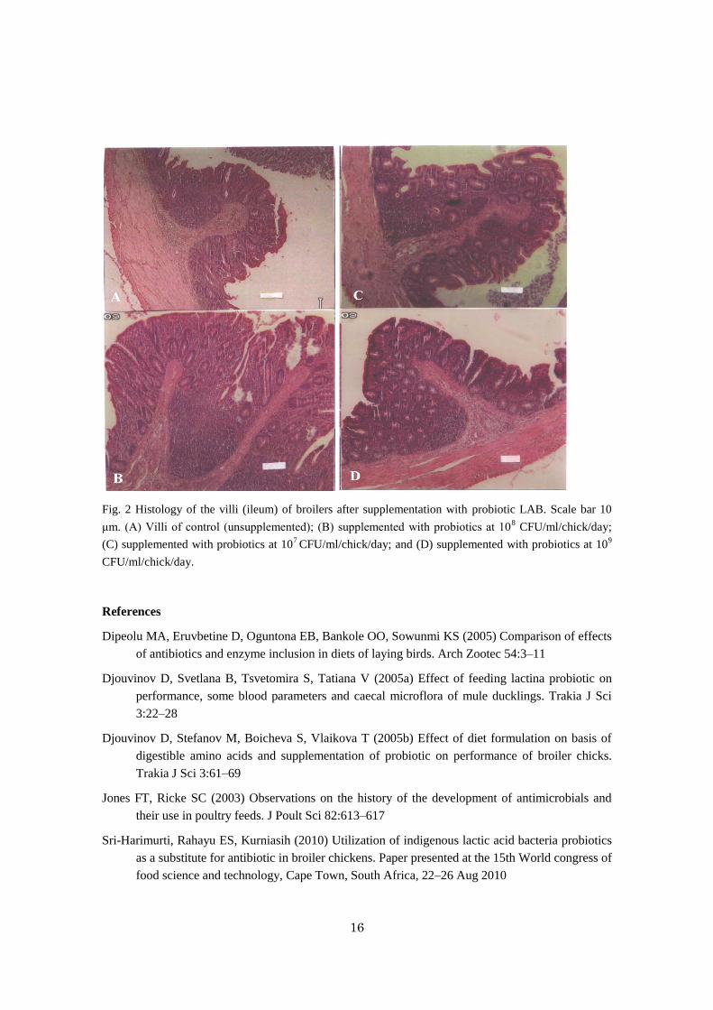
modulate the epithelial cells of the gastrointestinal tract, as presented in Fig. 2.
The in vitro adhesion assay showed that Lactobacillus murinus Ar3, Streptococcus thermophilus
Kp2, and Pediococcus acidilactici Kd6 had a good ability to adhere to IEC, as revealed by
phase-contrast microscopy (Fig. 3). Scanning electron micrographs showed a clear appearance of
Lactobacillus murinus Ar3, Streptococcus thermophilus Kp2, and Pediococcus acidilactici Kd6
attached firmly to the intestine of the supplemented broiler, but there was no attachment on the
intestine of unsupplemented chickens (Fig. 4).

15
Fig. 3 Phase-contrast microscopy of adherence of lactic acid bacteria on intestinal epithelial cells (IEC) of
the chicken. (1) IEC; (2) L. murinus (Ar3); (3) S. thermophilus (Kp2); and (4) P. acidilactici (Kd6)
Fig. 4 Scanning electron micrograph of Lactobacillus murinus (Ar3), Streptococcus thermophilus (Kp2),
and Pediococcus acidilactici (Kd6) in the chicken intestine. Left: unsupplemented chicken intestine;
right: supplemented by those three strains. Scale bar ±1 μm.
Effect on broiler productivity and immune system
Application of a mixture of Lactobacillus murinus Ar3, Streptococcus thermophilus Kd2,
and Pediococcus acidilactici Kp6 had significant effects on live weight, carcass yield, breast portion
weight, abdominal fat weight, and abdominal fat percentage but not in carcass percentage when
supplemented to broilers orally at 107 (T1), 10
8 (T2), and 10
9 CFU/ml/bird/day (T3). The carcass
percentages in this experiment were 68.26±0.60 (T2), 68.66±1.86 (T3), and 69.20±0.90 (T1), similar
to 70.40 observed with the application of a commercial Lactina probiotic. However, the live body
weights with the supplementation of these probiotics 1824.50±38.89(T2),1899±43.50(T3), and
1966.20±38.05(T1) were higher compared to 1688.9 g in broilers supplemented with a commercial
Lactina probiotic (Djouvinov et al. 2005a, b). The diameter of Peyer’s patches in the intestine, as
well as the weight of the bursa and spleen, as indicators of immune responses in chickens following
these probiotics supplementation.
16
Fig. 2 Histology of the villi (ileum) of broilers after supplementation with probiotic LAB. Scale bar 10
μm. (A) Villi of control (unsupplemented); (B) supplemented with probiotics at 108 CFU/ml/chick/day;
(C) supplemented with probiotics at 107 CFU/ml/chick/day; and (D) supplemented with probiotics at 10
9
CFU/ml/chick/day.
References
Dipeolu MA, Eruvbetine D, Oguntona EB, Bankole OO, Sowunmi KS (2005) Comparison of effects
of antibiotics and enzyme inclusion in diets of laying birds. Arch Zootec 54:3–11
Djouvinov D, Svetlana B, Tsvetomira S, Tatiana V (2005a) Effect of feeding lactina probiotic on
performance, some blood parameters and caecal microflora of mule ducklings. Trakia J Sci
3:22–28
Djouvinov D, Stefanov M, Boicheva S, Vlaikova T (2005b) Effect of diet formulation on basis of
digestible amino acids and supplementation of probiotic on performance of broiler chicks.
Trakia J Sci 3:61–69
Jones FT, Ricke SC (2003) Observations on the history of the development of antimicrobials and
their use in poultry feeds. J Poult Sci 82:613–617
Sri-Harimurti, Rahayu ES, Kurniasih (2010) Utilization of indigenous lactic acid bacteria probiotics
as a substitute for antibiotic in broiler chickens. Paper presented at the 15th World congress of
food science and technology, Cape Town, South Africa, 22–26 Aug 2010

17
Sri-Harimurti, Rahayu ES, Kurniasih (2012) The impact of indigenous lactic acid bacteria on the
ability to adhere on chicken ileal epithelial cells and productivity performance of the broiler.
In: The 15th Asian-Australasian animal production (AAAP) congress, Bangkok, Thailand,
26–30 Nov 2012
Sri-Harimurti, Sidadolog JHP, Yuwanta T, Wihandoyo, Sri-Sudaryati, Sasongko H, Ariyadi B
(2013a) Indigenous lactic acid bacteria probiotics: their effects on productive performance
and the effort for reducing the abdominal fat percentage of broiler chicken. Unpublished data
Sri-Harimurti, Huda M, Kistiani AD (2013b) The dynamics of indigenous lactic acid bacteria
probiotics on carcass yield, abdominal fat and intestinal morphology of broilers. In:
Proceeding of the 3rd AINI international seminar, Padang, West Sumatera, Indonesia, 24–26
Sept 2013
Sri-Harimurti, Sidadolog JHP, Yuwanta T, Wihandoyo, Sri-Sudaryati, Sasongko H, Ariyadi B
(2014) The dynamics of indigenous probiotics lactic acid bacteria on growth performance,
total adherence bacteria, and short-chain fatty acids production in the ileum of male quail.
Unpublished data.

18
Effects of probiotics on the innate immune-defense system by antimicrobial
peptides in the gut mucosa of chicks
Mohammed E.S.I. 1, Takumi Terada
1, Naoki Isobe
1,2, and Yukinori Yoshimura
1,2
1Graduate School of Biosphere Science, Hiroshima University;
2Research Center for Animal Science,
Hiroshima University, Higashi-Hiroshima 739-8528, Japan
Introduction
The mucosal of the gastrointestinal tract can be infected by various pathogenic
microorganisms. The gut-associated lymphoid tissues have not been fully developed during the first
week of life of chicks, whereas the immuno-protection could be provided during the first week of
life through maternal antibodies and innate defense system. In contrast, innate immune system may
play significant roles in the defense system since it is developed from early phase of life including
the embryonic stage.
The antimicrobial peptides including avian β-defensins (AvBDs) and cathelicidins (Caths)
are the part of the main components involved in the innate immune-defense system. They
demonstrate antimicrobial activities against a variety of microorganisms, including Gram-positive
and Gram-negative bacteria as well as fungi and enveloped viruses. Fourteen AvBDs and 4 Caths
have been identified in chickens. Innate immune responses are stimulated by pathogen-associated
molecular pathogens (PAMPs) via Toll-like receptors (TLRs). Chicken TLRs are important in the
recognition of PAMPs to induce the production of pro-inflammatory cytokines and antimicrobial
peptides. Ten TLRs have been reported in chickens (TLR1-type 1 and type 2 (1.1 and 1.2), 2.1 and
2.2, 3, 4, 5, 7, 15, and 21, and among them, TLR4 recognizes lipopolysaccharide (LPS) from
Gram-negative bacteria, and others also recognize their specific ligands of different viral and
bacterial molecular patterns.
Probiotic may have beneficial effects on broiler performance as well as on the modulation of
intestinal microflora and their genes (microbiome) to reduce pathogens. They have also been
reported to modulate the expression of AvBDs in the mucosal tissue of the gut mucosa. Akbari et al.
(2008) reported that the expression of AvBDs and cathelicidin was increased due to Salmonella
infection in the chick intestines, but the administration of probiotics eliminated the effects of
Salmonella infection on the expression of those antimicrobial peptides. Pro-inflammatory cytokines
such as IL1β, IL6, IFNγ, and tumor necrosis factor super family 15 (TNFSF15) may cause not only
the inflammation of tissues but also some of them may regulate the adaptive immune system and
AvBDs expression as reported by the studies in the blood and reproductive tissues. If probiotics
affect the expression of antimicrobial peptide and cytokines, they may be considered as the useful
treatment to enhance the gut innate immune system.
In this paper, we describe the effects of probiotics on the expression of AvBDs, Caths and
cytokines in the gut challenged with or without LPS.
Effects of probiotics on the expression of AvBDs, Caths and pro-inflammatory cytokines in
response to lipopolysaccharide stimulation in the proventriculus and cecum of broiler chicks
The aim of this study was to determine whether probiotic feeding affected the expression of
AvBDs, Caths and pro-inflammatory cytokines in response to lipopolysaccharide (LPS) challenge in
the proventriculus and cecum of broiler chicks. One-day-old male Chunky broiler chicks were fed

19
with or without 0.4% probiotics for 7 days (P-group and non-P-group, respectively). Then, they were
orally challenged with no LPS (0-LPS), 1 µg LPS (1-LPS), or 100 µg LPS (100-LPS) (n = 5 in all
groups) in Experiment 1, and with no LPS and 1 µg LPS (n = 6 in all groups) in Experiment 2.
Five hours after LPS challenge, the proventriculi and ceca were collected to analyze Toll-like
receptors (TLRs), AvBDs, Caths, and pro-inflammatory cytokines expression by reverse
transcription-PCR (RT-PCR) using 0-LPS chicks of experiment 1. The expressed TLRs, AvBDs,
CATHs and cytokines were furtherly analyzed by quantitative real-time PCR in all groups in
experiment 1 and 2. All TLRs (TLR1.1, TLR1.2, TLR2.1, TLR2.2, TLR3, TLR4, TLR7, TLR15 and
TLR21 and CD14 were expressed in the proventriculus and cecum of chicks. A total of 7 AvBDs
(AvBD1, 2, 4, 6, 7, 10, and 12) and 8 AvBDs (AvBD1, 2, 4-7, 10, and 12) were identified in the
proventriculus and cecum, respectively. All four types of Caths (Cath1, 2, 3 and 4) were expressed in
the proventriculus and cecum of broiler chicks. Interleukin (IL) 1β, IL6, interferon (IFN) γ and
TNFSF15 were expressed in the proventriculus and cecum of chicks. In Experiment 1, the
expression of 2 AvBDs in the proventriculus and 6 AvBDs in the cecum of 1-LPS chicks was higher
in P-group than in the non-P-group. In Experiment 2, the expression of AvBD1 in proventriculus and
5 AvBDs in cecum of 1-LPS- chicks was higher in P-group than in non-P-group. Challenge with
100-LPS did not cause differences in the AvBDs expression between P- and non-P-group. Expression
of Caths in cecum of 1-LPS- chicks was higher in P-group than in non-P-group. Although IL1β
expression was not affected, the expression of IL6 and TNFSF15 in the proventriculus and
expression of IFNγ in the cecum was lower in P-group than in non-P-group challenged with
100-LPS. These results suggest that probiotic feeding may enhance the immuno-defense system
mediated by AvBDs and Caths but not by cytokine, against infection by Gram-negative bacteria.
Conclusion
In response to LPS stimulation the expressions of AvBDs and Caths were higher in P-group
than non-P-groups, suggesting that probiotics feeding either enhance the expression of these
antimicrobial peptides or protect the reduction of their expression caused by LPS in the intestine.
Thus, probiotics can protect the chicks through antimicrobial peptides immuno-modulatory effects.
References
Akbari MR, Haghighi HR, Chambers JR, Brisbin J, Read LR, Sharif S. 2008. Expression of
antimicrobial peptides in cecal tonsils of chickens treated with probiotics and infected with
Salmonella enterica serovar typhimurium. Clin Vaccine Immunol. 15:1689-1693.
M 1.1 1.2 2.12.2 3 4 5 7 15 21
TLRs
bp
100
5001000
a) TLRs expression
M 1.11.2 2.1 2.2 3 4 5 7 15 21
TLRsbp
100
5001000
Proventriculus
Cecum
b) AvBDs expression
Proventriculus
M 1 2 3 4 5 6 7 8 9 10 11 12 13 14
AvBDs
500
100
bp
M 1 2 3 4 5 6 7 8 9 10 11 12 13 14
AvBDs
500
100
bp
Cecum
Fig.1 Expression of Toll-like receptors (TLRs) and avian
β-defensins (AvBDs) in the chick gut.

20
Effects of probiotics on the innate immune-defense system by antimicrobial
peptides in the gut mucosa of chicks
Takumi Terada1, Naoki Isobe
1,2, and Yukinori Yoshimura
1,2
1Graduate School of Biosphere Science, Hiroshima University;
2Research Center for Animal Science,
Hiroshima University, Higashi-Hiroshima 739-8528, Japan
Introduction
We have shown that avian β-defensins (AvBDs) and cathelicidins are expressed in the
gastrointestinal tract in chicks. The expression of AvBDs in response to LPS stimulation was higher
in the chick fed with probiotics than those fed without probiotics. Akbari et al. (2008) also reported
that the expression of AvBD1, AvBD2, AvBD4, AvBD6 was repressed by probiotics in combination
with Salmonella infection. The expressed AvBDs may play roles in the defense against
Gram-negative bacteria infection. Fourteen AvBDs have been identified in chickens till now. If the
AvBDs expressed by the gut mucosa differ among the different breeds of chickens, it may affect the
susceptibility of chicks. Hong et al. (2012) evaluated the changes in the expression levels of AvBD
mRNA in necrotic enteritis disease model in 2 genetically disparate commercial broiler chicken
lines: Ross and Cobb. They showed the differences in gene expression levels of AvBDs and
proinflammatory cytokines in the intestine, crop, and spleen, suggesting the differences in their
expression may affect the predisposed disease resistance and susceptibility to necrotic enteritis
disease in the 2 commercial broiler chicken lines.
For better understanding the regulation of AvBD expression and synthesis, it is necessary to
identify the cells responsible for the synthesis of AvBDs. Cuperus et al. (2016) reported that AvBD9
was predominantly found in enteroendocrine cells throughout the intestine, and cathelicidins-2 was
exclusively found in heterophils in the several tissues including the intestine of embryonic and early
posthatch chicks. They suggested that AvBD9 appears to be expressed in cell types strategically
located to respond to infectious stimuli, suggesting these peptides play a role in the intestinal defense.
However, the cells that synthesize other AvBDs remain unknown.
The aim of this study was to determine whether the expression profiles of AvBDs were
different in different chicken breeds, and to identify the cells responsible for the synthesis of AvBDs
in the chick intestine. We here examined the gene expression profiles of AvBDs in the gut of
different chicken breeds, including Japanese native chicks and broiler chicks. Then, the intestinal
cells immunoreactive for AvBD10 and 12 were identified.
Expression profiles of AvBDs in the gut of different breeds of chickens
The expression profiles of AvBDs in the proventriculus, ileum and cecum were compared
among three Japanese native chicks, including Tosa-jidori, Hinai-dori, Oh-Shamo, and Chunky
broiler chicks by RT-PCR. In the Chunky broilers, the expression of AvBD1, 2, 4, 7 and 10 in the
proventriculus,AvBD1, 2 ,4, 7 and 10 in the ileum, AvBD1, 2, 4, 6, 7 and 10 in the cecum were
identified. Most of these expression profiles were similarly observed also in the three Japanese
native chicks, suggesting that AvBDs are expressed commonly in different breeds of chickens.
Identification of intestinal cells containing immunoreactive AvBD10 and 12
Sections of the ileum and cecum of chicks (0, 7 or 14-d-old) were immunostained using
21
rabbit anti-AvBD12 raised in our lab. Immunoreaction products of AvBD12 were identified in the
epithelial cells of the crypts of both the cecum of the chicks.
Conclusion
We suggest there may be the polymorphism in the expression levels of AvBDs in the
intestine at the individual level rather than breed levels when compared within Japanese native
chicks. The epithelial cells of the crypts may be responsible to synthesize AvBD-10 and -12 in the
ileum and cecum. Thus the ability to synthesize AvBDs in these cells may be considered for the
strategy to enhance the resistance against infections in the chick intestine.
References
Akbari MR, Haghighi HR, Chambers JR, Brisbin J, Read LR, Sharif S. 2008. Expression of
antimicrobial peptides in cecal tonsils of chickens treated with probiotics and infected with
Salmonella enterica serovar typhimurium. Clin Vaccine Immunol. 15:1689-1693.
Cuperus T, van Dijk A, Dwars RM, Haagsman HP. 2016. Localization and developmental expression
of two chicken host defense peptides: cathelicidin-2 and avian β-defensin 9. Dev Comp
Immunol. 61:48-59.
Hong YH, Song W, Lee SH, Lillehoj HS. 2012. Differential gene expression profiles of β-defensins
in the crop, intestine, and spleen using a necrotic enteritis model in 2 commercial broiler
chicken lines. Poult Sci. 91:1081-1088.
![Gut microbiota and colorectal cancerin germfree animal models of these diseases, no symp-toms are observed [4, 5]. In this review, we discuss the roles of gut microbiota and the mucosal](https://cdn.vdocuments.net/doc/165x107/5f701813aa3b7c4e8c37c570/gut-microbiota-and-colorectal-cancer-in-germfree-animal-models-of-these-diseases.jpg)


