Download - Haemorrhage (original)


CONTENTS
Objectives Introduction Definition Normal Anatomy of Blood Vessels Composition of Blood Pathophysiology Types of Haemorrhage W.H.O Grading Classification of Haemorrhage Causes Signs & Symptoms Emergency Management Nursing Care Plan Summary References

OBJECTIVES
At the end of this presentation, participants willbe able to ; Gain knowledge about haemorrhage Define haemorrhage Describe anatomy of blood vessels & composition of
blood Differentiate different types of haemorrhage Elaborate classification Learn about W.H.O grading of haemorrhage Discuss causes and signs & symptoms of haemorrhage Manage haemorrhage in emergency

HAEMORRHAGE
INTRODUCTION Haemorrhage is the loss of blood escaping
from the circulatory system. Bleeding can occur internally, where blood leaks from blood vessels inside the body, or externally, either through a natural opening such as mouth, nose, ear, urethra, vagina or anus, or through a break in the skin. Uncontrolled bleeding can rapidly lead to shock and death.

HAEMORRHAGE
DEFINITION The term haemorrhage refers to a large amount of
bleeding in a short time. (Thygerson, Gulli & Krohmer 2006; pp:23)
An escape of blood from a ruptured blood vessel. ( Weller & Wells 1990; pp:217)
Haemorrhage is the loss of blood from a vessel. (Malcolm R. Colmer 1986 ; pp:98)

ANATOMY OF BLOOD VESSELS

BLOOD
PLASMA
55%
PROTEINS 7%
WATER 91.5%
PLATELETS1,50,000-400,000
OTHER SOLUTES 1.5%
WHITE BLOOD CELLS
RED BLOOD CELLS 4.8 – 5.4 millions
ALBUMIN 54%GLOBULIN 38%FIBRINOGEN 7%ALL OTHERS 1%
ELECTROLYTESNUTRIENTSGASESREGULATORY SUBSTANCESWASTE PRODUCTS
NEUTROPHILS60 – 70%
LYMPHOCYTES 20 – 25%
MONOCYTES 3 – 8%
EOSINOPHILS 2 – 4%
BASOPHILS 0.5 – 1.0%
COMPOSITION OF BLOOD

CLOTTING MECHANISM
Defense mechanism of circulatory system to leakage Involves complex series of reactions Adequate amount of calcium and all clotting factors are essential Clotting factors include; thromboplastin, prothrombin, thrombin and
fibrinogen Clot formed is called fibrin Platelets and other blood cells also play an important role in clot
formation
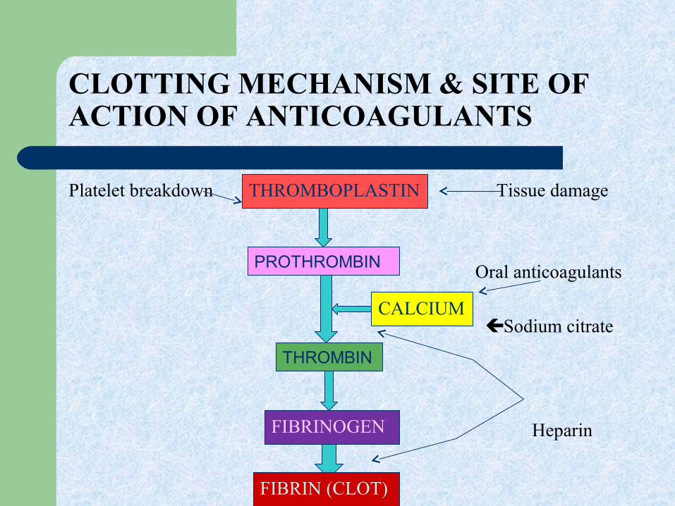
CLOTTING MECHANISM & SITE OF ACTION OF ANTICOAGULANTS
Platelet breakdown Tissue damage
Oral anticoagulants
Sodium citrate
Heparin
THROMBOPLASTIN
THROMBIN
CALCIUM
PROTHROMBIN
FIBRINOGEN
FIBRIN (CLOT)


TYPES OF HAEMORRHAGE

TYPES OF HAEMORRHAGE
CAPILLARY Bleeding oozes steadily but
slowlyVENOUS flow steadily under less
pressure doesn't spurtARTERIAL Bleeding spurts with each
heartbeat Difficult to control due to
pressure Most serious type as large
amount of blood may be lost in short time

TYPES (cont)
PRIMARY Occurs immediately A cut finger or an operation incision
REACTIONARY(INTERMEDIATE) Occurs in first 24-hrs after operation More severe the operation, more likely it is to occur Operations on kidney, thyroid and breasts as well as total
hysterectomy are more liable to be followed by reactionary haemorrhage
SECONDARY If infection is present, walls of blood vessels may be eroded and may
burst, causing what is known as secondary haemorrhage

TYPES (cont)
REVEALED OR EXTERNAL Bleeding can be seen From an open wound e.g.; abrasion, laceration, avulsion,
amputation etc. Through natural opening like mouth, nose, anus, vagina etc.
CONCEALED OR INTERNAL Bleeding cannot be seen Occurs in one of the body cavities such as abdomen Can result from;
Blunt trauma or penetrating injury Acute or chronic medical illness

W.H.O GRADING

CLASSIFICATION OF HAEMORRHAGECLASS I CLASS II CLASS III CLASS IV
up to 15%(<750ml) of total blood volume
15-30%(500-1500ml) of total blood volume
30-40%(2000ml) of total blood volume
>40%(>2000ml) of total blood volume
Compensation Early Decompensation Late Decompensation(Early irreversible)
Compensation Limited(Irreversible)
Normal BP, Pulse, Respirations •Unable to maintain BP•Tachycardia & tachypnea•Decreased pulse strength & narrow pulse pressure
•BP 70mmhg or below(systolic)•Weak , thready rapid pulse•Narrowing pulse pressure•Tachypnea
•Pulse barely palpable•Respirations : rapid, shallow and ineffective
•Vasoconstriction•Release of catecholamine• Epinephrine• Norepinephrine - Anxiety, slightly pale and clammy skin
•Significant release of catecholamine - Cool, clammy skin and thirst - Increased anxiety and agitation -Normal renal output
•Anxiety and restlessness•Increased LOC & AMS•Pale, cold and clammy skin•Decreased renal output
•Lethargic, confused and unresponsive•Extremely pale, cold and clammy skin•Diminished renal output
Fluid resuscitation is not usually required
-volume resuscitation with crystalloids is all that may be required- Blood transfusion is not usually required
fluid resuscitation with crystalloid-blood transfusions are usually required
-aggressive resuscitation is required to prevent death

CAUSES OF HAEMORRHAGE
Multiple trauma Injury to the highly vascular area involving lungs, liver,
spleen, or prostate Any surgical or obstetric emergency Aneurysms Hypertension Septicemia (Gram negative & Meningococcal) Widespread Carcinomas Bleeding disorders

SIGNS & SYMPTOMS
EARLY SIGNS & SYMPTOMS Restlessness and anxiety Coldness ; temp is slightly subnormal Blood pressure is lowered Pulse rate is slightly increased Pallor Increased thirstSIGNS & SYMPTOMS AFTER SEVERE HAEMORRHAGE Extreme pallor Coldness is profound Air hunger ; respirations are rapid & sighing Pulse rate is very rapid

SIGNS & SYMPTOMS (cont)
Blood pressure is extremely low Thirst is extreme Volume of urine output is diminished
SIGNS & SYMPTOMS OF INTERNAL BLEEDING May appear quickly or take days to appear Bruising Painful, tender area Vomiting or coughing up blood

NURSING MANAGEMENT MEDICAL MANAGEMENT
EVALUATION
INTERVENTION
PLANNING
NURSING DIAGNOSIS
ASSESSMENT
Dependent
Independent
HISTORY
EXAMINATION
INVESTIGATION
TREATMENT
PAST HX
PRESENT HX
INSPECTION
PALPATION
AUSCULTATION
GENERAL
SPECIFIC
GENERAL
SPECIFIC
SYMPTOMATIC

ASSESSMENT NURSING DIAGNOSIS PLANNING INTERVENTION EVALUATION
Obvious bleeding risk for deficient fluid volume r/t large amount of blood loss evidenced by trauma
To stop bleeding INDEPENDENT - Apply pressure bandage - Elevation(limbs - Shift the patient to OT (if needed) DEPENDENT - IV coagulation therapy(tranaxemic acid, vit.k, FFP)
Bleeding stopped
Restlessness and anxietySOBLips cyanoticDelayed or absent capillary refillBP below 70mmhg to unobtainable
Ineffective tissue perfusion related to hypotension evidenced by excessive blood loss
To make pt relax and comfortableTo build systolic BP upto above 90mmhgTo get strong peripheral pulses
INDEPENDENT - Counseling & psychotherapy - Attach cardiac monitor -O2 inhalation - Monitor SP02 continuously - Monitor patient for signs of shockDEPENDENT - IV fluid replacement according to blood loss
Patient is relaxedBreathing comfortableStrong peripheral pulsesBP above 90mmhg (systolic)No signs of shock
Cold clammy skinTemp below 35dc
Hypothermia related to ineffective tissue perfusion evidenced by hypotension
To build up body temperatureTo get skin warm
- cover patient with blanket to warm up body _ Monitor skin temperature every 15 mints
Skin warm and dryTemp 37dc or above

INVESTIGATIONS GENERAL All baseline investigations SPECIFIC PT, APTT to check clotting profile ABG’s to check perfusion CBC to review Hb, Hct levels, and platelet count RFT’s to review renal profile when urine output is less or
diminished

CONTROL OF EXTERNAL BLEEDING Place dressing over the wound and apply direct pressure If patient is bleeding from an arm or leg, elevate the injured area
above heart level to reduce blood flow Apply a pressure bandage (if bleeding is not controlled) If bleeding still cannot be controlled, apply pressure at a pressure
point (artery or vein) while keeping pressure on the wound


CONTROL OF INTERNAL BLEEDING For minor internal bleeding (such as bruise on the leg from
bumping into the corner of a table), follow the steps of the RICE procedure:
Rest the injured area Ice or cold pack application over the injury Compression over injured area by applying an elastic bandage Elevation of injured arm or leg, if it is not broken For serious internal bleeding follow these steps Care for shock by raising legs 6 to 12 inches, and cover the patient to
maintain warmth If vomiting occurs, roll the patient onto his/her side to keep airway
clear Monitor breathing Identification and correction of underlying problem

SUMMARY
Haemorrhage is loss of blood from any blood vessel due to some trauma or injury. It may also occur due to some bleeding disorder or tumors. Bleeding may be external or internal. Signs and symptoms depend on extent of blood loss. It is classified into four classes according to blood loss. W.H.O has set a standard grading schedule to assess level of blood loss. Uncontrolled bleeding can lead to hemorrhagic shock and even death. So immediate measures are taken to control bleeding and blood products and fluids are administered to replace fluid volume. Patient is monitored continuously and assessed to check patient’s response to therapy.

REFERENCES
Brunner & Suddhart’s Textbook of Medical Surgical Nursing, vol 2, 12th ed, 2010: pp 2161 – 2163
Colmer ; Moroney’s Surgery for Nurses, 16th ed, 1981 : pp 98 – 106
Howard, Steinmann, Sheehy’s emergency nursing principles & practice, 6th ed, 2003 pp ;
Thygerson, Gulli & Krohmer, First Aid, 5th ed, 2006 : pp 23 – 27
http://www.google.com/bleeding-wikipedia
http://europepmc.org/abstract/MED/6517266


