5
GENETIC D E A F N E S S
ROBERT J . R U B E N
Departments of Otorhinolaryngology and Pediatrics Albert Einstein College of Medicine
Yeshiva University New York, New York
I. Introduction History of the Genetics of Hearing Impairment
11. Nosology A. Anatomical B. Phenotype C. Mode of Inheritance D. Molecular Aberrations: Changes
in Nuclear DNA and Mitochondrial DNA (mtDNA)
III. Syndromic Hearing Impairment A. Alport Syndrome B. Branchio-Oto-Renal (BOR)
Syndrome
C. Jervell and Lange-Nielsen Syndrome
D. Mitochondrial Syndromes E. Norrie Disease F. Pendred Syndrome G. Stickler Syndrome H. Treacher Collins-Franceschetti
Syndrome I. Usher Syndrome J. Waardenburg Syndrome K. Nonsyndromic Hearing
Impairment L. Clinical Application M. Management
I. INTRODUCTION
The underlying processes that result in hearing loss and deafness are numerous and, in many instances, interact. The most common of these factors is the genetic make-up of the individual. It has been estimated that more than 50% of all hearing losses have a substantial genetic component. This is a low estimate, because for about 25% of those with hearing impairments the etiology is unknown, and many
The Handbook of Genetic Communicative Disorders 8 9
Copyright © 2001 by Academic Press. All rights of reproduction in any form reserved.

9 0 ROBERT J . RUBEN
of these will eventually be proven to be genetically derived (see Chapter 1). Additionally, as various acquired causes of hearing loss are prevented, such as congenital rubella and Haemophilus influenzae meningitis, the percentage of persons with genetically determined deafness will increase proportionally (Parv-ing & Christensen, 1996). Some of the acquired deafness (e.g., from a standard non-ototoxic dose of aminoglycosides) has now been determined to come about because of genetic predispositions (Fischel-Ghodsian et al, 1993). Another instance of an extrinsic factor that causes a gene to be activated so as to result in hearing loss may be the relationship between otosclerosis and the measles virus (McKenna & Mills, 1989; Niedermeyer & Arnold, 1995). It is anticipated that there will be an increase in the percentage of hearing loss in which a genetic disposition plays a substantial role. Genetic hearing loss is associated with all the different forms of hearing loss whether classified by age, progression, and site of lesion(s) or interactions with extrinsic factors, for example, aminoglycosides (Fischel-Ghodsian r̂ a/., 1993).
The information concerning genetics and hearing expands almost daily, and this is expected to continue for the foreseeable future. Much of this new information will have direct utility for the care of patients. These rapid advances are made available in a timely manner through a number of Internet websites. There are two among many (Table 5.1) that both contain substantial information in very usable format and provide links to other pertinent sites.
HISTORY OF THE GENETICS OF HEARING IMPAIRMENT (Ruben, 1991)
A famihal tendency for hearing loss has been recognized at least since the early seventeenth century. Bonet's book (1620), the first printed book concerning the education of the deaf, is based on the education of one child who had numerous hearing-impaired relatives (Plann, 1997). The first work to categorize systematically familial and nonfamilial deafness is Wilde's Otology (1853). Wilde's data,
TABLE 5 . 1 Pertinent Websites for Assessing Current Information Concerning the Genetics of Deafness
HTML
http://www3.ncbi.nlm.nih.gOv//
http://dnalab-www.uia.ac.be/dnalab/hhh
Name
Online Mendelian Inheritance in Man (OMIM)™
Hereditary Hearing Loss
Description
Information can be obtained by syndrome, characteristics. Has numerous links to literature. Substantial detail and depth. Excellent search engine.
Gives concise information of the state of the art of the known genes that result in hearing loss
Q ^ GENETIC DEAFNESS ^ *
developed before the recognition of Mendel's modes of inheritance, were not widely distributed. Love (1896) and others at the beginning of the twentieth century described and emphasized the role of inheritance as a significant etiology for hearing impairment.
The recognition of familial bases of hearing loss has been used to harm affected individuals through attempts to prevent marriage, sterilize, and exterminate. This tragic aspect of the history of the genetics of hearing impairment begins with A. G. Bell's On a Deaf Variety of the Human Race (1880), in which he states that to eliminate deafness the deaf should not marry nor have children. This work, and others of Bell's veiled works (he used sheep breeding as a model for human eugenics) contributed to the early policies of the Nazi regime to sterilize and then exterminate deaf children. These crimes are well documented and published in Biesold's (1988, 1999) Klagende Hande {Crying Hands).
Although there were some pertinent publications during the first half of the twentieth century, it was not until 1976, with the publication of two books, Fraser's (1976) and the posthumous publication of Konigsmark's work (Konigsmark & Gorlin, 1976), that there was widespread appreciation of the genetics underlying deafness. These works preceded the current enhanced ability to localize genes on chromosomes and to determine the aberrations in the protein sequences. A symposium held in 1990 under the auspices of the New York Academy of Sciences (Ruben et al, 1991) brought together the current information concerning the molecular basis of the genetics of deafness. Since the 1990 meeting, information on the genetics of deafness has vastly increased. During the period 1990 to 2000, the National Library of Medicine (as determined by computer search) has indexed more than 1500 articles that relate to genetics and deafness and/or hearing loss. This growth of information is expected to continue. The present era should be one of defining the molecular abnormalities resulting in deafness and lesser hearing losses, determining how these aberrations affect the hearing mechanisms, and constructing successful interventions to prevent, cure, or ameliorate the effects of the gene action.
I I . NOSOLOGY
A. ANATOMICAL
There are numerous ways by which the various genetically based hearing disorders can be classified. The anatomical site(s) of gene action can be used to organize the information in a useful manner. Knowledge of whether the outer ear, external ear canal, middle ear, cochlea, vestibular apparatus, statoacoustic nerve, and/or the central nervous system are affected is useful to the clinician in the care of the specific genetic disorder. One gene may affect a number of sites, and an abnormality at one locus can result in a variety of different anatomical defects.
9 2 "^'" ROBERT J . RUBEN
B. PHENOTYPE
The apparent characteristics of the gene action, the phenotype, are another useful means of classification. There are at least two general phenotypic modes of classification: syndromic and nonsyndromic. The first is defined in terms of whether there are other abnormalities associated with the deafness, such as are found in the various Waardenburg or Usher syndromes. If the deafness is determined to be of genetic etiology and there is no other detectable expression of the gene actions, then these are classified as nonsyndromic.
The second, nonsyndromic phenotypic classification concerns various features of the hearing loss. A time dimension can be applied to classify whether the loss was congenital, early onset, middle life, or presbycusis, thus mapping the presence of progression of the loss. The characteristics of the pure tone audiogram are also used to classify the abnormality. The audiometric profiles can be high tone, low tone, U-shaped audiograms, and others.
C. MODE OF INHERITANCE
The genetically determined hearing losses can be categorized as to whether they are dominant (DFNA#), recessive (DFNB#), X-linked (DFN#), or mitochondrial. (The suffixed numeral refers to the order of identification. Thus, DFNAl is the first dominant gene to be assigned a locus and DFNB5 is the fifth recessive gene to be.) There are both phenotypic and molecularly defined genetic hearing losses that appear to have more than one mode of inheritance. The difference between a recessive inheritance—carrying two identical abnormal genes or two different abnormal genes (compound heterozygous)—and a dominant mode of inheritance—carrying only one abnormal gene—may appear as a difference in the amount of the hearing impairment (Zlotogora, 1997).
D. MOLECULAR ABERRATIONS: CHANGES IN NUCLEAR DNA AND MITOCHONDRIAL DNA (mtDNA)
There are several types of abnormalities in the structure of an individual's DNA that result in a symptomatic form of hearing impairment. These consist of deletions or insertions of a portion of the DNA; substitution of one or more nucleic acids in a segment of DNA; a translocation, in which a piece of the DNA is exchanged for another piece of the DNA; and inversions, in which the orientation of a segment of DNA is flipped; or duplication of a stretch of DNA (see Gerber, 1998). Additionally, there may abnormalities in chromosome number.
Mitochondria, the energy-producing organelles, contain their own genetic material. Changes in mtDNA occur in a number of syndromic and nonsyndromic hearing impairments. The mitochondrial disorders are transmitted from the mother to the child.
Q 3 GENETIC DEAFNESS ^ ^
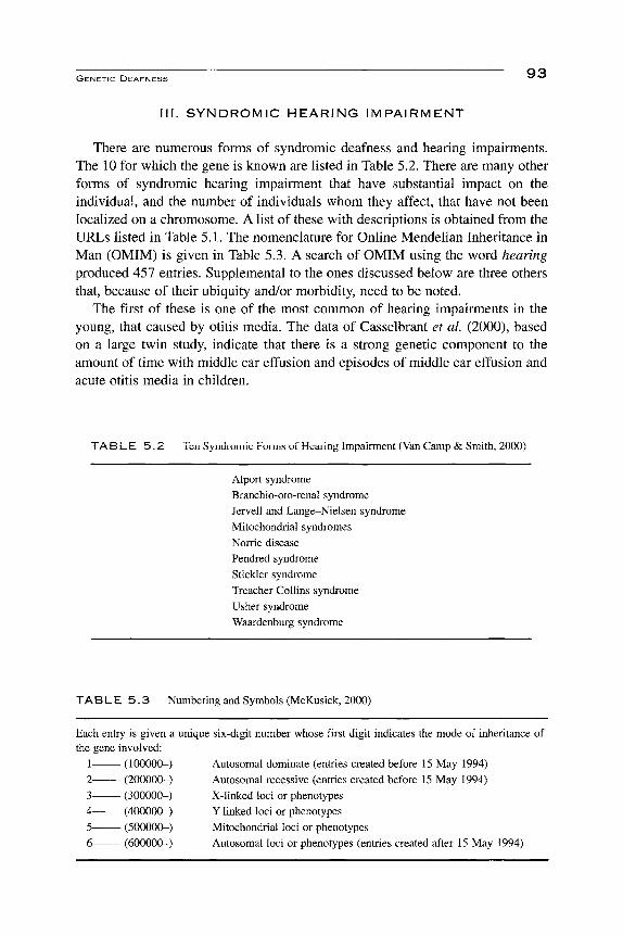
I I I . SYNDROMIC HEARING IMPAIRMENT
There are numerous forms of syndromic deafness and hearing impairments. The 10 for which the gene is known are Hsted in Table 5.2. There are many other forms of syndromic hearing impairment that have substantial impact on the individual, and the number of individuals whom they affect, that have not been locaHzed on a chromosome. A list of these with descriptions is obtained from the URLs listed in Table 5.1. The nomenclature for Online Mendelian Inheritance in Man (OMIM) is given in Table 5.3. A search of OMIM using the word hearing produced 457 entries. Supplemental to the ones discussed below are three others that, because of their ubiquity and/or morbidity, need to be noted.
The first of these is one of the most common of hearing impairments in the young, that caused by otitis media. The data of Casselbrant et al. (2000), based on a large twin study, indicate that there is a strong genetic component to the amount of time with middle ear effusion and episodes of middle ear effusion and acute otitis media in children.
TAB L E 5 . 2 Ten Synuiuiiiiu Funiis ui Hearing Impairment (Van Camp & Smith, 2000)
Alport syndrome Branchio-oto-renai syndrome Jervell and Lange-Nielsen syndrome Mitochondrial syndromes Norrie disease Pendred syndrome Stickler syndrome Treacher Collins syndrome Usher syndrome Waardenburg syndrome
TABLE 5 . 3 Numbering and Symbols (McKusick, 2000)
Each entry is given a unique six-digit number whose first digit indicates the mode of inheritance of the gene involved:
1 (100000-) Autosomal dominate (entries created before 15 May 1994) 2 (200000-) Autosomal recessive (entries created before 15 May 1994) 3 (300000-) X-linked loci or phenotypes 4 (400000-) Y-linked loci or phenotypes 5 (500000-) Mitochondrial loci or phenotypes 6 (600000-) Autosomal loci or phenotypes (entries created after 15 May 1994)
QA ^ ^ ROBERT J . RUBEN
The second is Shprintzen (velocardiofacial) syndrome (Shprintzen et«/., 1981) (OMIM Link 192430; this Hnk is the reference code from Online Mendelian Inheritance in Man). The locus for this syndrome is at 22qll and is similar to the DiGeorge syndrome. Shprintzen syndrome consists of cleft palate, pharyngeal hypotonia, medial displacement of the internal carotid arteries, cardiac anomalies, typical facies, learning disorders, slender hands and digits, umbilical hernia, hypospadias, and, in adolescents and adults, psychotic illness. Conductive hearing loss is found in 47% and a sensory-neural loss in 17% of patients with Shprintzen syndrome (Reyes etaL, 1999; Shprintzen etal, 1981; ID: 14). These patients need to be identified so as to avoid inappropriate adenoidectomy (and the possibility of damage to the internal carotid artery) and the exacerbation of their velopharyngeal insufficiency. Their hearing impairments need to be recognized and cared for.
The third is neurofibromatosis, type II (NFII) (OMIM Link 101000) whose locus is at 22ql2.2. These patients have the central form of neurofibromatosis, which is characterized by tumors of both statoacoustic nerves, and meningiomas and schwannomas of the dorsal roots of the spiral ganglia. There is often associated posterior capsular lens opacity. Almost half of these individuals will present with unilateral or bilateral hearing loss in the first two decades of life. Individuals will present in the first decade with severe morbidity due to the intracranial tumors and the subsequent loss of hearing, vestibular response, and, in some cases, blindness. Many of these patients' morbidities are the probable result of spontaneous mutation. The diagnosis of NFII should be considered in cases of asymmetrical hearing loss or those with poor discrimination. MRI is for diagnosis, and reveals small asymptomatic tumors in the statoacoustic nerve.
A. ALPORT SYNDROME (OMIM Link 104200)
This syndrome consists of nephritis, which can progress to renal failure. The hearing loss is sensory-neural, varying from modest to severe, and is usually progressive. Males are more severely affected than females (Admiraal, 1970; Turner, 1970). There are three forms of genetic inheritance: autosomal dominant, recessive at 2q36-37, and sex-linked at Xq22. All forms have variable penetrance.
B. BRANCHIO-OTO-RENAL (BOR) SYNDROME (OMIM Link 113560)
BOR consists of a number of variable anomalies of the face and neck, which may include abnormalities of cup-shaped and/or anteverted pinnae; bilateral pre-helical pits; bilateral bronchial fistulae; and bilateral renal dysplasia with anomalies of the collecting system. Additionally, there have been reported polycystic kidneys and abnormalities of the lachrymal ducts including stenosis and/or aplasia. The hearing loss may be conductive, mixed, or sensory neural (Cremers & Fikkers-van Noord, 1980). Many, if not most, of these patients have a spectrum of abnormalities of the bony labyrinth ranging from aplasia to deficient coils of
Q — GENETIC DEAFNESS ^ ^
the cochlea. The genetic transmission is dominant, and the gene has been located on 8ql3.3.
C. JERVELL AND LANGE-NIELSEN SYNDROME (OMIM Link 220400)
The Jervell and Lange-Nielsen syndrome consists of a cardiac conduction abnormality characterized by a prolonged QT interval. This is associated with syncopal attacks in the affected individual. The cardiac symptoms appear during the first 2 to 3 years of life and, if uncared for, can result in death (Cusimano et al, 1991). The hearing loss is typically a congenital severe to profound sensory-neural deafness. The mode of inheritance is recessive and there are two different gene locations: llpl5.5 and 21q22.1-22.2.
D. MITOCHONDRIAL SYNDROMES
Several forms of mitochondrial hearing loss with other associated defects have been reported. These are myoclonic epilepsy and ragged red fibers (MERRF) on tRNAlys at 8344>G. These patients have severe neurological symptoms including blindness and a variable amount of hearing loss. There is a variant of MERRF that occurs on tRNAlys but at 8356T>C that has an associated postlingual progressive deafness. Another is mitochondrial encephalopathy, lactic acidosis, and stroke-hke episodes (MELAS). The locus is on tRNAleu (UUR) at 3243A>G. About 30% of these patients will have an associated hearing loss. Keams-Sayre syndrome (KSS) consists of external ophthalmoplegia and retinopathy that begin by age 20. Additionally, there is an associated ataxia, heart block, and hearing loss. This hearing loss is thought to be central, based on auditory evoked potential data (Korres et aL, 1999). The defects on the mitochondria are large and are found on several genes.
E. NORRIE DISEASE (OMIM Link 310600)
This is an X-linked disorder that has congenital ocular symptoms, including retinal pseudotumor and necrosis of the inner layer of the retina, cataracts, and blindness (Black & Redmond, 1994). There is a progressive sensory-neural hearing loss and mental retardation in half of the patients. This disease begins with blindness, and the hearing loss comes on later in life. This is the reverse of most of the Usher disease variants. The gene is located at Xpll.3 (Berger et aL, 1992).
E PENDRED SYNDROME (OMIM Link 274600)
Pendred syndrome is a recessive disorder associated with a mild form of thyroid disease in that there can be a clinical goiter; it was first described by Vaughn Pendred in 1896, and a century later the gene was mapped to chromosome 7 (Coyle et aL, 1996). There is a reasonably specific test for this condition in that an affected patient will show only a partial discharge of iodine when thiocyanate or perchlorate is given. The histology of the affected thyroid gland is unusual and can be mistaken for cancer; cancers have been reported, however, in these patients.

9 6 *^ ^ ROBERT J. RUBEN
The hearing loss may be either a congenital or progressive sensory-neural loss and may be associated with defective vestibular function. The bony labyrinth of these patients may be abnormal, as has been seen in temporal bone histology and imaging. Many of these patients have an enlarged vestibular aqueduct (Cremers et aL, 1998). The gene is located at 7q21-34, and there is a number of allelic variants.
G. STICKLER SYNDROME (OMIM Links 108300,184840, and 121028)
There are three forms of this dominant syndrome reported (Table 5.4); the most common is STLl located at 12ql3.11-ql3.2. The classic syndrome consists of progressive myopia, vitreoretinal degeneration, premature joint degeneration with abnormal epiphyseal development, midfacial hypoplasia, irregularities of the vertebral bodies, and cleft palate, and many will have a sensory-neural hearing loss that can be exacerbated by the concomitant chronic or intermittent otitis media with effusion (Ruben & Math, 1978). Stickler syndrome affects vision and hearing, and these are less-than-total losses of either sensory modality. Additionally, it affects the expressive aspect of spoken language because of the cleft palate. Some of these children will have a tracheal or laryngeal stenosis, which further decreases the expressive faculties as they may have a tracheotomy in place for some time during their language formative years (Nowak, 1998).
TABLE 5 .4
Locus name
Three Forms of Stickler Syndrome (Van Camp & Smith, 2000)
Location Gene Entry
STLl
STL2
STL3
12ql3.11-ql3.2
6p2L3
lp21
COL2A1
COL11A2
COLllAl
108300
184840
121028
H. TREACHER COLLINS-FRANCESCHETTI SYNDROME (OMIM Link 154500)
The features of this autosomal-dominant syndrome are an antimongoloid slant of the eyes, coloboma (fissure) of the lower eyelid, micrognathia, hypoplastic zygomatic arches, macrostomia, and microtia. Most of these patients have an asymmetric hearing loss worse than moderate. All those with hearing loss have a conductive component, and about 10 to 20% have a mixed loss (Pron et ai, 1993; Jahrsdoerfer & Jacobson, 1995). The conductive loss is due to malformation of one or more of the ossicles. There are more than 35 different mutations at the locus 5q32-q33.1.

GENETIC DEAFNESS 9 7
I. USHER SYNDROME (OMIM Links 276900, 276903, 276904, 601067, 602097, 602083, 2376901, 276905, 276902; Table 5.5)
Usher syndrome affects vision and hearing. There are three cUnical types (Table 5.5) (Wagenaar, 2000). It is marked by a retinitis pigmentosa with a progressive loss of vision during the first decades of life and with either a congenital or progressive hearing loss of variable degree. Additionally, many of these patients will have a loss of vestibular function. The most common variety is Type I. These
T A B L E 5 . 5 Usher Syndrome: Clinical Classification (Wagenaar, 2000)
Usher Type I
Usher Type E Normal
Usher Type III
Hearing impairment
Congenital, Severe to profound
Congenital, mild to moderate; can be progressive
Congenital, Progressive
Visual impairment
RP starts before puberty
RP starts before or after puberty
RP starts before or after puberty
Vestibular function
Areflexia, Variable
Variable
patients should be identified as early as possible, for their disease affects three sensory systems: hearing, balance, and vision. Usher syndrome is one of the main reasons for ophthalmological screening of all hearing-impaired infants and children (see section III.L.T). The vestibular impairment will manifest itself as late walking that can be mistakenly interpreted as developmental delay, especially in a hearing- and visually impaired child. Understanding of the molecular bases of Usher syndrome is still evolving. There are at least ten different variations on six different chromosomes (Table 5.6) that are phenotypically classified as Usher syndrome. As with many forms of inherited deafness, there is a mouse model that is being used to further understand the molecular pathology (Saw et at, 1991 \ ID:6). The ten forms are all recessive, and are located at lq41, 3p23-24.2, 3q21-q25, 5ql4.3-q21-23, 10, lOq, l lpl5.1, l lql3.5, 14q32, and 21 q.
J. WAARDENBURG SYNDROME (OMIM Links 19350a, 193510,148820, 277850)
Waardenburg syndrome is presently classified into four types (Table 5.7), all with considerable variability of pigmentary and hearing abnormalities. Their pigmentary and facial features phenotypically and clinically identify these pa-
9 8
TABLE 5.6
Locus name
USHIA
USHIB
USHIC
USHID
USHIE
USHIF
USH2A
USH2B
0USH2C
USH3
Usher Syndrome
Location
14q32
llql3.5
llplS.l
lOq
21q
10
lq41
3p23-24.2.
5ql4.3^2L3
3q21-q25
ROBERT J . RUBEN
: Molecular Classification (Van Camp & Smith,
Gene
Unknown
MY07A
Unknown
Unknown
Unknown
Unknown
USH2A
Unknown
Unknown
Unknown
Screening markers
D14S250, D14S260, D14S292, D14S78
D11S906, D11S911, D11S52, OMP-CA
D11S902, D11S921, D11S899, D11S861
D10S529, D10S202, D10S573
D21S1884, D21S1257, D212S265, D21S1258
D10S199, D10S578, D10S596
D1S229, D1S490, D1S237, D1S474
D3S1578, D3S3647, D3S3658
D5S428, D5S421
D3S1299, D3S1555, D3S1280, D3S1279
2000)
Entry
276900
276903
276904
601067
602097
602083
276901
276905
276902
TABLE 5 . 7 Waardenburg Syndrome: Clinical Classification (Van Camp & Smith, 2000)
Type I Dystopia canthorum
Type II
Klein-Waardenburg syndrome (type III)
Waardenburg-Shah syndrome (type IV)
No dystopia canthorum
Type I and upper limb abnormalities
Type II and Hirschsprung disease (autosomal recessive inheritance)
tients. Many will have some combination of dystrophia canthorum (lateral displacement of the inner canthus of each eye), a white forelock, and heterochromia of the iris (Read & Newton, 1997; ID: 12). Additionally, they may have cleft palate or a submucosal cleft of the palate, and there is an association with Hirschsprung disease and upper limb abnormalities. The range of hearing will be from bilateral congenital profound loss to normal, with other varieties of sensory-neural losses including unilateral losses (DeStefano et al, 1998; Lalwani et ai, 1996; Liu et ai, 1995; Morell et al, 1997). There are six different molecular types (Table 5.8).
Q Q GENETIC DEAFNESS ^ ^
TABLE 5 . 8 Waardenburg Syndrome: Molecular Classification (Van Camp & Smith, 2000)
Type Location Gene Entry
WStypel(WSl)
WS type II (WS2)
WS type III
WS type IV
WS type IV
WS type IV
2q35
3pl4.1-pl2.3
2q35
13q22
20ql3.2-ql3.3
22ql3
PAX3
MITF (: 156M5)
PAX3
EDNRB (: 131244)
EDN3 (: 131242)
SOXIO (: 602229)
193500
193510
148820
277850
277580
277580
The mode of inheritance is dominant, three of the types with gene loci at 2q35 and 3pl4.1-pl2.3, and three types that are recessive with loci at 13q22, 20ql3.2-ql3.2, and 22ql3.
K. NONSYNDROMIC HEARING IMPAIRMENT
There is dominant, recessive, sex-linked, and mitochondrial transmission of nonsyndromic hearing impairments.
1. Dominant
The loci for the 31 DFNA hearing impairments are listed in Table 5.9. Each of these syndromes represents either one or just a few kindred. The forms of DFNA vary between and within each of the separate entities. There can be early-onset impairment (Kunst et al, 1999) or impairments later in life with progression (Van Camp et al, 1999), with many other variations. It is critical to make a genetic diagnosis, because at least one of these DFNA syndromes may be treatable with medication (Fukushima et al, 1999). It is expected that there will be many specific and generic effective interventions for the various forms of genetic hearing impairments as understanding of the molecular abnormalities increases.
2. Recessive
The loci for the 28 DFNB are listed in Table 5.10. The DFNBs manifest themselves similarly to the DFNAs in that there is heterogeneity of phenotype as to time of onset, progression, and severity. DFNBl—Connexin 26 (URL http://www.iro.es/cx26deaf.html)—is the most prevalent cause of all genetic hearing impairments. The abnormal gene is found in 3.01%, with a probable range of

lOO ROBERT J . RUBEN
T A B L E 5 . 9 Nonsyndromic Hearing Impairment, Autosomal Dominant Loci (DFNA) (Van Camp & Smith, 2000)
Locus name Location Gene
OMIM entry
DFNAl
DFNA2
DFNA3
DFNA4
DFNA5
DFNA6
DFNA7
DFNA8
DFNA9
DFNA 10
DFNAl 1
DFNA12
DFNA13
DFNA14
DFNA15
DFNA16
DFNA17
DFNA 18
DFNA19
DFNA20
DFNA21
DFNA22
DFNA23
DFNA24
DFNA25
DFNA26
DFNA27
DFNA28
DFNA29
DFNA30
DFNA31
5q31
lp34
13ql2
19ql3
7pl5
4pl6.3
Iq21-q23
llq22-24
14ql2-ql3
6q22-q23
Ilql2.3-q21
Ilq22-q24
6p21
4pl6
5q31
2q24
22q
3q22
10 (pericentr.)
17q25
HDIAl
GJB3
KCNQ4
GJB2
GJB6
Unknown
DFNA5
Unknown
Unknown
TECTA
COCH
Unknown
MY07A
TECTA
Unknown
Unknown
POU4F3
Unknown
Unknown
Unknown
Unknown
Unknown
14q
4q
12q21-24
17q25
4ql2
8q22
15q26
Unknown
Unknown
Unknown
Unknown
Unknown
Unknown
Unknown
124900
600101
601544
600652
600994
600965
601412
601543
601369
601316
601317
601842
601868
Unknown
602459
602460
Unknown
Unknown
Unknown
Unknown
Unknown
Unknown
Unknown
Unknown
Unknown
Unknown
Unknown
GENETIC DEAFNESS l O l
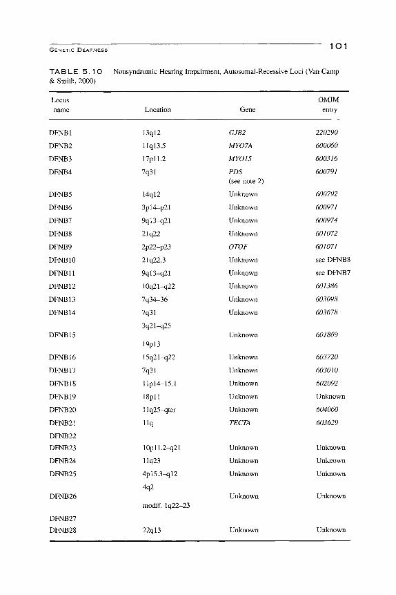
TA B L E 5 . 1 O Nonsyndromic Hearing Impairment, Autosomal-Recessive Loci (Van Camp & Smith, 2000)
Locus name Location Gene
OMIM entry
DFNBl
DFNB2
DFNB3
DFNB4
DFNB5
DFNB6
DFNB7
DFNB8
DFNB9
DFNBIO
DFNBll
DFNBl 2
DFNBl 3
DFNB14
DFNBl 5
DFNB16
DFNB17
DFNBl8
DFNB19
DFNB20
DFNB21
DFNB22
DFNB23
DFNB24
DFNB25
DFNB26
DFNB27
DFNB28
13ql2
llql3.5
17pll.2
7q31
14ql2
3pl4-p21
9ql3-q21
21q22
2p22-p23
21q22.3
9ql3-q21
10q21-q22
7q34-36
7q31
3q21-q25
19pl3
15q21-q22
7q31
llpl4-15.1
ISpll
llq25-qter
l lq
10pll.2-q21
llq23
4pl5.3-ql2
4q2
modif. lq22-23
22ql3
GJB2
MY07A
MY015
PDS (see note 2)
Unknown
Unknown
Unknown
Unknown
OTOF
Unknown
Unknown
Unknown
Unknown
Unknown
Unknown
Unknown
Unknown
Unknown
Unknown
Unknown
TECTA
Unknown
Unknown
Unknown
Unknown
Unknown
220290
600060
600316
600791
600792
600971
600974
601072
601071
see DFNB8
see DFNB7
601386
603098
603678
601869
603720
603010
602092
Unknown
604060
603629
Unknown
Unknown
Unknown
Unknown
Unknown

1 0 2 ROBERT J . RUBEN
2.5 to 3.5% of the population in the Midwestern United States (Green etal, 1999). It is found more frequently in populations whose origins are from the Mediterranean, and especially in Jewish populations of Ashkenazi origin (Morell et al, 1998), who have been found to have a tendency for a rare mutation of the gene 167delT, and an observed carrier rate of 4.03% (95% confidence interval of 2.5 to 6.0%). Connexin 26 appears to be associated with at least 50% of all nonsyn-dromal deafness (Murgia et ai, 1999). The hearing loss, sensory-neural, is characterized by being either congenital or early onset; it can be progressive, asymmetrical, and of varying degree (Cohn et al., 1999). This common, severe genetic disorder should be screened for as a part of a newborn hearing-screening program, because it is probable that neonates with Connexin 26 mutation who display early progressive loss would not be identified by the physiological techniques presently in use.
3. X-Linked
The loci for the five DFNs are listed in Table 5.11. These syndromes are similar to the other nonsyndromic hearing disorders in that they present with a myriad of phenotypes, most often but not exclusively in the male (Cremers & Huygen, 1983; Papadaki et al, 1998). The most common of these is DFN3: deafness mixed with perilymphatic gusher or Nance syndrome (Nance et al, 1970). These patients, primarily males, may have a progressive mixed hearing loss and a characteristic dilatation of the internal auditory meatus (Cremers etal, 1985). Attempts to repair the conductive loss with manipulation of the footplate of the stapes has resulted in a pouring out of perilymph—a gusher—and a resultant loss of hearing in the
TABLE 5 . 1 1 Nonsyndromic Hearing Impairment, X-Linked Recessive Loci (Van Camp & Smith, 2000)
Locus name
DFNl
DFN2
DFN3
DFN4
DFN5
Location
Xq22
Xq22
Xq2Ll
Xp2L2
Xp22
Gene
DDP
Unknown
POU3F4
Unknown
Unknown
Screening markers
DXSlOl
COL4A5
DXS26 DXS995 DXS232
DXS997
DXS992
DXS8036 DXS8022 DXS8019
OMIM entry
304700
304500
304400
300030
300066
1 O*^ GENETIC DEAFNESS * ^^^
operated ear. The presence of a progressive conductive or mixed hearing loss can be associated with a stapedial reflex in these patients (Snik et al, 1995), and this observation may be used so that interventions other than surgery are utilized for the restoration of hearing.
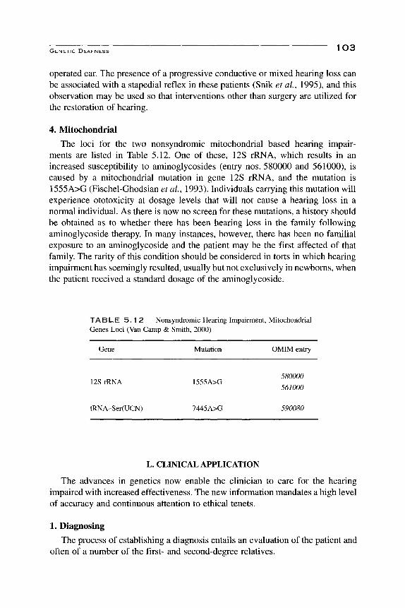
4. Mitochondrial
The loci for the two nonsyndromic mitochondrial based hearing impairments are listed in Table 5.12. One of these, 12S rRNA, which results in an increased susceptibility to aminoglycosides (entry nos. 580000 and 561000), is caused by a mitochondrial mutation in gene 12S rRNA, and the mutation is 1555A>G (Fischel-Ghodsian et al, 1993). Individuals carrying this mutation will experience ototoxicity at dosage levels that will not cause a hearing loss in a normal individual. As there is now no screen for these mutations, a history should be obtained as to whether there has been hearing loss in the family following aminoglycoside therapy. In many instances, however, there has been no familial exposure to an aminoglycoside and the patient may be the first affected of that family. The rarity of this condition should be considered in torts in which hearing impairment has seemingly resulted, usually but not exclusively in newborns, when the patient received a standard dosage of the aminoglycoside.
TABLE 5 . 1 2 Nonsyndromic Hearing Impairment, Mitochondrial Genes Loci (Van Camp & Smith, 2000)
Gene Mutation OMIM entry
12S rRNA 1555A>G 580000
561000
tRNA-Ser(UCN) 7445A>G 590080
L. CLINICAL APPLICATION
The advances in genetics now enable the clinician to care for the hearing impaired with increased effectiveness. The new information mandates a high level of accuracy and continuous attention to ethical tenets.
1. Diagnosing
The process of establishing a diagnosis entails an evaluation of the patient and often of a number of the first- and second-degree relatives.
* ^ ^ ROBERT J . RUBEN
2. History
Information should be acquired concerning the time of onset of the hearing loss, specific information about potential acquired causes such as ototoxic medications, especially salicylates, and sound trauma. A detailed family-genetic history is obtained for all hearing loss that is not obviously acquired. The genetic history should include siblings, children, and as many relatives as possible for two previous generations. Specific data are obtained about these relatives, not only concerning the hearing and whether there are any apparent physical signs— for example, different colored eyes as found in the Waardenburg syndrome—^but also what their last names may have been in the past and from which cities or region they and their progenitors came from. The ascertainment of last name and region of origin many times will indicate a possible relatedness of the parent of the affected individual. Health care providers working for patients who are members of circumscribed communities need to have knowledge of families that carry various genes for hearing impairment. The genetic history is obtained in the course of several visits. On the first visit, an outline is made and the person responsible for the patient—either the patient or a caregiver—is given the task of inquiring in detail about other members of the family. At a second visit, it is usually helpful, if possible, to have a member of the family from an older generation aid in the process of providing the pertinent history.
3. Functional Testing
Each patient requires a complete assessment of the auditory-vestibular system to include middle ear, inner ear, and the statoacoustic nerve. The techniques used depend on the age of the patient. Infants less than 6 months of age will be assessed with physiological measures that include middle ear impedance, cochlear emissions, and frequency-specific air and bone conduction auditory evoked potentials (Stapells & Ruben, 1989). Many infants and toddlers older than 6 months can be assessed with behavioral audiograms (Gravel & Traquina, 1992), in addition to the cochlear emissions and impedance measures. The initial hearing assessment must be accurate at all frequencies and, where appropriate, in discriminatory ability, as this evaluation serves to establish the extent of the loss and is the standard by which the patient is monitored for progression of the hearing impairment.
Audiometric testing of relatives, especially younger siblings, is carried out, as this will reveal other affected members. Many cases of hearing impairment will be nonsyndromal and have no obvious family history to indicate that the cause is genetic. Hearing evaluations of parents, siblings, or children may suggest a probable genetic origin, in that there can be frequency-specific notches in the transient evoked cochlear emission and/or the audiogram. This assessment appears to be more accurate in people carrying an autosomal dominant in which there is low penetrance, for example, Waardenburg syndrome (Liu et al, 1995), than in obligate carriers of recessive genes (Lina-Granade etai, 1998). Although there is less
-I r\c5 G E N E T I C D E A F N E S S • v-r*^
accuracy in the recessive carriers, the finding in a parent, sibling, or child of a propositus of a hearing loss that is not predicted based on the individual's age or hearing history is useful. It will indicate other possible investigations, and will benefit the presumably affected person who should be audiometrically monitored.
4. Serology
Viral and serological studies can be done for congenital infection. The incidence of congenital rubella and syphilis appears to be relatively rare. These studies should be obtained when the history and/or the physical examination indicate that such infection may be a possibility. Two hallmarks of congenital rubella are abnormalities of the retina and/or small infant size. Currently, the most common form of congenital infection is from cytomegalovirus (CMV), which, in some cases, can result in a progressive early-onset hearing loss (Ahlfors, Ivarsson, & Harris, 1999; Fowler et al, 1999).
5. Imaging
The CT will yield information on the bony structure of the ear. This is especially important in the diagnosis of labyrinthine malformations; such findings are used both to substantiate a diagnosis—for example, BOR, Pendred, Treacher Collins, DFN3 gusher—and to direct care with regard to the possibility of a perilymphatic fistula. MRI is used to evaluate patients with progressive unilateral or asymmetric losses, especially those with decreased speech discrimination.
6. Dysmorphology
Evaluation of the patient's facial features, pigmentation, and overall physical structure will add to the information necessary for diagnosis. Included in the routine assessment should be the intercanthal distance, the status of the palate, limb and digital morphology, and other facial features.
7. System Evaluation
The visual system needs to be evaluated to ensure that the patient has optimal visual acuity and/or to make a specific diagnosis (e.g.. Usher disease) and/or to provide for a care plan that would include a static or progressive visual impairment. Early diagnosis of Usher disease is critical and is facilitated by the use of the electroretinogram. This may not be accurate in a young child and is most sensitive at age l>-\ years. It has been found to be positive in a child of 18 months (Smith et al, 1998). The ophthalmologic evaluation can give information concerning congenital infections, such as rubella, toxoplasmosis, and so on. An important consideration is the greater adverse effect that a visual impairment will
^ ^ ^ ROBERT J . RUBEN
have on a person burdened with an auditory deficiency. The hearing-impaired individual is even more dependent on vision than others and needs vision to function optimally. The early diagnosis of myopia, astigmatism, or strabismus will be of considerable advantage to the hearing impaired, especially the young child. Ophthalmologic examination is required, for this author, in every case of static or progressive childhood hearing impairment.
The genital-urinary system is evaluated for anatomic malformation, with the use of ultrasound, in all patients with external and/or middle ear malformation, because of the high incidence of renal abnormalities found in these patients.
Renal function is assessed through the measure of creatinine and blood urea nitrogen in patients suspected of having Alport syndrome. Evaluation of thyroid function with the use of the perchlorate test is indicated if there is an indication of Pendred disease. The recording of an electrocardiogram is carried out for those who are suspected of having Jervell and Lange-Nielson disease. Most often there will be no indication that an individual will have any of the three above-mentioned disorders. It is suggested that these evaluations be carried out not as a screen but when there is information to suggest autosomal recessive inheritance, or when the patient and/or family show some finding that would indicate that there is a possibility of the disorder. The electrocardiogram and the renal function studies are the least invasive, whereas the thyroid studies are more invasive.
Vestibular studies are useful in establishing a diagnosis of Usher disease. They also contribute to the management of an infant in that they aid in better understanding of why motor development may be delayed. Patients who have no vestibular function and their caregivers need to be cautioned concerning swimming, as they have a decreased ability to orient in water. There have been drownings, which are thought to be attributable to the lack of vestibular function.
Patients with hearing impairment with onset during the period of language development should undergo a complete evaluation of expressive and receptive language functions and of their speech abilities. These data will provide a basehne for evaluations of therapy and are also useful in directing habiHtation. There is also the possibility that the patient may have a specific language disorder (see Chapter 6), which will be exacerbated by the hearing impairment. Those who appear to be cognitively delayed, with or without other neurological signs, require an appropriate neurological evaluation.
8. Molecular DNA and RNA Testing
Genetic disorders can be accurately and precisely diagnosed by molecular evaluation; presently, however, there is only limited availability for this testing. Each medical center has a number of genes that it may test. Many professionals will cooperate with one another to test samples. It is expected that, with the development of gene chip technology, there will be genetic tests widely available for many of the known genetic disorders of hearing impairment. These evaluations
GENETIC DEAFNESS I \J /
will be used to determine, in regard to a given patient, who in the family is affected or are carriers, and may be used to screen initially for abnormal genes.
M. MANAGEMENT
There are several special areas of patient management that are obligated when a diagnosis of a genetic basis for a hearing impairment is established.
1. FoUow-Up
Many genetic disorders are characterized by a progression of the hearing impairment and/or another clinical deficit. These patients require periodic evaluations so that appropriate care can be provided. The periodicity—duty cycle—of follow-up in some instances will be determined by previous reported experience with a particular genetic syndrome. If this information is not available, then periodicity of follow-up is determined by population studies (Ruben & Fishman, 1980). The rapid advances in medical genetics in the areas of diagnosis and, now, intervention, must be applied to patients who have been diagnosed. The new information is used as part of the continual care of the patient. Each patient should be aware of his or her diagnosis and be enabled to access the new developments, for example, through the use of the Internet.
2. Counseling
The action of the gene over time is an important dimension in the care of genetic disorders. There are two different aspects. The first is the way in which the gene will affect the individual. These issues are attended to both in the informing interview and at the periodic follow-up visits. The second concerns potential transmission of the gene to the patient's children, and how this may affect the progeny. Once the mode of inheritance is established, and the degree of penetrance is known, then a reasonable estimate of transmission can be communicated to the patient or the parents. Dominant genes will affect half of the progeny with usually variable penetrance. The recessive genes differ. If another child from the same couple is affected, then, in the main, 25% of the progeny will be homozygous. The offspring of a known recessive homozygote may or may not have a significantly increased probability of being affected, depending on the gene causing the impairment. The children of a homozygote or a compound heterozy-gote for Connexin 26 would be at a higher risk for hearing impairment because of the ubiquity of the abnormal Connexin gene. A child of a homozygote recessive, a rare recessive, would be at somewhat greater risk for impairment than the normal population if there were no assorted matings. Similarly, a hearing impairment with unknown etiology cannot be designated as not genetic; furthermore, children of this patient will be at a greater risk. The probability of having an affected child is greater than for the previous population based risk of 10% (Fraser, 1976), but less than the 25% of a known recessive.
' ^ ^ * - ' ROBERT J. RUBEN
3. Screening
Techniques are currently available to screen for a number of the genetic hearing impairments. These screening techniques have the potential to affect several purposes. One would be to screen all newborns for the disorders that are the most common and/or those with high morbidity, in the way the various hormonal screens are carried out, such as those for PKU and hypothyroidism. Such information would be beneficial for instituting prompt and effective care and effective preventive interventions. Other uses of a screen may or may not affect the conduct of a person's life, and thus bring ethical issues strongly into play. The screen could be used, for example, to determine who in a family carried a recessive gene, or whether a potential mate carried a gene synergetic for hearing impairment. There is also the potential use of a screen to determine the genetic makeup of a fetus.
4. Ethics
There is a population of people whose language is visual, because they have little or no access to auditory communication. Many of these individuals whose language is based on vision do not consider their lack of use of sound as an abnormality nor as a disease. From the end of the nineteenth century to the middle of the twentieth, there had been a number of programs directed not only at extinguishing their language, and in some cases their subculture, but also with the thrust of euthanizing, sterilizing, or prohibiting marriage so that those genetically so affected would cease to exist (Bell, 1883; Biesold, 1999; Ruben, 1991). This history underscores the importance of a critical tenet of self-determination, autonomy, in the ethics of genetic medicine. Individuals have the right to determine whether they will utilize any given intervention. The governing bodies can provide information, as from a screening program, but they cannot mandate a particular course of a biological therapy. In the case of hearing impairment and deafness, options exist that are sufficient to enable a person to achieve a normal life in the culture at large, and in a longstanding and recognized subculture of visually based language. Fetal testing for hearing-impairment genes raises an ethical issue. In assessing these, it must be kept in mind that the advent of gene therapies or other specific therapies directed at preventing or reversing deleterious gene action, should become a means to cure and to prevent, and not to destroy (see Chapter 11). The power that a deepening understanding of genetics provides cannot be used to eliminate any human group. Although such a statement may seem gratuitous, it is sobering to consider that past and recent history indicate the need for maintaining that idea in the forefront of awareness.
REFERENCES
Admiraal, R. J. (1970). Hereditary hearing loss with nephropathy (Alport's syndrome). Acta Otolaryn-gologica, SuppL, 271, 7-26.
1 OQ GENETIC DEAFNESS * v y ^
Ahlfors, K., Ivarsson, S. A., & Harris, S. (1999). Report on a long-term study of maternal and congenital cytomegalovirus infection in Sweden: Review of prospective studies available in the literature. Scandinavian Journal of Infectious Diseases, 31, 443-457.
Bell, A. G. (1880). On a deaf variety of the human race. Washington, DC: National Academy of Sciences.
Bell, A. G. (1883). Memoir upon the formation of a deaf variety of the human race. Proceedings of
the National Academy of Sciences, pp. 1-86.
Berger, W., Meindl, A., van de Pol, T. J., Cremers, F. R, Kopers, H. H., Doemer, C., Monaco, A., Bergen, A. A., Lebo, R., & Warburg, M. (1992). Isolation of a candidate gene for Norrie disease by positional cloning. Nature Genetics, 1, 199-203. [Published erratum appears m. Nature Genetics,
1992, 2, 84.]
Biesold, H. (1988). Klagende hande. Germany: Solms.
Biesold, H. (1999). Crying hands: Eugenics and deaf people in Nazi Germany. Washington, DC: Gallaudet University Press.
Black, G., & Redmond, R. M. (1994). The molecular biology of Norrie's disease. Eye, 8(Pt. 5), 491^96.
Bonet, J. P. (1620). Reduction de las letras y arte para ensehar a abler las mudos. Madrid: Francisco Abarca de Angulo.
Casselbrant, M. L., Mandel, E. M., Fall, P. A., Rockette, H. E., Kurslasky, M., Bluestone, C. D., & Ferrell, R. E. (2000). The heritability of otitis media: A twin and triplet study. Journal of the American Medical Association, 282, 2125-2130.
Cohn, E. S., Kelley, R M., Fowler, T. W, Gorga, M. R, Lefkowitz, D. M., Kuehn, H. J., Schaefer, G. B., Gobar, L. S., Hahn, F. J., Harris, D. J., & Kimberling, W. J. (1999). Clinical studies of families with hearing loss attributable to mutations in the connexin 26 gene (GJB2/DFNB1). Pediatrics, 103, 546-550.
Coyle, B., Coffey, R., Armour, J. A., Gausden, E., Hochberg, Z., Grossman, A., Britton, K., Pembrey, M., Reardon, W, & Trembath, R. (1996). Pendred syndrome (goiter and sensorineural hearing loss) maps to chromosome 7 in the region containing the nonsyndromic deafness gene DFNB4.
Nature Genetics, 12, 421-423.
Cremers, C. W., & Fikkers-Van Noord, N. M. (1980). The earpits-deafness syndrome. Clinical and genetic aspects. International Journal of Pediatric Otorhinolaryngology, 2, 309-322.
Cremers, C. W., & Huygen, P. L. (1983). Clinical features of female heterozygotes in the X-linked mixed deafness syndrome (with perilymphatic gusher during stapes surgery). International Journal of Pediatric Otorhinolaryngology, 6, 179-185.
Cremers, C. W, Hombergen, G. C , Scaf, J. J., Huygen, P L., Volkers, W S., & Pinckers, A. J. (1985). X-linked progressive mixed deafness with perilymphatic gusher during stapes surgery. Archives of Otolaryngology, 111, 249-254.
Cremers, C. W., Marres, H. A., & van Rijn, P. M. (1991). Nonsyndromal profound genetic deafness in childhood. In R. J. Ruben, T. R. Van De Watter, & K. P Steel (Eds.), Genetics of hearing impairment. Annals of the New York Academy of Sciences, 630, 191-196.
Cremers, C. W, Bolder, C, Admiraal, R. J., Everett, L. A., Joosten, F. B., VanHauwe, P., Green, E. D., & Otten, B. J. (1998). Progressive sensorineural hearing loss and a widened vestibular aqueduct in Pendred syndrome. Archives of Otolaryngology—Head & Neck Surgery, 124, 501-505.
Cusimano, F., Martines, E., & Rizzo, C. (1991). The Jervell and Lange-Nielsen syndrome. International Journal of Pediatric Otorhinolaryngology, 22, 49-58.
DeStefano, A. L., Cupples, L. A., Amos, K. S., Asher, J. H. J., Baldwin, C. T., Blanton, S., Carey, M. L., da Silva, E. O., Friedman, T. B., Greenberg, J., Lalwani, A. K., Milunsky, A., Nance, W. E., Pandya, A., Ramesar, R. S., Read, A. P, Tassabejhi, M., Wilcox, E. R., & Farrer, L. A. (1998). Correlation between Waardenburg syndrome phenotype and genotype in a population of individuals with identified PAX3 mutations. Human Genetics, 102, 499-506.
* * ^ ROBERT J . RUBEN
Fischel-Ghodsian, N., Prezant, T. R., Bu, X., & Oztas, S. (1993). Mitochondrial ribosomal RNA gene mutation in a patient with sporadic aminoglycoside ototoxicity. American Journal of Otolaryngology, 14, 399-403.
Fowler, K. B., Dahle, A. J., Boppana, S. B., & Pass, R. F. (1999). Newborn hearing screening: Will children with hearing loss caused by congenital cytomegalovirus infection be missed? Journal of Pediatrics, 135, 60-64.
Fraser, G. F. (1976). The causes of profound deafness in childhood. Baltimore: The Johns Hopkins
University Press.
Fukushima, K., Kasai, N., Ueki, Y, Nishizaki, K., Sugata, K., Hirakawa, S., Masuda, A., Gunduz, M., Ninomiya, Y., Masuda, Y, Sato, M., McGuirt, W. T., Coucke, P, VanCamp, G., & Smith, R. J. (1999). A gene for fluctuating, progressive autosomal dominant nonsyndromic hearing loss,
DFNA16, maps to chromosome 2q23-24.3. American Journal of Human Genetics, 65, 141-150.
Gerber, S. E. (1998). Etiology and prevention of communicative disorders. San Diego: Singular.
Gravel, J. S., & Traquina, D. N. (1992). Experience with audiologic assessment of infants and toddlers.
International Journal of Pediatric Otolaryngology, 23, 59-71.
Green, G. E., Scott, D. A., McDonald, J. M., Woodworth, G. G., Sheffield, V. C., & Smith, R. J. (1999). Carrier rates in the midwestern United States for GJB2 mutations causing inherited
deafness. Journal of the American Medical Association, 281, 2211-2216.
Jahrsdoerfer, R. A., & Jacobson, J. T. (1995). Treacher Collins syndrome: Otologic and auditory
management. Journal of the American Academy of Audiology, 6, 93-102.
Konigsmark, B. W., & Gorlin, R. J. (1976). Genetic and metabolic deafness. Philadelphia: Saunders.
Korres, S. G., Manta, P. B., Balatsouras, D. G., & Papageorgiou, C. T. (1999). Audiological assessment
in patients with mitochondrial myopathy. Scandinavian Audiology, 28, 231-240.
Kunst, H., Marres, H., Huygen, P., Ensink, R., VanCamp, G., VanHauwe, P., Coucke, P., Willems, P., & Cremers, C. (1998). Nonsyndromic autosomal dominant progressive sensorineural hearing loss:
Audiologic analysis of a pedigree linked to DFNA2. Laryngoscope, 108, 74-80.
Lalwani, A. K., Mhatre, A. N., San Agustin, T. B., & Wilcox, E. R. (1996). Genotype-phenotype
correlations in type 1 Waardenburg syndrome. Laryngoscope, 106, 895-902.
Lina-Granade, G., Kreiss, M., Gelas, T, Collet, L., & Morgan, A. (1998). Cochlear irregularities in obligate carriers of recessive genetic hearing impairment and in control subjects. In D. Stephens, A. Read, & A. Martini (Eds.), Developments in genetic hearing impairment (pp. 68-76). London: Whurr.
Liu, X. Z., Newton, V. E., & Read, A. P. (1995). Waardenburg syndrome type, II: Phenotypic findings
and diagnostic criteria. American Journal of Medical Genetics, 55, 95-100.
Love, J. K. (1896). Deaf mutism: A clinical and pathological study. Glasgow: James MacLehose and
Sons. McKenna, M. J., & Mills, B. G. (1989). Immunohistochemical evidence of measles virus antigens in
active otosclerosis. Otolaryngology-Head and Neck Surgery, 101, 415-421. McKusick, V. A. (2000). Mendelian inheritance in man [online]. 4-29-2000. Internet Communication. Morell, R., Friedman, T. B., Asher, J. H. J., & Robbins, L. G. (1997). The incidence of deafness is
nonrandomly distributed among families segregating for Waardenburg syndrome type 1 (WSl). Journal of Medical Genetics, 34, 447-452.
Morell, R. J., Kim, H. J., Hood, L. J., Goforth, L., Friderici, K., Fisher, R., VanCamp, G., Berlin, C. I., Oddoux, C, Ostrer, H., Keats, B., & Friedman, T. B. (1998). Mutations in the connexin 26 gene (GJB2) among Ashkenazi Jews with nonsyndromic recessive deafness. New England Journal of Medicine, 339, 1500-1505.
Murgia, A., Orzan, E., Polli, R., Martella, M., Vinanzi, C, Leonardi, E., Arslan, E., & Zacchello, F. (1999). Cx26 deafness: Mutation analysis and clinical variability. Journal of Medical Genetics, 36, 829-832.
GENETIC DEAFNESS * * *
Nance, W. E., Sweeney, A., McLeod, A. C, & Cooper, M. C. (1970). Hereditary deafness: A presentation of some recognized types, modes of inheritance, and aids in counseling. Southern Medical Bulletin, 58, 41-57.
Niedermeyer, H. P., & Arnold W. (1995). Otosclerosis: A measles virus associated inflammatory disease. Acta Otolaryngologic a, 115, 300-303.
Nowak, C. B. (1998). Genetics and hearing loss: A review of Stickler syndrome. Journal of Communication Disorders, 31, 437^53.
Papadaki, E., Prassopoulos, P., Bizakis, J., Karampekios, S., Papadakis, H., & Gourtsoyiannis, N. (1998). X-linked deafness with stapes gusher in females. European Journal of Radiology, 29, 71-75.
Parving, A., & Christensen, B. (1996). Epidemiology of permanent hearing impairment in children in relation to costs of a hearing health surveillance program. International Journal of Pediatric Otorhinolaryngology, 34, 9-23.
Phelps, P D., Coffey, R. A., Trembath, R. C , Luxon, L. M., Grossman, A. B., Britton, K. E., Kendall-Taylor, P, Graham, J. M., Cadge, B. C , Stephens, S. G., Pembrey, M. E., & Reardon, W. (1998). Radiological malformations of the ear in Pendred syndrome. Clinical Radiology, 53, 268-273.
Plann, S. (1997). Silent minority: Deaf education in Spain, 1550-1835. Berkeley: University of California Press.
Pron, G., Galloway, C , Armstrong, D., & Posnick, J. (1993). Ear malformation and hearing loss in patients with Treacher Collins syndrome. Cleft Palate and Craniofacial Journal, 30, 97-103.
Read, A. P., & Newton, V. E. (1997). Waardenburg syndrome. Journal of Medical Genetics, 34, 656-665.
Reyes, M. R., LeBlanc, E. M., & Bassila, M. K. (1999). Hearing loss and otitis media in velo-cardio-facial syndrome. International Journal of Pediatric Otorhinolaryngology, 47, 227-233.
Ruben, R. J. (1991). The history of the genetics of hearing impairment. Annals of the New York Academy of Sciences, 630, 6-15.
Ruben, R. J., & Fishman, G. (1980). Otological care of the hearing impaired child. In G. T. Mencher & S. E. Gerber (Eds.), Early management of hearing loss (pp. 105-120). New York: Grune & Stratton.
Ruben, R. J., & Math, R. (1978). Serous otitis media associated with sensorineural hearing loss in children. Laryngoscope, 88, 1139-1154.
Ruben, R. J., Van De Water, T. R., & Steel, K. P. (Eds.) (1991). Genetics of hearing impairment. New York: New York Academy of Sciences.
Saw, D. J., Steel, K. P., & Brown, S. D. (1997). Shaker mice and a peek into the House of Usher. Experiments in Animals, 46, 1-9.
Sheffield, V. C , Kraiem, Z., Beck, J. C, Nishimura, D., Stone, E. M., Salameh, M., Sadeh, O., & Glaser, M. (1996). Pendred syndrome maps to chromosome 7q21-34 and is caused by an intrinsic defect in thyroid iodine organification. Nature Genetics, 12, 424-426.
Shprintzen, R. J., Goldberg, R. B., Young, D., & Wolford, L. (1981). The velo-cardio-facial syndrome: A clinical and genetic analysis. Pediatrics, 67, 167-172.
Smith, S. D., Kimberling, W. J., Schaefer, G. B., Horton, M. B., & Tinley, S. (1998). Medical genetic evaluation for the etiology of hearing loss in children. Journal of Communication Disorders, 31, 371-389.
Snik, A. P., Hombergen, G. C , Mylanus, E. A., & Cremers, C. W. (1995). Air-bone gap in patients with X-linked stapes gusher syndrome. American Journal of Otology, 16, 241-246.
Stapells, D. R,, & Ruben, R. J. (1989). Auditory brain stem responses to bone-conducted tones in infants. Annals of Otology, Rhinology and Laryngology, 98, 941-949.
Turner, J. S. (1970). Hereditary hearing loss with nephropathy (Alport's syndrome). Acta Otolaryn-gologica, SuppL, 271, 7-26.
1 1 2 ^ ' * ^ ROBERT J. RUBEN
Van Camp, G., & Smith, R. J. H. (2000). Hereditary hearing loss homepage [onHne]. Internet Communication.
Van Camp, G., Kunst, H., Flothmann, K., McGuirt, W., Wauters, J., Marres, H., Verstreken, M., Bespalova, I. N., Burmeister, M., VandeHeyning, P. H., Smith, R. J., Willems, R J., Cremers, C. W., & Lesperance, M. M. (1999). A gene for autosomal dominant hearing impairment (DFNA14) maps to a region on chromosome 4pl6.3 that does not overlap the DFNA6 locus. Journal of Medical Genetics, 36, 532-536.
Wagenaar, M. (2000). The Usher syndrome: A clinical and genetic correlation. Den Haag: CIP— gegevens koninklijke bibliotheek.
Wilde, S. W. R. W. (1853). Particle observations on aural surgery and the nature and treatment of diseases of the ear with illustrations. Philadelphia: Blanchard & Lea.
Zlotogora, J. (1997). Dominance and homozygosity. American Journal of Medical Genetics, 68, 412-416.


