
Hematology in the ICU
Yoan Lamarche

Yoan Lamarche
• A 42 yo patient presents to the ER 3 weeks after allogenic hematopoietic stem cell transplant for acute myeloid leukemia. His principal complaint is fever and cough. 1. Classify hematologic malignancies
and outline the principles of treatment. (Noemie)

Yoan Lamarche
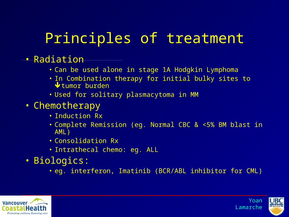
Principles of treatment• Radiation
• Can be used alone in stage 1A Hodgkin Lymphoma• In Combination therapy for initial bulky sites to tumor burden• Used for solitary plasmacytoma in MM
• Chemotherapy • Induction Rx• Complete Remission (eg. Normal CBC & <5% BM blast in
AML)• Consolidation Rx• Intrathecal chemo: eg. ALL
• Biologics: • eg. interferon, Imatinib (BCR/ABL inhibitor for CML)

Yoan Lamarche
Principles of treatment
• Bone Marrow Transplant– Source of cells: bone marrow, peripheral blood or
umbilical cord blood– Auto: Donor is also recipient
• No GVHD• Allows higher doses of chemo• Used for lymphoma and MM
– Allo: Immunologically distinct• Used for AML, ALL, CML, CLL, MDS, lymphoma• Used in nonmalignant diseases: eg.Aplastic anemia• Desired effect of Graft vs tumor BUT risk of GVHD• Followed by immunosuppression to prevent rejection

Yoan Lamarche
2. What are the complications of hematopoietic stem cell transplant leading to ICU admission (Todd)
3. Explain the principles of treatment of infectious and non infectious complications of hematopoietic stem cell transplant. (Todd)

Yoan Lamarche
Life-threatening complications of HSCT
• Non-infectious– Cardiogenic pulmonary edema
• From chemo conditioning (cyclophosphamide) or chronic chemo (anthracycline)
• Pericarditis (+/- tamponade or arrhythmias) from radiation
– Noncardiogenic pulmonary edema• Diffuse Alveolar Damage (analogous to ARDS)

Yoan Lamarche
Life-threatening complications of HSCT, continued
• Diffuse Alveolar Hemorrhage– Rarely with gross hemoptysis; usually progressive hypoxia,
dyspnea, tachycardia within 2 weeks of HSCT– CXR --> patchy interstitial +/- airspace opacification, initially
involving mid-zones, spreading outward– Dx is with bronchoscopy, where progressively bloodier returns are
noted with repeated BAL– Pathophysiology: ??? Pre-transplant airway inflammation– High mortality (up to 90%)– Treatment: Pulse steroids (according to a retrospective study in
1994…)

Yoan Lamarche
Life-threatening complications of HSCT, continued
• Interstitial lung disease (usually associated with aggressive chemo, including cyclophosphamide, doxorubicin and cisplatin)
• Pneumonitis:– Aspiration (ie mucositis and opiates)– Idiopathic interstitial pneumonitis and ARDS or
fibrosis (high mortality)
• Non-pulmonary: Hepatic veno-occlusive disease (may --> fulminant hepatic failure)

Yoan Lamarche
Infectious complications
• Preengraftment as well as immediately and late (> 3 mo) postengraftment
• Bacterial, fungal and viral pathogens lie in wait at each stage.
• Immunosuppression (including neutropenia) is the key factor– Including chemo conditioning period

Yoan Lamarche
The Usual Suspects• Bacterial:
– Aerobic gram positive and gram-negative organisms• Psuedomonas (<1% of allogeneic SCT pts), legionella and bacillus in
particular• Fungal:
– Candida (mucocutaneous or invasive) and aspergillus are most common• Fusarium, zygoycetes, and others are increasingly recognized
• Viral:– CMV (pneumonia, enteritis, retinitis, encephalitis, marrow suppression,
FUO…)– HSV (VZV late postengraftment)– HHV 6, 7,8 (fever, pneumonitis, encephalitis)
– EBV (pneumonia)– Adenovirus (pneumonitis, nephritis, hemorrrhagic colitis,
hemorrhagic cystitis)

Yoan Lamarche
The Usual Suspects (don’t forget Verbal Kint)
• Parasitic:– PCP (or PJP), rare due to chemoprophylaxis– Toxopasmosis– Mycobacteria (TB, avum)

Yoan Lamarche

Yoan Lamarche

Yoan Lamarche

Yoan Lamarche

Yoan Lamarche
DAH-Hemosiderin
laden macrophages

Yoan Lamarche

Yoan Lamarche

Yoan Lamarche
Prophylaxis
• Viral: Acyclovir through day 30 (HSV), gancyclovir through day 100 (CMV)
• Bacterial: Quinolone during neutropenia• Fungal: Fluconazole through day 75
(candida)• Parasitic: TMP/SMX through day 180 or end
of immunosuppression (PCP)

Acyclovir 30d
Gancyclovir 100d vs PCR q week
Quinolone x neutropenic phaseFluconazole x 75d
TMP/SMX x 180d or end immmunosupp

Yoan Lamarche
On initial assessment, the patient is anxious and coughing, his RR is 30 and HR 110, his BP is 95/40, he is 38ºC, O2 Sat is 88% on RA after administration of 2L NS by the emergency team.
Yoan Lamarche
The patient is admitted to the ICU for observation, investigation and treatment
4.What should be the initial workup and choice of Rx (Todd)

Yoan Lamarche
Treatment
• Low threshold for broad spectrum coverage, especially in critical care setting. Keep presenting syndrome in mind, but be prepared to jump on septic immunosuppressed patients like a spider monkey.

Yoan Lamarche
For our patient
• Early postengraftment (i.e. immunosuppressed), with pulmonary sepsis syndrome (RR, Temp, HR).– Prophylaxis, chemo and CBC/Diff unknown
• Initial broad coverage to include gram positive/negative bacteria (including psuedomonas– Imipenem 500 mg IV Q 6 hrs– Add antifungal or antiviral if no improvement by day
5, or if clinical picture evolves to suggest these pathogens

Yoan Lamarche
For our patient, continued
• Investigations: Pan-culture (not CSF at this point, but would have low threshold to LP if LOC changes or neurological complaints develop)
• Imaging: CXR initially

Yoan Lamarche
Tube?
• Goal directed therapy as with any other septic patient, with careful consideration of possible cardiomyopathy in setting of chemo
• Decision to intubate would be similar to any other septic patient; the poor outcome associated with multiorgan failure in this population would warrant consultation with heme regarding his prognosis (nb recent pt in SPH ICU)
Yoan Lamarche

Yoan Lamarche
Shorr, Pulmonary Infiltrates in the Non-HIV- Infected Immunocompromised Patient CHEST / 125/1/ JANUARY 2004

Yoan Lamarche
The patient is now in your ICU. His Sat is better with O2, his RR is now 25 and less labored. His CBC indicates Hb of 82, Plt of 19 000 and WBC count of 2, Creat is 200 and LFTs indicate N AST/ALT, Total Bilirubin is 100, Direct bili is 12, GGT N, LDH 200 . The patient has a urine output of 20 cc/hr for the first 2 hours, the patient appears more confused and complains of blurred vision.
5. What are the possible diagnoses leading to this constellation of symptoms (Noemie)

Yoan Lamarche
Q#5 What are the possible diagnoses leading to this constellation of symptoms?
• 42 yo 3 wks post ABMT for AML presents with fever and cough
• Becomes more confused and complaining of blurred vision
• T 38, RR 25, u/o 20 cc/hr in 2 hours• Hb 82, Plt 19, WBC 2, Cr 200, AST/ALT N,
T.Bili 100, D.Bili 12, GGT N, LDH 200

Yoan Lamarche
Q#5 What are the possible diagnoses leading to this constellation of symptoms?
• 42 yo 3 wks post ABMT for AML presents with fever and cough
• Becomes more confused and complaining of blurred vision(?)
• T 38, RR 25, u/o 20 cc/hr in 2 hours• Hb 82, Plt 19, WBC 2, Cr 200, AST/ALT N,
T.Bili 100, D.Bili 12, GGT N, LDH 200

Yoan Lamarche
What are the possible diagnoses leading to this constellation of symptoms?
• Fever in post BMT– ?infection, ?drug fever, ?TTP, rejection?
• Renal failure – ?Pre-renal, microvascular, toxins/meds, ATN
• Hemolysis– Transfusion reaction (Combs?), TTP?, DIC?
• Acute blurry vision– ?retinal detachment, neuritis, retinal vascular occlusion,
uveitis, acute glaucoma
• Cytopenia– ?rejection, hemolysis, malignancy, DIC/sepsis

Yoan Lamarche
What are the possible diagnoses leading to this constellation of symptoms?
• DDX:– TTP: classical pentad– DIC ? Coags, ? Fibrinogen– Transfusion rxn ? Combs, blurry vision
uncommon– GVHD no mucositis, ?hemolysis and blurry
vision uncommon

Yoan Lamarche
6. What are the risk factors associated with development of TTP (Scot)

Yoan Lamarche

Yoan Lamarche
5. What is the physiopathology of TTP (Naisan)

Yoan Lamarche

Yoan Lamarche

Yoan Lamarche
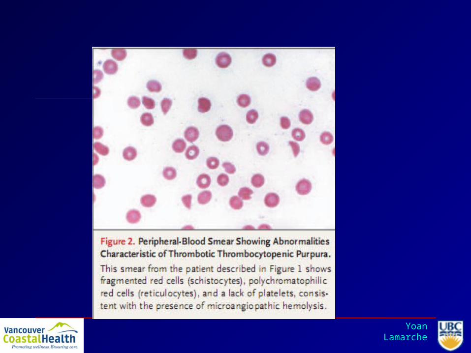
TTP blood smear
Yoan Lamarche
5. What are the diagnostic criteria for TTP (Naisan)

Yoan Lamarche
An update on the pathogenesis and management of acquiredthrombotic thrombocytopenic purpura
Helen Yarranton and Samuel J. Machin. Curr Opin Neurol 16:367–373.
Yoan Lamarche

Yoan Lamarche
5. Explain the treatment modalities for TTP, what is the outcome after treatment, are there reports of recurrence (Scot)

• PLEX – infusion presumably supplies missing ADAMTS-13 protease, and exchange removes any acquired autoantibody and unusually large VWF multimers.– Schedule – initially daily until platelet count has
normalized and hemolysis has ceased.– Recommended volume is one plasma volume.
• Adjunctive treatments – steroids, immunosupressants (rituximab, cyclosporine) may reduce production of autoantibodies in poorly responsive, relapsing or resistant disease.

• Prognosis - Relapses rate 36%, higher in those with a severe deficiency of ADAMTS 13 activity; half of such patients may have a relapse, most within a year.
• Long-term follow-up data suggest a diminished frequency of relapses over time, though a relapse can occur years after the initial episode.
• Small case series have suggested lower rates of relapse after splenectomy or the use of rituximab.
• Current recommended approach to pat ients in remission is only to ensure prompt medical attention, including a complete blood count , in the event of any systemic symptoms that may suggest relapse.

Yoan Lamarche

Yoan Lamarche
You manage the patient for two weeks in you ICU, the patient eventually needed a short intubation, no pressors, and no renal replacement therapy. The patient is now afebrile, his initial leucopenia has resolved and his platelet count is 182 000. You agree with the BMT team for a transfer on the hematology ward.
10.What blood tests should be monitored (Marios)

Yoan Lamarche
INVESTIGATIONS FOLLOWING TREATMENT OF HSCT RELATED THROMBOTIC MICROANGIOPATHY
The patient should have regular CBC checks with a repeat hemolysis work-up should his platelets and hemoglobin drop.
This would include a total and direct bilirubin, LDH, haptoglobin, and a peripheral blood smear.
Given this is most likely not idiopathic TTP, an ADAMTS13 level would be of no benefit, as levels are typically normal or not severely decreased in transplant-related thrombotic microangiopathy.

Yoan Lamarche
References
George JN. Thrombotic thrombocytopenic purpura. N Engl J Med 2006;354:1927-1935
Ho VT, Cutler C, Carter S, et al. Blood and Marrow Transplant Clinical Trials Network toxicity committee consensus summary: thrombotic microangiopathy after hematopoietic stem cell transplantation. Biol Blood Marrow Transplant 2005;11:571-575.
George JN, Li X, McMinn JR, Terrell DR, Vesely SK, Selby GB. Thrombotic thrombocytopenic purpura-hemolytic uremic syndrome following allogeneic HPC transplantation: a diagnostic dilemma. Transfusion 2004;44:294-304.
Kojouri K, George JN. Thrombotic microangiopathy following allogeneic hematopoietic stem cell transplantation. Curr. Opinion in Oncology 2007;19(2):148–154

Yoan Lamarche
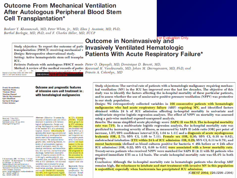
10. What are the long term outcomes of patients after BMT requiring ICU treatment (Neil)
Yoan Lamarche
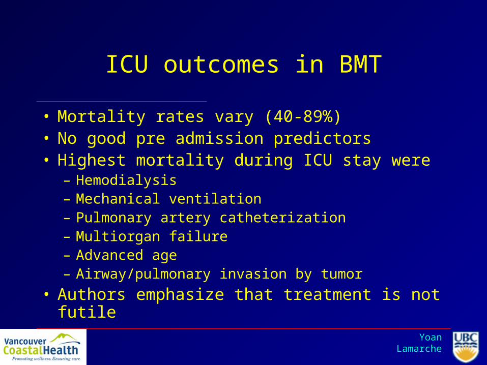
ICU outcomes in BMT
• Mortality rates vary (40-89%)• No good pre admission predictors• Highest mortality during ICU stay were
– Hemodialysis– Mechanical ventilation– Pulmonary artery catheterization– Multiorgan failure– Advanced age– Airway/pulmonary invasion by tumor
• Authors emphasize that treatment is not futile

Yoan Lamarche
Chest 2002

Yoan Lamarche

Yoan Lamarche
On its way up from the 2nd to the 5th floor, the elevator has a malfunction and falls 3 floors abruptly. The patient and his nurse are injured.
The double code blue is called as they open up the elevator on the 2nd foor.
• The nurse is unconscious, face down in a significant amount of blood. Her breathing is labored and she is tachycardic at 140. Her sBP is 80. You log roll her, her nose seems to be the source of the bleeding. You intubate her after having an IV access and you administer 2 liters of NS. The nose bleed remains serious. You administer 2 O- units of PRBCs to the patient and send stat blood work. She rapidly regains consciousness as your resuscitation progress. The ENT resident packs her nose. You get the CBC and coags back: Hb 100, WBC 14, Plt 150 000, INR 1.2 and PTT 42.

Yoan Lamarche
Your BMT patient was well braced in his stretcher and has no external injuries. He is intubated for hypotension. You transitorily loose his pulse during intubation. 1 minute of CPR and 3 L of NS later, the patient sBP is back at 100. The patients drops again, you administer 3 Units of PRBC and obtain a CXR that is N, a FAST US is negative, but suboptimal. A CT abdo reveals a significant pelvis hematoma (20cm x 15 cm)

Yoan Lamarche
12. Explain your initial approach to the coagulopathic patient (Neil)

Yoan Lamarche
Initial approach to Coagulopathic Patient
• ABC’s• Recussitation
– RBC’s– FFP
• II, V, VII, IX, X, XI, AT III– Platelets– Cryoprecipitate
• vWF, VIII, Fibrinogen, fibronectin– Factors (see Factor first card)

Yoan Lamarche
Initial approach to Coagulopathic Patient
• History– Nature of Bleeding– Sites of Bleeding– Patterns of Bleeding– Medications– Associated diseases– Family History
• Physical– Vitals– Skin– Mucosa– Lymphadenopathy– Liver size– Joints

Yoan Lamarche
Initial approach to Coagulopathic Patient
• Lab investigations– INR = extrinsic/common pathway– PTT = intrinsic/common pathway– Platelet count and bleeding time– Fibrinogen = hypoproduction or overconsumption
• Altered by preg, malignancy, sepsis– Thrombin time = fibrinogen and inhibitors– D-Dimer = Fibrin degradation products – Factor Level assays

Yoan Lamarche
12. Illustrate the coagulation cascade and the fibrinolytic pathway (Neil)

Yoan Lamarche

Yoan Lamarche
Fibrinolytic Pathway

Yoan Lamarche

Yoan Lamarche
..\Transfusion Therapy\Use of rFVII in Trauma.pdf

Yoan Lamarche
12. Identify the sites of action of anticoagulants, platelet inhibitors, fibrinolytics and antifibrinolytics (Noemie)

Yoan Lamarche
Q#14 Identify the sites of action of anticoagulants, platelet inhibitors, fibrinolytics and antifibrinolytics
• Heparin and LMWH– Binds to Antithrombin– Conformational change in AT– Potentiates binding capacity
to thrombin and Factor Xa

Yoan Lamarche
Identify the sites of action of anticoagulants, platelet inhibitors, fibrinolytics and antifibrinolytics
• Direct thrombin inhibitor– Lepirudin,
argatroban and bivalirudin
– Do not require a plasma cofactor like heparin

Yoan Lamarche
Identify the sites of action of anticoagulants, platelet inhibitors, fibrinolytics and antifibrinolytics
• Warfarin– Interferes with
synthesis of vitamin K depentdant factors
– Factors II, VII, IX, X– Prevents the
carboxylation process which is necessary to activate the clotting factors

Yoan Lamarche
Identify the sites of action of anticoagulants, platelet inhibitors, fibrinolytics and antifibrinolytics
• Aspirin– Inhibits thromboxane A2
synthesis activation and
recruitment to site• Clopidogrel and Ticlopidine
– Block ADP receptor on platelet surface
– Metabolized through P450delayed onset
• GPIIb/IIIa antagonists– Block binding of fibrinogen
to activated GP IIb/IIIa

Yoan Lamarche
Identify the sites of action of anticoagulants, platelet inhibitors, fibrinolytics and antifibrinolytics
• Fibrinolytics– Goal is rapid thrombus
dissolution– All promote conversion of
plasminogen to plasmin– Plasmin then degrades
the fibrin matrix of clot– New generation:
alteplase, tenecteplase and reteplase (activate fibrin bound plasminogen)
Yoan Lamarche
Identify the sites of action of anticoagulants, platelet inhibitors, fibrinolytics and antifibrinolytics
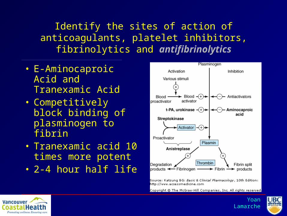
• E-Aminocaproic Acid and Tranexamic Acid
• Competitively block binding of plasminogen to fibrin
• Tranexamic acid 10 times more potent
• 2-4 hour half life

Yoan Lamarche
Summary
Aspirin
Clopidogrel
IIb/IIIa inhibitors
Warfarin
Fibrinolytics
AT Heparin
Argatroban

Yoan Lamarche

Yoan Lamarche
Fibrinolytic Pathway

Yoan Lamarche
• The nurse continues to bleed. You notice in her wallet a medic-alert card stating she has von Willebrand’s disease.
15. What are the principles of management of the bleeding von Willebrand disease patient (Marios)

Yoan Lamarche
Factors to consider before treating
1. Nature and severity of the type of VWD present (type 3 doesn’t respond to DDAVP, type 1 almost always does, and type 2 may or may not)
2. Location and severity of the bleeding episode or challenge (different treatments will be given depending on severity)
3. Response to the treatment of prior bleeding episodes(DDAVP should only be attempted in minor bleeds in pts known to have responded in the past)
4. Other medications and illnesses impacting hemostasis(Assess need for FFP, platelets, vitamin K, or protamine)

Yoan Lamarche
Minor bleeding
In patients with type 1 or 2 VWD who previously showed a good response to DDAVP:
Initial treatment should begin with DDAVP 0.3 mcg/kg (max. 20 mcg) IV or 150-300 mcg intranasally every 12 h for 2-4 doses.
Response can be assessed both clinically and with the ristocetin cofactor activity level which should reach at least 30 IU/dL, and preferably 50 IU/dL
The antifibrinolytics agents aminocaproic acid and tranexamic acid should be used as adjuvants (doses on next slide)

Yoan Lamarche
Minor bleeding
If the patient continues to bleed, has not had a previous trial of DDAVP, or is a known non-responder to DDAVP:
VWF concentrate should be given at 30-60 ristocetin cofactor units per kg followed by 20-40 ristocetin cofactor units per kg q 12-48 hr to keep VWF level >30 IU/dL for 3-5 days.

Yoan Lamarche
Major bleeding
1. Treatment should be initiated with VWF concentrate:Initial dose of 40-60 ristocetin cofactor units/kg x 1Then 20-40 ristocetin cofactor units/kg every 12-24 hr Goal VWF level 50-100 IU/dL for 7-14 days.
2. Antifibrinolytics should be given in addition to the concentrate: Tranexamic acid 50 mg/kg every 4 to 6 h ORAmnocaproic acid 10 mg/kg every 8 hours
3. Recombinant factor VIIa can be considered in type 3 VWD with serious refractory bleeding

Yoan Lamarche
Acquired VWD
1. Trial of DDAVP; levels of VWF activity should be monitored for possible rapid clearance
2. In patients who do not respond adequately to DDAVP, VWF concentrates should be used. Patients with circulating inhibitor may require very large doses of VWF (50 to 100 U/kg)
3. If the patient does not respond to DDAVP, and especially in patients who have documented antibody-mediated acquired VWD, a trial of high-dose IVIG (1g.kg per day for 2 days) can be attempted
4. Treatment of the underlying disease should be undertaken if feasible
5. If all else fails, recombinant Factor VIIa can be tried

Yoan Lamarche

Yoan Lamarche

Yoan Lamarche

Yoan Lamarche

Yoan Lamarche

Yoan Lamarche
NEJM351(7) 2004

Yoan Lamarche

Yoan Lamarche
• The patient’s condition gets better with significant blood component therapy. The junior resident in your team asks about alternatives to transfusions.

Yoan Lamarche
16. Even if you are busy resuscitating 2 patients, you take 1 minute to outline the options (Naisan)

Yoan Lamarche

Yoan Lamarche
Current Opinion in Anaesthesiology 2004, 17:139–143Blood substitutesFahim A. Habib and Stephen M. Cohn

Yoan Lamarche
Hb based carriers

Yoan Lamarche
Perfluorocarbons (PFCs)

Yoan Lamarche
Perfluorocarbons (PFCs)

Yoan Lamarche
Are they safe?
Natanson et al, JAMA, May 21, 2008—Vol 299, No. 19

Yoan Lamarche
As your resuscitation evolves, you notice progressive hypoxia in the patient.
17. What are the complications of blood transfusion and their physiopathology? (Marios)

Yoan Lamarche
ACUTE COMPLICATIONSOccur within minutes and up to 8 hours post-transfusion:
1. Acute hemolytic transfusion reaction
2. Febrile nonhemolytic transfusion reaction
3. Simple allergic transfusion reaction
4. Severe allergic or anaphylactic transfusion reaction
5. Transfusion-related circulatory overload (TACO)
6. Transfusion-related acute lung injury
7. Sepsis

Yoan Lamarche
DELAYED COMPLICATIONSOccur from 2 days to years post-transfusion
1. Delayed hemolytic transfusion reaction
2. Transfusion-associated graft-versus-host disease
3. Post-transfusion purpura
4. Transfusion-transmitted diseases

Yoan Lamarche
ACUTE HEMOLYTIC TRANSFUSION REACTION
Occurs within minutes or up to 4h post-transfusion
Incidence: 1:38 000 Mortality: 1:30
Etiology1. ABO incompatibilty: usually due to a clerical error2. Other blood group alloantibodies from prior pregnancy or transfusion3. Group O platelet transfusion with high anti-A or anti-B titers to non-O patient (rare)
Clinical presentationFever and chillsHemoglobinuria Less common: pain, hypotension, nausea/vomiting, dyspnea, renal failure, DIC

Yoan Lamarche
FEBRILE NON-HEMOLYTIC TRANSFUSION REACTION
Occurs during transfusion or up to 4-6h post-transfusion
Incidence RBC 1:526Platelets 1:10 per pool
EtiologyCirculating cytokines in the donor plasmaRecipient antibodies against donor leukocytes (less common since implementation of universal leukoreduction in 1999)
Clinical presentationFever is most commonMay be associated with chills, rigors, nausea, vomiting, and hypotension

Yoan Lamarche
SIMPLE ALLERGIC TRANSFUSION REACTION
Occurs during transfusion or up to a few hours post-transfusion
Incidence: 1:3 to 1:300
EtiologyRecognition of antigens in donor plasma by preformed recipient IgE antibodies.
Clinical presentationUrticarial rash that resolves with antihistamine administration. May also be associated pruritus, erythema, flushing, or mild airway symptoms (cough, wheezing), nausea, vomiting, abdominal crams, and diarrhea

Yoan Lamarche
ANAPHYLACTIC TRANSFUSION REACTION
Usually occurs within minutes but can occur up to 4h post transfusion.
Incidence 1:20 000 to 1:50 000Mortality 3%
Etiology (vast majority are unexplained)Transfusion of an allergen to a sensitized recipientPassive transfer of IgE (to drugs, food) to the recipientAnti-IgA in IgA-deficient patients
Antibodies to polymorphic forms of serum proteins (haptoglobin, IgG, albumin, C3, C4, transferrin, alpha-1-antitrypsin)
Clinical presentationUrticaria, stridor/wheezing, hypotension, nausea, vomiting
Yoan Lamarche
TRANSFUSION-ASSOCIATED CIRCULATORY OVERLOAD (TACO)
Occurs during transfusion or up to 6h post-transfusion
Incidence < 1:100 (higher in patients with CHF)
Etiology
Circulatory overload from impaired cardiac function and/or excessively rapid rate of transfusion
Clinical presentation
Dyspnea, orthopnea, hypoxia, tachycardia, increased venous pressure, increased PCWP, hypertension, elevated BNP
Yoan Lamarche
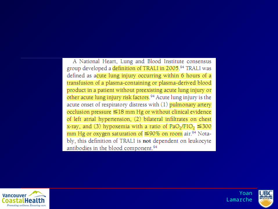
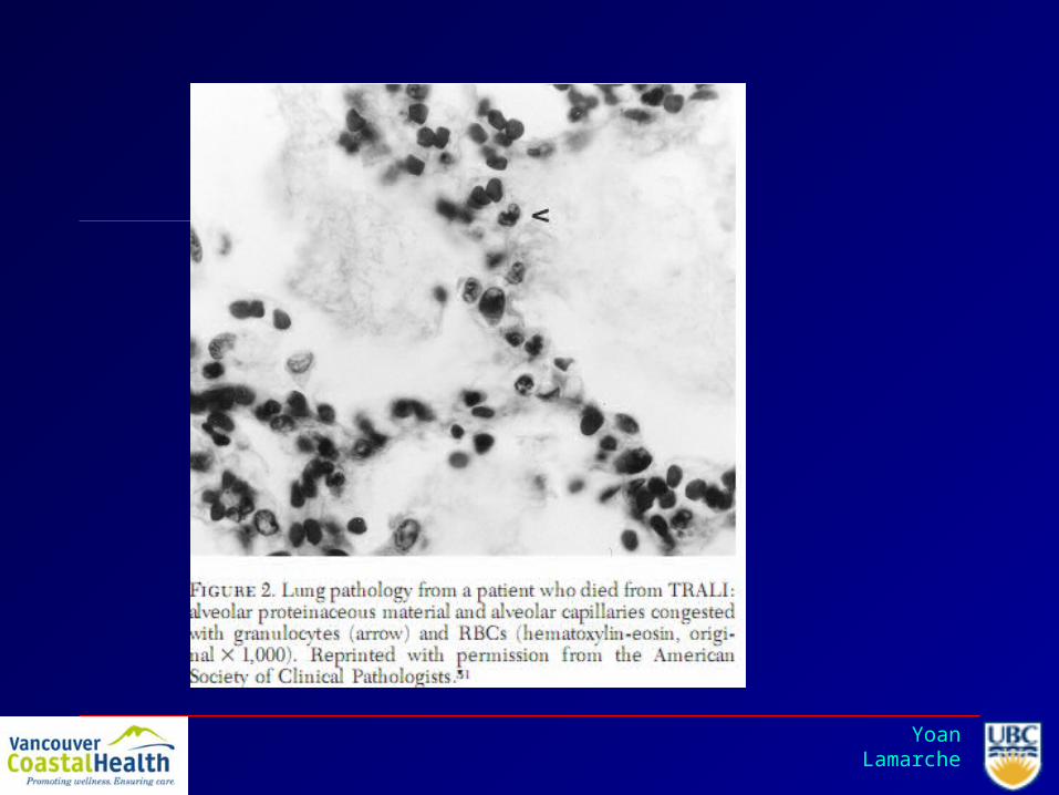
TRANSFUSION-RELATED ACUTE LUNG INJURY
Occurs during transfusion or up to 6h post-transfusion
Incidence 1:5000
Etiology (three major theories)1. Passive transfer of HLA or granulocyte antibodies that initiate an inflammatory response
in the pulmonary microvasculature2. Granulocyte priming by biologically active substances such as lipids and cytokines in the
transfused plasma 3. Granulocyte priming as above followed by activation by transfused antibodies (“two-
event” hypothesis)
DEFINITIONDefinite TRALI: New onset ALI within 6h of transfusion with no other risk factors for ALIPossible TRALI: New onset ALI within 6h of transfusion with one or more risk factors for ALI

Yoan Lamarche
TRANSFUSION-RELATED ACUTE LUNG INJURY
Clinical presentationDyspnea, hypoxia, fever, hypotension, and leukopeniaBilateral infiltrates with normal CVP, PCWP, and BNPUsually occurs with RBC, FFP and platelet transfusions, but can rarely occur with IVIG and cryoprecipitateAlmost always occurs within 1-2h after starting the transfusionUsually resolves in 24-72 hours
*Criteria for ALI: PO2:FiO2 < 300PCWP <= 18Bilateral chest infiltrates

Yoan Lamarche
SEPSISUsually occurs during or within an hour of transfusion but can beseen up to 4 or 8 h post transfusion
Incidence of symptomatic septic reactions: Platelets 1:10 000 (stored at 20-24 degrees)RBCs 1:100 000
Mortality Platelets 1:40 000RBCs 1:500 000
EtiologyContamination of blood components by donor skin plug, bacteremic donor, or improper handling of the blood product
Clinical presentationRigors, fever, tachycardia, hypotension, nausea and vomiting, dyspnea, DIC

Yoan Lamarche
DELAYED HEMOLYTIC TRANSFUSION REACTION
Occurs from 2 to 14 days post transfusion
Incidence 1:4000 to 1:11 000
EtiologyAnamnestic antibody response in a patient with previously formed antibodies (from prior transfusion, pregnancy, or transplant) that were undetectable on pre-transfusion screening
Clinical presentationExtravascular, gradual hemolysis with a falling Hb, slight fever, mild unconjugated hyperbilirubinemia, low haptoglobin, elevated LDH, and spherocytosis.

Yoan Lamarche
POST-TRANSFUSION PURPURA
Occurs 5 to 10 days post transfusion
Incidence is unknown; ~300 cases have been reported.Five (or 26) times more common in women then in men
EtiologyDelayed transfusion reaction to platelets in patients who have been sensitized from prior transfusions or pregnancy (human platelet antigen 1a)
Clinical presentationSevere thrombocytopenia (plts often < 10 000) that lasts days to weeks8% mortality - most often secondary to an intracranial hemorrhageShould be considered when a platelet refractory patient fails to respond to HLA-matched platelets

Yoan Lamarche
Transfusion-associated graft-versus-host disease
Occurs 2 to 30 days post transfusion (most commonly between 4 and 10d)
Incidence: 1:400 000Mortality: 80-90%
EtiologyDonor lymphocytes attack recipient’s lymphoid tissues (skin, liver, GI tract, bone marrow); occurs in two settings:
1. Immunosuppressed pt cannot mount a response against donor lymphocytes which then mount a response against the recipient
2. Recipient is heterozygous for an HLA haplotype for which the donor is homozygous (recipient does not recognize the donor lymphocytes as foreign. These then mount a response against the recipient.)
Clinical presentationFever, rash, liver dysfunction, and diarrhea, followed by pancytopeniaDiagnosis made by skin, liver, or bone marrow biopsy

Yoan Lamarche
TRANSFUSION-TRANSMITTED DISEASES
VIRAL:
HBV, HCV, HAV, HIV 1 & 2, HTLV 1 & 2, CMV, West Nile Virus
BACTERIAL (see sepsis)
Syphilis - no reported US cases since the 1960s
PARASITIC:
Malaria, babesia, chagas,
OTHER:
CJD, vCJD

Yoan Lamarche
Copyright ©2007 Canadian Medical Association or its licensors
Yoan Lamarche
REFERENCES
Bakdash S, Yazer MH. What every physician should know about transfusion reactions. CMAJ. July 17, 2007; 177(2):141-147
Callum JL, Pinkerton PH. Bloody easy: A guide to transfusion medicine. 1st ed. Toronto: Sunnybrook & Women's College Health Sciences Centre Press, 2003.

Yoan Lamarche

Yoan Lamarche

Yoan Lamarche
Complications of transfusions
– ABO mismatch (#1 cause of death post Transfusions)
– Hepatitis (#2)– TRALI (#3)
Yoan Lamarche
• TRALI– Criteria: transfusion- bilateral pulmonary infiltrates- low filling pressures-
aProtein/pProtein > 0.75– Causes
• Two-hit hypothesis– Recent surgery, sepsis, trauma, massive transfusions, hematologic malignancies,
and cardiac disease can all predispose– Parity of the blood donor(1 RCT), relationship to the blood donor (Ab against
father), and the age of the blood products• Bioactive lipids
– LysoPhosphatidylcholine: priming of neutrophils– Would require cellular products transfusion– TRALI has also been associated with FFP transfusion...
• Antibodies ± WBC– Ab from donor interacts with recipients ± donor’s neutrophils and creates ALI
• CD40 Ligand: also implicated as a potential cause of pulm. transf reactions– Prognosis: good: 5-10% mortality-clears in 24-48h
Yoan Lamarche

Yoan Lamarche
Yoan Lamarche

Yoan Lamarche
The patient deteriorates further and you notice the previous central line site is now bleeding, as is your new femoral central line.
18.How do you diagnose DIC? What are the treatment strategies for the patient presenting DIC? (Scot)

Yoan Lamarche

Yoan Lamarche
• Diagnosis – acute DIC;– Suggested by: Hx (eg. Sepsis, trauma, malignancy),
thrombocytopenia (< 100,000), and the presence of microangiopathic changes on smear.
– Clinical manifestations:• Bleeding (64 percent) • Renal dysfunction (25 percent) • Hepatic dysfunction (19 percent) • Respiratory dysfunction (16 percent) • Shock (14 percent) • Thromboembolism (7 percent) • Central nervous system involvement (2 percent)

Yoan Lamarche
Peripheral blood smear from a patient with a microangiopathic hemolytic anemia with marked red cell fragmentation. The smear shows multiple helmet cells (small black arrows), other fragmented red cells (large black arrow); microspherocytes are also seen (blue arrows). The platelet number is reduced; the large platelet in the center (red arrow) suggests that the thrombocytopenia is due to enhanced destruction. Courtesy of Carola von Kapff, SH (ASCP).

Yoan Lamarche
• Dx confirmed by tests showing increased thrombin generation (decreased fibrinogen), and increased fibrinolysis (elevated D-dimer and FDPs).– Degree of abnormality correlates with overall
organ involvement and mortality.

Yoan Lamarche

Yoan Lamarche

Yoan Lamarche
DIC - managment
• the management of DIC is based on the treatment of the underlying disease, supportive and replacement therapies and the control of coagulation mechanisms.
• Hemodynamic support is essential.• Many patients do not require specific therapy for the
coagulopathy associated with DIC, either because it is of short duration or because it is not severe enough to present a major risk of bleeding or thrombosis.
• There is no evidence to support the administration of platelets and coagulation factors in patients who are not bleeding or who are not at high risk of bleeding.

Yoan Lamarche
• Treatment with platelets and coagulation factors is justified in patients who have serious bleeding, are at high risk for bleeding (eg, after surgery), or require invasive procedures. Patients with marked or moderate thrombocytopenia (<50,000/microL) and serious bleeding should be given platelet transfusions (1 to 2 units per 10 kg per day).
• Actively bleeding patients with a significantly elevated prothrombin time (INR) and/or a fibrinogen concentration <50 mg/dL, should receive fresh frozen plasma or cryoprecipitate in order to keep the fibrinogen level >100 mg/dL.

Yoan Lamarche
• The administration of heparin is generally limited to the subset of patients with chronic, compensated DIC who have predominantly thrombotic manifestations. It is important to be sure that the patient's antithrombin (AT) level is near normal (ie, 80 to 100 percent) in order for heparin to be effective.

Yoan Lamarche
• The usual intravenous bolus heparin injection of 5000 to 10,000 units should be avoided. One may start with an IV dose of 500 units per hour, aiming for an aPTT of about 45 sec. If the patient's baseline aPTT is prolonged, the situation is more difficult, and one aims for further slight prolongation of the aPTT.
• Once there is evidence of heparin effect, replacement therapy with fresh frozen plasma or cryoprecipitate is pursued. In chronic DIC, a continuous infusion can be used, beginning at 500 units/hour. Low molecular weight heparins are also efficacious.

Thank You

Yoan Lamarche


