
JPET #208314
1
Title Page
Hepatic Basolateral Efflux Contributes Significantly to Rosuvastatin Disposition:
II. Characterization of Hepatic Elimination by Basolateral, Biliary, and Metabolic Clearance
Pathways in Rat Isolated Perfused Liver
Nathan D. Pfeifer, Arlene S. Bridges, Brian C. Ferslew, Rhiannon N. Hardwick, and Kim L.R. Brouwer
Division of Pharmacotherapy and Experimental Therapeutics, UNC Eshelman School of Pharmacy,
University of North Carolina at Chapel Hill, Chapel Hill, NC (N.D.P., B.C.F., K.L.R.B.)
Department of Pathology, UNC School of Medicine, University of North Carolina at Chapel Hill, Chapel
Hill, NC (A.S.B.)
Curriculum in Toxicology, UNC School of Medicine, University of North Carolina at Chapel Hill, Chapel
Hill, NC (R.N.H., K.L.R.B.)
JPET Fast Forward. Published on October 4, 2013 as DOI:10.1124/jpet.113.208314
Copyright 2013 by the American Society for Pharmacology and Experimental Therapeutics.
This article has not been copyedited and formatted. The final version may differ from this version.JPET Fast Forward. Published on September 30, 2013 as DOI: 10.1124/jpet.113.208314
at ASPE
T Journals on M
ay 11, 2018jpet.aspetjournals.org
Dow
nloaded from

JPET #208314
2
Running Title Page
Running Title: Rosuvastatin Disposition in Rat Isolated Perfused Liver
Address correspondence to:
Kim L.R. Brouwer
UNC Eshelman School of Pharmacy
University of North Carolina at Chapel Hill
CB #7569
Chapel Hill, NC 27599-7569
E-mail address: [email protected]
Telephone: 919 – 962 – 7030
Fax: 919 – 962 – 0644
Number of text pages: 31
Number of tables: 1
Number of figures: 5
Number of references: 60
Number of words in the Abstract: 250
Number of words in the Introduction: 745
Number of words in the Discussion: 1500
Abbreviations: IPL, isolated perfused liver; SCH, sandwich-cultured hepatocytes; DDI, drug-drug-
interaction; RSV, rosuvastatin; WT, wild-type; BCRP, breast cancer resistance protein; MRP, multidrug
resistance-associated protein; OATP, organic anion transporting polypeptide; CYP, Cytochrome P450.
This article has not been copyedited and formatted. The final version may differ from this version.JPET Fast Forward. Published on September 30, 2013 as DOI: 10.1124/jpet.113.208314
at ASPE
T Journals on M
ay 11, 2018jpet.aspetjournals.org
Dow
nloaded from

JPET #208314
3
Abstract
Basolateral efflux clearance (CLBL) contributes significantly to rosuvastatin (RSV) elimination in
sandwich-cultured hepatocytes (SCH). The contribution of CLBL to RSV hepatic elimination was
determined in single-pass isolated perfused livers (IPLs) from wild-type (WT) and multidrug resistance-
associated protein 2 (Mrp2)-deficient (TR-) rats in the absence and presence of the P-glycoprotein and
breast cancer resistance protein (Bcrp) inhibitor, GF120918; clearance values were compared to SCH.
RSV biliary clearance (CLBile) nearly was ablated by GF120918 in TR- IPLs, confirming that Mrp2 and
Bcrp primarily are responsible for RSV CLBile. RSV appearance in outflow perfusate was due primarily
to CLBL, which was impaired in TR- IPLs. CLBL was ~6-fold greater than CLBile in the linear range in WT
IPLs in the absence of GF120918. Recovery of unchanged RSV in liver tissue increased in TR-
compared to WT (~25% vs. 6% of the administered dose) due to impaired CLBL and CLBile. RSV
pentanoic acid, identified by high resolution LC-MS/MS, comprised ~40% of total liver content and
~16% of the administered dose in TR- livers at the end of perfusion compared to ~30% and 3% in WT
livers, consistent with impaired RSV excretion and “shunting” to the metabolic pathway. In vitro-ex vivo
extrapolation between WT (-GF120918) SCH and IPLs revealed that uptake clearance and CLBL were
4.2- and 6.4-fold lower, respectively, in rat SCH compared to IPLs; CLBile translated almost directly (1.1-
fold). The present IPL data confirmed the significant role of CLBL in RSV hepatic elimination, and
demonstrated that both CLBL and CLBile influence RSV hepatic and systemic exposure.
This article has not been copyedited and formatted. The final version may differ from this version.JPET Fast Forward. Published on September 30, 2013 as DOI: 10.1124/jpet.113.208314
at ASPE
T Journals on M
ay 11, 2018jpet.aspetjournals.org
Dow
nloaded from

JPET #208314
4
Introduction
The role of hepatic transport in the pharmacokinetics and pharmacodynamics of rosuvastatin
(RSV) has long been recognized (Nezasa et al., 2002; Ho et al., 2006; Zhang et al., 2006; Kitamura et al.,
2008). Mechanisms mediating hepatic uptake (OATPs, NTCP) and biliary excretion (MRP2, BCRP)
have been well-characterized (Ho et al., 2006; Huang et al., 2006; Zhang et al., 2006; Kitamura et al.,
2008; Keskitalo et al., 2009; Hobbs et al., 2012). Recently, it was shown that basolateral efflux represents
a significant elimination route from rat and human hepatocytes using a novel uptake/efflux protocol in
sandwich-cultured hepatocytes (SCH) (Pfeifer, 2013). Of the candidate transport proteins known to
mediate hepatic basolateral efflux of drugs and metabolites, RSV was shown to be a substrate of human
MRP4 (Pfeifer, 2013), which likely contributes to the basolateral efflux of RSV in human liver.
The importance of hepatic basolateral efflux in drug disposition remains largely unrecognized
except in the case of hepatically-derived drug conjugates (Zamek-Gliszczynski et al., 2006; Hardwick et
al., 2012). Exceptions include fexofenadine (Tian et al., 2008), enalaprilat (de Lannoy et al., 1993) and
methotrexate (Vlaming et al., 2009). As such, availability of tools and information for prediction and in
vitro-in vivo extrapolation (IVIVE) of hepatic basolateral efflux lags behind the development of these
tools for more-recognized pathways such as hepatic uptake and biliary excretion. Although isolated
expression systems have been employed for some proteins known to facilitate hepatic basolateral efflux,
such as Mrp3/MRP3 and Mrp4/MRP4 (Hirohashi et al., 1999; Akita et al., 2002; Chen et al., 2002), other
translational tools, such as quantitative proteomics data and identification of specific substrates/inhibitors,
are extremely limited. Therefore, it is important to assess the basolateral efflux of RSV and other drugs in
whole liver in order to ascertain the potential role of this pathway in vivo, and the predictive capability of
the aforementioned SCH method and other in vitro systems that may be developed.
Following oral administration of RSV to WT Sprague-Dawley and multidrug resistance-
associated protein 2 (Mrp2)-deficient Eisai hyperbilirubinemic rats (EHBR), biliary clearance (liver-to-
This article has not been copyedited and formatted. The final version may differ from this version.JPET Fast Forward. Published on September 30, 2013 as DOI: 10.1124/jpet.113.208314
at ASPE
T Journals on M
ay 11, 2018jpet.aspetjournals.org
Dow
nloaded from

JPET #208314
5
bile) was decreased without a significant increase in RSV liver concentrations, while systemic exposure
was increased more than 3-fold (Kitamura et al., 2008). While this may suggest efficient hepatic
basolateral efflux of RSV in the setting of impaired biliary excretion, decreased hepatic uptake and/or
impaired renal elimination in EHBR rats may contribute to the increased systemic exposure. An
important advantage of the IPL model is that the role of hepatic processes can be evaluated in isolation
from other organ systems (Brouwer and Thurman, 1996). RSV disposition was reported recently in
recirculating isolated perfused livers (IPLs) from WT and Mrp2-deficient (TR-) rats (Hobbs et al., 2012).
Increased perfusate concentrations in TR- compared to WT livers were attributed to decreased hepatic
uptake, however, the role of basolateral excretion was not considered. The single-pass IPL system used
in the present study allowed for direct evaluation of basolateral excretion from liver to perfusate.
RSV is not metabolized extensively in humans; identified metabolites include the inactive 5S-
lactone, as well as N-desmethyl-RSV, which is formed by Cytochrome P450 2C9 (CYP2C9) and retains
up to 50% of the HMG-CoA-reductase inhibitor activity of RSV (Martin et al., 2003b). Although
metabolism plays a similarly minor role in rats in terms of overall mass balance, metabolites have been
reported to account for a significant portion (~50%) of plasma and liver content in whole animal studies
(Nezasa et al., 2002; Kitamura et al., 2008). Biotransformation of RSV in rats is mediated primarily by β-
oxidation of the fatty acid chain, with no evidence of CYP involvement. The pentanoic acid derivative
(RSV-PA) has been suggested as the primary metabolite based on thin-layer chromatography of extracted
plasma and liver samples from rats administered [14C]RSV, co-spotted with an authentic standard of
RSV-PA (Nezasa et al., 2002); structural identification and confirmation of the RSV-PA metabolite by
mass-spectrometric analysis has not been reported.
Interplay between transporters and metabolizing enzymes has been recognized. This presents a
challenge in predicting the impact of altered function on hepatic and/or systemic exposure of drugs when
multiple elimination pathways are involved (Benet et al., 2004; Zamek-Gliszczynski et al., 2006; Parker
and Houston, 2008). This is clinically relevant for RSV because impaired hepatic transport due to drug-
This article has not been copyedited and formatted. The final version may differ from this version.JPET Fast Forward. Published on September 30, 2013 as DOI: 10.1124/jpet.113.208314
at ASPE
T Journals on M
ay 11, 2018jpet.aspetjournals.org
Dow
nloaded from

JPET #208314
6
drug interactions (DDIs) and genetic polymorphisms has been shown to alter the pharmacokinetics of
RSV (Schneck et al., 2004; Simonson et al., 2004; Zhang et al., 2006; Kiser et al., 2008; Keskitalo et al.,
2009). Some of these changes have been associated with altered efficacy (LDL-lowering) of RSV
(Simonson et al., 2003; Tomlinson et al., 2010), while increased systemic exposure has been associated
with life-threatening rhabdomyolysis related to statin use in general (Hamilton-Craig, 2001; Thompson et
al., 2003).
The present experiments were designed to quantify the contribution of the basolateral efflux
pathway to the hepatocellular elimination of RSV in single-pass rat IPLs. In addition, clearance values
generated using a novel uptake/efflux protocol that was developed in the SCH model were compared to
this dataset generated in whole liver. IPL data, combined with pharmacokinetic modeling, revealed that
basolateral efflux represents a significant route of RSV hepatocellular excretion from rat liver, similar to
findings in SCH. In addition, reduced CLBL and CLBile of RSV in TR- livers highlighted the contribution
of biotransformation as an alternative elimination pathway in the setting of impaired hepatic efflux in rat
liver, with the pentanoic acid derivative of RSV (RSV-PA) identified as the primary metabolite.
This article has not been copyedited and formatted. The final version may differ from this version.JPET Fast Forward. Published on September 30, 2013 as DOI: 10.1124/jpet.113.208314
at ASPE
T Journals on M
ay 11, 2018jpet.aspetjournals.org
Dow
nloaded from

JPET #208314
7
Materials and Methods
Materials
All chemicals were purchased from Sigma-Aldrich (St. Louis, MO) unless otherwise stated. RSV
and the deuterated internal standard (d6-RSV) were purchased from Moravek Biochemicals (Brea, CA).
GF120918 (elacridar) was a generous gift from GlaxoSmithKline (Research Triangle Park, NC).
Animals
Male Wistar wild-type (WT) rats (250-350 g) from Charles River Labs (Wilmington, MA) or
male Mrp2-deficient (TR-) rats bred at the University of North Carolina (250—350 g; breeding stock
obtained from Dr. Mary Vore, University of Kentucky, Lexington, KY) were used as donors for
hepatocyte and isolated perfused liver (IPL) studies. Rats were allowed water and food ad libitum, and
acclimated for a minimum of 1 week prior to experimentation. All animal procedures complied with the
guidelines of the Institutional Animal Care and Use Committee (University of North Carolina, Chapel
Hill, NC). All procedures were performed under full anesthesia with ketamine/xylazine (140/8 mg/kg
i.p.).
Isolated Perfused Livers (IPLs)
WT and TR- rat livers were perfused in a single-pass manner as described previously (Brouwer
and Thurman, 1996; Chandra et al., 2005). Briefly, following cannulation of the portal vein and bile duct,
livers were perfused in situ with continuously-oxygenated Krebs-Ringer bicarbonate buffer (35 mL/min)
containing 5 μM taurocholate to maintain bile flow. Livers were removed from the body cavity and
placed in a humidified perfusion chamber heated to maintain liver temperature at 37°C. Perfusion was
continued for a 15-min equilibration period, and then switched to a RSV-containing perfusate (0.5 μM)
for the 60-min loading phase; at 60 min, the buffer was switched to RSV-free perfusate, and perfusion
was continued for an additional 30 min. For conditions in the presence of inhibitor, the perfusate also
This article has not been copyedited and formatted. The final version may differ from this version.JPET Fast Forward. Published on September 30, 2013 as DOI: 10.1124/jpet.113.208314
at ASPE
T Journals on M
ay 11, 2018jpet.aspetjournals.org
Dow
nloaded from

JPET #208314
8
contained 0.5 μM GF120918 for the duration of the experiment (15-min equilibration period followed by
the 90-min perfusion; Fig 1). This concentration was sufficient to inhibit RSV biliary excretion in TR-
SCH with minimal impact on uptake (Pfeifer, 2013). Liver viability was assessed by monitoring inflow
perfusion pressure (<15 mm H2O), gross morphology, and maintenance of bile flow (within 30% of the
baseline rate during the equilibration period). Bile and perfusate were collected over 5-min intervals, and
bile volume was determined gravimetrically in pre-weighed tubes. After perfusion, livers were blotted
dry, weighed, and stored at -80°C until analysis.
Bioanalysis
RSV was quantified by LC-MS/MS as described previously for samples derived from IPL studies
(Abe et al., 2008). RSV-PA was identified by high resolution LC-MS/MS (TOF/TOF) using an ABSciex
5600 TripleTOF mass spectrometer (Fig. 2). Direct absolute quantification of RSV-PA was not possible
due to the lack of an analytical standard. However, estimating sample-to-sample difference in the relative
concentrations of RSV-PA was possible by comparing normalized peak area ratios (RSV-PA:d6-RSV
internal standard). The absolute concentration of RSV-PA was estimated from normalized peak area
ratios (RSV-PA:d6-RSV internal standard) by the calibration curve generated using the peak area ratios
(RSV-PA:d6-RSV internal standard) of samples with known concentrations of RSV, with the assumption
that RSV and RSV-PA have similar ionization efficiencies.
Pharmacokinetic Modeling
Pharmacokinetic modeling and simulation were used to evaluate RSV disposition in rat IPLs, and
to determine the effects of GF120918 and loss of Mrp2 function on RSV hepatobiliary disposition.
Models incorporating various combinations of linear and/or non-linear parameters governing RSV flux
were fit to rate versus time data from individual experiments (Fig. 3). The model fitting was performed
with WinNonlin Phoenix, v6.1 (St. Louis, MO) using the stiff estimation method and a mixed (additive
and proportional) model for residual error. The model scheme depicting the single-pass IPL system (Fig.
This article has not been copyedited and formatted. The final version may differ from this version.JPET Fast Forward. Published on September 30, 2013 as DOI: 10.1124/jpet.113.208314
at ASPE
T Journals on M
ay 11, 2018jpet.aspetjournals.org
Dow
nloaded from

JPET #208314
9
3) consisted of an extracellular (sinusoidal/perfusate) compartment, liver tissue, and a bile compartment,
each divided into 5 subcompartments in the semiphysiologically-based approximation of the dispersion
model, as reported by Watanabe, et al (Watanabe et al., 2009). The model was fit simultaneously to
biliary excretion rate and appearance rate in outflow perfusate data, as well as terminal recovery of RSV
and RSV-PA in liver tissue. Differential equations describing the model scheme in Fig. 3 are as follows:
Extracellular liver 1:
dXEC,1
dt=Q×Cin +
CLBL
5×Cu,L,1 −
CLUptake
5×CEC,1 − Q×CEC,1
Extracellular liver 2-5:
dXEC,n
dt = Q×CEC,n-1 +
CLBL
5×Cu,L,n −
CLUptake
5×CEC,n − Q×CEC,n
Intracellular liver 1-5:
dXL,n
dt =
CLUptake
5×CEC,n − ��
CLBL
5�+ �
CLMet
5�+ �
CLOther
5��×Cu,L,n −
Cu,L,n×(Vmax/5)�Km + Cu,L,n�
Bile 1-5:
dXBile,n
dt =
Cu,L,n×(Vmax/5)�Km + Cu,L,n�
where variables and parameters are defined as in Fig. 3 with further explanation as follows. CEC,n is the
extracellular concentration, calculated as XEC,n/(VEC/5) and VEC is the extracellular volume of the liver,
which was assumed to be in equilibration with the sinusoidal space and estimated at 20% of the total liver
mass, as reported previously (Watanabe et al., 2009; Hobbs et al., 2012). Q is the perfusate flow rate of
35 mL/min, and Cin is the concentration of RSV in the inflow perfusate, measured for each preparation;
binding of RSV to the perfusion tubing and apparatus was <10% and not considered further. Cu,Ln is the
unbound intracellular concentration of RSV in the liver based on binding of RSV to rat liver tissue, which
This article has not been copyedited and formatted. The final version may differ from this version.JPET Fast Forward. Published on September 30, 2013 as DOI: 10.1124/jpet.113.208314
at ASPE
T Journals on M
ay 11, 2018jpet.aspetjournals.org
Dow
nloaded from

JPET #208314
10
was determined by equilibrium dialysis with an unbound fraction of 0.25, corrected for dilution. The total
intracellular liver concentration was calculated as the sum of the mass in liver subcompartments 1-5,
divided by the intracellular volume (VL), calculated as total liver mass minus the VEC. The RSV
concentration resulting in half-maximal biliary excretion (Km) was set to 10 μM based on the reported
affinity of RSV for Mrp2 and Bcrp in isolated expression systems (Huang et al., 2006; Deng et al., 2008).
The CEC,5×Q was fit to the observed appearance rate in outflow perfusate, while the sum of the excretion
rate in bile (dXBile,n/dt) for liver subcompartments 1-5 was fit to the observed biliary excretion rate. The
sum of the RSV and RSV-PA mass in liver subcompartments 1-5 was fit to observed recovery of RSV
and RSV-PA in liver tissue at the end of the study. Initial parameter estimates were obtained from a
combination of direct extrapolation of IPL data and simulations in Berkeley-Madonna. Vmax was
estimated initially as the steady-state excretion rate in bile since the unbound liver concentrations were
estimated from mass balance to be ~3-5 times the Km for the biliary excretion process. Rapid attainment
of steady-state in the outflow perfusate of WT IPLs precluded accurate estimation of the CLUptake.
Therefore, CLUptake was fixed at 40 mL/min/g liver based on two independent reports of RSV initial
uptake (determined at timepoints <1 min) in freshly-isolated, suspended WT rat hepatocytes (Nezasa et
al., 2003; Yabe et al., 2011), using standard conversion factors of 200 mg protein/g liver and 100 million
cells/g liver (Swift et al., 2010). Metabolic clearance to the pentanoic acid derivative (CLMet) and other
potential metabolites (CLOther) were estimated initially from simulations in Berkeley-Madonna.
Transporter-mediated clearance values [CLUptake, CLBL and CLBile (Vmax/Km in the linear rangee)]
estimated from pharmacokinetic modeling of RSV IPL data were compared to analogous parameter
values reported previously in rat SCH (Pfeifer, 2013); scaling factors were reported for the WT Control
conditions. Scaling factors represent the respective clearance value in the IPL divided by the
corresponding value in SCH.
Data Analysis
This article has not been copyedited and formatted. The final version may differ from this version.JPET Fast Forward. Published on September 30, 2013 as DOI: 10.1124/jpet.113.208314
at ASPE
T Journals on M
ay 11, 2018jpet.aspetjournals.org
Dow
nloaded from

JPET #208314
11
All data are presented as mean ± SD of n=3 livers in each treatment group, unless otherwise
noted. The effects of Mrp2 status (WT or TR-) and GF120918 on RSV disposition were determined
independently by one-way ANOVA with Tukey’s post-hoc test. The effect of Mrp2 status was evaluated
at each level of inhibitor (absent or present), and the effect of GF120918 was evaluated at each level of
Mrp2 status (WT or TR-).
This article has not been copyedited and formatted. The final version may differ from this version.JPET Fast Forward. Published on September 30, 2013 as DOI: 10.1124/jpet.113.208314
at ASPE
T Journals on M
ay 11, 2018jpet.aspetjournals.org
Dow
nloaded from

JPET #208314
12
Results
Baseline bile flow in WT IPLs was 0.49±0.05 µL/min/g liver and 0.30±0.11 µL/min/g liver in the
absence and presence of GF120918, respectively, with corresponding values in TR- IPLs of 0.33±0.04
µL/min/g liver and 0.25±0.07 µL/min/g liver, respectively. Outflow perfusate concentrations of RSV
ranged from 0.003 to 0.23 µM in WT and TR- IPLs. The rates of RSV appearance in bile and outflow
perfusate are plotted in Fig. 4; recovery of RSV and RSV-PA at the end of the perfusion are summarized
in Fig. 5. The biliary excretion rate of RSV was increased slightly in WT livers in the presence of
GF120918 (Fig. 4A), while the addition of GF120918 in TR- livers markedly reduced the biliary
excretion rate of RSV (Fig. 4B). Interestingly, the initial appearance rate of RSV in the outflow perfusate
was reduced in TR- compared to WT livers (Fig 4C and D). Addition of GF120918 slightly reduced the
rate of RSV appearance in outflow perfusate of WT livers (Fig. 4C), but had no effect on the rate of RSV
appearance in outflow perfusate of TR- livers (Fig. 4D). The cumulative recovery of RSV in perfusate
over the 90-min study tended to be reduced in TR- compared to WT livers in the absence of GF120918,
and also by GF120918 in WT livers (Fig. 5); however, the individual effects of Mrp2 status (WT vs. TR-)
and GF120918 failed to reach significance after correcting for multiple comparisons. Biliary recovery of
RSV was not affected significantly by GF120918 in WT IPLs, but the effect of GF120918 on the reduced
biliary recovery in TR- IPLs was statistically significant (Fig. 5). The effect of Mrp2 status on RSV
biliary recovery was statistically significant in the presence of GF120918, but not in the absence of
GF120918 (Fig. 5). Total recovery of the dosed RSV in perfusate, bile, and liver tissue at the end of the
90-min studies was nearly complete following perfusion of WT IPLs (96±9% and 94±13% in the absence
and presence of GF120918, respectively, Fig. 5); however, it was reduced significantly in TR- IPLs
(71±4% and 56±7% in the absence and presence of GF120918, respectively; Fig. 5). This reduction in
total recovery of parent RSV in TR- IPLs was offset, in part, by the presence of the pentanoic acid
metabolite in TR- liver tissue.
This article has not been copyedited and formatted. The final version may differ from this version.JPET Fast Forward. Published on September 30, 2013 as DOI: 10.1124/jpet.113.208314
at ASPE
T Journals on M
ay 11, 2018jpet.aspetjournals.org
Dow
nloaded from

JPET #208314
13
The presence of the pentanoic acid metabolite of RSV (RSV-PA) was confirmed by both targeted
and untargeted high resolution LC-MS/MS. The targeted approach was based on a previous report that
suggested that RSV-PA was the primary metabolite in rat (Nezasa et al., 2002). First, targeted extracted
ion chromatograms (422.20±0.05 amu) from full-scan TOF revealed this parent ion was only present in
samples from RSV-perfused livers (Fig. 2A). Second, comparisons between the high-resolution product
ion spectra of RSV (482.20 amu) and the 422.20 amu parent ion further substantiated this unique analyte
to be RSV-PA (Fig. 2B). Using the proposed structure for the metabolite, there were common product
ions formed (242.10, 256.12 and 270.17 amu; Fig. 2B), which were independent of the changes within the
carboxylic acid side chain. Additionally, the paired product ions, 402.19/404.19 for RSV and
342.17/344.17 for RSV-PA, represent loss of CH4O2S while maintaining the Δ60 amu (C2H4O2) between
RSV and RSV-PA. RSV-PA also was identified independently by analyzing the extracted homogenates
from non-treated (blank) and RSV-treated TR- IPLs in an untargeted, or unbiased, manner using the
ABSciex PeakView and MetabolitePilot software packages. No other specific RSV metabolites were
identified by LC-MS/MS. Estimated concentrations of RSV-PA were low in perfusate and bile samples,
representing <2% of the total dose over the course of the study. As such, RSV-PA was reported only in
the liver tissue (Fig. 5). RSV-PA comprised approximately 40% of total RSV content (metabolite:parent
ratio of 0.70±0.24 and 0.70±0.30 in the absence and presence of GF120918, respectively) in TR- liver
tissue, or 16% of the total administered dose of RSV at the end of the perfusion (Fig. 5). In contrast,
while RSV-PA accounted for a similar proportion of total RSV content in WT liver tissue at the end of
the perfusion (~30%; metabolite:parent ratio of 0.58±0.28 and 0.50±0.10 in the absence and presence of
GF120918, respectively), this comprised only ~3% of the total administered dose of RSV (Fig. 5).
Parameter estimates recovered from fitting the differential equations based on the model scheme
depicted in Fig. 3 to the data are listed in Table 1. The estimated maximal velocity (Vmax) of the biliary
excretion process and resulting biliary clearance of RSV (CLBile = Vmax/Km) were reduced significantly in
TR- compared to WT livers in the absence and presence of GF120918. GF120918 reduced Vmax
This article has not been copyedited and formatted. The final version may differ from this version.JPET Fast Forward. Published on September 30, 2013 as DOI: 10.1124/jpet.113.208314
at ASPE
T Journals on M
ay 11, 2018jpet.aspetjournals.org
Dow
nloaded from

JPET #208314
14
significantly in TR- but not WT livers. CLBL was significantly decreased in TR- compared with WT livers
in the absence of GF120918. GF120918 tended to reduce CLBL in WT IPLs, with a minimal effect in
TR- livers, but these differences were not statistically significant. The CLMet and CLOther tended to be
increased in TR- compared to WT IPLs, and the presence of GF120918 also tended to increase the
metabolic clearance, but these differences were not statistically significant.
Transporter-mediated clearance values recovered from the current IPL studies were compared to
analogous values obtained in SCH studies reported previously. Comparison of WT (Control) IPL and
SCH data resulted in empirical scaling factors of 4.2 for CLUptake (40 mL/min/g liver in IPLs vs. 9.5
mL/min/g liver in SCH), 6.4 for CLBL (1.4 vs. 0.21 mL/min/g liver in IPLs compared to SCH), and 1.1 for
CLBile [0.26 mL/min/g liver (calculated as Vmax/Km) in IPLs vs. 0.23 mL/min/g liver in SCH].
This article has not been copyedited and formatted. The final version may differ from this version.JPET Fast Forward. Published on September 30, 2013 as DOI: 10.1124/jpet.113.208314
at ASPE
T Journals on M
ay 11, 2018jpet.aspetjournals.org
Dow
nloaded from

JPET #208314
15
Discussion
The present perfused liver studies confirm a significant role for basolateral efflux in the
hepatobiliary disposition of RSV in rat hepatocytes, as recently demonstrated in rat SCH (Pfeifer, 2013).
The effects of modulating Mrp2 and Bcrp function using TR- rat livers and GF120918, respectively, were
consistent with SCH data and previous reports suggesting that each transporter contributes to a similar
degree, and together comprise approximately 90-95% of RSV biliary excretion in rats (Table 1)
(Kitamura et al., 2008; Hobbs et al., 2012; Pfeifer, 2013).
It is clear that differences exist in the handling of RSV by WT and TR- livers beyond the expected
decrease in biliary excretion rate resulting from loss of Mrp2 function (Figs. 4 and 5, Table 1). The
biliary excretion rate appeared to reach steady-state by ~30 min in both WT and TR- IPLs, however, there
was a delay in the attainment of steady-state appearance rate of RSV in the outflow perfusate during the
loading phase of TR- compared to WT IPLs (Fig. 4C and 4D). Qualitatively, this supports saturation of
the biliary excretion capacity in TR- IPLs, which was allowed by parameterizing CLBile as [Vmax/(Km+C)]
in the model structure (Figs. 3, 4B and 4D). Quantitatively, it is curious that the excretion rate in the
outflow perfusate remains lower in TR- compared to WT throughout the loading phase (Figs. 4C and D),
suggesting that the basolateral efflux of RSV may be impaired in TR- livers. Rather, it would be expected
that RSV excretion in the outflow perfusate would be greater in TR- compared to WT livers due to
increased hepatocellular concentrations driving the efflux process(es). Basolateral efflux is regarded
commonly as a compensatory route of elimination to protect hepatocytes in the setting of cholestasis or
otherwise impaired biliary excretion (Ogawa et al., 2000; Scheffer et al., 2002; Denk et al., 2004;
Gradhand et al., 2008). Mrp3 exhibits increased expression in TR- rat livers, and Mrp4 expression is
increased following bile duct ligation, but remains unchanged in TR- livers (Akita et al., 2001; Donner
and Keppler, 2001; Chen et al., 2005; Johnson et al., 2006). Therefore, it would be expected that RSV
CLBL may be greater in TR- livers. Instead, the recovered CLBL value was reduced significantly in TR-
compared to WT livers in the absence of GF120918 (Table 1). This is entirely consistent with
This article has not been copyedited and formatted. The final version may differ from this version.JPET Fast Forward. Published on September 30, 2013 as DOI: 10.1124/jpet.113.208314
at ASPE
T Journals on M
ay 11, 2018jpet.aspetjournals.org
Dow
nloaded from

JPET #208314
16
acetaminophen-glutathione (APAP-GSH) data in WT and TR- rats, in which hepatic basolateral excretion
of APAP-GSH was impaired in TR- rats, along with biliary excretion, leading to pronounced retention of
APAP-GSH in TR- livers (Chen et al., 2003). APAP-GSH and RSV likely compete for basolateral
excretion with GSH and other organic anions that accumulate in TR- livers due to the absence of Mrp2
(Elferink et al., 1989). Similar to recent data reported by our group for RSV (Pfeifer, 2013), GSH and
GSH conjugates are poor substrates for Mrp3, with basolateral excretion mediated primarily by Mrp4
(Hirohashi et al., 1999; Rius et al., 2008).
The absence of impaired CLBL in TR- compared to WT SCH (Pfeifer, 2013) may be due to
decreased accumulation of endogenous anions in vitro. Although the SCH system retains much of the
synthetic function of the liver in vivo (Swift et al., 2010), bile acids and bilirubin are recycled extensively
from the intestine, which is absent in the SCH model (Chiang, 2009; Monte et al., 2009). Extrapolation
of transporter-mediated RSV clearance values between SCH and IPLs was confined to the WT Control
condition because of the difference observed between SCH and IPLs regarding the effect of Mrp2 status
(WT vs. TR-) on CLBL, as described above. The ~4-fold decrease in CLUptake in WT rat SCH compared to
IPLs (9.5 vs. 40 mL/min/g liver) corresponds with reduced Oatp expression over days in culture in rat
SCH (Tchaparian et al., 2011). CLBL was decreased ~6-fold (0.21 vs. 1.4 mL/min/g liver) in WT SCH
compared to IPLs. Interestingly, the CLBile translated almost directly [0.23 mL/min/g liver in SCH vs.
0.26 mL/min/g liver (Vmax/Km) in IPLs]. Bcrp and Mrp2 expression have been reported to increase and
decrease, respectively, in rat SCH compared to liver tissue (Li et al., 2009; Li et al., 2010). Therefore, the
effects of these changes appear to have a minimal impact on RSV CLBile.
Following i.v. administration of RSV to healthy humans, approximately 72% of the dose was
eliminated by the liver, with an estimated hepatic extraction (Eh) of 0.63 (Martin et al., 2003a). Similarly,
the mean Eh observed at steady-state (30-60 min) in WT rat IPLs without GF120918 in the current studies
was 0.66. However, perfusate outflow profiles from WT and TR- rat IPLs clearly indicated that RSV is
almost completely extracted by the liver in a single-pass, and that RSV appearance in outflow perfusate is
This article has not been copyedited and formatted. The final version may differ from this version.JPET Fast Forward. Published on September 30, 2013 as DOI: 10.1124/jpet.113.208314
at ASPE
T Journals on M
ay 11, 2018jpet.aspetjournals.org
Dow
nloaded from

JPET #208314
17
a result of basolateral efflux. This is evident from the low initial appearance rate of RSV in outflow
perfusate, especially from the TR- IPLs, and the absence of a “drop-off” in the outflow perfusate profile
when the RSV-containing inflow perfusate was switched to blank buffer at 30 min, thereby initiating the
efflux phase in single-pass IPL studies (Fig. 4C and D) (Akita et al., 2001; Chandra et al., 2005). These
studies demonstrate for the first time the important role of hepatic basolateral efflux in mediating
systemic RSV exposure, including impairment of this efflux pathway in TR- compared to WT IPLs.
In the present studies, the addition of GF120918 nearly ablated RSV CLBile in TR- livers, with
minimal effects on accumulation and CLBile in WT livers. This finding was expected based on the
fraction excreted (fe) concept (Zamek-Gliszczynski et al., 2009), which states that loss-of-function of a
transport pathway associated with fe<0.5 will have minor consequences on excretion and tissue exposure;
in contrast, exposure will change exponentially in response to loss-of-function of transport pathways with
fe>0.5. RSV is excreted into rat bile by Bcrp and Mrp2. In WT rat livers, GF120918 appeared to impair
<50% of RSV CLBile, which resulted in minimal changes in accumulation and biliary recovery of RSV. In
contrast, in TR- livers lacking Mrp2, the addition of GF120918 resulted in ≥90% impairment of RSV
CLBile, which appeared to be greater than proportional based on the loss of Mrp2 (TR- livers) and Bcrp
(GF120918) function in isolation; however, this effect is well-established (Zamek-Gliszczynski et al.,
2009).
Interestingly, in a whole animal study with WT Sprague-Dawley and Mrp2-deficient Eisai
hyperbilirubinemic rats (EHBR), liver concentrations of RSV were not increased significantly in EHBR
animals, as measured by total radioactivity with separation of metabolites by thin-layer chromatography
(Kitamura et al., 2008). RSV disposition also was reported recently in recirculating perfused WT and TR-
IPLs using LC-MS/MS detection (Hobbs et al., 2012). Increased liver accumulation was observed in TR-
compared to WT IPLs, similar to the present study, however, the roles of metabolism and/or basolateral
excretion were not considered. The present studies suggest that impaired CLBL, as well as CLBile,
contributes to the increased hepatic exposure of RSV in TR- compared to WT rat livers.
This article has not been copyedited and formatted. The final version may differ from this version.JPET Fast Forward. Published on September 30, 2013 as DOI: 10.1124/jpet.113.208314
at ASPE
T Journals on M
ay 11, 2018jpet.aspetjournals.org
Dow
nloaded from

JPET #208314
18
Results of the present study confirmed the presence of RSV-PA as the primary metabolite,
contributing ~30-40% of total RSV content in both WT and TR- livers following the 30-min efflux
period, consistent with previous reports in whole animals (Nezasa et al., 2002; Kitamura et al., 2008).
Delayed elimination of RSV metabolites, including RSV-PA, also is consistent with data in whole
animals(Nezasa et al., 2002). Measurement of total radioactivity in previous SCH experiments precluded
comparisons of RSV-PA formation between IPL and in vitro studies. However, the increase in hepatic
accumulation of RSV and RSV-PA in TR- compared to WT IPLs is consistent with impaired RSV
excretion and “shunting” to the metabolic pathway(s).
Tools and information remain scarce to help predict the consequences of altered function of
hepatic basolateral efflux mechanisms. Quantitative proteomics data and substrate/inhibitor specificity
for transport proteins mediating hepatic basolateral efflux lag behind availability of such information for
hepatic uptake and biliary excretion pathways. For example, quantitative proteomics data for MRP3 and
MRP4 in whole liver and isolated hepatocytes has become available only recently and is exclusive to
humans. Additionally, the data is based on a minimal number of samples compared to data for other
transporters (Ohtsuki et al., 2012; Schaefer et al., 2012). Similarly, screening for substrates and inhibitors
of MRP3 and MRP4 is extremely limited compared to other hepatic transporters (Koeck et al., 2012;
Sedykh et al., 2012). The number and role of precise proteins involved in basolateral efflux remains to
be elucidated. OATPs have been reported to function in a bidirectional manner (Li et al., 2000; Mahagita
et al., 2007), while emerging transporters include the organic solute transporter (OST) α/β, which is
localized to the basolateral membrane of the entero- and hepatocytes, serves as a bidirectional transporter
of bile acids between cells and blood (Ballatori et al., 2005) and has been postulated to transport RSV in
Caco-2 cells (Li et al., 2012). Given the limitations of current knowledge, it is important to assess the
basolateral efflux of RSV and other drugs in whole liver in order to ascertain the potential role of this
pathway in vivo.
This article has not been copyedited and formatted. The final version may differ from this version.JPET Fast Forward. Published on September 30, 2013 as DOI: 10.1124/jpet.113.208314
at ASPE
T Journals on M
ay 11, 2018jpet.aspetjournals.org
Dow
nloaded from

JPET #208314
19
Based on the present studies, impaired basolateral efflux clearly has the potential to impact
hepatic and systemic exposure of RSV, and shift routes of elimination through interplay between
transport and metabolism. Although the importance of hepatic basolateral efflux has long been
recognized for phase II conjugates, this work demonstrates the significance of this pathway in disposition
of the parent drug, RSV. Increasing recognition of the contribution of hepatic basolateral efflux
transporters to systemic and hepatic exposure of drugs/metabolites highlights the need to evaluate the
consequences of altered function of these transport proteins due to DDIs, genetic variation and/or disease
on drug disposition, which may ultimately impact the efficacy and/or toxicity of medications that are
substrates for these transporters.
This article has not been copyedited and formatted. The final version may differ from this version.JPET Fast Forward. Published on September 30, 2013 as DOI: 10.1124/jpet.113.208314
at ASPE
T Journals on M
ay 11, 2018jpet.aspetjournals.org
Dow
nloaded from
JPET #208314
20
Acknowledgements
Phoenix WinNonlin software was generously provided to the Division of Pharmacotherapy and
Experimental Therapeutics, UNC Eshelman School of Pharmacy, by Certara as a member of the Pharsight
Academic Center of Excellence Program. The insightful discussions with Drs. Gary Pollack and Dhiren
Thakker during analysis of the data and manuscript preparation are gratefully acknowledged.
This article has not been copyedited and formatted. The final version may differ from this version.JPET Fast Forward. Published on September 30, 2013 as DOI: 10.1124/jpet.113.208314
at ASPE
T Journals on M
ay 11, 2018jpet.aspetjournals.org
Dow
nloaded from

JPET #208314
21
Authorship Contributions
Participated in research design: Pfeifer, Bridges, Ferslew, Brouwer
Conducted experiments: Pfeifer, Hardwick, Bridges
Performed data analysis: Pfeifer, Bridges, Ferslew, Brouwer
Wrote or contributed to the writing of the manuscript: Pfeifer, Bridges, Hardwick, Brouwer
This article has not been copyedited and formatted. The final version may differ from this version.JPET Fast Forward. Published on September 30, 2013 as DOI: 10.1124/jpet.113.208314
at ASPE
T Journals on M
ay 11, 2018jpet.aspetjournals.org
Dow
nloaded from

JPET #208314
22
References
Abe K, Bridges AS, Yue W, and Brouwer KL (2008) In vitro biliary clearance of angiotensin II receptor
blockers and 3-hydroxy-3-methylglutaryl-coenzyme A reductase inhibitors in sandwich-cultured
rat hepatocytes: comparison with in vivo biliary clearance. J Pharmacol Exp Ther 326:983-990.
Akita H, Suzuki H, Hirohashi T, Takikawa H, and Sugiyama Y (2002) Transport activity of human
MRP3 expressed in Sf9 cells: comparative studies with rat MRP3. Pharm Res 19:34-41.
Akita H, Suzuki H, and Sugiyama Y (2001) Sinusoidal efflux of taurocholate is enhanced in Mrp2-
deficient rat liver. Pharm Res 18:1119-1125.
Ballatori N, Christian WV, Lee JY, Dawson PA, Soroka CJ, Boyer JL, Madejczyk MS, and Li N (2005)
OSTalpha-OSTbeta: a major basolateral bile acid and steroid transporter in human intestinal,
renal, and biliary epithelia. Hepatology 42:1270-1279.
Benet LZ, Cummins CL, and Wu CY (2004) Unmasking the dynamic interplay between efflux
transporters and metabolic enzymes. Int J Pharm 277:3-9.
Brouwer KL and Thurman RG (1996) Isolated perfused liver. Pharm Biotechnol 8:161-192.
Chandra P, Johnson BM, Zhang P, Pollack GM, and Brouwer KL (2005) Modulation of hepatic
canalicular or basolateral transport proteins alters hepatobiliary disposition of a model organic
anion in the isolated perfused rat liver. Drug Metab Dispos 33:1238-1243.
Chen C, Hennig GE, and Manautou JE (2003) Hepatobiliary excretion of acetaminophen glutathione
conjugate and its derivatives in transport-deficient (TR-) hyperbilirubinemic rats. Drug Metab
Dispos 31:798-804.
Chen C, Slitt AL, Dieter MZ, Tanaka Y, Scheffer GL, and Klaassen CD (2005) Up-regulation of Mrp4
expression in kidney of Mrp2-deficient TR- rats. Biochem Pharmacol 70:1088-1095.
Chen ZS, Lee K, Walther S, Raftogianis RB, Kuwano M, Zeng H, and Kruh GD (2002) Analysis of
methotrexate and folate transport by multidrug resistance protein 4 (ABCC4): MRP4 is a
component of the methotrexate efflux system. Cancer Res 62:3144-3150.
This article has not been copyedited and formatted. The final version may differ from this version.JPET Fast Forward. Published on September 30, 2013 as DOI: 10.1124/jpet.113.208314
at ASPE
T Journals on M
ay 11, 2018jpet.aspetjournals.org
Dow
nloaded from

JPET #208314
23
Chiang JY (2009) Bile acids: regulation of synthesis. J Lipid Res 50:1955-1966.
de Lannoy IA, Barker F, 3rd, and Pang KS (1993) Formed and preformed metabolite excretion clearances
in liver, a metabolite formation organ: studies on enalapril and enalaprilat in the single-pass and
recirculating perfused rat liver. J Pharmacokinet Biopharm 21:395-422.
Deng JW, Shon JH, Shin HJ, Park SJ, Yeo CW, Zhou HH, Song IS, and Shin JG (2008) Effect of
silymarin supplement on the pharmacokinetics of rosuvastatin. Pharm Res 25:1807-1814.
Denk GU, Soroka CJ, Takeyama Y, Chen WS, Schuetz JD, and Boyer JL (2004) Multidrug resistance-
associated protein 4 is up-regulated in liver but down-regulated in kidney in obstructive
cholestasis in the rat. J Hepatol 40:585-591.
Donner MG and Keppler D (2001) Up-regulation of basolateral multidrug resistance protein 3 (Mrp3) in
cholestatic rat liver. Hepatology 34:351-359.
Elferink RP, Ottenhoff R, Liefting W, de Haan J, and Jansen PL (1989) Hepatobiliary transport of
glutathione and glutathione conjugate in rats with hereditary hyperbilirubinemia. J Clin Invest
84:476-483.
Gradhand U, Lang T, Schaeffeler E, Glaeser H, Tegude H, Klein K, Fritz P, Jedlitschky G, Kroemer HK,
Bachmakov I, Anwald B, Kerb R, Zanger UM, Eichelbaum M, Schwab M, and Fromm MF
(2008) Variability in human hepatic MRP4 expression: influence of cholestasis and genotype.
Pharmacogenomics J 8:42-52.
Hamilton-Craig I (2001) Statin-associated myopathy. Med J Aust 175:486-489.
Hardwick RN, Fisher CD, Street SM, Canet MJ, and Cherrington NJ (2012) Molecular mechanism of
altered ezetimibe disposition in nonalcoholic steatohepatitis. Drug Metab Dispos 40:450-460.
Hirohashi T, Suzuki H, and Sugiyama Y (1999) Characterization of the transport properties of cloned rat
multidrug resistance-associated protein 3 (MRP3). J Biol Chem 274:15181-15185.
Ho RH, Tirona RG, Leake BF, Glaeser H, Lee W, Lemke CJ, Wang Y, and Kim RB (2006) Drug and bile
acid transporters in rosuvastatin hepatic uptake: function, expression, and pharmacogenetics.
Gastroenterology 130:1793-1806.
This article has not been copyedited and formatted. The final version may differ from this version.JPET Fast Forward. Published on September 30, 2013 as DOI: 10.1124/jpet.113.208314
at ASPE
T Journals on M
ay 11, 2018jpet.aspetjournals.org
Dow
nloaded from

JPET #208314
24
Hobbs M, Parker C, Birch H, and Kenworthy K (2012) Understanding the interplay of drug transporters
involved in the disposition of rosuvastatin in the isolated perfused rat liver using a
physiologically-based pharmacokinetic model. Xenobiotica 42:327-338.
Huang L, Wang Y, and Grimm S (2006) ATP-dependent transport of rosuvastatin in membrane vesicles
expressing breast cancer resistance protein. Drug Metab Dispos 34:738-742.
Johnson BM, Zhang P, Schuetz JD, and Brouwer KL (2006) Characterization of transport protein
expression in multidrug resistance-associated protein (Mrp) 2-deficient rats. Drug Metab Dispos
34:556-562.
Keskitalo JE, Zolk O, Fromm MF, Kurkinen KJ, Neuvonen PJ, and Niemi M (2009) ABCG2
polymorphism markedly affects the pharmacokinetics of atorvastatin and rosuvastatin. Clin
Pharmacol Ther 86:197-203.
Kiser JJ, Gerber JG, Predhomme JA, Wolfe P, Flynn DM, and Hoody DW (2008) Drug/Drug interaction
between lopinavir/ritonavir and rosuvastatin in healthy volunteers. J Acquir Immune Defic Syndr
47:570-578.
Kitamura S, Maeda K, Wang Y, and Sugiyama Y (2008) Involvement of multiple transporters in the
hepatobiliary transport of rosuvastatin. Drug Metab Dispos 36:2014-2023.
Koeck K, Ferslew BC, Netterberg I, Yang K, Urban TJ, Stewart PW, and Brouwer KLR (2012) Inhibition
of the hepatic basolateral bile acid transporter MRP4 predicts cholestatic drug-induced liver
injury (DILI). Hepatology 56:1530.
Li J, Wang Y, Zhang W, Huang Y, Hein K, and Hidalgo IJ (2012) The role of a basolateral transporter in
rosuvastatin transport and its interplay with apical breast cancer resistance protein in polarized
cell monolayer systems. Drug Metab Dispos 40:2102-2108.
Li L, Meier PJ, and Ballatori N (2000) Oatp2 mediates bidirectional organic solute transport: a role for
intracellular glutathione. Mol Pharmacol 58:335-340.
Li N, Bi YA, Duignan DB, and Lai Y (2009) Quantitative expression profile of hepatobiliary transporters
in sandwich cultured rat and human hepatocytes. Mol Pharm 6:1180-1189.
This article has not been copyedited and formatted. The final version may differ from this version.JPET Fast Forward. Published on September 30, 2013 as DOI: 10.1124/jpet.113.208314
at ASPE
T Journals on M
ay 11, 2018jpet.aspetjournals.org
Dow
nloaded from

JPET #208314
25
Li N, Singh P, Mandrell KM, and Lai Y (2010) Improved extrapolation of hepatobiliary clearance from in
vitro sandwich cultured rat hepatocytes through absolute quantification of hepatobiliary
transporters. Mol Pharm 7:630-641.
Mahagita C, Grassl SM, Piyachaturawat P, and Ballatori N (2007) Human organic anion transporter 1B1
and 1B3 function as bidirectional carriers and do not mediate GSH-bile acid cotransport. Am J
Physiol Gastrointest Liver Physiol 293:G271-278.
Martin PD, Warwick MJ, Dane AL, Brindley C, and Short T (2003a) Absolute oral bioavailability of
rosuvastatin in healthy white adult male volunteers. Clin Ther 25:2553-2563.
Martin PD, Warwick MJ, Dane AL, Hill SJ, Giles PB, Phillips PJ, and Lenz E (2003b) Metabolism,
excretion, and pharmacokinetics of rosuvastatin in healthy adult male volunteers. Clin Ther
25:2822-2835.
Monte MJ, Marin JJ, Antelo A, and Vazquez-Tato J (2009) Bile acids: chemistry, physiology, and
pathophysiology. World J Gastroenterol 15:804-816.
Nezasa K, Higaki K, Takeuchi M, Nakano M, and Koike M (2003) Uptake of rosuvastatin by isolated rat
hepatocytes: comparison with pravastatin. Xenobiotica 33:379-388.
Nezasa K, Takao A, Kimura K, Takaichi M, Inazawa K, and Koike M (2002) Pharmacokinetics and
disposition of rosuvastatin, a new 3-hydroxy-3-methylglutaryl coenzyme A reductase inhibitor, in
rat. Xenobiotica 32:715-727.
Ogawa K, Suzuki H, Hirohashi T, Ishikawa T, Meier PJ, Hirose K, Akizawa T, Yoshioka M, and
Sugiyama Y (2000) Characterization of inducible nature of MRP3 in rat liver. Am J Physiol
Gastrointest Liver Physiol 278:G438-446.
Ohtsuki S, Schaefer O, Kawakami H, Inoue T, Liehner S, Saito A, Ishiguro N, Kishimoto W, Ludwig-
Schwellinger E, Ebner T, and Terasaki T (2012) Simultaneous absolute protein quantification of
transporters, cytochromes P450, and UDP-glucuronosyltransferases as a novel approach for the
characterization of individual human liver: comparison with mRNA levels and activities. Drug
Metab Dispos 40:83-92.
This article has not been copyedited and formatted. The final version may differ from this version.JPET Fast Forward. Published on September 30, 2013 as DOI: 10.1124/jpet.113.208314
at ASPE
T Journals on M
ay 11, 2018jpet.aspetjournals.org
Dow
nloaded from

JPET #208314
26
Parker AJ and Houston JB (2008) Rate-limiting steps in hepatic drug clearance: comparison of
hepatocellular uptake and metabolism with microsomal metabolism of saquinavir, nelfinavir, and
ritonavir. Drug Metab Dispos 36:1375-1384.
Pfeifer NDY, K.; Brouwer, K.L.R. (2013) Hepatic Basolateral Efflux Contributes Significantly to
Rosuvastatin Disposition: Characterization of Basolateral vs. Biliary Clearance Using a Novel
Protocol in Sandwich-Cultured Hepatocytes. J Pharmacol Exp Ther Submitted for Review.
Rius M, Hummel-Eisenbeiss J, and Keppler D (2008) ATP-dependent transport of leukotrienes B4 and
C4 by the multidrug resistance protein ABCC4 (MRP4). J Pharmacol Exp Ther 324:86-94.
Schaefer O, Ohtsuki S, Kawakami H, Inoue T, Liehner S, Saito A, Sakamoto A, Ishiguro N, Matsumaru
T, Terasaki T, and Ebner T (2012) Absolute quantification and differential expression of drug
transporters, cytochrome P450 enzymes, and UDP-glucuronosyltransferases in cultured primary
human hepatocytes. Drug Metab Dispos 40:93-103.
Scheffer GL, Kool M, de Haas M, de Vree JM, Pijnenborg AC, Bosman DK, Elferink RP, van der Valk
P, Borst P, and Scheper RJ (2002) Tissue distribution and induction of human multidrug resistant
protein 3. Lab Invest 82:193-201.
Schneck DW, Birmingham BK, Zalikowski JA, Mitchell PD, Wang Y, Martin PD, Lasseter KC, Brown
CD, Windass AS, and Raza A (2004) The effect of gemfibrozil on the pharmacokinetics of
rosuvastatin. Clin Pharmacol Ther 75:455-463.
Sedykh A, Fourches D, Duan J, Hucke O, Garneau M, Zhu H, Bonneau P, and Tropsha A (2012) Human
Intestinal Transporter Database: QSAR Modeling and Virtual Profiling of Drug Uptake, Efflux
and Interactions. Pharm Res.
Simonson SG, Martin PD, Mitchell P, Schneck DW, Lasseter KC, and Warwick MJ (2003)
Pharmacokinetics and pharmacodynamics of rosuvastatin in subjects with hepatic impairment.
Eur J Clin Pharmacol 58:669-675.
This article has not been copyedited and formatted. The final version may differ from this version.JPET Fast Forward. Published on September 30, 2013 as DOI: 10.1124/jpet.113.208314
at ASPE
T Journals on M
ay 11, 2018jpet.aspetjournals.org
Dow
nloaded from

JPET #208314
27
Simonson SG, Raza A, Martin PD, Mitchell PD, Jarcho JA, Brown CD, Windass AS, and Schneck DW
(2004) Rosuvastatin pharmacokinetics in heart transplant recipients administered an antirejection
regimen including cyclosporine. Clin Pharmacol Ther 76:167-177.
Swift B, Pfeifer ND, and Brouwer KL (2010) Sandwich-cultured hepatocytes: an in vitro model to
evaluate hepatobiliary transporter-based drug interactions and hepatotoxicity. Drug Metab Rev
42:446-471.
Tchaparian EH, Houghton JS, Uyeda C, Grillo MP, and Jin L (2011) Effect of culture time on the basal
expression levels of drug transporters in sandwich-cultured primary rat hepatocytes. Drug Metab
Dispos 39:2387-2394.
Thompson PD, Clarkson P, and Karas RH (2003) Statin-associated myopathy. JAMA 289:1681-1690.
Tian X, Swift B, Zamek-Gliszczynski MJ, Belinsky MG, Kruh GD, and Brouwer KL (2008) Impact of
basolateral multidrug resistance-associated protein (Mrp) 3 and Mrp4 on the hepatobiliary
disposition of fexofenadine in perfused mouse livers. Drug Metab Dispos 36:911-915.
Tomlinson B, Hu M, Lee VW, Lui SS, Chu TT, Poon EW, Ko GT, Baum L, Tam LS, and Li EK (2010)
ABCG2 polymorphism is associated with the low-density lipoprotein cholesterol response to
rosuvastatin. Clin Pharmacol Ther 87:558-562.
Vlaming ML, van Esch A, Pala Z, Wagenaar E, van de Wetering K, van Tellingen O, and Schinkel AH
(2009) Abcc2 (Mrp2), Abcc3 (Mrp3), and Abcg2 (Bcrp1) are the main determinants for rapid
elimination of methotrexate and its toxic metabolite 7-hydroxymethotrexate in vivo. Mol Cancer
Ther 8:3350-3359.
Watanabe T, Kusuhara H, Maeda K, Shitara Y, and Sugiyama Y (2009) Physiologically based
pharmacokinetic modeling to predict transporter-mediated clearance and distribution of
pravastatin in humans. J Pharmacol Exp Ther 328:652-662.
Yabe Y, Galetin A, and Houston JB (2011) Kinetic characterization of rat hepatic uptake of 16 actively
transported drugs. Drug Metab Dispos 39:1808-1814.
This article has not been copyedited and formatted. The final version may differ from this version.JPET Fast Forward. Published on September 30, 2013 as DOI: 10.1124/jpet.113.208314
at ASPE
T Journals on M
ay 11, 2018jpet.aspetjournals.org
Dow
nloaded from

JPET #208314
28
Zamek-Gliszczynski MJ, Hoffmaster KA, Nezasa K, Tallman MN, and Brouwer KL (2006) Integration of
hepatic drug transporters and phase II metabolizing enzymes: mechanisms of hepatic excretion of
sulfate, glucuronide, and glutathione metabolites. Eur J Pharm Sci 27:447-486.
Zamek-Gliszczynski MJ, Kalvass JC, Pollack GM, and Brouwer KL (2009) Relationship between
drug/metabolite exposure and impairment of excretory transport function. Drug Metab Dispos
37:386-390.
Zhang W, Yu BN, He YJ, Fan L, Li Q, Liu ZQ, Wang A, Liu YL, Tan ZR, Fen J, Huang YF, and Zhou
HH (2006) Role of BCRP 421C>A polymorphism on rosuvastatin pharmacokinetics in healthy
Chinese males. Clin Chim Acta 373:99-103.
This article has not been copyedited and formatted. The final version may differ from this version.JPET Fast Forward. Published on September 30, 2013 as DOI: 10.1124/jpet.113.208314
at ASPE
T Journals on M
ay 11, 2018jpet.aspetjournals.org
Dow
nloaded from

JPET #208314
29
Footnotes
This research was supported by a grant [R01 GM41935] from the National Institute of General
Medical Sciences, National Institutes of Health awarded to K.L.R.B.. The content is solely the
responsibility of the authors and does not necessarily represent the official views of the National Institutes
of Health. This material is based upon work supported in whole or part by the North Carolina
Biotechnology Center [Institutional Development Grant #2012-IDG-1008] awarded to A.S.B. Any
opinions, findings, conclusions, or recommendation expressed in this publications are those of the authors
and do not necessarily reflect the views and policies of the North Carolina Biotechnology Center. N.D.P.
was supported, in part, by the UNC Royster Society of Fellows. R.N.H. was supported by a training grant
[T32 ES007126] from the National Institute of Environmental Health Sciences.
This article has not been copyedited and formatted. The final version may differ from this version.JPET Fast Forward. Published on September 30, 2013 as DOI: 10.1124/jpet.113.208314
at ASPE
T Journals on M
ay 11, 2018jpet.aspetjournals.org
Dow
nloaded from
JPET #208314
30
Tables
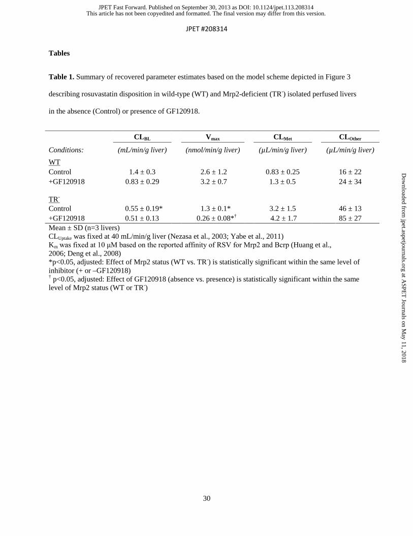
Table 1. Summary of recovered parameter estimates based on the model scheme depicted in Figure 3
describing rosuvastatin disposition in wild-type (WT) and Mrp2-deficient (TR-) isolated perfused livers
in the absence (Control) or presence of GF120918.
CLBL Vmax CLMet CLOther
Conditions: (mL/min/g liver) (nmol/min/g liver) (µL/min/g liver) (µL/min/g liver) WT Control 1.4 ± 0.3 2.6 ± 1.2 0.83 ± 0.25 16 ± 22 +GF120918 0.83 ± 0.29* 3.2 ± 0.7 1.3 ± 0.5† 24 ± 34 TR-
Control 0.55 ± 0.19*† 1.3 ± 0.1* 3.2 ± 1.5 46 ± 13 +GF120918 0.51 ± 0.13*† 0.26 ± 0.08*† 4.2 ± 1.7 85 ± 27 Mean ± SD (n=3 livers) CLUptake was fixed at 40 mL/min/g liver (Nezasa et al., 2003; Yabe et al., 2011) Km was fixed at 10 μM based on the reported affinity of RSV for Mrp2 and Bcrp (Huang et al., 2006; Deng et al., 2008) *p<0.05, adjusted: Effect of Mrp2 status (WT vs. TR-) is statistically significant within the same level of inhibitor (+ or –GF120918) † p<0.05, adjusted: Effect of GF120918 (absence vs. presence) is statistically significant within the same level of Mrp2 status (WT or TR-)
This article has not been copyedited and formatted. The final version may differ from this version.JPET Fast Forward. Published on September 30, 2013 as DOI: 10.1124/jpet.113.208314
at ASPE
T Journals on M
ay 11, 2018jpet.aspetjournals.org
Dow
nloaded from
JPET #208314
31
Figure Legends
Figure 1. Scheme depicting the experimental protocol in isolated perfused livers (IPLs). Gray shading
represents inclusion of rosuvastatin (RSV) in the Krebs’ Ringer Bicarbonate buffer during the uptake
phase. For conditions in the presence of inhibitor, the perfusate also contained 0.5 μM GF120918 for the
duration of the experiment (15-min equilibration period followed by loading and efflux phases).
Figure 2. A. Extracted ion current (XIC) chromatograph of rosuvastatin (RSV; blue), rosuvastatin
pentanoic acid (RSV-PA; purple), and d6-RSV (internal standard, red) in TR- Control liver tissue
compared to blank liver tissue (inset). B. High resolution product ion spectra of RSV (top, 482.20±0.05
amu), d6-RSV (middle, 488.20±0.05 amu), and RSV-PA (bottom, 422.20±0.05 amu). Note the expected
Δ6 amu between the products of RSV and its internal standard (258.14/264.17, 300.15/306.19,
314.14/320.20, 378.12/384.16, 404.19/410.23, 422.15/428.19 and 446.15/452.19), as well as common
product ions formed from RSV and RSV-PA (242.10, 256.12 and 270.17 amu), which are independent of
the changes within the carboxylic acid side chain.
Figure 3. Model scheme depicting the disposition of rosuvastatin (RSV) in rat IPL studies. Q denotes
the flow rate of perfusate, X denotes mass of RSV, V denotes compartmental volume, C denotes
compartmental concentration; subscripts on mass, flow and concentration terms denote the corresponding
compartment in the model scheme, as follows: inflow perfusate (in), outflow perfusate (out), extracellular
compartment (EC, assumed to be in rapid equilibration with the perfusate), intracellular liver
compartment (L), and bile (Bile); clearance values are designated as CLUptake for RSV uptake from the
extracellular to intracellular liver compartment, CLBL for RSV efflux from the intracellular to
extracellular liver compartment, CLMet for conversion from RSV to the pentanoic acid metabolite, and
CLOther for RSV conversion to metabolites other than the pentanoic acid derivative; Vmax is the maximum
velocity of RSV biliary excretion, Km is the RSV concentration resulting in half-maximal biliary
excretion; fu denotes unbound fraction.
This article has not been copyedited and formatted. The final version may differ from this version.JPET Fast Forward. Published on September 30, 2013 as DOI: 10.1124/jpet.113.208314
at ASPE
T Journals on M
ay 11, 2018jpet.aspetjournals.org
Dow
nloaded from

JPET #208314
32
Figure 4. Rosuvastatin (RSV) biliary excretion (A. and B.) and outflow perfusate (C. and D.) rate versus
time data in isolated perfused livers from wild-type (WT: A. and C.) and Mrp2-deficient (TR-: B. and D.)
rats in the absence (open symbols; dashed line) or presence (closed symbols; solid line) of GF120918.
The simulated excretion rate-time profiles were generated from the relevant equations based on the model
scheme in Figure 3 and the final parameter estimates reported in Table 1. Gray arrow indicates the switch
from RSV-containing to RSV-free perfusate at 60 min. Data are presented as mean±SD (n=3 livers).
Figure 5. Recovery of rosuvastatin (solid bars) and rosuvastatin pentanoic acid (open bars) in perfusate,
liver and bile, as well as total recovery from wild-type (WT) and Mrp2-deficient (TR-) isolated perfused
livers in the absence and presence of GF120918. Data are reported as the mean percentage ± SD based on
the total dose administered to each liver (n=3 per group) recovered at the end of the 90-min perfusion.
For recovery of parent drug: *p<0.05, adjusted: Effect of Mrp2 status (WT vs. TR-) is statistically
significant within the same level of inhibitor (+ or –GF120918); †p<0.05, adjusted: Effect of GF120918
(absence vs, presence) is statistically significant within the same level of Mrp2 status (WT or TR-). For
recovery of the metabolite: ‡p<0.05, adjusted: Effect of Mrp2 status (WT vs. TR-) is statistically
significant within the same level of inhibitor (+ or – GF120918).
This article has not been copyedited and formatted. The final version may differ from this version.JPET Fast Forward. Published on September 30, 2013 as DOI: 10.1124/jpet.113.208314
at ASPE
T Journals on M
ay 11, 2018jpet.aspetjournals.org
Dow
nloaded from

Figure 1
Equilibration Phase
15 min
Loading Phase
+ 0.5 µM RSV, 60 min
Efflux Phase
30 min
This article has not been copyedited and formatted. The final version may differ from this version.JPET Fast Forward. Published on September 30, 2013 as DOI: 10.1124/jpet.113.208314
at ASPE
T Journals on M
ay 11, 2018jpet.aspetjournals.org
Dow
nloaded from

Figure 2
dXout/dt =
Cout•Q
Vmax/Km
CLUptakeCLBL
Cin•Q =
dXin/dt
Intracellular
Space:
XL, Xmet
VL
fu,L
dXBile/dt = CBile•QBile
CLMet
CLOther
Extracellular Space: XEC, VEC
This article has not been copyedited and formatted. The final version may differ from this version.JPET Fast Forward. Published on September 30, 2013 as DOI: 10.1124/jpet.113.208314
at ASPE
T Journals on M
ay 11, 2018jpet.aspetjournals.org
Dow
nloaded from
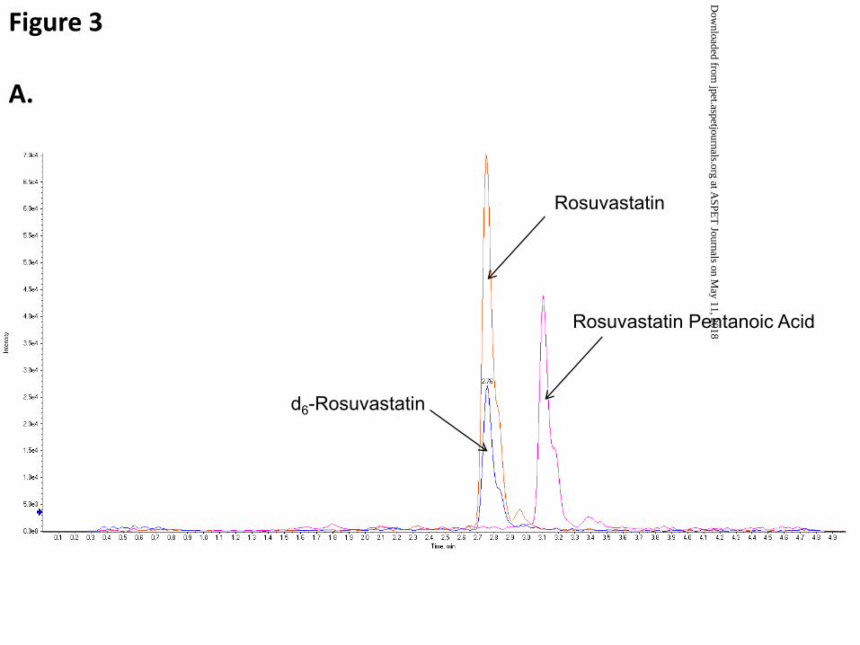
Figure 3
A.
Rosuvastatin
d6-Rosuvastatin
Rosuvastatin Pentanoic Acid
This article has not been copyedited and formatted. The final version may differ from this version.JPET Fast Forward. Published on September 30, 2013 as DOI: 10.1124/jpet.113.208314
at ASPE
T Journals on M
ay 11, 2018jpet.aspetjournals.org
Dow
nloaded from

B.
Figure 3
Rosuvastatin
d6-Rosuvastatin
Rosuvastatin Pentanoic Acid
CD3
CD3
This article has not been copyedited and formatted. The final version may differ from this version.JPET Fast Forward. Published on September 30, 2013 as DOI: 10.1124/jpet.113.208314
at ASPE
T Journals on M
ay 11, 2018jpet.aspetjournals.org
Dow
nloaded from

Figure 4
0.01
0.1
1
10
100
0 15 30 45 60 75 90
Bil
iary
Ex
cre
tio
n R
ate
(n
mo
l/m
in)
0.01
0.1
1
10
100
0 15 30 45 60 75 90
Bil
iary
Ex
cre
tio
n R
ate
(n
mo
l/m
in)
0.01
0.1
1
10
100
0 15 30 45 60 75 90
Ou
tflo
w P
erf
us
ate
Ra
te
(nm
ol/
min
)
Time (min)
0.01
0.1
1
10
100
0 15 30 45 60 75 90
Ou
tflo
w P
erf
us
ate
Ra
te
(nm
ol/
min
)
Time (min)
A. B.
C. D.
TR-WT
This article has not been copyedited and formatted. The final version may differ from this version.JPET Fast Forward. Published on September 30, 2013 as DOI: 10.1124/jpet.113.208314
at ASPE
T Journals on M
ay 11, 2018jpet.aspetjournals.org
Dow
nloaded from

Figure 5
0
20
40
60
80
100
120
% R
ec
ove
ry
Metabolite
Parent
Perfusate BileLiver Total
†
**
**
*
‡
‡
This article has not been copyedited and formatted. The final version may differ from this version.JPET Fast Forward. Published on September 30, 2013 as DOI: 10.1124/jpet.113.208314
at ASPE
T Journals on M
ay 11, 2018jpet.aspetjournals.org
Dow
nloaded from


