Issue N° 6 | March 2016
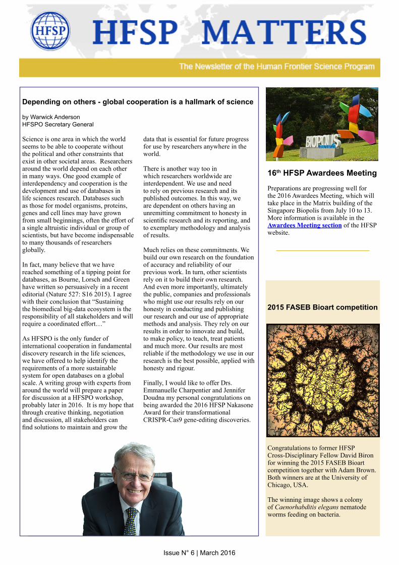
16th HFSP Awardees Meeting Preparations are progressing well for the 2016 Awardees Meeting, which will take place in the Matrix building of the Singapore Biopolis from July 10 to 13. More information is available in the Awardees Meeting section of the HFSP website.
Depending on others - global cooperation is a hallmark of science
by Warwick AndersonHFSPO Secretary General
Science is one area in which the world seems to be able to cooperate without the political and other constraints that exist in other societal areas. Researchers around the world depend on each other in many ways. One good example of interdependency and cooperation is the development and use of databases in life sciences research. Databases such as those for model organisms, proteins, genes and cell lines may have grown from small beginnings, often the effort of a single altruistic individual or group of scientists, but have become indispensable to many thousands of researchers globally.
In fact, many believe that we have reached something of a tipping point for databases, as Bourne, Lorsch and Green have written so persuasively in a recent editorial (Nature 527: S16 2015). I agree with their conclusion that “Sustaining the biomedical big-data ecosystem is the responsibility of all stakeholders and will require a coordinated effort…”
As HFSPO is the only funder of international cooperation in fundamental discovery research in the life sciences, we have offered to help identify the requirements of a more sustainable system for open databases on a global scale. A writing group with experts from around the world will prepare a paper for discussion at a HFSPO workshop, probably later in 2016. It is my hope that through creative thinking, negotiation and discussion, all stakeholders can find solutions to maintain and grow the
data that is essential for future progress for use by researchers anywhere in the world.
There is another way too in which researchers worldwide are interdependent. We use and need to rely on previous research and its published outcomes. In this way, we are dependent on others having an unremitting commitment to honesty in scientific research and its reporting, and to exemplary methodology and analysis of results.
Much relies on these commitments. We build our own research on the foundation of accuracy and reliability of our previous work. In turn, other scientists rely on it to build their own research. And even more importantly, ultimately the public, companies and professionals who might use our results rely on our honesty in conducting and publishing our research and our use of appropriate methods and analysis. They rely on our results in order to innovate and build, to make policy, to teach, treat patients and much more. Our results are most reliable if the methodology we use in our research is the best possible, applied with honesty and rigour.
Finally, I would like to offer Drs. Emmanuelle Charpentier and Jennifer Doudna my personal congratulations on being awarded the 2016 HFSP Nakasone Award for their transformational CRISPR-Cas9 gene-editing discoveries.
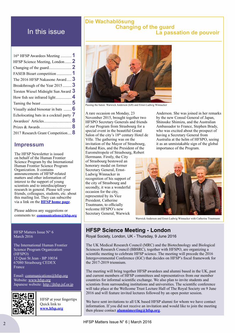
2015 FASEB Bioart competition
Congratulations to former HFSP Cross-Disciplinary Fellow David Biron for winning the 2015 FASEB Bioart competition together with Adam Brown. Both winners are at the University of Chicago, USA.
The winning image shows a colony of Caenorhabditis elegans nematode worms feeding on bacteria.
2 HFSP Matters Issue N° 6 | March 20162
In this issue
Impressum The HFSP Newsletter is issued on behalf of the Human Frontier Science Program by the International Human Frontier Science Program Organization. It contains announcements of HFSP-related matters and other information of interest to the support of young scientists and to interdisciplinary research in general. Please tell your friends, colleagues, students, etc. about this mailing list. They can subscribe via a link on the HFSP home page.
Please address any suggestions or comments to: [email protected]
16th HFSP Awardees Meeting ...........1 HFSP Science Meeting, London .......2Changing of the guard .......................2FASEB Bioart competition ...............1 The 2016 HFSP Nakasone Award .....3Breakthrough of the Year 2015 .........3Torsten Wiesel Midnight Sun Award 3How fish see infrared light ................4Taming the beast ...............................5Visually aided biosonar in bats ........6Echolocating bats in a cocktail party 7Awardees' Articles ............................8Prizes & Awards ................................82017 Research Grant Competition ....8
HFSP Matters Issue N° 6March 2016
The International Human Frontier Science Program Organization (HFSPO)12 Quai St Jean - BP 10034 67080 Strasbourg CEDEXFrance
Email: [email protected]: www.hfsp.orgJapanese website: http://jhfsp.jsf.or.jp
HFSP at your fingertipsQuick link to www.hfsp.org
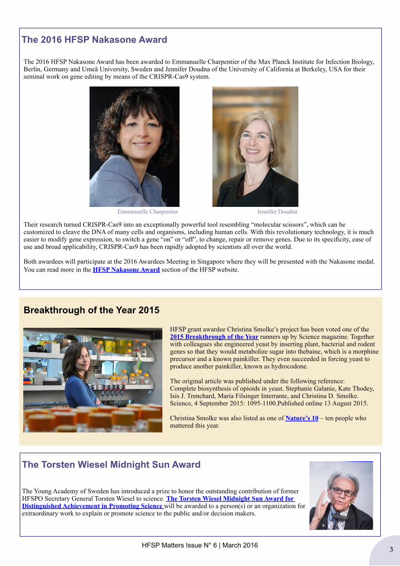
Passing the baton: Warwick Anderson (left) and Ernst-Ludwig Winnacker
A rare occasion on Monday, 23 November 2015, brought together two HFSPO Secretary Generals and friends of our Program from Strasbourg for a special event in the beautiful Grand Salon of the city’s 18th century Hotel de Ville. The gathering was on the invitation of the Mayor of Strasbourg, Roland Ries, and the President of the Eurométropole of Strasbourg, Robert Herrmann. Firstly, the City of Strasbourg bestowed an honorary medal on former Secretary General, Ernst-Ludwig Winnacker in recognition of his support of the city of Strasbourg and secondly, it was a wonderful occasion for the city, represented by its Vice President, Catherine Trautmann, to officially welcome HFSPO’s new Secretary General, Warwick
Anderson. She was joined in her remarks by the new Consul General of Japan, Shinsuke Shimizu, and the Australian Ambassador to France, Stephen Brady, who was excited about the prospect of having a Secretary General from Australia at the helm of HFSPO, seeing it as an unmistakable sign of the global importance of the Program.
Die Wachablösung Changing of the guard La passation de pouvoir
Warwick Anderson and Ernst-Ludwig Winnacker with Catherine Trautmann
HFSP Science Meeting - LondonRoyal Society, London, UK - Thursday, 9 June 2016
The UK Medical Research Council (MRC) and the Biotechnology and Biological Sciences Research Council (BBSRC), together with HFSPO, are organizing a scientific meeting to celebrate HFSP science. The meeting will precede the 2016 Intergovernmental Conference (IGC) that decides on HFSP’s fiscal framework for the 2017-2019 triennium.
The meeting will bring together HFSP awardees and alumni based in the UK, past and current members of HFSP committees and representatives from our member countries for informal scientific exchange. We also plan to invite students and scientists from surrounding institutions and universities. The scientific conference will take place at the Wellcome Trust Lecture Hall of The Royal Society on 9 June 2016 and will feature invited lectures followed by an open poster session.
We have sent invitations to all UK based HFSP alumni for whom we have contact information. If you did not receive an invitation and would like to join the meeting then please contact [email protected].
HFSP Matters Issue N° 6 | March 2016 3
Breakthrough of the Year 2015
HFSP grant awardee Christina Smolke’s project has been voted one of the 2015 Breakthrough of the Year runners up by Science magazine. Together with colleagues she engineered yeast by inserting plant, bacterial and rodent genes so that they would metabolize sugar into thebaine, which is a morphine precursor and a known painkiller. They even succeeded in forcing yeast to produce another painkiller, known as hydrocodone.
The original article was published under the following reference: Complete biosynthesis of opioids in yeast. Stephanie Galanie, Kate Thodey, Isis J. Trenchard, Maria Filsinger Interrante, and Christina D. Smolke.Science, 4 September 2015: 1095-1100.Published online 13 August 2015.
Christina Smolke was also listed as one of Nature’s 10 – ten people who mattered this year.
The Torsten Wiesel Midnight Sun Award
The Young Academy of Sweden has introduced a prize to honor the outstanding contribution of former HFSPO Secretary General Torsten Wiesel to science. The Torsten Wiesel Midnight Sun Award for Distinguished Achievement in Promoting Science will be awarded to a person(s) or an organization for extraordinary work to explain or promote science to the public and/or decision makers.
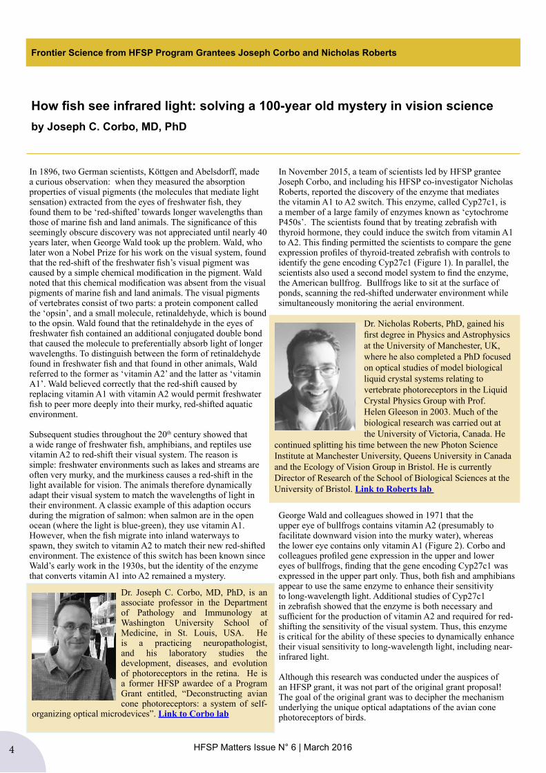
The 2016 HFSP Nakasone Award has been awarded to Emmanuelle Charpentier of the Max Planck Institute for Infection Biology, Berlin, Germany and Umeå University, Sweden and Jennifer Doudna of the University of California at Berkeley, USA for their seminal work on gene editing by means of the CRISPR-Cas9 system.
Their research turned CRISPR-Cas9 into an exceptionally powerful tool resembling “molecular scissors”, which can be customized to cleave the DNA of many cells and organisms, including human cells. With this revolutionary technology, it is much easier to modify gene expression, to switch a gene “on” or “off”, to change, repair or remove genes. Due to its specificity, ease of use and broad applicability, CRISPR-Cas9 has been rapidly adopted by scientists all over the world.
Both awardees will participate at the 2016 Awardees Meeting in Singapore where they will be presented with the Nakasone medal.You can read more in the HFSP Nakasone Award section of the HFSP website.
The 2016 HFSP Nakasone Award
Jennifer DoudnaEmmanuelle Charpentier
4 HFSP Matters Issue N° 6 | March 2016
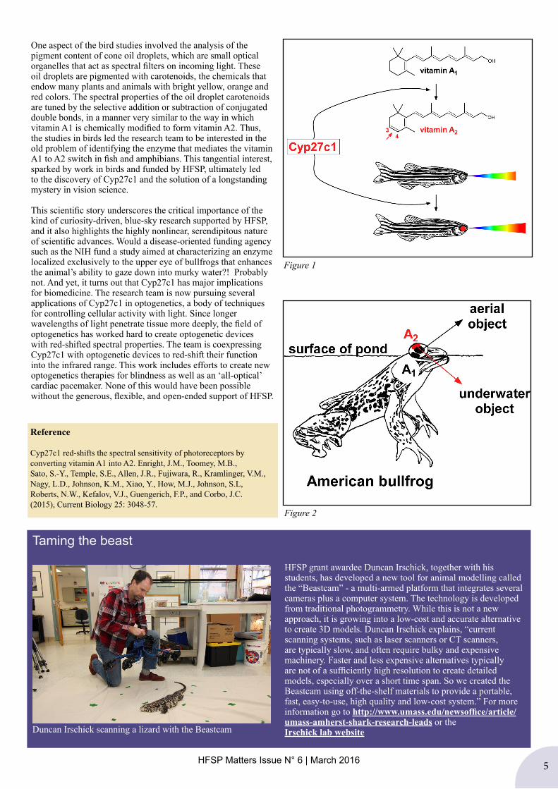
In 1896, two German scientists, Köttgen and Abelsdorff, made a curious observation: when they measured the absorption properties of visual pigments (the molecules that mediate light sensation) extracted from the eyes of freshwater fish, they found them to be ‘red-shifted’ towards longer wavelengths than those of marine fish and land animals. The significance of this seemingly obscure discovery was not appreciated until nearly 40 years later, when George Wald took up the problem. Wald, who later won a Nobel Prize for his work on the visual system, found that the red-shift of the freshwater fish’s visual pigment was caused by a simple chemical modification in the pigment. Wald noted that this chemical modification was absent from the visual pigments of marine fish and land animals. The visual pigments of vertebrates consist of two parts: a protein component called the ‘opsin’, and a small molecule, retinaldehyde, which is bound to the opsin. Wald found that the retinaldehyde in the eyes of freshwater fish contained an additional conjugated double bond that caused the molecule to preferentially absorb light of longer wavelengths. To distinguish between the form of retinaldehyde found in freshwater fish and that found in other animals, Wald referred to the former as ‘vitamin A2’ and the latter as ‘vitamin A1’. Wald believed correctly that the red-shift caused by replacing vitamin A1 with vitamin A2 would permit freshwater fish to peer more deeply into their murky, red-shifted aquatic environment. Subsequent studies throughout the 20th century showed that a wide range of freshwater fish, amphibians, and reptiles use vitamin A2 to red-shift their visual system. The reason is simple: freshwater environments such as lakes and streams are often very murky, and the murkiness causes a red-shift in the light available for vision. The animals therefore dynamically adapt their visual system to match the wavelengths of light in their environment. A classic example of this adaption occurs during the migration of salmon: when salmon are in the open ocean (where the light is blue-green), they use vitamin A1. However, when the fish migrate into inland waterways to spawn, they switch to vitamin A2 to match their new red-shifted environment. The existence of this switch has been known since Wald’s early work in the 1930s, but the identity of the enzyme that converts vitamin A1 into A2 remained a mystery.
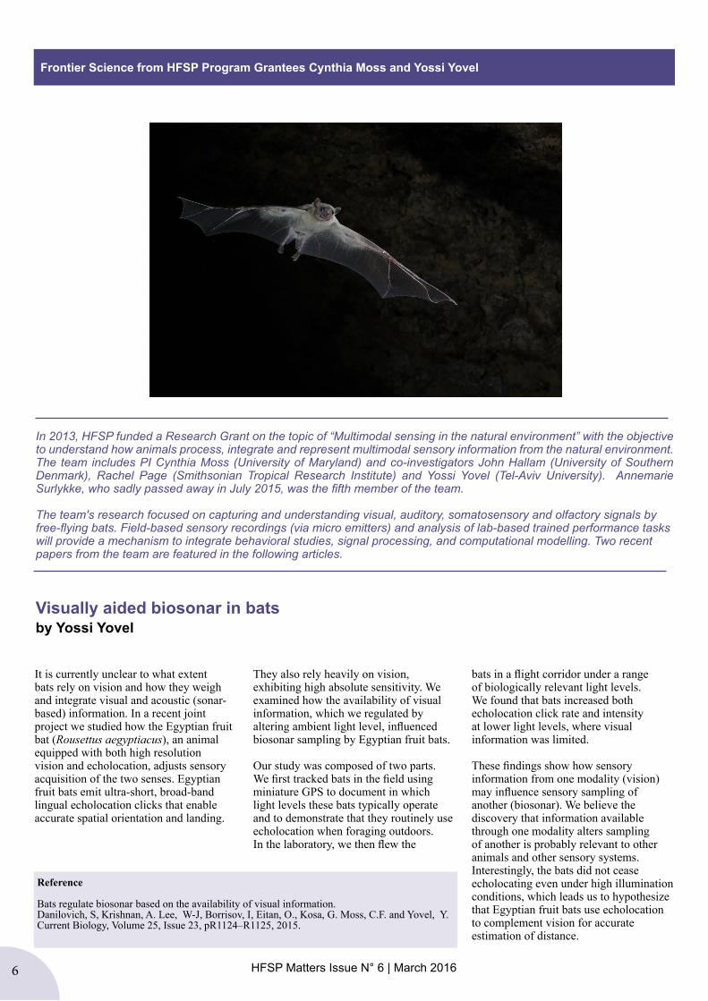
In November 2015, a team of scientists led by HFSP grantee Joseph Corbo, and including his HFSP co-investigator Nicholas Roberts, reported the discovery of the enzyme that mediates the vitamin A1 to A2 switch. This enzyme, called Cyp27c1, is a member of a large family of enzymes known as ‘cytochrome P450s’. The scientists found that by treating zebrafish with thyroid hormone, they could induce the switch from vitamin A1 to A2. This finding permitted the scientists to compare the gene expression profiles of thyroid-treated zebrafish with controls to identify the gene encoding Cyp27c1 (Figure 1). In parallel, the scientists also used a second model system to find the enzyme, the American bullfrog. Bullfrogs like to sit at the surface of ponds, scanning the red-shifted underwater environment while simultaneously monitoring the aerial environment.
George Wald and colleagues showed in 1971 that the upper eye of bullfrogs contains vitamin A2 (presumably to facilitate downward vision into the murky water), whereas the lower eye contains only vitamin A1 (Figure 2). Corbo and colleagues profiled gene expression in the upper and lower eyes of bullfrogs, finding that the gene encoding Cyp27c1 was expressed in the upper part only. Thus, both fish and amphibians appear to use the same enzyme to enhance their sensitivity to long-wavelength light. Additional studies of Cyp27c1 in zebrafish showed that the enzyme is both necessary and sufficient for the production of vitamin A2 and required for red-shifting the sensitivity of the visual system. Thus, this enzyme is critical for the ability of these species to dynamically enhance their visual sensitivity to long-wavelength light, including near-infrared light.
Although this research was conducted under the auspices of an HFSP grant, it was not part of the original grant proposal! The goal of the original grant was to decipher the mechanism underlying the unique optical adaptations of the avian cone photoreceptors of birds.
Frontier Science from HFSP Program Grantees Joseph Corbo and Nicholas Roberts
How fish see infrared light: solving a 100-year old mystery in vision scienceby Joseph C. Corbo, MD, PhD
Dr. Joseph C. Corbo, MD, PhD, is an associate professor in the Department of Pathology and Immunology at Washington University School of Medicine, in St. Louis, USA. He is a practicing neuropathologist, and his laboratory studies the development, diseases, and evolution of photoreceptors in the retina. He is a former HFSP awardee of a Program Grant entitled, “Deconstructing avian cone photoreceptors: a system of self-
organizing optical microdevices”. Link to Corbo lab
Dr. Nicholas Roberts, PhD, gained his first degree in Physics and Astrophysics at the University of Manchester, UK, where he also completed a PhD focused on optical studies of model biological liquid crystal systems relating to vertebrate photoreceptors in the Liquid Crystal Physics Group with Prof. Helen Gleeson in 2003. Much of the biological research was carried out at the University of Victoria, Canada. He
continued splitting his time between the new Photon Science Institute at Manchester University, Queens University in Canada and the Ecology of Vision Group in Bristol. He is currently Director of Research of the School of Biological Sciences at the University of Bristol. Link to Roberts lab
HFSP Matters Issue N° 6 | March 2016 5
One aspect of the bird studies involved the analysis of the pigment content of cone oil droplets, which are small optical organelles that act as spectral filters on incoming light. These oil droplets are pigmented with carotenoids, the chemicals that endow many plants and animals with bright yellow, orange and red colors. The spectral properties of the oil droplet carotenoids are tuned by the selective addition or subtraction of conjugated double bonds, in a manner very similar to the way in which vitamin A1 is chemically modified to form vitamin A2. Thus, the studies in birds led the research team to be interested in the old problem of identifying the enzyme that mediates the vitamin A1 to A2 switch in fish and amphibians. This tangential interest, sparked by work in birds and funded by HFSP, ultimately led to the discovery of Cyp27c1 and the solution of a longstanding mystery in vision science.
This scientific story underscores the critical importance of the kind of curiosity-driven, blue-sky research supported by HFSP, and it also highlights the highly nonlinear, serendipitous nature of scientific advances. Would a disease-oriented funding agency such as the NIH fund a study aimed at characterizing an enzyme localized exclusively to the upper eye of bullfrogs that enhances the animal’s ability to gaze down into murky water?! Probably not. And yet, it turns out that Cyp27c1 has major implications for biomedicine. The research team is now pursuing several applications of Cyp27c1 in optogenetics, a body of techniques for controlling cellular activity with light. Since longer wavelengths of light penetrate tissue more deeply, the field of optogenetics has worked hard to create optogenetic devices with red-shifted spectral properties. The team is coexpressing Cyp27c1 with optogenetic devices to red-shift their function into the infrared range. This work includes efforts to create new optogenetics therapies for blindness as well as an ‘all-optical’ cardiac pacemaker. None of this would have been possible without the generous, flexible, and open-ended support of HFSP.
Figure 1
Figure 2
Reference
Cyp27c1 red-shifts the spectral sensitivity of photoreceptors by converting vitamin A1 into A2. Enright, J.M., Toomey, M.B., Sato, S.-Y., Temple, S.E., Allen, J.R., Fujiwara, R., Kramlinger, V.M., Nagy, L.D., Johnson, K.M., Xiao, Y., How, M.J., Johnson, S.L, Roberts, N.W., Kefalov, V.J., Guengerich, F.P., and Corbo, J.C. (2015), Current Biology 25: 3048-57.
Taming the beast
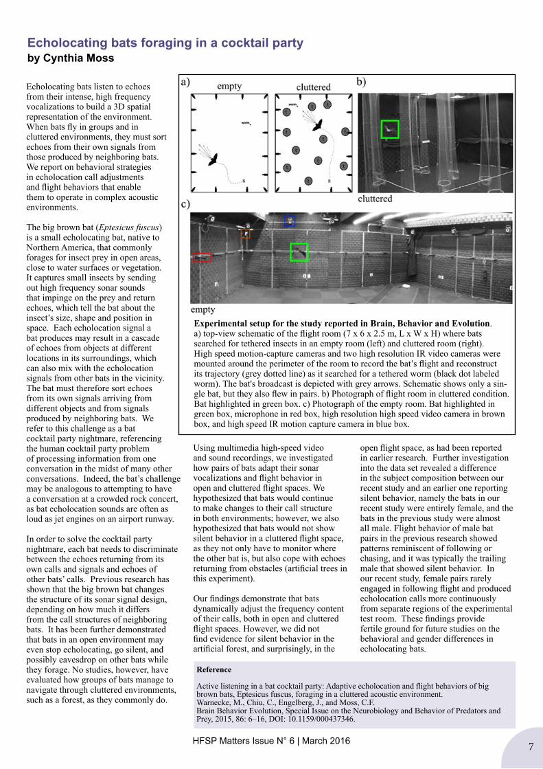
HFSP grant awardee Duncan Irschick, together with his students, has developed a new tool for animal modelling called the “Beastcam” - a multi-armed platform that integrates several cameras plus a computer system. The technology is developed from traditional photogrammetry. While this is not a new approach, it is growing into a low-cost and accurate alternative to create 3D models. Duncan Irschick explains, “current scanning systems, such as laser scanners or CT scanners, are typically slow, and often require bulky and expensive machinery. Faster and less expensive alternatives typically are not of a sufficiently high resolution to create detailed models, especially over a short time span. So we created the Beastcam using off-the-shelf materials to provide a portable, fast, easy-to-use, high quality and low-cost system.” For more information go to http://www.umass.edu/newsoffice/article/umass-amherst-shark-research-leads or the Irschick lab websiteDuncan Irschick scanning a lizard with the Beastcam
6 HFSP Matters Issue N° 6 | March 2016
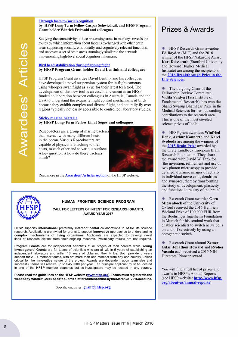
Frontier Science from HFSP Program Grantees Cynthia Moss and Yossi Yovel
In 2013, HFSP funded a Research Grant on the topic of “Multimodal sensing in the natural environment” with the objective to understand how animals process, integrate and represent multimodal sensory information from the natural environment. The team includes PI Cynthia Moss (University of Maryland) and co-investigators John Hallam (University of Southern Denmark), Rachel Page (Smithsonian Tropical Research Institute) and Yossi Yovel (Tel-Aviv University). Annemarie Surlykke, who sadly passed away in July 2015, was the fifth member of the team.
The team's research focused on capturing and understanding visual, auditory, somatosensory and olfactory signals by free-flying bats. Field-based sensory recordings (via micro emitters) and analysis of lab-based trained performance tasks will provide a mechanism to integrate behavioral studies, signal processing, and computational modelling. Two recent papers from the team are featured in the following articles.
Visually aided biosonar in bats by Yossi Yovel
It is currently unclear to what extent bats rely on vision and how they weigh and integrate visual and acoustic (sonar-based) information. In a recent joint project we studied how the Egyptian fruit bat (Rousettus aegyptiacus), an animal equipped with both high resolution vision and echolocation, adjusts sensory acquisition of the two senses. Egyptian fruit bats emit ultra-short, broad-band lingual echolocation clicks that enable accurate spatial orientation and landing.
They also rely heavily on vision, exhibiting high absolute sensitivity. We examined how the availability of visual information, which we regulated by altering ambient light level, influenced biosonar sampling by Egyptian fruit bats.
Our study was composed of two parts.We first tracked bats in the field using miniature GPS to document in which light levels these bats typically operate and to demonstrate that they routinely use echolocation when foraging outdoors. In the laboratory, we then flew the
bats in a flight corridor under a range of biologically relevant light levels. We found that bats increased both echolocation click rate and intensity at lower light levels, where visual information was limited.
These findings show how sensory information from one modality (vision) may influence sensory sampling of another (biosonar). We believe the discovery that information available through one modality alters sampling of another is probably relevant to other animals and other sensory systems. Interestingly, the bats did not cease echolocating even under high illumination conditions, which leads us to hypothesize that Egyptian fruit bats use echolocation to complement vision for accurate estimation of distance.
Reference
Bats regulate biosonar based on the availability of visual information. Danilovich, S, Krishnan, A. Lee, W-J, Borrisov, I, Eitan, O., Kosa, G. Moss, C.F. and Yovel, Y. Current Biology, Volume 25, Issue 23, pR1124–R1125, 2015.
HFSP Matters Issue N° 6 | March 2016 7
Echolocating bats listen to echoes from their intense, high frequency vocalizations to build a 3D spatial representation of the environment. When bats fly in groups and in cluttered environments, they must sort echoes from their own signals from those produced by neighboring bats. We report on behavioral strategies in echolocation call adjustments and flight behaviors that enable them to operate in complex acoustic environments.
The big brown bat (Eptesicus fuscus) is a small echolocating bat, native to Northern America, that commonly forages for insect prey in open areas, close to water surfaces or vegetation. It captures small insects by sending out high frequency sonar sounds that impinge on the prey and return echoes, which tell the bat about the insect’s size, shape and position in space. Each echolocation signal a bat produces may result in a cascade of echoes from objects at different locations in its surroundings, which can also mix with the echolocation signals from other bats in the vicinity. The bat must therefore sort echoes from its own signals arriving from different objects and from signals produced by neighboring bats. We refer to this challenge as a bat cocktail party nightmare, referencing the human cocktail party problem of processing information from one conversation in the midst of many other conversations. Indeed, the bat’s challenge may be analogous to attempting to have a conversation at a crowded rock concert, as bat echolocation sounds are often as loud as jet engines on an airport runway.
In order to solve the cocktail party nightmare, each bat needs to discriminate between the echoes returning from its own calls and signals and echoes of other bats’ calls. Previous research has shown that the big brown bat changes the structure of its sonar signal design, depending on how much it differs from the call structures of neighboring bats. It has been further demonstrated that bats in an open environment may even stop echolocating, go silent, and possibly eavesdrop on other bats while they forage. No studies, however, have evaluated how groups of bats manage to navigate through cluttered environments, such as a forest, as they commonly do.
Using multimedia high-speed video and sound recordings, we investigated how pairs of bats adapt their sonar vocalizations and flight behavior in open and cluttered flight spaces. We hypothesized that bats would continue to make changes to their call structure in both environments; however, we also hypothesized that bats would not show silent behavior in a cluttered flight space, as they not only have to monitor where the other bat is, but also cope with echoes returning from obstacles (artificial trees in this experiment).
Our findings demonstrate that bats dynamically adjust the frequency content of their calls, both in open and cluttered flight spaces. However, we did not find evidence for silent behavior in the artificial forest, and surprisingly, in the
open flight space, as had been reported in earlier research. Further investigation into the data set revealed a difference in the subject composition between our recent study and an earlier one reporting silent behavior, namely the bats in our recent study were entirely female, and the bats in the previous study were almost all male. Flight behavior of male bat pairs in the previous research showed patterns reminiscent of following or chasing, and it was typically the trailing male that showed silent behavior. In our recent study, female pairs rarely engaged in following flight and produced echolocation calls more continuously from separate regions of the experimental test room. These findings provide fertile ground for future studies on the behavioral and gender differences in echolocating bats.
Echolocating bats foraging in a cocktail partyby Cynthia Moss
Experimental setup for the study reported in Brain, Behavior and Evolution. a) top-view schematic of the flight room (7 x 6 x 2.5 m, L x W x H) where bats searched for tethered insects in an empty room (left) and cluttered room (right). High speed motion-capture cameras and two high resolution IR video cameras were mounted around the perimeter of the room to record the bat’s flight and reconstruct its trajectory (grey dotted line) as it searched for a tethered worm (black dot labeled worm). The bat's broadcast is depicted with grey arrows. Schematic shows only a sin-gle bat, but they also flew in pairs. b) Photograph of flight room in cluttered condition. Bat highlighted in green box. c) Photograph of the empty room. Bat highlighted in green box, microphone in red box, high resolution high speed video camera in brown box, and high speed IR motion capture camera in blue box.
Reference
Active listening in a bat cocktail party: Adaptive echolocation and flight behaviors of big brown bats, Eptesicus fuscus, foraging in a cluttered acoustic environment. Warnecke, M., Chiu, C., Engelberg, J., and Moss, C.F.Brain Behavior Evolution, Special Issue on the Neurobiology and Behavior of Predators and Prey, 2015, 86: 6–16, DOI: 10.1159/000437346.
8 HFSP Matters Issue N° 6 | March 2016HFSP Matters Issue N° 6 | March 2016
Through faces to (social) cognitionby HFSP Long-Term Fellow Caspar Schwiedrzik and HFSP Program Grant holder Winrich Freiwald and colleagues
Studying the connectivity of face processing areas in monkeys reveals the routes by which information about faces is exchanged with other brain areas supporting socially, emotionally, and cognitively relevant functions, and uncovers a set of brain areas stunningly similar to the network implementing high-level social cognition in humans.
Bird head stabilization during flapping flightby HFSP Program Grant holder David Lentink and colleagues
HFSP Program Grant awardee David Lentink and his colleagues have developed a novel suspension system for in-flight cameras using whooper swan flight as a cue for their latest tech tool. The development of this new tool is an essential element in an HFSP funded collaboration between colleagues in Australia, Canada and the USA to understand the exquisite flight control mechanisms of birds because they exhibit complex and diverse flight, and naturally fly over regions typically not easily accessible to controlled laboratory studies.
Sticky marine bacteriaby HFSP Long-Term Fellow Einat Segev and colleagues
Roseobacters are a group of marine bacteria that interact with many different hosts in the ocean. Various Roseobacters are capable of physically attaching to their hosts, to each other and to various surfaces. A key question is how do these bacteria attach?
Read more in the Awardees' Articles section of the HFSP website.
Aw
arde
es'
Art
icle
sPrizes & Awards
HFSP Research Grant awardee Ed Boyden (MIT) and the 2010 winner of the HFSP Nakasone Award Karl Deisseroth (Stanford University and Howard Hughes Medical Institute) are among the recipients of the 2016 Breakthrough Prize in the Life Sciences.
The outgoing Chair of the Fellowship Review Committee, Vidita Vaidya (Tata Institute of Fundamental Research), has won the Shanti Swarup Bhatnagar Prize in the Medical Sciences for her outstanding contributions to the research area. This is one of the most coveted science prizes of India.
HFSP grant awardees Winfried Denk, Arthur Konnerth and Karel Svoboda are among the winners of the 2015 Brain Prize awarded by the Grete Lundbeck European Brain Research Foundation. They share the award with David W. Tank for ‘the invention, refinement and use of two-photon microscopy to provide detailed, dynamic images of activity in individual nerve cells, dendrites and synapses, thereby transforming the study of development, plasticity and functional circuitry of the brain’.
Research Grant awardee Gero Miesenböck of the University of Oxford received the 2015 Heinrich Wieland Prize of 100,000 EUR from the Boehringer Ingelheim Foundation in Munich for his seminal work that enables scientists to switch nerve cells on and off selectively by using an optogenetic switch.
Research Grant alumni Zemer Gitai, Jonathon Howard and Ryohei Yasuda each received a 2015 NIH Directors’ Pioneer Award.
You will find a full list of prizes and awards in HFSP's Annual Reports (see HFSP website: http://www.hfsp.org/about-us/annual-reports)
HFSP supports international preferably intercontinental collaborations in basic life science research. Applications are invited for grants to support innovative approaches to understanding complex mechanisms of living organisms. Applicants are expected to develop novel lines of research distinct from their ongoing research. Preliminary results are not required.
Program Grants are for independent scientists at all stages of their careers while Young Investigators’ Grants are for teams of scientists who are all within 5 years of establishing an independent laboratory and within 10 years of obtaining their PhDs. Both provide 3 years support for 2 – 4 member teams, with not more than one member from any one country, unless critical for the innovative nature of the project. Awards are dependent upon team size and successful teams will receive up to $450,000 per year. The principal applicant must be located in one of the HFSP member countries but co-investigators may be located in any country.
Please read the guidelines on the HFSP website (www.hfsp.org). Teams must register via the website by March 21, 2016 so as to submit a letter of intent online by the March 31, 2016 deadline.
Specific enquiries: [email protected]
HUMAN FRONTIER SCIENCE PROGRAM
CALL FOR LETTERS OF INTENT FOR RESEARCH GRANTS:AWARD YEAR 2017


