Vol. 186, No. 1, 1992
July 15, 1992 BIOCHEMICAL AND BIOPHYSICAL RESEARCH COMMUNICATIONS
Pages 301-307
HUMAN INSULIN-LIKE G R O W T H FACTOR BINDING PROTEIN-6 IS O- GLYCOSYLATED
Leon A. Bach a, N. Rao Thotakura b, and Matthew M. Rechlera
aGrowth and Development Section, and bMolecular Regulation and Neuroendocrinology Section, Molecular and Cellular Endocrinology Branch, National Institute of Diabetes and Digestive and
Kidney Diseases, National Institutes of Health, Bethesda MD 20892
Received April 30, 1992
SUMMARY: Insulin-like growth factor binding protein-6 is abundant in cerebrospinal fluid and has a marked preferential binding affinity for IGF-II over IGF-I. The present study demonstrates that IGFBP-6 is O-glycosylated but not N-glycosylated. Carbohydrate analysis revealed the presence of ~20-30 carbohydrate residues/molecule. Galactosamine, galactose and sialic acid were most abundant, with glucosamine and fucose present in lower concentrations. Mannose was not detected. Enzymatic deglycosylation did not alter the high affinity of IGF binding protein-6 for IGF-II (Ka 4.4 _+ 2.2 x 10¹¹ M -1) or its preference for IGF-II over IGF-I. Glycosylation of IGFBP-6 may affect its secretion, in YivQ stability or localization, but does not affect its ligand binding properties. ~ 1992 A c a d e m i c Press, I n c .
Insulin-like growth factor binding protein-6 is one of a family of specific IGF binding proteins
which has recently been described and cloned (1,2). IGFBP-6 has been identified in human serum
(3), cerebrospinal fluid (4), and conditioned media from transformed and non-transformed human
lung fibroblasts (5,6). A distinctive property of IGFBP-6 is its marked preferential affinity for
IGF-II versus IGF-I (4-6).
Several observations suggest that IGFBP-6 may be glycosylated. The predicted molecular mass of
IGFBP-6 based on cDNA structure is 22.8 kDa (1), but IGFBP-6 migrates on SDS-PAGE with an
apparent molecular mass of 28-34 kDa as determined by ligand blotting (3-6). IGFBP-6 from
human fibroblasts binds to wheat germ agglutinin but not concanavalin A, suggesting that it
contains glucosamine but not mannose residues (5,6). Although human IGFBP-6 contains a
potential N-glycosylation site (1), N-glycanase does not alter the apparent molecular mass of
IGFBP-6 from human serum (3), indicating that IGFBP-6 from that source is not N-glycosylated.
Abbreviation~: IGF, insulin-like growth factor; IGFBP, insulin-like growth factor binding protein; D-PBS, Dulbecco's phosphate buffered saline; SDS-PAGE, sodium dodecyl sulfate- polyacrylamide gel electrophoresis.
301
0006-291X/92 $4.00 Copyright © 1992 by Academic Press, Inc.
All rights of reproduction in any form reserved.
Vol. 186, No. 1, 1992 BIOCHEMICAL AND BIOPHYSICAL RESEARCH COMMUNICATIONS
The present study demonstrates that IGFBP-6 purified from human cerebrospinal fluid is O-
glycosylated. Enzymatic removal of O-linked oligosaccharides does not affect its binding
properties.
METHODS
Preparation of IGF-II affinity column. IGF-II (1 mg, courtesy of Eli Lilly, Indianapolis, IN) in 3 ml 0.1 M Na HEPES (pH 7.4) was added to 3 ml of Affi-Gel 15 (BioRad, Richmond, CA) and incubated for 16 h at 4°C. The gel was then sequentially washed with 3 ml 1 M Tris-HC1 (pH 8.0) to block unbound sites. Prior to use, the gel was washed with 30 ml each of 0.1 M Na HEPES (pH 7.4), Dulbecco's phosphate buffered saline (138 mM NaC1, 8.1 mM Na2HPO4, 2.7 mM KC1, 1.2 mM KH2PO4) supplemented with NaC1 to 0.5 M, 0.5 M acetic acid, and D-PBS.
Purification of IGFBP-6 from human cerebrospinal fluid. Cerebrospinal fluid was obtained from postoperative drainage after surgical removal of pituitary adenomata from 2 patients and stored at -20°C prior to use. Results were similar from both patients. Ammonium sulfate was added to 930 rnl of cerebrospinal fluid from the first patient to a final saturation of 60% and mixed at room temperature for 16h. The solution was centrifuged at 3800 g in a Sorvall RC-5B centrifuge (DuPont Instruments, Wilmington, DE) for 15 min and the supernatant decanted. The pellet was resuspended in 30 ml D-PBS and dialyzed against D-PBS. The dialyzed sample was applied to the IGF-II affinity column and recycled overnight at 4°C. The column was washed with D-PBS supplemented with NaC1 to 0.5 M, and protein eluted with 0.5 M acetic acid. Eluate fractions that contained protein (based on absorbance at 280 nm) were further purified by reverse phase FPLC (ProRPC HR 5/10, Pharmacia, Piscataway, NJ) using a linear gradient of 24-40% acetonitrile over 60 min. Fractions containing protein (based on absorbance at 214 nm) were assayed for IGF-II binding activity as described below. IGFBP-6 eluted after 13 rain (acetonitrile concentration 28%). By comparison, IGFBP-2 was more hydrophobic, eluting at 21 min. The presence of IGFBP-6 was suggested by electrophoretic mobility on SDS-PAGE; purity was confirmed by silver staining. NH2-terminal amino acid sequencing of the first 10 residues and amino acid composition of the purified binding protein (80 pmol) was performed by Dr W Burgess (American Red Cross, Rockville, MD).
Carbohydrate analysis of IGFBP-6. Sialic acid content was determined by hydrolysis of IGFBP-6 (80 pmol) with 0.2 N HC1 (80°C, 1 h), followed by anion exchange HPLC at high pH (AS6 Ionpak, Dionex, Sunnyvale, CA) with pulsed amperometric detection (7). Neutral carbohydrate analysis was performed after hydrolysis in 2.75 N trifluoroacetic acid (100°C, 4 h) using the same HPLC detection system (7).
Deglycosylation of IGFBP-6. Desialylated IGFBP-6 was prepared by incubation of ~30 pmol of non-denatured IGFBP-6 with 50 mU neuraminidase (Genzyme, Boston, MA) in 50 ~tl 0.2 M sodium phosphate, 2 mM calcium acetate, pH 6.6 (37°C, 1 h). For complete removal of O- linked oligosaccharides, sialic acid and fucose were first removed by incubating IGFBP-6 (-100 pmol) with 50 mU neuraminidase (Genzyme, Boston, MA) and 40 mU fucosidase (Boehringer- Mannheim, Indianapolis, IN) in the above buffer. Core disaccharide was removed by subsequent incubation with 5 mU endo-a-N-acetyl-galactosaminidase (O-glycanase, Genzyme, Boston, MA) for 18.5 h at 37°C. Control aliquots of native, glycosylated IGFBP-6 were incubated with heat- inactivated enzymes under the same conditions. The presence of N-linked carbohydrate chains was assessed by incubation of native IGFBP-6 (-70 pmol) with 1.25 units of peptide: N- glycosidase F (N-glycanase, Genzyme) for 21 h at 37°C.
Lectin binding. Samples (-30 pmol) were fractionated by electrophoresis on SDS-PAGE (12% gels) using a discontinuous buffer system under reducing conditions (Novel Experimental Technology, San Diego, CA). Proteins were electroblotted to nitrocellulose membranes. Galactose 13(1-3) N-acetylgalactosamine core disaccharide was detected by binding of digoxigenin- conjugated peanut agglutinin, coupling with alkaline phosphatase labelled anti-digoxigenin antibodies, and color development, according to the manufacturer's instructions (Boehringer- Mannheim, Indianapolis, IN). The presence of glucosamine and mannose was examined by the same method (-10 pmol protein) using digoxigenin-labelled wheat germ agglutinin and concanavalin A respectively.
302
Vol. 186, No. 1, 1992 BIOCHEMICAL AND BIOPHYSICAL RESEARCH COMMUNICATIONS
Ligand blotting. Electrophoresis of samples (-1 pmol) under nonreducing conditions and electroblotting were performed as described for lectin binding. IGFBPs were identified by incubation with 15 pM [125I]IGF-II (specific activity, 2000 Ci/mmol, Amersham, Arlington Heights, IL) for 16 h at 4°C followed by autoradiography (8).
Competitive binding. Samples containing IGFBP were incubated with [125I]IGF-II (5 pM, Amersham, Arlington Heights, IL) and increasing concentrations (9 pM-2.4 nM) of unlabelled IGF-I (Amgen, Thousand Oaks, CA) or IGF-II (Upstate Biotechnology, Lake Placid, NY) in 0.1 M sodium phosphate buffer, pH 7.4 (4°C, 18 h, 0.4 ml final volume). Bound and free ligand were separated by addition of 0.5 ml ice-cold 5% charcoal/2% fatty acid-free bovine serum albumin (Sigma, St Louis, MI), incubation on ice for 10 min, and cenwifugation at 1300 g (4°C, 30 min). Bound radioactivity in supernatants was quantitated by T-counting (Beckman, Palo Alto, CA)(8). Results were analyzed using the Ligand program (9).
R E S U L T S
IGFBP-6 was purified from human cerebrospinal fluid by IGF-II affinity chromatography and
reverse phase FPLC. Its identity was established by earlier elution from the reverse phase column
than IGFBP-2 (3,4), electrophoretic mobility, marked preferential affinity for IGF-II versus IGF-I
(see below), and amino acid sequencing of the first 10 NH2-terminal residues (not shown) which
corresponded to amino acids 4-13 of the amino acid sequence deduced from the nucleotide
sequence (1). NH2-terminal truncation also has been observed for IGFBP-6 isolated from other
human sources (3,5,6).
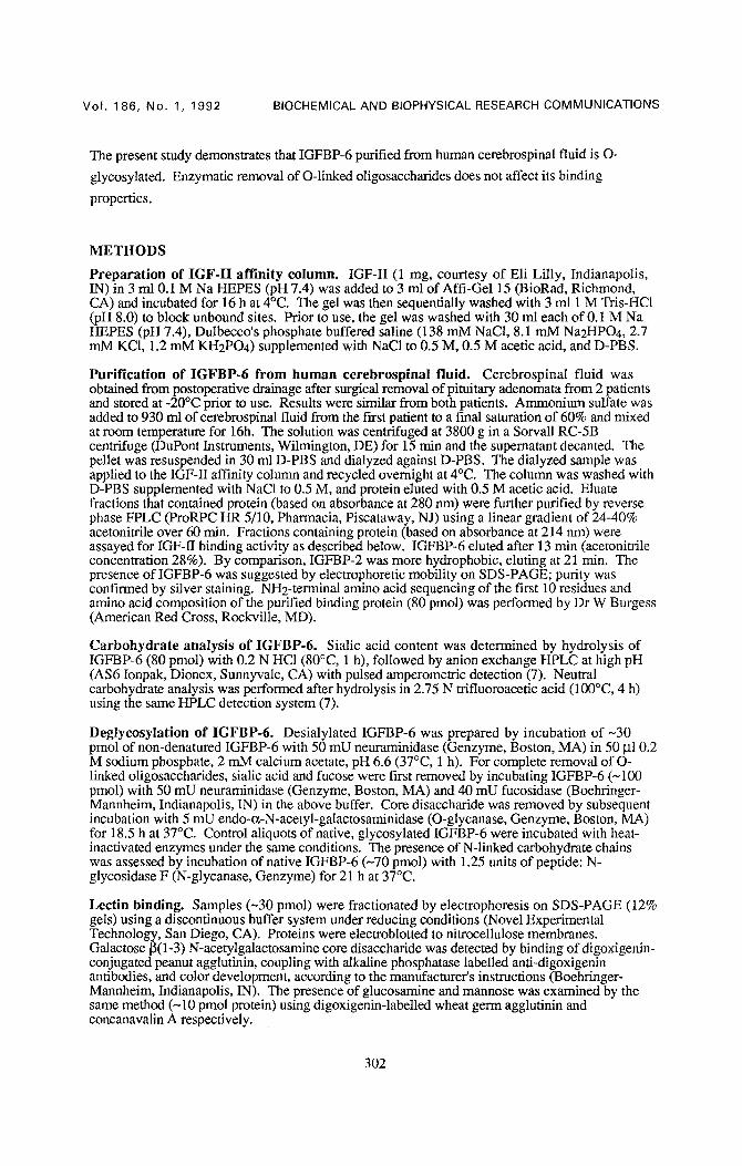
Ligand blotting of native glycosylated IGFBP-6 revealed a broad band with an apparent molecular
mass of 26.5-29.5 kDa (Fig. 1, lanes 1,5). Incubation with neuraminidase decreased the apparent
43
iiii!!i!!i! !i ili!ii!i iiliil ii i 1 2 3 4 5 6 7 8
Figure 1. Li~and blots of IGFBP-6 preparations following ine0bation with N- ~nd O-glycanas¢. Samples in nonreducing buffer were separated by SDS 12%-PAGE, electroblotted, and the blots incubated with 15 pM (125I]IGF-II (16h, 4°C) and exposed to film for 8 h (lanes 1-4) or 6.5 h (lanes 5-8). Experiment 1 (lanes 1-4): lane 1, native, glycosylated IGFBP-6; lane 2, IGFBP-6 after treatment with N-glycanase; lane 3, IGFBP-6 following treatment with neuraminidase, fucosidase and O-glycanase; lane 4, IGFBP-6 after treatment with neuraminidase, fucosidase, O- glycanase and N-glycanase. Experiment 2 (lanes 5-8): lane 5, native, glycosylated IGFBP-6; lane 6, IGFBP-6 following treatment with neuraminidase; lane 7, IGFBP-6 following treatment with neuraminidase, fucosidase and O-glycanase; lane 8: deglycosylation enzymes alone. Migration of reduced 14C-labeled molecular mass markers (kDa) is shown on the left. The relevant portions of the blots are shown.
303
Vol . 186, No. 1, 1992 BIOCHEMICAL AND BIOPHYSICAL RESEARCH COMMUNICATIONS
69
46
30 - -
21 ,5 - -
14 ,3 - -
1 2 3 4 5 6 7 8 9
- - 97.4
- - 69
- - 46
- - 30
- - 21 .5
1 0
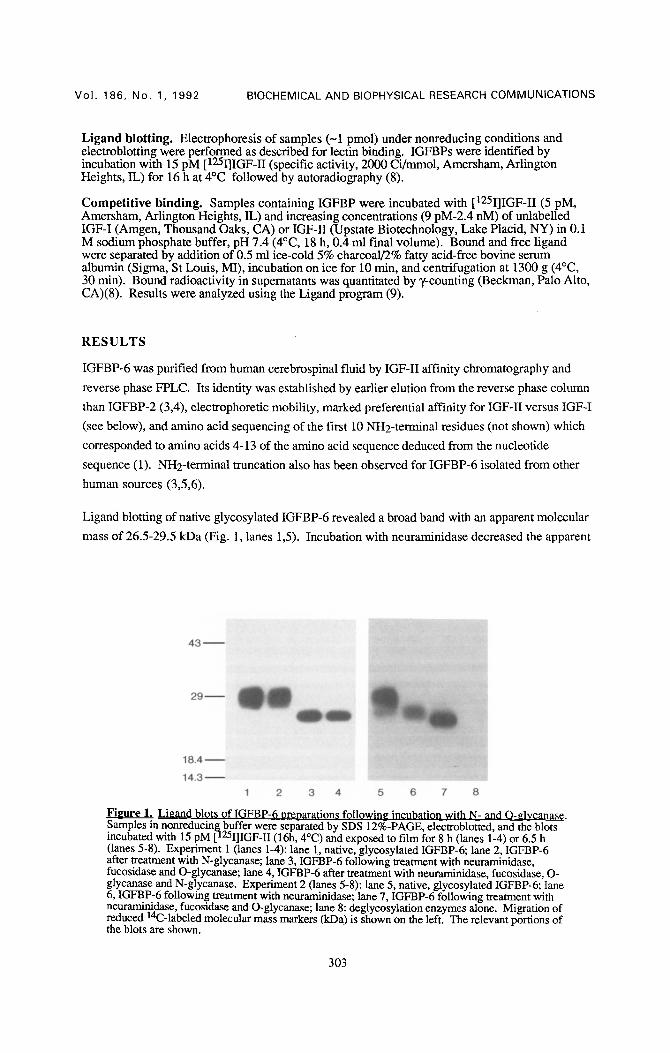
Figure 2, Lcctin binding to gly~osylated and deglycosylated IGFBP-6 oreparations. Samples in reducing buffer were separated by SDS 12%-PAGE, electroblotted and incubated with digoxigenin-coupled lectins. Binding was detected with antidigoxigenin antibody conjugated with alkaline phosphatase and subsequent color reaction. Binding to digoxigenin-coupled peanut agglutinin (lanes 1-3): lane 1, native, glycosylated IGFBP-6; lane 2, IGFBP-6 treated with neurarninidase; lane 3, IGFBP-6 following treatment with neuraminidase, fucosidase and O- glycanase. Binding to digoxigenin-coupled wheat germ agglutinin (lanes 4-6): lane 4, native, glycosylated IGFBP-6; lane 5, IGFBP-6 incubated with N-glycanase; lane 6, IGFBP-6 following treatment with neurarninidase, fucosidase and O-glycanase. Binding to digoxigenin-coupled concanavalin A (lanes 7-10): lane 7, Transferrin (-80 kDa), a positive control for concanavalin A binding; lane 8, native, glycosylated IGFBP-6; lane 9, IGFBP-6 incubated with N-glycanase; lane 10, IGFBP-6 following treatment with neuraminidase, fucosidase and O-glycanase. The bands seen at -50 kDa in lanes 6 and 10, and the faint bands seen at 31 and 43 kDa in lane 10 represent deglycosylation enzymes. Migration of reduced prestained molecular mass markers (kDa) for lanes 1-6 is shown on the left, and for lanes 7-10 on the right. The relevant portions of the blots are shown.
molecular mass to 25 kDa (Fig. 1, lane 6), whereas incubation with neuraminidase, fucosidase and
O-glycanase further decreased the apparent molecular mass to 23.5 kDa (Fig. 1, lanes 3,7). In
contrast, N-glycanase treatment did not affect the mobility of native or O-deglycosylated IGFBP-6
(Fig. 1, lanes 2,4). Incubation of IGFBP-6 with heat-inactivated enzymes did not alter
electrophoretic mobility (not shown).
Lectin blotting was performed to further characterize the carbohydrate content of IGFBP-6. Native
and enzymatically deglycosylated IGFBP-6 were fractionated by SDS-PAGE under reducing
conditions, blotted onto nitrocellulose membranes and incubated with digoxigenin-labeled lectins.
Peanut agglutinin did not bind to native glycosylated IGFBP-6 (Fig. 2, lane 1) but bound to
neuraminidase-treated IGFBP-6 (Fig. 2, lane 2), consistent with the removal of terminal sialic acid
residues and exposure of galactose [3(1-3) N-acetylgalactosamine, the core disaccharide of most O-
linked oligosaccharide chains. Following incubation of IGFBP-6 with neuraminidase, fucosidase
and O-glycanase, binding of peanut agglutinin was no longer observed, suggesting that O-
deglycosylation was complete (Fig. 2, lane 3). Wheat germ agglutinin bound to native and N-
glycanase-treated, but not O-glycanase treated, IGFBP-6 (Fig. 2, lanes 4-6), signifying that the O-
linked carbohydrate chains contained glucosamine. Concanavalin A did not bind to native or O-
glycanase treated IGFBP-6 (Fig. 2, lanes 7-10), indicating the absence of mannose in the
preparations. Since maxmose is an obligatory component of N-linked oligosaccharide chains, this
result verifies that IGFBP-6 is not N-glycosylated.
304
Vol. 186, No. 1, 1992 BIOCHEMICAL AND BIOPHYSICAL RESEARCH COMMUNICATIONS
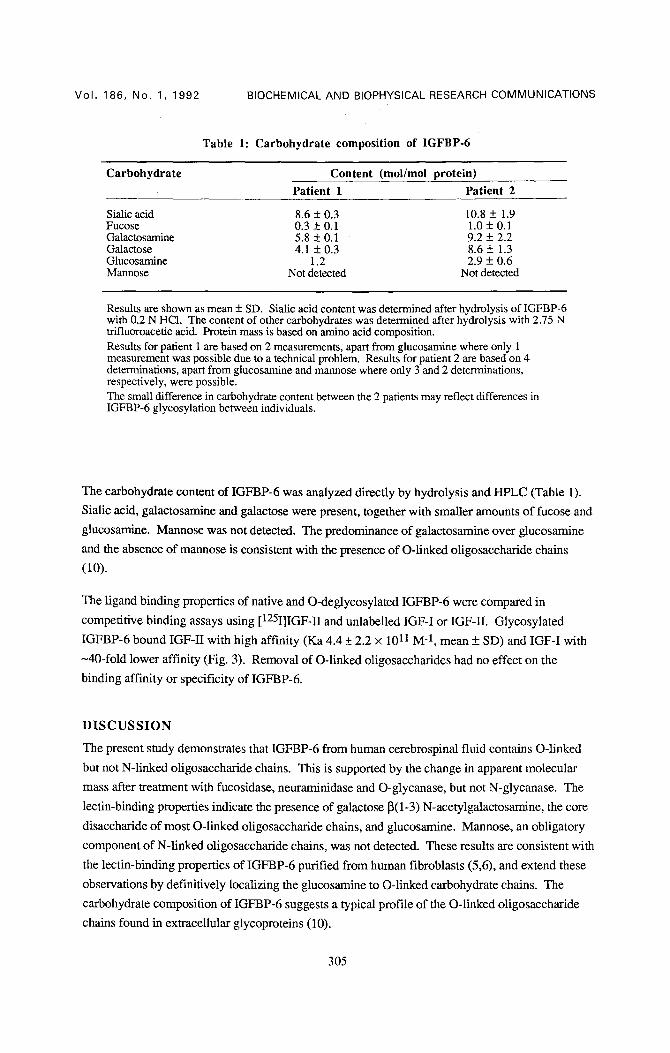
Table 1: Carbohydrate composition of IGFBP-6
Carbohydrate Content (mol/mol protein) Patient 1 Patient 2
Sialic acid 8.6 5:0.3 10.8 5:1.9 Fucose 0.3 5:0.1 1.0 + 0.1 Galactosamine 5.8 5:0.1 9.2 5:2.2 Galactose 4.1 5:0.3 8.6 5:1.3 Glucosamine 1.2 2.9 5:0.6 Mannose Not detected Not detected
Results are shown as mean + SD. Sialic acid content was determined after hydrolysis of IGFBP-6 with 0.2 N HC1. The content of other carbohydrates was determined after hydrolysis with 2.75 N trffiuoroacetic acid. Protein mass is based on amino acid composition. Results for patient 1 are based on 2 measurements, apart from glucosamine where only 1 measurement was possible due to a technical problem. Results for patient 2 are based on 4 determinations, apart from glucosamine and mannose where only 3 and 2 determinations, respectively, were possible. The small difference in carbohydrate content between the 2 patients may reflect differences in IGFBP-6 glycosylation between individuals.
The carbohydrate content of IGFBP-6 was analyzed directly by hydrolysis and HPLC (Table 1).
Sialic acid, galactosamine and galactose were present, together with smaller amounts of fucose and
glucosamine. Mannose was not detected. The predominance of galactosamine over glucosamine
and the absence of mannose is consistent with the presence of O-linked oligosaccharide chains
(10).
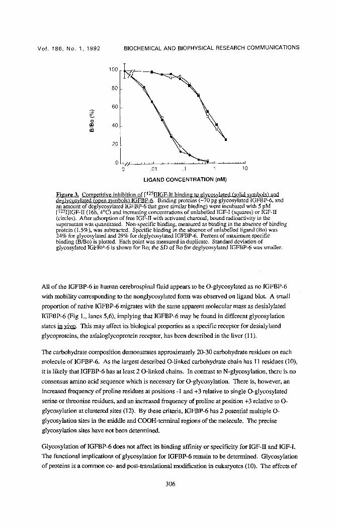
The ligand binding properties of native and O-deglycosylated IGFBP-6 were compared in
competitive binding assays using [125I]IGF-H and unlabelled IGF-I or IGF-II. Glycosylated
IGFBP-6 bound IGF-II with high affinity (Ka 4.4 _+ 2.2 x 10¹¹ M -1, mean _+ SD) and IGF-I with
~40-fold lower affinity (Fig. 3). Removal of O-linked oligosaccharides had no effect on the
binding affinity or specificity of IGFBP-6.
D I S C U S S I O N
The present study demonstrates that IGFBP-6 from human cerebrospinal fluid contains O-linked
but not N-linked oligosaccharide chains. This is supported by the change in apparent molecular
mass after treatment with fucosidase, neuraminidase and O-glycanase, but not N-glycanase. The
lectin-binding properties indicate the presence of galactose ~(1-3) N-acetylgalactosamine, the core
disaccharide of most O-linked oligosaccharide chains, and glucosamine. Mannose, an obligatory
component of N-linked oligosaccharide chains, was not detected. These results are consistent with
the lectin-binding properties of IGFBP-6 purified from human fibroblasts (5,6), and extend these
observations by definitively localizing the glucosamine to O-linked carbohydrate chains. The
carbohydrate composition of IGFBP-6 suggests a typical profile of the O-linked oligosaccharide
chains found in extracellular glycoproteins (10).
305
100
80
20
60
0 m 40
j j ,H I , , , , , =H I
0 .01 .1 1 10
Vo l . 186, No. 1, 1992 BIOCHEMICAL AND BIOPHYSICAL RESEARCH COMMUNICATIONS
LIGAND CONCENTRATION (nM)
Figure 3. Comoetitive inhibition of [125I]IGF-1I bindin~ to ~lvcosvlated (solid svmbols) and deglycosylated (open symbols) IGFBP-6. Binding proteins (L'10 pg glycosylatedIGFBP-6, and an amount of deglycos3~lated IGFBP-6 that gave similar binding) were incubated with 5 pM [125I]IGF-I/(16h, 4°C) and increasing concentrations of unlabelled IGF-I (squares) or IGF-II (circles). After adsorption of free IGF-II with activated charcoal, bound radioactivity in the supernatant was quantitated. Non-specific binding, measured as binding in the absence of binding protein (1.5%), was subtracted. Specific binding in the absence of unlabelled ligand (Bo) was 24% for glycosylated and 29% for deglycosylated IGFBP-6. Percent of maximum specific binding (B/Bo) is plotted. Each point was measured in duplicate. Standard deviation of glycosylated IGFBP-6 is shown for Bo; the SD of Bo for deglycosylated IGFBP-6 was smaller.
All of the IGFBP-6 in human cerebrospinal fluid appears to be O-glycosylated as no IGFBP-6
with mobility corresponding to the nonglycosylated form was observed on ligand blot. A small
proportion of native IGFBP-6 migrates with the same apparent molecular mass as desiaiylated
IGFBP-6 (Fig 1., lanes 5,6), implying that IGFBP-6 may be found in different glycosylation
states in vivo. This may affect its biological properties as a specific receptor for desialylated
glycoproteins, the asialoglycoprotein receptor, has been described in the liver (11).
The carbohydrate composition demonstrates approximately 20-30 carbohydrate residues on each
molecule of IGFBP-6. As the largest described O-linked carbohydrate chain has 11 residues (10),
it is likely that IGFBP-6 has at least 2 O-linked chains. In contrast to N-glycosylation, there is no
consensus amino acid sequence which is necessary for O-glycosylation. There is, however, an
increased frequency of proline residues at positions -1 and +3 relative to single O-glycosylated
serine or threonine residues, and an increased frequency of proline at position +3 relative to O-
glycosylation at clustered sites (12). By these criteria, IGFBP-6 has 2 potential multiple O-
glycosylation sites in the middle and COOH-ten'ninal regions of the molecule. The precise
glycosylation sites have not been determined.
Glycosylation of IGFBP-6 does not affect its binding affinity or specificity for IGF-II and IGF-I.
The functional implications of glycosylation for IGFBP-6 remain to be determined. Glycosylation
of proteins is a common co- and post-translational modification in eukaryotes (10). The effects of
306
Vol. 186, No. 1, 1992 BIOCHEMICAL AND BIOPHYSICAL RESEARCH COMMUNICATIONS
glycosylation are protean. Glycosylation may regulate intracellular protein trafficking (13). It may
affect protein conformation which in some instances may alter protein function such as ligand
binding, alter protein stability by conferring protection against proteases, or alter protein clearance
from the circulation (7,10,14). Receptors which recognise specific carbohydrate side chains in
glycoproteins, including the asialoglycoprotein receptor mentioned above (11), may have a role in
glycoprotein clearance (15). Glycosylation also may affect protein localization within tissues to the
cell surface or extracellular matrix (14). The latter may be of particular relevance for IGFBP-6 as
localization of IGFBPs may determine the way in which they modulate IGF action (16).
Acknowledgments: Leon Bach is the recipient of the J.J. Billings Travelling Fellowship of the Royal Australasian College of Physicians. We would like to thank Dr.E. Oldfield, NINDS, NIH, Bethesda, MD for providing cerebrospinal fluid samples and Dr.W. Burgess, American Red Cross, Rockville, MD for NH2-terminal amino acid sequence analysis and composition of IGFBP- 6, and Dr.B. Weintraub, NIDDK, NIH, Bethesda, MD for his critical reading of the manuscript.
R E F E R E N C E S
1. Shimasaki S., Gao L., Shimonaka M., and Ling N. (1991) Mol. Endocrinol. 5, 938-948. 2. Rechler M.M., and Brown A.L. (1992) Growth Regulation (in press). 3. Zapf J., Kiefer M., Merryweather J., et al. (1990) J. Biol. Chem. 265, 14892-14898. 4. Roghani M., Lassarre C., Zapf J., Povoa G., and Binoux M. (1991) J. Clin. Endocrinol.
Metab. 73, 658-666. 5. Martin J.L., Willetts K.E., and Baxter R.C. (1990) J. Biol. Chem. 265, 4124-4130. 6. Forbes B., Ballard F.J., and Wallace J.C. (1990) J. Endocr. 126, 497-506. 7. Thotakura N.R., Desai R.K., Bates L.G., et al. (1991) Endocrinology 128, 341-348. 8. Yang Y.W-H., Wang J-F., Orlowski C.C., Nissley S.P., and Rechler M.M. (1989)
Endocrinology 125, 1540-1555. 9. Munson P.J., and Rodbard D. (1980) Anal. Biochem. 70, 241-250. 10. Pan Y.T., and Elbein A.D. (1990) Progress in Drug Research 34, 163-207. 11. Ashwell G., and Hafford J. (1982) Ann. Rev. Biochem. 51,531-554. 12. Wilson I.B.H., Gavel Y., and von Heijne G. (1991) Biochem. J. 275,529-534. 13. Kuwano M., Seguchi T., and Ono M. (1991) J. Cell Science 98, 131-134. 14. Parekh R.B. (1991) Current Opinions in Structural Biology 1,750-754. 15. Drickamer K. (1991) Cell 67, 1029-1032. 16. Clemmons D.R., Camacho-Hubner C., Jones J.I., McCusker R.H., and Busby W.H. (1991)
In Modern Concepts of Insulin-like Growth Factors (E.M. Spencer, Ed), pp. 475-486. Elsevier, New York, NY.
307


