
INVESTIGATION OF BIOLOGICAL PROCESS FOR THE
CONVERSION OF BARK BIOMASS TO BIO-BASED
POLYPHENOLS
by
Muhammad Ferhan
A thesis submitted in conformity with the requirements
for the degree of Doctor of Philosophy
Faculty of Forestry
University of Toronto
© Copyright by Muhammad Ferhan 2016
ii
Investigation of biological process for the conversion of bark biomass to bio-based
polyphenols
Muhammad Ferhan
Doctor of Philosophy
Faculty of Forestry
University of Toronto
2016
Abstract
Due to increasing waste production and disposal problems arising from synthetic polymer production,
there is a critical need to substitute these materials with biodegradable and renewable resources. The
concept of green polymers has become more appealing due to the presence of large volumes of
processing residuals from the timber and pulp industries. This, in turn, supports the idea of developing
new polymers based on bark extractives. In this thesis, three comparative treatments i.e., enzymatic,
alkaline, and UV/H2O2, have been conducted for the extraction of beetle infested lodgepole pine
(BILP) and mixed aspen barks polyphenolic extractives. Use of laccases as biocatalysts to affect and
enhance the catalytic properties of enzymes has been shown to be a promising solution for bark
depolymerization. Furthermore, laccases are suitable for biotechnological applications that transform
bark biomass into high valued bark biochemicals. The industrial and biotechnological application of
ligninases is constantly increasing due to their multiple uses and applications in a diversity of
processes. Bark depolymerization was conducted in submerged fermentation (SF) and we identified
polyphenols/polyaromatic compounds after four weeks when the production media (PM) was induced
with 50mg/100ml of each type of bark during the lag-phase. During SF where honey was used as a
natural mediator substitute (NMS) in the PM, laccase activities were about 1.5 times higher than those
found in comparable cultures without honey in the PM. These samples were analyzed by GC-MS. The
laccase enzyme was purified using UNO® sphere Q-1 anion exchange chromatography and the
molecular weight was determined to be ~50kDa on 10% SDS-PAGE and laccase kinetic parameters
including maximal velocity (Vmax), Michaelis constant (Km), and turnover number (Kcat) were
calculated from a Lineweaver Burk plot. All calculated kinetic parameters of the laccase activity
are substrate (ABTS) specific. Py-GC-MS analysis of bark showed differing effects of fungal
activity on bark composition. Polyphenolics were separated in reverse-phase mode using HPLC with
two selected wavelengths of 290 and 340 nm to improve separation. The replacement of conventional
natural mediators (NM) by monofloral honey in production media, and investigation of the effect of
fungi-derived laccases on bark polyphenols are studied for the first time by this thesis work.

iii
Acknowledgments
In the name of Allah, the most Beneficent, the most Gracious, the most Merciful!!!
Thank you GOD for blessing me much more than I deserve. After GOD’s countless blessings, I
would like to express my sincere gratitude and appreciation to my respectable supervisor Prof.
Mohini M. Sain and co-supervisor Prof. Ning Yan for their guidance, and relentless support
throughout this research thesis. I would also like to acknowledge the valuable advice of
advisory board members, Profs. Martin Hubbes and Bradley Saville for their valuable
suggestions during my research work.
My heartfelt thanks to all the technical and administrative staff members for their assistance in
all possible ways which helped regarding accomplish my thesis in befitting manner. I also
would like to thanks to ORF-RE Bark Biorefinery Project and industry partners for their
financial support in carrying out this research work.
Special thanks are given to my best friend, Arslan Akhtar (late), Z. Iqbal, Asif, Adnan, Umar
and Uncle Hanif. Despite being far away, their constant support made my life in Canada much
easier. I cannot imagine what my life would have been without their help. I will remember their
kindness forever.
Special thanks to my family, my parents, brothers, sisters, nephew and niece for their
continuous prayers, moral support and patience during my research work.
Finally, special thanks to my all friends, lab colleagues, especially thanks to M. Pervaiz, S. Kunar,
S. Law, Suhara, Jason, Javad, Sadia, Scott, Greg, Jon Obnamia, J. Kaal, Mariana, and all my lab
colleagues for their encouragement, having lively discussions and helping in all possible ways to a
successful end of this project. In addition, would like to extend my thanks to administrative staff,
Deborah, Amalia, Susana, Ian and Tony.
At the end, I would like to share an elegy which I wrote in memory of my beloved friend
Arslan Akhtar, who passed away in April 6, 2014. May Allah bless his soul in heaven and save
him from the fire of hell, Aameen!!!

iv
Table of Contents
Acknowledgments ..................................................................................................................... iii
Table of Contents ...................................................................................................................... iv
List of Tables ............................................................................................................................ ix
List of Figures ............................................................................................................................ x
List of Acronyms ..................................................................................................................... xvi
List of Appendices................................................................................................................... xix
1 Chapter: Introduction ............................................................................................................. 1
1.1 Motivation and Significance ........................................................................................... 1
1.2 Scope of Research work .................................................................................................. 4
1.3 Hypothesis ...................................................................................................................... 4
1.4 Research Objectives ........................................................................................................ 5
1.5 Thesis overview .............................................................................................................. 5
2 Chapter: Literature review ..................................................................................................... 8
2.1 Bark ................................................................................................................................ 8
2.2 Chemical configuration ................................................................................................... 8
2.3 Bark polyphenolics ......................................................................................................... 9
2.3.1 Lignin ................................................................................................................. 9
2.3.2 Lignin valorization ............................................................................................ 10
2.3.3 Tannins ............................................................................................................. 11
2.4 Challenges in extraction of bark polyphenolics ............................................................. 13
2.4.1 Chemical and physical properties of polyphenolics ............................................ 13
2.4.2 Polyphenol–protein interactions ........................................................................ 15
2.4.3 Interaction of low molecular weight (LMW) phenolics ...................................... 17
2.5 Biopulping .................................................................................................................... 17

v
2.5.1 Role of ligninases in pulp delignification ........................................................... 18
2.5.2 Oxidation of PAHs by synthetic LMS ............................................................... 18
2.6 Fungal extracellular ligninases ...................................................................................... 19
2.6.1 Reaction mechanism of ligninases ..................................................................... 21
2.6.2 Laccase–Mediator Systems (LMS) .................................................................... 23
2.6.3 Natural mediators / phenolic enhancer ............................................................... 26
2.6.4 Biotransformation ............................................................................................. 26
2.7 Function of laccases with combination of natural mediators (NMs) for lignin
biodegradation .............................................................................................................. 28
2.7.1 Monofloral honey as mediator ........................................................................... 30
2.8 Usage of laccases and NM for future lignocellulosic biorefineries ................................. 30
3 Chapter: Experimental Methodology.................................................................................... 32
3.1 Chemicals and standards ............................................................................................... 32
3.2 Chemical Composition of Bark before and after Extraction ........................................... 33
3.2.1 Ash Contents in Bark ........................................................................................ 33
3.2.2 Preparation of Extractive-Free Bark .................................................................. 33
3.2.3 Lignin Content of Bark ...................................................................................... 33
3.2.4 Holocellulose and α-cellulose content of bark .................................................... 34
3.2.5 Enzymatic treatment .......................................................................................... 35
3.2.6 Alkaline Treatment ............................................................................................ 35
3.2.7 UV/H2O2 treatment ........................................................................................... 36
3.2.8 Sample preparation for HPLC ........................................................................... 36
3.2.9 HPLC analysis................................................................................................... 36
3.2.10 Folin-Denis method ........................................................................................... 37
3.2.11 SEM for BILP and Aspen bark .......................................................................... 37
3.3 Fungal strain isolation ................................................................................................... 38

vi
3.3.1 Substrate collection and preparation .................................................................. 39
3.3.2 Fermentative organism and culturing conditions ................................................ 39
3.3.3 Quantification of dye Remazol Brilliant Blue R (RBBR) oxidation in liquid
culture medium ................................................................................................. 39
3.3.4 Growth Media preparation ................................................................................. 40
3.3.5 Optimization of temperature and pH for MnP activities ..................................... 40
3.4 Ligninolytic enzyme assays........................................................................................... 40
3.5 18S rDNA amplification and sequencing ...................................................................... 41
3.6 Ligninases cocktail partial purification and characterization .......................................... 42
3.6.1 Scanning electron microscopy (SEM) ................................................................ 43
3.6.2 Determination of total proteins .......................................................................... 43
3.6.3 Gel electrophoresis and staining ........................................................................ 43
3.6.4 MS Analysis to determine delignification pattern .............................................. 43
3.7 Inoculum preparation .................................................................................................... 44
3.8 Monofloral honey used as a NM-substitute in PM ......................................................... 45
3.8.1 Time course study during SF ............................................................................. 45
3.9 Laccase purification by UNO® sphere Q-1 column ....................................................... 45
3.9.1 Laccase kinetics ................................................................................................ 46
3.10 Characterization techniques .......................................................................................... 46
3.10.1 Estimation of total polyphenolics....................................................................... 46
3.10.2 Sample preparation for HPLC ........................................................................... 47
3.11 HPLC Analysis ............................................................................................................. 47
3.12 Thermal Analysis .......................................................................................................... 48
3.13 GC-MS/MS .................................................................................................................. 48
3.14 MALDI-TOF-MS ......................................................................................................... 49
3.15 Sample preparation for Py-GC-MS ............................................................................... 49

vii
3.15.1 Py-GC-MS ........................................................................................................ 49
4 Chapter: Comparative treatments of bark to characterize bark .............................................. 51
polyphenols .............................................................................................................................. 51
4.1 Abstract ........................................................................................................................ 51
4.2 Introduction .................................................................................................................. 51
4.3 Results and discussion .................................................................................................. 54
4.3.1 Bark composition .............................................................................................. 54
4.3.2 Treated bark composition .................................................................................. 56
4.4 Conclusions .................................................................................................................. 63
5 Chapter: Brazilian isolated fungal strain for ligninases ......................................................... 65
production ................................................................................................................................ 65
5.1 Abstract ........................................................................................................................ 65
5.2 Introduction .................................................................................................................. 65
5.3 Results and Discussion.................................................................................................. 66
5.3.1 Ligninases production during time course studies .............................................. 67
5.3.2 Purification of MnP ........................................................................................... 70
5.3.3 Electron microscopy .......................................................................................... 72
5.3.4 MALDI-TOF MS .............................................................................................. 74
5.4 Conclusions .................................................................................................................. 76
6 Chapter: Optimization of temperature and pH for Manganese ............................................. 78
Peroxidase (MnP) ..................................................................................................................... 78
6.1 Abstract ........................................................................................................................ 78
6.2 Introduction .................................................................................................................. 78
6.3 Results and Discussion.................................................................................................. 79
6.3.1 Ligninases production during the time course studies ........................................ 79
6.4 Conclusions .................................................................................................................. 84

viii
7 Chapter: Depolymerization of bark with fungal laccases ..................................................... 85
into bark-derived polyphenols and comparative studies ............................................................ 85
between phenolic mediator vs. honey as a natural ..................................................................... 85
mediator substitute ................................................................................................................... 85
7.1 Abstract ........................................................................................................................ 85
7.2 Highlights ..................................................................................................................... 86
7.3 Introduction .................................................................................................................. 86
7.4 Results and discussion .................................................................................................. 88
7.4.1 Extracellular laccases and screening of potential strain ...................................... 88
7.4.2 Laccase purification and kinetics ....................................................................... 92
7.4.3 Bark decomposition by TG/DTG ....................................................................... 94
7.4.4 Mass spectrometry (GC-MS/MS, MALDI-TOF/MS)......................................... 95
7.4.5 Honey in PM as natural mediator substitute (NMS) ..........................................103
7.4.6 Polyphenolic chromatograms............................................................................105
7.4.7 Py-GC-MS .......................................................................................................109
7.5 Conclusions .................................................................................................................117
8 Chapter: Conclusions and Future work................................................................................118
8.1 Major contributions ......................................................................................................118
8.2 Recommendations for Future Work .............................................................................120
References ...............................................................................................................................122
Appendix .................................................................................................................................142

ix
List of Tables
Table 2.1: General chemical configuration in wood and bark for hardwoods and softwoods
(Harkin et al., 1971)................................................................................................................ 8
Table 4.1: Chemical composition of beetle infested lodgepole pine and aspen bark. ............. 54
Table 4.2: Extraction yield from soluble part and bark residues (insoluble part) of beetle
infested lodgepole pine (BILP) and aspen bark after extraction with different methods. ........ 56
Table 5.1: Measurement of ligninases activities during the time course study in three different
fungal strains. ....................................................................................................................... 68
Table 5.2: Partial purification summary of MnP produced during submerged fermentation by
isolated fungal strain under optimized conditions.................................................................. 72
Table 7.1: Bark polyphenols/polyaromatics identification. .................................................... 98
Table 7.2: Total polyphenolics in fermented sample when induced with 50mg of each bark
into 100ml of PM during lag-phase. .................................................................................... 109
Table 7.3: List of main pyrolysis products identified from the untreated/control, MS treated
........................................................................................................................................... 111
Table 7.4: Main pyrolytic compound groups and their cumulative relative proportions (%
TQPA). The calculated (% TQPA) values of each group were based on Table 7.3. ............. 112

x
List of Figures
Figure 1.1: Thesis overview and organization of thesis chapters. ............................................ 7
Figure 2.1: Phenylpropenoid units in lignin precursors ......................................................... 10
Figure 2.2: Conceptual theme of lignin valorization and application of this renewable resource
for value-added products by converting into carbon fibers, biopolymers, biochemicals, and
biofuels (Source: Oak Ridge National Laboratory, U.S. Department of Energy, Referred by:
Ragauskas, Science Reviews, 2014 with permission.) ........................................................... 11
Figure 2.3: Flavonoid unit .................................................................................................... 12
Figure 2.4: Wattle and pine tannin unit ................................................................................. 13
Figure 2.5: Structures of major identified polyphenols in bark (Source: Shi, J. et al., Food Rev
Int, 2005 with permission.) ................................................................................................... 14
Figure 2.6: A model for protein−polyphenol interactions that elucidates having two sides that
can join to protein. Proteins are defined as having a fixed number of polyphenol binding sites
(Source: Siebert, K.J et al., J. Agric. Food Chem, 1996 with permission.) ............................. 16
Figure 2.7: Degradation of oligomeric lignin model compounds by P. chrysosporium
ligninases (A) β-1 and (B) β-O-4 (Proposed by Tien and Kirk 1984, Proc Narl Acad. Sci.
USA., with permission.) ........................................................................................................ 21
Figure 2.8: Structural groups resulting from endways linking (Source: Wong, 2009, Appl
Biochem Biotechnol, with permission.) ................................................................................. 22
Figure 2.9: The sequential catalytic cycle of LMS (Source: Fabbrini, 2002, J Mol Catal B-
Enzym, with permission.) ...................................................................................................... 23
Figure 2.10: N–OH type laccase mediators and their chemical structures (A-E); A=1-
Hydroxybenzotriazole (HBT); B= N-Hydroxyphthalimide (HPI); C=N-Hydroxyacetanilide
(HAA); D= Violuric acid (VA); E= 2,2,6,6-Tetramethylpiperidin-1-oxyl (TMPO). .............. 25

xi
Figure 2.11: Oxidation of ABTS in presence of laccase (Source: Morozova, 2007, Appl
Biochem Microbiol Reviews, with permission.)..................................................................... 25
Figure 2.12: The catalytic cycle of LMS showing different paths implicated in development of
several phenolic species (Source: Rosado et al., Bioresource Technol, 2012 with permission.)
............................................................................................................................................. 27
Figure 2.13: Non-phenolic lignin model compound oxidation by laccase mediator system
(LMS) ensuing by two different oxidation methods: ET (electron transfer) and HAT (hydrogen
atom transfer) (Source: Fabbrini et al., J. Mol. Catal. B: Enzym, 2002 with permission.) ...... 28
Figure 2.14: Conceivable function of laccases with combination of phenolic natural mediators
(NMs) in decontamination of industrial effluents and soil bioremediation (Source: Cañas et al.,
Biotechnol Adv, 2010 with permission.) ................................................................................ 29
Figure 2.15: Strategic plan of Integrated Lignocellulosic Forest Products Biorefinery (ILFPB):
where, (1) different lignocellulosic reserves; (2) extraction of hemicelluloses to pulping; and
(3) biomass gasification. The main features where enzyme technology may help twice as
notably to this idea are also shown: (a) assembling of wood (or lignocellulose) products; (b)
create of more proficient and environmentally friendly industrial procedures for pulp and
paper production; (c) extraction of phenolic mediators and reusing of black liquors; (d)
production of cellulosic products with modified characteristics; or (e) enhanced biofuel
production (Source: Cañas et al., Biotechnol Adv, 2010 with permission.) ............................ 31
Figure 3.1: Design of strategic plan for objective-1. Reproduced by the permission of
American .............................................................................................................................. 32
Figure 3.2: Design of strategic plan for objective-2 ............................................................... 38
Figure 3.3: Design of strategic plan for objectives 3 & 4. Reproduced by the permission of.. 44
Figure 4.1: Bark residue composition of Beetle Infested Lodgepole Pine (BILP) and Aspen
after three different treatments. ............................................................................................. 57
Figure 4.2: Scanning electron microscopy of beetle-infested lodgepole pine (BILP) and aspen
barks at different magnifications. (A) Control BILP bark (magnification, x 3,500), (B) treated

xii
with alkaline (magnification, x 1,500), (C) treated with cocktail of enzymes (magnification, x
1,000), and (D) treated with UV/H2O2 (magnification, x 1,000). (E) Control aspen bark
(magnification, x 2,000), (F) treated with alkaline (magnification, x 1,000), (G) treated with
cocktail of enzymes (magnification, x 1,000), and (H) treated with UV/H2O2 (magnification, x
2,200). .................................................................................................................................. 59
Figure 4.3: Chromatograms of BILP bark (A, B, C) and aspen bark (D, E, F) polyphenolic
extractives.1 .......................................................................................................................... 62
Figure 5.1: Potential screening of three fungal strains out of ten based on the ligninases
production and degradation of RBBR dye, where, A - Pleurotus ostreatus POS 97/14, B-
Pycnoporus sanguineus and C was Brazilian isolated fungal strain. ...................................... 67
Figure 5.2: Size exclusion chromatography of MnP from isolated fungal strain (A): DEAE
fractionations from isolated fungal strain (B): MnP activity was eluted as a single peak. ...... 70
Figure 5.3: Polyacrylamide gel electrophoresis (5.3A): Ligninases with reduction and non-
reduction conditions (5.3B): Purified MnP from isolated fungal strain stained with Coomassie
Blue R-250. .......................................................................................................................... 71
Figure 5.4: Scanning electron microscopy of sugarcane bagasse (SCB) and Black liquor (BL)
of Eucalyptus. (A & B) Control SCB (magnification, x 400); (C & D) bagasse treated with
Brazilian isolated fungal strain grown after 2 weeks (magnification, x 2k); (E & F) Control BL
of Eucalyptus fibers (magnification, x 10k); (G & H) BL treated with local fungal isolate
grown after 2 weeks (magnification, x 5k); indicates the fungal growth where k denoted by
(x1000 magnification). ......................................................................................................... 74
Figure 5.5: MW distribution to determine the lignin monomers by using MALDI-TOF-MS
where α-cyano-4-hydroxy cinnamic acid was used as matrix compound. .............................. 76
Figure 6.1: Time course for ligninases production and cell biomass produced by Dothioraceae
sp. ........................................................................................................................................ 80
Figure 6.2: Plot between incubation time (days) vs. biomass (g/100 ml flask) production
among three fungal strains. ................................................................................................... 81
xiii
Figure 6.3: Effect of Temperature on MnP production; the test shake-flasks were inoculated
with Brazilian isolated fungal strain (Dothioraceae sp) and incubated at different temperatures
for 12 days in shake flasks. The maximum MnP activities (IU/L) and cell biomass (g/100ml)
were observed at 10th day of inoculation and plotted vs. different temperatures between (25-35
°C). Error bars represent the mean values of ± standard deviation from three replicates of
shake-flak experiments. All values were differed significantly at p˂0.05. ............................. 82
Figure 6.4 : Effect of pH on MnP production; the test flasks were inoculated with Brazilian
isolated fungal strain (Dothioraceae sp) and incubated at 30°C for 12 days in shake flasks.
The maximum MnP activities (IU/L) and biomass (g/100ml) were observed at 10th day of
inoculation and plotted against vs. different pH range (4.5-7.0). Error bars represent the mean
values of ± standard deviation from three replicates of shake-flak experiments. All values were
differed significantly at p˂0.05. ............................................................................................ 83
Figure 7.1: Representative time course of extracellular laccase activity (IU/ml/min) in the
production medium on glucose (10g/L) in shake flask cultures (pH 5.5, 30°C, 150 rpm)
following the growth of six different fungal strains induced with aspen bark during lag-phase
including: (◊) P. pulmonaris, (□) P. cornucopiae, (+) P. ostreatus, (○) P. chrysosporium, (▲)
T. versicolor, and ( ) G. mangiferae. Each point is a mean of three independent experiments.
Bars indicate standard deviation among three replicates. ....................................................... 89
Figure 7.2: Different fungal strains growth on balsam fir (A-E) and mixed aspen bark (F-J)
while, P. cornucopiae, G. mangiferae, P. pulmonarius (K-M) were exhibiting laccase activity
on agar plates containing (2.5% malt extract +0.05% RBBR). From (N-P) shows fungal
growth on the bark surface and (Q-R) presenting bark and P. pulmonarius surface morphology
was observed under AmScope-WF25X/9 (magnification 0.5X). ........................................... 92
Figure 7.3: Laccase purification using FPLC by UNO® sphere Q-1 anion exchange column
where (A-D) represents sequential purification steps. ........................................................... 93
Figure 7.4: Purified laccase molecular weight was determined on 10% SDS-PAGE from P.
pulmonarius stained with Coomassie Blue R-250 showed Mw~50kDa. Lanes 1–2 are serially
purified laccase enzyme, lane-3 shows purified laccase enzyme, Pp = total protein from
Pleurotus pulmonarius, Pc = total protein from Pleurotus cornucopiae, and TS26616 for

xiv
protein standard marker shown in (7.4A); Lineweaver-Burk plot for the calculation of laccase
kinetic parameters (7.4B). ..................................................................................................... 93
Figure 7.5: TG/DTG curves for control/untreated (solid-lines) and Pleurotus pulmonarius ... 95
Figure 7.6: TIC from GC-MS/MS of identified compounds w.r.t. their tr-values characterized
from both control and each treated bark species from Pleurotus pulmonarius are listed in
Table 7.1. ............................................................................................................................. 96
Figure 7.7: MWD of each bark treated by P. pulmonarius from MALDI-TOF/MS, where α-
cyano-4-hydroxy cinnamic acid was used as the matrix compound. .................................... 103
Figure 7.8: Time course study for laccase activity (IU/ml/min) in P. pulmonarius during SF
containing 5, 7 and 10% (v/v) of monofloral honey in production media mainly used as a
natural mediator substitute induced with 50mg of each bark in 100ml of PM during lag-phase
(A)-Balsam fir, (B)-Mixed aspen bark, and (C)-1% Methyl syringate as natural phenolic
mediator. Each point is a mean of three independent experiments. Bars indicate standard
deviation among three replicates. ........................................................................................ 105
Figure 7.9: HPLC chromatograms phenolic profiles of bark fermented samples at 7% honey in
production media (a-d at λ290) and (e-h at λ340). Peak identification: methyl syringate
(12.017), pinobanksin (13.100), 8-methoxykeampferol (24.567), pinocembrin (36.467),
chrysin (39.62), pinocembrin 7-Me (55.217), tetochrysin (57.142). All major peaks were
characterized based on their tr values of honey polyphenolics previously reported by
Pyrzynska et al. (2009). ...................................................................................................... 106
Figure 7.10: Total ion chromatograms (TIC) of untreated (control) bark samples of aspen and
........................................................................................................................................... 110
Figure 7.11: Partial ion chromatograms (PIC) of selected compounds of polyphenolic origin
from each bark fermented samples in PM with monofloral honey used as NMS analyzed by
Py-GC-MS. Selected polyphenolic compounds are: guaiacol (m/z 109+124), 4-
methylguaiacol (m/z 123 + 138), 4-vinylguaiacol (m/z 135 + 150) and syringic acid methyl
ester (m/z 181 + 212). Chromatograms were plotted between relative peak intensity vs.
retention time (tr). ............................................................................................................... 114

xv
Figure 7.12: Representation of the Lignin/Carbohydrate index (A) and Syringol/Guaiacol ratio
(B) for the different samples analyzed. ............................................................................... 116
xvi
List of Acronyms
ABTS 2,2 -azino-bis (3-ethylbenzothiazoline-6-sulphonic acid)
ALIPH Aliphatic
BF Balsam fir
BILP Beetle infested lodgepole pine
BL Black liquor
bnt Billion tons
BSA Bovine serum albumen
CARB Carbohydrate
α-C Alpha-cellulose
cat Catechol
CFIA Canadian Food Inspection Agency
DIPOA Departamento Nacional de Inspecão de Productos de Origen Animal
DEAE Diethylaminoethyl
DHB 2,5-dihydroxy benzoic acid
DTG Derivative thermogravimetry
FPLC Fast Protein liquid chromatography
FS Fermented samples
GC/MS Gas chromatography-mass spectrometry
gluc Glucose

xvii
H Honey
HC Holocellulose
HBT 1-hydroxybenzotriazole
HPLC High performance liquid chromatography
IU International unit
Kcat Turnover number
Km Michaelis constant
KL Klason lignin
Lcc Laccase
LGB Lignocellulosic green biotechnology
LIG Lignin
LiP Lignin peroxidase
LMS Laccase mediator system
LMW Low molecular weight
m/z Mass to charge ratio
MAH Monocyclic aromatic hydrocarbon
MALDI-TOF/MS Matrix assisted laser desorption ionization-time of flight-mass spectrometry
MnP Manganese peroxidase
MS Methyl syringate
NCOMP Nitrogen containing compound
NMS Natural mediator substitute

xviii
PAHs Polycyclic aromatic hydrocarbons
PCR Polymerase chain reaction
PHEN Phenol
PIC Partial ion chromatogram
PM Production medium
Py-GC/MS Pyrolysis gas chromatography-mass spectrometry
RBBR Remazol brilliant blue-R
RT Retention time
SDS-PAGE Sodium dodecyl sulfate-polyacrylamide gel electrophoresis
SCB Sugarcane bagasse
S.D Standard deviation
SEM Scanning electron microscope
SESQUI Sesquiterpenoid
SF Submerged fermentation
TGA Thermogravimetric analysis
TIC Total ion chromatogram
TP Total phenolics
Vmax Maximal velocity or reaction rate

xix
List of Appendices
1. Ferhan, M., Yan, N. and Sain, M. (2015) Bark depolymerization during submerged
fermentation using monofloral honey, a natural mediator substitute, and integration between
laccases vs. bark biopolymers, characterized by Py-GC-MS. RSC Adv. 5: 14937–14952.
DOI: 10.1039/C4RA13841D Published online: December 12, 2014
http://pubs.rsc.org/en/content/articlelanding/2014/ra/c4ra13841d#!divAbstract
2. Ferhan, M., Tanguy, N., Yan, N., Sain, M. (2014) Comparison of enzymatic, alkaline and
UV/H2O2 treatments for extraction of beetle infested lodgepole pine (BILP) and aspen bark
polyphenolic extractives. ACS Sustainable Chem Eng. 2: 165–172.
DOI: 10.1021/SC400184f. Publication Date (Web): October 22, 2013
http://pubs.acs.org/doi/abs/10.1021/sc400184f?journalCode=ascecg
3. Ferhan, M., Yan, N., and Sain, M. (2013) A new method for demethylation of lignin from
woody biomass using biophysical methods. J Chem Eng Process Technol. 4: 160
DOI:10.4172/2157-7048.1000160
http://omicsonline.org/a-new-method-for-demethylation-of-lignin-from-woody-biomass-
using-biophysical-methods-2157-7048.1000160.pdf
4. Ferhan, M., Santos, S. N. (2013) Identification of a potential fungal species by 18S
rDNA for ligninases production. World J Microbiol Biotechnol. 29: 2437-2440.
http://www.ncbi.nlm.nih.gov/pubmed/23744034
5. Ferhan, M., Leao, A.L., Itamar, S de Melo., Yan, N., Sain, M. (2012) Ligninases
production and partial purification of MnP from Brazilian fungal isolate in submerged
fermentation. Ferment Technol. 1:5, DOI:10.4172/2167-7972.1000106
http://www.omicsgroup.org/journals/ligninases-production-and-partial-purification-of-
mnp-from-brazilian-fungal-isolate-in-submerged-fermentation-2167-
7972.1000106.php?aid=19320

1
1 Chapter: Introduction
1.1 Motivation and Significance
This chapter describes background, perspective and composition of this thesis. Biotechnology is a
promising technology for conversion of biomass materials to value-added products. In this thesis,
integrated biological methods are developed in order to obtain bark-derived chemicals for
industrial applications.
Worldwide yearly phenol production has increased steadily (almost 2.5% per year), climbing
from 8.34 M tons in 2010 to in excess of 8.9 M tons in 2012. The five main phenol-producing
states in 2012 were China, USA, Japan, Taiwan and South Korea with a consolidated
production of over 5.5 M tons. Ineos Phenol, Mitsui Chemicals, SINOPEC, Cepsa Quimica,
Shell Chemical, LG Chem, Honeywell Resins & Chemicals, Versalis S.p.a., Formosa
Chemicals and Fiber Corp, Kumho P&B Chemicals, and Sabic Innovative are the main
producing companies. The major global firms involved in the phenol market manufacture
plastics. Overall yearly production of phenol was expected at over 11.5 M tons in 2012
(Merchant Research and Consulting Ltd Report, 2014).
Chemicals derived from renewable feedstock, e.g., agricultural wastes, lignocellulosic biomass,
pulp and paper mill effluents or biomass microorganisms make up the green chemicals market.
Market contributors are progressively encountering the need to make a change from
petrochemical feedstock to renewable feedstock, on the grounds that the chemicals business
sector is subject to the instability in unrefined petroleum costs (Market Publishers Report
Database, 2013).
Lignocellulosic biodegradation is a part of the carbon cycle. Development of humus (humic
acids) results from decayed lignin deposits through microbial degradation of biomass.
Breakdown of lignin in wood allows access to carbohydrates via attack of white-rot fungi on
the cell wall. Bond cleavage in lignin by enzymes supported by other ecological impacts (light,
temperature, and moisture) occurs in rotting woods or bacterial composting media (Hammel

2
1997). Production of free radicals results in cleavage of linkages among lignin molecules by
naturally occurring lignolytic enzymes (laccases, peroxidases) in white-rot fungi.
Tree bark contains naturally occurring polyphenolic compounds such as tannins. As a result of
having numerous biological functions, the polyphenolics are auxiliary metabolites and have a
wide range of properties, including anti-carcinogenic, antioxidant, antiviral, and anti-
inflammatory properties (Noferi et al., 1997). Tannins have low susceptibility to oxidation
because of their polyhydroxylated nature and are generally moderately insoluble in lipophilic
media, which conceivably restricts their applications. Diverse alteration techniques have been
investigated including esterification to enhance both their lipophilic solvency and relative
stability. Both chemical and enzymatic acylation methodologies, used to combine different
polyphenolic esters including longer chain unsaturated fatty acids, have been described (Suresh
Babu et al., 2005).
The significant source for lignin degrading enzymes is microorganisms. Ligninolytic enzymes
manufactured from distinctive microbial sources have been thoroughly recognized. For lignin
degrading enzymes, fungi are considered the most effective source. The best known producers
of these enzymes are the white rot fungi in contrast to brown rot and the soft rot fungi
(Niladevi 2009).
By going with product divisions, the enzyme industry in United States is a multi-million dollar
business. Products include: proteases, polymerases, carbohydrases and ligninases. The
particular end clients are in distinctive fields like: pharmaceuticals & diagnostics,
research/biotechnology, and other applications.
Forty-three main research groups are involved in industrial enzyme production as numerous
key contributors, e.g., Novozymes, DSM, Genzyme Corporation, Life Technologies
Corporation, Meteoric Life Sciences, National Enzyme Company, Roche Holding AG-
Genentech, Inc., Advanced Enzymes Technologies Ltd., BBI Solutions, Chr. Hansen Holding
AS, Codexis, Inc., Advanced Vital Enzymes Ltd., Amano Enzyme Inc., AST Enzymes,
Biocatalysts Ltd., and Specialty Enzymes & Biotechnologies Co. Market analysis and statistics
originate from main and auxiliary research study (Report Linker 2013).

3
Developments in genetic and protein engineering work are towards improving the stability,
specificity, and general application of laccase as a commercial enzyme. It is not surprising that the
total of viable applications of this enzyme, not only in textile industry, is expanding consistently,
when all the benefits of utilizing laccase are contemplated. The principle clients of the laccase-
based advances are developed enterprises, for example, Lion Corporation (Japan), L’Oréal
(France), Novo Nordisk (Denmark), and Henkel (Germany), and in addition, the textile industry
has been rapid to adopt new catalysts (Polak et al., 2012).
Different aromatic and phenolic compounds can be utilized as dyes, particularly within the textile
industry, through catalytic oxidation of laccases yielded from diverse sources. Laccases are
considered as green catalysts/enzymes, in biotechnological processes, because of their wide range
of substrates, versatile biochemical properties and high protein stability, and are useful for more
applications in textile, pulp and paper industries (Polak et al., 2012).
In honey, the main antioxidant compounds are polyphenolics, flavonoids, enzymes (catalase,
glucose oxidase), organic acids, ascorbic acid, carotenoid-like substances, amino acids and
proteins (Gheldof et al., 2002). Furthermore, it has been reported that manuka honey contains a
high amount of several phenolic compounds, such as syringic acid, 4-methoxyphenyllactic acid,
methyl syringate, kojic acid, 5-methyl-3-furancarboxylic acid, leptosin, and unedone. These
compounds were useful for characterizing manuka honey from the other kinds of investigated
monofloral honeys (Oelschlaegel et al., 2012).
The Canadian forest sector is the world’s second largest supplier of woody biomass, behind the
United States and as such, an annual supply of more than 200 million m3 of biomass through
commercial operations is reported (FAO, 2003; NRCAN, 2003). The pulp and paper industry is
the major end user of the forest biomass and has been a major contributor to the North American
economy for many years. Recently, the pulp and paper sector has been experiencing a downturn
due to global influence of concurrent impacts such as competition from paper industries producing
fibers from fast growing species, the high cost of the energy, and the reduced demand for news
print and pulp (Helmerius et al., 2010).
The pulp and paper industry needs to identify additional economic strategies in the future to
revitalize the forest sector and to strengthen its competitiveness in the current global market. The
4
concept of an integrated forest biorefinery has been suggested as an opportunity for the forest
products industry to increase revenues and improve environmental sustainability (Mabee at al.
(2005), Towers et al. (2007), Thorpe (2005), Carvalheiro (2008), and Mao et al. (2008).
According to this concept, all the components of biomass can be fractionated and be utilized in a
most profitable manner to make higher valued chemicals, fuels, materials, heat, and power in
addition to the traditional core products (van Heiningen, 2006, Huang et al., 2008).
Currently, chemicals are extracted mostly from petrochemical resources. Due to fossil fuel
depletion and climate change, there is a strong demand to develop an alternative woody feedstock
and establish green biological methods for extracting biomass-derived chemicals.
Conversion of bark biomass by biological modifications into bark biopolymers/chemicals could
be highly advantageous for the forest industry. However, no previous research has been reported
that studied identification and characterization of bark-derived chemicals / biopolymers during
submerged fermentation and particularly on replacement of natural mediators.
1.2 Scope of Research work
In this thesis study, bark was used as a raw material; bark polyphenolic extractives, fungal
ligninases and laccases were examined during the study and bark polyphenolics/polyaromatics
were characterized using different analytical and biotechnological approaches. Catalytic
oxidation of fungal laccases was enhanced in the presence of honey used as a novel natural
mediator substitute (NMS) in production media. Interaction of laccases and bark polyphenolics
/ polyaromatics eventually modified bark structure to produce bark derived chemicals.
1.3 Hypothesis
The following hypotheses were addressed in this thesis:
1. Ligninolytic fungal laccases could be highly efficient in depolymerize bark-biomass into
lower molecular weight polyphenols.
2. A Brazilian isolated fungal strain could produce ligninases and thus is suitable for
delignification of lignocellulosic biomass.

5
3. Honey can be a potentially used as a novel natural mediator substitute for bark
depolymerization.
1.4 Research Objectives
The overall objectives of this thesis are to explore fungal derived laccases for depolymerization
of bark into bark-derived polyphenols. Based on the research hypothesis, the following specific
set of objectives was identified:
1. To elucidate the comparative treatments of bark using fungal derived laccases vs. other
chemical conversion methods and to characterize bark polyphenols as a novel biochemical
feedstock.
2. To examine the efficacy of a Brazilian isolated fungal strain for ligninases production and
to identify the optimum temperature and pH for manganese peroxidase (MnP).1
3. To investigate the role of fungal laccases in Pleurotus pulmonarius during submerged
fermentation which converts bark into bark-derived polyphenols.
4. To conduct comparative studies between conventional phenolic mediator vs. honey as a
new natural mediator substitute (NMS).
1.5 Thesis overview
This thesis is divided into eight chapters. Chapter 1 introduces thesis scope, hypothesis and
research objectives. Chapter 2 reviews previous literature findings on fungal ligninolytic
1 In 2012, I was in Brazil as part of an international exchange program, and conducted all experimental work in
Embrapa Meio Ambiente, Jaguariuna, Sao Paulo. I found that a Brazilian isolated fungal strain was the best candidate
among other fungal strains for producing ligninases, particularly manganese peroxidases. Due to complicated official
procedure in transporting fungal strains between two countries authorities, Canadian Food Inspection Agency (CFIA),
Canada, and Departamento Nacional de Inspecão de Productos de Origen Animal (DIPOA), Brazil. I was not able to
bring the isolated fungal strain here in Canada to continue further work with bark using this particular type of fungal
strain.

6
enzymes, interaction of fungal laccases vs. bark polyphenolics / polyaromatics, use of honey as
natural mediator substitute (NMS), and compares synthetic and natural mediators. Knowledge
gaps linked to bark biopolymers biological treatment are also summarized. Chapter 3 gives a
detailed description of the materials, experimental methods, and microbial/analytical tools used
during the study, including: bark polyphenolic extractives, microbial techniques for selection
of potential fungal strains, time course study during submerged fermentation, ligninolytic
enzyme assays, enzyme purification, characterization techniques e.g., total polyphenolics
estimation, HPLC, thermal analysis, GC-MS/MS, MALDI-TOF/MS, and Py-GC/MS.
Chapter 4 focuses on comparative treatment of bark polyphenolic extractives. Chapter 5 and
Chapter 6 explore ligninases production, purification, characterization, optimization of process
parameters i.e., temperature and pH for extracellular manganese peroxidases and identification
of fungal species by 18S rDNA. Chapter 7 investigates monofloral honey as a NMS during
submerged fermentation for bark biopolymers characterization. Chapter 8 summarizes the
main conclusions of this thesis and provides recommendations for future studies. The main
thesis topics and their interrelationship are summarized in Figure 1.1.

7
Figure 1.1: Thesis overview and organization of thesis chapters.
8
2 Chapter: Literature review
2.1 Bark
Bark is considered as the outermost layers of stems and roots of woody plants existing outside
of the vascular cambium. It mainly consists of the inner and the outer bark. The plants with
bark include trees, shrubs, and woody vines. It makes up around 9-15% of the mill log by
volume (Harkin et al., 1971). Bark assumes a critical part in a living tree with a complex
structure and molecular arrangement. Bark has three primary roles: (1) giving supplement
transport from the leaves to the rest of the tree, (2) preventing the inward cambium from drying
up, and (3) providing the essential resistance of the tree against out of control conflagration,
mechanical wounds brought on by overwhelming wind, and assaults by phytopathogens,
phytophagous bugs, bigger beasts, and so on (Hon et al., 2000). In the wood industry, bark is a
residue in forest operations and it is mostly burned as a part of the hog fuel.
2.2 Chemical configuration
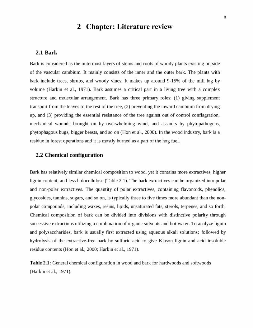
Bark has relatively similar chemical composition to wood, yet it contains more extractives, higher
lignin content, and less holocellulose (Table 2.1). The bark extractives can be organized into polar
and non-polar extractives. The quantity of polar extractives, containing flavonoids, phenolics,
glycosides, tannins, sugars, and so on, is typically three to five times more abundant than the non-
polar compounds, including waxes, resins, lipids, unsaturated fats, sterols, terpenes, and so forth.
Chemical composition of bark can be divided into divisions with distinctive polarity through
successive extractions utilizing a combination of organic solvents and hot water. To analyze lignin
and polysaccharides, bark is usually first extracted using aqueous alkali solutions; followed by
hydrolysis of the extractive-free bark by sulfuric acid to give Klason lignin and acid insoluble
residue contents (Hon et al., 2000; Harkin et al., 1971).
Table 2.1: General chemical configuration in wood and bark for hardwoods and softwoods
(Harkin et al., 1971).

9
2.3 Bark polyphenolics
2.3.1 Lignin
In bark, the polyphenolic part mainly consists of lignin, which is frequently depicted as a
complex 3-D polymer having different types of linked phenylpropenoid units. In nature, it has
an essential role to encase cellulose chains inside the ultra-structure of plant and wood fiber of
the cell walls. Softwood and hardwood lignins may be distinguished by the presence of an
extra methoxy group in the ortho-position of phenyl ring in hardwood lignins (Van Langenberg
et al., 2010).
Mostly guaiacyl units are found in softwood lignin starting from the precursor, trans-coniferyl
alcohol (Figure 2.1 (a), whereas hardwood lignin has combined guaiacyl and syringyl units
originating from trans-coniferyl and trans-sinapyl alcohols (Figure 2.1 (b). Overall, hardwood
bark lignins are mainly composed of syringyl, guaiacyl and small amounts of p-hydroxyphenyl
units while softwood bark lignins have quite similar composition of syringyl-guaiacyl ratio but
differ in higher proportion of p-hydroxyphenyl units, which come from trans-p-coumaryl
alcohol (Figure 2.1 (c) (Hon et al., 2000).

10
Figure 2.1: Phenylpropenoid units in lignin precursors
2.3.2 Lignin valorization
Recently, there are significant efforts worldwide in finding higher valued application for lignin,
especially lignin from the kraft pulping operation. Some research even focuses on finding
favorable genetic variation in local populations of bioenergy crops and direct manipulation of
biosynthesis pathways to create lignin feedstocks with favorable properties for recovery and
downstream conversion. Refinement of biomass pretreatment advances has further encouraged
lignin recovery. Coupled with biotechnology, there is a growing interests in uncovering new
uses for this biopolymer, including carbon fibers, plastics and thermoplastic elastomers,
polyurethane foams, biofuels, and biopolymers/chemicals as shown in (Figure 2.2) explained
by Ragauskas et al., (2014).

11
Figure 2.2: Conceptual theme of lignin valorization and application of this renewable resource
for value-added products by converting into carbon fibers, biopolymers, biochemicals, and
biofuels (Source: Oak Ridge National Laboratory, U.S. Department of Energy, Referred by:
Ragauskas, Science Reviews, 2014 with permission.)
2.3.3 Tannins
Tannins, with the molecular weights ranging from 500 to over 3000, are another important type
of natural polyphenolic compounds present in a relatively large quantity in coniferous tree
barks. The barks of some hardwood species, for example, Quercus, Eucalyptus, Acacia, and
Salix also contain a lot of tannin extractives. Tannins can be structured into hydrolysable and
condensed tannins based on their configuration and properties (Pizzi 1993; Van Langenberg et
al., 2010).
2.3.3.1 Hydrolysable tannins
Polyesters originated from glucose can be considered as hydrolysable tannins, which could be
sorted into: (1) gallotannins, which release gallic acid and its related products after acid
hydrolysis. (2) ellagitannins, which liberate ellagic and valonic acids upon hydrolysis. Caustic
hydrolysis of resorcinolic tannin has been reported to cleave the inter-flavonoid bond and open
the etherocyclic ring joining the A and B rings of the flavonoid unit (Figure 2.3). Acid

12
hydrolysis has been shown to easily open the heterocyclic ring of polyflavonoids with the
formation of a carbocation, which is capable of reacting with another nucleophile present (Pizzi
1993; Van Langenberg et al., 2010).
Figure 2.3: Flavonoid unit
2.3.3.2 Condensed tannins
Catechins (flavan-3-ols) and leucoantocyanidins (flavan-3,4-diols) recognized as condensed
tannins are comprised of flavonoid units. With the average degree of condensation ranging
from 4 to 12 flavonoid units, it is commonly present as polymer and does not undergo
hydrolysis. The condensed tannins constitute more than 90% of the total world production of
commercial tannins.
The main structure of tannin extractives from quebracho, mimosa (black wattle), hemlock and
Douglas-fir bark mainly composed of four to six linked flavonoid units where the A-ring is of
resorcinol type and B-ring of pyrogallol type units (Figure 2.4), with a few flavonoid units
entailing resorcinol A- and catechol B-ring.
In pine (taeda, aleppensis, patula, pinaster, radiata, eliotae, sylvestris, and so forth) species, the
flavonoid units are of phloroglucinol A-ring and catechol B-ring (catechin group) mainly
connected by four to eight bonds, with flavonoid units of phloroglucinol A-ring and phenol B-
ring to a much lesser extent. The structures of the fundamental polymeric constituents of wattle
and pine tannins are indicated in Figure 2.4 (Pizzi 1993; Van Langenberg et al., 2010).

13
Wattle tannin unit Pine tannin unit
Figure 2.4: Wattle and pine tannin unit
Besides the flavonoid units, non-tannins including carbohydrates, hydrocolloid gums, amino
and imino acid fractions also exist in the bark tannin extractives. The hydrocolloid gums with
hydrophilicity varying in concentration from 3 to 6% contribute significantly to the viscosity of
the extractives despite their low concentrations (Pizzi 1993; Van Langenberg et al., 2010).
2.4 Challenges in extraction of bark polyphenolics
Polyphenolic substances include many classes of compounds ranging from phenolic acids,
colored anthocyanins, simple and complex flavonoids. Similarly, pine bark has low levels of
monomers (Shi et al., 2005). During extraction, a solvent is blended with the plant material
(pine bark). Extraction might be accomplished by the evaporation of a solvent and the solvent
could be easily removed either by drying, or ultrafiltration (Shi et al., 2005). After any of these
methods, the concentrate must be dehydrated to get a powder form and quite a considerable
amount of organic solvents is required. Separation of polyphenols by the membrane method is
considered more efficient than the organic solvent extraction method. It is important to make
effective and productive extraction methods to ensure clean polyphenol product (Shi et al.,
2005).
2.4.1 Chemical and physical properties of polyphenolics
Two or more monomers are synthetically reinforced to make oligomeric proanthocyanidins.
The two proanthocyanidin monomers are catechin and epicatechin. A couple of procyanidins
are shown in (Figure 2.5) with the structures of catechins (monomers). These are made when

14
dimers, trimers, and tetramers each of these two monomers joined at or β position on their
molecular structures. Catechin and epicatechin can combine to produce esters, for instance,
catechin/epicatechin gallate, comparably, like the bonds between sugars and proteins to make
glycosidic and polyphenolic proteins. Around 162 dimers, including, gallic acid and glucose
esters that could be made are reported by Bagchi (1999).
Procyanidin B1: R'=OH, R=H Procyanidin B3: R'=OH, R=H
Procyanidin B2: R'=H, R=OH Procyanidin B4: R'=H, R=OH
Figure 2.5: Structures of major identified polyphenols in bark (Source: Shi, J. et al., Food Rev
Int, 2005 with permission.)
15
2.4.2 Polyphenol–protein interactions
Polyphenols interact with protein molecules through hydrophobic or hydrophilic interactions.
These interactions lead to formation of soluble or insoluble aggregates that depend on different
factors such as pH, temperature and ionic strength. The formations of these aggregates are
involved in hydrophobic stacking of aromatic groups of protein and polyphenols, or the
interaction between hydroxyl groups of polyphenols with protein chains. Proteins are recognized
to play a key role in many physiological activities owing to their stable 3-D structure. Afterwards,
unfolding of protein chains, upon binding with polyphenols, is assumed to affect the physiological
activity of protein molecules (Bennick, 2002; Naczk et al. 2006; Liang et al. 2008).
Proteins (dry weight ≥ 33%), linked by hydrogen bonding, can be involved in a variety of
supramolecular interactions (Loomis, 1969). Other than hydrogen bonding there are different
stable bonds that indicate spatial arrangement of polypeptide’s backbone and its subunits.
Distinctive substances react non-enzymatically with compounds, such as o-quinones. The
materials having amino, thiol, and activated methylene groups could be polymerized,
diminished, or maintained by nucleophilic attack (Pierpoint, 1970). Proteins and polyphenolic
compounds in this mode have similar bonding characteristics as the quinine and hydrogen
bonding reactions.
Polyphenols are known to form complexes with proteins leading to modifications in the
structural, functional and nutritional properties of both compounds. The different parameters
such as, temperature, pH, protein type, protein concentration, and the structure of phenolic
compounds can affect the protein–phenolic interactions (Ozdal et al., 2013). To measure
proteins, as, for instance, the haemoglobin, gelatin, and BSA assays, this property of
polyphenolic compounds might be utilized.
Several analytical techniques have been developed to characterize the polyphenol–protein
complex formation such as fluorescence, circular dichroism (CD) spectroscopy, dynamic light
scattering (DLS), Fourier transform infra-red (FTIR) spectroscopy, isothermal titration
calorimetry (ITC), and nuclear magnetic resonance (NMR) and mass spectroscopy (ESI-MS)
reported by Bandyopadhyay et al. (2012).

16
Figure 2.6: A model for protein−polyphenol interactions that elucidates having two sides that
can join to protein. Proteins are defined as having a fixed number of polyphenol binding sites
(Source: Siebert, K.J et al., J. Agric. Food Chem, 1996 with permission.)
A hypothetical model as shown in Figure 2.6 shows that proteins have several sites where
polyphenol can bind. In this situation, each polyphenolic molecule should have binding sites
where two proteins may attach. However, it is unlikely that there will be enough additional
polyphenolic molecules to bridge many of these “sandwiches” or “protein dimers” together.
Structure of proteins and polyphenols play a significant role to determine accumulated amount
between them.
With an extensive abundance of protein in respect to polyphenol, every polyphenol particle ought
to have the capacity to extend between two protein molecules; however it would be unlikely that
these proteins would be further connected to others. This would come about for the most part in
protein dimers. With excess polyphenol relative to protein, all of the protein binding sites would
be occupied, but probably that bridging would occur between polyhydroxyproline contents.
Polypeptides with higher percentages of proline tend to form more haze. The amount of haze

17
formed depends both on the concentrations of protein and polyphenol present in most beverage
samples (Siebert K.J et al., 1996).
Currently, the chemical and biological aspects of proteins and polyphenols are challenging
because of their applications in food, agriculture and their potential health benefits.
2.4.3 Interaction of low molecular weight (LMW) phenolics
There have generally been few studies conducted using protein and LMW-polyphenol
interactions. Certain protein concentrates from sunflower seeds can create an unwanted brown
color because of protein binding with the oxidation products of low molecular weight phenolic
compounds, such as, chlorogenic acid (Sastry and Rao 1990). Currently, BSA was examined
for its interaction with low molecular weight phenols (Bartolomé et al., 2000).
Protocatechuic and caffeic acid showed the most elevated binding for the protein, while p-
hydroxybenzoic acid displayed the least binding limit; though, p-coumaric acid and (+)-
catechin showed an irrelevant substance for protein- held phenols. The pH and the temperature
play important roles in protein-polyphenolics interactions. Phenolic acids with single aromatic
rings demonstrated much more significant interaction than multi-aromatic ring isoflavone.
BSA-phenolic acid indicated substantial contrast in electrophoretic movement, and displayed
total protein when contrasted with BSA (Bartolomé et al., 2000).
2.5 Biopulping
Biopulping is an industrial biotechnology process which utilizes different microorganisms,
especially lignin-degrading fungi and enzymes (ligninases and xylanases) for converting wood
chips into paper pulp. Biopulping provides an alternative solution to chemical and mechanical
pulping. Ligninolytic enzymes attack lignin and decompose it, while xylanases degrade
hemicelluloses and make the pulp more penetrable for the removal of remaining lignin (Ali and
Sreekrishnan, 2001). Named 'biopulping', this methodology displaces lignin as well as a portion of
the wood extractives, while reducing the pitch content and effluent toxicity (Ali and Sreekrishnan
2001).
Biotechnological tools are gradually used to replace chemical processes in a wide range of
industries. No pilot-scale biopulping plants are in operation right now as this procedure is still in

18
its earliest stages (Ali et al., 2001). Mixed hydrolytic and oxidative enzymes have been well
documented regarding biopulping and bio-modification of lignin, and thus provide an example
which is related to effectiveness, despite the susceptibility of xylanases to deactivate by laccase-
generated oxidants (Woolridge, E.M. 2014).
2.5.1 Role of ligninases in pulp delignification
LiP (lignin peroxidase, EC 1.11.1.7) and MnP (manganese peroxidase, EC 1.11.1.7) are Fe+2
-
containing glycoproteins which necessitate H2O2 as an oxidant. The fungal growth releases a few
isoenzymes into their development medium, although the enzymes may also be cell wall bound
(Lackner et al., 1991). Non-phenolic lignin substructures oxidize LiP to a radical cation followed
by a proton loss (Higuchi, 1989).
LiP catalyzes a large variety of reactions e.g., cleavage of β-O-4 ether bonds and C-Cβ bonds in
dimeric lignin model compounds (Higuchi, 1989). LiP is released during secondary metabolism
as response to nitrogen limitation. It is considered as strong oxidizer capable of catalyzing the
oxidation of phenols, aromatic amines, aromatic ethers and polycyclic aromatic hydrocarbons
(PAHs).
However, the degree of lignin biodegradation mainly depends on the environmental conditions
and the fungal species involved (Archibald 1992). Previous studies mainly emphasized the
mechanism of fungal degradation of lignin.
2.5.2 Oxidation of PAHs by synthetic LMS
The oxidation of polycyclic aromatic compounds (PAHs) was studied in laccase producing fungi
Trametes versicolor and synthetic mediators were examined by Johannes et al. (2000). Enzymatic
oxidation of acenaphthene, acenaphthylene, anthracene, and fluorene was mediated by different
laccase substrates such as phenols and substituted amines or compounds produced and secreted by
white rot fungi. There is an option of using wood-decay fungi able to produce hydrogen peroxide
in presence of multi-enzyme systems or as mixed fungal cultures to improve decolorization.
The best natural mediators, such as phenol, aniline, 4-hydroxybenzoic acid, and 4-
hydroxybenzyl alcohol were as effective as synthetic compounds e.g., ABTS [2,2′-azino-bis-
(3-ethylbenzothiazoline-6-sulfonic acid)] and 1-hydroxybenzotriazole. Natural compounds,

19
like methionine, cysteine, glutathione that contain sulfhydryl groups, were also observed as
mediator compounds.
2.6 Fungal extracellular ligninases
Extracellular ligninases may be categorized as either phenol oxidases (laccase) or heme containing
peroxidases (lignin peroxidase (LiP), manganese peroxidase (MnP) and versatile peroxidase
(VP)). Commonly, laccases use molecular oxygen as electron acceptors whereas; peroxidases use
H2O2 as a co-substrate. White-rot fungi secrete one or more of the lignin-modifying enzymes
(LMEs) as well as other compounds required for efficient lignin degradation (Mai et al., 2004).
Non-phenolic aromatic substrates have high redox potential ability to catalytically oxidize and
cleave C–C bonds and ether (C–O–C) linkages. Manganese peroxidases (MnP) oxidize Mn(II) to
Mn(III), to cleave non-phenolic compounds. Combination of LiP and MnP with a bifunctional part
was protected by versatile peroxidases (VP) reported by Wong (2009).
The oxidative enzymes prepared for modifying such complicated and mixed structures are
required. It was also felt that these enzymes did not require any cofactors, for example, NADPH
since extracellular unit of such cofactors seemed, by all reasons, to be inactive. In 1984, Kuwahara
et al. demonstrated that Phanerochaete chrysosporium excreted NADP+ into growth media and
hypothesized a reactant part for these cofactors play a main role in delignification.
Hall (1980) suggested that delignification can include non-enzymatic attack on the polymer by
"activated oxygen" species; H2O2, superoxide, oxygen, or -OH radicals have been found in lignin
degradation. Further studies including ligninases purification by use of different spectroscopy
methods, and recombinant DNA molecular techniques, have improved our understanding at the
molecular level of microbial delignification.
Relative molecular weight ranges between (41,000 to 42,000) had Fe+2
-containing ligninases
defined by Kirk and Tien (1983). The glycoprotein non stereospecific (42,000 Mw) was stated by
Tien and Kirk as detached from C-Cβ side chain in lignin, which, further oxidized benzyl
alcohol to aldehydes or ketones. Therefore, it separated aromatic ring, hydroxylated benzylic
methylene groups, and further catalyzed to oxidative coupling of phenols.
Ligninases can similarly catalyze H2O2- related oxidation of heterocyclic sulphur containing
compound, thianthrene, to thianthrene monosulfoxide. Kirk and Tien (1983), shown degradation

20
of β-1 and β-O-4 containing model compounds from lignin. Ligninases oxidize an extensive
variety of lignin model compounds. Kuwahara et al. (1984) additionally proved that molecular
oxygen factors in the cleavage of the C-Cβ link of 1-(3',4'-diethoxyphenyl)-1,3-dihydroxy-(4"-
methoxyphenyl) propane by ligninolytic enzymes.

21
Figure 2.7: Degradation of oligomeric lignin model compounds by P. chrysosporium
ligninases (A) β-1 and (B) β-O-4 (Proposed by Tien and Kirk 1984, Proc Narl Acad. Sci. USA.,
with author permission.)
Phlebia radiata produces lignin-modifying enzymes (LMEs), that showed to be an efficient
oxidant producing carbon-carbon bond cleavage of a dimeric, non-phenolic lignin model
compound (Lundell et al. 1993a). The antigens prepared from these three ligninases (I, II, III) with
molecular weights of 42,000, 45,000, and 44,000 cross-reacted with each other. Pleurotus sajor-
caju produces two flavins containing veratryl alcohol oxidases (Bourbonnais and Paice 1988). The
role of the oxidases in biodegradation might be to produce H2O2 and possibly superoxide anion
radical (O·̄2), during oxidation of lignin fragments (Leonowicz et al., 1999).
Enzymatic systems employed by microorganisms for oxidative transformation of various organic
molecules include laccases, ligninases, tyrosinases, monooxygenases, and dioxygenases. The
targeted enzymes are important to the carbon-cycle through either transformation or complete
mineralization of organic molecules (Sariaslani, 1989).
Microbial enzymatic systems involved in the oxidation of organic molecules were reviewed.
Enzymatic systems such as mono- and dioxygenases, with their inherent stereo- and regio
specificities, provide powerful tools for generating chemicals that are difficult to synthesize
through conventional chemical routes. Successful commercial applications of these catalysts will
be possible through new systems, e.g., immobilization of either integral microorganisms or
isolated enzyme preparations on various supports, utilization of organic solvents in the reaction
mixtures, and genetic engineering technology (Sariaslani, 1989).
2.6.1 Reaction mechanism of ligninases
The coupling reactions of a monomer at its β-position to chain polymer are present in β-linked
structures (Wong 2009). The attained polymers have 5-5 and 5-O-4 linked structures completing
coupling between two lignin oligomers. Changing lignin structure by dimerization to yield β-β
(resinol) is rarely found in nature. End groups arising from coupling reactions are not at the side
chain β-position of the monomer. The various linkages depend on the relative contribution of
monomers to the polymerization process during lignin biosynthesis. For instance, the monolignol
coupling in coniferyl alcohol to a guaiacyl (G) unit lignin oligomer/polymer yields G-β-O-4-G
and G-β-5-G linkages as shown in (Figure 2.8).
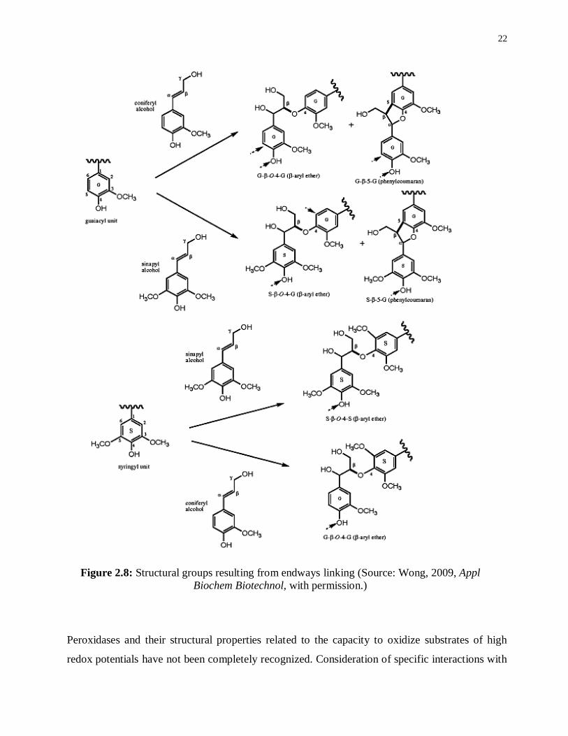
22
Figure 2.8: Structural groups resulting from endways linking (Source: Wong, 2009, Appl
Biochem Biotechnol, with permission.)
Peroxidases and their structural properties related to the capacity to oxidize substrates of high
redox potentials have not been completely recognized. Consideration of specific interactions with

23
mediators during biotransformation mainly supported that these catalysts are important for
delignification. Finally, it was concluded that polymerization is directed by proteinaceous sites on
a template, which, specify amongst linkage types and bond arrangements (Wong, 2009).
2.6.2 Laccase–Mediator Systems (LMS)
LMS perform as electron shuttles, offering oxidation of complex substrates (for example lignin
polymers) where enzymes can’t access the active sites due to steric interferences. Once it is
oxidized and stabilized, the mediators are dispersed far from enzymatic compartment because
of their high redox potential (Bourbonnais and Paice, 1990; Kawai et al., 1989). An ideal redox
mediator would be a small-size compound which is able to produce stable radicals so it also
works in recycling without disintegration (Canas et al., 2010).
Previous studies proved that the Trametes hirsute laccase with 2,2'-azino-bis-(3-
ethylbenzothiazoline-6-sulfonic acid) ABTS and Remazol blue used as mediators could oxidize
nonphenolic lignin parts with high redox potential to produce veratryl alcohol and 1-(3,4-
dimethoxy phenyl)-2-(2-methoxy phenoxy) propane-1,3-diol. Furthermore, laccase with ABTS
can disintegrate a model lignin dimer, 1-(3,4-dimethoxyphenyl)-2-phenoxyethane-1,2-diol, to
yield veratraldehyde and benzaldehyde. The oxidation of ABTS in presence of laccase is
shown in Figure 2.11.
The presence of redox mediators is required for a number of biotechnological applications, to
assist the oxidation of complex substrates not oxidized by the enzyme alone. However, it should
not hinder the enzymatic response (Johannes et al., 2000). However, the oxidizing mediator could
depend on the oxidation mechanism not on the enzyme, so it is possible to oxidize non-phenolic
substrates (Fabbrini et al., 2002), subsequently shutting down the sequential catalytic cycle
(Figure 2.9).
Figure 2.9: The sequential catalytic cycle of LMS (Source: Fabbrini, 2002, J Mol Catal
B-Enzym, with permission.)

24
The sequential catalytic cycle incorporates a set of several compounds, to meet the condition of
several reaction cycles in the redox process.
Distinctive structures of transition segments i.e. (K3[Mo(CN)8].H2O and K3[W(CN)8].H2O) FeII
complex with o-phenanthroline and 4,4'-dimethylbipyridine, and some natural laccase substrates,
e.g., ABTS and 2,2,6,6-tetramethyl-1-piperidinyloxyl (TMPO). For stable nitroxyl radical, TMPO
is different because it is oxidized by laccase and produced oxoammonium ion (oxidation to
>N=O+ that correspond to high redox fungal laccase), which follows a non-radical-ionic-
mechanism (Fabbrini et al., 2002). Conventional redox potentials have well defined transition
metal complexes and can oxidize non-phenolic lignin structures for the most part at low
concentrations in the reaction mixture.
ABTS is an organic compound suitable for the expression of "redox mediator". Oxidation of non-
phenolic lignin structures offers a way to look at new laccase mediators. The use of eco-friendly
mediators, can contribute to the industrial implementation of laccases and in the development of
lignocellulosic biorefineries. The following N-OH laccase mediators and their structural formulas
(A-E) are shown in Figure 2.10.

25
Figure 2.10: N–OH type laccase mediators and their chemical structures (A-E); A=1-
Hydroxybenzotriazole (HBT); B= N-Hydroxyphthalimide (HPI); C=N-Hydroxyacetanilide
(HAA); D= Violuric acid (VA); E= 2,2,6,6-Tetramethylpiperidin-1-oxyl (TMPO).
Figure 2.11: Oxidation of ABTS in presence of laccase (Source: Morozova, 2007, Appl
Biochem Microbiol Reviews, with permission.)
26
2.6.3 Natural mediators / phenolic enhancer
Natural mediators, in presence of laccases, expedite the oxidation of non-phenolics but it
depends on the phenolic compound structure, as well as the reactivity vs. stability of phenoxy
radicals produced (MS. > AS
. > SA
.) (Tania et al. 2012). Due to the complex nature of biomass,
degrading enzymes, and their interactions, researchers are trying to profile radical-coupling
routes involved in the development of several phenolic species that are classified as mediators.
Moreover, there is a need to improve relationship between biomass structure, effectiveness of
enzymatic hydrolysis and the biomass recalcitrance (Foston et al., 2012).
In presence of redox mediators, laccases enhance their substrate specificity. The enzymatic
oxidation of syringyl-type phenolics identified as natural mediators, such as methyl syringate
(MS), acetosyringone (AS) and syringaldehyde (SA), participate in phenolic oxidation
depending on the negatively charged residues similar to a substrate binding site of the enzyme.
2.6.4 Biotransformation
Biotransformation is a series of biological modifications / conversions made by a living organism
on a chemical compound. In the early 1990’s, the oxidation of phenolics by ligninases was
investigated for dimethoxylated compounds (veratryl alcohol and 3,4-dimethoxyphenyl acetic
acid). In all examples, polyphenolic compounds were found to be exclusively oxidized in contrast
with the dimethoxylated compounds. Ligninase catalyzes continuing delignification rather than
further polymerization; a model to define the framework is displayed by Harvey et al. (1990).
Furthermore, the ligninases, lignin and phenolic lignin are broken down into small fragments and
are connected to radical cations as mediators. If the free phenolic compounds are accumulated
then eventually it causes inhibition of ligninases, which prevents lignin cleavage and produces
more polyphenolics. Hence temporary inhibition of ligninases by phenols may play some role in
metabolic regulation of lignin (Harvey et al., 1990).
2.6.4.1 Coupling routes for polyphenolics oxidation
In the laccase mediator system (LMS), the catalytic cycle demonstrated an interchange of paths
including phenoxy radical as an intermediate. When they are oxidized in an enzymatic system, a
proton and an electron lead to discharge radicals and release phenolic substrates. In order to
oxidize non-phenolic compounds by laccases, the phenoxy radicals are involved in three different
possible routes. Moreover, it can either be combined with radical recombination, cross-coupling

27
or self-coupling to continue with or without release of substituent groups which further change
into other coupling structures (Figure 2.12).
Figure 2.12: The catalytic cycle of LMS showing different paths implicated in development of
several phenolic species (Source: Rosado et al., Bioresource Technol, 2012 with permission.)
The newly identified radical coupling routes which involved several C–O coupling reactions,
as well as, different dimeric and trimeric structures were proposed (Tania et al. 2012). The
catalytic cycle supports the demand of added phenolic compounds in laccase mediator system
which explains the role of phenolics as ‘‘mediators’’ rather than ‘‘enhancers’’ (Díaz-González
et al., 2011).
2.6.4.2 Coupling routes for non-phenolics oxidation
For oxidation of recalcitrant aromatic compounds, the >N–OH mediators, for instance, 1-
hydroxybenzotriazole (HBT), N-hydroxyphthalimide (HPI), violuric acid (VA) or N-
hydroxyacetanilide (NHA) (Xu et al. 2000) are the possible laccase mediators; their mechanism is

28
shown in (Figure 2.13). Laccase produces an extremely reactive nitroxyl radical (> N–O•), owing
to the enzymatic removal of an electron by release of a proton and an oxidation of this type of
mediators. The target substrate oxidized nitroxyl radicals by hydrogen atom transfer (HAT) route
(Xu et al., 2000).
A mediator-to-substrate molar ratio can affect the efficiency of oxidation procedure. The low
redox potential substrates could be responsible for the oxidation of non-phenolic substrates under
non-catalytic conditions (Cantarella et al., 2003).
Figure 2.13: Non-phenolic lignin model compound oxidation by laccase mediator system
(LMS) ensuing by two different oxidation methods: ET (electron transfer) and HAT (hydrogen
atom transfer) (Source: Fabbrini et al., J. Mol. Catal. B: Enzym, 2002 with permission.)
2.7 Function of laccases with combination of natural mediators (NMs) for
lignin biodegradation
The phenolic compounds related to phenolic structures in lignin are able to help oxidation of non-
phenolic compounds by radical mechanism. This finding may support the role of laccases in lignin
biodegradation with no other mediators. Therefore, oxidation of phenolic lignin units with
laccases or ligninolytic peroxidases would release phenolic natural mediators as shown in (Figure

29
2.14) that could help in start oxidation of more recalcitrant non-phenolic lignin moiety (Canas et
al., 2010).
Lignin removal from biomass by using laccases might be achieved by various methods, depending
on different types of lignocellulosic material and its further use. Alternatively, the phenolic
residues in lignocellulosic material i.e., (p-hydroxycinnamic acids) highly abundant in herbaceous
plants, could act as in situ laccase mediators which assist to remove recalcitrant lignin moiety as
has already been indicated with model compounds (Cho et al., 2008; Nousiainen et al., 2009).
Figure 2.14: Conceivable function of laccases with combination of phenolic natural mediators
(NMs) in decontamination of industrial effluents and soil bioremediation (Source: Cañas et al.,
Biotechnol Adv, 2010 with permission.)

30
2.7.1 Monofloral honey as mediator
The composition of honey, a natural bee product, mainly depends on different botanical
sources, topographical origin and atmospheric conditions (Chen et al., 2000; Vela et al., 2007;
Alvarez et al., 2010). In honey, the main antioxidant compounds are polyphenolics, flavonoids,
enzymes (catalase, glucose oxidase), organic acids, ascorbic acid, carotenoid-like substances,
amino acids and proteins. The antioxidant value varies significantly depending on the floral
source (Gheldof et al., 2002). Several phenolic compounds are found in honey, including
syringic acid (SA) and methyl syringate (MS). Lately, a compound was obtained having
substantial antibacterial activity against Staphylococcus aureus (Alvarez-Suarez et al., 2010).
2.8 Usage of laccases and NM for future lignocellulosic biorefineries
The utilization of laccases and their natural mediators represents a promising alternative for
environmentally friendly delignification of paper pulp. These enzymatic frameworks could be
incorporated at different phases of biopulping and paper production (Camarero et al., 2007). As
by-products from pulp production, these natural phenolic mediators can be obtained. Eucalypt
kraft pulping black liquor possesses a large amount of some of these phenolic compounds (e.g.,
acetosyringone and syringaldehyde) which may be sufficiently separated by little efforts from
pulp and paper industrial effluents (Camarero et al., 2005).
Functionalization of wood and non-wood by laccases has not been completely explored (Chandra
and Ragauskas, 2002). Figure 2.15 indicates biotechnological application of laccases (and natural
mediators), which can contribute most significantly to develop Integrated Lignocellulosic Forest
Products Biorefineries (ILFPB). Wood products (boards and panels) used in furniture
manufacturing are bonded with high cost adhesives derived from petroleum. Most of these
adhesives are formaldehyde-based resins, due to its harmful emissions to limit the reuse of
fiberboards (Felby et al., 1997). Therefore, laccases can contribute to reduce (or eliminate) the use
of these synthetic adhesives by catalyzing the cross-linking of polyphenolic residues in lignin
based materials to produce medium density fiberboards (Canas et al., 2010).
Biografting of phenols onto different types of pulps can provide new physico-mechanical
properties to paper such as improved strength due to promotion of new hydrogen bonding and
crosslinking between phenoxyl groups on pulp surface (Chandra et al., 2004). Laccase-assisted
grafting of phenols can also modify optical properties like color. Protection against

31
antimicrobial activity can be obtained using an enzymatic method to make cellulosic products
for food packaging or cleansing materials (Elegir et al., 2008).
Figure 2.15: Strategic plan of Integrated Lignocellulosic Forest Products Biorefinery (ILFPB):
where, (1) different lignocellulosic reserves; (2) extraction of hemicelluloses to pulping; and
(3) biomass gasification. The main features where enzyme technology may help twice as
notably to this idea are also shown: (a) assembling of wood (or lignocellulose) products; (b)
create of more proficient and environmentally friendly industrial procedures for pulp and paper
production; (c) extraction of phenolic mediators and reusing of black liquors; (d) production of
cellulosic products with modified characteristics; or (e) enhanced biofuel production (Source:
Cañas et al., Biotechnol Adv, 2010 with permission.)
In nature, these phenolic compounds (released during lignin depolymerization or present as free
acids in plants) perform as natural mediators. Moreover, by the application of these enzymes,
sustainable industrial processes are able to move forward by the presence of these
environmentally-friendly mediators, easily available from lignocellulosic biomass (Canas et al.,
2010). Therefore, there are important economical, ecological and environmental reasons for
achieving better understanding of fungal activities and their implementation in lignocellulosic
green biotechnology (LGB) (Harms et al., 2011).

32
3 Chapter: Experimental Methodology
Research approach for objective-1
Synopsis
Comparative studies were conducted among treatments to characterize bark polyphenolic
extractives that can be effectively used for green adhesive production, leading to environmental
sustainability.
Figure 3.1: Design of strategic plan for objective-1. Reproduced by the permission of American
Chemical Society (ACS) http://pubs.acs.org/doi/abs/10.1021/sc400184f
3.1 Chemicals and standards
Sodium hydroxide (NaOH), sulphuric acid (H2SO4) and acetic acid (CH3COOH) were
purchased from Caledon Laboratory Chemicals. Catechol, tannin, Folin-Denis reagent and
sodium chlorite (NaClO2) were purchased from Sigma-Aldrich. Cellulose extraction thimbles
33
and grade 42 filters were purchased from Whatman. Mountain pine beetle-infested lodgepole
pine and mixed aspen barks were provided by FP Innovations and ground to a particle size
below 0.212 mm (US70 mesh size). All of the chemicals used in this study were reagent grade
and were used without further purification.
3.2 Chemical Composition of Bark before and after Extraction
3.2.1 Ash Contents in Bark
Ash contents in the bark were determined by oxidizing dry bark at 580C with a TGA
instrument. About 10 mg of oven-dried sample was weighed in a platinum pan and heated from
room temperature to 580C, at a heating rate of 10
C/min using a thermal gravimetric analyzer
(TGA-Q500, TA Instruments, USA). The final ignition temperature was 575C according to the
ASTM D1102-84 method-suggested temperature for the determination of ash contents in
wood. Bark ash contents were reported as the percentage of residues remaining after treatment.
Five samples of each bark were tested to determine the standard deviation.
3.2.2 Preparation of Extractive-Free Bark
Extractive-free bark was prepared according to ASTM D1105-96. Ethanol−toluene extractives
and hot water extractives contents in bark were then reported. The ethanol−toluene extractive
values listed in this paper refer to the bark extraction with successive ethanol−toluene mixtures
and 95% ethanol.
3.2.3 Lignin Content of Bark
The lignin contents of beetle-infested lodgepole pine (BILP) and aspen bark were determined
according to a modified procedure for the evaluation of acid-insoluble lignin content in wood and
pulp according to standard TAAPI method T 222 om-02 (TAPPI, 2002).
Briefly, 0.2 g of oven-dried extractive-free bark was combined with 2 mL of 72% sulfuric acid
and incubated at 30 °C for 1 h. Fifty mL of distilled water was then added, and the mixture was
autoclaved at 120 °C. The solution was then filtered into glass crucibles, grade 40−60, and lignin
content was determined using eq 1.

34
Equation 1
where mbe is the oven-dry weight of extractive-free bark, mb is the oven-dry weight of bark; mlc
is the oven-dry weight of crucible and of lignin, and mc the oven-dry weight of crucible.
3.2.4 Holocellulose and α-cellulose content of bark
Holocellulose and α-cellulose contents in extracted bark were determined following a procedure
developed by Browning, (1967).
0.5 g of oven-dried extractive-free bark was weighed and 16 mL of buffer solution (60 ml of
acetic acid and 1.3 g of sodium hydroxide in 1 litre of water) was added. The mixture was
placed in a water bath at 70°C and 1 mL of 27% (w/v) NaClO2 was added every 30 minutes for
the following 4 hours. The filtered holocellulose was separated in two sets of experiments. The
first set was oven-dried at 65°C and weighed while the second set of filtered holocellulose was
kept in the desiccator for 24 hours, and then transferred into beakers for the α-cellulose
determination. Three mL of 17.5% NaOH solution was added to the prepared holocellulose and
incubated at 20°C, followed by another 6 mL after 5 min. The total treatment lasted 45
minutes. Then distilled water was added, and the mixture was allowed to stand for 1 hour.
Once the caustic treatment was completed, the solution was filtered under vacuum into 40-60
grade crucibles and washed with 30 mL of distilled water. Crucibles and samples were then
oven-dried overnight at 65°C. Holocellulose and α-cellulose contents were determined
according to equations 2 and 3.
Equation 2
Equation 3

35
where mbe is the oven-dry weight of extractive-free bark, mb is the oven-dry weight of bark,
mhc is the oven-dry weight of holocellulose and crucibles, mαc is the oven-dry weight of α-
cellulose and of crucible and mc is the oven-dry weight of the crucible.
3.2.5 Enzymatic treatment
Enzymatic hydrolysis was performed using a cocktail of three enzymes. They included
cellulase (E.C. 3.2.1.4) from T. reesei (6.3 U/mg) and β-glucosidase (E.C. 3.2.1.21) from
almonds (7.55U/mg) (both enzymes were purchased from Biochemika-Fluka; the purity of
both enzymes is not given in the certificate of analysis describing the product specifications
from Sigma-Aldrich (Appendix). Novozym®51003 laccase (E.C.1.10.3.2) from genetically
modified Aspergillus sp. (1000 LAMU/g = 3.57 IU/ml/min) was obtained from Novozymes
(Franklinton, NC, USA). Enzyme reactions were performed at a solid:liquor ratio of 1:10 (w/v)
by inserting ground bark (BILP and aspen bark) in 0.2 M sodium acetate / acetic acid buffer
(pH 5.6), containing 10 U of each enzyme, per gram of bark at 45C for 48 h. The enzymatic
treated flasks were agitated in a rotary shaker at 150 rpm. After hydrolysis, 600µl of extractives
were removed from the treated barks, passed through a 0.22µm filter and stored for subsequent
analysis of phenolic compounds.
3.2.6 Alkaline Treatment
Bark powders were extracted using 1% NaOH solution with a solid:liquor ratio of 1:10 (w/v),
at 100°C for 120 min. Bark soluble fractions were filtered and the liquid fractions were
collected and stored at -10°C. Bark residues were washed with excessive hot water and oven-
dried at 65°C to constant weight. The extraction yield was calculated based on equation 4.
Equation 4
Where mb is the oven-dry bark weight, mbef is the weight of the oven-dried bark after extraction
and mf is the oven-dry filter weight.

36
3.2.7 UV/H2O2 treatment
Beetle-infested lodgepole pine (BILP) and aspen bark samples were treated with UV/H2O2 in
300 ml Erlenmeyer flasks under room temperature conditions. The ground barks were
combined with 1% NaOH at a solid: liquor ratio of 1:10, and then exposed with conventional
UV lamps (Philips, 40Watt) for 3 hours with 100mM of H2O2. Hydrogen peroxide
concentration of 100mM was adjusted due to acting as a free-radical eliminator at higher
concentrations, as reported by Kusic et al. (2006), which decreases the amount of hydroxyl
radicals in the solution.
3.2.8 Sample preparation for HPLC
After three consecutive treatments, the total phenolic contents from bark extract were assessed
by dissolving the extractives in hot-water, according to the method reported by Yu et al.
(2001). Bark extractive samples (0.1 g) were mixed with 2 mL of hot water in test tubes and
heated for 1h in a boiling water bath. The samples were cooled to room temperature and
centrifuged at 10000g for 10 min (model Avanti® J-E Centrifuge, Beckman Coulter, USA).
Samples were filtered through a 0.45-µm filter and analyzed by HPLC. The treatments selected
in this study allowed extracting both polar and non-polar fractions from bark, and, thus, a tiny
fraction of the extractives was not completely dissolved as the HPLC sample preparation stage
was completed. Consequently, the following study on bark polyphenolic extractives only
considered the hot-water soluble fractions.
3.2.9 HPLC analysis
The HPLC system included a Dionex BioLC 20 Series HPLC instrument equipped with a
GP50 gradient pump, AS40 auto sampler, AD25 absorbance detector and Chromeleon for data
collection and analysis (CMS), all of them from Dionex Technologies (USA). 10µl of sample
was injected by the auto-sampler. A Hypersil ODS C18 column (100 mmX4.6 mm, particle
size 3µ) from Alltech (USA) was used for chromatographic separation.
For elution, the two mobile phase gradients were used with a flow rate of 1.3mL/min: 0.1%
formic acid in aqueous solution as (eluent A) and acetonitrile (ACN) as organic mobile phase
(eluent B). The initial isocratic range started from 0-4 min at 5% ACN, and pursued a linear
increase until reaching 23% after 10 min. To obtain better separation of polyphenols, an
isocratic range from 10-15 min at 23% ACN was applied. ACN concentration was further

37
increased, linearly, to 50% within 4 minutes to elute the most preserved analytes directly,
followed by reverting back to the initial conditions from 50-95% for 1 min. Chromatograms
were recorded at three different wavelengths, 280, 310 and 370 nm. The bark polyphenolic
extractives were separated and characterized based on their reported retention time (t r) values
(Aznar et al., 2011).
3.2.10 Folin-Denis method
The Folin-Denis method is a common spectrophotometric method to assess the total phenolic
content in the samples. An intense blue colour was produced after 30 min in the reaction
mixture due to the reduction of phenol (Iolanda da Cruz et al., 1998). The Folin-Denis reagent
is a combination of phosphomolybdic - phosphotungstic acid in alkaline solution. In this study,
catechol was used as a reference standard in place of tannic acid or gallic acid. Thus, the total
phenolic contents were calculated as catechol equivalents from the calibration curve (mg
catechol equiv/g of treated dry extract).
3.2.11 SEM for BILP and Aspen bark
Samples of beetle-infested lodgepole pine (BILP) and aspen bark produced after enzyme,
alkaline and UV/H2O2 treatments were oven-dried at 50C for 1 h and a dense layer of samples
was kept in the sample-holder fixed on a carbon ribbon. The sample assembly was maintained
in plasma for 60 seconds in a SC7620 Mini Sputter Coater (Polaron) purged with Argon to
eliminate air from the samples. SEM with a Jeol model JSM-6610LV microscope
supplemented by an Oxford/SDD EDS detector was used for observing the bark samples
before and after treatments (Ferhan et al., 2012).
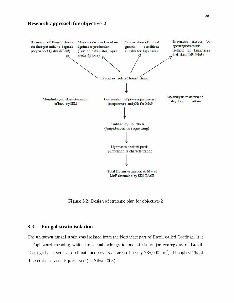
38
Research approach for objective-2
Figure 3.2: Design of strategic plan for objective-2
3.3 Fungal strain isolation
The unknown fungal strain was isolated from the Northeast part of Brazil called Caatinga. It is
a Tupi word meaning white-forest and belongs to one of six major ecoregions of Brazil.
Caatinga has a semi-arid climate and covers an area of nearly 735,000 km2, although < 1% of
this semi-arid zone is preserved (da Silva 2003).

39
The leaf samples of Combretum leprosum were collected in the tropical dry forest (Caatinga),
in the Brazilian semiarid region (S05°55′33,7″, W39°31′44,4″) (Santos et al. 2012). The leaf
samples were brought to the laboratory in sterile polythene bags and processed within 24 h of
collection.
The leaf samples were cut into 2 cm pieces and left on a Petri dish containing PDA (potato
dextrose agar) and 150 mgL−1
chloramphenicol with approximately 4–5 pieces on each dish.
The material was incubated at 25C for 10 days and hyphal tips of the fungus protruding from
the inner leaf segments on the plates were further purified and transferred to slants. The fungal
strain was banked in (CCMAEI) Culture Collection of Microorganisms of Agricultural and
Environmental Importance, Embrapa Meio Ambiente, Jaguariuna, SP with accession number:
CFE044.
3.3.1 Substrate collection and preparation
Sugarcane bagasse and Eucalyptus lignin were used for delignification. Sugarcane bagasse and
Eucalyptus were collected from LWART Quimica, Brazil. All substrates were dried in an oven
at 80C to constant weight and were powdered using an electric grinder and stored in airtight
glass containers to keep out moisture.
3.3.2 Fermentative organism and culturing conditions
Pleurotus ostreatus and Pycnoporus sanguineus were obtained from the Mycology Collection
Lab, Department of Plant Protection, UNESP, Botucatu, SP, whereas the local fungal strain
was isolated from the Northeast part of the country in the Caatinga forest. All fungal strains
were plated on malt extract agar medium (malt extract 25 g; agar 20 g; distilled water 1L)
containing 0.05% of the dye Remazol Brilliant Blue R (RBBR). Plates were incubated in the
dark for 14 days at 25°C. Ligninolytic activity was assessed by scoring for the presence of a
halo of decolorized dye surrounding the fungal colonies.
3.3.3 Quantification of dye Remazol Brilliant Blue R (RBBR) oxidation in liquid culture
medium
Out of ten strains, seven exhibiting ligninolytic activity were then grown in broth culture media
containing (2.5% malt extract and 0.05% RBBR). All the cultures were kept in the dark and
incubated at 30C, aerated by constant shaking. The presence of ligninase activity, as evidenced
by decolorization following oxidation of polymeric dye, was determined at 12 days by
40
monitoring the decrease in the absorption peak at λ595 nm using a Shimadzu UV-1601PC
spectrophotometer. Cell biomass was measured gravimetrically by oven-drying at 70°C. All
shake flask experiments were tested for ligninolytic activity in replicates of three.
3.3.4 Growth Media preparation
Growth media for P. ostreatus contained: Glucose 10g/L, L-Asparagine monohydrate 3g/L,
MgSO4.7H2O 0.05g/L, KH2PO4 0.5g/L, K2HPO4 0.6g/L, CuSO4.5H2O 0.4mg/L, MnCl2.4H2O
0.09mg/L, H3BO3 0.07mg/L, NaMoO4.2H2O 0.02mg/L, FeCl3 1mg/L, ZnCl2 3.5mg/L,
Thiamine-HCl 0.1mg/L, Biotin 5μg/L (Trupkin et al., 2003). The medium was dispersed into
250 mL Erlenmeyer flasks at a rate of 50 mL of medium per flask adjusted pH to 6.5 with 1N
NaOH and autoclaved at (121C) for fifteen minutes. A loop with fungal strains was transferred
to the sterilized growth medium under sterile conditions and the flasks were incubated at 30C
in a shaker (130 rpm) with continuous shaking.
3.3.5 Optimization of temperature and pH for MnP activities
During submerged fermentation the process parameters, like temperature and pH, involved in the
production of both fungal growth and ligninolytic activities were optimized in shake-flask
experiments. Mainly two variable process parameters were examined i.e., incubation temperature
(25-35°C), and pH of production media (4.5-7). The two production process parameters of
Brazilian isolated fungal strain were optimized mainly for manganese peroxidase (MnP) activities.
3.4 Ligninolytic enzyme assays
The extracellular enzyme manganese peroxidase (MnP) activity was quantified
spectrophotometrically at λ610 nm (ɛ = 4460 M−1
cm−1
) using the methodology described by de
Souza Silva, C.M.M. et al., (2005) The reaction mixture / ml contained: 500 μl of culture
medium supernatant; 100 μl of phenol red (substrate); 100 μl of 250-mM sodium lactate
solution; 200 μl of 0.5% bovine serum albumen (BSA); 50 μl of 2-mM MnSO4; and 50 μl of
H2O2 from 2- mM sodium succinate buffer prepared from 20-mM, pH-4.0 stock solution. The
reaction mixture was kept in a water bath for 5 min and incubated at 30C; reactions were
terminated by the addition of 40 μl of 2N NaOH solution.
The oxidation of 2,2'-azino-bis(3-ethylbenzthiazoline)-6-sulfonate (ABTS) at λ420nm
(ε=36,000M-1
cm-1
), which is indicative of laccase (Lcc) activity, was measured

41
spectrophotometrically according to the method used by Machado et al., (2006). The reaction
mixture included 100µL of 0.3 mM-ABTS (substrate) in 100 mM of NaOAc-buffer (pH-3.5)
and 100µL of crude enzyme from culture supernatant solution, was incubated at 40C for 1
min. The oxidation of ABTS was observed by an increase in absorbance at λ420 nm as well as
changes in blue colour intensity due to presence of the Cu+2
ion in laccases. For all peroxidases,
the unit activity is defined as the amount of enzyme required to oxidize 1 μmol of substrate /
minute.
Lignin peroxidase (LiP) activity was assayed by measuring the oxidation of veratryl alcohol to
veratryl aldehyde by a UV spectrophotometer at λ310 nm (ε = 9300 M−1
cm−1
) as reported by
Tien and Kirk (1984). The reaction mixture contained 375μl of 0.33-M sodium tartrate buffer
pH-3.0; 125 μl of 4 mM-veratryl alcohol (substrate); 50 μl of 10 mM-H2O2; 450μl distilled
water and 250 μl of culture medium supernatant for a final volume of 1250 μL.
3.5 18S rDNA amplification and sequencing
The family identification of the unknown fungal strain was carried out on the basis of 18S-
rRNA homology carried out by National Centre for Biotechnology Information (NCBI)
BLAST service and Gene Bank. The genomic DNA was extracted according to the procedures
of Power kit, DNA isolation kit (MOBIO, Carlsbad, USA), and ITS4 (5′
TCCTCCGCTTATTGATATGC 3′) - EF4 (5′ GGAAGGGRTGTATTTATTAG 3′) primers set
was chosen to amplify a major part of the 18S rDNA, resulted in intense 183 PCR product of
approximately 550–820 bp in size for each sample.
Polymerase chain reactions were carried out on a Dyad DNA Engine thermal cycler (MJ
Research, Waltham, USA) using 50 ml reaction volumes containing approximately 50 ng of
template DNA, 20 pmol of each primer, 2 mM MgCl2, 250 mM of each of dATP, dCTP, dGTP
and dTTP, 10 buffer [20 mM Tris–HCl (pH 7.5), 100 mM KCl, 1 mM dithiothreitol (DTT), 0.1
mM EDTA, 0.5 % Tween 20 (v/v), 0.5 % Nonidet P40 (v/v), 50% glycerol (v/v)], 1ml of a
bovine serum albumin (BSA) solution (20 mg ml−1
) and 2.5 U of Expand High Fidelity DNA
polymerase. Cycling parameters were: (1) 94 °C for 2 min; (2) 35 cycles of 94 °C for 1 min, 55
°C for 1 min and 72 °C for 2 min, and (3) 72 °C for 10 min (Santos et al., 2012; White et al.
1990).
42
The purified PCR products were used in sequencing reactions with the same set of primers
using a BigDye Terminator Cycle Sequencing Ready Reaction Kit version 3.0 (Applied
Biosystems). Sequencing was performed on an ABI 3100 sequencer (Applied Biosystems).
The obtained sequence was subjected to BLAST queries using the ‘‘blastn’’ algorithm
implemented at NCBI to determine the putative identity of the strain
(www.ncbi.nlm.nih.gov/BLAST). Based on sequence homology the first hit on BLAST was
Dothioraceae sp. LM572 with accession number EF060858.1 (Mahdi et al., 2008; Ferhan et
al., 2013).
3.6 Ligninases cocktail partial purification and characterization
The total proteins in 2L of broth culture filtrate were precipitated by the addition of ammonium
sulfate (80% saturation); all the steps were carried out at 4C. The ammonium sulfate
precipitate was collected by centrifugation (5000 g x 30 min) and dissolved in 50 mM
potassium phosphate buffer, pH 6.5 (buffer A). The dissolved precipitate was dialyzed
overnight in Buffer A and then loaded onto a DEAE Sepharose Fast Flow column (10 x 300
mm) equilibrated with buffer A.
The column was washed with buffer A and then eluted at a flow rate of 0.5 ml/min with a 0 to
1.0 M linear gradient of sodium chloride in buffer A. MnP fractions were collected, assayed,
and dialyzed overnight against buffer A and then loaded onto a Superdex 75 column (10 x 300
mm), which was also equilibrated with buffer A.
30 ml MnP fractions were collected and concentrated to 5 ml by ultrafiltration with a
Centriprep-3 (3 kDa cut-off, Amicon). The concentrated MnP fraction was adjusted to 100 mM
sodium chloride and loaded onto a Sephadex G-100 column equilibrated with buffer A, in a
fast protein liquid chromatography (FPLC) system (Pharmacia, AKTA purifier). Fractions
were observed by a UV detector using Unicorn 5.11 software of Pharmacia. At this phase, the
enzyme activity corresponded to a peak of absorbance observed at 280nm and eluted as a
single peak. The purified and concentrated enzyme was preserved at -20C and did not exhibit
any noteworthy loss of enzymatic activity over several months (Kenji at al., 2000).

43
3.6.1 Scanning electron microscopy (SEM)
Sugarcane bagasse and the eucalyptus black liquor samples were kept in an oven and dried at
50C for 1h; thick layers of the samples were spread on a carbon ribbon in the sample holder.
Until analysis, the sample assembly was maintained in a vacuum desiccator. The SEM analysis
of lignin samples before and after fungal treatment was observed using a Jeol model JSM-
6360LV microscope (Goncalves et al., 1998).
3.6.2 Determination of total proteins
Total proteins were quantified by absorbance at 595 nm according to the Bradford assay
method (1976) using BSA as a standard.
3.6.3 Gel electrophoresis and staining
SDS-PAGE (sodium dodecyl sulfate-polyacrylamide gel electrophoresis) was performed as
described by (Hofer et al., 1999) to determine the molecular weight of proteins. Gels were
stained with Coomassie brilliant blue R-250 and commercially available low molecular weight
size markers (Bio-Rad, Munich, Germany) were used as standards.
3.6.4 MS Analysis to determine delignification pattern
An Autoflex III Series MALDI-TOF, (Bruker Daltonic Leipzig, Germany) was used to
determine the delignification pattern with pulsed ion extraction of 130 ns. The specification of
the mass spectrometer was set up with N2 laser (337 nm, 3 ns pulse width with pulse energy of
200 mJ). The acceleration voltage of ion source-1=19.48 kV; ion source-2=18.2 kV, Lens: 6.5
kV Reflector: 21 kV Reflector-1= 9.7 kV was 19 kV and reflectron voltage was 15.1 kV. -
cyano-4-hydroxy cinnamic acid (Sigma-Aldrich) was used as the matrix compound.
The matrix compound (10mg/ml) was dissolved 1:1 (v/v) in acetonitrile/Milli-Q-water with a
concentration of 2.5% trifluoroacetic acid reagent. The samples were diluted 9:1 (v/v) in
acetone/Milli-Q-water with a concentration of 0.1–0.5% (w/v) and mixed 1:1 (v/v) with the
matrix compound solution used for analysis. The MS measurements were performed in the
reflection mode (Ferhan et al., 2012; Bayerbach et al., 2006).
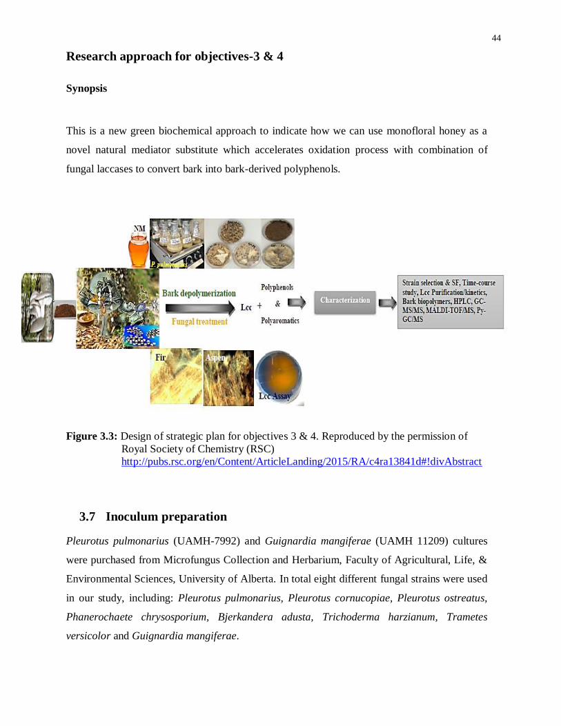
44
Research approach for objectives-3 & 4
Synopsis
This is a new green biochemical approach to indicate how we can use monofloral honey as a
novel natural mediator substitute which accelerates oxidation process with combination of
fungal laccases to convert bark into bark-derived polyphenols.
Figure 3.3: Design of strategic plan for objectives 3 & 4. Reproduced by the permission of
Royal Society of Chemistry (RSC)
http://pubs.rsc.org/en/Content/ArticleLanding/2015/RA/c4ra13841d#!divAbstract
3.7 Inoculum preparation
Pleurotus pulmonarius (UAMH-7992) and Guignardia mangiferae (UAMH 11209) cultures
were purchased from Microfungus Collection and Herbarium, Faculty of Agricultural, Life, &
Environmental Sciences, University of Alberta. In total eight different fungal strains were used
in our study, including: Pleurotus pulmonarius, Pleurotus cornucopiae, Pleurotus ostreatus,
Phanerochaete chrysosporium, Bjerkandera adusta, Trichoderma harzianum, Trametes
versicolor and Guignardia mangiferae.

45
Out of eight, six cultures were obtained from the Applied Mycology Lab, Faculty of Forestry,
University of Toronto. First the cultures were plated on potato dextrose agar media from
HiMedia® contains (Potatoes, infusion from 200g; dextrose 20g; agar 15g; water 1L; final pH-
5.6) and stored at 4oC. A loop with fungal strains was transmitted into disinfected potato
dextrose broth media pH-5.1 under antiseptic conditions and the flasks were incubated at 300C
in an orbital shaking incubator (150 rpm) aerated with continuous shaking.
3.8 Monofloral honey used as a NM-substitute in PM
Production media (PM) for P. pulmonarius contained: Monofloral Buckwheat Honey (Burke’s
Honey Ltd) mainly used as a substitute for natural mediators along with carbon, sugars,
minerals, vitamin source in the production medium (PM) at three different concentrations 5%,
7% and 10% (w/v), L-Asparagine monohydrate 3g/L, MgSO4.7H2O 0.05g/L, KH2PO4 0.5g/L,
K2HPO4 0.6g/L, CuSO4.5H2O 0.4mg/L, MnCl2.4H2O 0.09mg/L, NaMoO4.2H2O 0.02mg/L,
FeCl3 1mg/L, Thiamine-HCl 0.1mg/L (Ferhan et al., 2012).
50mL of PM were distributed into each 250 mL Erlenmeyer flasks and pH set to 5.5 with 1N
NaOH, autoclaved at (121°C) for 15 minutes. 10% (v/v) inoculum was transferred into PM
under sterile conditions and the flasks were incubated at 30C in a shaker (150 rpm) aerated
with constant shaking. Bark containing lignophenols were introduced into the PM after three
days during the lag-phase of submerged fermentation.
3.8.1 Time course study during SF
During submerged fermentation, time-courses were run for 24-days and each day samples were
stored and kept at 4oC for laccase assays. For optimization, three samples were collected at
each time from all various experimental shake flasks contain different concentrations of honey
in production medium (5%, 7%, and 10%). During submerged fermentation, the collected
samples were centrifuged in a Heraeus Biofuge®
Pico for about five minutes at 13,000 rpm and
the supernatant was used for laccase assays.
3.9 Laccase purification by UNO® sphere Q-1 column
UNO®
sphere Q-1 anion exchange column constructed for single-step polymerization was
used. The Q-1 anion exchange column specifications are: column bed volume (1.3mL),
maximum protein loading (20 mg), flow rate (0.5ml/min), column dimension (7 x 35mm), and

46
maximum operating pressure (4.5 MPa). The separation method was initiated by equilibrating
the UNO Q-1 column with Buffer A (25mM Tris-HCl; pH-8.1). Crude laccase was injected
onto a 50µl pre-column loop, after which, the sample in the loop was loaded onto the UNO Q-1
column using Buffer A.
The fluid flow in the system was incrementally changed to Buffer B (25mM Tris-HCl pH-8.1 +
0.5M NaCl) to elute the proteins captured in the column. At this phase, the laccase activity
resembled a peak of absorbance monitored at 280 nm and eluted as a single peak. Fractions
were observed at 280 nm using ChromLab™ software. The FPLC system (Bio-Rad, USA)
comprised a Biologic Duoflow pump system, BioFrac fraction collector, and UV detector.
The purified and concentrated enzyme was preserved at -20C and did not show any significant
loss of enzymatic activity over several months. The molecular weight of laccase enzyme was
determined by running 10% SDS-PAGE as earlier described by Höfer et al., (1999). The
protein gel was stained with Coomassie brilliant blue R-250 and commercially available
prestained protein ladder (Fermentas, USA) was used as a standard.
3.9.1 Laccase kinetics
Laccase kinetic constants (Vmax, Km, Kcat and Kcat/Km) were controlled by incubating the set
quantity of purified laccase enzyme with different concentrations of substrate (ABTS) ranging
from 200-1200 µM at 40oC, pH-3.5 as depicted previously (Li et al., 1999; Mishra et al., 2009).
3.10 Characterization techniques
3.10.1 Estimation of total polyphenolics
The total polyphenolic (TP) content in the samples was estimated by spectrophotometric
method using Folin-Denis reagent. 20 μl of sample of standard solution (catechol) was diluted
with 1.58 ml of distilled water and thoroughly mixed with 100 μl of Folin-Denis reagent (Fluka
Analytical, Sigma-Aldrich, St Louis, MO). The reaction mixture was left for 5 minutes at room
temperature and 300 μl of 25% (w/v) Na2CO3 stock solution was added into the reaction
mixture.
47
The reaction mixture was incubated at 40°C in a waterbath for 30 minutes and the absorbance
measured at λ765 nm with a UV-160 UV/Vis spectrometer (Shimadzu, USA). A standard curve
was plotted using 100, 200, 400, 600, 800 and 1000 ppm (mg/L) concentrations of catechol vs.
absorbance at 765 nm. Results were expressed as (mg catechol-equiv/100ml of sample) (Yen-
Peng 2011; Ferhan et al., 2013).
3.10.2 Sample preparation for HPLC
Fermented bark samples were centrifuged for 10 min at 10,000 rpm. The supernatant was
filtered through a 0.45 μm filter and analyzed by HPLC.
3.11 HPLC Analysis
The HPLC system comprised a Dionex BioLC 20 Series equipped with a GP50 gradient pump,
AS40 auto sampler, AD25 absorbance detector, and Chromeleon for information accumulation
and examination (CMS); all of them were from Dionex Technologies (USA). Ten μL of sample
were injected into a column by an autosampler. A Pinnacle DB Biphenyl C18 column (100 ×
4.6 mm, particle size 3 μm) from Restek (USA) was used for chromatographic separation. Two
mobile phase gradients were used for elution with a flow rate of 1 mL/min: 5% (v/v) HCOOH
in aqueous solution as (eluent A), and CH3OH as organic mobile phase (eluent B).
Chromatograms were chronicled at two different wavelengths, 290 and 340 nm (Pyrzynska et
al., 2009; Lachman et al., 2010).
The fermented samples were introduced with 50 mg of each bark sample during lag-phase, and
polyphenolics were determined spectrophotometrically and characterized based on their
reported retention time (tr) values.
Chromatographic separation was performed with gradient elution and the following steps were
required (Lachman et al., 2010): 70% eluent A + 30% eluent B by an isocratic elution for 0–15
min, 60% eluent A + 40% eluent B by a linear increase for 16–20 min, 55% eluent A + 45%
eluent B by a linear increase for 21– 30 min, 40% eluent A + 60% eluent B by a linear increase
for 31–50 min, 20% eluent A + 80% eluent B by a linear increase for 51–52 min, 10% eluent A
+ 90% eluent B by a linear increase for 52–60 min, 10% eluent A + 90% eluent B by an
isocratic elution for 61–63 min, 70% eluent A + 30% eluent B by a linear increase for 64–73
min and lastly 70% eluent A + 30% eluent B by an isocratic elution for 74–75 min,
respectively.

48
3.12 Thermal Analysis
Thermogravimetric analysis (TGA) of fungal treated and untreated/control bark samples were
performed on a computer controlled (TGA-Q500, TA Instruments, USA). Bark powder
samples were loaded into alumina pans and samples were heated from room temperature to
800C at a rate of 10
C min
-1 under N2 atmosphere.
3.13 GC-MS/MS
Fermented bark samples were centrifuged (10,000 rpm for 15 min) to detach cell biomass.
Supernatants were acidified with concentrated HCl to pH 2.5–3 and then completely separated
with three volumes (1:3 ml) of CH3COOC2H5. An organic layer was collected; moisture was
removed over anhydrous Na2SO4 and the sample filtered, and then samples were dried over
night to keep in the oven at 50C. According to the Lundquist method (1971) the ethyl acetate
extracts residues were examined as trimethyl silyl (TMS) derivatives. Dioxane and pyridine
(10:1 v/v) were added in the samples which were then silylated with 5-ml of silylation
derivatizing reagent trimethylchlorosilane (TMCS). To dissolve residual particles, the mixture
was heated to 60 oC for 15 min with regular shaking.
An aliquot of 1 ml of silylated compound was injected into a Saturn 2100T GC/MS/MS
(Varian, Inc. USA) equipped with a Varian 3900 (GC oven) and Saturn®
200MS workstation
software. PE–5MS capillary column (20m x 0.18mm i.d; 0.18 mm film thickness) and helium
was utilized as a carrier gas with flow rate of 1ml min-1
. The column temperature was
maintained (50 oC for 5 min); 50–300
oC (10
C min
-1, hold time for 5 min). 3 min was chosen
for solvent delay.
The exchange line and particle source temperatures were sustained at 200 and 250 oC. In full-
examine mode, electron ionization (EI) mass spectra within the scope of 30–550 (m/z) were
recorded at electron energy of 70 eV (Raj et al., 2007). Characterization of lignin-derived low
molecular weight (LMW) compounds, which are isolated from fungal treatment, was
interpreted by comparing their retention time (tr) with an existing database of the original
compounds.

49
3.14 MALDI-TOF-MS
Mass spectra of bark samples were analyzed using 4800 MALDI TOF/TOF Analyzer with
4000 Series Explorer Workflow software, by a fixed laser intensity and with an evenly
spontaneous spot pattern (Applied Biosystems / MDS Sciex). Before analysis, the spot position
was fixed carefully. The positive ion reflector mode for all MS measurements acquired for
identical repetition whereas -cyano-4-hydroxy cinnamic acid (Sigma-Aldrich) was used as
the matrix compound (Ferhan et al., 2012).
3.15 Sample preparation for Py-GC-MS
During the time course study, the bark fermented samples were harvested in the late
exponential-phase (after 18-20 days of inoculation) and afterwards dried overnight in an oven
(Thelco GCA Precision Scientific 18 Mechanical Convection Oven) at 60C. The oven-dried
fermented samples were milled into a fine powder and further analyzed by Py-GC-MS.
3.15.1 Py-GC-MS
Pyrolysis-GC-MS analysis was conducted using a Pt-filament loop test Pyroprobe 5250
pyrolyzer (CDS Analytical, Oxford, USA). For that purpose, 1 mg of specimen was installed in
quartz tubes applying glass wool, and pyrolyzed at 650 oC for 10 seconds (heating rate 10
oC/ms), with the pyrolysis valve oven and transfer line set at 325C.
Typically for thermostable materials such as charcoal or charred condensed tannins 750C is
suggested (Kaal et al., 2009), but 650C is recommended for uncharred biomass (Eckmeier et
al., 2007), which does not need so much energy to pyrolyze adequately. The pyrolysis products
were swept online into a 6890N GC instrument, with the inlet set at 325 C, in splitless mode.
The GC instrument was fixed with non-polar 5% phenyl, 95% dimethylpolysiloxane (HP-5MS)
column (30 m x 0.25 mm internal diameter; film thickness 0.25 µm) and helium was used as a
carrier gas (flow rate 1 ml/min). The GC oven remained heated from 50 to 325 C (held for 5
min) at 20 C/min.
The transfer line of GC–MS was maintained at 325 oC. The ion source (electron impact mode,
70 eV) of the 5975 MSD (Agilent Technologies, Palo Alto, USA) was controlled at 230 oC and
scanning of quadrupole detector at 150 oC, a range between m/z 50 and 550. Relative

50
proportions of the pyrolysis products were estimated from their peak areas, built on one or two
characteristic or major fragment ions. The aggregate of total quantified peak area (TQPA) was
fixed to 100%.
51
4 Chapter: Comparative treatments of bark to characterize bark
polyphenols
4.1 Abstract
This chapter describes the comparison of enzymatic, alkaline and UV/H2O2 treatments for the
extraction of polyphenolic extractives from beetle infested lodgepole pine (BILP) and mixed
aspen barks. The concept of green polymers has become more appealing due to the presence
of large volumes of processing residuals from timber and pulp industries. This, in turn,
supports the idea of developing new polymers based on bark extractives. Here we used a
chromatographic method to determine the chemical composition of some of the polyphenolic
compounds in bark extractives and observed the effect of different extraction methods on
extraction yield. Polyphenolic compounds separation was performed using HPLC in reverse-
phase mode with an Octadecylsilane (ODS), C18 column (3µm particle size) and an UV
detector. Detection wavelengths of 280, 310 and 370 nm were selected to allow better
separation of each compound. The comparative studies and effects of enzymatic, alkaline and
UV/H2O2 treatments on extractives yield and component contents were investigated. UV/H2O2
treatment exhibited the highest yield with 54% of dry bark weight extracted and was found to
degrade larger amounts of lignins/tannins than enzymatic and alkaline treatments. Conversely,
enzymatic treatment was good for holocellulose recovery.
4.2 Introduction
Today, we are facing an environmental crisis, and its associated socio-ecological burden,
mainly due to the extraction and processing of fossil fuels. Under these circumstances, it is
urgent to reduce fossil resource consumption. As such, replacing petrochemical products with
green biomaterials based on cost-effective renewable resources appears as a viable solution.
Petrochemical compounds such as phenol and its derivatives are produced in quantities over 10
million tons on an annual basis (essentialchemicalindustry.org/chemicals/phenol.html).
Therefore, to alleviate environmental concerns, it is important to find new natural raw
materials to produce polyphenolic compounds. Such raw materials are woody biomass and
bark, which are available in abundance as by-products from wood conversion industries.

52
Bark, similar to wood, is composed of cellulose, hemicellulose, lignin, extractives and ash.
Compared to wood, bark contains higher amounts of phenolic extractives, such as lignan, and
hydrolysable and condensed tannins, which have been of great interest to scientists and green
technologists during the last two decades. The main idea of this study to produce value-added
chemicals that are rich in phenolic structures, and thus suitable for wood adhesives (Wen-Jau et
al., 2003), PF resins (Yong et al., 2012), polyols (D’ Souza et al., 2013), polyurethane foams
(Yan et al., 2012), and Novolak resins (Wen-Jau et al., 2008). In addition, the lower toxicity of
bark based phenolics as compared to pure phenol would favour resin production
(Athanassiadou et al., 2010).
In British Columbia, lodgepole pine (Pinus contorta var. latifolia) accounts for 24% of the
total forest growing stock (Reports: COFI 2005). Large numbers of mature lodgepole pines
have been damaged by the attack of the mountain pine beetle (Dendroctonus ponderosae) and
its fungal associates. The scale of the mountain pine beetle attack in British Columbia is
unprecedented. In 2010, the beetle attack had affected over 18 million hectares of forest and
had killed 710 million m3
of lodgepole pine (Report: 2000-2010).
Aspen (Populus tremuloides) is considered as a source of abundant forage and important for
maintaining biodiversity in western and boreal regions in North America (Hogg et al., 2005). It
is one of the main timber sources in Canada, and in some Scandinavian and Baltic nations
(Johansson et al., 2002). Aspen accounts for 39.5% of the growing stock volume of the
forested land-base in these countries, which is almost equal to the 42.1% of the stock that is
attributable to all coniferous species (Forestry Canada 1993). In order to better understand the
bark extraction process and to provide better direction for the application of bark extractives, it
is important to explore and compare the effect of fundamentally different treatments such as
alkaline, enzymatic and UV/H2O2 on bark.
Due to the versatile chemical composition of bark, researchers have expended tremendous
efforts in developing extraction procedures that allow the isolation of specific phenolic
compounds. Among these procedures, extraction using organic solvents such as methanol and
ethanol has emerged as one of the most promising techniques for extracting phenolic
compounds, especially condensed tannins, with high purity and intact structural properties
(Jerez et al., 2009; Maimoona et al., 2011).

53
However, bark extraction with organic solvents has severe limitations, with notably low
extractives yield, generally ranging from 10-20% in bark weight (Sakai, 2001). In this sense,
the use of alkaline solvents, such as 1% NaOH, significantly increases the extractives yield and
releases large amounts of phenolic compounds (Vazquez et al., 1986; Vazquez et al., 2001).
Nevertheless, extraction with alkaline solvents is a complex process, which involves, as with
polar-solvents, the swelling and dissolution of polymers in cell walls, and also hydrolysis
reactions. Hence, the extractives obtained using alkaline solvents not only contain phenolic
compounds but also fractions of aliphatic acids and hemicelluloses (Yazaki et al., 1988). The
low selectivity of alkaline extraction, combined with the large diversity in composition of tree
barks, requires a systematic and thorough investigation of extraction process effects on each
species of tree bark.
In addition to these extractives, tree bark mainly contains cellulose, hemicellulose and lignin;
owing to the very selective nature of enzymes, to proceed to the extraction it is necessary to use
a cocktail of enzymes that include exoglucanase, endogluconase, β-glucosidase and ligninases.
These enzymes are already widely used for process integration to reduce the number of process
steps and increase yield (Galbe et al. 2002). Using a combination of cellulases, hemicellulases
and ligninases initiates hydrolytic degradation of the plant cell wall, which retains phenolics in
the polysaccharide-lignin network by hydrogen or hydrophobic bonding. Direct enzyme
catalysis is another approach to cleave ether and ester linkages between phenol and plant cell
wall polymers, as explained by Pinelo et al. (2008).
The mechanism of photocatalysis by UV/TiO2 has been described by several authors (Selli et
al. 1999; Tanaka et al. 1999; Lanzalunga et al. 2000; Castellan et al. 1990; Chang et al. 2004)
who have studied the degradation of various wastes, in particular lignin. Because of the
multifunctionality of lignin compared to phenol, wavelengths ranges of lower energy are
sufficient to initiate the degradation (Castellan et al., 1990). The main reactive species
produced in photocatalytic process by UV/H2O2 are hydroxyl and superoxide radicals
(Machado et al., 2000), which are proposed to allow the degradation of bark components.
The aim of the work was to determine and compare the effectiveness of an alkaline solution, a
cocktail of enzymes and UV/H2O2 treatment of BILP and mixed aspen barks to improve the
extraction efficiency and polyphenolic extractive yields. The raw materials considered for this

54
study were beetle infested lodgepole pine (BILP) bark and mixed aspen bark; both bark species
have a world-wide distribution but are most abundant in North America. In addition, we sought
to select the most appropriate wavelengths for each phenolic compound in bark polyphenolic
extractives. Biotechnological processes enable the discerning separation of main fractions
(cellulose, hemicellulose and lignin) from bark constituents.
4.3 Results and discussion
4.3.1 Bark composition
The major components of bark from infested lodgepole pine and aspen are summarized in
Table 4.1.
Table 4.1: Chemical composition of beetle infested lodgepole pine and aspen bark.
Composition (%) BILP Bark Aspen Bark
Ethanol-Toluene extractives 19.3 ± 0.5 18.4 ± 0.7
Water extractivesa 9.4 ± 0.4 17.1 ± 1.2
Total extractivesb 28.6 35.5
Klason Ligninc 33.1 ± 0.6 34.3 ± 0.6
Holocellulosec 39.2 ± 1.7 31.5 ± 0.7
α-cellulosec 24.8 ± 1.0 19.2 ± 1.4
Ash contents 4.1 ± 0.7 5.7 ± 0.5
Total extractsd 100.9 101.3
Mean ± S.D (n = 3)
a Yield obtained after extraction using ethanol-toluene.
b Addition of ethanol-toluene and water
extractives. c Calculated as the percentage of initial dry bark weight.
dAddition of total extracts,
Klason lignin, Holocellulose and Ash contents.

55
Bark major components analysis was reported in terms of extractives, which correspond to the
soluble fractions in ethanol/toluene mixture and 95% ethanol, water and the bark main
components, which refer to lignin, holocellulose and α-cellulose. The addition of bark fractions
from infested lodgepole pine and aspen resulted in 100.9 and 101.3 % of total bark weight,
respectively. The slight deviation observed may be due to some experimental errors. Infested
lodgepole pine bark and aspen bark contained 4.1% and 5.7% of ash, respectively, which falls
between the values obtained for other softwood and hardwood species such as Tsuga
heterophylla and Quercus mongolica (Herrick, 1980; Kofujita et al., 1999).
Infested lodgepole pine and aspen bark total extractives contents were 28.6% and 35.5% of dry
bark weight, which is generally high compared to the 16.6% obtained for Pinus pinaster after
dichloromethane, ethanol and water extraction and the 12.1% reported for the bark belly of
cork oak (Quercus suber L.), after successive extraction with ethanol and water, yet within the
range of the 43.40% reported for Populus hybrid bark (Fradinho et al., 2002; Jove et al., 2011;
Blankenhorn et al., 1988).
The extractives content in barks was assessed by subtracting the weight before and after
extraction and not by direct evaporation of the concentrated extractives. As a result, the values
could be slightly overestimated due to possible weight losses during manipulations. In addition,
the high extractives values may be due to other effects, such as bark particles size, which
drastically affect the fractionation and extraction yield, resulting in general difficulties in
precisely comparing the extractives content obtained with values in the literature (Miranda et
al., 2013). It is interesting to note that our results revealed higher extractives content in the
mixed aspen bark compare to lodgepole pine, which is unusual although such high values are
reported for Populus hybrid (Blankenhorn et al., 1988).
Aspen bark exhibited considerably higher amounts of water extractives compared to infested
lodgepole pine bark, indicating a larger presence of polar extractives such as lignans,
neolignans, and hydrolysable tannins (Sakai, 2001). Infested lodgepole pine and aspen bark’s
main components exhibited similar content of klason lignin but aspen holocellulose content
was lower (Rowell et al., 2005).

56
4.3.2 Treated bark composition
Infested lodgepole pine and aspen barks were extracted using alkaline, enzymatic, and
UV/H2O2 treatment and the influence of these treatments on bark chemical composition is
listed in Table 4.2 as well as shown in Figure 4.1. Infested lodgepole pine bark extractives
yield ranged from 49-55% depending on the treatment used, whereas aspen bark extractives
yield was slightly lower, ranging from 48.3-53%.
Table 4.2: Extraction yield from soluble part and bark residues (insoluble part) of beetle
infested lodgepole pine (BILP) and aspen bark after extraction with different methods.
Bark
species
Extraction
method
Soluble fraction
after extraction Insoluble fraction after extraction
Extraction Yield
(% w/w)
Klason Lignin
(KL)
(% w/w)
Holocellulose
(HC)
(% w/w)
α-Cellulose (α-C)
(% w/w)
BILP
Alkaline 52.3 ± 1.4 19.6 ± 0.3 26.7 ± 2.4 22.2 ± 1.6
Enzymatic 49.5 ± 2.2 25.1 ± 0.5 16.7 ± 2.6 8.9 ± 2.1
UV/H2O2 54.1 ± 2.1 14.8 ± 1.7 22.4 ± 0.3 13.2 ± 0.7
Aspen
Alkaline 48.3 ± 0.5 22.8 ± 0.2 26.4 ± 1.3 18.6 ± 1.9
Enzymatic 48.8 ± 2.7 21.7 ± 1.2 20.4 ± 2.6 10.8 ± 0.8
UV/H2O2 53.6 ± 2.7 15.9 ± 0.8 26.6 ± 2.1 15.8 ± 2.3
Mean ± S.D (n = 3)
The α-cellulose value is a fraction of the holocellulose and the percentage (% w/w) of bark
residues was calculated based on the relative proportion of total bark weight.

57
UV/H2O2 was an efficient treatment due to improvement of 1.13-fold extraction yield, as
compared to alkaline and enzymatic treatments, but the difference in extraction yields between the
different treatments did not exceed 6% in weight. Although only slight differences in extraction
yields were observed depending on the treatments used, we found different effects on the main
component constituents. The one-way ANOVA showed that the extraction yield of BILP among
three treatments represents no-significant difference (p > 0.05); however, bark residues of BILP
(KL, HC and α-C) were significantly different (p < 0.05). The extraction yield of aspen among
three treatments significantly different (p ˂ 0.05) and bark residues of aspen (KL, HC and α-C)
were also significantly different (p < 0.05). All the samples were precisely evaluated at the
beginning and then at the end of each treatment step to follow the mass balance. All the
experiments were performed in triplicate.
Mean ± S.D (n = 3)
Figure 4.1: Bark residue composition of Beetle Infested Lodgepole Pine (BILP) and Aspen
after three different treatments.

58
The lower holocellulose and α-cellulose content observed in enzymatic treated bark residues is
possibly due to the presence of cellulase and β-glucosidase in the enzyme cocktail, which
resulted in the removal of large amounts of polysaccharides from the bark. Matsushita et al.
(2010) previously observed the swelling of the phloem parenchyma’s primary cell wall and the
presence of many nano-clefts in the phloem fiber’s secondary wall after hydrothermal pre-
treatment, which may allow greater access of enzymes to the secondary cell wall cellulose,
supporting enzymatic hydrolysis.
In addition, the lower klason lignin content observed after UV/H2O2 treatment supports its
strong oxidative nature, which would preferentially degrade phenolic structures in bark such as
lignin and condensed tannins (Ugurlu et al., 2007). In general, alkaline treatments resulted in
the extraction of klason lignin and holocellulose with yields very similar to that of enzymatic
treatment, although the α-cellulose content did not vary, which suggests that the decrease in
holocellulose is mainly due to the degradation of hemicellulose.
SEM images at different magnifications were obtained for control, alkaline, enzymatically and
UV/H2O2 treated BILP and aspen barks reported in this work. Figure 4.2 illustrates the surface
morphology; the treated bark revealed the surface roughness while the untreated surface
appears smooth.
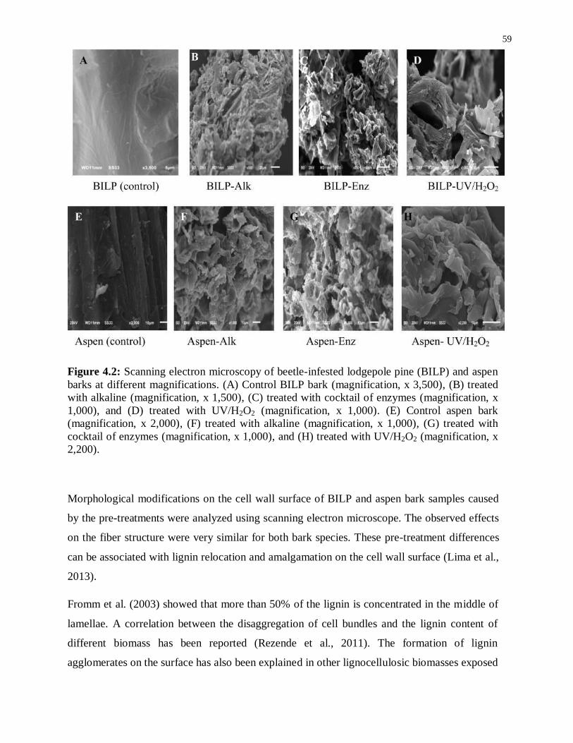
59
Figure 4.2: Scanning electron microscopy of beetle-infested lodgepole pine (BILP) and aspen
barks at different magnifications. (A) Control BILP bark (magnification, x 3,500), (B) treated
with alkaline (magnification, x 1,500), (C) treated with cocktail of enzymes (magnification, x
1,000), and (D) treated with UV/H2O2 (magnification, x 1,000). (E) Control aspen bark
(magnification, x 2,000), (F) treated with alkaline (magnification, x 1,000), (G) treated with
cocktail of enzymes (magnification, x 1,000), and (H) treated with UV/H2O2 (magnification, x
2,200).
Morphological modifications on the cell wall surface of BILP and aspen bark samples caused
by the pre-treatments were analyzed using scanning electron microscope. The observed effects
on the fiber structure were very similar for both bark species. These pre-treatment differences
can be associated with lignin relocation and amalgamation on the cell wall surface (Lima et al.,
2013).
Fromm et al. (2003) showed that more than 50% of the lignin is concentrated in the middle of
lamellae. A correlation between the disaggregation of cell bundles and the lignin content of
different biomass has been reported (Rezende et al., 2011). The formation of lignin
agglomerates on the surface has also been explained in other lignocellulosic biomasses exposed

60
to steam-explosion, diluted acid or organosolv pretreatments (Koo et al., 2012; Heiss-Blanquet
et al., 2011).
Following the different treatments, the total phenolic compounds in aspen and BILP bark
extractives were assessed. The given values in the above mentioned tables (Table 4.1 and Table
4.2) are the means of three replicates of each. Enzymatic treatment resulted in 6.95±0.40 mg/g
and 8.82±0.51 mg/g, alkali treatment provided 4.3±0.66 mg/g and 4.64±0.44 mg/g, and
UV/H2O2 treatment yielded 5.62±0.30 mg/g and 8.0±0.52 mg/g, respectively. Generally,
extractives from BILP exhibited higher amounts of phenolic compounds compared to aspen.
The extractive levels in barks are higher than in wood and, similarly, the total phenols level in
bark has been found to be higher (Chow et al., 2008), although some studies have shown higher
polyphenols levels in wood than in bark (Chang et al., 2001; Wang et al., 2004). The total
polyphenolic level attained by sequential extraction of wood of Maclura tinctoria was higher than
compared to bark, although these results were similar, and the differences when acetone: water
was used as extraction solvents were not significant (P > 0.05) reported by Lamounier et al.,
(2012). Bark of Eucalyptus globulus has less favourable chemical composition with more
extractives, pentosans, and ash contents, although the pulp obtained with tops was similar to the
wood pulp in regards to yield and strength properties (Miranda et al., 2012).
The values found in the literature for the total phenols content of methanol: water (80:20, v/v)
extracts of Eucalyptus bark (E. camaldulensis, E. globulus, and E. rudis) were in the range of
0.25 to 9.16 % (Conde et al., 1997). Vázquez et al. (2008) reported a value of 1.48 mg/g for
Eucalyptus globulus bark extracted with this solvent. Subsequently, the estimation of total
phenolic compounds should be reflected as a polyphenol index, depending upon the applied
method.
The values obtained after the different treatments of BILP and mixed aspen bark were
generally low compared to the values found in the literature for the total phenols. However, the
extraction with organic solvents generally resulted in low extractives yield (20.8%) in bark
(Lamounier et al., 2012), obtained with cyclohexane: ethanol (1:2, v/v) solvent mixture
compared to the treatments reported in this chapter.
Most co-elutions occurred between the peaks of ferulic acid and quercetin, so the gradient was
modified in this part of the chromatogram to allow a progressive elution of components and

61
minimize overlapping. The chromatograms corresponding to the better separation of
polyphenolic extractives treated with a cocktail of enzymes are shown in Figure 4.3 from (A-
C) for BILP and (D-F) for aspen bark, respectively. This demonstrates that all analytes can be
successfully separated at three different wavelengths. Polyphenolic constituents of the
extractives obtained after enzymatic cocktail treatment of BILP and aspen bark exhibited better
separation at three different wavelengths as compared to the extractives from other treatments.
In all cases, the yields from enzymatic cocktails were comparable to the other two treatments,
but the enzymatic treatment resulted in better separations of polyphenolics, while the
chromatograms of the other treatments showed overlapping separations. The separation of
polyphenolic complex molecules in bark under acidic mobile phase with the combination of
acetonitrile gradient is a common method in reverse-phase liquid chromatography.
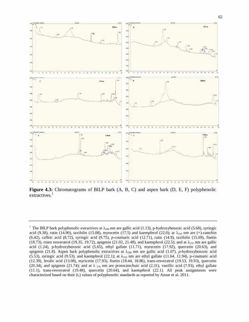
62
Figure 4.3: Chromatograms of BILP bark (A, B, C) and aspen bark (D, E, F) polyphenolic
extractives.1
1 The BILP bark polyphenolic extractives at λ280 nm are gallic acid (1.13), p-hydroxybenzoic acid (5.68), syringic
acid (9.38), rutin (14.90), taxifolin (15.08), myrecetin (17.5) and kaempferol (22.0); at λ310 nm are (+)-catechin
(6.42), caffeic acid (8.72), syringic acid (9.75), p-coumaric acid (12.71), rutin (14.9), taxifolin (15.69), fisetin
(18.73), trans resveratrol (19.35, 19.72), apigenin (21.02, 21.48), and kaempferol (22.5); and at λ370 nm are gallic
acid (1.24), p-hydroxybenzoic acid (5.65), ethyl gallate (11.71), myrecetin (17.92), quercetin (20.63), and
apigenin (21.8). Aspen bark polyphenolic extractives at λ280 nm are gallic acid (1.07), p-hydroxybenzoic acid
(5.53), syringic acid (9.53), and kaempferol (22.1); at λ310 nm are ethyl gallate (11.64, 11.94), p-coumaric acid
(12.39), ferulic acid (13.08), myricetin (17.93), fisetin (18.44, 18.86), trans-resveratrol (19.53, 19.93), quercetin
(20.34), and apigenin (21.74); and at λ 370 nm are protocatechuic acid (2.31), vanillic acid (7.93), ethyl gallate
(11.1), trans-resveratrol (19.48), quercetin (20.64), and kaempferol (22.1). All peak assignments were characterized based on their (tr) values of polyphenolic standards as reported by Aznar et al. 2011.

63
It was reported that under alkaline conditions, the caffeic and syringic acids may overlap, as
can p-coumaric acid and vanillic acid. Under alkaline conditions, phenolic acid tends to
complex with sodium and prevents an efficient separation, due to alteration of the
hydrophilicity of analytes, thereby reducing its interaction with the stationary phase (Hemstrom
et al., 2006). Furthermore, under alkaline conditions, syringic acid and ferulic acid were not
completely separated; moreover, p-hydroxybenzoic acid shifted as an unsymmetrical peak. As
a result, syringic acid and ferulic acid, as well as caffeic acid and hydroxycinnamic acid, nearly
overlapped (Pomponio et al., 2002).
Although the difficulties in separating analytes may be due to the samples basicity, samples
dilution during preparation, as well as the use of 0.1% formic acid as eluent may prevent these
phenomena. The very low selectivity of alkaline and UV/H2O2 compared to enzymatic
treatment lead to obtaining a very diverse range of molecules with close structural features,
which could also complicate analytes separation.
Degradation of various products treated by AOPs using UV light and H2O2, linked to specific
pH dependent mechanisms, produces very reactive hydroxyl radicals (•OH) in the reaction
mixture, thus making it more susceptible to oxidation (Machado et al., 1994). Nevertheless, the
chromatogram and the chromatographic peak patterns were comparable to those reported in a
previously published article (Aznar et al., 2011).
As a result, these two treatments have low selectivity embedded in very diverse structures that
might be difficult to separate during chromatography. Moreover, UV/H2O2 and alkaline
treatment lead to highly basic extractives, which could prevent an effective separation due to
possible interactions between analytes and column. However, our results indicate that
enzymatic treatment, due to its highly specificity, low pH, and cleavage of selective bonds
among the molecules can probably affect the polyphenolic compounds in bark.
4.4 Conclusions
The effect of three different treatments on the surface morphology and chemical composition
of bark residues and polyphenolic extractives from BILP and aspen barks were studied. The
total chemical composition of BILP bark was ash (ca. 4%), extractives (ca. 28%), Klason lignin
(ca. 33%), holocellulose (ca. 39%), while mixed aspen bark residues consisted of ash (ca. 5%),
extractives (ca. 35%), Klason lignin (ca. 34%), and holocellulose (ca. 31%), respectively. The

64
HPLC analysis shows fast, selective, sensitive and reliable determination of the most common
polyphenolics in bark extractives. UV/H2O2 treatment was able to preferentially remove
phenolic compounds, with limited effect on the sugar concentration of the bark hydrolysates.
The removal of phenolic compounds (catechol equivalent) was found to be 0.8% in BILP and
0.6% in aspen bark under UV/H2O2 treatment. Furthermore, these results elucidate the effects
of different treatments on bark-derived polyphenols, which can be considered as promising
methods for applications in bioconversion and polymer industrial processes.

65
5 Chapter: Brazilian isolated fungal strain for ligninases
production
5.1 Abstract
The potential of ligninases in green processes for effective valorization of lignin can be shown
through investigation of enzymatic cocktails containing different lignin degrading enzymes.
The present study deals with the screening of potential fungal strains useful for the liquefaction
of bark containing lignin. Three different local isolates (Pleurotus ostreatus POS97/14,
Pycnoporus sanguineus and the Brazilian isolated fungal strain) were selected out of ten
different strains for ligninases production. Maximum production of enzymes was observed in
the local isolated fungal strain after ten days in submerged fermentation. The isolated fungal
strain produces ligninases, mainly manganese peroxidase (MnP). The enzyme oxidized a
variety of the usual MnP substrates, including lignin related phenols. Furthermore, the partial
purification for MnP was determined by fast protein liquid chromatography (FPLC) and the
molecular weight was evaluated by sodium dodecyl sulphate polyacrylamide gel
electrophoresis (SDS-PAGE).
5.2 Introduction
Among microorganisms that colonize wood, white-rot fungi are regarded as considerable lignin
degraders. They produce extracellular enzymes, such as MnP, LiP and laccase, which play
important roles in lignin biodegradation (Mester et al., 2000). Pleurotus ostreatus is a white rot
fungus belonging to the basidiomycetes. Pleurotus ostreatus is an edible mushroom, as well as
a food supplement for hypercholesterolemia patients due to presence of higher concentrations
of lovastatins (Alarcon et al., 2003). It was also noticed that due to lack of lignin peroxidases in
some genera of basidiomycetes, such as Pleurotus spp, laccases are involved in the degradation
of lignin (Mansur et al., 2003).
The combined action of laccase and aryl alcohol oxidase decreases the molecular weights of
soluble lignosulphonates by P. ostreatus (Mansur et al., 2003). Ligninolytic system
configuration is complicated and species-restricted (Baldrian 2008). White rot fungi produce
ligninases, including MnPs, LiPs and Lcc, which are possible contributors to fungal

66
ligninolysis. White-rot fungi were mainly found to produce consistent products following the
ligninolysis of model lignin compounds (Cullen et al., 2008).
MnP belongs to the family of peroxidases, and the methodical name of this enzyme is
Mn(II):H2O2 oxidoreductase. According to protein databank, the MnP containing cofactor is
protoporphyrin IX having Fe (C34H32FeN4O4) incorporated with Mn+2
and Ca+2
ions required
for enzyme activity.
Eventually, peroxidases oxidize phenolic compounds and reduce molecular oxygen to water
(Mester et al., 2000). Ligninases oxidize several environmental pollutants such as polycyclic
aromatic hydrocarbons, dyes and chlorophenols. Heme containing enzymes such as LiP and
MnP also have catalytic cycles characteristic of other peroxidases. LiP has an ability to oxidize
many aromatic compounds, whereas MnP oxidizes Mn (II) to Mn (III) (Sadhasivam et al.,
2008; Wang et al., 2009).
Laccases [benzenediol: oxygen oxidoreductase, EC (1. 10. 3. 2)] are multi-copper blue
oxidases commonly found in higher plants, several bacteria and some insects. Nevertheless, the
well-characterized laccases are fungal in origin (Sadhasivam et al., 2008). Laccases have
significant importance in many industrial areas due to their remarkable catalytic properties.
Potential applications include immunoassay bio-labeling, biosensors, biocatalysts, and
advancement of oxygen cathodes in biofuel cells. In addition, they have good prospects in the
environmental sector, including use in textile dye bleaching, pulp delignification and
xenobiotic compound degradation due to their wide-ranged substrate specificity (Sadhasivam
et al., 2008; Mazumder et al., 2009). The present applications of this enzyme motivated us to
do new basic research.
The current research activity of ligninases includes utilizing the local lignin sources
(Eucalyptus and sugarcane bagasse) and checking their delignification pattern. The
characterization of ligninases (MnPs, LiPs and Lcc,) from Brazilian isolated fungal strain, with
respect to production, partial purification and time course studies, is reported in this study.
5.3 Results and Discussion
Due to broad substrate specificity, the ligninases potential of white rot fungi has been reported
as liable for the alteration and mineralization of organic pollutants that are structurally similar

67
to lignin. Ligninases are the combination of three peroxidases, MnP, LiP and laccases, which
are characterized as lignin degrading enzymes. The distribution of MnP and laccases are very
frequent in white rot fungi, while LiP is not (Palaez et al., 1995).
Degradation and decolorization of many organic pollutants by white-rot fungi have been
reported by many researchers (Clemente et al., 2001), but MnP and LiP are noted mainly for
the degradation of polymeric dyes (Moreira et al., 2001). The white rot fungi have the ability to
decolorize polymeric dyes because of existing ligninolytic enzymes. An anthracene derivative
like Remazol Brilliant Blue R (RBBR), which is considered to be an organic pollutant and is
similar to the lignin polymers, has been used as a model compound by many researchers to
measure ligninases activity (Moreira et al., 2001).
Therefore, we examined a petri plate assay to identify ligninases-producing fungal strains,
based on their ability to decolorize RBBR. Using this system, we determined that the fungal
strains Pleurotus ostreatus, Pycnoporus sanguineus and the new Brazilian isolate all exhibited
ligninolytic activity (Figure 5.1).
Figure 5.1: Potential screening of three fungal strains out of ten based on the ligninases
production and degradation of RBBR dye, where, A - Pleurotus ostreatus POS 97/14, B-
Pycnoporus sanguineus and C was Brazilian isolated fungal strain.
5.3.1 Ligninases production during time course studies
To better quantify ligninases production, the ligninolytic activity of the aforementioned strains
was measured in liquid cultures. During the time course study, triplicate fermentation flasks
A B C

68
were harvested after every 48h and the culture supernatants were analyzed for ligninases
activity and cell biomass was also noted. The maximum production of ligninolytic activity by
the newly isolated Brazilian fungal strain, MnP (64 IU l−1
), LiP (26.35 IU l−1
) and laccase (5.44
IU l−1
), respectively, was attained in 10 days. In contrast, the ligninolytic activity of Pleurotus
ostreatus, and Pycnoporus sanguineus (Table 5.1) had minimal Remazol Brilliant Blue R
(RBBR) decolorization efficiency and surprisingly did not show any peroxidase activity. The
low peroxidase activity and decolorization efficiency, especially as compared to that which has
been reported for other strains (Ganoderma spp, Stereum ostrea, and Trametes versicolor)
(Pointing 2001) raises the possibility that the growing conditions or the growth medium were
not optimal for these fungal strains.
Table 5.1: Measurement of ligninases activities during the time course study in three different
fungal strains.
Brazilian isolated fungal strain
Days Laccase (IU l−1
) LiP (IU l−1
) MnP (IU l−1
)
2 1.42±0.01f 17.6±0.05
e 16.7±0.21
f
4 3.17±0.01e 19.7±0.09
d 36.8±0.09
d
6 4.45±0.00d 21.8±0.03
c 56.8±0.09
c
8 5.31±0.01b 23.8±0.09
b 59.3±0.09
b
10 5.44±0.01a 26.4±0.07
a 64.1±0.12
a
12 5.22±0.01c 14.9±0.10
f 30.9±0.14
e
Pleurotus ostreatus POS97/14
Days Laccase (IU l−1
) LiP (IU l−1
) MnP (IU l−1
)
2 0.14±0.01d 0.82±0.07
f 5.17±0.11
e
4 0.18±0.02d 2.93±0.05
e 7.62±0.09
d
6 0.31±0.01c 4.03±0.07
d 10.09±0.16
c
8 0.56±0.01b 5.84±0.04
c 13.32±0.14
b

69
10 0.80±0.01a 6.77±0.09
a 17.91±0.45
a
12 0.54±0.01b 6.58±0.06
b 12.93±0.14
b
Pycnoporus sanguineus
Days Laccase (IU l−1
) LiP (IU l−1
) MnP (IU l−1
)
2 0.31±0.01d 0.72±0.04
f 1.09±0.14
f
4 0.81±0.02c 1.73±0.13
e 6.11±0.11
d
6 1.07±0.01b 2.41±0.05
c 9.82±0.14
c
8 1.13±0.00a 2.77±0.08
b 10.93±0.14
b
10 1.15±0.01a 3.41±0.09
a 13.21±0.14
a
12 1.12±0.01a 2.06±0.13
d 1.66±0.20
e
Mean ± S.D (n = 3)
It was also observed that cell biomass increased with increasing fermentation time. In addition,
ligninases activity also increased with increasing fermentation time for 10 days, but a further
increase (12 days) in fermentation time (Table 5.1) decreased ligninase activities (Table 5.1).
The local isolated fungal strain was the best producer of MnP (64 IU l−1
) as the main enzyme
activity, followed by LiP (26.35 IU l−1
) and laccase (5.44 IU l−1
), respectively, as evidenced by
the high percentage (94%) of RBBR decolorization in the broth medium. The given values of
ligninases activities in Table 5.1 are the mean of three replicates of each. The least significant
difference among the experimental values was calculated by ANOVA using Assistat-Statistical
Assistance version 7.6-beta software.
During exponential phase, the increase of cell biomass resulted in an increase in secretory
proteins in the growth medium. Increase of cell biomass yield was associated with increased
extracellular secretory protein in the fermentation medium up to the 12th day, when biomass
started to decrease. However, the specific activities of MnP and LiP constantly increased
(Pointing 2001).
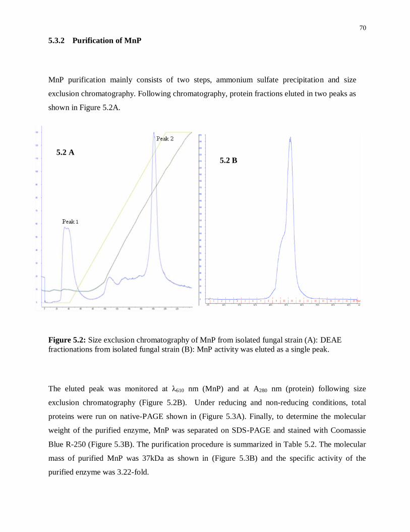
70
5.3.2 Purification of MnP
MnP purification mainly consists of two steps, ammonium sulfate precipitation and size
exclusion chromatography. Following chromatography, protein fractions eluted in two peaks as
shown in Figure 5.2A.
Figure 5.2: Size exclusion chromatography of MnP from isolated fungal strain (A): DEAE
fractionations from isolated fungal strain (B): MnP activity was eluted as a single peak.
The eluted peak was monitored at λ610 nm (MnP) and at A280 nm (protein) following size
exclusion chromatography (Figure 5.2B). Under reducing and non-reducing conditions, total
proteins were run on native-PAGE shown in (Figure 5.3A). Finally, to determine the molecular
weight of the purified enzyme, MnP was separated on SDS-PAGE and stained with Coomassie
Blue R-250 (Figure 5.3B). The purification procedure is summarized in Table 5.2. The molecular
mass of purified MnP was 37kDa as shown in (Figure 5.3B) and the specific activity of the
purified enzyme was 3.22-fold.
5.2 A 5.2 B
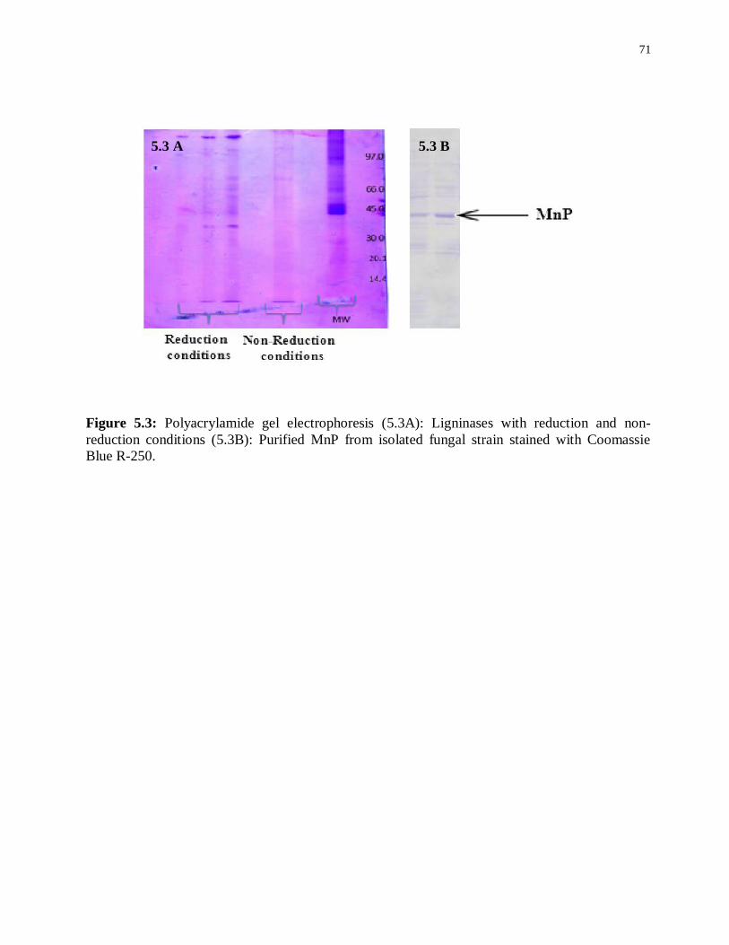
71
Figure 5.3: Polyacrylamide gel electrophoresis (5.3A): Ligninases with reduction and non-
reduction conditions (5.3B): Purified MnP from isolated fungal strain stained with Coomassie
Blue R-250.
5.3 A
5.3 A 5.3 B

72
Table 5.2: Partial purification summary of MnP produced during submerged fermentation by
isolated fungal strain under optimized conditions.
5.3.3 Electron microscopy
Scanning electron microscopy (SEM) was performed to determine the surface morphology of
lignin samples of sugarcane bagasse and eucalyptus black liquor that were treated with the
isolated fungal strain (Figure 5.4). Abundant fungal growths were seen on bagasse and
eucalyptus black liquor, which indicated the decay of lignin samples. After fungal
modification, the fiber structure of bagasse specifically shows the presence of bore holes,
indicated by arrows in Figure 5.4 (C and D).
Purification steps Activity/L Protein
content
(U/mg)
Specific
activity
(U/mg)
Purification
fold
Crude enzyme 4467.8 228.9 98.6 1
Ammonium
Sulphate
1634.9 157.8 182.1 1.8
Dialysis 1442.6 101.2 190.1 1.9
1st DEAE-
Sepharose Fast
Flow column (10 x
300mm)
1381.5 89.0 219.8 2.2
2nd
DEAE Superdex 75 (10 x
300mm)
1254.1 19.9 293.6 2.9
Sephadex G-100 1085.9 5.6 317.9 3.2

73

74
Figure 5.4: Scanning electron microscopy of sugarcane bagasse (SCB) and Black liquor (BL) of
Eucalyptus. (A & B) Control SCB (magnification, x 400); (C & D) bagasse treated with Brazilian
isolated fungal strain grown after 2 weeks (magnification, x 2k); (E & F) Control BL of
Eucalyptus fibers (magnification, x 10k); (G & H) BL treated with local fungal isolate grown after
2 weeks (magnification, x 5k); indicates the fungal growth where k denoted by (x1000
magnification).
Sugarcane bagasse contains cellulose (40–50%), hemicellulose (20–30%), lignin (20–25%) and
ash (1.5–3.0%). Hemicelluloses are complex polysaccharides that appear in correlation with
cellulose in the cell wall. It is a mixture of polysaccharides, comprised of almost all sugars, such
as glucose, mannose, xylose, arabinose, methylglucoronic and galaturonic acids, with an average
molecular weight < 30,000 Da (Saxena et al., 2009). Mushroom growth is encouraged on
sugarcane bagasse in some countries, including Brazil. This is in response to the increase in global
demands for ethanol (from 66-125 million m3), that is required in the production of bagasse
(Pandey et al., 2000; Balat et al., 2009). By utilizing agricultural wastes and converting them into
high valued products, this is an alternative approach to decreasing the waste disposal costs (Sidana
et al., 2014).
Moreover, the main monosaccharides (glucose and xylose) were found in Eucalyptus grandis bark
( 40% glucose, and 9% xylose, respectively) reported by Yu et al. (2010). The edible
mushroom production with eucalyptus is considered to be a raw material for the pulp and paper
mill. Eucalypt farming competes with agricultural crops, is more suitable to farmers to consider as
a substitute for agricultural crops, e.g. cassava. The farmers are also supportive of the idea of
eucalypt farming (Reungchai, 1996).
5.3.4 MALDI-TOF MS
The MALDI-TOF method provides information regarding the interpretation of lignin
composition. This method also helps to determine the lignin structure. After biological
modification, it illustrates the effectiveness of fungal attack on the lignin bio-molecule and the
impact on its compositional arrangement. The fungal attack generally affected phenolic units
of the lignin molecule, as observed by the reduction in the relative frequency of β-O-4-linked H
units, which are mainly terminal units with free phenolic groups (Lapierre et al., 1988).
After delignification, the complete structures of molar mass distribution of lignophenols by
MALDI-TOF-MS spectra are shown in (Figure 5.5). In delignification spectra, the lignin

75
monomers specified the dominant signals at m/z 171, 188 (coniferyl aldehyde), m/z 227, 229,
233 (syringyl propene), m/z 334, 378, 397 (phenyl coumaran), m/z 453 (resinol), m/z 655, 715
(dimethoxyphenol) reported by Bayerbach et al. (2006). Most monomers were by-products
from guaiacol and syringol. Most dimers were assumed to be a phenylcoumaran structure.
Biphenyl and resinol were infrequent.
171: Coniferyl aldehyde
227, 249: Syringylpropene
334, 378: Phenylcoumaran
188: Coniferyl aldehyde
233: Syringylpropene 397: Phenylcoumaran
453: Resinol
655, 715: DMP

76
Figure 5.5: MW distribution to determine the lignin monomers by using MALDI-TOF-MS where
α-cyano-4-hydroxy cinnamic acid was used as matrix compound.
5.4 Conclusions
With the increasing global anxiety over fossil fuel use and its environmental footprint, there is
a strong interest in using biorenewable materials as substitute feedstocks for making more
environmental-friendly biomaterials. The present study screens potential fungal strains for
ligninases production, and includes partial purification of MnP and the degradation pattern of
local lignin resources found in Brazil. The industrial and biotechnological application of
ligninases is constantly increasing due to their multiple uses and applications in a diversity of
378.9171.1
211.2
334.8
144.8
293.6
227.3
440.9 568.2265.5644.3 701.8 855.4 929.5494.7 1060.4757.0 1157.7
a-ciano_branco_23_08_12 0:F1 MS Raw
0.00
0.25
0.50
0.75
1.00
1.25
5x10
Inte
ns. [a
.u.]
171.0
144.8
265.4 378.9
334.8
211.2
440.9 522.6
SCB_a ciano_23_08_12 0:C3 MS Raw
0
2
4
6
4x10
Inte
ns. [a
.u.]
264.5
165.9
170.9 310.6440.8227.2
144.7
715.2403.9 616.2 843.4563.2 666.1500.0347.7 972.5 1066.2
SCB+O_a ciano_23_08_12 0:E3 MS Raw
0.00
0.25
0.50
0.75
1.00
1.25
5x10
Inte
ns. [a
.u.]
715.3
843.5655.3
759.3 972.5625.2688.2
1066.3
SCB+O_a ciano_23_08_12_620_1600_E3 0:E3 MS Raw
0.0
0.5
1.0
1.5
2.0
4x10
Inte
ns. [a
.u.]
715.5
655.5
843.7759.5625.4 688.4972.7905.6 1060.7
SCB+O_a ciano_23_08_12_620_1600_E4 0:E4 MS Raw
0
2
4
6
4x10
Inte
ns. [a
.u.]
200 400 600 800 1000 1200m/z
378: Phenylcoumaran
440: Resinol
715: DMP

77
processes. Their ability to remove xenobiotic pollutants and produce polymeric products makes
them a beneficial tool for bioremediation purposes. The unknown isolated fungal strain has the
potential for delignification. Nevertheless, further molecular biology studies are needed to
identify the species by 18S-rDNA.
78
6 Chapter: Optimization of temperature and pH for Manganese
Peroxidase (MnP)
6.1 Abstract
In this study, a fungal species selected for ligninases production was investigated by 18S
ribosomal DNA sequence analysis. Two primers sets were chosen to amplify a major part of the
18S rDNA, which resulted in intense PCR product of approximately 550–820 bp in size per
sample. The results suggest that the 18S rDNA-based approach is a useful tool for identification
of unknown potential fungal species for ligninases production. The isolated fungal species
produces mainly manganese peroxidase (MnP). It oxidizes a wide range of typical MnP substrates
as well as lignin-related polyphenols. The process parameters such as temperature and pH of the
medium in shake flask experiments were optimized for the maximum production of manganese
peroxidases. Time course studies showed that maximum production of ligninolytic enzymes MnP
(64 IUL-1
), LiP (26.4 IUL-1
), and laccase (5.44 IUL-1
), respectively, were achieved after 10 days
of cultivation under optimum conditions (temperature and pH). Furthermore, the biological
decolorization of Remazol Brilliant Blue R (RBBR) dye following 10 days of cultivation was
94%. NCBI BLAST was used to search for closest matched sequences in the GenBank database
and based on sequence homology the first BLAST hit was Dothioraceae sp. LM572 with
accession number EF060858.1.
6.2 Introduction
Filamentous fungi play a prominent role in decomposition of lignocellulose. There are three
types of fungi living on dead wood that better degrade one or more wood components: soft-rot
fungi, brown-rot fungi, and white-rot fungi. Soft-rot fungi (Ascomycetes and Fungi Imperfecti)
can decay cellulose as well as also partially degrade lignin (Grazyna et al., 1989).
Extracellular oxidoreductases, including lignin peroxidase (LiP), manganese peroxidase (MnP)
and laccase produced by wood-decomposing microorganism, are directly involved in the
degradation of lignin in natural lignocellulosic substrates and various xenobiotic compounds
including dyes (Abrahao et al. 2008). Some wood-degrading fungi contain all three classes of
ligninases, while the others include only one or two of these ligninolytic enzymes (Dhouib et
al. 2005). The ligninases produce a colourless halo around microbial growth in media

79
containing coloured indicator compounds (Dhouib et al. 2005). So, the lignin degradation
capability of microbes is initially screened indirectly on solid media containing different
indicator compounds.
Although, wood decay fungi are primarily basidiomycetes, other microorganisms are also
involved in the lignocellulosic decaying processes (Nilsson et al. 1989). Wood biodegradation
by certain ascomycetes was first described in detail and designated as “soft rot” by Savory
(1954). Nilsson et al. (1989) showed that, some higher ascomycetes, mainly Daldinia
concentrica, degraded aspen wood with the same strength as Trametes versicolor, a
basidiomycete characteristically classified as white-rot fungus.
The ascomycete, Chrysonilia sitophila could degrade rice hull and Pinus radiata bark products
and also produce ligninolytic and cellulolytic enzymes (Ferraz et al. 1991). However, some of
ligninolytic microorganisms have ability to degrade a wide range of pollutant substances which
are similar to lignin or its derivative. This is an interesting method due to low cost, specificity
and likelihood of a total mineralization of these compounds (Van der Zee et al. 2005).
In this study, an ascomycete producing ligninolytic enzyme, was isolated from leaf samples of
Combretum leprosum in the tropical dry forest (Caatinga), in Brazil. The fungal species was
identified by 18S rDNA and its ability for lignin degradation was characterized by production
of ligninases and decolorization of polyaromatic ligninolytic indicator dye, RBBR respectively
(as explained above in Chapter 3; Section 3.3).
6.3 Results and Discussion
6.3.1 Ligninases production during the time course studies
The Brazilian isolated fungal strain was identified as Dothioraceae sp. using 18 S rDNA
sequence analysis (as explained above in Chapter 3; Section 3.5) was used for the production
of ligninases in submerged fermentation. Dothioraceae belongs to Phylum of Ascomycota as
recognised by NCBI Taxonomy in the Encyclopedia of Life. The triplicate fermentation flasks
were harvested every 48 h, and the culture supernatants were analyzed for ligninases activity
and the dry weight of biomass (residue) was also recorded. The results of Dothioraceae fungal
species during time course study proved that the highest production of MnP (64 IUl−1
), LiP

80
(26.35 IUl−1
) and laccase (5.44 IUl−1
) were achieved in 10 days as shown in (Table 5.1, Figure
6.1).
Figure 6.1: Time course for ligninases production and cell biomass produced by Dothioraceae sp.
The enzymatic pattern for Dothioraceae fungal species was distinguished among the known
strains P. ostreatus, and P. sanguineus. It showed ligninolytic activities that were exceptional
as compared to other strains (Ganoderma spp, Stereum ostrea, and Trametes versicolor) which
had the lowest remazol decolorization efficacy and did not show any peroxidase activity.
Dothioraceae had the highest percentage of RBBR decolorization (94%) in liquid medium.
As a control, the fungi were cultivated in broth culture medium without supplementation.
During the development period, a record was carried out for biomass and protein yield in the
broth culture medium. The concentration of proteins was comparable to the biomass yield,
until the 12th day, when biomass started to decrease (Figure 6.2). The specific activities of
MnP and LiP conversely continually increased.
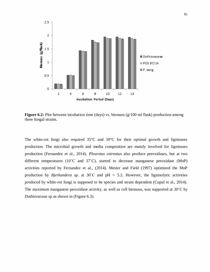
81
Figure 6.2: Plot between incubation time (days) vs. biomass (g/100 ml flask) production among
three fungal strains.
The white-rot fungi also required 35°C and 30°C for their optimal growth and ligninases
production. The microbial growth and media composition are mainly involved for ligninases
production (Fernandez et al., 2014). Pleurotus ostreatus also produce peroxidases, but at two
different temperatures (10˚C and 37˚C), started to decrease manganese peroxidase (MnP)
activities reported by Fernandez et al., (2014). Mester and Field (1997) optimized the MnP
production by Bjerkandera sp. at 30˚C and pH = 5.2. However, the ligninolytic activities
produced by white-rot fungi is supposed to be species and strain dependent (Cupul et al., 2014).
The maximum manganese peroxidase activity, as well as cell biomass, was supported at 30˚C by
Dothioraceae sp as shown in (Figure 6.3).
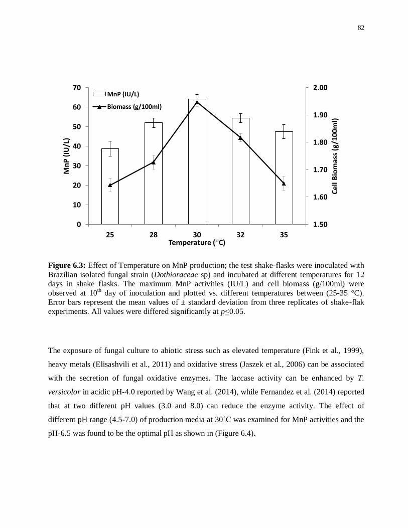
82
Figure 6.3: Effect of Temperature on MnP production; the test shake-flasks were inoculated with
Brazilian isolated fungal strain (Dothioraceae sp) and incubated at different temperatures for 12
days in shake flasks. The maximum MnP activities (IU/L) and cell biomass (g/100ml) were
observed at 10th day of inoculation and plotted vs. different temperatures between (25-35 °C).
Error bars represent the mean values of ± standard deviation from three replicates of shake-flak
experiments. All values were differed significantly at p˂0.05.
The exposure of fungal culture to abiotic stress such as elevated temperature (Fink et al., 1999),
heavy metals (Elisashvili et al., 2011) and oxidative stress (Jaszek et al., 2006) can be associated
with the secretion of fungal oxidative enzymes. The laccase activity can be enhanced by T.
versicolor in acidic pH-4.0 reported by Wang et al. (2014), while Fernandez et al. (2014) reported
that at two different pH values (3.0 and 8.0) can reduce the enzyme activity. The effect of
different pH range (4.5-7.0) of production media at 30˚C was examined for MnP activities and the
pH-6.5 was found to be the optimal pH as shown in (Figure 6.4).
1.50
1.60
1.70
1.80
1.90
2.00
0
10
20
30
40
50
60
70
25 28 30 32 35
Cel
l Bio
mas
s (g
/100
ml)
Mn
P (
IU/L
)
Temperature (°C)
MnP (IU/L)
Biomass (g/100ml)
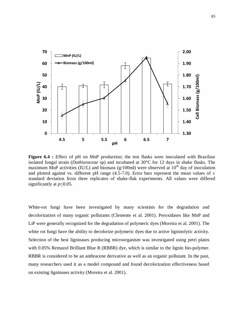
83
Figure 6.4 : Effect of pH on MnP production; the test flasks were inoculated with Brazilian
isolated fungal strain (Dothioraceae sp) and incubated at 30°C for 12 days in shake flasks. The
maximum MnP activities (IU/L) and biomass (g/100ml) were observed at 10th day of inoculation
and plotted against vs. different pH range (4.5-7.0). Error bars represent the mean values of ±
standard deviation from three replicates of shake-flak experiments. All values were differed
significantly at p˂0.05.
White-rot fungi have been investigated by many scientists for the degradation and
decolorization of many organic pollutants (Clemente et al. 2001). Peroxidases like MnP and
LiP were generally recognized for the degradation of polymeric dyes (Moreira et al. 2001). The
white rot fungi have the ability to decolorize polymeric dyes due to active ligninolytic activity.
Selection of the best ligninases producing microorganism was investigated using petri plates
with 0.05% Remazol Brilliant Blue R (RBBR) dye, which is similar to the lignin bio-polymer.
RBBR is considered to be an anthracene derivative as well as an organic pollutant. In the past,
many researchers used it as a model compound and found decolorization effectiveness based
on existing ligninases activity (Moreira et al. 2001).
1.30
1.40
1.50
1.60
1.70
1.80
1.90
2.00
0
10
20
30
40
50
60
70
4.5 5 5.5 6 6.5 7
Cel
l Bio
mas
s (g
/100
ml)
Mn
P (
IU/L
)
pH
MnP (IU/L)
Biomass (g/100ml)

84
Our results indicated that, the newly isolated ascomycete Dothioraceae sp. is the first on record
of producing extracellular oxidoreductases including laccase (Lcc), lignin peroxidase (LiP) and
manganese peroxidase (MnP). The basidiomycetous white-rot fungi and related litter
decomposing fungi are the most efficient lignin-degrading organisms in nature (Frackler et al.
2006). Nevertheless, the degradation of lignocellulose by ascomycetes is the major path for
carbon cycling in soil and plant wastes (Nilsson et al. 1989; Liers et al. 2006). Species in this
family have broad distribution, and are biotrophic or necrotrophic, usually linked with woody
plants (Cannon et al. 2007). The ability to use several lignin model compounds and the
decoloration of aromatic dyes by Dothioraceae sp. LM572 provided additional evidence of its
potential to degrade lignin (Huiju et al. 2011).
6.4 Conclusions
The new isolated Dothioraceae sp. LM572 had a certain lignin degradation capacity and
provided a new microorganism resource for lignin degradation. The ability of this fungal strain
could be of interest for lignocellulosic biomass degradation as well as treatments of pulp and
paper mill effluent. In addition, it would be more useful to compare the ligninolytic potential of
this fungus with other lignin-degrading ascomycetes and basidiomycetes and more research
regarding the role of ascomycetes on lignin biodegradation must carried out using
Dothioraceae sp. LM572 and other related microorganisms.

85
7 Chapter: Depolymerization of bark with fungal laccases
into bark-derived polyphenols and comparative studies
between phenolic mediator vs. honey as a natural
mediator substitute
7.1 Abstract
Due to increasing waste production and disposal problems arising from synthetic polymer
production, there is a critical need to substitute materials with biodegradable and renewable
resources. Use of laccases as biocatalysts to affect and enhance the catalytic properties of enzymes
has been shown to be a promising solution for bark depolymerization. Initially eight different
fungal strains were tested during submerged fermentation (SF) for laccase enzyme production,
with the Pleurotus species found to be good producers compared to other fungal strains
investigated. P. pulmonarius mainly produces laccase enzyme in production medium (PM) at
initial conditions of pH-5.5 and 30°C. Bark depolymerization was conducted in SF and we
identified polyphenols/polyaromatic compounds after four weeks when the PM was induced with
50mg/100ml of each bark during the lag-phase. During SF where honey was used as a natural
mediator substitute (NMS) in the PM, laccase activities were about 1.5 times higher than those
found in comparable cultures without honey in the PM. These samples were analyzed by GC-
MS/MS. The laccase enzyme was purified using UNO® sphere Q-1 anion exchange
chromatography and the molecular weight was determined to be ~50kDa on 10% SDS-PAGE.
The laccase kinetic parameters were determined by using 2,2′-azinobis-(3-ethylbenzthiazoline-6-
sulfonate) (ABTS) as a substrate. The maximal velocity (Vmax), Michaelis constant (Km), and
turnover number (Kcat) were found to be 76.9µM min-1
, 909µM and 739min-1
, respectively, from a
Lineweaver Burk plot. Furthermore, laccases are suitable for biotechnological applications that
transform bark biomass into high value bark biopolymers / biochemicals. The differences
observed among identified aromatic compound GC-MS profiles were due to the utilization of two
different bark species. Py-GC-MS analysis of bark showed differing effects of fungal activity on
bark composition. Polyphenolics were separated in reverse-phase mode using HPLC with a
pinnacle DB Biphenyl, C18 column, and UV detector. Two recognition wavelengths of 290 and
340 nm were selected to improve the separation of each single compound in monofloral honey
and bark-fermented samples. This study is novel because it replaces natural mediators (NM) with

86
monofloral honey in PM and bark materials impregnated with honey, and studies the effects of
fungi-derived laccases on bark-derived polyphenols.
7.2 Highlights
• Biochemical and catalytic properties of purified fungal laccases.
• Characterization of LMW polyphenols via GC-MS/MS & MALDI-TOF/MS.
• Use of monofloral honey as natural mediator substitute and compared with MS.
• Polyphenols separated by HPLC using two different detection wavelengths.
• Biotransformation of bark biomass into high value biopolymers/biochemicals.
• Integration between laccases vs. bark biopolymers and characterized by Py-GC/MS.
7.3 Introduction
The biobased economy relies on sustainable resources, and is likely to have an enormous
impact comparable to the fossil fuel-based economy. The biobased economy is not about
simply utilizing renewable resources and applying them to cutting-edge innovations; its
principles can be used to modify procedures with far-reaching effects on society (Langeveld et
al., 2010).
Nations have set managed goals to replace certain fuel and chemical commodities with biomass to
help the biobased economy. Woody biomass provides lignocellulosic feedstock for the energy
sector and chemical industry. Because of its inexhaustible accessibility, useful chemical
composition, and reasonably low costs, woody biomass can improve products economically and
sustainably. Global annual lignocellulosic biomass production was reported by Zhang (2008) to be
about 200 billion tons (bnt). Haveren et al. (2008) found that 0.3 billion tons (bnt) is
differentiated to synthetic chemicals every year.
In Europe and North America, P. pulmonarius is the most cultivated fungal species (Fungal Guide,
2012), and is commonly found throughout the world, particularly in temperate and subtropical
forest. This species is frequently found on hardwoods in eastern North American forests; however,
it is also found on conifers in the western United States (Stamets, 2000). The cultivation method,

87
which is comparable to that of other Pleurotus species, is by spreading spores in grain and then
dispersing the inoculated seeds in cellulosic biomass as substrate, like straw, coffee grounds,
wood chips, sawdust and cardboard.
Pleurotus is considered an effective lignin degrader that can grow fairly fast on different types of
lignocellulosic biomass. Laccase (EC 1.10.3.2) belongs to the multicopper oxidase family. Fungal
growth conditions, media composition and cultivation method play an important role for the
laccase production (Lee et al., 1999).
The composition of honey, a natural bee product, depends mainly on the botanical source,
topographical origin, and dispensing and atmospheric conditions (Chen et al., 2000; Vela et al.,
2007; Alvarez et al., 2010). In honey, the main antioxidant compounds are polyphenolics,
flavonoids, enzymes (catalase, glucose oxidase), organic acids, ascorbic acid, carotenoid-like
substances, amino acids and proteins. The antioxidant value varies significantly depending on the
floral source (Gheldof et al., 2002). Several phenolic compounds are found in honey, including
syringic acid (SA) and methyl syringate (MS) found in honey. Lately, a compound was found
having substantial antibacterial activity against Staphylococcus aureus (Alvarez-Suarez et al.,
2010).
The tree species balsam fir (Abies balsamea) is one of the more important conifers of the boreal
forest. It is mainly used in pulp, light frame construction, and as well is a food source and shelter
for wildlife (http://www.borealforest.org/).
Mixed aspen (Populus tremuloides and Populus grandidentata) are the most widespread forest
species for managing biodiversity in the western and boreal zones in North America (Hogg et al.,
2005). Polyaromatics are hydrocarbons, containing mainly C and H with multiple aromatic rings
in which the electrons are delocalized. They are primarily found in fossil fuels (oil, coal and tar
deposits) and are produced by partial burning of organic matter.
Laccase alone does not polymerize, nor depolymerize nor delignify pulp until it consolidates with
a mediator like hydroxybenzotriazole (HBT). Laccases, owing to high redox-potential in
basidiomycetes from genus Trametes, assist in lignin removal when combined with HBT from
complete (Gutierrez et al., 2012) and preserved (Chen et al., 2012) lignocellulosic biomass,
producing cellulose available to hydrolysis. Generally examined mediators are synthetic

88
compounds based on nitrogen heterocycles. Because of high toxicity and high price, it is hard to
make laccase-mediator systems (LMS) at an industrial scale.
Natural mediators, in the presence of laccase, facilitate the oxidation of non-phenolics depending
on the phenolic compound structure, as well as, the reactivity vs. stability of phenoxy radicals
produced (MS. > AS
. > SA
.) reported by Tania et al. (2012). Due to complex nature of woody
biomass, degrading enzymes, and their interactions, researchers are trying to profile radical-
coupling routes which are involved in the formation of different phenolic species and identified as
mediators (Foston et al., 2012).
Molecular properties of bark/biomass combustion (BC) can be assessed by pyrolysis–gas
chromatography–mass spectrometry (Py–GC–MS) (De la Rosa et al., 2008; Kaal et al., 2009; Kaal
et al., 2009; Fabri et al., 2012). Heat-affected reaction and resulting changes produce structures that
may be like the pyrolysis artifacts of BC (Saiz-jimenez 1995; Wampler 1999). Py-GC-MS is
considered a quick and quite inexpensive method for the bark characterization. The hydrolyzable
bonds are cleaved and, subsequently, CO2H and OH groups are changed in-situ by pyrolysis to
related methyl esters and methyl ethers, respectively, which are more amenable to GC than their
underivatized complements (Hatcher et al., 2001; Shadkami et al., 2010). It also provides
supplementary data on structure over position of derivatized multifunctional rings (Shadkami et
al., 2010).
There is need to focus on bark depolymerization and realizing the effect of honey in production
media where it can mainly be used as replacement of natural phenolic mediators.
7.4 Results and discussion
7.4.1 Extracellular laccases and screening of potential strain
Extracellular laccase was produced during submerged growth conditions and screening of the
best fungal strain was done based on the laccase production and growth conditions. These
experiments were conducted both in solid as well as in liquid media. Among other competitor
fungal strains, P. pulmonarius was screened for further examination as it possesses a strong
laccase activity. This was confirmed using malt extract agar media plates (malt extract 25g;
agar 20g; distilled water 1L, and 0.05% of the dye Remazol Brilliant Blue R), wrapped with
aluminum foil to avoid light and incubated at 30oC for two weeks. Ligninolytic activity was
assessed on agar plates by observing decolorizing of polyaromatic anthraquinone dye and

89
scoring attainable halos surrounding the fungus growing colonies as shown in Figure 7.2 (K-
M).
After the agar plate prescreening, all fungal strains were transferred into the production media
and fungal growth observed during submerged fermentation. 50mg of each bark was
introduced in 100ml of production medium (PM) during lag-phase, when the fungus starts to
produce laccase activity in the PM; this occurs about five days after the inoculum is placed in
the PM. Maximum laccase activity was recorded after 25 days in submerged fermentation as
52 IU/ml/min in P. pulmonarius, 46 IU/ml/min in P. cornucopiae, 35 IU/ml/min P. ostreatus,
28 IU/ml/min in P. chrysosporium, 27 IU/ml/min in T. versicolor, and 23 IU/ml/min in G.
mangiferae, respectively as shown in (Figure 7.1).
Figure 7.1: Representative time course of extracellular laccase activity (IU/ml/min) in the
production medium on glucose (10g/L) in shake flask cultures (pH 5.5, 30°C, 150 rpm) following
the growth of six different fungal strains induced with aspen bark during lag-phase including: (◊)
P. pulmonaris, (□) P. cornucopiae, (+) P. ostreatus, (○) P. chrysosporium, (▲) T. versicolor, and
( ) G. mangiferae. Each point is a mean of three independent experiments. Bars indicate standard
deviation among three replicates.

90
In nature, the lignocellulosic biomass is mainly composed of carbohydrate polymers (cellulose,
hemicellulose) and an aromatic polymer (lignin) (Henriksson et al., 2000; Bernards et al.,
2001). By utilizing fungal degrading enzymes, the polyphenolic–carbohydrate complex
compounds disrupt it into smaller fragments. As lignin retains high antioxidant activity, (Cruz
et al., 2001; Oki et al., 2002) hypothetically a decrease in size of lignin-like polyphenolics
degraded fungus is able to increase soluble bark phenolic contents (Cohen et al., 2002; Jeandet
et al., 2002; McCue et al., 2003).
It was observed that fungal growth increased through mid (10–25 days) to late (20–30 days)
phases, with improved soluble phenolic contents. P. pulmonarius had the maximum virtual
laccase activity among investigated fungal strains. In general, the phenolic compounds
oxidized easily due to phenoxy radical formation as compared to non-phenolic compounds.
Increased phenolic concentration and oxygen availability assist polymerization. Lignin
degrading rate via feedback control of laccases with phenolics directs an enzyme into a latent
catalytic state explained by Harvey et al., 1993. Therefore, it starts a decline in enzyme activity
after a certain time period owing to feedback inhibition due to accumulation of secondary
metabolites and toxic compounds to a certain level. Morphological characterization of different
fungal growth patterns on each bark was observed using AmScope-WF25X/9 (magnification
0.5X) attached with cold-light source haloid lamp 150W (Figure 7.2).

91

92
Figure 7.2: Different fungal strains growth on balsam fir (A-E) and mixed aspen bark (F-J) while,
P. cornucopiae, G. mangiferae, P. pulmonarius (K-M) were exhibiting laccase activity on agar
plates containing (2.5% malt extract +0.05% RBBR). From (N-P) shows fungal growth on the
bark surface and (Q-R) presenting bark and P. pulmonarius surface morphology was observed
under AmScope-WF25X/9 (magnification 0.5X).
7.4.2 Laccase purification and kinetics
The extracellular laccases from P. pulmonarius cultivated under submerged fermentation were
purified. The purification method has already been explained in the methodology section. The
sequential purification steps were summed up in (Figure 7.3) and its purification on 10% SDS-
PAGE stained with Comassie Blue R-250 (Figure 7.4A). With the help of Lineweaver-Burk
plot, the Michaelis constant (Km) and maximal velocity (Vmax) values of laccase from Pleurotus
pulmonaris were obtained 909µM and 76.9µMmin-1
, respectively, and these constants are
specific to a substrate (ABTS) (Figure 7.4B).

93
Figure 7.3: Laccase purification using FPLC by UNO®
sphere Q-1 anion exchange column where
(A-D) represents sequential purification steps.
Figure 7.4: Purified laccase molecular weight was determined on 10% SDS-PAGE from P.
pulmonarius stained with Coomassie Blue R-250 showed Mw~50kDa. Lanes 1–2 are serially
purified laccase enzyme, lane-3 shows purified laccase enzyme, Pp = total protein from Pleurotus
pulmonarius, Pc = total protein from Pleurotus cornucopiae, and TS26616 for protein standard
-0.02
-0.01
0
0.01
0.02
0.03
0.04
0.05
0.06
0.07
0.08
-0.002 -0.001 0 0.001 0.002 0.003 0.004 0.005 0.006
1/V
[m
in /
µM
]
1/substrate [1/µM]
7.4B
7.4A
3 2 1

94
marker shown in (7.4A); Lineweaver-Burk plot for the calculation of laccase kinetic parameters
where ABTS used as a substrate (7.4B).
The turnover number (Kcat) and specificity constant (Kcat/Km) was 739 min-1
and 0.81 µM-1
min-1
respectively. The similar specific constant was investigated for laccase enzyme from P. ostreatus
where ABTS used as substrate (Palmieri et al., 1997). As a result of high catalytic efficiency and
binding affinity with substrate (ABTS) it is possible to compare with other laccase producing
microorganisms (Palmieri et al., 1997).
7.4.3 Bark decomposition by TG/DTG
Thermal degradation of bark samples was estimated using TGA. All experiments were done
under nitrogen atmosphere. Both control and fungal (P. pulmonarius) treated bark samples
were examined (Figure 7.5). Cellulose decomposition is denoted by the main DTG peak, while
the shoulder peak mainly represents hemicellulose decomposition assigned at lower
temperature (around 160 °C) (Varhegyi et al., 1989) while, lignin starts to decomposes from
lower to wide temperature range (200–400 °C) (Jakab et al., 1997).
Aspen and fungal treated aspen bark samples started to decompose at about 190 o
C and 140oC
respectively, while, balsam fir and fungal treated fir bark samples were activated to decay at
about 200oC and 170
oC respectively. The solid-lines in both TG/DTG (thermograms- shown in
Figure 7.5) indicate control or untreated barks whereas the dotted-lines specify fungal-treated
bark samples. The weight loss of fungal degraded bark samples was faster between 140-400°C
as also observed by Meszaros et al. (2004).
Relatively large amounts of water, carbon monoxide and carbon dioxide are formed during
thermal decomposition of bark materials due to a large number of hydroxyl groups and oxygen
atoms present in the natural polymers that form cell wall (cellulose, hemicellulose and lignin).
Besides decomposition, smaller amounts of gases are released from the natural polymer. Carbon
monoxide is formed around 500 °C, while carbon dioxide is released around 500 °C and also
between 600-700°C from the bark. The occurrence of these smaller peaks is characteristic to
oxalate decomposition due to presence of higher amount of oxalate ions in the bark compared to
wood (Szekely et al., 1987).
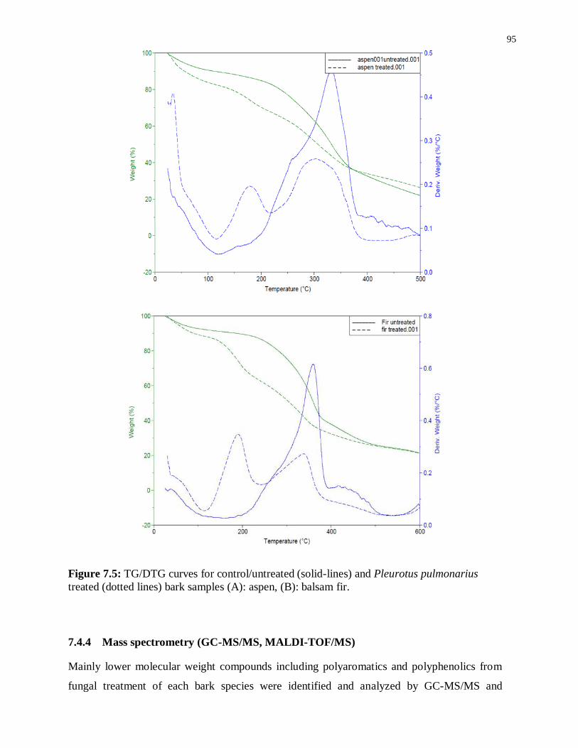
95
Figure 7.5: TG/DTG curves for control/untreated (solid-lines) and Pleurotus pulmonarius
treated (dotted lines) bark samples (A): aspen, (B): balsam fir.
7.4.4 Mass spectrometry (GC-MS/MS, MALDI-TOF/MS)
Mainly lower molecular weight compounds including polyaromatics and polyphenolics from
fungal treatment of each bark species were identified and analyzed by GC-MS/MS and

96
MALDI-TOF-MS methods. In this study, GC–MS/MS was considered to analyze LMW
compounds liberated from fungal decayed bark. The total ion chromatograph (TIC) of GC-
MS/MS relating to ethyl acetate extracted compounds from acidified supernatants according to
the method reported by Ksibi et al. (2003) were obtained from both control and fungal treated
bark samples with P. pulmonarius and are shown in (Figure 7.6). Major identified peak
retention time (tr) values were marked in (green) while, all others in (black) are summarized in
Table 7.1.
A Control-fir
B Fungal treated fir (Sample collected after two weeks of lag-phase)

97
C Fungal treated fir (Sample collected after three weeks of lag-phase)
D Control-aspen
E Fungal treated aspen (Sample collected after two weeks of lag-phase)

98
Figure 7.6: TIC from GC-MS/MS of identified compounds w.r.t. their tr-values characterized
from both control and each treated bark species from Pleurotus pulmonarius are listed in Table
7.1: where (A) balsam fir, (B and C) fungal treated fir, while, (D) aspen and (E and F) fungal
treated aspen. The fungal treated samples of each bark exhibited a variable number of new
peaks due to fungal degradation and changes in chemical composition.
Table 7.1: Bark polyphenols/polyaromatics identification.
m/z Polyaromatics/polyphenols m/z Polyaromatics/polyphenols
39, 39.6 CH2=C=CH anion, Propyne 124.9 Guaiacol, 4-methoxy-1-oxide
isocyanato-
41.2 Methyl Isocyanide 127.83
2-Propenoic acid,
Oxiranylmethyl ester, 4-
Pentenoic acid,
42.1 Propene 128.8 Isoquinoline, 2-propanoic acid
43.01, 44 Iso-cyanic acid 139.97 1-propanol, 3-phenoxy-
56.9 CH2COCH3 143.84 2-Butenedioic acid, dimethyl
ester
F Fungal treated aspen (Sample collected after three weeks of lag-phase)

99
59 CH3COO-, Glyoxal 150.9, 151 benzaldehyde, benzoic acid
68 1,3 butadiene, 2-methyl- 128.8 Isoquinoline, 2-propanoic acid
69
vinyl Isocyanate 153
Vanillin, Biphenylene
protonated ethyl ester, 3,4-
dimethoxy-
CH2=CHCH=CHO anion 128.8 Isoquinoline, 2-propanoic acid
74 Methyl propyl ether 160.1
Succinic acid, Isopropyl-
Propanedioic acid, ethyl-
dimethyl ester, 1,3 propanediol,
tert-butyl Propanedioic acid,
77 Phenyl radical, Isopropyl
methyl-d3-ether 182.1 Syringaldehyde (SA)
79.9 2-vinyl-1,3 butadiene 183 1,2-benzenediol
81 2-furanyl-CH2 anion, C6H9 187.1 2-ethylhexyl ester
87.9 2-butene-1,4 diol 196.2 Acetosyringone (AS)
96 furaldehyde 199 Propyl ester
97 Isooxazole, 3,5 dimethyl- 202.83 Butanoic acid, Valeric acid,
trimethyl silyl ester
98 1,3 butadiene-1-carboxylic acid 212.23 Methyl Syringate (MS),
Phenylmethyl ester
104 Propanedioic acid 216
Benzoic acid, benzal-barbituric
acid, Diethyl methyl isopropyl
malonate, malonic acid, di-
isobutyl ester, glutaric acid,
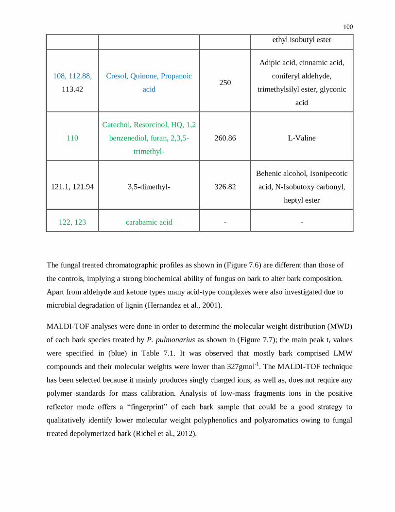
100
ethyl isobutyl ester
108, 112.88,
113.42
Cresol, Quinone, Propanoic
acid 250
Adipic acid, cinnamic acid,
coniferyl aldehyde,
trimethylsilyl ester, glyconic
acid
110
Catechol, Resorcinol, HQ, 1,2
benzenediol, furan, 2,3,5-
trimethyl-
260.86 L-Valine
121.1, 121.94 3,5-dimethyl- 326.82
Behenic alcohol, Isonipecotic
acid, N-Isobutoxy carbonyl,
heptyl ester
122, 123 carabamic acid - -
The fungal treated chromatographic profiles as shown in (Figure 7.6) are different than those of
the controls, implying a strong biochemical ability of fungus on bark to alter bark composition.
Apart from aldehyde and ketone types many acid-type complexes were also investigated due to
microbial degradation of lignin (Hernandez et al., 2001).
MALDI-TOF analyses were done in order to determine the molecular weight distribution (MWD)
of each bark species treated by P. pulmonarius as shown in (Figure 7.7); the main peak tr values
were specified in (blue) in Table 7.1. It was observed that mostly bark comprised LMW
compounds and their molecular weights were lower than 327gmol-1
. The MALDI-TOF technique
has been selected because it mainly produces singly charged ions, as well as, does not require any
polymer standards for mass calibration. Analysis of low-mass fragments ions in the positive
reflector mode offers a “fingerprint” of each bark sample that could be a good strategy to
qualitatively identify lower molecular weight polyphenolics and polyaromatics owing to fungal
treated depolymerized bark (Richel et al., 2012).

101
The molecular weight distribution (MWD) patterns were very similar within each bark species.
During depolymerization, β-O-4 ether bond is heterolytically cleaved via formation of sodium
phenolate and carbenium ion in transition state, which is instantly neutralized by hydroxide ion. In
addition, the heterolytic cleavage of β-O-4 ether bond, dealkylation of side chains and hydrolysis
of methoxy groups took place and lead to the formation of phenol, cresols and catechol reported
by Erdocia et al. (2014).
16.0 102.4 188.8 275.2 361.6 448.0
Mass (m/z )
1.3E+4
0
10
20
30
40
50
60
70
80
90
100
% I
nte
ns
ity
4700 Linear Spec #1[BP = 38.9, 13271]
38.9321
22.9336
39.6590
41.2053
37.6891
103.851023.8221
87.9604
112.8843
104.3624
42.1454 113.4240
42.6410 121.942098.907074.0052 197.2408122.951525.3463 97.9869
181.302563.0483 139.9702
21.0 63.4 105.8 148.2 190.6 233.0
Mass (m/z )
1.5E+4
0
10
20
30
40
50
60
70
80
90
100
% I
nte
ns
ity
4700 Linear Spec #1[BP = 38.9, 15449]
38.9281
22.9419
103.8499
112.849487.9100
41.0252
104.893037.9505
120.9942
98.8647119.9296
105.914088.9446 196.980123.2628 71.9670 213.0480123.014440.3679 181.0271 199.015989.9399 106.948973.987362.9805
A Balsam fir (Sample collected after two weeks of lag-phase)
B Balsam fir (Sample collected after three weeks of lag-phase)

102
17.0 94.4 171.8 249.2 326.6 404.0
Mass (m/z )
1134.1
0
10
20
30
40
50
60
70
80
90
100
% I
nte
ns
ity
4700 Linear Spec #1[BP = 22.9, 1134]
22.9429
55.9133
51.9174
98.9028
202.8360
38.8799156.9118111.8111
260.8665
20.0 92.8 165.6 238.4 311.2 384.0
Mass (m/z )
5109.0
0
10
20
30
40
50
60
70
80
90
100
% In
te
ns
ity
4700 Linear Spec #1[BP = 22.9, 5109]
22.9500
55.9587
51.9536
98.8798
143.8488
80.8297
64.892138.8836
82.8298 127.831253.969823.2895326.8204
C Aspen (Sample collected after two weeks of lag-phase)
D Aspen (Sample collected after three weeks of lag-phase)
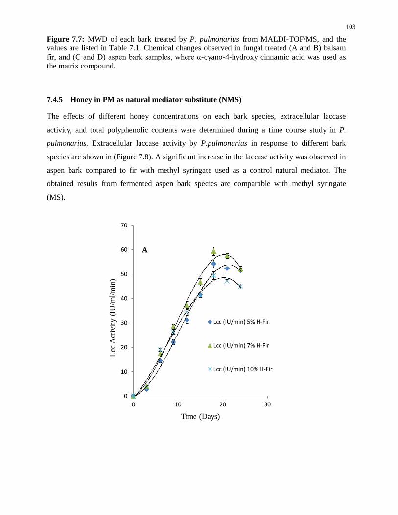
103
Figure 7.7: MWD of each bark treated by P. pulmonarius from MALDI-TOF/MS, and the
values are listed in Table 7.1. Chemical changes observed in fungal treated (A and B) balsam
fir, and (C and D) aspen bark samples, where α-cyano-4-hydroxy cinnamic acid was used as
the matrix compound.
7.4.5 Honey in PM as natural mediator substitute (NMS)
The effects of different honey concentrations on each bark species, extracellular laccase
activity, and total polyphenolic contents were determined during a time course study in P.
pulmonarius. Extracellular laccase activity by P.pulmonarius in response to different bark
species are shown in (Figure 7.8). A significant increase in the laccase activity was observed in
aspen bark compared to fir with methyl syringate used as a control natural mediator. The
obtained results from fermented aspen bark species are comparable with methyl syringate
(MS).
0
10
20
30
40
50
60
70
0 10 20 30
Lcc
Act
ivit
y (
IU/m
l/m
in)
Time (Days)
Lcc (IU/min) 5% H-Fir
Lcc (IU/min) 7% H-Fir
Lcc (IU/min) 10% H-Fir
A

104
0
10
20
30
40
50
60
70
80
0 10 20 30
Lcc
Act
ivit
y (
IU/m
l/m
in)
Time (Days)
Lcc (IU/min) 5% H-Aspen
Lcc (IU/min) 7% H-Aspen
Lcc (IU/min) 10% H-Aspen
B
0
10
20
30
40
50
60
70
80
90
0 5 10 15 20 25 30
Lcc
Act
ivit
y (
IU/m
l/m
in)
Time (Days)
Lcc (IU/min) 1% MS-Aspen
Lcc (IU/min) 1% MS-Fir
C

105
Figure 7.8: Time course study for laccase activity (IU/ml/min) in P. pulmonarius during SF
containing 5, 7 and 10% (v/v) of monofloral honey in production media mainly used as a
natural mediator substitute induced with 50mg of each bark in 100ml of PM during lag-phase
(A)-Balsam fir, (B)-Mixed aspen bark, and (C)-1% Methyl syringate as natural phenolic
mediator. Each point is a mean of three independent experiments. Bars indicate standard
deviation among three replicates.
In the presence of different applied honey concentrations, after 18-days of incubation, the
laccase activity began to slightly decrease. In our study, we used three different (%, w/v)
concentrations of honey but we observed the highest laccase activity 68 IU/ml/min at 7% (w/v)
honey concentration after 18-days of growth (Figure 7.8). During fermentation, a higher
substrate concentration in the production medium leads to catabolite repression which
ultimately affects enzyme productivity yield. In contrast, it has been suggested that during
laccase production in the presence of phenolic compounds, oxidation occurs to form quinone
which is considered as toxic to fungal growth (Haars et al., 1980; Arora et al., 1985).
Furthermore, induction with bark during the lag-phase may decrease the extracellular
proteolytic activity as well as ligninolytic enzymes including LiP, and MnP; therefore, it may
increase the laccase activity. Thus, we can propose that improved laccase activity owing to
ready availability of phenolics and aromatic compounds in fungal degraded aspen bark might
help to improve enzyme stability (Yang et al., 2013).
In presence of ligninases, the oxidation mechanism of synthetic mediators like
hydroxybenzotriazole (HBT) is similar to phenolic type mediators such as methyl syringate.
During oxidation, highly reactive phenoxy radicals are produced which assist to remove one
proton and one electron from the target substrate (Cantarella et al., 2003). Bark related free
radicals and reactive oxygen species (ROS) are associated with laccase and laccase mediator
system where honey is used as a natural mediator substitute.
7.4.6 Polyphenolic chromatograms
Reversed phase-high performance liquid chromatography (RP-HPLC) has been used for
qualitative and quantitative analysis such as identification of separated analytes, which are
precisely analyzed by UV/Vis detector and the spectral peaks are identified by their reported
retention time (tr) values (Chen et al., 2000; Romani et al., 2000; Inoue et al., 2005; Zielinska
et al., 2008). Romani et al. (2000) compared electrochemical detection methods with HPLC for

106
polyphenolics in natural extract. The HPLC procedure was found to be more precise as
compared to differential pulse voltammetry method which was suitable for fast screening. The
polyphenolics separation was conducted by HPLC and the chromatograms are shown in
(Figure 7.9).
0.0 5.0 10.0 15.0 20.0 25.0 30.0 35.0 40.0 45.0 50.0 55.0 60.0 64.0
-0.20
0.25
0.50
0.75
1.00
1.25
1.50
1.80Javad #339 [modif ied by Administrator] ms-d UV_VIS_1AU
min
0.0 5.0 10.0 15.0 20.0 25.0 30.0 35.0 40.0 45.0 50.0 55.0 60.0 64.0
-0.050
0.100
0.200
0.300
0.450Javad #341 fs-d UV_VIS_1AU
min
1 - 2.217
2 - 5.367
3 - 12.550
4 - 13.100
5 - 20.6176 - 36.100 7 - 52.867
8 - 55.033
9 - 55.850
0.0 5.0 10.0 15.0 20.0 25.0 30.0 35.0 40.0 45.0 50.0 55.0 60.0 65.0 70.0 75.0
-0.020
0.000
0.020
0.040
0.060
0.080
0.100
0.120
0.140
0.160Javad #335 500ppm Honey UV_VIS_1AU
min
1 - 16.717 2 - 20.550
3 - 24.567
4 - 36.467
5 - 54.567
6 - 55.217
7 - 58.134
8 - 66.600
(a)
(b)
(c)
λ=290nm; MS
Dil. Honey sample
FS-Mixed fir

107
0.0 5.0 10.0 15.0 20.0 25.0 30.0 35.0 40.0 45.0 50.0 55.0 60.0 65.0 70.0 75.0
-0.020
0.000
0.020
0.040
0.060
0.080
0.100
0.120
0.140
0.160Javad #331 std UV_VIS_1AU
min
1 - 11.883
2 - 24.0003 - 25.983 4 - 30.3505 - 32.183
6 - 33.433
7 - 55.067
8 - 57.433
9 - 59.283
10 - 59.667
11 - 66.333
0.0 5.0 10.0 15.0 20.0 25.0 30.0 35.0 40.0 45.0 50.0 55.0 60.0 65.0 70.0 75.0
-0.0020
0.0000
0.0020
0.0040
0.0060
0.0080
0.0100
0.0120Javad #363 repeat 1 UV_VIS_1AU
min
1 - 36.550
2 - 54.817
0.0 5.0 10.0 15.0 20.0 25.0 30.0 35.0 40.0 45.0 50.0 55.0 60.0 65.0 70.0 75.0
-0.00100
0.00000
0.00125
0.00250
0.00375
0.00500
0.00625
0.00800Javad #362 8 UV_VIS_1AU
min
1 - 37.100
0.0 5.0 10.0 15.0 20.0 25.0 30.0 35.0 40.0 45.0 50.0 55.0 60.0 65.0 70.0 75.0
-0.00100
0.00000
0.00100
0.00200
0.00300
0.00400
0.00500
0.00600Javad #358 [modif ied by Administrator] 4 UV_VIS_1AU
min
1 - 36.134
(d) FS-Mixed aspen
(e) λ=340nm; Dil. Honey
(f) FS-Mixed fir
(g) FS-Mixed aspen

108
Figure 7.9: HPLC chromatograms phenolic profiles of bark fermented samples at 7% honey in
production media (a-d at λ290) and (e-h at λ340). Peak identification: methyl syringate
(12.017), pinobanksin (13.100), 8-methoxykeampferol (24.567), pinocembrin (36.467), chrysin
(39.62), pinocembrin 7-Me (55.217), tetochrysin (57.142). All major peaks were characterized
based on their tr values of honey polyphenolics previously reported by Pyrzynska et al. (2009).
Remarkably, it was noticed that the standard methyl syringate (MS) peak appeared at 290nm
but vanished at 340nm. Similarly, the diluted honey sample had MS peak at 290nm, but, when
the same sample was run at 340nm the peak was missing and different peaks were seen. Our
HPLC results confirmed that the wavelength at λ=290 nm was more suitable for the separation
of MS natural mediator compounds. All major peaks in the chromatograms were compared and
characterized based on their tr values of honey polyphenolic compounds as earlier reported by
Pyrzynska et al. (2009).
0.0 5.0 10.0 15.0 20.0 25.0 30.0 35.0 40.0 45.0 50.0 55.0 60.0 65.0 70.0 75.0
-0.020
0.000
0.020
0.040
0.060
0.080
0.100
0.120
0.140
0.160Javad #335 500ppm Honey UV_VIS_1AU
min
1 - 16.717 2 - 20.550
3 - 24.567
4 - 36.467
5 - 54.567
6 - 55.217
7 - 58.134
8 - 66.600
(h) FS-aspen with 1% MS

109
Table 7.2: Total polyphenolics in fermented sample when induced with 50mg of each bark into
100ml of PM during lag-phase.
Mean ± S.D (n = 5); Catechol was used as a standard for the calibration of total polyphenols in
fermented samples during late-log phase (after 20-days of fermentation). Calibration curve was
prepared by using catechol solutions at concentrations (10 to 50 mg / 100 ml) in distilled water.
Total phenolic content of 0.114 ± 0.09 mg cat equiv/g was found in a buckwheat monofloral
honey. Wood degrading fungi play an important role by attacking protein-polyphenolic
complexes which possibly changes substrate properties (Bending et al., 1997). Polyphenolics,
mainly condensed tannins with protein complexes assist to develop in fungal growth
(Mutabaruka et al., 2007). During fermentation in PM with honey, it was also noticed that total
polyphenols in each bark species were significantly degraded as compared to glucose and
natural mediator (MS) samples because of fungal biomass accumulation.
7.4.7 Py-GC-MS
Py–GC–MS total ion chromatograms (TIC) are represented in Figure 7.10. The major peaks in
TIC-fingerprints are the pyrolysis products reported in (Table 7.3), with contrasting retention
times (RT), characteristic ion fragments (m/z) utilized for the quantification and their relative
proportions. Flags are used to organize the compounds and are mainly divided into seven
groups i.e., ALIPH = aliphatic compound, CARB = carbohydrate product, LIG = lignin, MAH
Samples
Mean total
polyphenolics
(mg cat equiv/100ml)
Aspen
PM-Gluc 18.4 ± 0.54
PM-H 23.7 ± 0.61
PM-MS 27.8 ± 0.48
Balsam
Fir
PM-Gluc 20.1 ± 0.81
PM-H 28.8 ± 0.36
PM-MS 32.1 ± 0.74

110
= monocyclic aromatic hydrocarbon, NCOMP = nitrogen-containing compound, PHEN =
phenol, SESQUI = sesquiterpenoid. Total quantified peak area (TQPA) values of each group
are given in Table 7.4, and the values of each group were calculated based on Table 7.3.
Figure 7.10: Total ion chromatograms (TIC) of untreated (control) bark samples of aspen and
fir, MS-treated and bark fermented samples in honey production medium. Peak labels refer to
peak numbers in Table 7.3.

111
Table 7.3: List of main pyrolysis products identified from the untreated/control, MS treated
and honey fermented bark samples of Aspen and Fir. Percentages refer to the proportion of total
quantified peak area (TQPA) accounted for by each compound (sum 100%). Peak numbers refer
to labels in (Fig.7.10). Retention time = (tr), Dominant ion fragments = m/z. Flags are used to
organize the compounds in main groups: ALIPH = aliphatic compound, CARB = carbohydrate
product, LIG = lignin product, MAH = monocyclic aromatic hydrocarbon, NCOMP = Nitrogen
containing compound, PHEN = phenol, SESQUI = sesquiterpenoid.
Peak
No Pyrolysis
product
Retention
time (tr)
min m/z Flag
Mixed Aspen Bark Mixed Balsam Fir Bark
Control MS
treated
samples
Honey
treated
samples Control
MS
treated
samples
Honey
treated
samples 1 toluene 2.535 91, 92 MAH 23.2 19.7 31.4 13.0 20.2 25.1 2 3/2-furaldehyde 2.918 95, 96 CARB 15.0 11.0 24.9 18.5 18.1 46.9 3 C1-pyridine 3.107 93, 66 NCOMP 0.0 0.0 2.9 0.0 0.0 0.0 4 C2-benzene 3.107 91,
106 MAH
7.3 5.6 2.2 3.7 6.6 5.3
5 2-furanmethanol 3.147 98, 97 CARB 0.0 0.0 8.0 0.0 0.0 2.0 6 Styrene 3.404 104,
78 MAH
2.1 1.8 1.8 0.9 1.4 2.0
7 2,3 dihydro-5-
methylfuran-2-one 3.896 98, 55 CARB
6.9 7.0 3.6 11.4 10.2 1.4
8 5-methyl-2-
furaldehyde 3.939 110,
109 CARB
1.8 1.0 4.5 2.5 3.1 9.0
9 2-methyl-2-
cyclopenten-1-one 4.091 96, 67 CARB
0.9 0.7 2.2 8.9 1.6 0.4
10 phenol 4.234 94, 66 PHEN 15.0 15.9 6.8 4.2 4.9 1.8 11 3-hydroxy-2-methyl-
2-cyclopenten-1-one 4.486 112,
55 CARB
2.4 3.5 3.5 3.9 4.5 0.8
12 2-
hydroxybenzaldehyde 4.583 121,
122 CARB
2.3 1.1 0.3 0.2 0.6 0.2
13 4-methylphenol 4.749 107,
108 PHEN
4.2 4.6 2.9 2.0 2.6 0.6
14 unidentified aliphatic
compound 4.766 57, 70 ALIPH
6.5 5.7 0.5 1.4 2.6 0.2
15 guaiacol 5.046 109,
124 LIG
5.0 8.7 2.4 8.4 7.9 1.3
16 4-methylguaiacol 5.784 123,
138 LIG
1.7 2.6 0.2 6.1 5.5 0.9
17 4-ethylguaiacol 6.380 137,
152 LIG
0.9 1.3 0.1 1.7 1.7 1.7
18 4-vinylguaiacol 6.643 150,
135 LIG
2.6 6.5 0.3 8.1 4.6 0.4
19 C3-guaiacol 6.912 164,
149 LIG
0.5 0.9 0.1 1.4 1.1 0.1
20 alkene 7.015 55, 69 ALIPH 0.7 0.4 0.3 0.1 0.2 0.3 21 C3-guaiacol 7.244 164,
149 LIG
0.3 0.5 0.0 0.6 0.4 0.1
22 C3-guaiacol 7.530 164,
149 LIG
0.8 1.5 0.1 2.8 1.7 0.2
23 cf.-muurolene
(sesquiterpenoid) 7.833 105,
204 SESQUI
0.0 0.0 0.0 0.1 0.3 0.0
24 unidentified aliphatic
compound 10.010 81, 95 ALIPH
0.0 0.0 1.2 0.1 0.1 0.3

112
Table 7.4: Main pyrolytic compound groups and their cumulative relative proportions (% TQPA).
The calculated (% TQPA) values of each group were based on Table 7.3.
Flag Aspen (% TQPA) Fir (% TQPA)
control MS honey control MS honey
ALIPH 8.1 6.2 2 1.6 2.9 0.8
CARB 30.2 23.2 46.7 45.2 37.5 60.5
LIG 13.2 22 3.2 29 22.9 3.7
MAH 24.5 27 35.4 17.7 28.2 32.3
NCOMP 0.0 0.0 2.9 0.0 0.0 0.0
PHEN 24 21.6 9.9 6.4 8.1 2.6
SESQUI 0.0 0.0 0.0 0.1 0.3 0.0
7.4.7.1 Aspen bark
The pyrolysis fingerprints of untreated/control aspen bark is dominated by phenols
(compounds 10, 13) and guaiacols (15-19, 21, 22), in addition to an unidentified aliphatic
compound (14). The guaiacols originate predominantly from guaiacyl-based lignin, while
syringyl-type markers were virtually absent. Such dominance of guaiacyl groups is typical of
lignin in bark materials (Marquez et al., 2013).
The MS-treated bark sample from aspen produced a set of pyrolysis products that is rather
similar to that of the control sample, even though there are minor differences such as a smaller
relative proportion of phenols and higher proportion of guaiacols, especially guaiacol (15) and
4-vinylguaiacol (18). This might suggest that the material that is primarily affected by
fermentation in MS is the phenol precursors while the lignin component is relatively
unaffected.
113
The pyrolysis fingerprint of the aspen bark sample fermented in honey medium was very
different from the control and MS-treated analogues. It is dominated by a set of furans,
furaldehydes and cyclopentenones (2, 5, 7-9, 11), which are typical products of
polysaccharides (Pouwels et al., 1989). Guaiacol represents a lignin fraction that is relatively
unaffected, but the intensities of the guaiacols are much lower than in the control and MS-
treated samples.
Moreover, several N-containing pyrolysis products such as methyl pyridine (compound 3), in
combination with the chitin marker acetamide (intensity too low for representation in Figure
7.10, but identified unambiguously) are indicative of a major increase in the amount of
microbial biomass in this sample. This makes it very likely that the carbohydrate products do
not represent a recalcitrant polysaccharide component but rather that they originate from the
honey source. Despite the small proportion of the lignin products, they can be traced from
partial ion chromatograms (PIC) as shown in Figure 7.11.

114
Figure 7.11: Partial ion chromatograms (PIC) of selected compounds of polyphenolic origin
from each bark fermented samples in PM with monofloral honey used as NMS analyzed by Py-
GC-MS. Selected polyphenolic compounds are: guaiacol (m/z 109+124), 4-methylguaiacol (m/z
123 + 138), 4-vinylguaiacol (m/z 135 + 150) and syringic acid methyl ester (m/z 181 + 212).
Chromatograms were plotted between relative peak intensity vs. retention time (t r).
7.4.7.2 Balsam fir bark
The pyrolysate of the untreated bark material from fir was strongly dominated by guaiacol
products (15-19, 21-23), suggesting that the sample consists almost completely of guaiacyl-
type lignin material.

115
The MS-treated sample gave a similar pyrolysis fingerprint, even though minor increases of
monocyclic aromatic hydrocarbons (MAHs, 1, 4, 6) can be witnessed. This suggests that
fermentation in MS produced some degradation of the lignin backbone. The relative loss of 4-
vinylguaiacol in comparison to other guaiacols might indicate that the sample contained a non-
lignin polyphenolic material that was preferentially decomposed to a slight extent, as 4-
vinylguaiacol is not only a product of guaiacyl-type lignin but also of cinnamic acids (Ralph et
al., 1991).
The fir bark material submerged in honey medium produced a very different set of pyrolysis
products, dominated by MAHs (1, 4) and furaldehydes (2, 8). Some remains of lignin can still
be recognized from the total ion chromatograms (TIC), e.g. 4-methylguaiacol (compound 16)
and 4-ethylguaiacol (17), and additional compounds can be traced from partial ion
chromatograms (Figure 7.11), but it is clear that this fermentation treatment almost completely
degraded the original polyphenolic structures and the sample became dominated by microbial
biomass.
The lignin products are very abundant in control/untreated and MS-treated samples, and they can
be identified directly from the TIC (with the peak labels). There would be no added scientific
value to presenting the partial ion chromatograms (PIC) of these compounds. As compared to bark
fermented samples in PM with honey as NMS, the lignin products are not very abundant;
therefore, partial ion chromatograms (PIC) were supported for the better separation of
polyphenolic compounds. However, most of the syringol (S-type lignin) products were below the
detection limit and the syringol was quantified at (m/z 154 and 139 at 6.8 min) as shown in Figure
7.11.
Possibly, the polyphenols in honey production media are below the detection limit of Py-GC-
MS or these polyphenols could have been degraded by pyrolysis, producing aromatics such as
toluene and benzene. Traces of catechol can be detected by the Folin-Denis method but it
should be below approximately ~1mg/100mg of catechol and below the detection limit of the
pyrolysis method.
Both control samples produced typical pyrolysis fingerprints of bark materials consisting
primarily of lignin and polysaccharides. The MS-treatment seemed to preferentially degrade a
non-lignin component in the aspen bark, causing the enrichment of guaiacol markers from

116
lignin, while the opposite trend was observed for the fir bark fermented in MS medium. This
difference may be explained by the differences in original sample composition, with the fir
bark being composed almost purely of lignin while the aspen bark sample also contains
polyphenolic precursors producing phenols rather that guaiacols and a more abundant aliphatic
component.
These additional components in fir bark materials are probably more heavily affected by
fermentation in MS than the lignin component. Anyway, the effects of fermentation on the
pyrolysis fingerprints of both bark samples were rather small. By contrast, fermentation in
honey medium eliminated most of the recognisable polyphenols (lignin) and both samples were
almost completely converted into microbial cells which mainly composed of carbohydrates as
shown in Figure 7.12 (A).
Figure 7.12: Representation of the Lignin/Carbohydrate index (A) and Syringol/Guaiacol ratio
(B) for the different analyzed samples.
A
B

117
The lignin is strongly dominated by guaiacol (G-type lignin) which is why it can be concluded
that proportionately very small amount of syringol (S-type lignin) was found (less than 0.20
%/%) in all samples and the relative proportion of syringol/guaiacol (S/G) ratio of different
treated samples are shown in Figure 7.12 (B). MS-treated sample seems to slightly increase
(S/G, %/%) ratio while honey medium has opposite effects.
7.5 Conclusions
In the present study, we demonstrated that monofloral honey can be used as a natural mediator
substitute in PM during submerged fermentation for the production of fungal laccases. We also
found a methyl syringate in the diluted honey sample through HPLC analysis that separated at
a wavelength of 290 nm. The time course study showed diverse laccase production among
different fungal cultures. Moreover, GC-MS/MS and MALDI-TOF/MS analyses were found to
be suitable methods to analyze LMW polyaromatics and polyphenolics due to fungal
degradation of bark. Methyl syringate is considered to be the most effective natural mediator
for the oxidation of non-phenolic lignin units, but our results during submerged fermentation
with 7% honey in production medium was comparable to the natural mediator. Py-GC-MS of
bark fermented samples in a honey medium indicated evidence of a profound effect on biotic
degradation to an extent that a large proportion of the pyrolyzate can be traced back to
microbial biomass. This microbial biomass may be related to the formation of thermolabile
substances (proteins, polysaccharides, and microbial cell wall components such as chitin)
detected by thermogravimetry. The abundance of phenols and unidentified products combined
with the polysaccharide fingerprint of degraded material indicate significant fungal degradation
and depolymerisation of bark biopolymers. Hence, biotechnological procedures were
effectively used for the conversion of bark biomass into high value bark-derived polyphenols.

118
8 Chapter: Conclusions and Future work
8.1 Major contributions
This thesis describes a comprehensive and systematic study on bark depolymerization during
submerged fermentation using monofloral honey, a natural mediator substitute, and investigates
the interaction between laccases and bark biopolymers. It incorporates comparative studies of
enzymatic, chemical and physical treatments for the extraction of two bark species and
characterizes bark polyphenolic extractives. Potential fungal strains for ligninases production were
identified, and purification, characterization and effective valorization of lignin and bark were
investigated.
The major findings of this thesis are as follows:
1. The HPLC analysis shows fast, selective, sensitive, and reliable determination of the most
common polyphenolics in bark extractives. UV/H2O2 treatment was able to preferentially
remove phenolic compounds, with limited effect on the sugar concentration of the bark
hydrolysates. Under these conditions, high removal of phenolic compounds, expressed in
terms of catechol equivalent (0.8% in BILP and 0.6% in aspen) bark extractives were
observed. Furthermore, these results elucidate the effects of different treatments on bark
derived polyphenols, which can be considered as promising methods for applications in
bioconversion and polymer industrial processes.
2. Potential fungal strain for ligninolytic activities were screened, MnP partially purified and
the degrading pattern of local lignin resources found in Brazil was observed. In addition to
this, potential products from various lignin-degrading enzymes and substrates were
identified. Application of ligninases in the biotech industry will be possible if these
enzymes and mediators are commercially developed. The unknown isolated fungal strain
was Dothioraceae sp. LM572 by 18S-rDNA.

119
3. The process parameters such as temperature and pH of the medium in shake flask
experiments were optimized for the maximum production of manganese peroxidases. The
maximum manganese peroxidase activity, as well as cell biomass, was supported at 30˚C
and pH 6.5 by Dothioraceae sp.
4. The new isolated Dothioraceae sp. LM572 has high lignin degradation capacity and is a
new microorganism resource found for lignin degradation. The ability of this fungus strain
could be of interest for lignocellulosic biomass degradation as well as for treatment of pulp
and paper mill effluent. Furthermore, it would be more informative to compare the
ligninolytic capabilities of this fungus with other lignin-degrading ascomycetes and
basidiomycetes and further research concerning the role of ascomycetes on delignification
should be carried out using Dothioraceae sp. LM572 and other microorganisms of this
class.
5. GC-MS/MS and MALDI-TOF/MS analyses were found to be suitable methods to analyze
LMW polyaromatics and polyphenolics from fungal degradation of bark. Methyl syringate
is considered as the most effective natural mediator used for the oxidation of non-phenolic
lignin units but our obtained result during submerged fermentation with combination of
7% honey in production media increased laccase activity 1.5-times, which was comparable
to the natural phenolic mediator.
6. Pyrolysis-GC/MS of bark fermented samples in honey medium indicated evidence of a
profound effect of biotic degradation, to an extent that a large proportion of pyrolyzate (~
60-90%) was converted into microbial cells. This microbial biomass may be related to the
formation of the thermolabile component detected by thermogravimetry. The abundance of
phenols and unidentified products, in combination with the polysaccharide fingerprint of
degraded material, are indicative of significant fungal degradation and depolymerisation of
bark biopolymers.
In this thesis, significant data about bark depolymerization and for application development are
presented. This research has explored the feasibility of bark conversion into bio-based green
chemicals; additional research is required to obtain the optimum bioprocess conditions either
using chemoenzymatic or biocatalysis approaches applying natural mediator sources.
120
To further develop this process there has to be more focused research to find low-cost natural
mediator sources. Further investigation into effect of concentrations on bark biological
modifications is needed to better explore biotransformation routes for green biochemicals
production.
8.2 Recommendations for Future Work
Further work is needed in the research area of creating new bio-based chemicals and functional
biopolymers. There is a need to improve technology through pilot scales to facilitate future
applications and commercialization processes. Key areas proposed for further work are outlined as
follows:
1. The fungal strains can be improved for industrial scale ligninases production through
random mutagenesis (uv-irradiation, gamma-ray irradiation, chemical mutagens), protoplast
fusion or by genetic manipulation which might be involved in the re-routing of metabolic
pathways. Comparative studies can be conducted between parent and the mutated strains.
2. Another potential research area associated with microbial metabolism is to isolate and
identify lignin- and furfural-tolerant bacteria from the gut of wood feeding termites and to
screen various ligninolytic enzymes for different industrial bioprocesses. Consequently, in
fungus or anaerobic microorganisms, the metabolism of polymeric / furanic compounds
which eventually lead to discover alternative metabolic pathways.
3. Scale-up of current process from lab scale to industrial scale, and the optimization of
fermentation parameters for the process development of bark depolymerization, with
emphasis on honey to produce more polyphenolic compounds and should motivate future
research.
4. Moreover, difficulties exist in the purification and recovery of polyphenolic compounds
after bark depolymerization during submerged fermentation, which is a big challenge.
Nanofiltration membrane might be used for the separation of natural polyphenolic
compounds which can be used as food additives, active cosmetics or as drugs.

121
5. There needs to be more focus on eco-friendly methods of extracting bark biopolymers from
bark biomass as an alternative way to synthetically derive molecules. Based on the
functionality of materials from bark, they can be developed in applications for adhesives,
coatings and plastic additives.

122
References
Abrahão, M.C., Gugliotta, A.M., Silva, R.D., Fujieda, R.J.Y., Boscolo, M., Gomes, E.
(2008) Ligninolytic activity from newly isolated basidiomycete strains and effect of these
enzymes on the azo dye orange II decolourisation. Ann. Microbiol. 58: 427-432.
Abou-Zeid, R.E., Hassan, E.A., Bettaieb, F., Khiari, R., Hassan, L.M (2015) Use of
Cellulose and Oxidized Cellulose Nanocrystals from Olive Stones in Chitosan
Bionanocomposites. J. Nanomater Article ID 687490, 11 pages,
http://dx.doi.org/10.1155/2015/687490.
Alarcon, J., Aguila, S., Arancibia, A.P., Funtes, O., Zamorano, P.E., Hernandez, M. (2003)
Production and purification of statins from Pleurotus ostreatus (Basidiomycetes) strains. Z
Naturforsch C 58: 62-64.
Alvarez-Suarez, J.M., Tulipani, S., Dıaz, D., Estevez, Y., Romandini, S., Giampieri, F.,
Damiani, E., Astolfi, P., Bompadre, S., and Battino, M. (2010) Antioxidant and
antimicrobial capacity of several monofloral Cuban honeys and their correlation with color,
polyphenol content and other chemical compounds. Food Chem Toxicol 48: 2490–2499.
Alvarez-Suarez, J.M., Tulipani, S., Romandini, S., Bertoli, E., and Battino, M. (2010)
Contribution of honey in nutrition and human health: a review. Mediterr J Nutr Metab 3:
15–23.
Ali, M., Sreekrishnan, T.R. (2001) Aquatic toxicity from pulp and paper mill effluents: a
review. Adv Environ Res 5: 175–196.
Archibald, F.S. (1992) Lignin peroxidase activity is not important in biological bleaching
and delignification of unbleached kraft pulp by Trametes versicolor. Appl. Environ.
Microbiol. 58, 3101-3109.
Arora, D. S., Sandhut, D. K. (1985) Laccase production and wood degradation by a white-
rot fungus Daedalea flavida. Enzyme Microb Technol 7: 405-408.
Athanassiadou, E., Tsiantzi, S., Nakos, P. (2000) Wood adhesives made with pyrolysis oils.
Accessed 20 July 2010.
http://www.chimarhellas.com/wp-content/uploads/2008/07/paper1.PDF
Aznar, O., Checa, A., Oliver, R., Hernández-Cassou, S., Saurina, J. (2011)
Determination of polyphenols in wines by liquid chromatography with UV
spectrophotometric detection. J Sep Sci. 34: 527-535.
Bagchi, D. (1999). The truth about OPC’s. Concord, CA, USA: InterHealth Nutraceuticals.

123
Baldrian, P. (2008) Enzymes of saprotrophic Basidiomycetes. British Mycological Society
Symposia Series 28: 19-41.
Balat M., Balat, H. (2009) Recent trends in global production and utilization of bio-ethanol
fuel. Appl Energy. 86: 2273–2282.
Bartolome, B., Estrella, I., and Hernandez, M.T. (2000) Interaction of low molecular weight
phenolics with protein (BSA). J Food Sci. 65: 617-621.
Bayerbach, R., Dy Nguyen, V., Schurrb, U., Meier, D. (2006) Characterization of the water-
insoluble fraction from fast pyrolysis liquids (pyrolytic lignin) Part III. Molar mass
characteristics by SEC, MALDI-TOF-MS, LDI-TOF-MS, and Py-FIMS. J. Anal. Appl.
Pyrolysis 77: 95–101.
Bending, G., Read, D.J. (1997) Lignin and soluble phenolic degradation by ectomycorrhizal
and ericoid mycorrhizal fungi. Mycol Res., 101: 1348–1354.
Bennick, A. (2002) Interaction of plant polyphenols with salivary proteins. Crit. Rev. Oral
Biol. Med 13: 184–196.
Bernards, M.A., Razem, F.A. (2001) The poly(phenolic) domain of potato suberin: A non-
lignin plant cell wall bio-polymer. Phytochem 57: 1115–1122.
Biopolymer Network:
http://www.biopolymernetwork.com/content/Tannins/38.aspx#_ftnref1
Blankenhorn, P.R., Bowersox, T.W., Strauss, C.H., Stimely, G.L., Stover, L.R., and Di
Cola, M.L. (1988) Effects of management strategy and site on selected properties of first
rotation populus hybrid NE-388. Wood Fiber Sci. 20: 74-81.
Bourbonnais, R., and Paice, M. (1988) Veratryl alcohol oxidases from the lignin-degrading
basidiomycete Pleurotus sajor-caju, Biochem. J. 255: 445-450.
Bradford, M.M. (1976) A rapid and sensitive method for the quantitation of microgram
quantities of protein utilizing the principle of protein-dye binding. Anal Biochem 72: 248-
254.
Bandyopadhyay, P., Ghosh, A.K., Chandrasekhar, G. (2012) Recent developments on
polyphenol–protein interactions: effects on tea and coffee taste, antioxidant properties and
the digestive system. Review Article: Food Funct. 3: 592-605.
Browning, B.L., (1967) Methods of Wood Chemistry, Vol. 2. Interscience / Wiley, New
York.
Boris N. Kuznetsov, B.N., Svetlana A. Kuznetsova, S.A., Levdansky, V.A., Levdansky,
A.V., Vasil’eva, N.Y., Chesnokov, N.V., Ivanchenko, N.M., Djakovitch, L., Pinel, C.

124
(2015) Optimized methods for obtaining cellulose and cellulose sulfates from birch wood.
Wood Sci Technol 49:825–843.
Camarero, S., Ibarra, D., Martínez, A.T., Romero, J., Gutiérrez, A., del-Río, J.C. (2007)
Paper pulp delignification using laccase and natural mediators. Enzyme Microb Technol.
40:1264–1271.
Camarero, S., Ibarra, D., Martínez, M.J., Martínez, A.T. (2005) Lignin-derived compounds
as efficient laccase mediators for decolorization of different types of recalcitrant dyes. Appl
Environ Microbiol. 71:1775–84.
Cañas, A.I., Camarero, S. (2010) Laccases and their natural mediators: Biotechnological
tools for sustainable eco-friendly processes. Biotechnol Adv. 28: 694–705.
Cannon, P.F., Kirk, P.M. (2007) Fungal Families of the World. Wallingford: CABI.
pp. 109–110. ISBN 0-85199-827-5.
Cantarella, G., Galli, C., Gentili, P. (2003) Free radical versus electron-transfer routes of
oxidation of hydrocarbons by laccase–mediator systems. Catalytic and stoichiometric
procedures. J Mol Cat B: Enzym. 22:135–44.
Carvalheiro, F., Duarte, L. C., Girio, F. M. (2008). Hemicelluloses biorefineries: A review
on biomass pretreatments. Sci Ind Res J. 67: 849-864.
Castellan, A., Colombo, N., Vanucci, C., Fornier de Violet, P., Bouas Laurent, H. (1990) A
photochemical study of an O-methylated α-carbonyl β-1 lignin model dimmer: 1,2- di (3',4'-
dimethoxyphenyl) ethanone (deoxyveratroin). J. Photochem. Photobiol. 51: 451–467.
Chandra, R., Raj, A., Purohit, H.J., Kapley, A. (2005) 25th
Intl Symp Toxicol Environ Occup
Health, 14–17 Nov 2005. Industrial Toxicology Research Centre, Lucknow, India, p. 18.
Chandra, R.P., Lehtonen, L.K., Ragauskas, A.J. (2004) Modification of high lignin content
kraft pulps with laccase to improve paper strength properties. 1. Laccase treatment in the
presence of gallic acid. Biotechnol Prog 20:255–61.
Chandra, R.P., Ragauskas, A.J. (2002) Evaluating laccase-facilitated coupling of phenolic
acids to high-yield kraft pulps. Enzyme Microb Technol. 30:855–61.
Chang, C.N., Ma, Y.S., Fang, G.C., Chao, A.C., Tsai, M.C., Sung, H.F. (2004) Decolorizing
of lignin wastewater using the photochemical UV/TiO2 process. Chemosphere. 56: 1011-
1017.
Chang, S. T., Wu, J. H., Wang, S. Y., Kang, P. L., Yang, N. S., Shyur, L. F.(2001)
Antioxidant activity of extracts from Acacia confusa bark and Heartwood. J. Agric. Food
Chem. 2001, 49, 3420–3424.
125
Chen, G., Zhang, H., Ye, J. (2000) Determination of rutin and quercetin in plants by
capillary electrophoresis with electrochemical detection. Anal Chim Acta 423: 69-76.
Chen, L., Mehta, A., Berenbaum, M., Zangerl, A.R., and Engeseth, N.J. (2000) Honeys
from different floral sources as inhibitors of enzymatic browning in fruit and vegetable
homogenates. J Agric Food Chem 48: 4997–5000.
Chen, Q., Marshall, M.N., Geib, S.M., Tien, M., and Richard, T.L. (2012) Effects of
laccase on lignin depolymerization and enzymatic hydrolysis of ensiled corn
stover. Bioresource Technol 117: 186-192.
Cho, N.S., Leonowicz, A., Jarosz-Wilkolazka, A., Ginalska, G., Cho, H.Y., Shin, S.J., et al.
(2008) Degradation of a non-phenolic β-0-4 lignin model dimer by Cerrena unicolor laccase
and mediators, acetovanillone and acetosyringone. J Fac Agric Kyushu Univ. 53:7-12.
Chow, P., Nakayama, F. S., Blahnik, B., Youngquist, J. A., Coffelt T. A. (2008) Chemical
constituents and physical properties of guayule wood and bark. Ind. Crops Prod. 28: 303–
308.
Clemente, A.R., Anazawa, T.A., Durrant, L.R. (2001) Biodegradation of polycyclic
aromatic hydrocarbons by soil fungi. Braz J Microbiol 32: 255–61.
Cohen, R., Persky, L., Hadar, Y. (2002) Biotechnological applications and potential of
wood-degrading mushrooms of the genus Pleurotus. Appl Microbiol Biot 58: 582–594.
Conde, E., Cadahía, E., García-Vallejo, M. C., Fernández de Simón, B. (1997) Tannin
composition of Eucalyptus camaldulensis, E. globulus and E. rudis: part I. Wood.
Holzforschung. 51: 119–124.
Cruz, J.M., Dominguez, J.M., Dominguez, H., Parajo, J.C. (2001) Antioxidant and
antimicrobial effects of extracts from hydrolysates of lignocellulosic materials. J Agr Food
Chem 49: 2459–2464.
Cullen, D., Hammel, K.E. (2008) Role of fungal peroxidases in biological ligninolysis.
Curr. Opin. Plant Biol 11: 349-355.
Cupul, W.C., Abarca, G.H., Carrera, D.M. Vázquez, R.R. (2014) Enhancement of
Ligninolytic Enzyme Activities in a Trametes maxima-Paecilomyces carneus Co-Culture:
Key Factors Revealed after Screening Using a Plackett-Burman Experimental Design.
Electron J Biotechn. 17: 114-121.
d’Acunzo, F., Galli, C., Gentili, P., Sergi, F. (2006) Mechanistic and steric issues in the
oxidation of phenolic and nonphenolic compounds by laccase or laccase-mediator systems.
The case of bifunctional substrates. New J Chem 30: 583–591.

126
D’Souza, J., Yan, N. (2013) Producing bark-based polyols through liquefaction: Effect of
liquefaction temperature. ACS Sust. Chem. Eng. 5: 534–540. DOI:
10.1021/sc400013e.
da Silva, J.M.C (2004) Biodiversidade da Caatinga: Áreas e Ações Prioritárias para
Conservação. Ministério do Meio Ambiente, Brasília-DF (orgs).
De la Rosa, J.M., Knicker, H., López-Capel, E., Manning, D.A.C., González-Pérez, J.A.,
González-Vila, F.J. (2008) Direct detection of black carbon in soils by Py-GC/MS, carbon-
13 NMR spectroscopy and thermogravimetric techniques. Soil Sci Soc Am J 72: 258–267.
de Souza Silva, C.M.M., Itamar, S de Melo., de Oliveira, P.R. (2005) Ligninolytic enzyme
production by Ganoderma spp. Enzyme Microbial Technol 37: 324-329.
Desai, S.S., Nityanand, C. (2011) Microbial Laccases and their Applications: A Review.
Asian J Biotechnol 3: 98–124.
Dhouib, A., Hamza, M., Zouari, H., Mechichi, T., Hmidi, R., Labat, M., Martinez, M.J.,
Sayadi, S. (2005) Screening for ligninolytic enzyme production by diverse fungi from
Tunisia. World J Microbiol Biotechnol 21: 1415-1423.
Díaz-González, M., Vidal, T., Tzanov, T. (2011) Phenolic compounds as enhancers in
enzymatic and electrochemical oxidation of veratryl alcohol and lignins. Appl. Microbiol.
Biotechnol. 89: 1693–1700.
Eckmeier, E., Rösch, M., Ehrmann, O., Schmidt, M.W.I., Schier, W., and Gerlach, R.
(2007) Conversion of biomass to charcoal and the carbon mass balance from a slash-and-
burn experiment in a temperate deciduous forest. Holocene 17: 539-542.
Elisashvili, V., Torok, T., Kachlishvili, E., Khardziani, T. and Metreveli, E. (2011) Evaluation
and Regulation of the Lignocellulolytic Activity of Novel White-Rot Basidiomycetes. Global J
Biochem 2: 134-141.
Elegir, G., Kindl, A., Sadocco, P., Orlandi, M. (2008) Development of antimicrobial
cellulose packaging through laccase-mediated grafting of phenolic compounds. Enzyme
Microb Technol. 43:84–92.
Erdocia, X., Prado, R., Corcuera, M.A., Labidi, J. (2014) Base catalyzed depolymerization
of lignin: Influence of organosolv lignin nature. Biomass Bioenergy 66: 379-386.
Fabbri, D., Torri, C., Spokas, K.A. (2012) Analytical pyrolysis of synthetic chars derived
from biomass with potential agronomic application (biochar). Relationships with impacts on
microbial carbon dioxide production. J Anal Appl Pyrol 93: 77–84.

127
Fabbrini, M., Galli, C., and Gentili, P. (2002) Comparing the catalytic efficiency of some
mediators of laccase. J. Mol. Catal. B: Enzym.16: 231–240.
Fackler, K., Gradinger, C., Hinterstoisser, B., Messner, K., Schwanninger, M. (2006) Lignin
degradation by white rot fungi on spruce wood shavings during short-time solid-state
fermentations monitored by near infrared spectroscopy. Enzyme Microb Tech 39: 1476-
1483.
FAO. (2003). State of the World’s Forests 2003, FAO, Rome, Italy, pp. 143–145.
Felby, C., Pedersen, L.S., Nielsen, B.R. (1997) Enhanced auto adhesion of wood fibers using
phenol oxidases. Holzforschung 51: 281–286.
Ferhan, M., Leao, A.L., Itamar, S de Melo., Yan, N., and Sain, M. (2012) Ligninases
production and partial purification of Mnp from Brazilian fungal isolate in submerged
fermentation. Ferment Technol. 1:106. doi:10.4172/2167-7972.1000106.
Ferhan, M., Santos, S. N. (2013) Identification of a potential fungal species by 18S rDNA
for ligninases production. World J Microbiol Biotechnol. 29: 2437-2440.
Ferhan, M., Tanguy, N., Yan, N., and Sain, M. (2013) Comparison of enzymatic, alkaline,
and UV/H2O2 treatments for extraction of beetle-infested lodgepole pine (BILP) and aspen
bark polyphenolic extractives. ACS Sustainable Chem Eng DOI: 10.1021/SC400184f
Published online: October 22, 2013.
Fernandez, F.E., Castanera, R., Ruiz-Dueñas, F.J., López-Lucendo, M.F., Ramírez, L.,
Pisabarro, A.G., Martínez, A.T. (2014) Ligninolytic Peroxidase Gene Expression by Pleurotus
ostreatus: Differential Regulation in Lignocellulose Medium and Effect of Temperature and
pH. Fungal Genet Biol. 72, 150-161.
Ferraz, A., Baeza, J., Duran, N. (1991) Softwood biodegradation by an ascomycete
Chrysoniliasitophila (TFB 27441 strain). Lett Appl Microbiol 13: 82-86.
Fink, B.M., Malarczyk, E. and Leonowicz, A. (1999) Increased Enzymatic Activities and
Levels of Superoxide Anion and Phenolic Compounds in Cultures of Basidiomycetes after
Temperature Stress. Acta Biotechnol 19: 319-330.
Forestry Canada (1993), National Forestry Database. Can. Council For. Mins. Ottawa,
Canada.
Foston, M., Ragauskas, A.J. (2012) Biomass characterization: Recent progress in
understanding biomass recalcitrance. Ind Biotechnol 8: 191-208.

128
Fradinho, D.M., Pascoal Neto, C., Evtuguin, D., Jorge, F.C., Irle, M.A., Gil, M.H., Pedrosa
de Jesus, J. (2002) Chemical characterisation of bark and of alkaline bark extracts from
maritime pine grown in Portugal. Ind. Crops Prod. 16: 23-32.
Fromm, J., Rockel, B., Lautner, S., Windeisen, E., Wanner, G. (2003) Lignin distribution in
wood cell walls determined by TEM and backscattered SEM techniques. J Struct Biol.
143:77–84.
Galbe, M., Zacchi, G. (2002) A review of the production of ethanol from softwood. Appl
Microbiol Biotechnol. 59: 618–628.
Gheldof, N., Wang, X., and Engeseth, N.J. (2002) Identification and quantification of
antioxidant components of honeys from various floral sources. J. Agric. Food Chem. 50:
5870–5877.
Gliszczynska-Swiglo, A., Muzolf, M. (2007) pH-Dependent radical scavenging activity of
folates. J Agric Food Chem 55: 8237–8242.
Goncalves, A.R., Esposito, E., Benar, P. (1998) Evaluation of Panus tigrinus in the
delignification of sugarcane bagasse by FTIR-PCA and pulp properties. J. Biotechnol. 66:
177-185.
Grazyna, D., Szklarz, R.K., Antibus, R., Sinsabaugh, L., and Arthur, E. L. (1989)
Production of Phenol oxidases and Peroxidases by wood-rotting fungi. Mycologia 81: 234-
240.
Guadalupe, S.O., Manuel, M., José, M.R.M., Rafael, R.A. (2012) New Biosensor for
Phenols Compounds Based on Gold Nanoparticle-Modified PVC/TTF-TCNQ Composite
Electrode. Int. J. Electrochem. Sci. 7: 10952 – 10964.
Gutiérrez, A., Rencoret, J., Cadena, E.M., Rico, A., Barth, D., del Río, J.C., Martínez, A.T.
(2012) Demonstration of laccase-based removal of lignin from wood and non-wood plant
feedstocks. Bioresource Technol 119: 114–122.
Haars, A., Huttermann, A. (1980) Function of laccase in the white-rot fungus Fomes
annosus. Arch Microbiol 125: 233-237.
Hall, P. L. (1980) Enzymatic transformation of lignin. Enzyme Microb.Technol, 2: 170-176.
Hammel, K.E. (1997) Fungal degradation of lignin. In: Driven by Nature: Plant Litter
Quality and Decomposition, Eds. G. Cadisch, K.E. Giller, CAB International, pp. 33-45.
Harkin, J.M., and Rowe, J.W. (1971) Bark and its possible uses, Forest Research Note-091,
4-056-9 71, Forest Products Laboratory, Forest Service, U.S. Department of Agriculture.

129
Harms, H., Schlosser, D., Wick, Y.L. (2011) Untapped potential: exploiting fungi in
bioremediation of hazardous chemicals. Nat Rev Microbiol 9: 177-192.
Harvey, P.J., Gilardi, G-F., Goble M.L., and Palmer, J.M. (1993) Charge-transfer reactions
and feedback-control of lignin peroxidase by phenolic-compounds-significance in lignin
degradation. J Biotechnol 30: 57-69.
Harvey, P.J., Palmer, J.M. (1990) Oxidation of phenolic compounds by ligninase. J.
Biotechnol. 13:169-179.
Hatcher, P.G., Dria, K.J., Kim, S., Frazier, S.W. (2001) Modern analytical studies of humic
substances. Soil Sci 166: 770–794.
Haveren, J.V., Scott, E.L., Sanders, J.P.M. (2008) Bulk chemicals from biomass. Biofuels
Bioprod Bioref 2: 41-57.
Heiss-Blanquet, S., Zheng, D., Ferreira, N.L., Lapierre, C., Baumberger, S. (2011) Effect of
pretreatment and enzymatic hydrolysis of wheat straw on cell wall composition,
hydrophobicity and cellulase adsorption. Bioresour Technol.102:5938–5946.
Hemström, P., Irgum, K. (2006) Hydrophilic interaction chromatography. J Sep Sci. 29:
1784-821.
Helmerius, J., Von Walter, V. J., Rova, U., Berglund, K. A., Hodge, D. B. (2010). Impact of
hemicelluloses pre-extraction for bioconversion on birch kraft pulp properties. Bioresource
Technol. 101: 5996-6005.
Henriksson, G., Johansson, G., Pettersson, G. (2000) A critical review of cellobiose
dehydrogenases. J Biotechnol 78: 93–113.
Hernandez, M., Hernandez-Coronado, M.J.C., Montiel, M.D., Rodriguez, J., Arias, M.E.
(2001) Analysis of alkali-lignin in a paper mill Zuent decolourised with two Streptomyces
strains by gas chromatography-mass spectrometry after cupric oxide degradation. J
Chromatogr 919: 389–394.
Herrick, F.W. (1980) Chemistry and utilization of western hemlock bark extractives. J.
Agric. Food Chem. 28: 228–237.
Higuchi, T. (1989) Mechanisms of lignin degradation by lignin peroxidases and laccase of
white-rot fungi. In: Plant Cell Wall Polymers. Biogenesis and Biodegradation. (Lewis, N.G.
and Paice, M.G., Eds.), pp. 482-502. ACS Symp. Ser. 399, ACS, Washington, DC.
Höfer, C., Schlosser, D. (1999) Novel enzymatic oxidation of Mn2+
to Mn3+
catalyzed by a
fungal laccase. FEBS Lett 451:186-190.

130
Hogg, E.H., Brandt, J.P., Kochtubajda, B. (2005) Factors affecting interannual variation in
growth of western Canadian aspen forests during 1951-2000. Can. J. For. Res. 35: 610-622.
Hogg, E.H., Brandt, J.P., Kochtubajda, B. (2005) Factors affecting interannual variation in
growth of western Canadian aspen forests during 1951–2000.Can J For Res 35: 610−622.
Hon, D.N.S., Shiraishi, N. (2005) Wood and Cellulose Chemistry, Second ed., Rev. and
Expanded, Marcel Dekker. Inc., New York, pp. 243-274.
Huiju, G., Yanwen, W., Wenting, Z., Weile, W., and Mu, Z. (2011) Isolation, identification
and application in lignin degradation of an ascomycete GHJ-4. African J Biotechnol 10:
4166-4174.
Huang, H.J., Ramaswamy, S., Tschirner, U.W., Ramrao, B.V. (2008). A review of
separation technologies in current and future biorefineries. Sep Purif Technol. 62: 1-21
Inoue, K., Murayama, S., Seshimo, F., Takeba, K., Yoshimura, Y., Nakazawa, H. (2005)
Identification of phenolic compound in manuka honey as specific superoxide anion radical
scavenger using electron spin resonance (ESR) and liquid chromatography with coulometric
array detection. J Sci Food Agric 85: 872-878.
Iolanda da Cruz, V., Orlando, F.F. (1998) Flow injection spectrophotometric determination
of total phenols using a crude extract of sweet potato root (Ipomoea batatas (L.)
Lam.) as enzymatic source. Anal. Chim. Acta. 366: 111–118.
Jakab, E., Faix, O., Till, F. (1997) Thermal decomposition of milled wood lignins studied by
thermogravimetry/mass spectrometry. J Anal Appl Pyrol 40/41: 171-186.
Jaszek, M., Grzywnowicz, K., Malarczyk, E. and Leonowicz, A. (2006) Enhanced
Extracellular Laccase Activity as a Part of the Response System of White Rot Fungi
Trametes versicolor and Abortiporus biennis to Paraquat-Caused Oxidative Stress
Conditions. Pesticide Biochem Physiol 85: 147-154.
Jeandet, P., Douillet-Breuil, A.C., Bessis, R., Debord, S., Sbaghi, M., Adrian, M. (2002)
Phytoalexins from the Vitaceae: Biosynthesis, phytoalexin gene expression in transgenic
plants, antifungal activity and metabolism. J Agr Food Chem 50: 2731-2741.
Jerez, M., Sineiro, J., Nuñez, M.J. (2009) Fractionation of pine bark extracts: selecting
procyanidins. Eur. Food Res. Technol. 229: 651-659.
Johannes, C., Majcherczyk, A. (2000) Natural mediators in the oxidation of polycyclic
aromatic hydrocarbons by laccase mediator systems. Appl. Environ. Microbiol. 66:524-528.
Johansson, T. (2002) Increment and biomass in 26- to 91- year old European aspen and
some practical implications. Biomass Bioenergy. 23: 245-255.

131
Jové, P., Angels, M. O., Cano, L. (2011) Study of the variability in chemical
composition of bark layers of Quercus suber L. from different production area.
BioRes. 6: 1806-1815.
Kaal, J., Martínez Cortizas, A., Nierop, K.G.J. (2009) Characterisation of aged charcoal
using a coil probe pyrolysis-GC/MS method optimised for black carbon. J Anal Appl Pyrol.
85: 408–416.
Kaal, J., Martínez Cortizas, A., Nierop, K.G.J. (2009) Characterisation of aged charcoal
using a coil probe pyrolysis-GC/MS method optimised for black carbon. J Anal Appl Pyrol
85: 408–416.
Kaal, J., Rumpel, C. (2009) Can Pyrolysis-GC/MS be used to estimate the degree of thermal
alteration of black carbon? Org Geochem 40: 1179–1187.
Kenji, O., Yanagi, S., Sakai, T. (2000) Purification and characterization of extracellular
laccase from Pleurotus ostreatus. Mycoscience 41: 7-13.
Kirk, T. K., and Tien, M. (1983) Biochemistry of lignin degradation by Phanerochaete
chrysosporium: investigations with non-phenolic model compounds, in Recent Advances in
Lignin Biodegradation Research, Higuchi, T., Chang, H., and Kirk, T. K., Eds., Uni, Tokyo,
233-245.
Kofujita, H., Ettyu, K., Ota, M. (1999) Characterization of the major components in bark
from five Japanese tree species for chemical utilization. Wood Sci. Technol. 33:
223-228.
Kokki S. (2001) Chemistry of Bark. Wood and cellulosic chemistry, Second Edition,
Revised and Expanded, David N.-S. Hon; Nobuo Shiraishi; Chapter 7, 2001, Marcel Dekker
Inc. USA ISBN: 0-8247-00244.
Koo, B.W., Min, B.C, Gwak, K.S., Lee, S.M., Choi, J.W., Yeo, H., Choi, I.G. (2012)
Structural changes in lignin during organosolv pretreatment of Liriodendron tulipifera and
the effect on enzymatic hydrolysis. Biomass Bioenergy. 42:24–32.
Ksibi, M., Amor, S.B., Cherif, S., Elaloui, E., Houas, A., Elaloui, M. (2003)
Photodegradation of lignin from black liquor using a UV/TiO2 system. J Photoch Photobio
A 154: 211–218.
Kusic, H., Koprivanac, N., Bozic, A.L. (2006) Minimization of organic pollutant content in
aqueous solution by means of AOPs: UV- and ozone-based technologies. Chem. Env. J.
123: 127–137.

132
Kuwahara, M., Glenn, J.K., Morgan, M.A., Gold. M.H. (1984) Separation and
characterization of two extracellular H2O2-dependent oxidases from lignolytic cultures of
Phanerochaete chrysosporium. FEBS Lett 169: 247–250.
Lachman, J., Hejtmankova, A., Sykora, J., Karban, J., Orsak, M., Rygerova, B. (2010)
Contents of major phenolic and flavonoid antioxidants in selected Czech honey. Czech J
Food Sci 28: 412–426.
Lackner, R., Srebotnik, E. and Messner, K. (1991) Oxidative degradation of high molecular
weight chlorolignin by manganese peroxidase of Phanerochaete chrysosporium. Biochem.
Biophys. Res. Commun. 178, 1092-1098.
Lamounier, K. C., Cunha, L. C. S., de Morais, S. A. L., de Aquino, F. J. T., Chang, R., do
Nascimento, E. A., de Souza, M. G. M., Martins, C. H. G., Cunha, W. R. (2012) Chemical
Analysis and Study of Phenolics, Antioxidant Activity, and Antibacterial Effect of the
Wood and Bark of Maclura tinctoria (L.) D. Don ex Steud. Evid. Based Complement.
Alternat. Med. 2012: Article ID: 451039, doi:10.1155/2012/451039.
Langeveld, H., Sanders, J., Meeusen, M. (2010) The Biobased Economy: Biofuels,
Materials and Chemicals in the Past-oil Era. Edited by: Langeveld, J.W.A. Earthscan
Publications, UK.
Lanzalunga, O., Bietti, M. (2000) Photo-radiation chemical induces degradation of lignin
model compounds. J. Photochem. Photobiol. 56: 85–108.
Lapierre, C., Rolando, C. (1988) Thioacidolysis of pre-methylated lignin samples from pine
compression and poplar woods. Holzforschung 42:1–4.
Lee, I.Y., Jung, K.H., Lee, C.H., and Park, Y.H. (1999) Enhanced production of laccase in
Trametes vesicolor by the addition of ethanol. Biotechnol Lett 21: 965–968.
Leonowicz, A., Matuszewska, A., Luterek, J., Ziegenhagen, D., Wasilewska, M.W., Cho, N.S.,
Hofrichter, M., Rogalski, J. (1999) Biodegradation of Lignin by White Rot Fungi. Fungal
Genet Biol 27: 175–185.
Li, K., Xu, F., and Karl-Erik, L.E. (1999) Comparison of Fungal Laccases and Redox
Mediators in Oxidation of a Nonphenolic Lignin Model Compound. Appl Environ
Microbiol. 65: 2654-660.
Liang, L., Tajmir-Riahi, H. A., and Subirade, M. (2008) Interaction of β-lactoglobulin with
resveratrol and its biological implications. Biomacromolecules 9: 50–56.
Liers, C., Ullrich, R., Steffen, K.T., Hatakka, A., Hofrichter, M. (2006) Mineralization of
14C-labelled synthetic lignin and extracellular enzyme activities of the wood-colonizing
ascomycetes Xylaria hypoxylon and Xylaria polymorpha. Appl Microbiol Biotechnol 69:
573-579.
133
Lima, M.A., Lavorente, G.B., da Silva, H.K.P., Bragatto, J., Rezende, C.A. et al (2013)
Effects of pretreatment on morphology, chemical composition and enzymatic digestibility of
eucalyptus bark: a potentially valuable source of fermentable sugars for biofuel production –
part 1. Biotechnol Biofuels. 6:75.
Loomis, W. D. (1969). Methods in Enzymology. Vol. 13. New York: Academic Press.
Lundquist, K., Kirk, T.K. (1971) Acid degradation of lignin. IV. Analysis of lignin
acidolysis products by gas chromatography, using trimethylsilyl derivatives. Acta Chem
Scand. 25: 889–894.
Lundell, T., Schoemaker, H., Hatakka, A., Brunow, G. (1993a) New mechanism of the Cα-
Cβ cleavage in non-phenolic aryl glycerol β-aryl ether lignin substructures catalyzed by
lignin peroxidase. Holzforschung 47: 219-224.
Mabee, W.E., Gregg, D.J., Saddler, J.N. (2005). Assessing the emerging biorefinery sector
in Canada. Appl Biochem Biotechnol. 121: 765–778.
Machado, A.E.H., Furuyama, A.M., Falone, S.Z., Ruggiero, R., da Silva Perez, D.,
Castellan, A. (2000) Photocatalytic degradation of lignin models, using titanium dioxide:
the role of the hydroxyl radical. Chemosphere. 40: 115–124.
Machado, A.E.H., Ruggiero, R., Neumann, M.G. (1994) Fotodegradação de ligninas
acelerada por peróxido de hidrogênio: evidências de participação do 1O2(
1Δg) nas reações
em meio alcalino. Quím. Nova. 17: 111-118.
Machado, K.M.G., Matheus, D.R. (2006) Biodegradation of Remazol Brilliant Blue R by
ligninolytic enzymatic complex produced by Pleurotus ostreatus. Braz J Microbiol 37: 468-
473.
Mahdi, L.E., Donachie, S.P. (2008) Dothioraceae sp. LM572 18S ribosomal RNA gene,
partial sequence. EF060858.1, GI: 117653263, 791 bp DNA linear ENV 11-DEC-2008.
Mai, C., Kues, U., Militz, H. (2004) Biotechnology in the wood industry. Appl Microbiol
Biotechnol. 63: 477-494.
Mao, H., Genco, J.M., Yoon, S.H., van Heiningen, A., Pendse, H. (2008). Technical economic
evaluation of a hardwood biorefinery using the “near neutral” hemicelluloses pre-extraction
process. JBMB 2:177–85.
Maimoona, A., Naeem, I., Saddiqe, Z., Ali, N., Ahmed, G., Shah, I. (2011) Analysis of total
flavonoids and phenolics in different fractions of bark and needle extracts of Pinus
roxburghii and Pinus wallichiana. J. Med. Plants Res. 13: 2724-2728.
Mansur, M., Arias, M.E., Flardh, M., Gonzalez, A.E. (2003) The white rot fungus
Pleurotus ostreatus secretes laccase isozymes with different substrate specificities.
Mycologia 95: 1013-1020.

134
Marquez, A.V., Pereira, H. (2013) Lignin monomeric composition of corks from the barks
of Betula pendula, Quercus suber and Quercus cerris determined by Py-GC–MS/FID. J
Anal Appl Pyrol 100: 88-94.
Matsushita, Y., Yamauchi, K., Takabe, K., Awano, T., Yoshinaga, A., Kato, M.,
Kobayashi, T., Asada, T., Furujyo, A., Fukushima, K. (2010) Enzymatic saccharification of
Eucalyptus bark using hydrothermal pre-treatment with carbon dioxide. Bioresour.
Technol. 101: 4936–4939.
Mazumder, S., Basu, K.S., Mina, M. (2009) Laccase production in solid-state and
submerged fermentation by Pleurotus ostreatus. Eng. Life Sci 9: 45-52.
McCue, P., Shetty, K. (2003) Role of carbohydrate-cleaving enzymes in phenolic
antioxidant mobilization from whole soybean fermented with Rhizopus oligosprorus. Food
Biotechnol 17: 27-37.
Mester, T., Ming, T. (2000) Oxidation mechanism of ligninolytic enzymes involved in the
degradation of environmental pollutants. Int. Biodeterior. Biodegradation 46: 51-59.
Mester, T., Field, J.A. (1997) Optimization of manganese peroxidase production by the
white rot fungus Bjerkandera sp. strain BOS55. FEMS Microbiol Lett 155: 161-168.
Meszaros, E., Jakab, E., Varhegyi, G., Szepesvary, P., Marosvolgyi, B. (2004) Comparative
study of the thermal behavior of wood and bark of young shoots obtained from an energy
plantation. J Anal Appl Pyrolysis 72: 317–328.
Miranda, I., Gominho, J., Mirra, I., Pereira, H. (2013) Fractioning and chemical
characterization of barks of Betula pendula and Eucalyptus globulus. Ind. Crops Prod. 41:
299-305.
Miranda, I., Gominho, J., Pereira, H. (2012) Incorporation of bark and tops in Eucalyptus
globulus wood pulping. BioResources 7: 4350-4361.
Mishra A., Kumar, S. (2009) Kinetic studies of laccase enzyme of Coriolus versicolor
MTCC 138 in an inexpensive culture medium. Biochem Eng J. 46: 252-256.
Moreira, P.R., Almeida-Vara, E., Sena-Martins, G., Polonia, I., Malcata, F.X., Duarte, J.C.
(2001) Decolorisation of Remazol Brilliant Blue R via novel Bjerkandera sp. strain. J
Biotechnol 89:107–111.
Moreira, P.R., Almeida-Vara, E., Sena-Martins, G., Polónia, I., Malcata, F.X. (2001)
Decolorisation of Remazol Brilliant Blue R via novel Bjerkandera sp. strain. J Biotechnol
89: 107-111.
Morozova, O. V., Shumakovich, G. P., Shleev, S. V., and Yaropolov, Y. I. (2007) Laccase–
Mediator Systems and Their Applications: A Review. Appl Biochem Microbiol. 43: 523–
535.

135
Mutabaruka, R., Hairiah, K., Cadisch, G. (2007) Microbial degradation of hydrolysable and
condensed tannin polyphenol–protein complexes in soils from different land-use histories.
Soil Biol Biochem 39: 1479–1492.
Naczk, M., Grant, S., Zadernowski, R., Barre, E. (2006) Protein precipitating capacity of
phenolics of wild blueberry leaves and fruits. Food Chem 96:640–647.
Niladevi K.N. (2009) Ligninolytic enzymes. In: Singh-Nee N, Pandey A, Ed. Biotechnology
for agro-industrial residues utilization. Dordrecht: Springer, 397–414.
Nilsson, T., Daniel, G., Kirk, T.K., Obst, J.R. (1989) Chemistry and microscopy of wood
decay by some higher ascomycetes. Holzforschung 43: 11-18.
Noferi, M., Masson, E., Merlin, A., Pizzi, A., Deglise, X. (1997) Antioxidant Characteristics
of Hydrolysable and Polyflavonoid Tannins: An ESR Kinetics Study. J. Appl Polym Sci 63:
475-482.
Nousiainen, P., Maijala, P., Hatakka, A., Martinez, A.T., and Sipila, J. (2009) Syringyl-type
simple plant phenolics as mediating oxidants in laccase catalyzed degradation of
lignocellulosic materials: model compound studies 10th EWLP, Stockholm, Sweden, August
25–28, 2008. Holzforschung 63: 699–704.
NRCan. (2003). The State of Canada’s Forests 2002–2003, Natural Resources Canada,
Ottawa, ON
Oelschlaegel, S., Gruner, M., Wang, P.N., Boettcher, A., Speer, I.K., Speer, K. (2012)
Classification and Characterization of Manuka Honeys Based on Phenolic Compounds and
Methylglyoxal. J. Agric. Food Chem. 60: 7229−7237.
Oki, T., Masuda, M., Kobayashi, M., Nishiba, Y., Furuta, S., Suda, I., Sato, T. (2002)
Polymeric procyanidins as radical-scavenging components in red-hulled rice. J Agr Food
Chem 50: 7524-7529.
Ozdal, T., Capanoglu, E., Altay, F. (2013) A review on protein – phenolic interactions and
associated changes. Food Res Int 51: 954-970.
Palmieri, G., Giardina, P., Bianco, C., Scaloni, A., Capasso, A., Sannia, G. (1997) A novel
white laccase from Pleurotus ostreatus. J Biol Chem 272: 31301–31307.
Pandey, A., Soccol, C. R., Nigam, P., Soccol, V. T. (2000) Biotechnological potential of
agro-industrial residues. I: sugarcane bagasse. Bioresour Technol. 74: 69–80.
Pelaez, F., Martinez, M.J., Martinez, A.T. (1995) Screening of 68 species of basidiomycetes
for enzymes involved in lignin degradation. Mycol Res 99: 37–42.
Pierpoint, W. S. (1970) Rep. Rothamsted exp. Stn. 1970. Part 2, pp. 199-218.

136
Pinelo, M., Arnous, A., Meyer, A.S. (2008) Enzyme-assisted extraction of antioxidants:
release of phenols of vegetable matrixes. Electron J. Environ. Agric. Food Chem. 7: 3217-
3220.
Pizzi, A. (1993) Wood Adhesives Chemistry and Technology, Vol.1. Marcel Dekker, New
York.
Pleurotus-Fungal Guide. (2012) New Zealand: Landcare Research. Pleurotus pulmonarius.
Retrieved 26 January 2012, ed. P. Kummer, 1871, Der Fuhrer in die Pilzkunde, C. Luppe.
Pointing, S.B. (2001) Feasibility of bioremediation by white-rot fungi. Appl Microb Technol
57:20–33.
Polak, J., Wilkolazka, A.J. (2012) Fungal laccases as green catalysts for dye synthesis.
Process Biochem. 47: 1295–1307.
Pomponio, R., Gotti, R., Hudaib, M., Vanni, C. (2002) Analysis of phenolic acids by
micellar electrokinetic chromatography: application to Echinacea purpurea plant
extracts. J. Chromatogr. A. 945: 239–247.
Poppius-Levlin, K., Wang, W., Tamminen, T., Hortling, B., Viikari, L., Niku-Paavola, M.L.
(1999) Effects of laccase/HBT treatment on pulp and lignin structures. J Pulp Paper Sci 25:
90-94.
Pouwels, A.D., Eijkel, G.B., Boon, J.J. (1989) Curie-point pyrolysis capillary gas
chromatography high-resolution mass spectrometry of microcrystalline cellulose. J Anal
Appl Pyrol 14: 237-280.
Pyrzynska, K., Biesaga, M. (2009) Analysis of phenolic acids and flavonoids in honey. Trends Anal Chem 28: 893-902.
Ragauskas, A.J., Beckham,G.T., Biddy,M.J., Chandra,R., Chen, F., Davis, M.F.,
Davison,B.H., Dixon, R.A., Gilna, P., et al. (2014) Lignin Valorization: Improving Lignin
Processing in the Biorefinery. Science 344: 1246843 DOI: 10.1126/science.1246843.
Raj, A., Krishna Reddy, M.M., Chandra, R. (2007) Identification of low molecular weight
aromatic compounds by gas chromatography–mass spectrometry (GC–MS) from kraft
lignin degradation by three Bacillus sp. Int Biodeterior Biodegrad. 59: 292-296.
Ralph, J., Hatfield, R.D. (1991) Pyrolysis-GC-MS characterization of forage materials. J
Agric Food Chem 39: 1426-1437.
Reungchai Pousajja (1996) Eucalyptus Plantations in Thailand. Reports submitted to the
regional expert consultation on eucalyptus. Volume II, Bangladesh.
http://www.fao.org/DOCREP/005/AC772E/ac772e0m.htm
137
Richel, A., Vanderghem, C., Simon, M., Wathelet, B., Paquot, M. (2012) Evaluation of
Matrix-Assisted Laser Desorption/Ionization Mass Spectrometry for Second-Generation
Lignin Analysis. Anal Chem Insights. 7:79-89. doi:10.4137/ACI.S10799.
Report: A history of battle against the mountain pine beetle 2000-2012. BC Ministry of
Forests Lands & Natural Resource Operations.
http://www.for.gov.bc.ca/hfp/mountain_pine_beetle/Pine%20Beetle%20Response%20Brief
%20History%20May%2023%202012.pdf
Report: Global Phenol Supply to Exceed 10.7 Mln Tonnes in 2016 by Merchant Research
and Consulting Ltd. http://mcgroup.co.uk/news/20140131/global-phenol-supply-exceed-
107-mln-tonnes.html Jan 31, 2014.
Report: Renewable Chemicals Market – Alcohols (Ethanol, Methanol), Biopolymers
(Starch, PLA, PHA, Polyethylene & Others), Platform Chemicals & Others - Global Trends
& Forecasts to 2018.
http://marketpublishers.com/report/chemicals_petrochemicals/renewable-chemicals-market-
alcohols-ethanol-methanol-biopolymers-starch-pla-pha-polyethylene-others-platform-
chemicals-others-global-trends.html Market Publishers Report Database June 28, 2013.
ReportLinker (2013): Global Specialty Enzymes Industry, September 2013, Global Industry
Analysts-298 pages http://www.reportlinker.com/p090599-summary/World-Specialty-
Enzymes-Market.html
Reports: COFI facts books. Part III. BC's forests and forest industry. Council of Forest
Industries. 2005, http://www.cfs.nrcan.gc.ca/pubwarehouse/pdfs/26286.pdf
Rezende, C.A., Lima, M.A., Maziero, P., de Azevedo, E.R., Garcia, W., Polikarpov, I.
(2011) Chemical and morphological characterization of sugarcane bagasse submitted to a
delignification process for enhanced enzymatic digestibility. Biotechnol Biofuels. 4:54.
Romani, A., Minunni, M., Mulinacci, N., Pinelli, P., Vinecieri, F.F. (2000) Comparison
among differential pulse voltammetry, amperometric biosensor, and HPLC/DAD analysis
for polyphenol determination. J Agric Food Chem 48: 1197-1203.
Rowell, Roger M., Pettersen, R., Han, J.S., Rowell, J.S., Tshabalala, M.A. (2005) Cell wall
chemistry. Handbook of wood chemistry and wood composites. Boca Raton, FL, CRC Press,
35-74.
Sadhasivam, S., Savitha, S., Swaminathan, K., Lin, F.H. (2008) Production, purification and
characterization of mid-redox potential laccase from a newly isolated Trichoderma
harzianum W L1. Process Biochem 43: 736-742.
Saiz-Jiménez, C. (1995) Production of alkylbenzenes and alkylnaphthalenes upon pyrolysis
of unsaturated fatty acids. Natuurwet 81: 451–453.
138
Santos, S.N., Perraris, F.K., de Souza, A.O., das Graças, M., Henriques, M.O., Melo, I.S.
(2012) Endophytic fungi from Combretum leprosum with potential anticancer and
antifungal activities. Symbiosis 58: 109-117.
Sariaslani, F.S. (1989) Microbial enzymes for oxidation of organic molecules. Cr Rev
Biotechn. 9: 171-257.
Sastry, M.C.S., Rao, M.S.N. (1990) Binding of chlorogenic acid by the isolated polyphenol-
free 11S protein of sunflower (Helianthus annuus) seed. J Agri Food Chem 38: 2103-2110.
Savory, J.G. (1954) Breakdown of timber by ascomycetes and fungi imperfecti. Ann Appl
Biol 41: 336-357.
Saxena, R.C., Adhikari, D.K., Goyal, H.B. (2009) Biomass-based energy fuel through
biochemical routes: A review. Renew Sust Energ Rev. 13: 167–178.
Selli, E., Baglio, D., Montanarella, L., Bidoglio, G. (1999) Role of humic acids in the TiO2-
photocatalyzed degradation of tetrachloroethene in water. Water Res. 33: 1827–1836.
Shadkami, F., Helleur, R. (2010) Recent applications in analytical thermochemolysis. J Anal
Appl Pyrol 89: 2–16.
Shi, J., Haseeb, N., Joseph, P., Gauri, M., Yukio, K., and Yueming J. (2005). Extraction of
Polyphenolics from Plant Material for Functional Foods—Engineering and Technology.
Food Reviews International, 21: 139–166.
Sidana, A., Farooq, U. (2014) Sugarcane Bagasse: A Potential Medium for Fungal Cultures.
Chin J Biol. Article ID 840505, 5 pages, 2014. doi:10.1155/2014/840505
Siebert, K.J., Troukhanova, N.V., and Lynn, P.Y. (1996) Nature of Polyphenol−Protein
Interactions. J. Agric. Food Chem. 44: 80–85.
Stamets, P. (2000) Chapter 21: Growth parameters for gourmet and medicinal mushroom
species. Growing gourmet and medicinal mushrooms (3rd
Ed.). Berkeley, California, USA:
Ten Speed Press. pp. 316–320. ISBN 978-1-58008-175-7.
Suresh Babu, K., Hari Babu, T., Srinivas, P.V., Sastry, B.S. et al., (2005) Synthesis and in
vitro study of novel 7- O -acyl derivatives of Oroxylin A as antibacterial agents. Bioorg
Med Chem Lett 15: 3953-3956.
Szekely, T., Varhegyi, G., Till, F., Szabo, P., Jakab, E. (1987) The effects of heat and mass
transport on the results of thermal decomposition studies. Part 1. The three reactions of
calcium oxalate monohydrate. J Anal Appl Pyrol 11: 71-81.

139
Szklarz, G.D., Antibus, R.K., Sinsabaugh, R.L., Linkins, A.E. (1989a) Production of phenol
oxidases and peroxidase by wood-rotting fungi. Mycologia 81: 234-240.
Szklarz, G.D., Antibus, R.K., Sinsabaugh, R.L., Linkins, A.E. (1989b) Production of phenol
oxidases and peroxidase by wood-rotting fungi. Mycologia 81: 234-240.
Tanaka, K., Calanag, R.C.R., Hisanaga, T. (1999) Photacatalyzed degradation of lignin on
TiO2. J. Mol. Catal. 138: 287–294.
Technical Association of the Pulp and Paper Industry- Acid- insoluble lignin in wood and pulp
in: 2002-2003 TAPPI Test Methods, Tappi Press, Atlanta, GA, USA.
Tânia, R., Pedro, B., Kamila, K., Ana, V.C., Paula-Robalo, M., Lígia, O.M. (2012) Methyl
syringate: An efficient phenolic mediator for bacterial and fungal laccases. Bioresource
Technol 124: 371–378.
Tien, M., Kirk, T.K. (1984) Lignin degrading enzyme from Phanerochaete chrysosporium.
Purification, characterization and catalytic properties of a unique H2O2-requirning enzyme.
Proc Natl Acad Sci USA 81: 2280–2284.
Thorpe, B. (2005). Biorefinery offers industry leaders business model for major change.
Pulp and Paper Canada. 79(11): 35-39.
Towers, M., Browne, T., Kerekes, R., Paris, J., Tran, H. (2007). Biorefinery opportunities for
the Canadian pulp and paper industry. Pulp Paper Canada. 108 (6): 26–29.
Trupkin, S., Levin, L., Forchiassin, F., Viale, A. (2003) Optimization of a culture medium for
ligninolytic enzyme production and synthetic dye decolorization using response surface
methodology. J Ind Microbiol Biotechnol 30: 682–690.
Ugurlu, M., Kula, I. (2007) Decolourization and removal of some organic compounds from
olive mill wastewater by advanced oxidation processes and lime treatment. Environ. Sci
Pollut. Res. 14: 319.
Van der Zee, F.P., Villaverde, S. (2005) Combined anaerobic-aerobic treatment of azo dyes-
A short review of bioreactor studies. Water Res., 39: 1425-1440.
Van Langenberg, K., Grigsby, W., Ryan, G. (2010) Green adhesives: options for the
Australian industry-summary of recent research into green adhesives from renewable
materials and identification of those that are closet to commercial uptake, Forest & Wood
Products for Australia, PNB158-0910, June (2010).

140
Várhegyi, G., Jakab, E., Till, F., Székely, T. (1989) Thermogravimetric - mass spectrometric
characterization of the thermal decomposition of sunflower stem. Energ Fuel 3: 755-760.
Vázquez, G., Fontenla, E., Santos, J., Freire, M. S., González-Álvarez, J., Antorrena, G.
(2008) Antioxidant activity and phenolic content of chestnut (Castanea sativa) shell and
Eucalyptus (Eucalyptus globulus) bark extracts. Ind. Crops Prod. 28: 279–285.
Vazquez, G., Antorrena, G., Parajo, J.C. (1986) Selection of operational conditions in
alkaline lixiviation of Pinus pinaster bark. Holz Roh-und Werkstoff. 44: 415-418.
Vazquez, G., Gonzalez-Alvarez, J., Freire, S., Lopez-Suevos, F., Antorrena, G. (2001)
Characteristics of Pinus pinaster bark extracts obtained under various extraction conditions.
Holz Roh-und Werkstoff. 59: 451-456.
Vela, L., De Lorenzo, C., and Perez, R.A. (2007) Antioxidant capacity of Spanish honeys
and its correlation with polyphenol content and other physicochemical properties. J Sci
Food Agric 87: 1069–1075.
van Heiningen, A. (2006). Converting a kraft pulp mill into an integrated forest biorefinery.
Pulp and Paper Magazine Canada. 10(6): 38-46.
Wampler, T.P. (1999) Introduction to pyrolysis-capillary gas chromatography. J
Chromatogr A 842: 207–220.
Wang, F., Hu, J., Guo, C. and Liu, C. (2014) Enhanced Laccase Production by Trametes
versicolor Using Corn Steep Liquor as Both Nitrogen Source and Inducer. Bioresource
Technol 166: 602-605.
Wang, L., Zhiwei, L., Teng, Z., Ling, L., Chuanqi, Z., Zhanxi, L., Wang, S. (2009)
Fermentation optimization and characterization of the laccase from Pleurotus ostreatus
strain 10969. Enzyme Microbial Technol 44: 426-433.
Wang, S. Y., Wu, J. H., Cheng S. S. (2004) Antioxidant activity of extracts from
Calocedrus formosana leaf, bark, and heartwood. J. Wood Sci. 50: 422–426.
Wen-Jau, L., Cheng-Tzu, L. (2003) Preparation of liquefied bark-based resol resin and its
application to particle board. J. Appl Polym Sci. 87: 1837–1841.
Wen-Jau, L., Yi-Chun, C. (2008) Novolak PF resins prepared from phenol liquefied
Cryptomeria japonica and used in manufacturing moldings. Bioresour. Technol. 15: 7247–
7254.
White, T.J., Bruns, T., Lee, S., Taylor, J. (1990) Amplification and direct sequencing of
fungal ribosomal RNA genes for phylogenetics. In: Innis MA, Gelfand DH, Sninsky JJ,
White T.J. (Eds) PCR Protocols: a guide to methods and applications. Academic, New
York, pp 315–322.

141
Wong, D.W.S. (2009) Structure and action mechanism of ligninolytic enzymes. Appl
Biochem Biotechnol. 157:174–209.
Woolridge, E.M. (2014) Mixed enzyme systems for delignification of lignocellulosic
biomass. Catalysts 4, 1-35; doi: 10.3390/catal4010001.
Xu, F., Kulys, J.J., Duke, K., Krikstopaitis, K., Deussen, H-J.W., Abbate, E., Galinyte, V.,
Schneider, P. (2000) Redox chemistry in laccase-catalyzed oxidation of N-hydroxy
compounds. Appl. Environ. Microbiol.66: 2052–2056.
Yan, N. (2012) Bark-based polyurethane foams through solvent liquefaction. Biobased
Materials II: Lignin-Based Materials, AIChE-Proc. 2012, Annual Meeting, Pittsburgh, PA.
Yang, Y., Wei, F., Zhuo, R., Fan, F., Liu, H., Zhang, C., Ma, L., Jiang, M., Zhang, X.
(2013) Enhancing the laccase production and laccase gene expression in the white-rot
fungus Trametes velutina 5930 with great potential for biotechnological applications by
different metal ions and aromatic compounds. PLoS ONE 8: e79307.
doi:10.1371/journal.pone.0079307.
Yazaki, Y., Aung, T. (1988) Alkaline extraction of Pinus radiata bark and isolation of
aliphatic dicarboxylic acids. Holzforschung. 42: 357-360.
Yen-Peng T. (2011) Master’s Thesis, Louisiana State University, USA.
Yong, Z., Ning, Y., Martin, W. F. (2012) Bark extractives-based phenol–formaldehyde
resins from beetle-infested lodgepole pine. J Adhes Sci Technol.
DOI:10.1080/01694243.2012.697689.
Yu, J., Vasanthan, T., Temelli, F. (2001) Analysis of phenolic acids in barley by High
Performance Liquid Chromatography. J. Agric. Food Chem. 49: 4352-4358.
Yu, Q., Zhuang, X., Yuan, Z., Wang, Q., Qi, W., Wang, W., Zhang, Y. et al (2010) Two-
step liquid hot water pretreatment of Eucalyptus grandis to enhance sugar recovery and
enzymatic digestibility of cellulose. Bioresour Technol. 101:4895–4899.
Zhang, Y.H.P. (2008) Reviving the carbohydrate economy via multi-product lignocellulose
biorefineries. J Ind Microbiol Biotechnol 35: 367-375.
Zielinska, D., Nagles, L., Piskuła, M.K. (2008) Determination of quercetin and its
glucosides in onion by electrochemical methods. Anal Chim Acta 617: 22-31.

142
Appendix
Figure 1. Representative time course of extracellular lignin peroxidases (LiP) production in shake-
flask experiments among three fungal strains.
Figure 2. Representative time course of extracellular laccase (Lcc) production in shake-
flask experiments among three fungal strains.
e
d
c b
a
f
e
d c
b a b
f e c b a
d
0.00
5.00
10.00
15.00
20.00
25.00
30.00
2 4 6 8 10 12
LiP
(IU
/L)
Time (Days)
Brazilian isolated strain
POS 97/14
P. sang.
f
e
d
b a c
d d c b
a b
d
c b a a a
0.00
1.00
2.00
3.00
4.00
5.00
6.00
2 4 6 8 10 12
Lacc
ase
(IU
/L)
Time (Days)
Brazilian Isolated strain
POS 97/14
P. sang.

143
Figure 3. Representative time course of extracellular manganese peroxidases (MnP) production
and cell biomass in shake-flask experiments among three fungal strains.
Figure 4. Determinations of decolorization ability of RBBR dye in the broth medium.
0.0
0.5
1.0
1.5
2.0
2.5
0.0
10.0
20.0
30.0
40.0
50.0
60.0
70.0
2 4 6 8 10 12
Bio
mas
s (g
/100
ml)
Mn
P (I
U/L
)
Incubation Period (Days)
Brazilian Isolated strain POS 97/14
P. sang. Biomass Brazilian Isolated strain
Biomass POS 97/14 Biomass P. sang.
Control 0.05%RBBR 1st 2nd 4th 6th 8th 10th 12th day
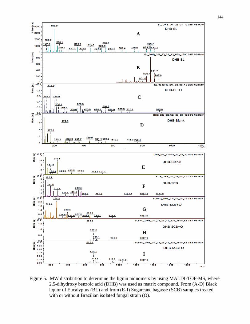
144
Figure 5. MW distribution to determine the lignin monomers by using MALDI-TOF-MS, where
2,5-dihydroxy benzoic acid (DHB) was used as matrix compound. From (A-D) Black
liquor of Eucalyptus (BL) and from (E-I) Sugarcane bagasse (SCB) samples treated
with or without Brazilian isolated fungal strain (O).
A
B
C
D
E
F
G
H
I
145

146
147

148

149
150
151


