BI 231 Lab Survival Guide http://spot.pcc.edu/anatomy/lab.htm Fall 2008 0
Biology 231
Anatomy and Physiology I
Sylvania Laboratory Survival Guide
Lab Objectives and Worksheets to accompany
Fundamentals of Human Anatomy and Physiology, By Frederic Martini, 8th Edition
Summer 2009 Update

BI 231 Lab Survival Guide http://spot.pcc.edu/anatomy/lab.htm Summer 2009 1
Table of Contents
Week (Summer Schedule) Martini Chapter
Lab Guide Page
Wee
ks
1-3
Safety Guidelines ---- 2
Disposal Guidelines ---- 3
1. Language of Anatomy Chapter 1 4
2. Organ Systems & Rat Dissection Chapter 1 12
3. The Microscope and
4. Classification of Tissues
Part I: Introduction, Epithelial Tissue,
and Neural Tissue
Part II: Connective Tissue and Muscle
Tissue
-----
Chapter 4
(Chapter 12)
(Chapter 10)
20
22
28
5. Body Membranes Chapter 4
pp. 135-137 33
6. Integumentary System Chapter 5 34
Week 4: Review and LABORATORY MIDTERM 1
Wee
ks
5-7
7. Bone Histology Chapter 6 39
8. Axial Skeleton Chapter 7 42
9. Appendicular Skeleton Chapter 8 57
10. Articulations and Body Movements Chapter 9 67
11. Muscles
Group 1 Chapter 11 69
Group 2 Chapter 11 76
Week 8: Review and LABORATORY MIDTERM 2
Lab PowerPoint slides can be viewed at: http://spot.pcc.edu/anatomy/lab.htm

BI 231 Lab Survival Guide http://spot.pcc.edu/anatomy/lab.htm Summer 2009 2
PCC-Sylvania BI 231 Laboratory Supplement
1. Upon entering the laboratory, please locate the exits, fire extinguisher, eyewash station, and clean up materials for chemical spills. Your instructor will demonstrate the location of fire blanket, safety kit, and showers.
2. Read the general laboratory directions and any objectives before coming to lab.
3. Food and drink, including water, are prohibited in laboratory. This is per Federal laboratory guidelines
and per College Safety Policy. Do not chew gum, use tobacco products of any kind, store food or apply cosmetics in the laboratory. No drink containers of any kind may be on the benches.
4. Please keep all personal materials off the working area. Store backpacks and purses at the rear of the
laboratory, not beside or under benches. Some laboratory spaces have shelving in rear for this purpose.
5. For your safety, please restrain long hair, loose fitting clothing and dangling jewelry. Hair ties are available, ask your instructor. Hats and bare midriffs are not acceptable in the laboratory. Shoes, not sandals, must be worn at all times in laboratory. You may wear a laboratory apron or lab coat if you desire, but it is not required.
6. We do not wish to invade your privacy, but for your safety if you are pregnant, taking
immunosuppressive drugs or who have any other medical conditions (e.g. diabetes, immunological defect) that might necessitate special precautions in the laboratory must inform the instructor immediately. If you know you have an allergy to latex or chemicals, please inform instructor.
7. Decontaminate work surfaces at the beginning of every lab period using Amphyl solution.
Decontaminate bench following any practical quiz, when given, and after labs involving the dissection of preserved material.
8. Use safety goggles in all experiments in which solutions or chemicals are heated or when instructed to
do so. Never leave heat sources unattended: hot plates or Bunsen burners.
9. Wear disposable gloves when handling blood and other body fluids or when touching items or surfaces soiled with blood or other body fluids such as saliva and urine. (NOTE: cover open cuts or scrapes with a sterile bandage before donning gloves.) Wash your hands immediately after removing gloves.
10. Keep all liquids away from the edge of the lab bench to avoid spills. Immediately notify your instructor
of any spills. Keep test tubes in racks provided, except when necessary to transfer to water baths or hot plate. You will be advised of the proper clean-up procedures for any spill.
11. Report all chemical or liquid spills and all accidents, such as cuts or burns, no matter how minor, to the
instructor immediately.
12. Use mechanical pipetting devices only. Mouth pipetting is prohibited.
Students who do not comply with these safety guidelines will be excluded from the Laboratory

BI 231 Lab Survival Guide http://spot.pcc.edu/anatomy/lab.htm Summer 2009 3
Safe Disposal of Contaminated Materials
• Place disposable materials such as gloves, mouth pieces, swabs, toothpicks and paper towels that have come into contact with blood or other body fluids into a disposable Autoclave bag for decontamination by autoclaving. This bucket is not for general trash. • Place glassware contaminated with blood and other body fluids directly into a labeled bucket of 10% bleach solution. ONLY glass or plastic-ware is to be placed in this bucket, not trash. • Sharp’s container is for used lancets only. It is bright red. When using disposable lancets do not replace their covers.
1. Properly label glassware and slides, using china markers provided.
2. Wear disposable gloves when handling blood and other body fluids or when touching items or surfaces soiled with blood or other body fluids such as saliva and urine. (NOTE: cover open cuts or scrapes with a sterile bandage before donning gloves.) Wash your hands immediately after removing gloves.
3. Wear disposable gloves when handling or dissecting specimens fixed with formaldehyde or stored in
Carosafe/Wardsafe.
4. Wear disposable gloves when handling chemicals denoted as hazardous or carcinogenic by your instructor. Read labels on dropper bottles provided for an experiment, they will indicate the need for gloves or goggles, etc. Upon request, detailed written information is available on every chemical used (MSDS). Ask your instructor.
5. No pen or pencil is to be used at any time on any model or bone. The bones are fragile, hard to replace and
used by hundreds of students every year. To protect them and keep them in the best condition, please use pipe cleaners and probes provided instead of a writing instrument.
a. Probes may be used on models as well. The bones are very difficult and costly to replace, as are the models and may take a long time to replace.
6. At the end of an experiment:
a. Clean glassware and place where designated. Remove china marker labels at this time. b. Return solutions & chemicals to designated area. Do not put solutions or chemicals in cupboards!
7. You cannot work alone or unsupervised in the laboratory. 8. Microscopes should be cleaned before returning to numbered cabinet. Be sure objectives are clean, use lens
paper. Place objectives into storage position, and return to the storage cabinet. Be sure cord has been coiled and restrained. Your instructor may require microscope be checked before you put it away. Be sure it is in assigned cupboard.
9. Please replace your prepared slides into the box from which they came (slides and boxes are numbered), so
students using them after you will be able to find the same slide. Before placing slides in box, clean it with Kimwipes if it is dirty or covered with oil. If you break a slide, please, inform you instructor so the slide can be replaced. Please be aware that there is hundreds of dollars worth of slides in each box and handle the boxes with care when carrying to and from your workbench.
10. Be sure all paper towels used in cleaning lab benches and washing hands are disposed of in trash container
provided. Students who do not comply with these safety guidelines
and directions will be excluded from the Laboratory

BI 231 Lab Survival Guide http://spot.pcc.edu/anatomy/lab.htm Summer 2009 4
Lab Activity 1: Language of Anatomy Martini Chapter 1
Define these terms:
1. Gross anatomy
2. Histology
3. Anatomical position.
4. Be able to describe, define, and locate the following regions:
Abdominal Acromial
Antebrachial Antecubital
Axillary Brachial
Buccal Calcaneal
Carpal Cephalic
Cervical Deltoid
Digital Dorsum
Femoral Frontal

BI 231 Lab Survival Guide http://spot.pcc.edu/anatomy/lab.htm Summer 2009 5
Gluteal Hallux
Inguinal Lumbar
Mammary Mental
Nasal Occipital
Olecranal Oral
Orbital Otic
Palmar Patellar
Pedal Pelvic
Perineal Plantar
Pollex Popliteal
Pubic Sacral
Scapular Tarsal
Vertebral Thoracic

BI 231 Lab Survival Guide http://spot.pcc.edu/anatomy/lab.htm Summer 2009 6

BI 231 Lab Survival Guide http://spot.pcc.edu/anatomy/lab.htm Summer 2009 7
5. Define the following terms:
Superior Inferior
Anterior Posterior
Medial Lateral
Cephalic / Cephalad Caudal
Ventral Dorsal
Superficial Deep
Proximal Distal

BI 231 Lab Survival Guide http://spot.pcc.edu/anatomy/lab.htm Summer 2009 8
6. Describe the following anatomical
planes; give alternate terms if possible.
a. Frontal
b. Midsagittal
c. Parasagittal
d. Transverse
i. Which plane could never cut through the kidneys?
ii. Which plane would slice you like a loaf of bread?
7. Define these serous membranes:
a. Visceral b. Parietal
8. Locate the following body cavities. a. Dorsal
i. What organs are located within this cavity?
b. Ventral: subdivided into thoracic and abdominopelvic. (The thoracic cavity contains
smaller cavities inside: Name three. Here are your hints:) i. Which cavity is in between the lungs and contains the heart, trachea, and
esophagus?
ii. Which cavity surrounds the heart, and is located in between the parietal and visceral pericardium?

BI 231 Lab Survival Guide http://spot.pcc.edu/anatomy/lab.htm Summer 2009 9
iii. Which cavity surrounds the lungs, and is located in between the parietal and visceral pleura?
iv. Which cavity is in the abdominal cavity, and is in between the parietal and
visceral peritoneum?
v. Is the liver actually in this cavity, or is it better to say that the liver is surrounded by this cavity? Look at the balloon. Is the fist in the balloon (the cavity containing the air) or does the balloon surround it?
9. Abdominal quadrants a. Right upper quadrant b. Right lower quadrant c. Left upper quadrant d. Left lower quadrant
10. Abdominal regions. a. Epigastric region b. Right and left hypochondriac regions c. Umbilical region d. Right and left lumbar regions e. Hypogastric region f. Right and left inguinal regions

BI 231 Lab Survival Guide http://spot.pcc.edu/anatomy/lab.htm Summer 2009 10
Superficial Muscles
11. Identify the following
Muscles a. Sternocleidomastoid b. Deltoid c. Pectoralis major d. External abdominal
oblique e. Rectus abdominis f. Biceps brachii g. Sartorius h. Rectus femoris i. Tibialis anterior
(Tip: learn the meaning of each term below:
sternum mastoid delta pectoral major external abdominal oblique rectus biceps brachial sartor = “tailor” femoral tibial anterior

BI 231 Lab Survival Guide http://spot.pcc.edu/anatomy/lab.htm Summer 2009 11
12. Identify the following Muscles
a. Trapezius b. Deltoid c. Triceps brachii d. Latissimus dorsi e. Gluteus maximus f. Semitendinosus g. Biceps femoris h. Gastrocnemius

BI 231 Lab Survival Guide http://spot.pcc.edu/anatomy/lab.htm Summer 2009 12
Lab Activity 2: Organ Systems Martini Chapter 1, pages 9-11
1. Human organ systems List the chief functions of each. List two or three organs of each system.
a. Integumentary
b. Skeletal
c. Muscular
d. Nervous
e. Endocrine
f. Cardiovascular
g. Lymphatic/immunity
h. Respiratory
i. Digestive
j. Urinary
k. Reproductive

BI 231 Lab Survival Guide http://spot.pcc.edu/anatomy/lab.htm Summer 2009 13
2. Identify the following organs and structures on models and/or rats:
a. Thymus b. Heart c. Lungs d. Trachea e. Esophagus f. Diaphragm g. Stomach h. Small Intestine i. Mesentery j. Large Intestine
i. Cecum (first part of large intestine) k. Pancreas l. Spleen m. Liver n. Kidneys o. Adrenal glands p. Ureter q. Urinary Bladder r. Male
i. Scrotum ii. Testes
iii. Vas deferens iv. Penis
s. Female
i. Ovaries
ii. Fallopian tubes
iii. Uterine horns in rat iv. Uterus in human

BI 231 Lab Survival Guide http://spot.pcc.edu/anatomy/lab.htm Summer 2009 14
Rat Dissection
1. Lay the rat on the dissection tray.
2. Using forceps, grasp the skin and pull upwards.
3. Using the sharp point of the scissors, pierce the skin to start the cut. Only cut skin, do not puncture into the deeper tissues.
4. Using the scissors, cut the skin along the midline from the pelvis to the chin.

BI 231 Lab Survival Guide http://spot.pcc.edu/anatomy/lab.htm Summer 2009 15
5. Make horizontal cuts at the top and bottom. (Indicated by dashed lines)
6. Peel the skin back with the scalpel as shown above.
7. Once the skin is peeled back, grasp the abdominal wall with the forceps and pull upward.
8. Using the scissors, carefully puncture the abdominal wall and cut along the midline. Be very careful not to cut the underlying structures.
9. Expose the abdominal, pelvic and thoracic cavities to identify the organs.

BI 231 Lab Survival Guide http://spot.pcc.edu/anatomy/lab.htm Summer 2009 16
Diaphragm
Liver
Stomach
Spleen
Pancreas
Kidney
Small Intestine
Large Intestine
Bladder
Mesentery
Cecum

BI 231 Lab Survival Guide http://spot.pcc.edu/anatomy/lab.htm Summer 2009 17
Cecum
Liver
Liver
Ureter
Bladder
Kidney
Spleen
Pancreas
Stomach

BI 231 Lab Survival Guide http://spot.pcc.edu/anatomy/lab.htm Summer 2009 18
Trachea
Thymus
Heart
Lung
Diaphragm

BI 231 Lab Survival Guide http://spot.pcc.edu/anatomy/lab.htm Summer 2009 19
Small Intestine
Large Intestine
Seminal Vesicle
Bladder
Penis
Vas Deferens
Testicle
Ovary
Uterine Horn
Uterus
Bladder

BI 231 Lab Survival Guide http://spot.pcc.edu/anatomy/lab.htm Summer 2009 20
Lab Activity 3: The Microscope
1. Care of the microscope
a. When transporting microscope, hold in upright position with one hand on the arm and the other supporting the base
b. Only use lens paper to clean the lens. NEVER USE KIMWIPES.
c. Always begin the focusing process with the lowest-power objective and change to
higher-power lenses as necessary. Which objectives should never be used with oil? Which objective can only be used with oil?
d. Use coarse adjustment knob only with the lowest power lens e. Always use a coverslip with temporary preparations
2. Putting the microscope away a. Remove slides from stage and place in appropriate place
b. Rotate the lowest-power objective lens into position
c. Move stage to the lowest position
d. Turn down light brightness
e. Turn off power
f. Wipe microscope (not the lens) with Kimwipes or alcohol wipe if needed
g. Wrap the cord neatly around the base
h. Lock the cabinet
3. Basic Parts of a Compound Light Microscope:
a. Eyepiece (Ocular): Usually contains a 10X lens.
b. Arm: contains the housing for the fine and coarse adjustments and connects the base of the microscope to the nosepiece and ocular.
c. Nosepiece: A rotating head that has the objective lenses attached to it. The lens to be
used should "click" into position when the wheel is gently turned so that it is directly over the specimen slide.
d. Objective: Basically a housing for different powers of lenses (usually 4X, 10X, 40X,
100X)
e. Stage: The specimen slides rests on this part of the microscope.

BI 231 Lab Survival Guide http://spot.pcc.edu/anatomy/lab.htm Summer 2009 21
f. Coarse adjustment knobs: The larger of two sets of knobs located on either side of the
arm, just above the base. This adjustment is used to make large adjustments in focusing by moving the lenses up and down.
g. Fine adjustment knobs: The smaller of two sets of knobs located on either side of the arm. This adjustment is used to make small adjustments in focusing. It has a limited amount of movement and is most efficiently used after focusing with the 4X objective and coarse focus, then increasing magnification and making final adjustments with the fine focus knob.
h. Light source: Located directly under the stage. i. Adjustable diaphragm: This rotating wheel on the underside of the stage allows the
user to adjust the amount of light that passes through the specimen. As a general rule, the lowest intensity of light that allows you to resolve the structure of the object you are viewing should be used.
4. Calculating magnification: a. Total magnification = (magnification of ocular) x (magnification of objective).
Example: the total magnification is 450x when using a 10x ocular is 10x and 45x objective.
5. Learn how to focus the microscope using the 10x and 40x. Do not use the 100x objective lens.

BI 231 Lab Survival Guide http://spot.pcc.edu/anatomy/lab.htm Summer 2009 22
Lab Activity 4: Classification of Tissues
Part I: Introduction, Epithelial Tissue, and Neural Tissue
Martini Chapter 4 (and Part of Chapter 12)
1. Rearrange the following from highest to lowest order of magnitude:
______Atoms ______Cells ______Molecules ______Organs ______Tissues
2. What are the 4 primary tissue types?
a.
b.
c.
d.
3. Describe 5 distinguishing features of epithelial tissue.
a.
b.
c.
d.
e.
4. Describe the way that epithelial tissues are named 1st part of name:
2nd part of name:
5. Two epithelial tissues do not follow the naming process in #3, and explain the meaning of their names:
a. Pseudostratified columnar epithelium

BI 231 Lab Survival Guide http://spot.pcc.edu/anatomy/lab.htm Summer 2009 23
b. Transitional epithelium
6. Complete this table on epithelial tissues, and be able to identify these tissues from microscope
slides. Epithelial Tissue Sketch a picture
Functions this type is especially suited for and why.
One location that helps you remember its function
Simple squamous
Simple cuboidal
Simple columnar

BI 231 Lab Survival Guide http://spot.pcc.edu/anatomy/lab.htm Summer 2009 24
Ciliated simple columnar
Ciliated pseudostratified columnar
Non-keratinized stratified squamous
Keratinized stratified squamous

BI 231 Lab Survival Guide http://spot.pcc.edu/anatomy/lab.htm Summer 2009 25
Transitional
Stratified Cuboidal
Nervous Tissue
1. Basic cell structure of a multipolar neuron. Identify and describe function a. Cell body
b. Nucleus
c. Axon hillock
d. Dendrite
e. Axon
f. Schwann cells and myelin sheath
g. Nodes of Ranvier
h. Telodendria
i. Axon Terminals=Synaptic end bulbs

BI 231 Lab Survival Guide http://spot.pcc.edu/anatomy/lab.htm Summer 2009 26
2. Distinguish structurally and functionally between unipolar, bipolar, and multipolar neurons. a. Where can each be located?
3. What is another word for a sensory neuron?
4. What is another word for a motor neuron?
5. What type of neuron is between sensory and motor neurons?

BI 231 Lab Survival Guide http://spot.pcc.edu/anatomy/lab.htm Summer 2009 27
6. Identify the following structures a. Epineurium
b. Perineurium
c. Endoneurium
d. Fascicle
e. Axon
f. Myelin sheath
7. Glial Cells: Identify structure and function.
a. CNS i. Astrocytes
ii. Oligodendrocytes
iii. Microglia
b. PNS i. Schwann cells

BI 231 Lab Survival Guide http://spot.pcc.edu/anatomy/lab.htm Summer 2009 28
Lab Activity 4: Classification of Tissues
Part II: Connective Tissue and Muscle Tissue
Martini Chapter 4 (and Part of Chapter 10)
1. What are the 4 types of connective tissue? a.
b.
c.
d.
2. List the distinguishing features of connective tissue, especially focusing on how it differs from epithelial tissue:
3. Define these connective tissue terms: matrix, ground substance, and fibers. a. Matrix:
i. Ground Substance:
ii. Fibers
4. Give an example of where you can find this type of ground substance. a. Fluid
b. Gel
c. Solid
5. Define/describe these cells
a. Fibroblasts
b. Macrophages
c. Adipocytes

BI 231 Lab Survival Guide http://spot.pcc.edu/anatomy/lab.htm Summer 2009 29
d. Mesenchymal cells
e. Mast cells
f. Lymphocytes
6. Complete this table for connective tissue fibers
Fiber type, sketch
Function
Example of where found
Elastic
Reticular
Collagen
7. Complete this table for connective tissues Connective Tissue type Sketch cells & matrix
Functions this type is especially suited for and why.
One location that helps you remember its function
Areolar CT

BI 231 Lab Survival Guide http://spot.pcc.edu/anatomy/lab.htm Summer 2009 30
Adipose CT
Reticular CT
Dense Regular CT
Dense Irregular CT
Elastic CT

BI 231 Lab Survival Guide http://spot.pcc.edu/anatomy/lab.htm Summer 2009 31
Blood
Bone
Hyaline Cartilage
Elastic Cartilage
Fibrocartilage

BI 231 Lab Survival Guide http://spot.pcc.edu/anatomy/lab.htm Summer 2009 32
Muscle Tissue Histology
8. Complete this table on muscle tissue.
Muscle type Sketch
# Nuclei per cell Visible striations?
Locations Voluntary or Involuntary
Skeletal
Cardiac
Smooth

BI 231 Lab Survival Guide http://spot.pcc.edu/anatomy/lab.htm Summer 2009 33
Lab Activity 5: Body Membranes Martini Chapter 4, page 135-137
Epithelial membranes are simple organs consisting of an epithelial sheet bound to an underling
layer of connective tissue
1. Define these epithelial membranes a. Mucous membranes
i. Do all mucous membranes secrete mucus?
b. Serous membranes c. Cutaneous membrane
Synovial membranes are composed entirely of connective tissue; they contain no epithelial
cells. 2. Where can you find synovial membranes?
a. What is the function of synovial membranes?
b. What is the function of synovial fluid?

BI 231 Lab Survival Guide http://spot.pcc.edu/anatomy/lab.htm Summer 2009 34
Lab Activity 6: Integumentary System Martini Chapter 5
Identify from microscope slide
1. Epidermis: (Keratinized stratified squamous epithelium)
a. Stratum corneum b. Stratum lucidum in palms and soles. c. Stratum granulosum d. Stratum spinosum e. Stratum basale.
2. Dermis:
a. Papillary layer i. What type of connective
tissue is this?
b. Reticular layer i. What type of connective
tissue is this?
c. Hypodermis: (not part of the skin) i. Adipose tissue
Draw a picture:
1. Describe/define these cells associated with the epidermis and indicate which layer they are in. a. Keratinocytes
1. What is the role of keratin?
b. Melanocytes
1. What is the role of melanin?
c. Langerhans’ cells 1. Are these epithelial cells? 2. Where do they originally come from?
BI 231 Lab Survival Guide http://spot.pcc.edu/anatomy/lab.htm Summer 2009 35
2. Describe these features of the dermis and which layer they are associated with. a. Fingerprints
b. Dermal papilla
c. Cleavage lines
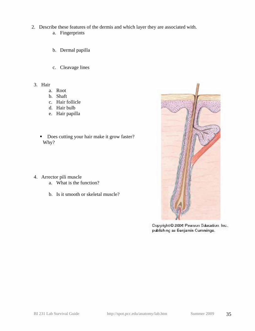
3. Hair a. Root b. Shaft c. Hair follicle d. Hair bulb e. Hair papilla
Does cutting your hair make it grow faster? Why?
4. Arrector pili muscle a. What is the function? b. Is it smooth or skeletal muscle?

BI 231 Lab Survival Guide http://spot.pcc.edu/anatomy/lab.htm Summer 2009 36
5. Sweat glands
a. Apocrine sweat glands b. Eccrine (merocrine) sweat glands
c. What is the difference between eccrine (merocrine) sweat glands and apocrine sweat glands?
d. What are mammary glands?
e. What are ceruminous glands?
6. Sebaceous gland
a. What does it secrete?
7. Sebaceous follicle a. Where on the body are these
located?

BI 231 Lab Survival Guide http://spot.pcc.edu/anatomy/lab.htm Summer 2009 37
8. Pacinian (lamellated) corpuscle (look at fetal palm slide)
a. Which layer are they located in?
b. What is the function?
9. Meissner’s (tactile) corpuscle (found in dermal papillae)
a. What is the function?
10. Merkel cells a. Which layer are they located in?
b. What is the function?
11. Fingernail a. Free edge b. Body c. Lateral fold d. Lunula e. Cuticle f. Root of the nail g. Matrix

BI 231 Lab Survival Guide http://spot.pcc.edu/anatomy/lab.htm Summer 2009 38
BI 231 Lab Survival Guide http://spot.pcc.edu/anatomy/lab.htm Summer 2009 39
Lab Activity 7: Bone Histology Martini Chapter 6
1. Describe these types of bone a. Spongy bone
i. Where can it be located?
ii. What are trabeculae?
b. Compact bone
i. Where can it be located?
2. Compact bone features: Identify on model and tissue slides. a. Concentric lamellae b. Haversian (Central) Canal c. Volkmann’s (perforating) Canal d. Interstitial lamellae e. Circumferential lamellae f. Lacunae g. Canaliculi
3. Cells: Describe the function of these bone cells:
a. Osteoblasts
b. Osteocytes
c. Osteoclasts
3. Coverings: Describe the location of these:
a. Periosteum
i. Which cells are located on the periosteum?
b. Endosteum
i. Which cells are located on the endosteum? 4. Marrow types, where are these located in adults and what are their function?
a. Red bone marrow
b. Yellow bone marrow

BI 231 Lab Survival Guide http://spot.pcc.edu/anatomy/lab.htm Summer 2009 40
Bone Histology Worksheet
Instructions: Label all of these structures. Volkmann’s canal (perforating canal) Trabeculae Haversian canal (central canal) Canaliculi Lacunae Osteocyte in lacunae Osteocyte in lacunae Periosteum Compact bone Endosteum Medullary cavity Interstitial lamellae Osteon Concentric lamellae Identify where you would find: Circumferential lamellae
Bone marrow, osteoblasts, and osteoclasts

BI 231 Lab Survival Guide http://spot.pcc.edu/anatomy/lab.htm Summer 2009 41
5. Describe these types of bones and give examples a. Short bones
b. Flat bones
c. Irregular bones
d. Sesamoid bones
e. Wormian or sutural bones
f. Long bones
6. Long bone structure: from long bone, identify
a. Diaphysis b. Epiphyses
i. What type of bone makes up the center of the epiphyses?
c. Epiphyseal line d. Epiphyseal plate (not in this picture)
i. What type of cartilage is this?
ii. What process happens at the diaphyseal side of the epiphyseal plate?
e. Medullary cavity f. Endosteum g. Periosteum h. Articular surface
i. What type of cartilage is this?
Label:

BI 231 Lab Survival Guide http://spot.pcc.edu/anatomy/lab.htm Summer 2009 42
Lab Activity 8: Axial Skeleton Martini Chapter 7
1. Axial skeleton components: skull, spine, thoracic cage, and hyoid bone
2. Spine: 33 separate bones in fetus & infant; in children & adults it's made of 26 bones:
a. Coccyx (4 fused vertebrae) b. Sacrum (5 fused vertebrae) c. 24 individual vertebrae.
Know number of vertebrae in each part of back (7 cervical, 12 thoracic, 5 lumbar)
Direction of curves:
d. Cervical (concave posterior) e. Thoracic (convex posterior) f. Lumbar (concave posterior)
3. Structures of Typical Vertebra a. Body b. Vertebral foramen c. Spinous process d. Transverse process e. Superior & inferior articular processes f. Vertebral arch: made of laminae & pedicles g. Intervertebral foraminae between vertebrae h. Transverse foraminae in cervical vertebrae i. Facets: on super & inferior articular processes: smooth surfaces for joints between
processes

BI 231 Lab Survival Guide http://spot.pcc.edu/anatomy/lab.htm Summer 2009 43
4. Intervertebral disk: i. Anulus fibrosus
(fibrocartilage) ii. Nucleus pulposus
5. Cervical Vertebrae a. There are 7 cervical vertebrae b. Direction of curve- concave posterior c. Foraminae in transverse processes for vertebral artery to the brain d. Note bifid spinous processes

BI 231 Lab Survival Guide http://spot.pcc.edu/anatomy/lab.htm Summer 2009 44
e. C1=atlas: superior articular facet for articulation with occipital condyle
f. C2=axis i. Odontoid process
(dens)
BI 231 Lab Survival Guide http://spot.pcc.edu/anatomy/lab.htm Summer 2009 45
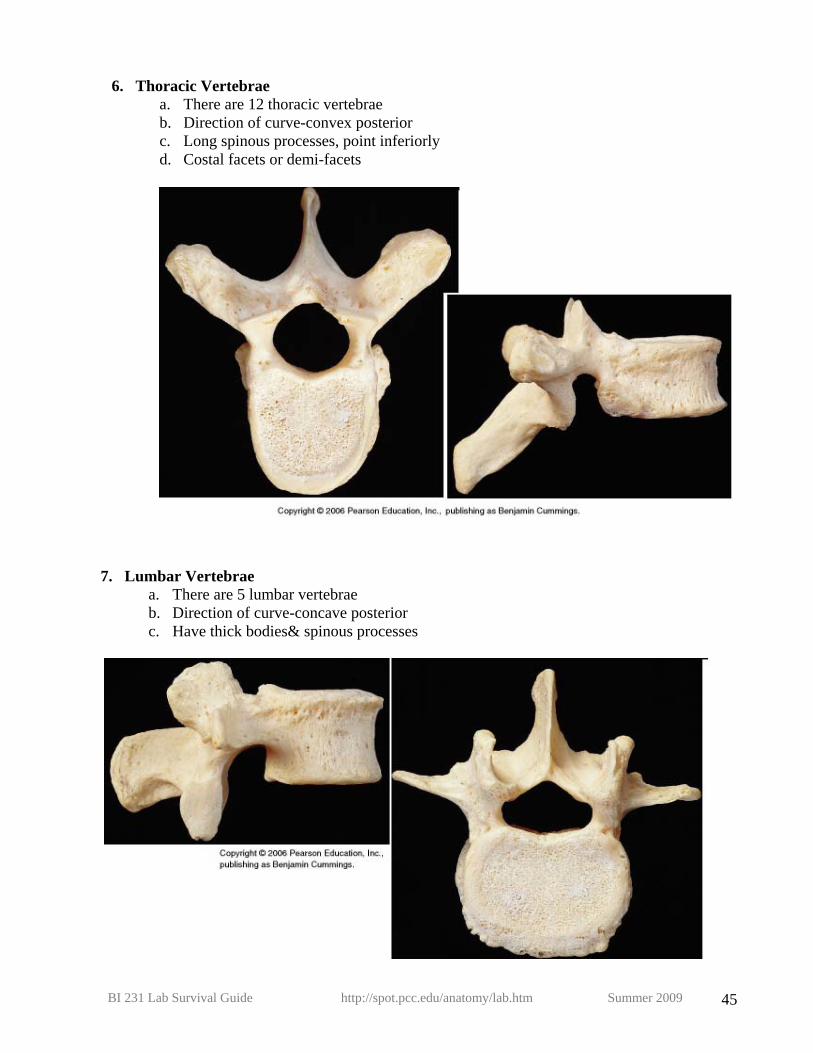
6. Thoracic Vertebrae a. There are 12 thoracic vertebrae b. Direction of curve-convex posterior c. Long spinous processes, point inferiorly d. Costal facets or demi-facets
7. Lumbar Vertebrae a. There are 5 lumbar vertebrae b. Direction of curve-concave posterior c. Have thick bodies& spinous processes

BI 231 Lab Survival Guide http://spot.pcc.edu/anatomy/lab.htm Summer 2009 46
8. Sacrum: a. 5 fused vertebrae b. Convex posterior c. Sacral promontory d. Sacroiliac joints e. Anterior sacral foramina f. Posterior sacral foramina
9. Coccyx-4 rudimentary vertebrae
10. Hyoid Bone: attachment for muscles of tongue, neck, & pharynx
a. Greater and lesser horns b. Body

BI 231 Lab Survival Guide http://spot.pcc.edu/anatomy/lab.htm Summer 2009 47
11. Thoracic cage = Bony Thorax a. Sternum:
i. Manubrium ii. Sternal angle: separates manubrium from body
iii. Body iv. Xiphoid process (cartilage when young,
ossifies about age 40)
b. Ribs: i.True (1st 7 pairs), cartilage of rib
(costal cartilage) articulates with sternum
ii.False A. Pair 8-10: cartilage of rib
articulates with cartilage above B. Lowest 2 pairs are floating
iii. Parts of a typical rib: A. Head B. Tubercle C. Angle D. Costal groove
c. Note how the ribs attach to the vertebrae. Looking at them from the posterior side of the back, there is an acute angle created between the 12th rib and the spine due to the downward direction of the 12th rib as it heads anteriorly. This angle is the costovertebral angle.

BI 231 Lab Survival Guide http://spot.pcc.edu/anatomy/lab.htm Summer 2009 48
12. Cranial Bones: recognize from inside & outside a. Frontal
i. Supraorbital ridge b. Parietal (left and right) c. Temporal (left and right)
i. Mandibular fossa ii. External acoustic canal
iii. Styloid process iv. Mastoid processes v. Zygomatic process
vi. Carotid canal vii. Jugular foramen
viii. Internal acoustic canal (seen on the cranial floor) d. Occipital
i. Occipital Condyles ii. Superior and inferior nuchal lines
iii. External occipital protuberance iv. Foramen Magnum
e. Sphenoid i. Greater wing
ii. Lesser wing iii. Medial and lateral pterygoid plates iv. Pterygoid processes v. Sella turcica
A. Hypophyseal fossa (shallow depression in middle of sella turcica) B. Dorsum sella (ridge on posterior aspect of sella)
f. Ethmoid i. Cribriform plate
ii. Cristae galli (Sail of Christ) iii. Perpendicular plate (boney nasal septum) iv. Superior and middle nasal conchae
13. Cranium-Floor
a. Fossae: Anterior, middle, and posterior cranial fossae
14. Cranium-Sutures: a. Sagittal b. Coronal c. Squamous d. Lambdoid e. Wormian (sutural) bones: these are small islands of bone that fill gaps in sutures; not
always present.

BI 231 Lab Survival Guide http://spot.pcc.edu/anatomy/lab.htm Summer 2009 49
15. Infant/fetal skull a. Anterior fontanel b. Occipital fontanel c. Sphenoidal fontanel d. Mastoid fontanel
16. Orbit (Bony eye socket) a. Frontal bone b. Sphenoid bone
i. Superior orbital fissure ii. Optic canal
c. Zygomatic bone d. Maxillary bone e. Palatine bone f. Lacrimal bone g. Ethmoid bone
17. Face: Bones
a. Maxilla b. Mandible
i. Body ii. Ramus
iii. Coronoid process iv. Mandibular condyle v. Angle
vi. Mandibular foramen vii. Mylohyoid line
viii. Alveolar process ix. Mental foramen
c. Palatine (left and right) d. Zygomatic (left and right)
i. Temporal process e. Lacrimal (left and right) f. Nasals (left and right) g. Vomer h. Superior & Middle nasal conchae (part of ethmoid bone) i. Inferior nasal conchae
18. Face: special structures
a. Alveoli (tooth sockets) b. Sinuses:
i. Frontal ii. Maxillary
iii. Sphenoidal iv. Ethmoid air cells
c. Zygomatic arch

BI 231 Lab Survival Guide http://spot.pcc.edu/anatomy/lab.htm Summer 2009 50

BI 231 Lab Survival Guide http://spot.pcc.edu/anatomy/lab.htm Summer 2009 51

BI 231 Lab Survival Guide http://spot.pcc.edu/anatomy/lab.htm Summer 2009 52
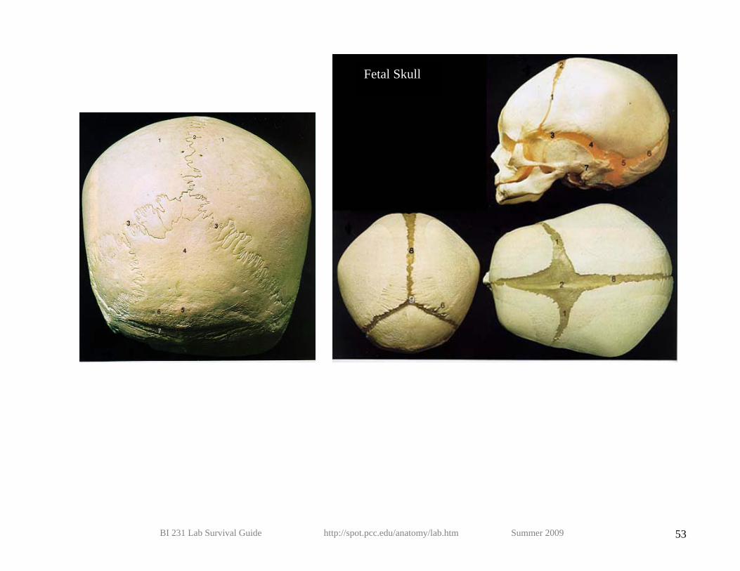
BI 231 Lab Survival Guide http://spot.pcc.edu/anatomy/lab.htm Summer 2009 53
Fetal Skull

BI 231 Lab Survival Guide http://spot.pcc.edu/anatomy/lab.htm Summer 2009 54

BI 231 Lab Survival Guide http://spot.pcc.edu/anatomy/lab.htm Summer 2009 55

BI 231 Lab Survival Guide http://spot.pcc.edu/anatomy/lab.htm Summer 2009 56

BI 231 Lab Survival Guide http://spot.pcc.edu/anatomy/lab.htm Summer 2009 57
Lab Activity 9: Appendicular Skeleton Martini Chapter 8
1. Appendicular Skeleton: Upper & Lower extremities, Shoulder Girdle, Pelvic Girdle Upper extremity Humerus:
A. Head B. Greater & lesser tubercle C. Intertubercular groove D. Anatomical & surgical necks E. Deltoid tubercle F. Radial groove G. Trochlea H. Capitulum I. Olecranon fossa J. Medial & lateral epicondyles K. Radial fossa L. Coronoid fossa M. Supracondylar ridges (medial
and lateral)—region proximal to each epicondyle

BI 231 Lab Survival Guide http://spot.pcc.edu/anatomy/lab.htm Summer 2009 58
Radius
A. Radial head B. Radial tuberosity C. Ulnar notch of the radius D. Interosseous ridge of the radius E. Styloid process of the radius
Ulna:
A. Head of the ulna B. Styloid process of the ulna C. Olecranon D. Coronoid process E. Semilunar or trochlear notch F. Radial notch of the ulna G. Supinator crest H. Interosseous ridge of the ulna I. Ulnar tuberosity

BI 231 Lab Survival Guide http://spot.pcc.edu/anatomy/lab.htm Summer 2009 59
Carpals:
A. Proximal row, lateral to medial: scaphoid, lunate, triquetrum, and pisiform B. Distal row, lateral to medial: trapezium, trapezoid, capitate, hamate
Metacarpals: #1-thumb, through#5=to baby finger
Phalanges: proximal & distal on thumb, proximal, middle, & distal on rest. (Toe bones also called phalanges)
Dorsal Palmar

BI 231 Lab Survival Guide http://spot.pcc.edu/anatomy/lab.htm Summer 2009 60
Pectoral girdle
Scapula:
A. Spine B. Medial, lateral, & superior borders and angles (superior, lateral, inferior) C. Acromion or acromial process D. Coracoid process E. Fossa (supraspinous, infraspinous & subscapular) F. Glenoid fossa (cavity) G. Supraglenoid tubercle H. Infra glenoid tubercle I. Scapular notch
Clavicle
A. Acromial end B. Sternal end C. Conoid tubercle

BI 231 Lab Survival Guide http://spot.pcc.edu/anatomy/lab.htm Summer 2009 61
Lower extremity
Femur:
A. Head B. Greater trochanter C. Lesser trochanter D. Neck E. Intertrochanteric line F. Intertrochanteric crest G. Linea aspera H. Fovea capitis I. Medial and lateral condyles of
the femur J. Adductor tubercle—located
proximal to medial epicondyle K. Medial & lateral epicondyles of
the femur L. Patellar surface M. Intercondylar notch or fossa N. Gluteal tubercle

BI 231 Lab Survival Guide http://spot.pcc.edu/anatomy/lab.htm Summer 2009 62
Tibia:
A. Medial & lateral condyles of the tibia B. Intercondylar eminence C. Tibial tuberosity D. Fibular surface of the tibia E. Medial malleolus F. Anterior crest of the tibia G. Fibular notch H. Tibular articular surface for the talus
Fibula:
A. Head of the fibula B. Styloid process (apex) of the fibula C. Lateral malleolus D. Fibular articular surface for the talus

BI 231 Lab Survival Guide http://spot.pcc.edu/anatomy/lab.htm Summer 2009 63
Patella: a sesamoid bone.
A. Medial facet (articular surface) B. Lateral facet (articular surface)
Tarsal bones:
A. Talus-bears body's weight B. Calcaneus C. Navicular D. Cuboid E. Lateral, Intermediate & Medial Cuneiform
Metatarsals: 1st-5th
Phalanges: two for big toe, rest have 3

BI 231 Lab Survival Guide http://spot.pcc.edu/anatomy/lab.htm Summer 2009 64
Pelvic Girdle: two hipbones (os coxae) Unite anteriorly at pubic symphysis A. Ilium:
a. Iliac crests b. Anterior superior iliac spine c. Anterior inferior iliac spine d. Posterior superior iliac spine e. Posterior inferior iliac spine f. Greater sciatic notch g. Iliac fossa h. Body of the ilium i. Auricular surface j. Sacroiliac joint k. Arcuate line l. Inferior gluteal line
B. Ischium a. Ischial tuberosity b. Ischial Spine c. Body of the ischium d. Ischial ramus e. Lesser sciatic notch
C. Pubic bone a. Pubic crest b. Body of the pubis c. Pubic symphysis: What kind of
cartilage is it? d. Superior & inferior rami e. Pectineal line f. Pubic tubercle g. Pubic arch h. Pubic angle (calculate this)

BI 231 Lab Survival Guide http://spot.pcc.edu/anatomy/lab.htm Summer 2009 65
D. Other regions: a. Obturator foramen b. Pelvic brim c. Lesser (true) pelvis below pelvic brim & d. Greater (false) pelvis above pelvic brim e. Acetabulum

BI 231 Lab Survival Guide http://spot.pcc.edu/anatomy/lab.htm Summer 2009 66
E. Male pelvis: a. Vertical iliac bones b. Pubic arch <90 degrees c. Deep iliac fossa d. Heart shaped pelvic inlet
F. Female pelvis: a. Less vertical iliacs b. Arch >90, shallow iliac fossa c. Round pelvic brim (inlet)

BI 231 Lab Survival Guide http://spot.pcc.edu/anatomy/lab.htm Summer 2009 67
Lab Activity 10: Articulations and Body Movements Martini Chapter 9
1. Knee a. Medial & lateral collateral ligaments (=tibial & fibular collaterals) b. Anterior & posterior cruciates c. Medial & lateral menisci d. Quadriceps tendon, patellar ligament
Anterior
Posterior

BI 231 Lab Survival Guide http://spot.pcc.edu/anatomy/lab.htm Summer 2009 68
2. Define these movements a. Flexion
b. Extension
c. Hyperextension
d. Adduction
e. Abduction
f. Circumduction
g. Rotation
h. Pronation & supination
i. Inversion & eversion
j. Dorsiflexion
k. Plantar flexion

BI 231 Lab Survival Guide http://spot.pcc.edu/anatomy/lab.htm Summer 2009 69
Lab Activity 11: Muscles Martini Chapter 11
**GROUP I**
MUSCLES OF THE
HEAD ORIGIN INSERTION ACTION
Frontalis Epicranial aponeurosis Skin over forehead Elevates eyebrows and wrinkles skin of forehead
Occipitalis Occipital and temporal bones Epicranial aponeurosis Fixes epicranial aponeurosis and pulls scalp posteriorly
Orbicularis oculi Medial orbital margin and zygomatic bone
Skin surrounding eye Closes eyelids and depresses skin of forehead
Temporalis Temporal fossa Coronoid process of mandible Elevates and retracts mandible Masseter Zygomatic process and arch Angle and ramus of mandible Elevates mandible Orbicularis oris By muscular skips to surround
muscles Muscles interlace to surround mouth Closes and purses lips
Zygomaticus major Zygomatic arch Corner of mouth Elevates corner of mouth Risorius Fascia of masseter Corner of mouth Draws corner of mouth laterally
SUPERFICIAL MUSCULATURE OF THE NECK
ORIGIN INSERTION ACTION
Omohyoid Medial tip of suprascapular notch Body of hyoid bone Depresses hyoid bone Sternohyoid Posterior surface of manubrium, and
medial clavicle Hyoid bone Depresses hyoid bone
Mylohyoid Mylohyoid line of mandible Hyoid bone Elevates hyoid bone and floor of mouth, depresses mandible
Digastric - Anterior belly -Posterior belly
Anterior: Lower border of mandible near midline Posterior: Mastoid notch of temporal bone
Intermediate tendon Intermediate tendon
Ant.: Elevates hyoid bone and base of tongue, depresses mandible Post: Moves hyoid bone back

BI 231 Lab Survival Guide http://spot.pcc.edu/anatomy/lab.htm Summer 2009 70
MUSCLES OF THE NECK & BACK
ORIGIN INSERTION ACTION
Sternocleidomastoid Manubrium and medial third of clavicle
Mastoid process Rotates head to opposite side, extends head, and flexes vertebral column
Trapezius External occipital protuberance, superior nuchal line, and spinous process of C7-T12
Anterior border of scapular spine, acromion process, lateral third of clavicle
Elevates, adducts, and depresses scapula
Latissimus dorsi Spinous processes of lower 6 thoracic vertebrae, thoracolumbar fascia, crest of ilium
Intertubercular groove of humerus Extends, rotates humerus medially, draws shoulder down and backward
Levator scapula Transverse processes of C1-4 Superior angle spine of scapula Elevates scapula Rhomboid major Spinous process of T1-5 and
supraspinous ligament Medial border below spine of scapula
Adducts scapula and performs downward rotation
Rhomboid minor Spinous process of C7-T1 Medial border of scapula at base of spine
Adducts scapula and performs downward rotation
Serratus anterior Lateral surface of upper 8 ribs Anterior lip of medial border of scapula
Holds scapula to chest wall, medially rotates scapula in abducting or extending humerus
Erector Spinae
Sacrum, iliac crest, spinous processes of lumbar vertebrae and T11,12
Angles of the ribs Spinous & Transverse processes of vertebrae
Extension of vertebral column

BI 231 Lab Survival Guide http://spot.pcc.edu/anatomy/lab.htm Summer 2009 71
MUSCLES OF THORACIC WALL
ORIGIN INSERTION ACTION
External intercostal Inferior border of rib above Superior border of rib below Elevates rib cage during inspiration Internal intercostal Superior border of rib below Inferior border of rib above Depresses rib cage during expiration Pectoralis major Medial half of clavicle, sternum,
costal cartilages, aponeurosis of external abdominal oblique
Intertubercular groove of humerus Flexes, adducts, and medially rotates humerus, draws body upward in climbing
Pectoralis minor Anterior surface of ribs 3 to 5 Coracoid process of scapula Draws scapula down and forward and elevates ribs
MUSCLES OF THE ANTERIOR ABDOMINAL WALL
ORIGIN INSERTION ACTION
Rectus abdominis
Pubis symphysis and crest of pubis Xiphoid process and cartilages of ribs 5 to 7
Tenses abdominal wall and flexes vertebral column
External abdominal oblique
External surface of lower 8 ribs Anterior half of iliac crest and linea alba
Compresses abdomen, contralaterally rotates and flexes vertebral column
Internal abdominal oblique
Lateral half of inguinal ligament, anterior iliac crest and thoracolumbar fascia
Lower four ribs, linea alba and by conjoined tendon to pubis
Compresses abdomen, ipsilaterally rotates and flexes vertebral column
Transverse abdominis Lateral third of inguinal ligament, anterior iliac crest, and thoracolumbar fascia
Linea alba, and by conjoined tendon to pubis
Compresses abdomen

BI 231 Lab Survival Guide http://spot.pcc.edu/anatomy/lab.htm Summer 2009 72

BI 231 Lab Survival Guide http://spot.pcc.edu/anatomy/lab.htm Summer 2009 73

BI 231 Lab Survival Guide http://spot.pcc.edu/anatomy/lab.htm Summer 2009 74

BI 231 Lab Survival Guide http://spot.pcc.edu/anatomy/lab.htm Summer 2009 75

BI 231 Lab Survival Guide http://spot.pcc.edu/anatomy/lab.htm Summer 2009 76
MUSCLES OF THE SHOULDER AND ARM
ORIGIN INSERTION ACTION
Deltoid Anterior surface of clavicle, acromion process and spine of scapula
Deltoid tubercle of humerus Abducts humerus; aids in flexion, extension, and internal and external rotation
Supraspinatus Supraspinous fossa Greater tubercle of humerus Abducts humerus Infraspinatus Infraspinous fossa Greater tubercle of humerus Rotates humerus laterally Teres minor Axillary border of scapula Greater tubercle of humerus Rotates humerus laterally Teres major Axillary border at inferior angle of
scapula Intertubercular groove of humerus Extends, adduction and medial
rotates humerus. Subscapularis Subscapular fossa Lesser tubercle of humerus Rotates humerus medially Biceps brachii
Long head, supraglenoid tubercle; Short head, coracoid process scapula
Radial tuberosity of the radius Flexes radius and humerus, and supinates forearm
Coracobrachialis Coracoid process of scapula Middle third of humerus Flexes and adducts humerus Triceps brachii
Long head, infraglenoid tubercle; Lateral head, proximal portion of posterior humerus; Medial head, distal half of posterior humerus
Olecranon process of ulna Extends humerus and ulna
Brachialis Anterior distal two-thirds of humerus
Coronoid process of ulna Flexes ulna
Group II
MUSCLES OF ANT. FOREARM
ORIGIN INSERTION ACTION
Pronator teres Medial epicondyle of humerus and coronoid process of ulna
Middle portion of radius Pronates and flexes forearm
Flexor carpi radialis Medial epicondyle of humerus Base of second metacarpal Flexes wrist & elbow; abducts wrist Palmaris longus Medial epicondyle of the humerus Palmar aponeurosis Weak flexion of wrist Flexor carpi ulnaris Medial epicondyle of humerus,
olecranon process, & posterior ulna Pisiform, hamate, and fifth metacarpal
Flexes and adducts wrist
Flexor digitorum superficialis
Medial epicondyle of humerus and coronoid process of ulna
Middle phalanges of fingers Flexes fingers and wrist

BI 231 Lab Survival Guide http://spot.pcc.edu/anatomy/lab.htm Summer 2009 77
Flexor pollicis longus Middle half of radius, interosseous membrane, coronoid process of ulna
Distal phalanx of thumb Flexes thumb and wrist
MUSCLES OF POSTEROLATERAL FOREARM
ORIGIN INSERTION ACTION
Brachioradialis Lateral supracondylar ridge Styloid process of radius Flexes forearm Extensor carpi radialis longus
Lateral supracondylar ridge of humerus
Second metacarpal Extends and abducts hand
Extensor carpi radialis brevis
Lateral epicondyle of humerus Third metacarpal Extends and abducts hand
Extensor digitorum Lateral epicondyle of humerus Into distal phalanx by 4 tendons Extends fingers and hand Extensor carpi ulnaris Lateral epicondyle of humerus and
posterior border of ulna Fifth metacarpal Extends and adducts hand
Extensor digiti minimi Lateral epicondyle of humerus Extensor expansion of little finger Extends 5th digit and hand Supinator Lateral epicondyle of humerus,
supinator crest of ulna Lateral surface and posterior border of radius
Supinates forearm
Abductor pollicis longus
Posterior surface of ulna and radius, and interosseous membrane
First metacarpal Abducts thumb and hand
Extensor pollicis brevis Middle third of radius and interosseous membrane
Base of proximal phalanx of thumb Extends thumb
Extensor pollicis longus Middle third of ulna and interosseous membrane
Base of distal phalanx of thumb Extends thumb

BI 231 Lab Survival Guide http://spot.pcc.edu/anatomy/lab.htm Summer 2009 78

BI 231 Lab Survival Guide http://spot.pcc.edu/anatomy/lab.htm Summer 2009 79

BI 231 Lab Survival Guide http://spot.pcc.edu/anatomy/lab.htm Summer 2009 80

BI 231 Lab Survival Guide http://spot.pcc.edu/anatomy/lab.htm Summer 2009 81
MUSCLES OF BACK AND GLUTEAL REGION
ORIGIN INSERTION ACTION
Gluteus maximus Upper portion of ilium, the sacrum and coccyx
Gluteal tuberosity and iliotibial tract Principal extensor and lateral rotator of femur
Gluteus medius Middle portion of ilium Oblique ridge on greater trochanter of femur
Abducts femur, stabilizes contralateral hip while standing on one leg
Piriformis Pelvic surface of sacrum Greater trochanter of femur Rotates femur laterally Quadratus femoris Ischial tuberosity Greater trochanter and shaft of femur Rotates thigh laterally Psoas Major Transverse processes of bodies of
lumbar vertebrae Lesser trochanter of femur with iliacus
Flexes trunk and flexes and laterally rotates thigh
Iliacus Iliac fossa and lateral margin of sacrum
Lesser trochanter of femur with psoas major
Flexes and laterally rotates femur
POSTERIOR COMPARTMENT OF THE THIGH
ORIGIN INSERTION ACTION
Biceps femoris
Long head, ischial tuberosity; Short head, lateral supracondylar ridge of femur
Head of fibula and lateral condyle of tibia
Extends femur and flexes leg
Semitendinosus Ischial tuberosity Medial condyle of tibia Extends femur and flexes leg Semimembranosus Ischial tuberosity Medial condyle of tibia Extends femur and flexes leg

BI 231 Lab Survival Guide http://spot.pcc.edu/anatomy/lab.htm Summer 2009 82
ANTERIOR AND MEDIAL THIGH
ORIGIN INSERTION ACTION
Anterior Compartment Sartorius Anterior superior iliac spine Medial margin of tibial tuberosity Flexes both femur and tibia Quadriceps femoris Rectus femoris
Anterior inferior iliac spine and upper margin of acetabulum
Tibial tuberosity Extends tibia and flexes femur
Vastus lateralis Intertrochanteric line and linea aspera of femur
Tibial tuberosity Extends tibia
Vastus medialis Intertrochanteric line and linea aspera of femur
Tibial tuberosity Extends tibia
Vastus intermedius Upper shaft of femur Tibial tuberosity Extends tibia Posterior Compartment
Adductor brevis Inferior pubic ramus Upper part of linea aspera Adducts, flexes, and medially rotates femur
Adductor longus Between pubic rami near symphysis Middle third of linea aspera Adducts, flexes & medially rotates femur
Adductor magnus Pubic arch and ischial tuberosity Linea aspera and adductor tubercle Adducts, flexes, and laterally and medially rotates femur
Gracilis Inferior pubis near symphysis Upper portion of tibia Adducts, flexes, medially rotates tibia Other Tensor fasciae latae Iliac crest Iliotibial tract Flexes thigh, stabilizes knee
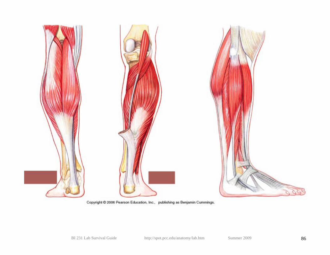
SUPERFICIAL POSTERIOR
COMPARTMENT OF THE LEG
ORIGIN INSERTION ACTION
Gastrocnemius Medial and lateral condyles of femur With soleus into calcaneus via calcaneal tendon
Flexes tibia and plantar; flexes foot
Soleus Upper third of fibula and soleal line of tibia
With gastrocnemius into calcaneus via calcaneal tendon
Flexes foot
Plantaris Lateral supracondylar ridge of femur Posterior calcaneus via calcaneal tendon
Weakly assists gastroc in plantar flexing ankle and flexing knee

BI 231 Lab Survival Guide http://spot.pcc.edu/anatomy/lab.htm Summer 2009 83
DEEP POSTERIOR COMPARTMENT
OF THE LEG
ORIGIN INSERTION ACTION
Popliteus Lateral surface of lateral condyle Posterior surface of tibia just below condyles
Flexes and unlocks knee joint
Tibialis posterior Interosseous membrane and tibia and fibula on either side
Navicular, with slips to cuneiform; cuboid; metatarsals 2-4
Adducts and inverts foot and aids in plantar flexion
Flexor digitorum longus
Middle half of tibia By four tendons into distal phalanges of lateral four toes
Flexes lateral four toes
Flexor hallucis longus Distal two-thirds of fibula Distal phalanx of great toe Flexes great toe
ANTERIOR COMPARTMENT
OF THE LEG
ORIGIN INSERTION ACTION
Tibialis anterior Upper half of tibia and interosseous membrane
Base of first cuneiform and first metatarsal
Dorsiflexes and inverts foot
Extensor hallucis longus
Middle half of fibula and interosseous membrane
Distal phalanx of great toe Dorsiflexes foot and extends great toe
Extensor digitorum longus
Tibia, proximal three-fourths of fibula, & interosseous membrane
Tendons to middle & terminal phalanges of four lateral toes by extensor expansion
Dorsiflexes foot and extends toes
LATERAL
COMPARTMENT OF THE LEG
ORIGIN INSERTION ACTION
Fibularis longus
Upper two-thirds of fibula and intermuscular septa
Plantar base of first metatarsal and first cuneiform and
Plantar flexes and everts foot
Fibularis brevis
Lower two-thirds of fibula Plantar base of fifth metatarsal Plantar flexes and everts foot

BI 231 Lab Survival Guide http://spot.pcc.edu/anatomy/lab.htm Summer 2009 84

BI 231 Lab Survival Guide http://spot.pcc.edu/anatomy/lab.htm Summer 2009 85
BI 231 Lab Survival Guide http://spot.pcc.edu/anatomy/lab.htm Summer 2009 86

BI 231 Lab Survival Guide http://spot.pcc.edu/anatomy/lab.htm Summer 2009 87

BI 231 Lab Survival Guide http://spot.pcc.edu/anatomy/lab.htm Summer 2009 88

BI 231 Lab Survival Guide http://spot.pcc.edu/anatomy/lab.htm Summer 2009 89


BI 231 Lab Survival Guide http://spot.pcc.edu/anatomy/lab.htm Summer 2009 91


