
Braz. J. Biol., 66(1A): 109-116, 2006
LARVAL DEVELOPMENT OF Brachidontes solisianus (BIVALVIA, MYTILIDAE), WITH NOTES ON DIFFERENCES BETWEEN ITS
HINGE SYSTEM AND THAT OF THE MOLLUSK Perna perna
MONTEIRO-RIBAS, W.1, ROCHA-MIRANDA, F.2, ROMANO, R. C.1 and QUINTANILHA, J.1
1Departamento de Oceanografia, Instituto de Estudos do Mar Almirante Paulo Moreira, IEAPM,
Rua Kioto, 253, Praia dos Anjos, CEP 28930-000, Arraial do Cabo, RJ, Brazil2Laboratório de Bentos, Departamento de Zoologia, Universidade de Brasília, ICC Ala Sul, Asa Norte, Brasília, DF, Brazil
Correspondence to: Fabio Miranda da Rocha, Laboratório de Bentos, Departamento de Zoologia, ICC Ala Sul, UnB, Asa Norte, Brasília, DF, Brazil, e-mail: [email protected]
Received October 17, 2003 – Accepted December 17, 2003 – Distributed February 28, 2006
(With 11 figures)
ABSTRACT
This work, which is part of a study program on meroplankton larvae, aims to gain more in-depth knowledge about planktonic larvae. This study began with the mollusk Brachidontes solisianus (Bivalvia – Mytilidae), which is abundant on the rocky shores of the Cabo Frio region (state of Rio de Janeiro, Brazil). Brachidontes solisianus larvae were grown under controlled conditions for a period of 26 days and were fed with Isochrysis galbana and Tetraselmis chui. The temperature was kept at 26 °C and the saltiness at 28‰. Images of the larvae were taken daily with a light camera and measured with a micrometric lens until settlement occurred. The average size of the first D-shaped veliger stage was 90 µm in length and 70 µm in height, while the size in the last stage before settlement (pediveliger) was 273 µm in length and 257 µm in height. The comparative study of the hinge system involved the most abundant intertidal species of the study area: Brachidontes solisianus and Perna perna. The B. solisianus species were found to have more visible denticles at the extremities of the provinculum, whereas the denticles of the P. perna species occur along the entire provinculum.
Keywords: prodissoconch morphology, larval growth, hinge system.
ReSumo
Desenvolvimento larvar de Brachidontes solisianus: com notas sobre as diferenças do seu sistema ligamentar quando comparado ao de Perna perna
Este trabalho faz parte de um programa de estudo sobre larvas meroplanctônicas que tem como objetivo o reconhecimento mais preciso das larvas no plâncton. Este estudo foi iniciado com a espécie Brachidontes solisianus (Bivalvia – Mytilidae) que é muito abundante nos costões rochosos da região de Cabo Frio. O desenvolvimento larvar foi realizado sob condições controladas durante 26 dias. A alimentação foi feita com Isochrysis galbana e Tetraselmis chui. A temperatura e a salinidade foram mantidas a 26° C e 28 ‰ , respectivamente. Diariamente, as larvas foram desenhadas em câmara clara e medidas com ocular micrométrica até a fixação. A primeira fase de véliger em forma de “D” ou Prodissoconcha I, mediu em média, 90 µm de comprimento por 70 µm de altura e a última, antes da fase de fixação (Pedivéliger), mediu 273 µm de comprimento e 257 µm de altura. No estudo comparativo das charneiras, duas espécies foram consideradas: Brachidontes solisianus e Perna perna. Observou-se que a espécie B. solisianus apresenta dentes mais evidentes nas extremidades do provinculum, enquanto na espécie P. perna aparecem ao longo de todo o provinculum.
Palavras-chave: prodissonconcha , crescimento larvar, ligamento.

110 MONTEIRO-RIBAS, W. et al.
Braz. J. Biol., 66(1A): 109-116, 2006
INTRoDuCTIoN
Some Lammelibranch larvae have been described by several authors but, despite the many exhaustive reports on the subject (Lebour, 1938; Werner, 1939; Jorgensen, 1946; Rees, 1950; Loosanoff & Davis, 1963; Martinez, 1967; Camacho & Cabello, 1973; Schweinitz & Lutz, 1976; Romero, 1977; Fernandes, 1988), the literature lacks any reference to the larvae of Brachidontes solisianus. B. solisianus is a small seawater bivalve with free-swimming plankton larvae. Notwithstanding the numerous reports in the literature on mussels and their ecological significance, few studies have concentrated on larval shell morphology.
The mussel Brachidontes solisianus (Bivalvia, Mytilidae) characteristically attaches itself to rocky shores at depths of < 3 m (mainly intertidal) in Arraial do Cabo, RJ, Brazil. This species comprises sedentary and filtering organisms, which are capable of resisting the hot sun throughout the low tide and attach themselves by a stout byssus to the basal regions of algae or rocks.
At first glance, no remarkable differences are distinguishable between the larvae of Brachidontes solisianus and other mussels (Martinez, 1967); in fact, the initial stages are very similar. At Arraial do Cabo, the two most abundant intertidal mussels are Perna perna and B. solisianus. Therefore, the primary aim of this paper is to enable planktologists to identify and differentiate these two Mytilidae larvae by their hinge systems. The secondary purpose of this study is to provide information on i) the larval shell morphology of these species, using light microscopy; ii) the morphometry of B. solisianus larval stages (prodissoconch I and II, settlers); and iii) differences in the hinge systems of B. solisianus and P. perna.
This study focused on the most outstanding aspects of each stage of Brachidontes solisianus development.
mATeRIAL AND meTHoDS
Thirty specimens of Brachidontes solisianus were collected from the rocky shores of the Praia dos Anjos at Arraial do Cabo (Fig. 1) and placed in
Fig. 1 — Map of the area showing the study site – Praia dos Anjos.
111LARVAL DEVELOPMENT OF Brachidontes solisianus
Braz. J. Biol., 66(1A): 109-116, 2006
a 2000 mL beaker with seawater, which had been filtered through 5, 3 and 1 µm Cuno filters and sterilized with ultra-violet radiation. The samples were maintained at 25 °C and 28‰ (salinity).
The thermal shock method was applied to release the gametes. To this end, the specimens were placed in a beaker containing seawater at a temperature of 15 °C for 10 min, after which they were returned to the beaker containing seawater at 25 °C. The females released their ovules 30 to 90 min after application of the thermal shock, followed soon thereafter by the males, who released their spermatozoon, enabling fertilization to occur.
B. solisianus adults were removed from the spawning beaker and the seawater with gametes was filtered through a 45 µm sieve. The gametes were washed and put into a 2000 mL beaker containing filtered and sterilized seawater. They were fed daily with Isochrysis galbana (40 cel/mL) and Tetraselmis chui (15 cel/mL), and the seawater was changed daily.
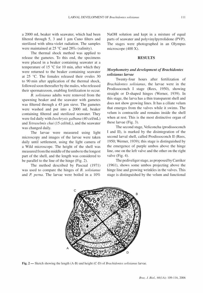
The larvae were measured using light microscopy and images of the larvae were taken daily until settlement, using the light camera of a Wild microscope. The height of the shell was measured from the middle of the umbo to the longest part of the shell, and the length was considered to be parallel to the line of the hinge (Fig. 2).
The method described by Pascual (1971) was used to compare the hinges of B. solisianus and P. perna. The larvae were boiled in a 10%
NaOH solution and kept in a mixture of equal parts of seawater and polyvinylpyrrolidone (PVP). The stages were photographed in an Olympus microscope (400 X).
ReSuLTS
Morphometry and development of Brachidontes solisianus larvae
Twenty-four hours after fertilization of Brachidontes solisianus, the larvae were in the Prodissoconch I stage (Rees, 1950), showing straight or D-shaped hinges (Werner, 1939). In this stage, the larva has a thin transparent shell and does not show growing lines. It has a ciliate velum that emerges from the valves while it swims. The velum is contractile and remains inside the shell when at rest. This is the most distinctive organ of these larvae (Fig. 3).
The second stage, Veliconcha (prodissoconch I and II), is marked by the disintegration of the second larval shell, called Prodissoconch II (Rees, 1950; Werner, 1939); this stage is distinguished by the emergence of purple umbos above the hinge line, one on the left valve and the other on the right valve (Fig. 4).
The pediveliger stage, as proposed by Carriker (1961), shows some umbos projecting above the hinge line and growing wrinkles in the valves. This stage is distinguished by the velum and functional
A
B
D
C
Fig. 2 — Sketch showing the length (A-B) and height (C-D) of Brachidontes solisianus larvae.

112 MONTEIRO-RIBAS, W. et al.
Braz. J. Biol., 66(1A): 109-116, 2006
feet and by the appearance of an ocellus-like spot in the center of each valve. Anterior and posterior adductor muscles are visible (Fig. 5).
Brachidontes solisianus settled onto the rock from day 23 on. From then on, their planktonic life ended and their benthic life began (Fig. 6).
50
Fig. 3 — Brachidontes solisianus larvae - Prodissoconch I.
50
Fig. 4 — Brachidontes solisianus larvae - Prodissoconch II.
50
Fig. 5 — Brachidontes solisianus larvae – Pediveliger phase.
50
Fig. 6 — Brachidontes solisianus larvae - Settler.

113LARVAL DEVELOPMENT OF Brachidontes solisianus
Braz. J. Biol., 66(1A): 109-116, 2006
0
50
100
150
200
250
300
1 3 5 7 9 11 13 15 17 19 21 23
Days
Hei
ght (
µm)
0
50
100
150
200
250
300
1 3 5 7 9 11 13 15 17 19 21 23Days
Leng
th (µ
m)
Prodissoconch I Prodissoconch II Pediveliger
Fig. 7 — Length and height of Brachidontes solisianus larvae during the experiment.
day 13 on and lasted 11 days, with an average length of 237 to 273 µm (Fig. 7).
During the experiment, the length of Brachidontes solisianus larvae was always greater than their height (Fig. 8), and there was a
The prodissoconch I stage lasted four days, during which the average length varied from 90 to 117 µm, while the prodissoconch II stage lasted 8 days, with an average length of 123 to 228 µm (Fig. 7). The pediveliger stage was observed from
0
30
60
90
120
150
180
210
240
270
300
1 3 5 7 9 11 13 15 17 19 21 23
Days
Siz
es (
m)
Length
Height
Fig. 8 — Larval length and height of Brachidontes solisianus.

114 MONTEIRO-RIBAS, W. et al.
Braz. J. Biol., 66(1A): 109-116, 2006
Height ( m)
Leng
th (
m)
80
120
160
200
240
280
320
40 80 120 160 200 240 280
Length = 35.889 + 0.92728 * HeightCorrelation: r = 0.99929
Fig. 9 — Correlation between length and height of Brachidontes solisianus larvae.
strong correlation between these two parameters: y = 35,889 + 0,92728x (r = 0.99) (Fig. 9).
Study of Brachidontes solisianus and Perna perna hinges
The study of the Mytilidae larvae hinge is important because it is the only way to differentiate mussel species in plankton samples (Camacho & Cabello 1973). Two regions can be distinguished in the larval shells: the provinculum and the lateral hinge system. By definition, if any denticles or tooth-like projections are visible beneath the straight part of the shell margin, a provinculum is present. This is formed by the thickening of the region of the shell that forms the straight part of the valve. Hinge structures, apart from denticles that are present beyond the limits of the provinculum, constitute the lateral hinge system, which comprises prominent lateral structures and calcareous channels from where projections known as lateral denticles can develop.
A comparison of the hinge structures of Perna perna and Brachidontes solisianus revealed that P. perna has visible denticles along the entire length of the provinculum in the prodissoconch II stage (Fig. 10). In contrast, the most visible denticles of B. solisianus occur in the early part of the prodissoconch II stage at the extremities of the provinculum, which were less visible in the middle region after day 5 of this study (Fig. 11).
The lateral hinge system was found to be more developed in the Perna perna larvae, comprising two prominently visible teeth in both valves.
DISCuSSIoN
In this study, the prodissoconch I stage lasted four days and the average length varied from 90 to 117 µm, which is quite similar to the length recorded for the Perna perna species. Fernandes (1988) recorded an average length of 91 µm in P. perna species in the first days of this stage and 117 µm at the end of the stage, which lasted eight days.
The prodissoconch II stage of B. solisianus lasted 8 days and the average length was 123 to 228 µm. For the Perna perna species, Fernandes (1988) recorded an average length of 141 µm on the first day and 216 µm on the tenth day. Similarly to the development of the Perna perna species (Romero, 1977), the shell grows as concentric wrinkles build up along its edge. According to Martinez (1967), Perna perna’s veliconch presents an A-type provincular structure, which is typical of Mytilidae as described by Rees (1950), and presents several rectangular denticles of equal or unequal size and set in a regular or irregular pattern.
The pediveliger stage of B. solisianus was observed from day 13 on, lasting for 11 days and

115LARVAL DEVELOPMENT OF Brachidontes solisianus
Braz. J. Biol., 66(1A): 109-116, 2006
Fig. 10 — Perna perna larvae on day 5 – Prodissoconch II, showing denticles along the hinge (400 X).
Fig. 11 — Brachidontes solisianus larvae in the 5th day– Prodissoconch II, showing teeth at hinge extremities (400 X).

116 MONTEIRO-RIBAS, W. et al.
Braz. J. Biol., 66(1A): 109-116, 2006
showing an average length of 237 to 273 µm (Fig. 7). In the pediveliger shell, Fernandes (1988) detected an average length of 256 µm at the beginning of the stage, on day 2, and 397 µm on day 30.
The most noticeable difference between the larvae of Brachidontes solisianus and Perna perna species was found in the hinge system of the Prodissoconch II stage. P. perna species has dentricles along the entire provinculum, whereas B. solisianus species has denticles only at the extremities of the provinculum. The lateral hinge system is more developed in the larvae of Perna perna and consists of three prominent and apparent denticles. Despite the many references on the ecology of mussels, few studies have focused on larval levels (Lebour, 1938; Werner, 1939; Jorgensen, 1946; Rees, 1950; Loosanoff & Davis, 1963; Martinez, 1967; Camacho & Cabello, 1973; Romero, 1977; Fernandes, 1988).
In conclusion, this paper offers the first description of differences in Brazilian Mytilidae larvae, and aims to facilitate the larval identification of mussels.
Acknowledgments – The authors would like to express their gratitude to Dr. Flávio Fernandes and all their colleagues at the IEAPM. This work was supported by the Instituto de Estudos do Mar Almirante Paulo Moreira.
ReFeReNCeS
CAMACHO, A. P. Y., CABELLO, G. R., 1973, Desarrollo larvario de Venerupis pullastra. Boletin del Instituto Espanol de Oceanografia, 165: 3-31.
CARRIKER, M.R. 1961. Interrelation of funcional morphology, behavior and autoecology in early stages of the bivalve
Mercenaria mercenaria. Journal of the Elisha Mitchell Scientific Society, 77(2): 168-241.
FERNANDES, A. C. B., 1988, Larvicultura do Mexilhão Perna perna (Linné, 1758). Dissertação de Mestrado. Instituto de Biofísica da UFRJ, 85p.
JORGENSEN, C. B., 1946, Lamellibranchia. In G. Thorson. Reproduction and larval development of Danish bottom invertebrates. Medd. Komm. Havundersog., Kbh., Ser Plankton, 4: 277-311.
LEBOUR, M., 1938, Notes on the breeding of some lamellibranchs from Plymouth and their larvae. Journ. Mar biol. Ass., 23:119-4.
LOOSANOFF, V. L. & DAVIS, H. C., 1963, Rearing of Bivalve mollusks. Advances in Marine Biol. Academic Press. Edited by F. S. Russell, 1: 1-136.
MARTINEZ, E. R., 1967, Identificación y descripción de la larva veloconcha y dissoconcha del mejillon comestible Perna perna (L) del Oriente de Venezuela. Ser. Recur. Explot. Pesq. Venezuela, 1(3): 79-113.
PASCUAL, E., 1971. Morfologia de la charnela larveria de Crassostrea angulata (Lmk) en diferentes fases de su desarrollo. Inv. Pesq., 35(2): 549-563.
REES, C. B., 1950, The identification and classification of lamellibranch larval. Hull Bull. Mar Ecol., 3(19): 73-104.
ROMERO, S. M. B., 1977, Efeitos Combinados de Salinidade e temperatura sobre embriões e larvas de Perna perna (Linné, 1758). Dissertação de Mestrado. Inst. de Biociências. USP. Mimeografado, 50p.
SCHWEINITZ, E. H. & LUTZ, R. A., 1976, Larval development of the northern horse mussel, Modiolus modiolis (L.) including a comparison with the larvae of Mytilus edulis L. as an aid in planktonic identification. Biol. Bull. mar. biol. Lab., Woods Hole, 150: 348-360
WERNER, B., 1939, Uber die Entwicklung und Artuntersc-heidung Von Muschellarven dos Nordsuplanktons, unter besonderer Berucksichtigung der Schalen entwwicklung. Zoll. Jahrb., Abteilung anatomie und ontogenie, 66: 1-54.


