Download - Mathur A Biochimie 2016
lable at ScienceDirect
Biochimie xxx (2016) 1e12
Contents lists avai
Biochimie
journal homepage: www.elsevier .com/locate/biochi
Research paper
Nelfinavir targets multiple drug resistance mechanisms to increasethe efficacy of doxorubicin in MCF-7/Dox breast cancer cells
Geetika Chakravarty a, Aditi Mathur a, Pallavi Mallade a, Samantha Gerlach a,Joniece Willis a, Amrita Datta b, Sudesh Srivastav c, Asim B. Abdel-Mageed b,Debasis Mondal a, *
a Department of Pharmacology, Tulane University Medical Center, USAb Department of Urology, Tulane University Medical Center, USAc Department of Biostatistics, Tulane University School of Public Health and Tropical Medicine, USA
a r t i c l e i n f o
Article history:Received 15 July 2015Accepted 29 January 2016Available online xxx
Keywords:Breast cancerMDRDoxorubicinNelfinavirChemosensitizationTumor xenograft
* Corresponding author. Department of PharmacolSchool of Medicine, 1430 Tulane Avenue, New Orlean
E-mail address: [email protected] (D. Mondal)
http://dx.doi.org/10.1016/j.biochi.2016.01.0140300-9084/© 2016 Published by Elsevier B.V.
Please cite this article in press as: G. Chakrdoxorubicin in MCF-7/Dox breast cancer cel
a b s t r a c t
Development of multidrug resistance (MDR) remains a significant problem in cancer chemotherapy andunderscores the importance of using chemosensitizers. Well known MDR mechanisms include: (i)upregulation of drug-efflux; (ii) increased signaling via AKT; and (iii) decreased apoptosis. Therefore,chemosensitizers should target multiple resistance mechanisms. We investigated the efficacy of nelfi-navir (NFV), a clinically approved anti-HIV drug, in increasing doxorubicin (DOX) toxicity in a MDR breastcancer cell line, MCF-7/Dox. As compared to parental MCF-7 cells, the MCF-7/Dox were 15e20 fold moreresistant to DOX-induced cytotoxicity at 48 h post-exposure (DOX IC50 ¼ 1.8 mM vs. 32.4 mM). Coex-posures to NFV could significantly (p < 0.05) decrease DOX-IC50 in MCF-7/Dox cells. Multiple exposuresto physiologic concentrations of NFV (2.25 mM or 6.75 mM) decreased DOX-IC50 by 21-fold and 50-fold,respectively. Interestingly, although single exposure to NFV transiently induced P-glycoprotein (P-gp)levels, multiple treatments with NFV inhibited both P-gp expression and efflux function, which increasedintracellular DOX concentrations. Single exposure to NFV augmented the markers of cell-survival (AKT)and autophagy (LC3-II), whereas multiple exposures enabled suppression of both total AKT (t-AKT) andinsulin like growth factor-1 (IGF-1)-induced phosphorylated AKT (p-AKT) levels. Multiple exposures toNFV also resulted in increased unfolded protein response (UPR) transducers, e.g. Grp78, p-PERK, p-eIF2a,and ATF-4; and endoplasmic reticulum (ER) stress induced death sensors, e.g. CHOP & TRIB-3. Multipleexposures to NFV also abrogated the mitogenic effects of IGF-1. In mice carrying MCF-7/Dox tumorxenografts, intraperitoneal (i.p.) injection of NFV (20 mg/kg/day) and DOX (2 mg/kg/twice/wk) decreasedtumor growth more significantly (p < 0.01) than either agent alone. Immunohistochemical (IHC) analysisrevealed decreased p-AKT and Ki-67 levels. Thus, NFV overcomes MDR in breast cancer cells and shouldbe tested as an adjunct to chemotherapy.
© 2016 Published by Elsevier B.V.
1. Introduction
Breast cancer is the second leading cause of cancer-relatedmortality in women in the United States [1]. Patient's age, raceand body mass index (BMI) are crucial determinants of cancerprogression to aggressive disease [2]. Despite recent advances inthe early detection of malignancy and improvements in surgery
ogy, SL-83, Tulane Universitys, LA, 70112, USA..
avarty, et al., Nelfinavir targls, Biochimie (2016), http://d
and radiation for localized tumors [3,4] a significant number ofpatients manifest recurrent and metastatic disease [5]. Further-more, even with the numerous treatments currently availableagainstmetastatic breast cancer, e.g. anti-estrogens, tyrosine kinaseinhibitors (TKIs) and immunotherapeutics, evolution of resistanttumor clones poses a significant challenge [6,7]. A cytotoxicchemotherapy regimen, primarily consisting of anthracyclines (e.g.doxorubicin) and taxanes (e.g. paclitaxel) is the last resort in thesepatients [8,9]. Approximately 60% of all patients with breast cancerultimately undergo chemotherapy. However, only a minority ofpatients on chemotherapy show long-term remission and theirsuboptimal response accounts for high rate of failure and selection
ets multiple drug resistance mechanisms to increase the efficacy ofx.doi.org/10.1016/j.biochi.2016.01.014
G. Chakravarty et al. / Biochimie xxx (2016) 1e122
of chemoresistant tumors [10e12]. Therefore, significant efforts arebeing made to develop agents that can suppress the MDR pheno-type of breast cancer cells [13,14].
One of the mechanisms by which tumor cells acquire MDRphenotype is induction of drug-efflux pumps [15e17]. The ATP-binding cassette (ABC) transporters such as P-glycoprotein (P-gpor ABCB1/MDR1), MDR related proteins (MRP), lung resistanceprotein (LRP), and breast cancer resistance protein (BCRP) canefflux a number of chemotherapeutic agents. Indeed, augmentedlevels of both P-gp and BCRP have been associated with resistanceto both anthracyclines and taxanes [18,19]. Therefore, a strategy tocircumvent drug resistance via coadministration of competitiveinhibitors is being actively investigated [17]. However, in order toovercome the high constitutive expression of ABC-transporters inMDR tumor clones, a sustained chemosensitizing effect would needto be achieved. Furthermore alternate survival pathways respon-sible for the resurgence of aggressive cancers will need to be tar-geted. Chemosensitizers should both act as efflux-pump inhibitors(EPI) and should also downregulate these crucial survival mecha-nisms [20,21].
Growth factors and cytokines within inflammatory tumor mi-croenvironments can facilitate tumor growth and survival [2].Frequent overexpression of insulin/insulin like growth factor-1(IGF-1) receptors on breast cancer cells can lead to the stimula-tion of survival signaling via the activation of PI3K/AKT/mTORpathway [22,23]. Indeed, tumor cells with acquired resistancedisplay constitutive AKT activation (phosphorylation) and chemo-therapeutic agents are known to activate this survival mechanismin cancer cells [24,25]. The PI3K/AKT/mTOR signaling pathway isalso known to upregulate P-gp (MDR-1) expression and function[26,27]. In addition to the PI3K/AKT/mTOR signaling axis, a numberof other crucial survival mechanisms are also becoming importantdrug resistance mechanisms. Two newly discovered metabolicpathways, i.e. autophagy and unfolded protein response (UPR) areessential for cancer cell survival in stressful tumor microenviron-ments [28,29]. Autophagy is a catabolic process that maintainshomeostasis in response to increased metabolic demands [28].Tumor cells activate autophagy by preventing the accumulation ofdamaged proteins via increased protein degradation and turnoverof cell organelles. Autophagy-mediated cell survival or cell deathdecisions can also be exquisitely regulated by the UPR pathways[29]. In aggressive cancer cells, escape from chemo-therapyeinduced apoptosis is facilitated by a mild UPR, and thecross-talk between autophagy and PI3K/AKT/mTOR cascades. Onthe other hand, although the UPR cascade initially favors cellularhomeostasis and survival, uncontrolled UPR results in the ensuingendoplasmic reticulum (ER)-stress that dictates apoptosis [30].Indeed, pharmacological agents that target ER-stress are becomingpromising anticancer agents [31,32].
A number of natural compounds, e.g. curcumin, genistein andresveratrol, are being tested as relatively safe chemosensitizers.However, the preclinical successes with these phytochemicals havenot translated to the clinic, primarily due to their low bioavailabilityin vivo [33,34]. Furthermore, clinical approval of new agents thatare able to target multiple drug resistance mechanisms in aggres-sive cancer cells would be an expensive and time-consuming pro-cess. Hence, drug repositioning strategies are becoming a viablealternative and a highly rewarding approach [35,36]. Indeed,numerous studies have shown that the clinically approved drugnelfinavir (NFV) may inhibit AKT phosphorylation and induce ER-stress in cancer cells [35e37]. In contrast to the side effects seenwith experimental MDR-modulators, NFV has been used chroni-cally in HIV-positive patients and has relatively fewer side effects.The enthusiasm to use NFV as a chemosensitizing agent is clearlyreflected by several ongoing cancer clinical trials [38]. The
Please cite this article in press as: G. Chakravarty, et al., Nelfinavir targdoxorubicin in MCF-7/Dox breast cancer cells, Biochimie (2016), http://d
availability of chemosensitizers with known pharmacokinetics andtoxicity profiles will be of significant benefit in the clinic [15].However, although NFV is an inhibitor of P-gp [39] it may alsoinduce P-gp expression since it is a P-gp substrate as well [40,41],thus compromising its anticancer efficacy.
Using the MCF-7/Dox cells, an aggressive breast cancer cell linethat overexpresses P-gp and shows constitutive AKT and UPR levels[42], we have documented that physiologically achievable con-centrations of NFV (<7.0 mM) can increase doxorubicin (DOX)-induced cytotoxicity in these cells. Molecular mechanistic studiesdemonstrated that coexposure to NFV can overcome multiplecompensatory drug resistance mechanisms in vitro. Using in vivoMCF-7/Dox tumor xenografts, we have also established that coex-posure to NFV can enhance the anti-tumor efficacy of DOX.
2. Materials and methods
2.1. Cell culture
The human breast adenocarcinoma cell line, MCF-7 was pur-chased from American Type Culture Collection (ATCC; Manassas,VA, USA). The DOX resistant MCF-7/Dox cells were a generous giftfrom Dr. Kapil Mehta (M. D. Anderson Cancer Center, Houston, TX,USA) [42]. Both cell lines were maintained in Eagle's MinimumEssential Medium (EMEM), with Earle's balanced salt solution(EBSS), containing 10% fetal bovine serum (FBS), penicillin andstreptomycin, L-glutamine, non-essential amino acids (NEAA), andsodium pyruvate (Gibco-BRL, Grand Island, NY). Cells were grownat 37� C in a humidified incubator with 5% CO2. TheMDR phenotypeof MCF-7/Dox cells was maintained by continuous selective pres-sure using medium supplemented with 50 ng/mL of doxorubicin.
2.2. Reagents
Experiments were carried out using both pure nelfinavir pur-chased from Sigma (St. Louis, MO) and extracted from Viracept™tablets (250 mg) obtained from Agouron pharmaceuticals (SanDiego, CA). Doxorubicin was purchased from SigmaeAldrich (St.Louis, MO). Calcein-AM was purchased from Molecular Probes(Eugene, OR USA). Verapamil was purchased from EMD Chemicals(Gibbstown, NJ USA). Primary antibody against P-gp [Mdr1 Anti-body (C-19): sc-1517] was purchased from Santa Cruz Biotech-nology (Santa Cruz, CA). Antibodies against phosphor-PERK(Threonine-980), ATF-4, TRIB-3 and Goat anti-rabbit secondaryantibody were purchased from Santa-Cruz Biotechnology. Anti-bodies against total-AKT and phospho-AKT (Serine-473), total-eIF2a and phospho-eIF2a (Serine-51), Grp78/BiP, CHOP, and LC3,were all obtained from Cell Signaling (Danvers, MA). Antibodyagainst b-Actin and GAPDH were purchased from Fisher Scientific(Waltham, MA) and antibody against Ki-67 was from Spring Bio-sciences (Pleasanton, CA).
2.3. Cell viability assays
Cell viability was measured using the MTT reagent {3-(4, 5-dimethylthiazol-2-yl)-2, 5-diphenyltetrazolium bromide} and ac-cording to our published protocols [32]. In brief, cells (5 � 103 cells/well) were plated in 96-well plates and allowed to adhere over-night. Desired concentrations of compounds were added to cells insix replicate wells and the cytotoxic effects of NFV, alone and incombination with DOX, were determined by MTT-assays. DOX(0.1e30 mM) was either added at the same time as NFV(0.75e6.75 mM) or was added after overnight pre-exposure to NFV.In ‘single’ exposure experiments, cells were coexposed to NFV andDOX for 48 h; and in the ‘multiple’ exposures experiments, cells
ets multiple drug resistance mechanisms to increase the efficacy ofx.doi.org/10.1016/j.biochi.2016.01.014
G. Chakravarty et al. / Biochimie xxx (2016) 1e12 3
were first treated with NFV for 16 h and then re-exposed to bothNFV and DOX for another 48 h. Following drug treatments, MTTwasadded to each well and incubated for 3 h. Formazan crystals weredetected by measuring the absorbance at 540 nm with a mQuantplate reader (Bio-Tek; Seattle, WA). Percent change in cell survivalcompared to untreated controls was calculated.
2.4. Drug-efflux assays
Calcein-AM (a P-gp substrate) was used to measure drug-effluxfunction [17,43]. Briefly, cells (1 � 104) were plated in 24-wellKrystal black plates (Labnet International; Woodbridge, NJ) anddrug exposures initiated the next day. Each plate contained back-ground wells (no cell), cells with no treatment (negative control),cells exposed to Verapamil (positive control) or with NFV and/orDOX (experimental). For acute effects of drug exposure, cells wereincubated with NFV or Verapamil (0.75e6.75 mM) for 15 min priorto the addition of Calcein-AM (0.25 mM) and further incubation foranother 15 min before fluorescence measurements. For prolongedeffects of ‘single or multiple’ drug exposures, cells were treatedwith NFV for 16 h, and either the drug was removed or replenishedfor another 30 min before the addition of Calcein-AM. Following15 min incubation with Calcein, cells were washed 3-times withcold phosphate-buffered saline (PBS) and intracellular Calceinretention was measured by using an Flx800 fluorescence micro-plate reader (BioTek). Excitation and emission wavelengths wereset at 485 ± 20 nm and 528 ± 20 nm, respectively. Cells were thenlysed and total protein contents measured in each well. Meanfluorescence units per mg of protein (MFU/mg) were calculated andnormalized to controls.
2.5. Fluorescence microscopy
Intracellular DOX accumulation in the presence or absence ofNFV was monitored by fluorescence microscopy [43]. The MCF7/Dox cells (5 � 104) were grown on coverslips that were submergedin media in 6-well tissue culture plates. Cells were pre-incubatedfor 30 min with NFV (0e6.75 mM) prior to the addition of DOX(3.0 mM) and further incubation was carried out for another 1 h.Cells were washed with PBS, coverslips were removed and treatedwith Vectashield mounting medium containing DAPI (Vector Labs,Burlingame, CA USA). Intracellular DOX retention, which fluorescesred upon excitation with green light (lex ¼ 470 ± 20 nm,lem ¼ 590 ± 20 nm), were measured using a fluorescent NikonEclipse E400microscope (Melville, NY USA) attached to the EXFOX-Cite 120 fluorescence illuminator system (Mississauga, ON, CAN).
2.6. Immunoblot analysis
Immunoblot studies were carried out according to our pastpublication [32]. Both short-term (8e16 h) and long-term (64 h)effects of NFV and/or DOX were evaluated. In specified experi-ments, to measure activated AKT (p-AKT) levels, cells were firstexposed to NFV for 48 h and then stimulated either acutely(30 min) or long-term (16 h) with IGF-1 (10 ng/ml). Cells wereharvested and whole cell lysates were obtained using cell lysisbuffer (Cell Signaling; Danvers, MA). Proteins were quantified withBCA protein assay reagent (Thermo Scientific; Rockford, IL).Approximately 30 mg of proteins were electrophoresed on 10% so-dium dodecyl sulfate-polyacrylamide (SDS-PAGE) gels from Bio-Rad (Hercules, CA). Proteins were wet transferred to a PVDFmembrane (Bio-Rad) at 90V for 2 h and blocked with bløk™-CHchemiluminescent blocker (Millipore) for 1 h at room temperature.Membranes were then probed with the desired primary (1:200 to1:1000 dilutions) and secondary antibodies (1:1000 dilution).
Please cite this article in press as: G. Chakravarty, et al., Nelfinavir targdoxorubicin in MCF-7/Dox breast cancer cells, Biochimie (2016), http://d
Bands were detected using SuperSignal West Pico chemilumines-cent substrate (Thermo Scientific) and band intensities werequantified using the Image-J software (NIH). Densitometric valueswere normalized to corresponding b-actin or GAPDH levels andfold changes were calculated.
2.7. Flow cytometry
Flow cytometry assays were carried out to measure cell surfaceP-gp levels by using a phycoerythrine (PE)-labeled P-gp antibody(C-19). Briefly, MCF7/Dox cells (1 � 106) were treated with NFV(0.75e6.75 mM) for 16 h. Next day, cells were washed with PBS,harvested with 0.05% trypsin/0.025% EDTA and re-suspended inwash buffer. The PE-labeled P-gp antibody or its isotype (IgG)control (1:200 dilution) was added to this cell suspension andincubated at 4 �C for 30 min in the dark. Cells were washed in PBScontaining 0.05% Tween-20, resuspended in ice cold PBS, andanalyzed on a Beckman Coulter Epics FC-500 flow cytometer withCXP software (Brea, CA). Both forward and side scatter gatings wereperformed with unlabeled cells.
2.8. Tumor xenografts
Tumor xenograft studies were carried out according to our pastpublications [32,43]. All animal protocols were approved by theAnimal Care and Use Committee (IACUC) of Tulane University andwere conducted in accordance with NIH guidelines. Animals werehoused in sterile filtered cages and given food and water ad libitumand maintained at 21e22 �C with a 12 h/12 h light and dark cycle.Briefly, female athymic nude BALB/c mice (6e8 week old) wereobtained from Charles River Laboratories (Wilmington, MA USA)and were allowed to acclimate for one week prior to experimen-tations. Prior to injection, the MCF7/Dox cells (5 � 106) wereresuspended in equal volume of serum free medium (SFM) con-taining Matrigel, and 100 ml of suspension was injected orthotopi-cally into the fourth inguinal mammary gland of each mouse.Tumors were allowed to grow to 50 mm3 (~2 weeks) at which timethe intraperitoneal (i.p.) injection of drugs were initiated. Tumorbearingmicewere randomly divided into 4 treatment groups (n¼ 6mice/grp), (i) vehicle control; (ii) NFV (20 mg/kg daily); (iii) DOX(2mg/kg every 3 days), or (iv)NFV (20mg/kg daily) and DOX (2mg/kg every 3 days) combination. Both drugs were diluted in vehicle(4% DMSO, 5% polyethylene glycol, 5% Tween 80 in saline) imme-diately before administration. At the end of treatment (6 weeks)tumor sizes were measured using a Vernier caliper and tumorvolumes were calculated using the formula: length �width2 � 0.4.Mice were then sacrificed and tumors harvested and sectioned forimmunohistochemical (IHC) analysis.
2.9. Immunohistochemistry
The IHC staining of tumor sections were carried to measure theproliferation marker Ki-67 and the survival marker p-AKT [32].Briefly, resected tumors were fixed in 10% neutral buffered formalinfollowed by incubation in 70% ethanol and embedding in paraffin.Paraffin blocks were sectioned using a microtome (~5 mm) andstained with hematoxylin and eosin (H&E). Slides were deparaffi-nized, hydrated, and antigen retrieval carried out using 10 mMcitrate buffer (pH 6.0) and then incubated with 3% H2O2 and 1.5%blocking serum (Vector laboratories; Burlingame, CA). Slides werethen incubated with antibodies against Ki-67 or p-AKT for 60 minat room temperature, washed twice in PBS and incubated withbiotinylated secondary antibody. Slides were treated with theVectastain enzyme reagent (Vector laboratories) and then stainedwith diaminobenzidine (DAB) and counterstained using
ets multiple drug resistance mechanisms to increase the efficacy ofx.doi.org/10.1016/j.biochi.2016.01.014
G. Chakravarty et al. / Biochimie xxx (2016) 1e124
Hematoxylin nuclear stain (Vector laboratories). Staining wasvisualized and captured using an Eclipse E-400 microscope (NikonInstruments, Melville, NY). Five different fields from each slidewere used to analyze and quantify p-AKT and Ki-67 positive cells byImage-J software.
2.10. Statistical analysis
Data was summarized using descriptive statistics such as mean,standard deviation, proportions and standard errors. Man-neWhitney U tests were used to compare between control, NFV orDOX alone, and with NFVþDOX combination groups. One-wayANOVA was performed to compare the differences in tumor vol-ume in mice treated with NFV, DOX, and NFVþDOX. All statisticalanalyses were performed at the 5% level of significance (p < 0.05)using the GraphPad Prism software (La Jolla, CA).
3. Results
3.1. Coexposure to NFV potentiates DOX-induced cytotoxicity
We compared DOX (0.1e100 mM) induced cytotoxicity in bothMCF-7 and MCF-7/Dox cells and fifty percent inhibitory concen-tration (IC50) was determined. The DOX-IC50 values at 64 h post-exposure were 1.8 mM in MCF-7 cells and 32.4 mM in MCF-7/Doxcells (Fig. 1A). This confirmed the resistant phenotype of MCF-7/Dox cells. We also measured the concentration dependent cyto-toxicity of NFV (0.75e22.5 mM) following either single (48 h) ormultiple (16 h þ 48 h) exposures (Fig. 1B). The physiologic con-centrations of NFV (0.75e6.75 mM) showed less than 20% cellkilling; however, the supra-physiologic concentration of NFV(22.5 mM) showed 60e70% toxicity. Therefore, all of our experi-ments were carried out using the physiologically achievable con-centrations of NFV (<7 mM) and using DOX concentrations lowerthan its IC50 (<3 mM).
To investigate the chemosensitizing ability of NFV, wemeasuredthe effect of ‘single’ or ‘multiple’ exposures on DOX-induced cyto-toxicity. For ‘single’ exposure studies, cells were coexposed to bothDOX and NFV for 48 h and for ‘multiple’ exposure studies cells werefirst pre-exposed to NFV for 16 h and then re-exposed to both NFV& DOX for another 48 h. Both single (Fig. 1C) and multiple (Fig. 1D)exposures to NFV were able to significantly (p < 0.01) decreaseDOX-IC50 in MCF-7/Dox cells. Single exposure to the low (2.25 mM)or high dose (6.75 mM) of NFV decreased DOX-IC50 from 32.4 mM to13.5 mM and 5.2 mM, respectively. However, multiple treatmentswith both low and high dose NFV further decreased DOX-IC50 to6.4 mM & 1.85 mM, respectively. Indeed, a precipitous drop in cellviability was clearly evident even with the lower concentrations ofDOX when cells were coexposed to NFV. This demonstrated theability of NFV to chemosensitize MCF-7/Dox cells. Furthermore,cells exposed multiple times to NFV showed significantly higherkilling ability. Therefore, mechanistic studies were carried out tomonitor the temporal effects of NFV in suppressing different drugresistance mechanisms in the MCF-7/Dox cells.
3.2. Acute effects of NFV on drug-efflux function and intracellularDOX levels
We quantified drug efflux function in MCF-7/Dox cells by usingthe fluorescent dye Calcein-AM and the known P-gp inhibitor,verapamil, [43]. Several of the HIV protease inhibitors, includingNFV, are known to be competitive inhibitors of P-gp [39,44].Therefore, we first tested whether NFV can suppress drug-effluxand increase DOX retention in these MDR cells (Fig. 2). Cells werepre-incubated with either verapamil or NFV (0.75e6.75 mM) for
Please cite this article in press as: G. Chakravarty, et al., Nelfinavir targdoxorubicin in MCF-7/Dox breast cancer cells, Biochimie (2016), http://d
30 min before the addition of Calcein-AM or DOX. Intracellularfluorescence was measured after another 15 min for Calcein incu-bation (Fig. 2A) or after 2 h for DOX incubation (Fig. 2B). As shownin Fig. 2A, equimolar concentrations of verapamil or NFV pre-incubation could suppress Calcein efflux and clearly establishedNFV as a potent inhibitor of drug-efflux function. As shown inFig. 2B, fluorescence microscopy images similarly showedincreased DOX retention inMCF7/Dox cells following coexposure toincreasing concentrations of NFV (Fig. 2B). Cells exposed to DOX(3 mM) alone had very little intracellular DOX accumulation evenafter 2 h incubation (Fig. 2B, top). However, a profound increase inintracellular DOX-fluorescence (red) was observed in cells thatwere pre-exposed to NFV (Fig. 2B, bottom panels). Therefore, acuteexposure to NFV shows a potent inhibitory effect on P-gp mediateddrug efflux to increase DOX levels in cells.
3.3. Differential effects of low vs. high dose NFV on P-gp expressionand drug-efflux function
Although acute exposure to NFV may decrease P-gp function viacompetitive inhibition. However, by being a P-gp substrate, pro-longed exposure to NFVmay also result in increased P-gp levels andthus augment MDR function instead [39]. Therefore, we monitoredthe effects of both acute and long-term exposures to NFV on Calceinefflux and P-gp expression (Fig. 3). We compared the effects ofeither acute (30 min) or prolonged treatments with NFV, in eithersingle (16 h) or multiple (16 h þ 30 min) exposure studies. Exper-iments on intracellular Calcein retention showed drasticallydifferent dose- and time-dependent effects of NFV on drug-effluxfunction. Similar to our previous study (Fig. 2), acute exposure toNFV (30 min) decreased drug-efflux function (Fig. 3A). However,single pre-exposure to low-dose of NFV (2.25 mM) for as short as16 h was shown to increase drug-efflux function, as evident bylower Calcein retention (Fig. 3A). This increased drug-efflux func-tion was potent so that it could not be fully suppressed bycompetitive inhibition with acute (30 min) coexposure to NFV. Onthe other hand, in both ‘single’ and ‘multiple’ exposure studies, highdose of NFV (6.75 mM) showed significant (p < 0.01) suppression inefflux function (15-fold). More importantly, this sustained sup-pressive effect on P-gp function was further enhanced followinganother acute exposure to NFV, as evident from a 30-fold increasein Calcein retention. This clearly showed the advantage of multipleexposures to high dose NFV as a far superior approach for sup-pression of drug-efflux mechanisms in the MDR cancer cells.
In order to understand this differential dose- and time-dependent effect of NFV on drug-efflux function, we monitoredP-gp protein levels in NFV exposed MCF7/Dox cells (Fig. 3).Immunoblot studies were carried out to measure total P-gp proteinexpression (Fig. 3B& C) and cell surface P-gp levels were quantifiedby flow cytometry (Fig. 3D). We documented increases in both alower molecular-weight P-gp band (unglycosylated) and a highermolecular-weight P-gp band (glycosylated) in these immunoblots(Fig. 3B). Exposure to low-dose NFV (2.25 mM) showed a temporalincrease in P-gp protein (140e170 kDa) levels, which was evidentas early as 4 h post-treatment (1.3-fold) and was significantlyhigher at 16 h post-exposure. Prolonged NFV exposure showed a2.5 fold increase in the lower P-gp band and a 5.2-fold increase inthe higher P-gp band (Fig. 3B, lane-4). Data from Fig. 3B clearlyindicated that low dose NFV (2.25 mM) may increase both total andglycosylated P-gp levels. Since the dose- and time-dependent ef-fects of exposure to NFV had differential effects on drug-effluxfunction, we measured the effects of high dose NFV (6.75 mM) onP-gp protein levels (Fig. 3C). Although prolonged exposure to thelower concentrations of NFV (0.75 mM & 2.25 mM) increased bothtotal and glycosylated P-gp (Fig. 3C, lane-3) the high dose of NFV
ets multiple drug resistance mechanisms to increase the efficacy ofx.doi.org/10.1016/j.biochi.2016.01.014
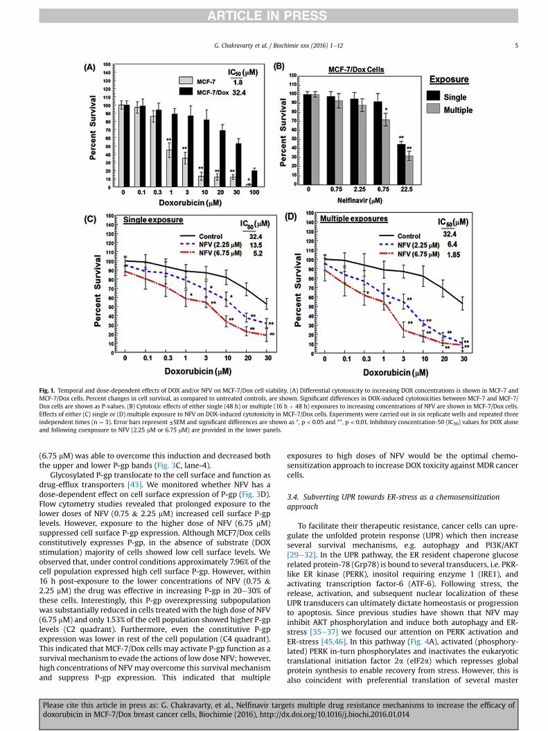
Fig. 1. Temporal and dose-dependent effects of DOX and/or NFV on MCF-7/Dox cell viability. (A) Differential cytotoxicity to increasing DOX concentrations is shown in MCF-7 andMCF-7/Dox cells. Percent changes in cell survival, as compared to untreated controls, are shown. Significant differences in DOX-induced cytotoxicities between MCF-7 and MCF-7/Dox cells are shown as P-values. (B) Cytotoxic effects of either single (48 h) or multiple (16 h þ 48 h) exposures to increasing concentrations of NFV are shown in MCF-7/Dox cells.Effects of either (C) single or (D) multiple exposure to NFV on DOX-induced cytotoxicity in MCF-7/Dox cells. Experiments were carried out in six replicate wells and repeated threeindependent times (n ¼ 3). Error bars represent ±SEM and significant differences are shown as *, p < 0.05 and **, p < 0.01. Inhibitory concentration-50 (IC50) values for DOX aloneand following coexposure to NFV (2.25 mM or 6.75 mM) are provided in the lower panels.
G. Chakravarty et al. / Biochimie xxx (2016) 1e12 5
(6.75 mM) was able to overcome this induction and decreased boththe upper and lower P-gp bands (Fig. 3C, lane-4).
Glycosylated P-gp translocate to the cell surface and function asdrug-efflux transporters [43]. We monitored whether NFV has adose-dependent effect on cell surface expression of P-gp (Fig. 3D).Flow cytometry studies revealed that prolonged exposure to thelower doses of NFV (0.75 & 2.25 mM) increased cell surface P-gplevels. However, exposure to the higher dose of NFV (6.75 mM)suppressed cell surface P-gp expression. Although MCF7/Dox cellsconstitutively expresses P-gp, in the absence of substrate (DOXstimulation) majority of cells showed low cell surface levels. Weobserved that, under control conditions approximately 7.96% of thecell population expressed high cell surface P-gp. However, within16 h post-exposure to the lower concentrations of NFV (0.75 &2.25 mM) the drug was effective in increasing P-gp in 20e30% ofthese cells. Interestingly, this P-gp overexpressing subpopulationwas substantially reduced in cells treatedwith the high dose of NFV(6.75 mM) and only 1.53% of the cell population showed higher P-gplevels (C2 quadrant). Furthermore, even the constitutive P-gpexpression was lower in rest of the cell population (C4 quadrant).This indicated that MCF-7/Dox cells may activate P-gp function as asurvival mechanism to evade the actions of low dose NFV; however,high concentrations of NFVmay overcome this survival mechanismand suppress P-gp expression. This indicated that multiple
Please cite this article in press as: G. Chakravarty, et al., Nelfinavir targdoxorubicin in MCF-7/Dox breast cancer cells, Biochimie (2016), http://d
exposures to high doses of NFV would be the optimal chemo-sensitization approach to increase DOX toxicity against MDR cancercells.
3.4. Subverting UPR towards ER-stress as a chemosensitizationapproach
To facilitate their therapeutic resistance, cancer cells can upre-gulate the unfolded protein response (UPR) which then increaseseveral survival mechanisms, e.g. autophagy and PI3K/AKT[29e32]. In the UPR pathway, the ER resident chaperone glucoserelated protein-78 (Grp78) is bound to several transducers, i.e. PKR-like ER kinase (PERK), inositol requiring enzyme 1 (IRE1), andactivating transcription factor-6 (ATF-6). Following stress, therelease, activation, and subsequent nuclear localization of theseUPR transducers can ultimately dictate homeostasis or progressionto apoptosis. Since previous studies have shown that NFV mayinhibit AKT phosphorylation and induce both autophagy and ER-stress [35e37] we focused our attention on PERK activation andER-stress [45,46]. In this pathway (Fig. 4A), activated (phosphory-lated) PERK in-turn phosphorylates and inactivates the eukaryotictranslational initiation factor 2a (eIF2a) which represses globalprotein synthesis to enable recovery from stress. However, this isalso coincident with preferential translation of several master
ets multiple drug resistance mechanisms to increase the efficacy ofx.doi.org/10.1016/j.biochi.2016.01.014
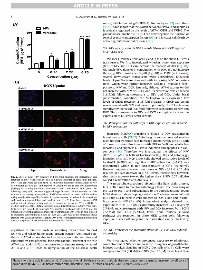
Fig. 2. Effect of acute NFV exposure on P-gp efflux function and intracellular DOXretention in MCF-7/Dox cells. (A) NFV is a potent inhibitor of drug-efflux function.MCF-7/Dox cells were pre-incubated (30 min) with equimolar concentrations of NFVor Verapamil (0e6.75 mM) and exposed to Calcein-AM for 15 min and fluorescence(MFU/mg of protein) measured. Increased Calcein retention in MCF-7/Dox cellsfollowing verapamil treatment suggested P-gp function. Calcein retention in cellsexposed to increasing concentrations of NFV demonstrated its potent inhibitory effecton P-gp mediated drug-efflux function. Experiments were carried out in three replicatewells and were repeated three independent times (n ¼ 3). Error bars represent ±SEMand significant differences from untreated controls are shown as; ***, p < 0.001; **,p < 0.01 and *, p < 0.05. (B) NFV increases intracellular DOX levels in MCF-7/Dox cells.Cells were pre-treated (30 min) with NFV and then incubated with DOX (5 mM) for60 min. Fluorescent microscopy images show intracellular DOX levels in cells exposedto increasing concentrations of NFV (0e6.75 mM). Each row in the composite showsstaining with DAPI alone (nuclear stain), DOX alone (red fluorescence) and the mergedimage. Coexposure to NFV increased intracellular DOX levels.
G. Chakravarty et al. / Biochimie xxx (2016) 1e126
regulators of ER-stress such as activating transcription factor-4(ATF-4) and C/EBP homologous protein (CHOP). Continued syn-thesis of ATF-4 occurs due to low translation initiation rates andribosomal by-pass of several false start codons upstream of the trueATF-4 start codon [47]. In response to continuous stress, increasedATF-4 and CHOP leads to upregulation of the ER-stress death
Please cite this article in press as: G. Chakravarty, et al., Nelfinavir targdoxorubicin in MCF-7/Dox breast cancer cells, Biochimie (2016), http://d
sensor, tribbles homolog-3 (TRIB-3). Studies by us [32] and others[48,49] have shown that the switch between survival and apoptosisis critically regulated by the levels of ATF-4, CHOP and TRIB-3. Thepseudokinase function of TRIB-3 can downregulate the function ofseveral crucial transcription factors [50] and initiates cell death byactivating mitochondrial caspases [51].
3.5. NFV rapidly subverts UPR towards ER-stress in DOX exposedMCF-7/Dox cells
Wemeasured the effects of NFV and DOX on the above ER-stresstransducers. We first investigated whether short-term exposure(8 h) to NFV and DOX can increase the markers of UPR (Fig. 4B).Although NFV, alone or in combination with DOX, did not increasethe early UPR transducers Grp78 (Fig. 4B) or PERK (not shown),several downstream transducers were upregulated. Enhancedlevels of p-eIF2a were observed with increasing NFV concentra-tions, which were further increased (2.8-fold) following coex-posure to NFV and DOX. Similarly, although ATF-4 expression didnot increase with NFV or DOX alone, its expression was enhanced(1.8-fold) following coexposure to NFV and DOX. Under basal(unstimulated) conditions, the MCF-7/Dox cells expressed lowlevels of CHOP. However, a 1.5-fold increase in CHOP expressionwas observed with NFV and, most importantly, CHOP levels weresignificantly increased (3.6-fold) following coexposure to NFV andDOX. Thus, coexposure to NFV and DOX can rapidly increase theexpression of ER-stress death sensors.
3.6. Resurgent survival pathways in DOX exposed cells are blockedby NFV coexposure
Increased PI3K/AKT signaling is linked to DOX resistance inbreast cancer cells [21,52]. Autophagy is another survival mecha-nism utilized by cancer cells to escape chemotherapy [28,53]. Bothof these pathways also interact with UPR to facilitate cellular ho-meostasis, and suppress ER-stress induction and apoptosis in can-cer cells [54]. Therefore, we investigated the effects of NFV(0.75e6.75 mM) on both AKT-activation (Fig. 4C) and autophagy-induction (Fig. 4D). MCF-7/Dox cells showed constitutive levels oftotal-AKT (t-AKT) and significant AKT activation (p-AKT) wasdocumented within 15 min post-exposure to IGF-1 (10 ng/ml).However, exposure to even low doses of NFV (0.75 & 2.25 mM)resulted in a 50% decrease in p-AKT levels. Interestingly however,short-term exposure to even the highest dose of NFV (6.75 mM) alsocaused a reactivation of p-AKT levels.
The microtubule-associated ubiquitin-like light chain protein,LC3 is often used to monitor autophagy [36,48]. The processing ofpro-LC3 to LC3-I, and subsequently to the autophagosome boundLC3-II demonstrates autophagy induction. Therefore, wemonitoredLC3-I and LC3-II levels in cells exposed to DOX, alone and in com-bination with NFV (Fig. 4D). Immunoblot analysis showed thatexposure to NFV (6.75 mM) significantly increased LC3-I levels by2.3-fold, and cotreatment with NFV and DOX increased both LC3-I(7.2-fold) and LC3-II (5.2-fold) levels. Thus, multiple survivalpathways are resurgent in these MDR cancer cells followingexposure to chemotherapy and their activation can be blocked byNFV.
3.7. NFV overcomes the protective effects of IGF-1 on DOX-inducedcytotoxicity
We investigated whether prolonged exposure to physiologicconcentrations of NFV can suppress the resurgence of growth factorinduced survival signaling in MCF-7/Dox cells (Fig. 5). Cells werecoexposed to DOX (3.0 mM) and NFV (0e6.75 mM) for 48 h and then
ets multiple drug resistance mechanisms to increase the efficacy ofx.doi.org/10.1016/j.biochi.2016.01.014
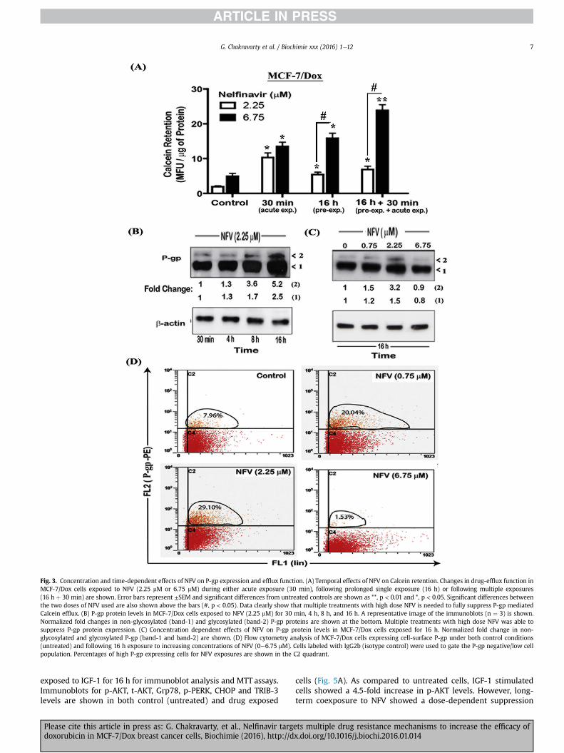
Fig. 3. Concentration and time-dependent effects of NFV on P-gp expression and efflux function. (A) Temporal effects of NFV on Calcein retention. Changes in drug-efflux function inMCF-7/Dox cells exposed to NFV (2.25 mM or 6.75 mM) during either acute exposure (30 min), following prolonged single exposure (16 h) or following multiple exposures(16 h þ 30 min) are shown. Error bars represent ±SEM and significant differences from untreated controls are shown as **, p < 0.01 and *, p < 0.05. Significant differences betweenthe two doses of NFV used are also shown above the bars (#, p < 0.05). Data clearly show that multiple treatments with high dose NFV is needed to fully suppress P-gp mediatedCalcein efflux. (B) P-gp protein levels in MCF-7/Dox cells exposed to NFV (2.25 mM) for 30 min, 4 h, 8 h, and 16 h. A representative image of the immunoblots (n ¼ 3) is shown.Normalized fold changes in non-glycosylated (band-1) and glycosylated (band-2) P-gp proteins are shown at the bottom. Multiple treatments with high dose NFV was able tosuppress P-gp protein expression. (C) Concentration dependent effects of NFV on P-gp protein levels in MCF-7/Dox cells exposed for 16 h. Normalized fold change in non-glycosylated and glycosylated P-gp (band-1 and band-2) are shown. (D) Flow cytometry analysis of MCF-7/Dox cells expressing cell-surface P-gp under both control conditions(untreated) and following 16 h exposure to increasing concentrations of NFV (0e6.75 mM). Cells labeled with IgG2b (isotype control) were used to gate the P-gp negative/low cellpopulation. Percentages of high P-gp expressing cells for NFV exposures are shown in the C2 quadrant.
G. Chakravarty et al. / Biochimie xxx (2016) 1e12 7
exposed to IGF-1 for 16 h for immunoblot analysis and MTT assays.Immunoblots for p-AKT, t-AKT, Grp78, p-PERK, CHOP and TRIB-3levels are shown in both control (untreated) and drug exposed
Please cite this article in press as: G. Chakravarty, et al., Nelfinavir targdoxorubicin in MCF-7/Dox breast cancer cells, Biochimie (2016), http://d
cells (Fig. 5A). As compared to untreated cells, IGF-1 stimulatedcells showed a 4.5-fold increase in p-AKT levels. However, long-term coexposure to NFV showed a dose-dependent suppression
ets multiple drug resistance mechanisms to increase the efficacy ofx.doi.org/10.1016/j.biochi.2016.01.014
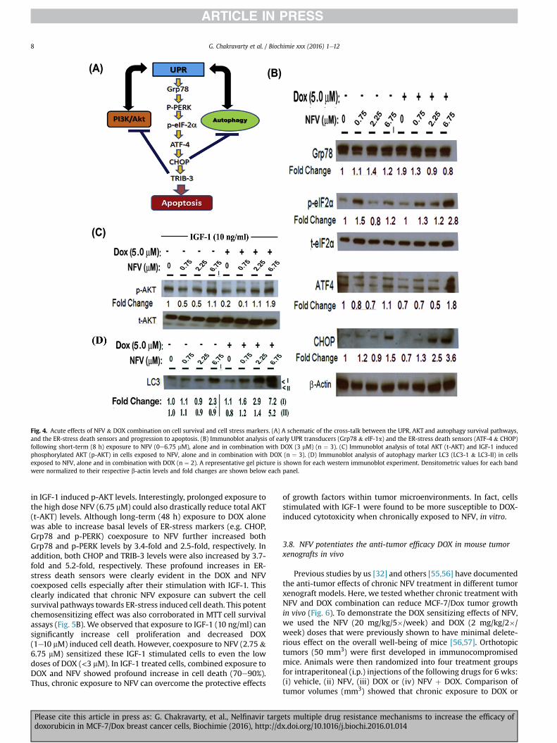
Fig. 4. Acute effects of NFV & DOX combination on cell survival and cell stress markers. (A) A schematic of the cross-talk between the UPR, AKT and autophagy survival pathways,and the ER-stress death sensors and progression to apoptosis. (B) Immunoblot analysis of early UPR transducers (Grp78 & eIF-1a) and the ER-stress death sensors (ATF-4 & CHOP)following short-term (8 h) exposure to NFV (0e6.75 mM), alone and in combination with DOX (3 mM) (n ¼ 3). (C) Immunoblot analysis of total AKT (t-AKT) and IGF-1 inducedphosphorylated AKT (p-AKT) in cells exposed to NFV, alone and in combination with DOX (n ¼ 3). (D) Immunoblot analysis of autophagy marker LC3 (LC3-1 & LC3-II) in cellsexposed to NFV, alone and in combination with DOX (n ¼ 2). A representative gel picture is shown for each western immunoblot experiment. Densitometric values for each bandwere normalized to their respective b-actin levels and fold changes are shown below each panel.
G. Chakravarty et al. / Biochimie xxx (2016) 1e128
in IGF-1 induced p-AKT levels. Interestingly, prolonged exposure tothe high dose NFV (6.75 mM) could also drastically reduce total AKT(t-AKT) levels. Although long-term (48 h) exposure to DOX alonewas able to increase basal levels of ER-stress markers (e.g. CHOP,Grp78 and p-PERK) coexposure to NFV further increased bothGrp78 and p-PERK levels by 3.4-fold and 2.5-fold, respectively. Inaddition, both CHOP and TRIB-3 levels were also increased by 3.7-fold and 5.2-fold, respectively. These profound increases in ER-stress death sensors were clearly evident in the DOX and NFVcoexposed cells especially after their stimulation with IGF-1. Thisclearly indicated that chronic NFV exposure can subvert the cellsurvival pathways towards ER-stress induced cell death. This potentchemosensitizing effect was also corroborated in MTT cell survivalassays (Fig. 5B). We observed that exposure to IGF-1 (10 ng/ml) cansignificantly increase cell proliferation and decreased DOX(1e10 mM) induced cell death. However, coexposure to NFV (2.75 &6.75 mM) sensitized these IGF-1 stimulated cells to even the lowdoses of DOX (<3 mM). In IGF-1 treated cells, combined exposure toDOX and NFV showed profound increase in cell death (70e90%).Thus, chronic exposure to NFV can overcome the protective effects
Please cite this article in press as: G. Chakravarty, et al., Nelfinavir targdoxorubicin in MCF-7/Dox breast cancer cells, Biochimie (2016), http://d
of growth factors within tumor microenvironments. In fact, cellsstimulated with IGF-1 were found to be more susceptible to DOX-induced cytotoxicity when chronically exposed to NFV, in vitro.
3.8. NFV potentiates the anti-tumor efficacy DOX in mouse tumorxenografts in vivo
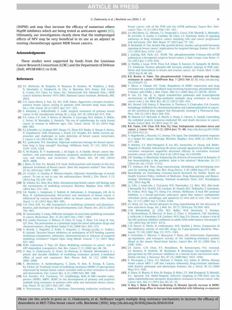
Previous studies by us [32] and others [55,56] have documentedthe anti-tumor effects of chronic NFV treatment in different tumorxenograft models. Here, we tested whether chronic treatment withNFV and DOX combination can reduce MCF-7/Dox tumor growthin vivo (Fig. 6). To demonstrate the DOX sensitizing effects of NFV,we used the NFV (20 mg/kg/5�/week) and DOX (2 mg/kg/2�/week) doses that were previously shown to have minimal delete-rious effect on the overall well-being of mice [56,57]. Orthotopictumors (50 mm3) were first developed in immunocompromisedmice. Animals were then randomized into four treatment groupsfor intraperitoneal (i.p.) injections of the following drugs for 6 wks:(i) vehicle, (ii) NFV, (iii) DOX or (iv) NFV þ DOX. Comparison oftumor volumes (mm3) showed that chronic exposure to DOX or
ets multiple drug resistance mechanisms to increase the efficacy ofx.doi.org/10.1016/j.biochi.2016.01.014
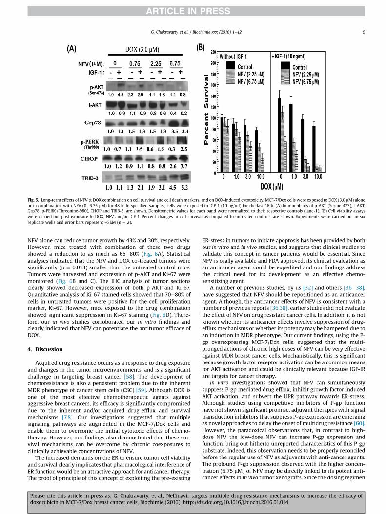
Fig. 5. Long-term effects of NFV & DOX combination on cell survival and cell death markers, and on DOX-induced cytotoxicity. MCF-7/Dox cells were exposed to DOX (3.0 mM) aloneor in combination with NFV (0e6.75 mM) for 48 h. In specified samples, cells were exposed to IGF-1 (10 ng/ml) for the last 16 h. (A) Immunoblots of p-AKT (Serine-473), t-AKT,Grp78, p-PERK (Threonine-980), CHOP and TRIB-3, are shown. Densitometric values for each band were normalized to their respective controls (lane-1). (B) Cell viability assayswere carried out post-exposure to DOX, NFV and/or IGF-1. Percent changes in cell survival as compared to untreated controls, are shown. Experiments were carried out in sixreplicate wells and error bars represent ±SEM (n ¼ 2).
G. Chakravarty et al. / Biochimie xxx (2016) 1e12 9
NFV alone can reduce tumor growth by 43% and 30%, respectively.However, mice treated with combination of these two drugsshowed a reduction to as much as 65e80% (Fig. 6A). Statisticalanalyses indicated that the NFV and DOX co-treated tumors weresignificantly (p ¼ 0.013) smaller than the untreated control mice.Tumors were harvested and expression of p-AKT and Ki-67 weremonitored (Fig. 6B and C). The IHC analysis of tumor sectionsclearly showed decreased expression of both p-AKT and Ki-67.Quantitative analysis of Ki-67 stained cells showed that 70e80% ofcells in untreated tumors were positive for the cell proliferationmarker, Ki-67. However, mice exposed to the drug combinationshowed significant suppression in Ki-67 staining (Fig. 6D). There-fore, our in vivo studies corroborated our in vitro findings andclearly indicated that NFV can potentiate the antitumor efficacy ofDOX.
4. Discussion
Acquired drug resistance occurs as a response to drug exposureand changes in the tumor microenvironments, and is a significantchallenge in targeting breast cancer [58]. The development ofchemoresistance is also a persistent problem due to the inherentMDR phenotype of cancer stem cells (CSC) [59]. Although DOX isone of the most effective chemotherapeutic agents againstaggressive breast cancers, its efficacy is significantly compromiseddue to the inherent and/or acquired drug-efflux and survivalmechanisms [7,8]. Our investigations suggested that multiplesignaling pathways are augmented in the MCF-7/Dox cells andenable them to overcome the initial cytotoxic effects of chemo-therapy. However, our findings also demonstrated that these sur-vival mechanisms can be overcome by chronic coexposures toclinically achievable concentrations of NFV.
The increased demands on the ER to ensure tumor cell viabilityand survival clearly implicates that pharmacological interference ofER functionwould be an attractive approach for anticancer therapy.The proof of principle of this concept of exploiting the pre-existing
Please cite this article in press as: G. Chakravarty, et al., Nelfinavir targdoxorubicin in MCF-7/Dox breast cancer cells, Biochimie (2016), http://d
ER-stress in tumors to initiate apoptosis has been provided by bothour in vitro and in vivo studies, and suggests that clinical studies tovalidate this concept in cancer patients would be essential. SinceNFV is orally available and FDA approved, its clinical evaluation asan anticancer agent could be expedited and our findings addressthe critical need for its development as an effective chemo-sensitizing agent.
A number of previous studies, by us [32] and others [36e38],have suggested that NFV should be repositioned as an anticanceragent. Although, the anticancer effects of NFV is consistent with anumber of previous reports [36,38], earlier studies did not evaluatethe effect of NFV on drug resistant cancer cells. In addition, it is notknown whether its anticancer effects involve suppression of drug-effluxmechanisms or whether its potencymay be hampered due toan induction in MDR phenotype. Our current findings, using the P-gp overexpressing MCF-7/Dox cells, suggested that the multi-pronged actions of chronic high doses of NFV can be very effectiveagainst MDR breast cancer cells. Mechanistically, this is significantbecause growth factor receptor activation can be a common meansfor AKT activation and could be clinically relevant because IGF-IRare targets for cancer therapy.
In vitro investigations showed that NFV can simultaneouslysuppress P-gp mediated drug efflux, inhibit growth factor inducedAKT activation, and subvert the UPR pathway towards ER-stress.Although studies using competitive inhibitors of P-gp functionhave not shown significant promise, adjuvant therapies with signaltransduction inhibitors that suppress P-gp expression are emergingas novel approaches to delay the onset of multidrug resistance [60].However, the paradoxical observations that, in contrast to high-dose NFV the low-dose NFV can increase P-gp expression andfunction, bring out hitherto unreported characteristics of this P-gpsubstrate. Indeed, this observation needs to be properly reconciledbefore the regular use of NFV as adjuvants with anti-cancer agents.The profound P-gp suppression observed with the higher concen-tration (6.75 mM) of NFV may be directly linked to its potent anti-cancer effects in in vivo tumor xenografts. Since the dosing regimen
ets multiple drug resistance mechanisms to increase the efficacy ofx.doi.org/10.1016/j.biochi.2016.01.014
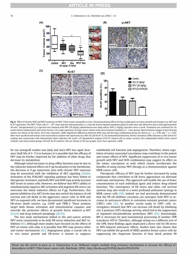
Fig. 6. Effect of chronic DOX and NFV treatment on MCF-7/Dox tumor xenografts in mice. Chemosensitizing effects of drug combination on tumor growth and changes in p-AKT andKi-67 expression. The MCF-7/Dox cells (5 � 106) were injected subcutaneously (s.c.) into the fourth inguinal mammary gland of nude mice and allowed to grow until approximately50 mm3. Intraperitoneal (i.p.) injection was initiated with NFV (20 mg/kg) administered once daily and/or DOX (2 mg/kg) injection twice a week. Treatment was continued for sixweeks before euthanization and tumor harvest. (A) Graph represents average tumor volume with each treatment modality (n ¼ 6 per group). Representative images of mice bearingtumors are shown in the insets. Error bars represent ±SEM. Significant differences between DOX-only and the drug combination group are shown as, *, p < 0.05 and **, p < 0.01.Mice were sacrificed and tumors were harvested to monitor the expression of p-AKT (B) and Ki-67 (C) by immunohistochemistry. Brown coloration (DAB) indicates p-AKT and Ki-67staining and counterstain with Hematoxylin (blue) indicates the nuclei. (D) Quantitative analysis of Ki-67 stained cells in tumor sections (five independent fields) from control(vehicle) and drug treated groups. Percent Ki-67 positive cells are shown in the bar graphs. Error bars represent ±SEM.
G. Chakravarty et al. / Biochimie xxx (2016) 1e1210
for our xenograft studies was daily and since NFV has rapid clear-ance (half-life of 3e5 h in humans) it is possible that the efficacy ofNFV may be further improved by the addition of other drugs thatdecrease its metabolism.
Although initial increases in drug-efflux function may be due tothe substrate induced effects on P-gp localization to themembrane,the decrease in P-gp expression seen with chronic NFV exposuremay be associated with the inhibition of AKT signaling [24,60].Activation of the PI3K/AKT signaling pathway has been linked totherapeutic resistance, and both NFV and DOXmay acutely increaseAKT levels in tumor cells. However, we believe that NFV's ability tosimultaneously suppress AKT activation and augment ER-stress canovercome the initial inductive effects on P-gp. Furthermore, thispotent inhibition of p-AKT levels may also switch the balance of cellsurvival and cell death in the aggressive cancer cells. In DOX andNFV co-exposed cells, we have documented significant increases inER-stress death sensors, e.g. CHOP and TRIB-3. These proteinstarget both kinase activation and mitochondrial caspases andoverride the protective effects of growth factor induced p-AKT[23,24] and drug-induced autophagy [28,53].
The two main mechanisms linked to the anti-cancer activityNFV seem consistently to be both ER-stress induction and AKT in-hibition [61]. Although most studies have focused on the effects ofNFV on tumor cells only, it is possible that NFV may possess otheranti-tumor mechanisms [62]. Angiogenesis plays a crucial role inin vivo tumor growth and ER-stress is known to suppress
Please cite this article in press as: G. Chakravarty, et al., Nelfinavir targdoxorubicin in MCF-7/Dox breast cancer cells, Biochimie (2016), http://d
endothelial cell function and angiogenesis. Therefore, down regu-lation of tumor associated vasculature may contribute to the potentanti-tumor effects of NFV. Significant suppression of in vivo tumorgrowth with NFV and DOX combination may suggest its effect onthe tumor vasculature as well, which clearly corroborates thebenefits of using chronic NFV therapy as a chemosensitizer againstMDR cancer cells.
Therapeutic efficacy of NFV may be further increased by usingcompounds that contribute to ER stress aggravation via alternatemolecular mechanisms. This approach will enable the use of lowerconcentrations of each individual agent and reduce drug-relatedtoxicities. The convergence of ER stress and other cell survivalsystems may also result in a more profound antitumor synergy inMDR cancer cells [58]. Indeed, we had previously demonstratedthat the NF-kB inhibitor curcumin can synergize with NFV to in-crease its anticancer effects in castration-resistant prostate cancer(CRPC) cells [32]. In another recent study in CRPC cells, in-vestigators showed that NFV can increase cell death by inhibitingsite-2 protease (S2P) cleavage activity, which led to the suppressionof regulated intramembrane proteolysis (RIP) [63]. Interestingly,RIP is necessary for post-translational processing of another UPRtransducer ATF-6. Therefore, although acting via a different arm ofthe UPR, these findings again implicated a direct role for ER-stressin NFV-induced anticancer effects. Studies have also shown thatNFV can inhibit the growth of HER2-positive breast cancer cells bysuppressing the chaperone function of heat shock protein 90
ets multiple drug resistance mechanisms to increase the efficacy ofx.doi.org/10.1016/j.biochi.2016.01.014
G. Chakravarty et al. / Biochimie xxx (2016) 1e12 11
(HSP90) and may thus increase the efficacy of numerous othersHsp90 inhibitors which are being tested as anticancer agents [63].Ultimately, our investigations clearly show that the multiprongedeffects of NFV may be very beneficial in its use as an adjunct toexisting chemotherapy against MDR breast cancers.
Acknowledgments
These studies were supported by funds from the LouisianaCancer Research Consortium (LCRC) and the Department of Defense(DoD, #PC0810811) to D.M.
References
[1] S.F. Altekruse, M. Krapcho, N. Neyman, R. Aminou, W. Waldron, J. Ruhl,N. Howlader, Z. Tatalovich, H. Cho, A. Mariotto, M.P. Eisner, D.R. Lewis,K. Cronin, H.S. Chen, E.J. Feuer, D.G. Stinchcomb, B.K. Edwards (Eds.), SEERCancer Statistics Review 1975-2007, National Cancer Institute, Bethesda, MD,2010.
[2] C.A. Santa-Maria, J. Yan, X.J. Xie, D.M. Euhus, Aggressive estrogen-receptor-positive breast cancer arising in patients with elevated body mass index,Int. J. Clin. Oncol. 20 (2) (2015 Apr) 317e323.
[3] S. Kümmel, J. Holtschmidt, S. Loibl, Surgical treatment of primary breastcancer in the neoadjuvant setting, Br. J. Surg. 101 (8) (2014 Jul) 912e924.
[4] F.A. Calvo, C.V. Sole, S. Rivera, R. Meiri~no, S. Lizarraga, M.A. Infante, E. Boldo,C. Ferrer, H. Marsiglia, E. Deutsch, The use of radiotherapy for early breastcancer in woman at different ages, Clin. Transl. Oncol. 16 (8) (2014 Aug)680e685.
[5] E.J. Schneble, L.J. Graham, M.P. Shupe, F.L. Flynt, K.P. Banks, A. Nissan, L. Henry,A. Stojadinovic, N.M. Shumway, I. Avital, G.E. Peoples, R.F. Setlik, Current ap-proaches and challenges in early detection of breast cancer recurrence,J. Cancer 5 (4) (2014 Mar 16) 281e290.
[6] R.C. Jankowitz, N.E. Davidson, Adjuvant endocrine therapy for breast cancer:how long is long enough? Oncology (Williston Park) 27 (12) (2013 Dec)1210e1216, 1224.
[7] A. M Braden, R. V Stankowski, J. M Engel, A. A Onitilo, Breast cancer bio-markers: risk assessment, diagnosis, prognosis, prediction of treatment effi-cacy and toxicity, and recurrence, Curr. Pharm. Des. 20 (30) (2014)4879e4898.
[8] J. Bines, H. Earl, A.C. Buzaid, E.D. Saad, Anthracyclines and taxanes in the neo/adjuvant treatment of breast cancer: does the sequence matter? Ann. Oncol.25 (6) (2014 Jun) 1079e1085.
[9] J.A. Crozier, A. Swaika, A. Moreno-Aspitia, Adjuvant chemotherapy in breastcancer: To use or not to use, the anthracyclines, World J. Clin. Oncol. 5 (3)(2014 Aug 10) 529e538.
[10] M. Videira, R.L. Reis, M.A. Brito, Deconstructing breast cancer cell biology andthe mechanisms of multidrug resistance, Biochim. Biophys. Acta 1846 (2)(2014 Dec) 312e325.
[11] S.B. Nandy, L. Gangwani, Z. Nahleh, R. Subramani, A. Arumugam, J.M. de laRosa, R. Lakshmanaswamy, Recurrence and metastasis of breast cancer isinfluenced by ovarian hormone's effect on breast cancer stem cells, FutureOncol. 11 (6) (2015 Mar) 983e995.
[12] Y.H. Choi, A.M. Yu, ABC transporters in multidrug resistance and pharmaco-kinetics, and strategies for drug development, Curr. Pharm. Des. 20 (5) (2014)793e807.
[13] M. Saraswathy, S. Gong, Different strategies to overcome multidrug resistancein cancer, Biotechnol. Adv. 31 (8) (2013 Dec) 1397e1407.
[14] K.R. Landis-Piwowar, V. Milacic, D. Chen, H. Yang, Y. Zhao, T.H. Chan, B. Yan,Q.P. Dou, The proteasome as a potential target for novel anticancer drugs andchemosensitizers, Drug Resist Updat 9 (6) (2006 Dec) 263e273.
[15] A. Br�ozik, C. Hegedüs, Z. Erdei, T. Hegedus, C. €Ozvegy-Laczka, G. Szak�acs,B. Sarkadi, Tyrosine kinase inhibitors as modulators of ATP binding cassettemultidrug transporters: substrates, chemosensitizers or inducers of acquiredmultidrug resistance? Expert Opin. Drug Metab. Toxicol. 7 (5) (2011 May)623e642.
[16] M.M. Gottesman, T. Fojo, S.E. Bates, Multidrug resistance in cancer: role ofATP-dependent transporters, Nat. Rev. Cancer 2 (1) (2002 Jan) 48e58.
[17] U. Roy, G. Chakravarty, K. Honer Zu Bentrup, D. Mondal, Montelukast is apotent and durable inhibitor of multidrug resistance protein 2-mediatedefflux of taxol and saquinavir, Biol. Pharm. Bull. 32 (12) (2009 Dec)2002e2009.
[18] E. Mechetner, A. Kyshtoobayeva, S. Zonis, H. Kim, R. Stroup, R. Garcia,R.J. Parker, J.P. Fruehauf, Levels of multidrug resistance (MDR1) P-glycoproteinexpression by human breast cancer correlate with in vitro resistance to taxoland doxorubicin, Clin. Cancer Res. 4 (2) (1998 Feb) 389e398.
[19] A.A. Kovalev, D.A. Tsvetaeva, T.V. Grudinskaja, Role of ABC-cassette trans-porters (MDR1, MRP1, BCRP) in the development of primary and acquiredmultiple drug resistance in patients with early and metastatic breast cancer,Exp. Oncol. 35 (4) (2013 Dec) 287e290.
[20] A. Provenzano, S. Kurian, J. Abraham, Overcoming endocrine resistance in
Please cite this article in press as: G. Chakravarty, et al., Nelfinavir targdoxorubicin in MCF-7/Dox breast cancer cells, Biochimie (2016), http://d
breast cancer: role of the PI3K and the mTOR pathways, Expert Rev. Anti-cancer Ther. 13 (2) (2013 Feb) 143e147.
[21] J.A. McCubrey, S.L. Abrams, T.L. Fitzgerald, L. Cocco, A.M. Martelli, G. Montalto,M. Cervello, A. Scalisi, S. Candido, M. Libra, L.S. Steelman, Roles of signalingpathways in drug resistance, cancer initiating cells and cancer progressionand metastasis, Adv. Biol. Regul. 57 (2015 Jan) 75e101.
[22] H. Beckwith, D. Yee, Insulin-like growth factors, insulin, and growth hormonesignaling in breast cancer: implications for targeted therapy, Endocr. Pract. 20(11) (2014 Nov) 1214e1221.
[23] J. Lauring, B.H. Park, A.C. Wolff, The phosphoinositide-3-kinase-Akt-mTORpathway as a therapeutic target in breast cancer, J. Natl. Compr. Canc Netw. 11(6) (2013 Jun 1) 670e678.
[24] J.J. Wallin, J. Guan, W.W. Prior, K.A. Edgar, R. Kassees, D. Sampath, M. Belvin,L.S. Friedman, Nuclear phospho-Akt increase predicts synergy of PI3K inhi-bition and doxorubicin in breast and ovarian cancer, Sci. Transl. Med. 2 (48)(2010 Sep 8) 48e66.
[25] K.K. Brown, A. Toker, The phosphoinositide 3-kinase pathway and therapyresistance in cancer, F1000Prime Rep. 7 (2015 Feb 3) 13, http://dx.doi.org/10.12703/P7-13.
[26] S. Misra, S. Ghatak, B.P. Toole, Regulation of MDR1 expression and drugresistance by a positive feedback loop involving hyaluronan, phosphoinositide3-kinase, and ErbB2, J. Biol. Chem. 280 (21) (2005 May 27) 20310e20315.
[27] H. Sui, Z.Z. Fan, Q. Li, Signal transduction pathways and transcriptionalmechanisms of ABCB1/Pgp-mediated multiple drug resistance in humancancer cells, J. Int. Med. Res. 40 (2) (2012) 426e435.
[28] M.L. Bristol, S.M. Emery, P. Maycotte, A. Thorburn, S. Chakradeo, D.A. Gewirtz,Autophagy inhibition for chemosensitization and radiosensitization in cancer:do the preclinical data support this therapeutic strategy? J. Pharmacol. Exp.Ther. 344 (3) (2013 Mar) 544e552.
[29] M. Maurel, E.P. McGrath, K. Mnich, S. Healy, E. Chevet, A. Samali, Controllingthe unfolded protein response-mediated life and death decisions in cancer,Semin. Cancer Biol. 33 (2015 Aug) 57e66.
[30] R.K. Yadav, S.W. Chae, H.R. Kim, H.J. Chae, Endoplasmic reticulum stress andcancer, J. Cancer Prev. 19 (2) (2014 Jun) 75e88, http://dx.doi.org/10.15430/JCP.2014.19.2.75.
[31] A. Nagelkerke, J. Bussink, F.C. Sweep, P.N. Span, The unfolded protein responseas a target for cancer therapy, Biochim. Biophys. Acta 1846 (2) (2014 Dec)277e284.
[32] A. Mathur, Z.Y. Abd Elmageed, X. Liu, M.L. Kostochka, H. Zhang, A.B. Abdel-Mageed, D. Mondal, Subverting ER-stress towards apoptosis by nelfinavir andcurcumin coexposure augments docetaxel efficacy in castration resistantprostate cancer cells, PLoS One 9 (8) (2014 Aug 14) e103109.
[33] J.M. Smoliga, O. Blanchard, Enhancing the delivery of resveratrol in humans: iflow bioavailability is the problem, what is the solution? Molecules 19 (11)(2014 Oct 24) 17154e17172.
[34] T.T. Ashburn, K.B. Thor, Drug repositioning: identifying and developing newuses for existing drugs, Nat. Rev. Drug Discov. 3 (8) (2004 Aug) 673e683.
[35] Roundtable on Translating Genomic-based Research for Health, Board onHealth Sciences Policy, Institute of Medicine. Drug Repurposing and Reposi-tioning: Workshop Summary, National Academies Press (US), Washington(DC), 2014 Aug 08.
[36] J.J. Gills, J. Lopiccolo, J. Tsurutani, R.H. Shoemaker, C.J. Best, M.S. Abu-Asab,J. Borojerdi, N.A. Warfel, E.R. Gardner, M. Danish, M.C. Hollander, S. Kawabata,M. Tsokos, W.D. Figg, P.S. Steeg, P.A. Dennis, Nelfinavir, A lead HIV proteaseinhibitor, is a broad-spectrum, anticancer agent that induces endoplasmicreticulum stress, autophagy, and apoptosis in vitro and in vivo, Clin. CancerRes. 13 (17) (2007 Sep 1) 5183e5194.
[37] J.S. Shim, J.O. Liu, Recent advances in drug repositioning for the discovery ofnew anticancer drugs, Int. J. Biol. Sci. 10 (7) (2014 Jun 10) 654e663.
[38] G.M. Blumenthal, J.J. Gills, M.S. Ballas, W.B. Bernstein, T. Komiya,R. Dechowdhury, B. Morrow, H. Root, G. Chun, C. Helsabeck, S.M. Steinberg,J. LoPiccolo, S. Kawabata, E.R. Gardner, W.D. Figg, P.A. Dennis, A phase I trial ofthe HIV protease inhibitor nelfinavir in adults with solid tumors, Oncotarget 5(18) (2014 Sep 30) 8161e8172.
[39] C.H. Storch, D. Theile, H. Lindenmaier, W.E. Haefeli, J. Weiss, Comparison ofthe inhibitory activity of anti-HIV drugs on P-glycoprotein, Biochem. Phar-macol. 73 (10) (2007 May 15) 1573e1581.
[40] S. Cisternino, C. Mercier, F. Bourasset, F. Roux, J.M. Scherrmann, Expression,up-regulation, and transport activity of the multidrug-resistance proteinAbcg2 at the mouse blood-brain barrier, Cancer Res. 64 (9) (2004 May 1)3296e3301.
[41] J.A. Zastre, G.N. Chan, P.T. Ronaldson, M. Ramaswamy, P.O. Couraud,I.A. Romero, B. Weksler, M. Bendayan, R. Bendayan, Up-regulation of P-glycoprotein by HIV protease inhibitors in a human brain microvessel endo-thelial cell line, J. Neurosci. Res. 87 (4) (2009 Mar) 1023e1036.
[42] E. Devarajan, J. Chen, A.S. Multani, S. Pathak, A.A. Sahin, K. Mehta, Humanbreast cancer MCF-7 cell line contains inherently drug-resistant subcloneswith distinct genotypic and phenotypic features, Int. J. Oncol. 20 (5) (2002May) 913e920.
[43] A. Datta, N. Bhasin, H. Kim, M. Ranjan, B. Rider, Z.Y. Abd Elmageed, D. Mondal,K.C. Agrawal, A.B. Abdel-Mageed, Selective targeting of FAK-Pyk2 axis byalpha-naphthoflavone abrogates doxorubicin resistance in breast cancer cells,Cancer Lett. 362 (1) (2015 Jun 28) 25e35.
[44] U. Roy, C. Bulot, K. Honer zu Bentrup, D. Mondal, Specific increase in MDR1mediated drug-efflux in human brain endothelial cells following co-exposure
ets multiple drug resistance mechanisms to increase the efficacy ofx.doi.org/10.1016/j.biochi.2016.01.014
G. Chakravarty et al. / Biochimie xxx (2016) 1e1212
to HIV-1 and saquinavir, PLoS One 8 (10) (2013 Oct 3) e75374, http://dx.doi.org/10.1371/journal.pone.0075374.
[45] T. Verfaillie, N. Rubio, A.D. Garg, G. Bultynck, R. Rizzuto, J.P. Decuypere,J. Piette, C. Linehan, S. Gupta, A. Samali, P. Agostinis, PERK is required at theER-mitochondrial contact sites to convey apoptosis after ROS-based ER stress,Cell Death Differ. 19 (11) (2012 Nov) 1880e1891.
[46] D. Cojocari, R.N. Vellanki, B. Sit, D. Uehling, M. Koritzinsky, B.G. Wouters, Newsmall molecule inhibitors of UPR activation demonstrate that PERK, but notIRE1a signaling is essential for promoting adaptation and survival to hypoxia,Radiother. Oncol. 108 (3) (2013 Sep) 541e547.
[47] A. Brüning, M. Rahmeh, K. Friese, Nelfinavir and bortezomib inhibit mTORactivity via ATF4-mediated sestrin-2 regulation, Mol. Oncol. 7 (6) (2013 Dec)1012e1018.
[48] M. Salazar, A. Carracedo, I.J. Salanueva, S. Hern�andez-Tiedra, A. Egia,M. Lorente, P. V�azquez, S. Torres, J.L. Iovanna, M. Guzm�an, P. Boya, G. Velasco,TRB3 links ER stress to autophagy in cannabinoid anti-tumoral action, Auto-phagy 5 (7) (2009 Oct) 1048e1049.
[49] C.A. Corcoran, X. Luo, Q. He, C. Jiang, Y. Huang, M.S. Sheikh, Genotoxic andendoplasmic reticulum stresses differentially regulate TRB3 expression, Can-cer Biol. Ther. 4 (10) (2005 Oct) 1063e1067.
[50] N. Zareen, S.C. Biswas, L.A. Greene, A feed-forward loop involving Trib3, Aktand FoxO mediates death of NGF-deprived neurons, Cell Death Differ. 20 (12)(2013 Dec) 1719e1730.
[51] B. Qian, H. Wang, X. Men, W. Zhang, H. Cai, S. Xu, Y. Xu, L. Ye, C.B. Wollheim,J. Lou, TRIB3 is implicated in glucotoxicity- and endoplasmic reticulum-stress-induced beta-cell apoptosis, J. Endocrinol. 199 (3) (2008 Dec) 407e416.
[52] J.A. McCubrey, L.S. Steelman, W.H. Chappell, S.L. Abrams, E.W. Wong, F. Chang,B. Lehmann, D.M. Terrian, M. Milella, A. Tafuri, F. Stivala, M. Libra, J. Basecke,C. Evangelisti, A.M. Martelli, R.A. Franklin, Roles of the Raf/MEK/ERK pathwayin cell growth, malignant transformation and drug resistance, Biochim. Bio-phys. Acta 1773 (8) (2007 Aug) 1263e1284.
[53] W.L. Sun, J. Chen, Y.P. Wang, H. Zheng, Autophagy protects breast cancer cellsfrom epirubicin-induced apoptosis and facilitates epirubicin-resistancedevelopment, Autophagy 7 (9) (2011 Sep) 1035e1044.
[54] C. Appenzeller-Herzog, M.N. Hall, Bidirectional crosstalk between
Please cite this article in press as: G. Chakravarty, et al., Nelfinavir targdoxorubicin in MCF-7/Dox breast cancer cells, Biochimie (2016), http://d
endoplasmic reticulum stress and mTOR signaling, Trends Cell Biol. 22 (5)(2012 May) 274e282.
[55] M.B. Braga-Neto, B.M. Oliveira, R.S. Rodrigues, F.J. Noronha, R.F. Leitao,G.A. Brito, A.A. Lima, R.L. Guerrant, C.A. Warren, Protective effects of alanyl-glutamine supplementation against nelfinavir-induced epithelial impair-ment in IEC-6 cells and in mouse intestinal mucosa, Cancer Biol. Ther. 13 (14)(2012 Dec) 1482e1490.
[56] S. Kawabata, J.J. Gills, J.R. Mercado-Matos, J. Lopiccolo, W. Wilson 3rd,M.C. Hollander, P.A. Dennis, Synergistic effects of nelfinavir and bortezomibon proteotoxic death of NSCLC and multiple myeloma cells, Cell Death Dis. 3(2012 Jul 19) e353.
[57] P.D. Ottewell, H. M€onkk€onen, M. Jones, D.V. Lefley, R.E. Coleman, I. Holen,Antitumor effects of doxorubicin followed by zoledronic acid in a mousemodel of breast cancer, J. Natl. Cancer Inst. 100 (16) (2008 Aug 20)1167e1178.
[58] J.S. Chen, M. Konopleva, M. Andreeff, A.S. Multani, S. Pathak, K. Mehta, Drug-resistant breast carcinoma (MCF-7) cells are paradoxically sensitive toapoptosis, J. Cell. Physiol. 200 (2) (2004 Aug) 223e234.
[59] K.M. Britton, J.A. Kirby, T.W. Lennard, A.P. Meeson, Cancer stem cells and sidepopulation cells in breast cancer and metastasis, Cancers (Basel) 3 (2) (2011Apr) 2106e2130.
[60] X. Lin, X. Zhang, Q. Wang, J. Li, P. Zhang, M. Zhao, X. Li, Perifosine down-regulates MDR1 gene expression and reverses multidrug-resistant phenotypeby inhibiting PI3K/Akt/NF-kB signaling pathway in a human breast cancer cellline, Neoplasma 59 (3) (2012) 248e256.
[61] T. Koltai, Nelfinavir and other protease inhibitors in cancer: mechanismsinvolved in anticancer activity. Version 2, F1000Res (2015), http://dx.doi.org/10.12688/f1000research.5827.2.
[62] M. Guan, L. Su, Y.C. Yuan, H. Li, W.A. Chow, Nelfinavir and nelfinavir analogsblock site-2 protease cleavage to inhibit castration-resistant prostate cancer,Sci. Rep. 5 (2015 Apr 16) 9698, http://dx.doi.org/10.1038/srep09698.
[63] J.S. Shim, R. Rao, K. Beebe, L. Neckers, I. Han, R. Nahta, J.O. Liu, Selective in-hibition of HER2-positive breast cancer cells by the HIV protease inhibitornelfinavir, J. Natl. Cancer Inst. 104 (20) (2012 Oct 17) 1576e1590.
ets multiple drug resistance mechanisms to increase the efficacy ofx.doi.org/10.1016/j.biochi.2016.01.014


