Download - Medical Devices Sheba
Tel Hashomer Medical Research
Infrastructure and Services Ltd.
Medical Devices Innovations
Contact : Sylvie Luria PhD. CEO
Technology Transfer Company
Tel Hashomer Medical Research, Infrastructure and Services Ltd.
Tel: +972-3-5305998 Fax: +972-3-5305944 Cell: 052-6667277
[email protected] http://research.sheba.co.il/e/
Sheba Medical Center - Technology Transfer Company
Tel Hashomer Medical Research, Infrastructure and Services Ltd.
http://rnd.sheba.co.il/
Business:
Tel Hashomer Medical Research, Infrastructure and Services Ltd. (THM) the technology transfer
arm of Sheba Medical Center, is responsible for managing the intellectual property assets of Sheba
Medical Center and to promote the transfer of technologies, innovation and professional know-how for
society's use and benefit, and for the development of the medical and health care delivery fields. Sheba
Medical Center facilities, experience, human resources and regulations enable the development of a novel
idea from its basic science to its product development and prototype, thus rapidly generating value to its
IP for commercialization.
Main Activities:
Scientific insights and academic breakthroughs often translate into inventions for the benefit of the
marketplace. THM bridges the gap between Academia Research and Industry Needs, since the industry is
product-based, business-oriented, and focused on time-framed missions, THM helps turn scientific
progress into tangible products, while returning income to the inventor and to Sheba Medical Center to
support further research.
THM receives invention disclosures from faculty, staff and students. We evaluate the innovations for
patent applications and develop licensing strategy, consider the technical and market risks.
Patentable inventions constitute the majority of THM's licensing activities; however, we also handle
collaborations with industrial partners and Tangible Research Property (TRP) such as Tissue Bank,
Genomics and Bio-Markers, Cell Therapy, Computational Imaging and more.
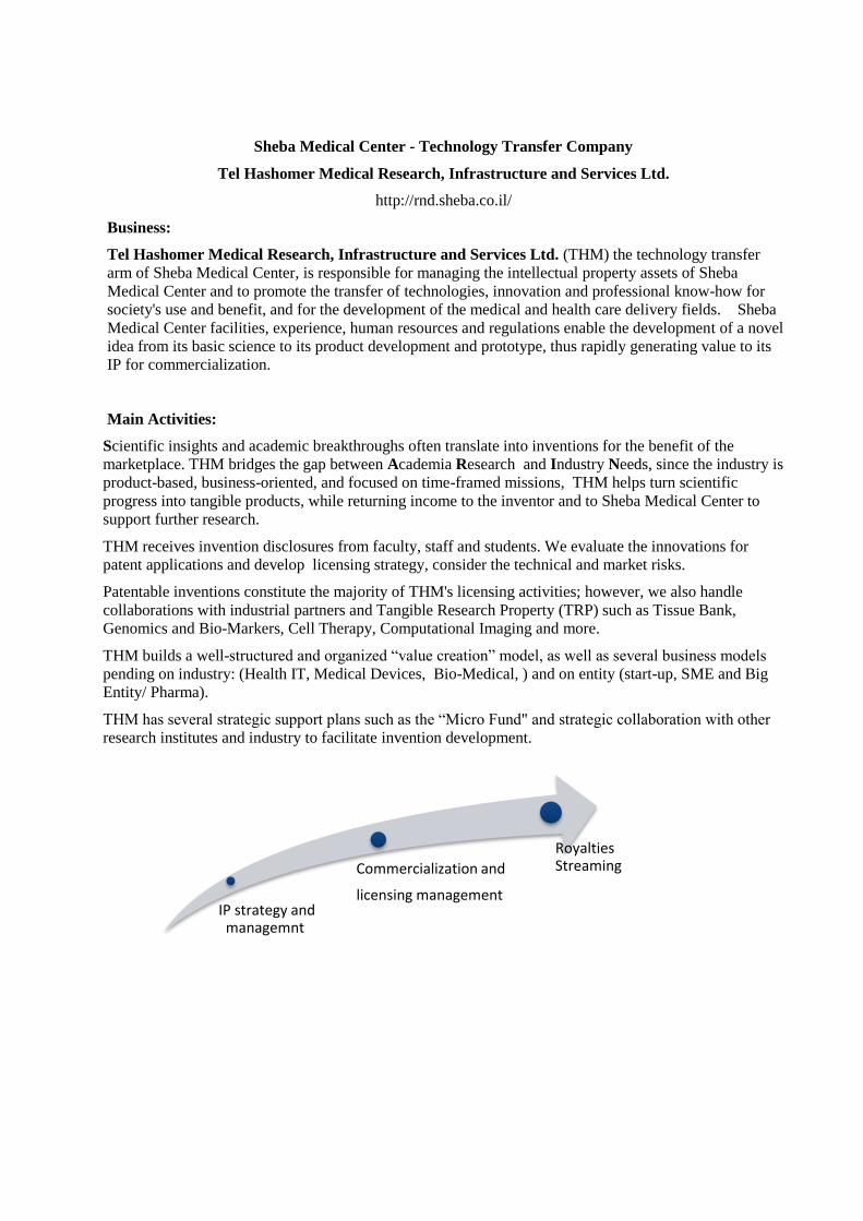
THM builds a well-structured and organized “value creation” model, as well as several business models
pending on industry: (Health IT, Medical Devices, Bio-Medical, ) and on entity (start-up, SME and Big
Entity/ Pharma).
THM has several strategic support plans such as the “Micro Fund" and strategic collaboration with other
research institutes and industry to facilitate invention development.
IP strategy and managemnt
Commercialization and
licensing management
Royalties Streaming
THM Strategic Principles to the Success of our Tech Transfer
► We bridge basic research to commercial Value
► We develop close interaction with researchers and industry
► We Build Strategic Capabilities
► We are a “Learning Organization” and Flexible Organization
► We understand the stakeholders need and Value creation
► We Build Collaboration & Alliances
► Our stream: Identify Need from the bedside, Basic and applicable Research-
► We develop broad and Multi-national view
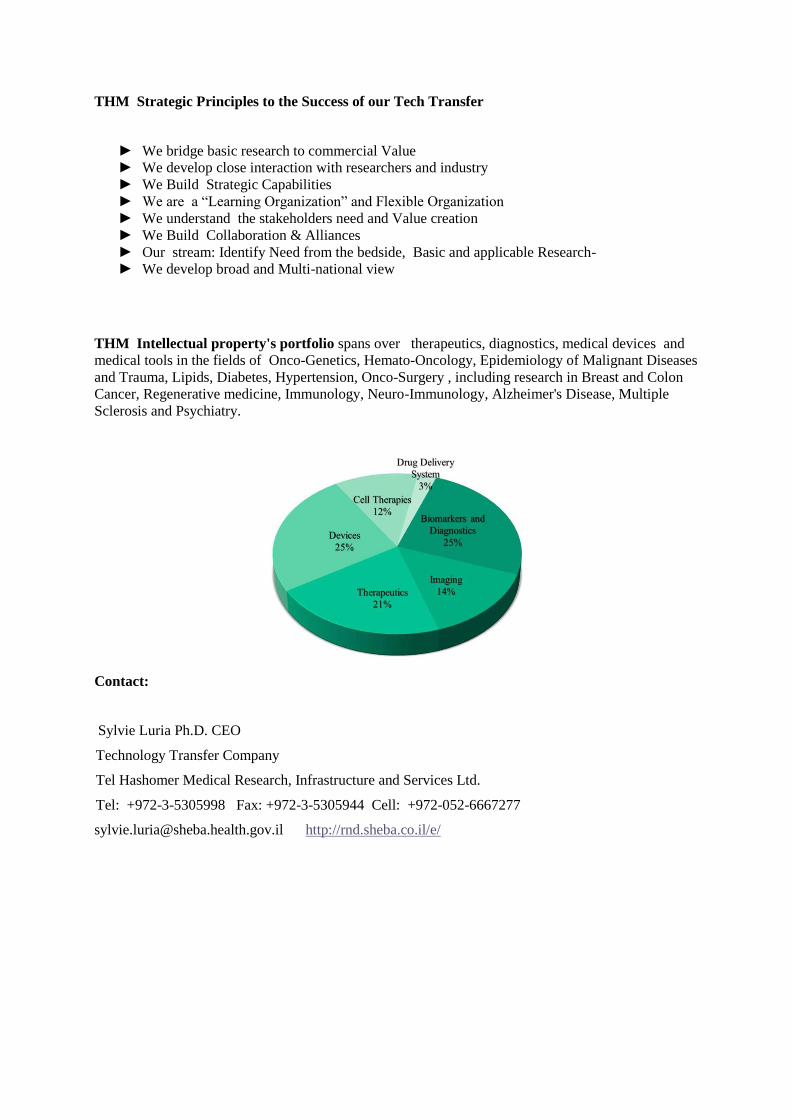
THM Intellectual property's portfolio spans over therapeutics, diagnostics, medical devices and
medical tools in the fields of Onco-Genetics, Hemato-Oncology, Epidemiology of Malignant Diseases
and Trauma, Lipids, Diabetes, Hypertension, Onco-Surgery , including research in Breast and Colon
Cancer, Regenerative medicine, Immunology, Neuro-Immunology, Alzheimer's Disease, Multiple
Sclerosis and Psychiatry.
Contact:
Sylvie Luria Ph.D. CEO
Technology Transfer Company
Tel Hashomer Medical Research, Infrastructure and Services Ltd.
Tel: +972-3-5305998 Fax: +972-3-5305944 Cell: +972-052-6667277
[email protected] http://rnd.sheba.co.il/e/
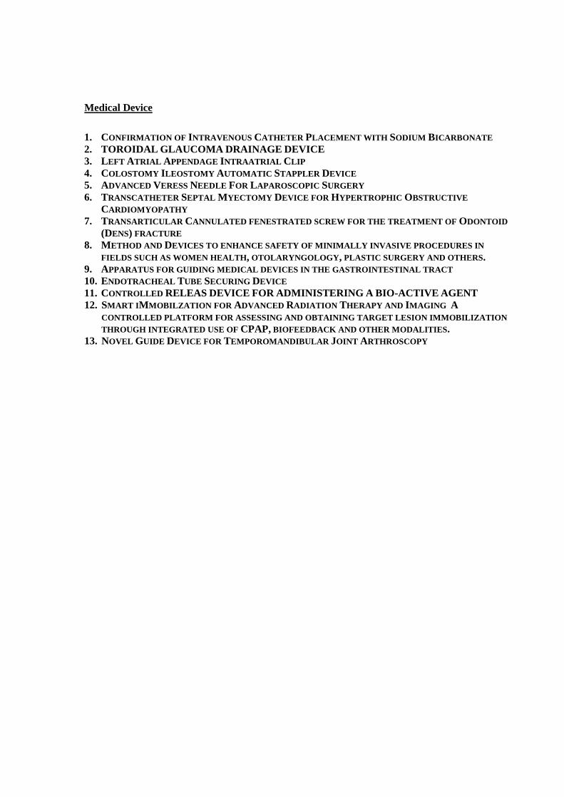
Medical Device
1. CONFIRMATION OF INTRAVENOUS CATHETER PLACEMENT WITH SODIUM BICARBONATE
2. TOROIDAL GLAUCOMA DRAINAGE DEVICE
3. LEFT ATRIAL APPENDAGE INTRAATRIAL CLIP
4. COLOSTOMY ILEOSTOMY AUTOMATIC STAPPLER DEVICE
5. ADVANCED VERESS NEEDLE FOR LAPAROSCOPIC SURGERY
6. TRANSCATHETER SEPTAL MYECTOMY DEVICE FOR HYPERTROPHIC OBSTRUCTIVE
CARDIOMYOPATHY
7. TRANSARTICULAR CANNULATED FENESTRATED SCREW FOR THE TREATMENT OF ODONTOID
(DENS) FRACTURE
8. METHOD AND DEVICES TO ENHANCE SAFETY OF MINIMALLY INVASIVE PROCEDURES IN
FIELDS SUCH AS WOMEN HEALTH, OTOLARYNGOLOGY, PLASTIC SURGERY AND OTHERS.
9. APPARATUS FOR GUIDING MEDICAL DEVICES IN THE GASTROINTESTINAL TRACT
10. ENDOTRACHEAL TUBE SECURING DEVICE
11. CONTROLLED RELEAS DEVICE FOR ADMINISTERING A BIO-ACTIVE AGENT
12. SMART IMMOBILZATION FOR ADVANCED RADIATION THERAPY AND IMAGING A
CONTROLLED PLATFORM FOR ASSESSING AND OBTAINING TARGET LESION IMMOBILIZATION
THROUGH INTEGRATED USE OF CPAP, BIOFEEDBACK AND OTHER MODALITIES.
13. NOVEL GUIDE DEVICE FOR TEMPOROMANDIBULAR JOINT ARTHROSCOPY

Method and System to Confirm Intravenous Catheter Placement and Positioning
Dr. Ilan Keidan, Sheba Medical Center, Israel
Categories Method and Algorithm
Development Stage Clinical Stage
Patent Status
PCT/IB2012/052288 :
"PROVIDING EVIDENCE WHETHER AN INTRAVASCULAR
CONDUIT ISCORRECTLY POSITIONED"
Background and Technology
INFILTRATION AND EXTRAVASATION are common complications of intravenous (I.V.) infusion
therapy. Extravasation can cause accidental administration of intravenously infused medicinal drugs into
the surrounding tissue, either by leakage (e.g., because of brittle veins in very elderly patients), or direct
exposure (e.g. because the needle has punctured the vein and the infusion goes directly into the arm
tissue). For example, Extravasation of medicinal drugs highly irritating solutions, such as those containing
calcium, potassium, contrast media, some antibiotics, vasopressors, or chemotherapeutic agents.during
intravenous therapy is a side effect that should be avoided. In mild cases, extravasation can cause pain,
reddening, or irritation on the arm with the infusion needle. Severe damage may include tissue necrosis. In
extreme cases, it even can lead to loss of an arm. The best "treatment" of extravasation is prevention.
While there is no real treatment per se, there are some techniques that can be applied in case of
extravasation, though their efficacy is modest. We have developed a simple method and system to
monitor intravenous position of catheters via periodically administration of a simple composition and a
monitoring device. (Sodium bicarbonate solution and end-tidal carbon dioxide monitor). The rationale for
using bicarbonate is based on the well-known phenomenon of increased exhaled carbon dioxide (CO2)
after its IV administration.
The Need
Extravasation is a serious condition that warrants special attention from the healthcare professionals
involved in administering intravenous medications. Over 100,000 doses of chemotherapy and in excess of
1,000,000 intravenous (IV) infusions given every day around the world, keeping adverse events and
complications of these procedures to a minimum is important both for the patients receiving them and the
healthcare systems in which they take place. It is critical that an extravasation is recognized and diagnosed
early. The tools available today to recognize and detect extravasation in its early stages are mainly
subjective and awareness to all relevant signs and symptoms. Infiltration rates were reported to be high,
with as many as 20-30% of IV catheters in adults resulting in infiltration, with higher rates seen in
children. Analysis of the American Society of Anesthesiologists Closed Claims database revealed 2% of
all claims were related to peripheral IV catheterization and over half of these were due to extravasation,
and higher rates could be expected with other health care providers given the presumed expertise of
anesthesiologists in IV cannulation.
Development Stage and Technology
We developed a novel technique that can differentiate between an infiltrated and a correctly sited IV
catheter in both anesthetized ventilated and spontaneously breathing volunteers. We have demonstrated
the efficacy of the novel method as very useful in providing information to monitor and assist in
determining whether or not an intravenous conduit is in a correct position. The method is simple to
integrate in various monitoring systems in the hospital set-up. We have initiated clinical studies that
demonstrate the efficacy and specificity of the concept and system in patients from age 2-35 years old.
Currently we are expending the study to the general patient's population. We target those patients which
extravasations/ infiltration rate is high and the consequences are grave.
Advantages
The technology relates to a specific tool to be implemented in clinical monitors. Our technology is simple
to integrate into existing monitoring devices, Capnometers and monitoring systems with critical added
value of CO2 monitoring..
The Market
The market is add on capnometry devices for the determination of the end-tidal partial pressure of carbon
dioxideThe carbon dioxide (CO2) monitors market is witnessing an increasing trend over the last few
years, primarily driven by enhanced requirements in patient monitoring for safety and disease
management. Although majority of capnography applications are in the operating rooms for detecting and
identifying the end-tidal CO2 levels, new and emerging applications including critical care units, recovery
rooms, labor and delivery rooms, emergency rooms, post-anesthesia care units, intensive care units and
daily care units in Oncology and autoimmune diseases are instigating the use of capnography equipment.
Capnography market worldwide is presently considered a segment with rich opportunities, increasing
simultaneously with the continuously evolving ways and methods of patient care. Our new feature to be
incorporated into an existing commercial end-tidal CO2 monitor may contribute the market growth, with
the rise of aging population as well as increasing IV bio-pharmaceutical therapies and the augmented
safety regulations.
The worldwide markets for Carbon Dioxide (CO2) Monitors include the following Product Segments:
End-tidal Carbon Dioxide (EtCO2) Monitors, and Transcutaneous Carbon Dioxide (tcpCO2) Monitors.
There are 40 companies including many key and niche players such as B. Braun Melsungen AG, CAS
Medical Systems, Inc., Criticare Systems, Inc., Dräger Medical AG & Co. KG, GE Healthcare Life
Support Solutions, Heinen + Löwenstein GmbH, Invivo Corporation, Ivy Biomedical Systems, Inc.,
Mindray North America, Nellcor Puritan Bennett, LLC, Nihon Kohden Corporation, Nonin Medical, Inc.,
Oridion Systems, Ltd., OSI Systems, Inc., Philips Healthcare, Physio-Control, Inc., Radiometer Basel AG,
Radiometer Medical ApS, Respironics, Smiths Medical, Thames Medical, Weinmann Geräte Für Medizin
GmbH + Co. KG, and Welch Allyn Inc.
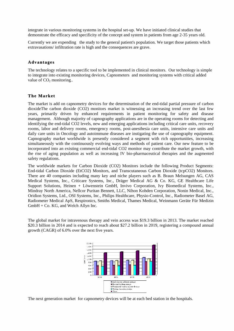
The global market for intravenous therapy and vein access was $19.3 billion in 2013. The market reached
$20.3 billion in 2014 and is expected to reach about $27.2 billion in 2019, registering a compound annual
growth (CAGR) of 6.0% over the next five years.
The next generation market for capnometry devices will be at each bed station in the hospitals.
P atent
METHODS AND DEVICES USEFUL FOR DETERMINING CORRECT PLACEMENT OF INTRA-VASCULARTURE "
CONDUIT "
PATENT PENDING
TOROIDAL GLAUCOMA DRAINAGE DEVICE
Dr. Ilia Piven, Sheba Medical Center.
Categories Implantable Medical device for ocular drainage, Ophthalmology
Development Stage Initial prototype design
Patent Status Patent Pending – TOROIDAL GLAUCOMA DRAINAGE DEVICE
Background of the Invent ion
Ocular hypertension has been associated with a number of eye conditions, including Glaucoma, eye
trauma, pseudoexfoliation syndrome, pigment dispersion syndrome and corneal arcus. In the majority of
cases, vision loss usually occurs when the eye pressure is too high for the specific individual and damages
the optic nerve. Any resultant damage cannot be reversed. In eyes with glaucoma, peripheral (side) vision
is affected first. The changes in vision may be so gradual that they are not noticed until a lot of vision loss
has already occurred. Ocular hypertension must be monitored and treated to save vision lost.
The modern goals of glaucoma management are to avoid glaucomatous damage and nerve damage, and
preserve visual field and total quality of life for patients, with minimal side effects. Although intraocular
pressure is only one of the major risk factors for glaucoma, lowering it via various pharmaceuticals and/or
surgical techniques is currently the mainstay of glaucoma treatment.
Vascular flow and neurodegenerative theories of glaucomatous optic neuropathy have prompted studies
on various neuroprotective therapeutic strategies, including nutritional compounds, some of which may be
regarded by clinicians as safe for use now, while others are on trial.
Currently approved treatments for glaucoma include a number of pharmaceutical drugs, laser therapies
and surgical procedures. Each of these approaches to treating this disease has both side-effects and risks:
Pharmaceuticals (usually formulated as eye-drops): Glaucoma is often treated initially with medicated
eye-drops that lower IOP by increasing the outflow or reducing the inflow of fluids in the eye. However,
many patients experience significant side-effects with these drugs, which results in poor compliance.
There are a number of categories of glaucoma drugs on the market and others in development, but over
50% of glaucoma patients are non-compliant within 6 months of use, primarily due to side effects.
Laser Surgery: “Laser Trabeculoplasty” is an established treatment for glaucoma that uses a high-energy
laser beam to open the clogged drainage canals in the eye, thereby allowing the aqueous humour to drain
more readily. However, improvements in drainage may take a few weeks to become apparent and
eventually the intraocular pressure will increase again as the drainage channels become blocked again.
This procedure usually can only be done 2 times in each eye.
Filtering Surgery: This is a surgical technique also known as “Trabulectomy” that is used to create a
new opening in the sclera (i.e. the “white” of the eye) by removing a small piece of the trabecular
network. This permits the aqueous humour to drain more normally and thus lowers the eye pressure. This
procedure usually remains effective for 2-5 years, but complications can occur.
Drainage Implants: Drainage implant surgery installs a shunt or other device in the eye to create a new
drain for fluids in the trabecular meshwork. Glaucoma drainage implant devices (GDDs) create an
alternate aqueous pathway from the anterior chamber (AC) by channeling aqueous out of the eye through
a tube to a subconjunctival bleb or to the suprachoroidal space. This tube is usually connected to an
equatorial plate under the conjunctiva. GDDs are being used more frequently in the treatment of glaucoma
that is not responding to medications and trabeculectomy operations. In certain conditions, such as
neovascular glaucoma, iridocorneal endothelial (ICE) syndrome, penetrating keratoplasty (PKP) with
glaucoma, and glaucoma following retinal detachment surgery, it has become the preferred operation.
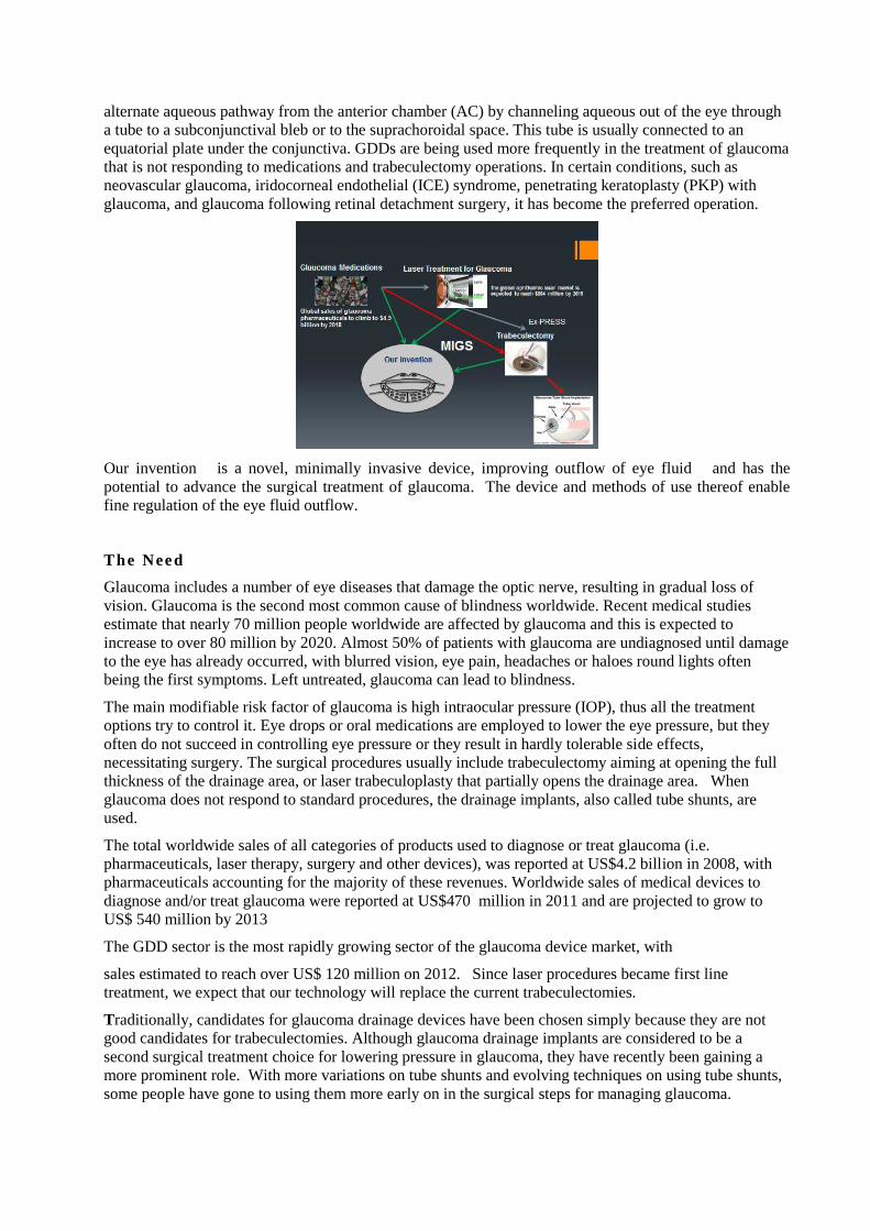
Our invention is a novel, minimally invasive device, improving outflow of eye fluid and has the
potential to advance the surgical treatment of glaucoma. The device and methods of use thereof enable
fine regulation of the eye fluid outflow.
The Need
Glaucoma includes a number of eye diseases that damage the optic nerve, resulting in gradual loss of
vision. Glaucoma is the second most common cause of blindness worldwide. Recent medical studies
estimate that nearly 70 million people worldwide are affected by glaucoma and this is expected to
increase to over 80 million by 2020. Almost 50% of patients with glaucoma are undiagnosed until damage
to the eye has already occurred, with blurred vision, eye pain, headaches or haloes round lights often
being the first symptoms. Left untreated, glaucoma can lead to blindness.
The main modifiable risk factor of glaucoma is high intraocular pressure (IOP), thus all the treatment
options try to control it. Eye drops or oral medications are employed to lower the eye pressure, but they
often do not succeed in controlling eye pressure or they result in hardly tolerable side effects,
necessitating surgery. The surgical procedures usually include trabeculectomy aiming at opening the full
thickness of the drainage area, or laser trabeculoplasty that partially opens the drainage area. When
glaucoma does not respond to standard procedures, the drainage implants, also called tube shunts, are
used.
The total worldwide sales of all categories of products used to diagnose or treat glaucoma (i.e.
pharmaceuticals, laser therapy, surgery and other devices), was reported at US$4.2 billion in 2008, with
pharmaceuticals accounting for the majority of these revenues. Worldwide sales of medical devices to
diagnose and/or treat glaucoma were reported at US$470 million in 2011 and are projected to grow to
US$ 540 million by 2013
The GDD sector is the most rapidly growing sector of the glaucoma device market, with
sales estimated to reach over US$ 120 million on 2012. Since laser procedures became first line
treatment, we expect that our technology will replace the current trabeculectomies.
Traditionally, candidates for glaucoma drainage devices have been chosen simply because they are not
good candidates for trabeculectomies. Although glaucoma drainage implants are considered to be a
second surgical treatment choice for lowering pressure in glaucoma, they have recently been gaining a
more prominent role. With more variations on tube shunts and evolving techniques on using tube shunts,
some people have gone to using them more early on in the surgical steps for managing glaucoma.
P otent ia l Appl icat ions
Glaucoma drainage device implantation is usually reserved for cases with refractory glaucoma, or those
unlikely to respond successfully to a conventional filtration surgery. The indications for GDD
implantation include the following:
Neovascular glaucoma
Penetrating keratoplasty with glaucoma
Retinal detachment surgery with glaucoma
Iridocorneal endothelial syndrome
Traumatic glaucoma
Uveitic glaucoma
Open angle glaucoma with failed trabeculectomy
Epithelial down growth
Refractory infantile glaucoma
Contact lens wearers who need glaucoma filtration surgery
Sturge-Weber's syndrome.
Contraindications:
Eyes with severe scleral or sclera-limbal thinning
Extensive fibrosis of conjunctiva
Ciliary block glaucoma.
Relative Contraindications:
Vitreous in AC
Intra-ocular silicone oil-Implant if required is placed in inferio-temporal quadrant
Advantages
In a study published in 2012 (Gedde SJet al. in the Am J Ophthalmol 2012;153:789-803) , they
have demonstrated that tube shunt surgery had a higher success rate compared to trabeculectomy
with MMC during 5 years of follow-up in the TVT Study. Both procedures were associated with
similar IOP reduction and use of supplemental medical therapy at 5 years. A total of 212 eyes of
212 patients were enrolled, including 107 in the tube group and 105 in the trabeculectomy group.
At 5 years, IOP (mean ± SD) was 14.4 ± 6.9 mmHg in the tube group and 12.6 ± 5.9 mmHg in the
trabeculectomy group (P = 0.12). The number of glaucoma medications (mean ± SD) was 1.4 ±
1.3 in the tube group and 1.2 ± 1.5 in the trabeculectomy group (P = 0.23). The cumulative
probability of failure during 5 years of follow-up was 29.8% in the tube group and 46.9% in the
trabeculectomy group (P = 0.002; hazard ratio = 2.15; 95% confidence interval = 1.30-3.56). The
rate of reoperation for glaucoma was 9% in the tube group and 29% in the trabeculectomy group
(P = 0.025).
GDD have been successful in controlling IOP in eyes with previously failed trabeculotomy and
for cases with refractory glaucoma. Since their introduction, numerous modifications in design
and improvements in surgical technique have enhanced clinical outcomes and minimized

complications.
Patent:
" Toroidal Glaucoma Drainage Device" , US provisional, Pending.
Left Atrial Appendage Intraatrial Transcatheter Suturless Closure
Leonid Sternik, Sheba Medical Center
Categories Medical Device for cardiologic application
Development Stage Proven Concept using experimental prototype
Patent Status" BODY PART REPOSITIONING APPARATUS AND METHOD" WO
2013/008231A1
Background and Technology
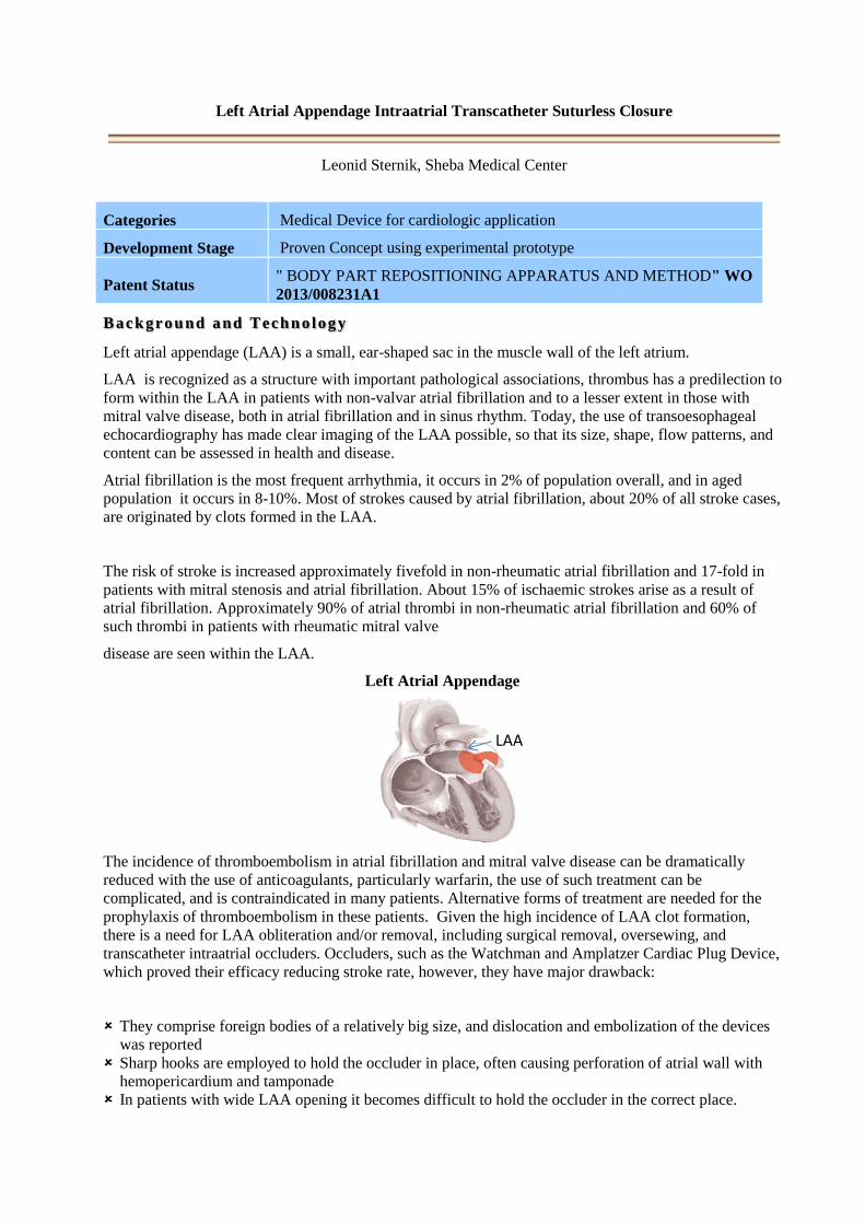
Left atrial appendage (LAA) is a small, ear-shaped sac in the muscle wall of the left atrium.
LAA is recognized as a structure with important pathological associations, thrombus has a predilection to
form within the LAA in patients with non-valvar atrial fibrillation and to a lesser extent in those with
mitral valve disease, both in atrial fibrillation and in sinus rhythm. Today, the use of transoesophageal
echocardiography has made clear imaging of the LAA possible, so that its size, shape, flow patterns, and
content can be assessed in health and disease.
Atrial fibrillation is the most frequent arrhythmia, it occurs in 2% of population overall, and in aged
population it occurs in 8-10%. Most of strokes caused by atrial fibrillation, about 20% of all stroke cases,
are originated by clots formed in the LAA.
The risk of stroke is increased approximately fivefold in non-rheumatic atrial fibrillation and 17-fold in
patients with mitral stenosis and atrial fibrillation. About 15% of ischaemic strokes arise as a result of
atrial fibrillation. Approximately 90% of atrial thrombi in non-rheumatic atrial fibrillation and 60% of
such thrombi in patients with rheumatic mitral valve
disease are seen within the LAA.
Left Atrial Appendage
The incidence of thromboembolism in atrial fibrillation and mitral valve disease can be dramatically
reduced with the use of anticoagulants, particularly warfarin, the use of such treatment can be
complicated, and is contraindicated in many patients. Alternative forms of treatment are needed for the
prophylaxis of thromboembolism in these patients. Given the high incidence of LAA clot formation,
there is a need for LAA obliteration and/or removal, including surgical removal, oversewing, and
transcatheter intraatrial occluders. Occluders, such as the Watchman and Amplatzer Cardiac Plug Device,
which proved their efficacy reducing stroke rate, however, they have major drawback:
They comprise foreign bodies of a relatively big size, and dislocation and embolization of the devices
was reported
Sharp hooks are employed to hold the occluder in place, often causing perforation of atrial wall with
hemopericardium and tamponade
In patients with wide LAA opening it becomes difficult to hold the occluder in the correct place.
LAA
LAA
Size and the shape of the appendage differ amongst patients, so occluders must be of different size to fit
the appendage.
We propose a Novel device for Left Atrial Appendage occlusion by invagination.
The Technology and Development Stage
We have developed a novel trans-septal catheter for LAA closure device.
The apparatus principally used together with a multipurpose coronary catheter with our novel addition of
a soft Teflon cone on its tip enable the septal penetration. The invagination of the LAA procedure is
completed with our innovative detachable lasso catheter apparatus.
The device was tested in an in -vitro testing model using pig and human hearts.
Advantages
Even though an external ligation of LAA is a very well-known surgical procedure for the last 40 years it
has many drawbacks. We propose to ligate the LAA intraluminally and achieve the following advantages:
Transcatheter Device - The procedure based on the device would be percutaneous catheter
based.
No Anchors, Atraumatic - Contrary to currently used occluders (as Watchman ® by Atritech
Inc., Amplatzer Cardiac Plug by AGA Medical Corporation) no pins, hooks or other sharp and
penetrating parts on the atrial wall are needed.
Minimal Invasive - No penetration of atrial wall outside to pericardium is performed.
Minimal Foreign Material - No bulky appendage occluder is used, the only foreign material in
the left atrium is a tiny loop or clip on the base of the appendage.
Complete Occlusion - Invaginated appendage cannot holds blood clots at all, as it is washed with
blood and covered naturally with endocardium.
The Need and The Market
The procedures for Left Atrial Appendage (LAA) devices, intended to minimize stroke risk in patients
with atrial fibrillation, have grown by over 200 percent since 2010. This procedure will compete for
market share against the new generation of anticoagulants, which provide an alternative method of
decreasing stroke risk.
Stroke is a common consequence of atrial fibrillation, and approximately 15 percent of all strokes are
caused by atrial fibrillation, and as many as one third of strokes occurring in individuals aged 65 or older.
The standard preventive treatment has long been anticoagulants, typically warfarin, in clinical use since
the 1950s. Warfarin has a number of drawbacks, including excessive bleeding, dosage difficulties and
frequent interactions with food and other drugs.
Nearly 90 percent of stroke-causing thrombi in atrial fibrillation form in a part of the heart called the left
atrial appendage (LAA). Devices that close off the LAA have proved effective in minimizing stroke risk.
These devices fall into two main categories: endocardial and epicardial, generally referred to as
“occlusion” and “exclusion,” respectively. Endocardial devices have continuously proved to be
significantly more popular than the epicardial alternatives.
LAA procedures are relatively new, and have been growing strongly. More than 30 percent of the
physicians performing LAA procedures started to perform them only within the past year. These
procedures will be competing against the next generation of oral anticoagulants with clinical profiles
significantly superior to warfarin’s. According to a recent report from Decision Resources, these drugs
include apixaban (Bristol-Myers Squibb/Pfizer’s Eliquis), dabigatran etexilate (Boehringer Ingelheim’s
Pradaxa) and rivaroxaban (Bayer/Janssen’s Xarelto). Worldwide, these novel anticoagulants are expected
to capture 72 percent of the atrial fibrillation drug market in 2020.
Despite improvement in anticoagulants, LAA devices are expected to continue to increase their share in
this market. Decision Resources projects the number of diagnosed prevalent cases of atrial fibrillation
worldwide to reach over 30 million in 2015 and will continue to grow at a rate of 1.6 percent annually.
The adoption of LAA devices in this market will continue to grow, since the implantation of an LAA
device, whether endocardial or epicardial, is a one-time procedure, while drug therapy must be ongoing.
According to Millennium Research Group (MRG), the global authority on medical technology market
intelligence, procedures for Left Atrial Appendage (LAA) devices, intended to minimize stroke risk in
patients with atrial fibrillation, have grown by over 2 folds since 2010.
Next Step Deve lopment
1- Acute and Chronic experiment in open heart surgery in pigs to validate to concept.
2- Complete the final Prototype.
3- Chronic Pig experiment for up to 20 weeks.
Inte l lectual Property
WO 2013/008231 A1 - "Left Atrial Appendage Intraatrial Clip" National Phase,
Priority date-July, 2012
A Novel Colostomy/Ileostomy Automated Stapler Device
Avinoam Nevler, Sheba Medical Center, Israel
Categories Surgery Device, Medical Device, Colostomy/Ileostomy device
Development Stage Initial Stage of Concept
Patent Status " Colostomy Ileostomy Automatic Stappler Device" PCT 61/680,329
Background and Technology
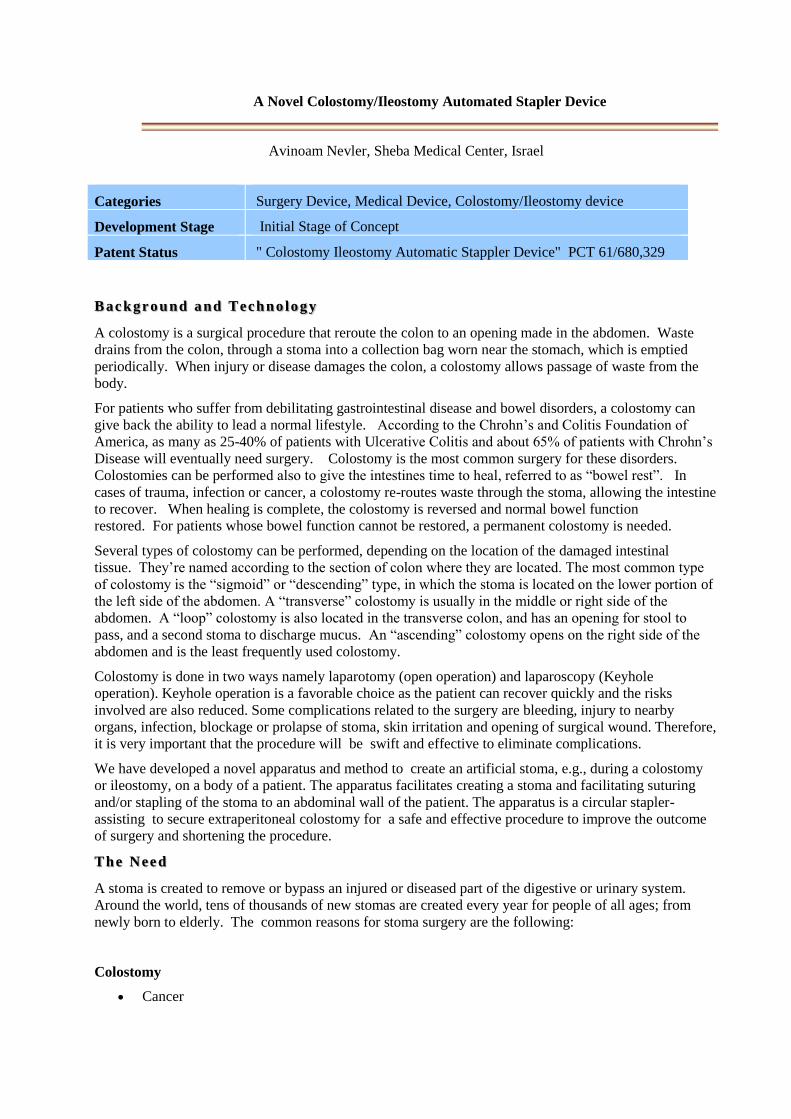
A colostomy is a surgical procedure that reroute the colon to an opening made in the abdomen. Waste
drains from the colon, through a stoma into a collection bag worn near the stomach, which is emptied
periodically. When injury or disease damages the colon, a colostomy allows passage of waste from the
body.
For patients who suffer from debilitating gastrointestinal disease and bowel disorders, a colostomy can
give back the ability to lead a normal lifestyle. According to the Chrohn’s and Colitis Foundation of
America, as many as 25-40% of patients with Ulcerative Colitis and about 65% of patients with Chrohn’s
Disease will eventually need surgery. Colostomy is the most common surgery for these disorders.
Colostomies can be performed also to give the intestines time to heal, referred to as “bowel rest”. In
cases of trauma, infection or cancer, a colostomy re-routes waste through the stoma, allowing the intestine
to recover. When healing is complete, the colostomy is reversed and normal bowel function
restored. For patients whose bowel function cannot be restored, a permanent colostomy is needed.
Several types of colostomy can be performed, depending on the location of the damaged intestinal
tissue. They’re named according to the section of colon where they are located. The most common type
of colostomy is the “sigmoid” or “descending” type, in which the stoma is located on the lower portion of
the left side of the abdomen. A “transverse” colostomy is usually in the middle or right side of the
abdomen. A “loop” colostomy is also located in the transverse colon, and has an opening for stool to
pass, and a second stoma to discharge mucus. An “ascending” colostomy opens on the right side of the
abdomen and is the least frequently used colostomy.
Colostomy is done in two ways namely laparotomy (open operation) and laparoscopy (Keyhole
operation). Keyhole operation is a favorable choice as the patient can recover quickly and the risks
involved are also reduced. Some complications related to the surgery are bleeding, injury to nearby
organs, infection, blockage or prolapse of stoma, skin irritation and opening of surgical wound. Therefore,
it is very important that the procedure will be swift and effective to eliminate complications.
We have developed a novel apparatus and method to create an artificial stoma, e.g., during a colostomy
or ileostomy, on a body of a patient. The apparatus facilitates creating a stoma and facilitating suturing
and/or stapling of the stoma to an abdominal wall of the patient. The apparatus is a circular stapler-
assisting to secure extraperitoneal colostomy for a safe and effective procedure to improve the outcome
of surgery and shortening the procedure.
The Need
A stoma is created to remove or bypass an injured or diseased part of the digestive or urinary system.
Around the world, tens of thousands of new stomas are created every year for people of all ages; from
newly born to elderly. The common reasons for stoma surgery are the following:
Colostomy
Cancer
Diverticular Disease
Trauma
Congenital (present at birth)
Incontinence
Ileostomy
Ulcerative Colitis
Crohn's Disease
Familial Polyposis Coli (FPC)
Congenital (present at birth)
Staged Process for Other Surgery (e.g. Loop stoma - which is reversed later)
Urostomy
Bladder Cancer
Trauma
Congenital (present at birth)
Incontinence/Repeated Infection
Interstitial Cystitis
The Market
The loss of continence individuals suffer as a result of ostomy surgery is often a life-changing experience.
The impact on the quality of life of patients and their families can be profound. Access to ostomy supplies
that are fitted and prescribed by a healthcare provider is critical to maintaining the health and well-being
of the person with an ostomy. New innovations that provide enhanced skin protection and/or prosthetic
functionality are very important for these individuals. Ostomy surgery impacts approximately 700,000
people (0.14% of the total population) in Europe and over 2.5 million ostomy surgery are performed
worldwide and the market for the innovated device may be up to 250 M USD. More than half of ostomy
surgeries (55%) are considered permanent surgeries, meaning the patient will be permanently unable to
control the output of effluent and will require a collection device attached to their abdomen.
World Market for Gastrointestinal Devices (Rigid, Flexible and Capsule Endoscopes, Bariatric Surgery,
Ostomy Products, Enteral Feeding Pumps and Other Devices) is rapidly growing. There are many factors
behind the growth of this market. Cancer is one of leading reasons for surgery with more than 1 million
new cases of colorectal cancer reported each year around the world. Obesity is another reason for growth.
More and more people worldwide are turning to gastric bypass (bariatric surgery) and gastric banding to
aide in their weight loss, thus increasing the need for gastrointestinal endoscopy. Improved technology is
another driver for the market.
תל השומר מחקרים תשתיות ושירותים רפואיים
Tel Hashomer Medical Research, Infrastructure and Services Tel: +972-3-5305998 Fax: +972-3-5305944 [email protected]
Advanced Veress Needle For Laparoscopic Surgery
Avinoam Nevler, Nir Horesh, Haya Shwartz and Gil Har, Sheba Medical Center
Categories Medical Device, Laparoscopic Surgery
Development Stage Proof of concept demonstrated in animal miodels.
Patent Status " IMPROVED VERESS NEEDLE" 61/682,321
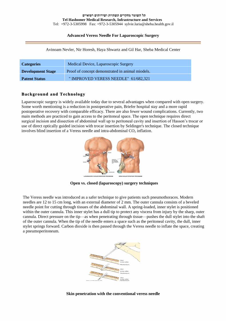
B ackground and Technology
Laparoscopic surgery is widely available today due to several advantages when compared with open surgery.
Some worth mentioning is a reduction in postoperative pain, Briefer hospital stay and a more rapid
postoperative recovery with comparable efficacy. There are also fewer wound complications. Currently, two
main methods are practiced to gain access to the peritoneal space. The open technique requires direct
surgical incision and dissection of abdominal wall up to peritoneal cavity and insertion of Hasson’s trocar or
use of direct optically guided incision with trocar insertion by Seldinger's technique. The closed technique
involves blind insertion of a Veress needle and intra-abdominal CO2 inflation.
Open vs. closed (laparoscopy) surgery techniques
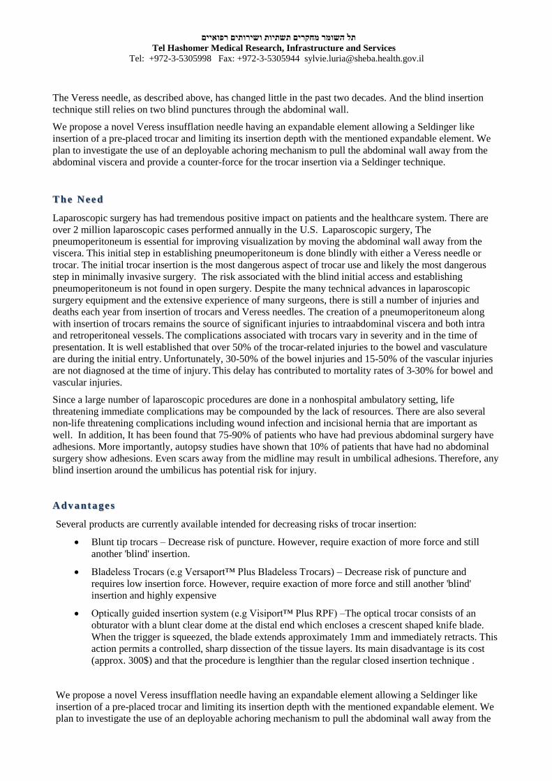
The Veress needle was introduced as a safer technique to give patients such pneumothoraces. Modern
needles are 12 to 15 cm long, with an external diameter of 2 mm. The outer cannula consists of a beveled
needle point for cutting through tissues of the abdominal wall. A spring-loaded, inner stylet is positioned
within the outer cannula. This inner stylet has a dull tip to protect any viscera from injury by the sharp, outer
cannula. Direct pressure on the tip—as when penetrating through tissue—pushes the dull stylet into the shaft
of the outer cannula. When the tip of the needle enters a space such as the peritoneal cavity, the dull, inner
stylet springs forward. Carbon dioxide is then passed through the Veress needle to inflate the space, creating
a pneumoperitoneum.
Skin penetration with the conventional veress needle
תל השומר מחקרים תשתיות ושירותים רפואיים
Tel Hashomer Medical Research, Infrastructure and Services Tel: +972-3-5305998 Fax: +972-3-5305944 [email protected]
The Veress needle, as described above, has changed little in the past two decades. And the blind insertion
technique still relies on two blind punctures through the abdominal wall.
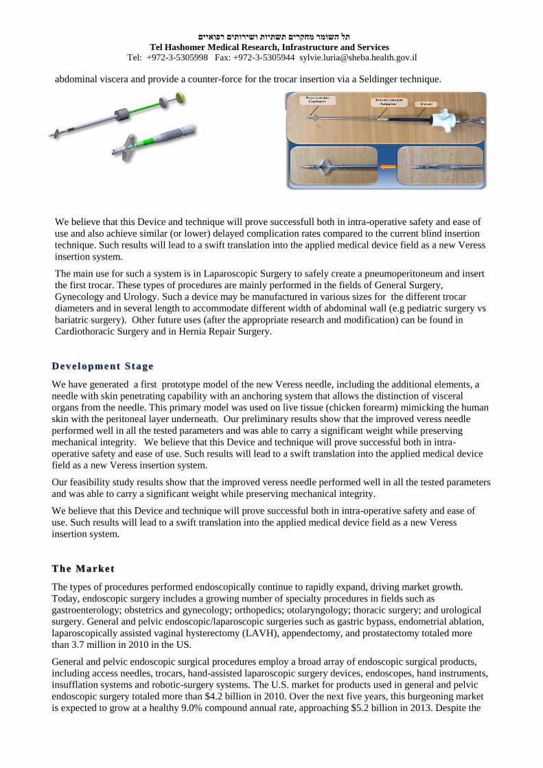
We propose a novel Veress insufflation needle having an expandable element allowing a Seldinger like
insertion of a pre-placed trocar and limiting its insertion depth with the mentioned expandable element. We
plan to investigate the use of an deployable achoring mechanism to pull the abdominal wall away from the
abdominal viscera and provide a counter-force for the trocar insertion via a Seldinger technique.
The Need
Laparoscopic surgery has had tremendous positive impact on patients and the healthcare system. There are
over 2 million laparoscopic cases performed annually in the U.S. Laparoscopic surgery, The
pneumoperitoneum is essential for improving visualization by moving the abdominal wall away from the
viscera. This initial step in establishing pneumoperitoneum is done blindly with either a Veress needle or
trocar. The initial trocar insertion is the most dangerous aspect of trocar use and likely the most dangerous
step in minimally invasive surgery. The risk associated with the blind initial access and establishing
pneumoperitoneum is not found in open surgery. Despite the many technical advances in laparoscopic
surgery equipment and the extensive experience of many surgeons, there is still a number of injuries and
deaths each year from insertion of trocars and Veress needles. The creation of a pneumoperitoneum along
with insertion of trocars remains the source of significant injuries to intraabdominal viscera and both intra
and retroperitoneal vessels. The complications associated with trocars vary in severity and in the time of
presentation. It is well established that over 50% of the trocar-related injuries to the bowel and vasculature
are during the initial entry. Unfortunately, 30-50% of the bowel injuries and 15-50% of the vascular injuries
are not diagnosed at the time of injury. This delay has contributed to mortality rates of 3-30% for bowel and
vascular injuries.
Since a large number of laparoscopic procedures are done in a nonhospital ambulatory setting, life
threatening immediate complications may be compounded by the lack of resources. There are also several
non-life threatening complications including wound infection and incisional hernia that are important as
well. In addition, It has been found that 75-90% of patients who have had previous abdominal surgery have
adhesions. More importantly, autopsy studies have shown that 10% of patients that have had no abdominal
surgery show adhesions. Even scars away from the midline may result in umbilical adhesions. Therefore, any
blind insertion around the umbilicus has potential risk for injury.
Advantages
Several products are currently available intended for decreasing risks of trocar insertion:
Blunt tip trocars – Decrease risk of puncture. However, require exaction of more force and still
another 'blind' insertion.
Bladeless Trocars (e.g Versaport™ Plus Bladeless Trocars) – Decrease risk of puncture and
requires low insertion force. However, require exaction of more force and still another 'blind'
insertion and highly expensive
Optically guided insertion system (e.g Visiport™ Plus RPF) –The optical trocar consists of an
obturator with a blunt clear dome at the distal end which encloses a crescent shaped knife blade.
When the trigger is squeezed, the blade extends approximately 1mm and immediately retracts. This
action permits a controlled, sharp dissection of the tissue layers. Its main disadvantage is its cost
(approx. 300$) and that the procedure is lengthier than the regular closed insertion technique .
We propose a novel Veress insufflation needle having an expandable element allowing a Seldinger like
insertion of a pre-placed trocar and limiting its insertion depth with the mentioned expandable element. We
plan to investigate the use of an deployable achoring mechanism to pull the abdominal wall away from the
תל השומר מחקרים תשתיות ושירותים רפואיים
Tel Hashomer Medical Research, Infrastructure and Services Tel: +972-3-5305998 Fax: +972-3-5305944 [email protected]
abdominal viscera and provide a counter-force for the trocar insertion via a Seldinger technique.
We believe that this Device and technique will prove successfull both in intra-operative safety and ease of
use and also achieve similar (or lower) delayed complication rates compared to the current blind insertion
technique. Such results will lead to a swift translation into the applied medical device field as a new Veress
insertion system.
The main use for such a system is in Laparoscopic Surgery to safely create a pneumoperitoneum and insert
the first trocar. These types of procedures are mainly performed in the fields of General Surgery,
Gynecology and Urology. Such a device may be manufactured in various sizes for the different trocar
diameters and in several length to accommodate different width of abdominal wall (e.g pediatric surgery vs
bariatric surgery). Other future uses (after the appropriate research and modification) can be found in
Cardiothoracic Surgery and in Hernia Repair Surgery.
Development Stage
We have generated a first prototype model of the new Veress needle, including the additional elements, a
needle with skin penetrating capability with an anchoring system that allows the distinction of visceral
organs from the needle. This primary model was used on live tissue (chicken forearm) mimicking the human
skin with the peritoneal layer underneath. Our preliminary results show that the improved veress needle
performed well in all the tested parameters and was able to carry a significant weight while preserving
mechanical integrity. We believe that this Device and technique will prove successful both in intra-
operative safety and ease of use. Such results will lead to a swift translation into the applied medical device
field as a new Veress insertion system.
Our feasibility study results show that the improved veress needle performed well in all the tested parameters
and was able to carry a significant weight while preserving mechanical integrity.
We believe that this Device and technique will prove successful both in intra-operative safety and ease of
use. Such results will lead to a swift translation into the applied medical device field as a new Veress
insertion system.
The Market
The types of procedures performed endoscopically continue to rapidly expand, driving market growth.
Today, endoscopic surgery includes a growing number of specialty procedures in fields such as
gastroenterology; obstetrics and gynecology; orthopedics; otolaryngology; thoracic surgery; and urological
surgery. General and pelvic endoscopic/laparoscopic surgeries such as gastric bypass, endometrial ablation,
laparoscopically assisted vaginal hysterectomy (LAVH), appendectomy, and prostatectomy totaled more
than 3.7 million in 2010 in the US.
General and pelvic endoscopic surgical procedures employ a broad array of endoscopic surgical products,
including access needles, trocars, hand-assisted laparoscopic surgery devices, endoscopes, hand instruments,
insufflation systems and robotic-surgery systems. The U.S. market for products used in general and pelvic
endoscopic surgery totaled more than $4.2 billion in 2010. Over the next five years, this burgeoning market
is expected to grow at a healthy 9.0% compound annual rate, approaching $5.2 billion in 2013. Despite the
תל השומר מחקרים תשתיות ושירותים רפואיים
Tel Hashomer Medical Research, Infrastructure and Services Tel: +972-3-5305998 Fax: +972-3-5305944 [email protected]
current economic recession, strong pricing pressure and competition, this market is expected to exhibit
healthier-than-average growth due to demographics, ongoing demand for less invasive, less-costly surgery,
increased surgeon training, and adaption of next-generation endoscopic technologies, including robotic
surgical systems and next-generation endoscopic instrumentation. In addition, emerging “scarless”
techniques, including reducing the surgical access to a single incision or via natural orifices such as the
umbilicus, have resulted in the development of several novel surgical instruments and systems that may
significantly impact the field of laparoscopy.
P atent
A patent application was submitted on 13/08/2012 to the USPTO (Application 61682321) securing all
aspects of the potential technology and applications.
Tech Transfer Of f icer
Dr. Sylvie Luria
Tel Hashomer Medical Research, Infrastructure and Services
Tel: +972-3-5305998 Fax: +972-3-5305944 ; [email protected]
תל השומר מחקרים תשתיות ושירותים רפואיים
Tel Hashomer Medical Research, Infrastructure and Services Tel: +972-3-5305998 Fax: +972-3-5305944 [email protected]
Transcatheter Septal Myectomy Device for Hypertrophic Obstructive Cardiomyopathy
Dr. Elad Maor, Sheba Medical Center, Tel Hashomer
Categories Interventional Cardiology, Structural Heart Disease
Development Stage Proved Concept – initial design of prototype
Patent Status Pending
B ackground and Technology
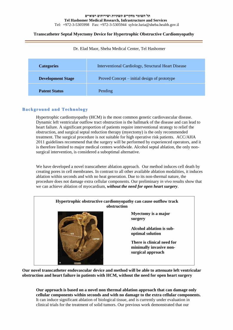
Hypertrophic cardiomyopathy (HCM) is the most common genetic cardiovascular disease.
Dynamic left ventricular outflow tract obstruction is the hallmark of the disease and can lead to
heart failure. A significant proportion of patients require interventional strategy to relief the
obstruction, and surgical septal reduction therapy (myectomy) is the only recommended
treatment. The surgical procedure is not suitable for high operative risk patients. ACC/AHA
2011 guidelines recommend that the surgery will be performed by experienced operators, and it
is therefore limited to major medical centers worldwide. Alcohol septal ablation, the only non-
surgical intervention, is considered a suboptimal alternative.
We have developed a novel transcatheter ablation approach. Our method induces cell death by
creating pores in cell membranes. In contrast to all other available ablation modalities, it induces
ablation within seconds and with no heat generation. Due to its non-thermal nature, the
procedure does not damage extra cellular components. Our preliminary in vivo results show that
we can achieve ablation of myocardium, without the need for open heart surgery.
Hypertrophic obstructive cardiomyopathy can cause outflow track
obstruction
Myectomy is a major
surgery
Alcohol ablation is sub-
optimal solution
There is clinical need for
minimally invasive non-
surgical approach
Our novel transcatheter endovascular device and method will be able to attenuate left ventricular
obstruction and heart failure in patients with HCM, without the need for open heart surgery
Our approach is based on a novel non thermal ablation approach that can damage only
cellular components within seconds and with no damage to the extra cellular components. It can induce significant ablation of biological tissue, and is currently under evaluation in
clinical trials for the treatment of solid tumors. Our previous work demonstrated that our
תל השומר מחקרים תשתיות ושירותים רפואיים
Tel Hashomer Medical Research, Infrastructure and Services Tel: +972-3-5305998 Fax: +972-3-5305944 [email protected]
technology can be delivered in an endovascular approach, and that it can cause significant
ablation of myocardial tissue in the beating heart.
The Need
Approximately 1 in 500 adults have HCM. The disease is associated with normal longevity in
the vast majority of diagnosed patients. However, in up to 70% of the patients HCM is
complicated by outflow tract obstruction that can lead to clinical symptoms. Among those
patients who do develop symptoms, the most common complaints include: chest pain, shortness
of breath with exertion, fatigue, palpitations, and lightheadedness. Some people with
hypertrophic cardiomyopathy have an increased risk of developing a dangerous heart rhythm
(ventricular arrhythmias), which can lead to sudden cardiac death.
Patients with symptomatic obstructive HCM who have failed medical therapy require an
interventional therapy. Current surgical procedures require operator and institutional experience
that are crucial to successful outcomes and low peri-procedural morbidity and mortality. Given
the substantial learning curve associated with invasive surgical procedures for HCM, these
should be performed in centers with adequate procedural volumes to ensure good early and long
term results. Surgical myectomy remains the gold standard by which other procedures need to
be compared to, as the results for myectomy are unmatched for early, long term benefit.
The Advantages
The main advantage of our system and device is the replacement of heart surgery procedure
with a minimal invasive one to achieve the same results with shorter recovery time and
diminished complications and costs. In addition, more patients can benefit from the treatment,
since our transcatheter device therapy is not limited to low operative risk subjects. The
procedure has an extremely short duration (seconds). It affects only the cell membrane and has
the potential of sparing the tissue scaffold. We expect a very short learning curve due to the
simplicity of our transcatheter approach.
Add i t i ona l potent ia l appl i ca t i ons i nc lude :
• Septal ablation for HOCM
• Congenital heart disease
• Tissue scaffolding
• Cardiac arrhythmias
Endovascular drug delivery•
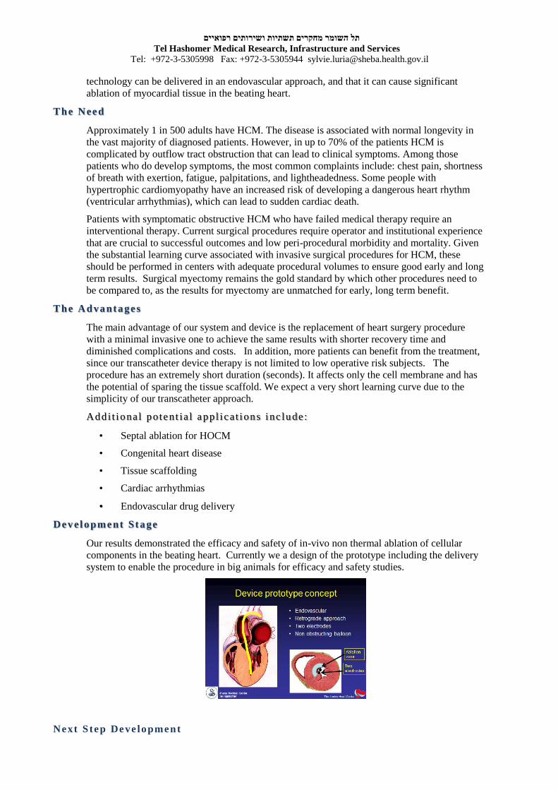
Development Stage
Our results demonstrated the efficacy and safety of in-vivo non thermal ablation of cellular
components in the beating heart. Currently we a design of the prototype including the delivery
system to enable the procedure in big animals for efficacy and safety studies.
Next S tep Deve lopment
תל השומר מחקרים תשתיות ושירותים רפואיים
Tel Hashomer Medical Research, Infrastructure and Services Tel: +972-3-5305998 Fax: +972-3-5305944 [email protected]
4- Acute and Chronic experiment in open heart surgery in pigs to validate to concept.
5- Complete the final Prototype.
6- Chronic Pig experiment for up to 20 weeks.
The Market
Hypertrophic Obstructive Cardiomyopathy:
• The most common cardiac genetic disorder
• Prevalence 1:500 adults (600,000 in the U.S.)
• A major cause of sudden death and heart failure in young people.
• 2.5 Million individual worldwide with HCM
• 10% will benefit intervention
• Estimated price target 5K-10K USD
• 1-2 B USD potential market
Inte l lectual Property
WO2014/195933 - "Myocardial Ablation by Irreversible Electroporation", pending
תל השומר מחקרים תשתיות ושירותים רפואיים
Tel Hashomer Medical Research, Infrastructure and Services Tel: +972-3-5305998 Fax: +972-3-5305944 [email protected]
Transarticular Cannulated fenestrated screw for the treatment of Odontoid (Dens) fracture
Dr. Ran Harel, Sheba Medical center, Tel Hashomer
Categories Medical Device – Neurosurgery, cervical spine injuries
Development Stage Conceptualization
Patent Status Pending
B ackground and Technology
Cervical spine injuries are common in elderly falls, accidents and athletes, particularly those engaged in
contact sports. American football and diving are the sports most often associated with these injuries.
Although most cervical spine injuries have a benign natural history with limited morbidity, catastrophic
spine injuries, along with head injuries, account for 70% of the traumatic deaths and 20% of the permanent
disability related to sports. The first two cervical vertebrae, C1 and C2, are especially vulnerable to injury,
both because they directly support the weight of the skull and because they have the greatest range of motion
to allow the head to move freely in all directions. Spinal cord injury (SCI) in elderly are strong predictors of
mortality in elderly patients with trauma-related cervical spine injury (CSI).
The odontoid process (Dens) is a protuberance of the Axis (second cervical vertebra ). Odontoid fractures
occur as a result of traumatic cervical spine injuries. In younger patients the fracture is a result of motor
vehicle accidents and in older patients as a result of falls. In these patients there is a risk of ongoing damage
to the spinal cord and paralysis. Odontoid fractures are associated with respiratory problems, nonunion and
pain.
The goal of treatment is fracture healing with cervical spine stabilization and fusion. Treatment options
include either external immobilization or surgery.
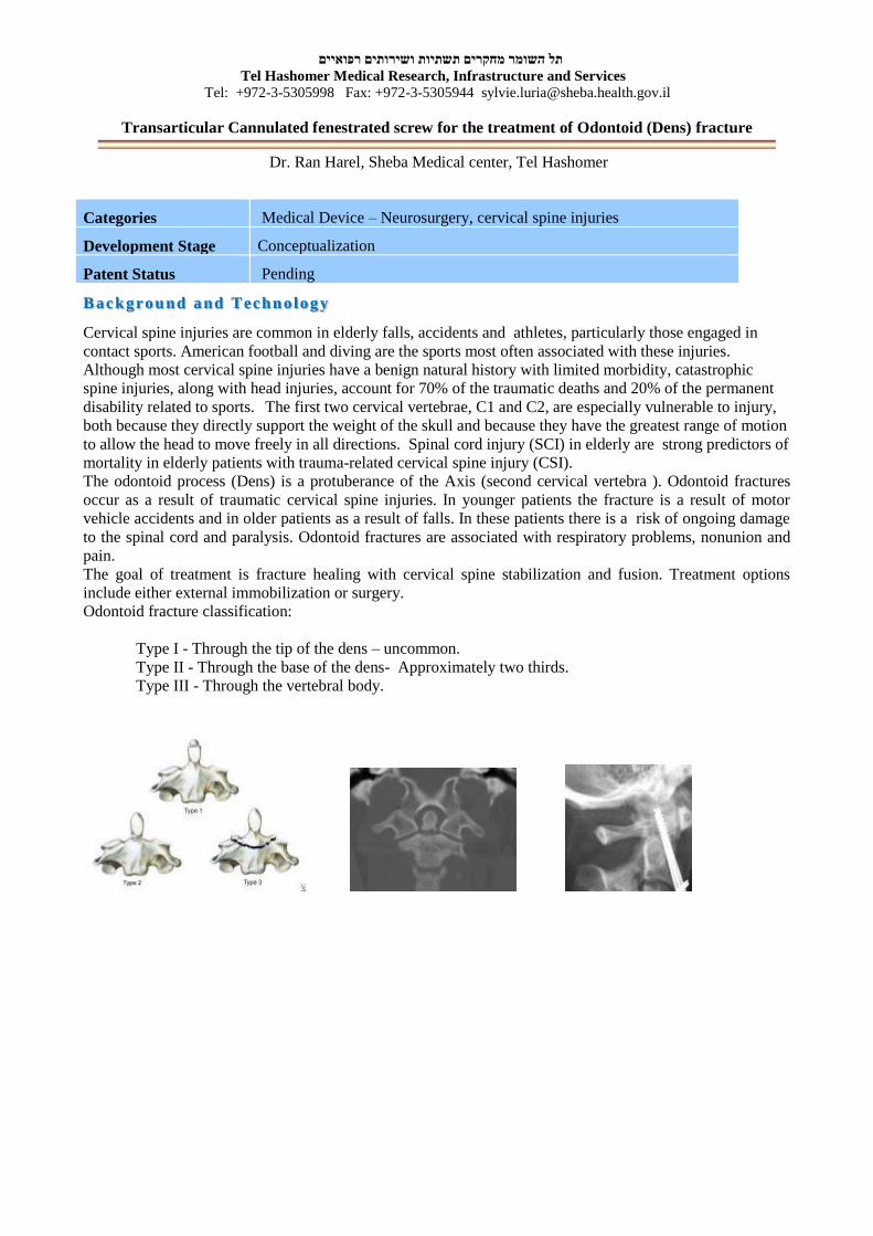
Odontoid fracture classification:
Type I - Through the tip of the dens – uncommon.
Type II - Through the base of the dens- Approximately two thirds.
Type III - Through the vertebral body.
תל השומר מחקרים תשתיות ושירותים רפואיים
Tel Hashomer Medical Research, Infrastructure and Services Tel: +972-3-5305998 Fax: +972-3-5305944 [email protected]
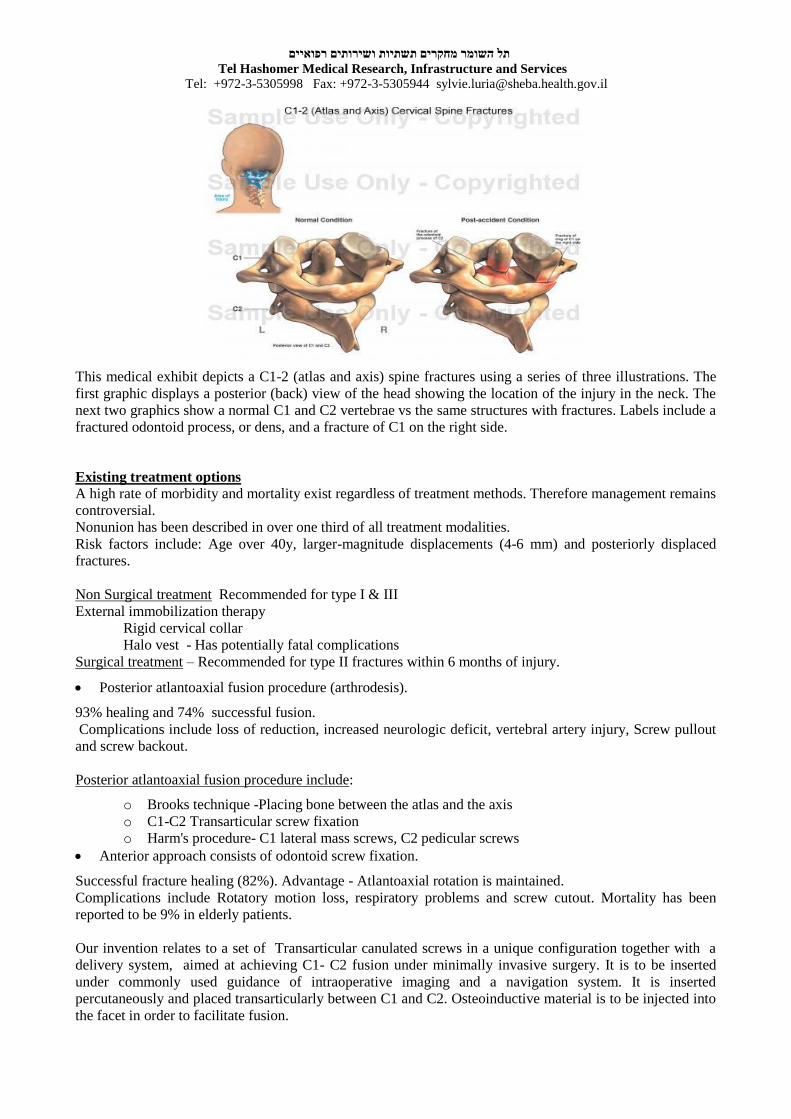
This medical exhibit depicts a C1-2 (atlas and axis) spine fractures using a series of three illustrations. The
first graphic displays a posterior (back) view of the head showing the location of the injury in the neck. The
next two graphics show a normal C1 and C2 vertebrae vs the same structures with fractures. Labels include a
fractured odontoid process, or dens, and a fracture of C1 on the right side.
Existing treatment options
A high rate of morbidity and mortality exist regardless of treatment methods. Therefore management remains
controversial.
Nonunion has been described in over one third of all treatment modalities.
Risk factors include: Age over 40y, larger-magnitude displacements (4-6 mm) and posteriorly displaced
fractures.
Non Surgical treatment Recommended for type I & III
External immobilization therapy
Rigid cervical collar
Halo vest - Has potentially fatal complications
Surgical treatment – Recommended for type II fractures within 6 months of injury.
Posterior atlantoaxial fusion procedure (arthrodesis).
93% healing and 74% successful fusion.
Complications include loss of reduction, increased neurologic deficit, vertebral artery injury, Screw pullout
and screw backout.
Posterior atlantoaxial fusion procedure include:
o Brooks technique -Placing bone between the atlas and the axis
o C1-C2 Transarticular screw fixation
o Harm's procedure- C1 lateral mass screws, C2 pedicular screws
Anterior approach consists of odontoid screw fixation.
Successful fracture healing (82%). Advantage - Atlantoaxial rotation is maintained.
Complications include Rotatory motion loss, respiratory problems and screw cutout. Mortality has been
reported to be 9% in elderly patients.
Our invention relates to a set of Transarticular canulated screws in a unique configuration together with a
delivery system, aimed at achieving C1- C2 fusion under minimally invasive surgery. It is to be inserted
under commonly used guidance of intraoperative imaging and a navigation system. It is inserted
percutaneously and placed transarticularly between C1 and C2. Osteoinductive material is to be injected into
the facet in order to facilitate fusion.
תל השומר מחקרים תשתיות ושירותים רפואיים
Tel Hashomer Medical Research, Infrastructure and Services Tel: +972-3-5305998 Fax: +972-3-5305944 [email protected]
Summary
Odontoid fractures occur as a result of traumatic cervical spine injuries. Patient harbor the risk of
ongoing damage to the spinal cord and paralysis.
The invention regards cannulated screws to perform fixation procedures in the spinal cord. After
implanting the screw in the desired position, therapeutic compositions can be injected through the
cannula to the site of intervention.
The goal of treatment is cervical spine stabilization and fusion. Treatment options are either External
immobilization or surgery. High rates of morbidity and mortality exist regardless of treatment methods.
Therefore management remains controversial.
o External immobilization therapies include Rigid cervical collar and a Halo vest.
o Surgical options include: 1. Posterior approach atlantoaxial fusion (arthrodesis). 2.
Anterior approach odontoid screw fixation.
In Elderly patients comorbidities limit surgical practice. High in-hospital mortality also occur in the
elderly after surgical stabilization.
Cervical fusion surgery involves instrumentation which include metal screws, rods, plates and interbody
fusion devices that are used in an open surgery.
The suggested medical Device is a Transarticular screw with a cannula aimed at achieving C1- C2 fusion
under minimally invasive surgery and injection of osteoinductive material into the facet in order to
facilitate fusion of the odontoid fracture.
The Need
Because of the growing ageing population and degenerative disc disease spinal disorders are the major driver
for spinal surgery. Other disorders include (In the following order) disc herniation, abnormal curvature of
the spine, spondylosis, stenosis, tumors and vertebral fractures . Spine implants are the fastest growing
segment in the orthopedic market. Devices in spinal surgery include minimally invasive spinal fusion
devices, prosthetic discs, nucleus replacement products, bone morphogenic proteins, intradiscal thermal
therapies and kyphoplasty. MIS devices include Interbody cages, pedicle screw systems and spinal plating
systems.
According to the National Spinal Cord Injury Association, as many as 450,000 people in the United States
are living with a spinal cord injury (SCI). According to the Centers for Diseases Control and Prevention
(CDC), SCI costs the nation an estimated $9.7 billion each year. Pressure sores alone, a common secondary
condition among people with SCI, cost an estimated $1.2 billion.
The US spinal surgery device market was valued at $4.6 billion in 2010 with a CAGR of 10%. Medtronic,
Inc. dominates the global market with a 34% share, DePuy, Inc. from Johnson & Johnson shares 13% and
Synthes Inc. 12%. Others companies in the market include Stryker Corporation, NuVasive, Inc., Zimmer,
Aesculap/B. Braun Melsungen, Biomet and Ulrich Medical.
Cervical fusion surgery involves instrumentation which includes metal screws, rods, plates, and interbody
fusion devices. Spine surgeons perform 230,000 cervical fusions in the US annually .
o A Cervical Fusion Surgery survey in 45 California Hospitals in 2008
The annual volume of cervical fusion procedures in hospitals (surgical procedure) ranged from 2 to
169, with an average of 54. Average cervical fusion implant costs in these hospitals ranged from $2,053 to
$14,382, with a mean of $4,868. Total surgical costs for cervical fusion varied from $6,907 to $24,689 with
an average of $13,450. One should consider that above age of 70 years only few percentage of the patients
benefit from the procedure and over 90 % are sent with Rigid cervical collar or Halo vest, with potentially
fatal complications.
http://www.berkeleyhealthtech.org/docs/Vol.2.5.Cervical_Fusion.pdf
תל השומר מחקרים תשתיות ושירותים רפואיים
Tel Hashomer Medical Research, Infrastructure and Services Tel: +972-3-5305998 Fax: +972-3-5305944 [email protected]
Comorbidities occasionally limit the practice of surgical stabilization techniques.
In patients over the of 70, high in-hospital mortality occur after surgery due to poor rehabilitative,
comorbidities and fracture management - 40% with anterior screw fixation, 13% with hard collar
immobilization and 33% with a halo vest.
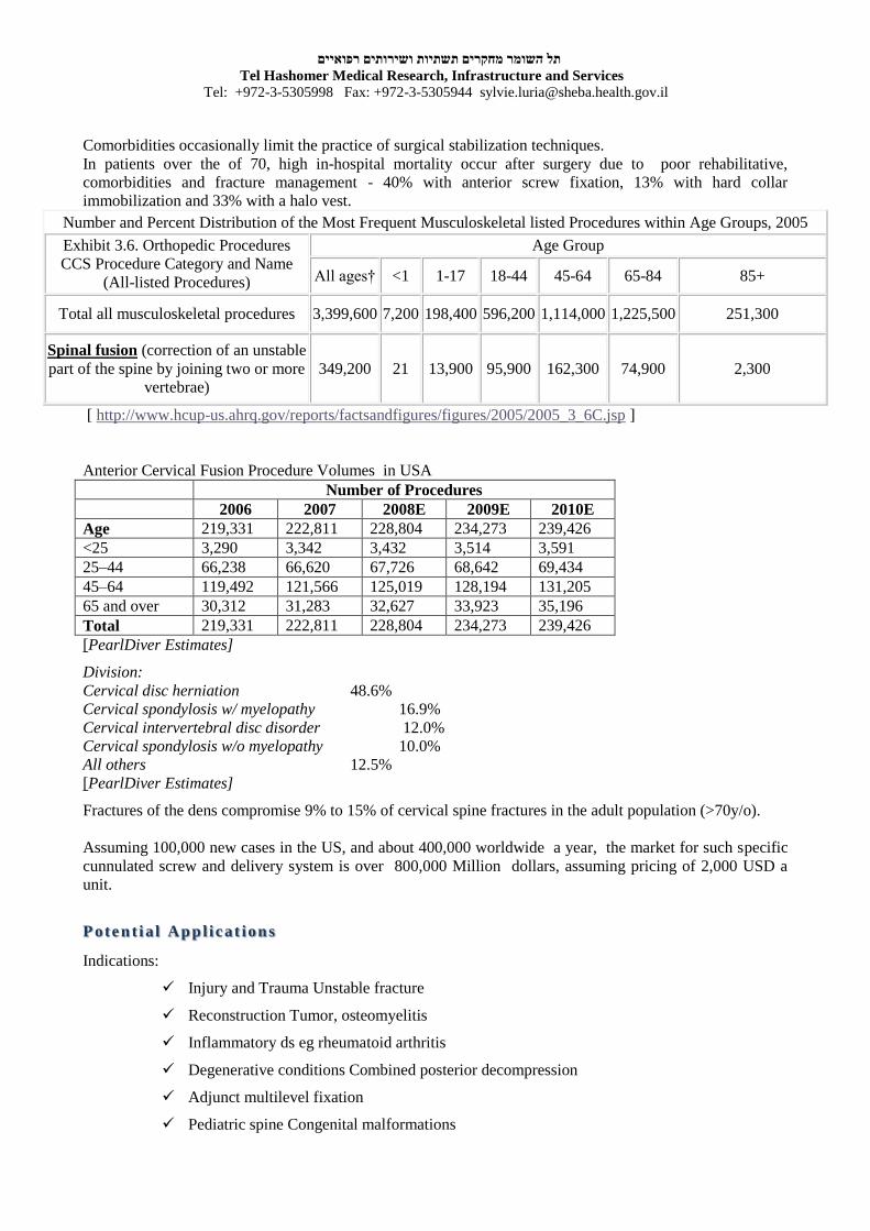
Number and Percent Distribution of the Most Frequent Musculoskeletal listed Procedures within Age Groups, 2005
Exhibit 3.6. Orthopedic Procedures
CCS Procedure Category and Name
(All-listed Procedures)
Age Group
All ages† <1 1-17 18-44 45-64 65-84 85+
Total all musculoskeletal procedures 3,399,600 7,200 198,400 596,200 1,114,000 1,225,500 251,300
Spinal fusion (correction of an unstable
part of the spine by joining two or more
vertebrae)
349,200 21 13,900 95,900 162,300 74,900 2,300
[ http://www.hcup-us.ahrq.gov/reports/factsandfigures/figures/2005/2005_3_6C.jsp ]
Anterior Cervical Fusion Procedure Volumes in USA
Number of Procedures
2006 2007 2008E 2009E 2010E
Age 219,331 222,811 228,804 234,273 239,426
<25 3,290 3,342 3,432 3,514 3,591
25–44 66,238 66,620 67,726 68,642 69,434
45–64 119,492 121,566 125,019 128,194 131,205
65 and over 30,312 31,283 32,627 33,923 35,196
Total 219,331 222,811 228,804 234,273 239,426
[PearlDiver Estimates]
Division:
Cervical disc herniation 48.6%
Cervical spondylosis w/ myelopathy 16.9%
Cervical intervertebral disc disorder 12.0%
Cervical spondylosis w/o myelopathy 10.0%
All others 12.5%
[PearlDiver Estimates]
Fractures of the dens compromise 9% to 15% of cervical spine fractures in the adult population (>70y/o).
Assuming 100,000 new cases in the US, and about 400,000 worldwide a year, the market for such specific
cunnulated screw and delivery system is over 800,000 Million dollars, assuming pricing of 2,000 USD a
unit.
P otent ia l Appl icat ions
Indications:
Injury and Trauma Unstable fracture
Reconstruction Tumor, osteomyelitis
Inflammatory ds eg rheumatoid arthritis
Degenerative conditions Combined posterior decompression
Adjunct multilevel fixation
Pediatric spine Congenital malformations
תל השומר מחקרים תשתיות ושירותים רפואיים
Tel Hashomer Medical Research, Infrastructure and Services Tel: +972-3-5305998 Fax: +972-3-5305944 [email protected]
Advantages
Low cost and Minimal invasive procedure.
Safe procedure
Benefit for the patient life quality
P atent
We have done patent search and found several patents that describe cannulated screw, however no one of
them aim for minimal invasive procedure, no one of the is applicable for the C1/C2 injuries, and no one of
them has the design and features we are aiming.
The following are the patents we have found:
US20110093020A1 Poly-porous hollow screw for target delivery of growth factors and stem cells
describes a poly-porous hollow screw useful as carrier for bioactive materials such as growth factors and
stem cells, for treating few fractures with high incidence of nonunion due to disruption of local circulation.
US20100298836A1 Pressured syringe for the injection of a viscous liquid through a cannulated
surgical screw bone filler adapter discloses the use of a cannulated screw as a delivery means for the
injection of a liquid or gel into a bone void.
US20080177334A1 SCREW AND METHOD OF USE teaches a surgical fixing device for use in bone
wherein the first portion is a cannulated screw having self tapping threads.
EP1898841A2 CANNULATED SCREW ACCESS SYSTEM provides a minimally invasive surgical
system and method for introducing instruments and/or biomaterial into the interior of a bone, particularly the
interior of a vertebral body, using a cannulated screw that is sized and configured to penetrate the cortical
bone and thereby provide access to the interior of the bone through the integral cannula of the screw.
US4760844A Cannulated screw dye injector regards a cannulated screw injector that transfers a material
(such as dye) from syringe to cannulated screw facilitating use of X-ray photography.
WO2005020833A2 SYSTEM AND KIT FOR DELIVERY OF RESTORATIVE MATERIALS Kit for
delivery of a composition into an intraosseous space comprises a cannula, a movable styletinsertable into the
cannula, a catheter having high-porosity tip and a system for delivery of a composition.
There are abundant publications regarding cannulated screws as well as injection of therapeutic compounds
through cannulated screws for orthopedic procedures. However, no art was found anticipating the use of said
devices in Dense fractures in C2, instability between C1 and C2, lumbar spondylolisthesis, instability and
listhesis in S1-L5.
Competing products
There is no device specifically manufactured for Odontoid fracture treatment.
1. DePuy Spine, Inc., A Johnson & Johnson company
1) SKYLINE™ Anterior Cervical Plate System
2) UNIPLATE™, Anterior Cervical Plate System
3) EAGLE™ Plus Anterior Cervical Plate System
2. Zimmer Holdings, Inc.
1) TM-S Trabecular Metal™ Cervical Interbody Fusion Device - A Porous metal biomaterial with
structural and mechanical properties similar to cancellous bone. Trabecular Metal Material
provides an osteoconductive scaffold which supports bony in-growth and vascularization into
the implant. Utilized in cervical interbody fusion.
3. Medtronics

תל השומר מחקרים תשתיות ושירותים רפואיים
Tel Hashomer Medical Research, Infrastructure and Services Tel: +972-3-5305998 Fax: +972-3-5305944 [email protected]
1) PEEK PREVAIL® Cervical Interbody Device. Indicated for anterior cervical interbody fusion
procedures.
2) rhBMP-2 INFUSE® / Medtronics
A biologic compound that stimulates the body to regrow bone and eliminates the need to harvest
bone from another area of the patient’s body.
תל השומר מחקרים תשתיות ושירותים רפואיים
Tel Hashomer Medical Research, Infrastructure and Services Tel: +972-3-5305998 Fax: +972-3-5305944 [email protected]
METHOD AND DEVICES TO ENHANCE SAFETY OF MINIMALLY INVASIVE PROCEDURES IN FIELDS SUCH AS
WOMEN HEALTH, OTOLARYNGOLOGY, PLASTIC SURGERY AND OTHERS.
Dr. Orgad Rosenblat, Sheba Medical Center
Categories Medical device, Women Health
Development Stage Initial designed prototype
Patent Status Pending
B ackground and Technology
Many surgical procedures and especially minimally invasive one are done in a confined and well defined
anatomical space wherein any breach of that space has the potential of causing serious medical
complications.
As a hallmark of such procedures we chose medical curettage, with curettes as a model device.
Curettes are surgical tools typically used for scraping away of debridement or removal of unwanted
diagnostic or therapeutic purposes.
For example, curettes may be used for removing necrotic or infected tissues in plastic surgery, removal of
earwax or adenoids in otolaryngology, or removing neoplastic tissue in orthopedic and general surgery
Curettes are also used in dilation and curettage (D&C) gynecological procedures, wherein the cervix is
dilated and uterine contents and/or its lining are removed. D&Cs are common gynecological procedures and
can be used as both a diagnostic and a therapeutic procedure. D&Cs may be used with patients who
experience instances of abnormal uterine bleeding, with patients who have experienced a missed or
incomplete miscarriage, or to prevent hemorrhage and infection as the result of a retained placenta post birth,
Our invention relates to a novel platform technology enabling the development of variety of medical devices
with enhanced safety and controlled surgical procedures to minimize the risk of applying excess force on to
the tissue involved and consecutively causing damage by perforation or excess scraping.
The Need
In women health alone, curettage is one of the most common procedures and is the second most common
reason for admission after labor
a) Over 650,000 abortions by curettages were performed in 2009 in the US.
b) 44,000,000 abortions per year world wide
c) In the US, 75.9% of abortion are performed by curettage
D & C is a commonly performed procedure, but one that encompasses risk of iatrogenic damage such as
uterine perforation and late complication such as intra uterine adhesions.
Uterine perforation- occurs when one of the surgical instruments makes a hole in and through the uterine
wall. The potential hazards caused by perforation are bleeding from injury to a blood vessel and injury to
other internal organs and subsequent infections.
Intrauterine adhesions — Adhesions (areas of scar tissue) can sometimes form in the uterus following
D&C risk of adhesions might be increased if excess force is used.
In some cases, these adhesions can lead to abnormalities in the menstrual cycle, painful menstrual cycles,
infertility, or miscarriage.
a) Curettage procedure has up to 1% overall risk for perforation
תל השומר מחקרים תשתיות ושירותים רפואיים
Tel Hashomer Medical Research, Infrastructure and Services Tel: +972-3-5305998 Fax: +972-3-5305944 [email protected]
b) Occurs more frequently in women who were recently pregnant and in older women who have gone
through menopause.
c) Most perforations heal on their own but up to 50% of perforation will require diagnostic or curative
surgery
In other fields such as Otolaryngology, plastic surgery, orthopedics, etc. excess force might cause similar
damage and be associated with increased blood loss and tissue damage
The Innovat ion
The proposed medical devices are in essence an expansion or upgrade to existing devices. Newly designed
devices consist of an additional a safety component termed "limiter"
The proposed limiter acts as a barrier between surgeon and tissue and is designed to absorb excess power
from being transferred on to the patient
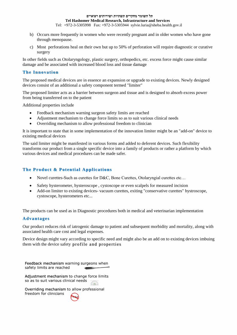
Additional properties include
Feedback mechanism warning surgeon safety limits are reached
Adjustment mechanism to change force limits so as to suit various clinical needs
Overriding mechanism to allow professional freedom to clinician
It is important to state that in some implementation of the innovation limiter might be an "add-on" device to
existing medical devices
The said limiter might be manifested in various forms and added to deferent devices. Such flexibility
transforms our product from a single specific device into a family of products or rather a platform by which
various devices and medical procedures can be made safer.
The Product & P otent ia l Appl icat ions
Novel curettes-Such as curettes for D&C, Bone Curettes, Otolaryngial curettes etc…
Safety hysterometer, hysteroscope , cystoscope or even scalpels for measured incision
Add-on limiter to existing devices- vacuum curettes, exiting "conservative curettes" hystroscope,
cystoscope, hysterometers etc...
The products can be used as in Diagnostic procedures both in medical and veterinarian implementation
Advantages
Our product reduces risk of iatrogenic damage to patient and subsequent morbidity and mortality, along with
associated health care cost and legal expenses.
Device design might vary according to specific need and might also be an add on to existing devices imbuing
them with the device safety p ro f i l e and prope r t i es
תל השומר מחקרים תשתיות ושירותים רפואיים
Tel Hashomer Medical Research, Infrastructure and Services Tel: +972-3-5305998 Fax: +972-3-5305944 [email protected]
Development Stage –des igned s tage
- PendingP atent
The Market
The global gynecological devices market holds prime importance in the overall medical devices market due
to increase in specialized gynecological procedures and rising gynecological conditions with increasing
awareness being the major factors driving the growth of this market. The market is classified into four major
segments and is estimated in terms of USD million, for the period 2012 - 2018, keeping 2011 as the base
year. The gynecological surgical devices market is classified on the basis of the types of endoscopy devices,
endometrial ablation devices, fluid management systems, and female sterilization and contraceptive devices
used by gynecology surgeons.
The global gynecological devices market is expected to reach USD 5.3 Billion by 2018 USD 3.4 compare to
billion in 2011. The market is expected to reach USD 5.3 billion in 2018, growing at a CAGR of 6.4% from
2012 to 2018. In the gynecological devices market, the surgical devices segment is the largest revenue
generator.
The major drivers of this market are growth in acceptance of minimally invasive surgical procedures as a
viable substitute for hysterectomy, increase in the prevalence of gynecological conditions, growth in
women's preference for more innovative and effective gynecological procedures, rise in the global healthcare
expenditure and the aging baby boomer population. On the other hand, lack of capital availability to small
manufacturers and delays in the approval procedures in North America are expected to hold back the growth
of this market.
Key Market Participants
• ACMI Corp.
• American Medical
• Boston Scientific
• CR Bard, Inc.
• Caldera Medical
• Conceptus
• Cook Urological
• Cytyc Corp.
• Ethicon
• Karl Storz
• MDMI Technologies
• Mentor
• Microsulis
• Novasys
• Richard Wolf
• Rochester
• Stryker Corp.
• U.S Surgical/TYCO
• Welch Allyn
תל השומר מחקרים תשתיות ושירותים רפואיים
Tel Hashomer Medical Research, Infrastructure and Services Tel: +972-3-5305998 Fax: +972-3-5305944
Apparatus for guiding medical devices in the gastrointestinal tract
Shomron Ben-Horin, Gastroenterology Department, Chaim Sheba Medical Center, Israel
Background of the Invent ion
GI endoscopy is gaining wide acceptance as a method for visualizing the GI tract. This device comprises
of an ingestible capsule with imaging capabilities, which records images of the intestinal lumen while
moving along it by force of peristalsis. A major limitation is that the endoscopes and capsule
advancement along the GI tract is determined solely by the intestinal motility. This precludes operator-
controlled visualization of particular segments of interest, and makes impossible the obtainment of tissue
samples (biopsies). Another shortcoming of capsule dependency on peristalsis for movement is that in
some individuals slow motility may cause the battery to run-out and imaging to cease, before the capsule
traverses the entire small intestine. This caveat also hampers the ability to design effective colonic
capsules. Similarly, there is often difficulty in advancing endoscope to remote segments of the GI tract,
particularly of the small intestine. One solution is double balloon enteroscopy, but this method requires
significant expertise and time.
The Need
From the above shortcoming it is obvious that there is great need for steerable diagnostic capsules, as well
as for better devices to facilitate conventional endoscopy of the small intestine.
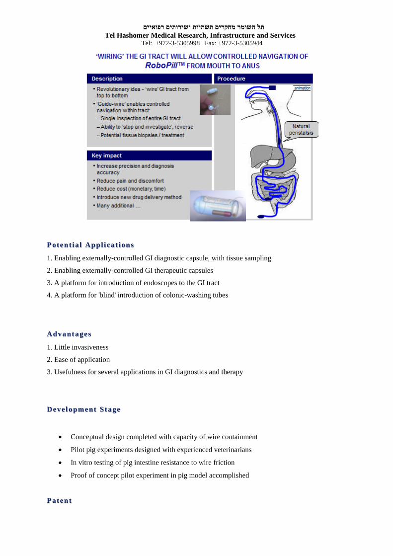
Invent ion summary
We have devised methods and apparatus for external-control of diagnostic/therapeutic capsule movement
in the GI tract, as well as to facilitate the introduction of conventional endoscopes into the alimentary
tract. These comprise of a subject first ingesting a leading capsule containing a spooled wire, whose
proximal end is anchored on the subjects' body. As the leading capsule is propelled along the GI tract by
peristalsis, the wire is played out to be laid along the entire GI tract. Alternatively, the wire is spooled and
anchored to an apparatus mounted on the patient body, and is unspooled by the dragging force of the
ingested leading capsule. Regardless of the specific embodiment, once this wire extends along the GI
tract, it can serve as a guide-wire to facilitate controlled movement of diagnostic capsules by various
embodiments. Alternatively, it can serve as a guide-wire upon which a conventional endoscope can be
introduced to remote segments of the GI tract.
תל השומר מחקרים תשתיות ושירותים רפואיים
Tel Hashomer Medical Research, Infrastructure and Services Tel: +972-3-5305998 Fax: +972-3-5305944
P otent ia l Appl icat ions
1. Enabling externally-controlled GI diagnostic capsule, with tissue sampling
2. Enabling externally-controlled GI therapeutic capsules
3. A platform for introduction of endoscopes to the GI tract
4. A platform for 'blind' introduction of colonic-washing tubes
Advantages
1. Little invasiveness
2. Ease of application
3. Usefulness for several applications in GI diagnostics and therapy
Development Stage
Conceptual design completed with capacity of wire containment
Pilot pig experiments designed with experienced veterinarians
In vitro testing of pig intestine resistance to wire friction
Proof of concept pilot experiment in pig model accomplished
P atent
תל השומר מחקרים תשתיות ושירותים רפואיים
Tel Hashomer Medical Research, Infrastructure and Services Tel: +972-3-5305998 Fax: +972-3-5305944
Apparatus for guiding medical devices in the gastrointestinal tract – patent pending
Tech Transfer Of f icer
Dr. Sylvie Luria
Tel Hashomer Medical Research, Infrastructure and Services
Tel: +972-3-5305998 Fax: +972-3-5305944 ; [email protected]
ENDOTRACHEAL TUBE SECURING DEVICE
Arkadiy Belenkiy , Naharia Medical Center
Categories Tracheal Intubation, Ototracheal Tubes, Holders for Ototracheal Tubes
Development Stage Prototype design
Patent Status Pending
B A C K G R O U N D A N D TE C H N O L O G Y
Intubation and Unplanned extubation (UE) is a life-threatening event, and in recent years has been a focus
of continuous quality improvement (CQI) programs. While CQI programs and research have improved
the care of the intubated patient, relatively little attention has been given to experimental comparisons
between methods for endotracheal tube (ETT) securement. Tracheal tube holders are essential to secure
tube fixation and provides the solution to keep the tube in place and to ensure patient safety.
The more acute Issue is UE, a multi-factorial problem, and one that affects many disciplines, notably
anesthesia, critical care, and pre-hospital and emergency medicine. While intubated patients may spend
less time in Emergency Departments (EDs) than they do in Intensive Care Units (ICUs) or Operating
Rooms (ORs), the ED stay represents a vulnerable period, and UE is a topic worthy of the attention of
Emergency Physicians (EPs).
Endotracheal Tubes (ETTs) are commonly secured using adhesive tape, cloth tape, or commercial
devices. We have used different methods of ETT securement and analysed the rates of failure using
several commercial devices manufactured specifically for securing ETTs including non-adhesive cloth
tape to tie ETTs in place.
UE has a long list of complications of endotracheal intubation. Even assuming proper tube placement and
cuff pressure, there are many potential complications: tissue trauma may affect the ears and scalp (from
passing over them); nasal tissues; lips, oral mucosa, gingivae, pharynx, glottis and subglottic tissues.
Leverage upon the external end of the ETT may traumatize the lower respiratory tract. ETT securement
methods may produce difficulties to keep the diameter of ET, in access for suction of the upper and lower
respiratory tract. Infections in many areas of the respiratory tract are common. There is also the potential
for further traumatizing and fractures.
Among all these potential problems, UE is one which Emergency Physicians and Pre-Hospital providers
must grapple with on unique terms. In Pre-Hospital and ED conditions, patients may not always be
adequately sedated, they may be moved frequently, and physical monitoring may not always be
consistent. Tube dislodgement during Pre-Hospital transport has been noted as an important problem. UE
is a potential disaster, and loss of airway is not the only complication of UE. Other morbidities include
tissue trauma to the face and airway; bronchospasm; aspiration pneumonia; and dysrhythmias.
Methods for securing ETTs can be broadly classified into four groups: First, there is adhesive tape,
applied to the face and head in a variety of ways; Second is cloth tape, tied around the tube and around the
תל השומר מחקרים תשתיות ושירותים רפואיים
Tel Hashomer Medical Research, Infrastructure and Services Tel: +972-3-5305998 Fax: +972-3-5305944
neck and occiput. Third, there are specialized devices, both purpose-built commercial devices, and
devices or arrangements fashioned by clinicians using a variety of hospital products. Finally, there are
securement methods usually reserved for specialized situations such as facial burns, fractures and oral and
maxillofacial surgery.
The first two methods, cloth tape and adhesive tape, are by far the most commonly employed.
Commercial devices are used in only a small percentage of intubated patients. We have analysed several
of them, and came to a conclusion that modifications and improvements are needed for a friendly use to
the patient and to the medical staff.
DEVELOPMENT STAGE
We have design a novel Tracheal tube holders to secure tube fixation and to eliminate potential UE. Our
novel device has flexible features enabling stabilization of the ET, with two anchorage sites and an
anchorage for an orogastric tube, which incorporates an element to secure and keep the diameter of ET
and protect the ET from the patients' bites to ensure effective mouth care, suction procedure and
ventilation.
T H E N E E D
Every year in the United States, millions of patients are being intubated from different reasons during
staying at hospitals. Many times, the use of an endotracheal tube is required for surgeries. The
endotracheal tube needs to be held in place during the surgery and intensive care, to prevent complications
or harm to the patient. The methods currently used to keep the tube in place are straps made from various
materials. These straps function properly in many surgical procedures, but there are times when their use
can cause complications.
Securing the endotracheal tube is a common procedure in the adult and neonatal intensive care unit.
Adequate fixation of the tube is essential to ensure effective ventilation while minimizing any potential
complications secondary to the intervention. Methods used to secure the endotracheal tube often vary
between units and sometimes even between healthcare providers in the same hospital. Complications
varies from minor symptoms to extubation and mortality and may affect several issues, as for example:
Mortality
tube re-taping (number of episodes per patient-days of intubation)
Total or partial lung collapse (number of episodes per patient-days of intubation)
Air leak (e.g., pneumothorax, pulmonary interstitial emphysema)
Subglottic stenosis or post-extubation stridor
Perioral or facial pressure areas and skin trauma
Extension of duration of hospital stay (days)
Extension of ventilation (days and hours, or hours)
Extension of oxygen therapy (days and hours, or hours)
Enhance Incidence of an adverse neurodevelopmental outcome
Long-term dentition problems
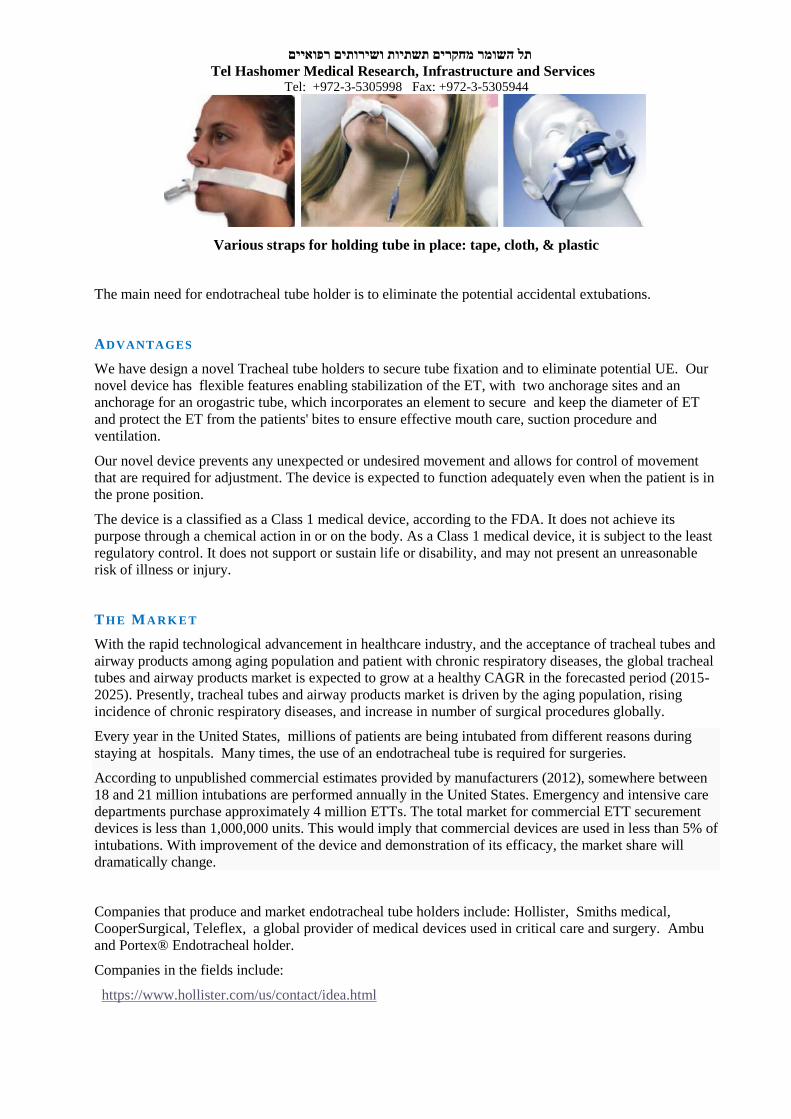
תל השומר מחקרים תשתיות ושירותים רפואיים
Tel Hashomer Medical Research, Infrastructure and Services Tel: +972-3-5305998 Fax: +972-3-5305944
Various straps for holding tube in place: tape, cloth, & plastic
The main need for endotracheal tube holder is to eliminate the potential accidental extubations.
ADVANTAGES
We have design a novel Tracheal tube holders to secure tube fixation and to eliminate potential UE. Our
novel device has flexible features enabling stabilization of the ET, with two anchorage sites and an
anchorage for an orogastric tube, which incorporates an element to secure and keep the diameter of ET
and protect the ET from the patients' bites to ensure effective mouth care, suction procedure and
ventilation.
Our novel device prevents any unexpected or undesired movement and allows for control of movement
that are required for adjustment. The device is expected to function adequately even when the patient is in
the prone position.
The device is a classified as a Class 1 medical device, according to the FDA. It does not achieve its
purpose through a chemical action in or on the body. As a Class 1 medical device, it is subject to the least
regulatory control. It does not support or sustain life or disability, and may not present an unreasonable
risk of illness or injury.
T H E M A R K E T
With the rapid technological advancement in healthcare industry, and the acceptance of tracheal tubes and
airway products among aging population and patient with chronic respiratory diseases, the global tracheal
tubes and airway products market is expected to grow at a healthy CAGR in the forecasted period (2015-
2025). Presently, tracheal tubes and airway products market is driven by the aging population, rising
incidence of chronic respiratory diseases, and increase in number of surgical procedures globally.
Every year in the United States, millions of patients are being intubated from different reasons during
staying at hospitals. Many times, the use of an endotracheal tube is required for surgeries.
According to unpublished commercial estimates provided by manufacturers (2012), somewhere between
18 and 21 million intubations are performed annually in the United States. Emergency and intensive care
departments purchase approximately 4 million ETTs. The total market for commercial ETT securement
devices is less than 1,000,000 units. This would imply that commercial devices are used in less than 5% of
intubations. With improvement of the device and demonstration of its efficacy, the market share will
dramatically change.
Companies that produce and market endotracheal tube holders include: Hollister, Smiths medical,
CooperSurgical, Teleflex, a global provider of medical devices used in critical care and surgery. Ambu
and Portex® Endotracheal holder.
Companies in the fields include:
https://www.hollister.com/us/contact/idea.html

תל השומר מחקרים תשתיות ושירותים רפואיים
Tel Hashomer Medical Research, Infrastructure and Services Tel: +972-3-5305998 Fax: +972-3-5305944
http://www.ambuusa.com/usa/ideas/propose_an_idea.aspx
http://www.smiths-medical.com/customer-support/contact-us.html
http://www.coopersurgical.com/Contact-Us
http://www.teleflex.com/en/usa/contact/index.html
IP S T A T U S
" Endotracheal Tube Holder" – Pending
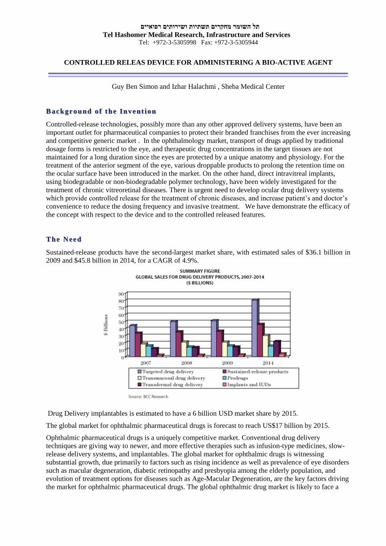
תל השומר מחקרים תשתיות ושירותים רפואיים
Tel Hashomer Medical Research, Infrastructure and Services Tel: +972-3-5305998 Fax: +972-3-5305944
CONTROLLED RELEAS DEVICE FOR ADMINISTERING A BIO-ACTIVE AGENT
Guy Ben Simon and Izhar Halachmi , Sheba Medical Center
Background of the Invent ion
Controlled-release technologies, possibly more than any other approved delivery systems, have been an
important outlet for pharmaceutical companies to protect their branded franchises from the ever increasing
and competitive generic market . In the ophthalmology market, transport of drugs applied by traditional
dosage forms is restricted to the eye, and therapeutic drug concentrations in the target tissues are not
maintained for a long duration since the eyes are protected by a unique anatomy and physiology. For the
treatment of the anterior segment of the eye, various droppable products to prolong the retention time on
the ocular surface have been introduced in the market. On the other hand, direct intravitreal implants,
using biodegradable or non-biodegradable polymer technology, have been widely investigated for the
treatment of chronic vitreoretinal diseases. There is urgent need to develop ocular drug delivery systems
which provide controlled release for the treatment of chronic diseases, and increase patient’s and doctor’s
convenience to reduce the dosing frequency and invasive treatment. We have demonstrate the efficacy of
the concept with respect to the device and to the controlled released features.
The Need
Sustained-release products have the second-largest market share, with estimated sales of $36.1 billion in
2009 and $45.8 billion in 2014, for a CAGR of 4.9%.
Drug Delivery implantables is estimated to have a 6 billion USD market share by 2015.
The global market for ophthalmic pharmaceutical drugs is forecast to reach US$17 billion by 2015.
Ophthalmic pharmaceutical drugs is a uniquely competitive market. Conventional drug delivery
techniques are giving way to newer, and more effective therapies such as infusion-type medicines, slow-
release delivery systems, and implantables. The global market for ophthalmic drugs is witnessing
substantial growth, due primarily to factors such as rising incidence as well as prevalence of eye disorders
such as macular degeneration, diabetic retinopathy and presbyopia among the elderly population, and
evolution of treatment options for diseases such as Age-Macular Degeneration, are the key factors driving
the market for ophthalmic pharmaceutical drugs. The global ophthalmic drug market is likely to face a
תל השומר מחקרים תשתיות ושירותים רפואיים
Tel Hashomer Medical Research, Infrastructure and Services Tel: +972-3-5305998 Fax: +972-3-5305944
patent cliff after 2010 as patents of major products within ocular anti-allergic, anti-glaucoma, as well as
anti-inflammatory/anti-inflective therapies expire.
The global ophthalmic drug market is expected to witness introduction of a range of new therapies
beginning 2010, which include complex biotechnological agents, small-molecule anti-infective and many
more. Besides, these therapies are also expected to capitalize on various novel drug delivery systems that
are a result of significant growth in technology. We have developed a unique miniature device that can be
implanted in the eye for prolong controlled released of various drug in the eye for variety of eye
pathologies.
The Innovat ion and Technology
Our novel approach and method is a cost-effective, versatile passive controlled release device, for
slow release of drugs, hormones and bio-active agents. The device is useful for medical treatment
of humans and animals, as well as for agriculture.
The device comprising a core layer (refer as CL) and outer barrier layer (refer as BL).
The CL comprises three major components:
Porous ceramic or ceramic-polymeric composite. The ceramic and polymer are bio-inert and
selected from groups certified for implantation in humans.
Bio-active agent, loaded inside pores and on surface of ceramic particles.
The BL comprises of noble metal layer that is deposited onto drug-loaded CL, by vacuum deposition,
evaporation, sputtering or by wet-deposition process.
The process of surface activation of CL and metal deposition has been developed and demonstrated in
many prior art publications, such as semiconductor and printed wiring boards.
The release of bio-active ingredients is controlled by perforation of the BL, so as size of holes, and count
of holes, controlling release rate. The perforation process is very accurate and usually provided by laser
drilling. Unlike polymer based controlled released devices, here release rate is constant and very accurate
vs. time.
Bio active agent is loaded in an inert carrier. CL capacity is greater than polymer, because of
internal porosity and surface area. The loading is less sensitive to drug nature (hydrophilic,
hydrophobic...etc)
Wide range of Bio active agents is enabled (ambient temperature process, neutral chemicals)
Release rate is controlled very accurately
Very wide spectrum of release rates
Release rate is not affected by interaction of the device and tissue (swelling of polymers,
hydrolysis...etc)
Very simple and cost-effective process
All materials are non-toxic and medical grade
P otent ia l Appl icat ions
In addition to the ophthalmic applications, the miniature device that can be implanted, placed, or
attached to any organ of a human, animal, plant, tissue culture cells for the slow release of a bio-active
agent for most medical applications.
Advantages
תל השומר מחקרים תשתיות ושירותים רפואיים
Tel Hashomer Medical Research, Infrastructure and Services Tel: +972-3-5305998 Fax: +972-3-5305944
We have developed a miniature device that can be implanted, placed, or attached to the organ of a
human, animal, or plant, then slowly release a bio-active agent. The concepts, using miniature devices for
delivery and slow release of drugs have been used for many years and are under continuous development
around the world. However, most technologies implement various polymers which has a major drawback
– especially when long term release is required: their relatively high permeability. Some polymers, such
as polytetrafluoroethylene (PTFE), are relatively impermeable, but are also difficult to process, have weak
structural integrity, and are soft. Practically, a device constructed from, or encapsulated by, such
polymers may not be useful. Our innovation implement unique metals and organic compounds that
enable manufacturing of small, lightweight devices, that are resistant to puncture, impregnation, etc.; The
materials can be loaded with bio-active agents and are characterized by a slow release rate of the bio-
active agent to the surrounding biological medium abutting the device. Such biological medium includes,
but is not limited to, a body organ such as blood, tissue, fat, liver, muscle, eye fluid, and the like, as well
as animal and plant tissues. The bio-active agent can be released for a long period of time up to several of
months without the need to reload the device with additional bio-active agent.
P atent
Patent application pending: RELEASING DEVICE FOR ADMINISTERING A BIO-ACTIVE
AGENT
Tech Transfer Of f icer
Dr. Sylvie Luria
Tel Hashomer Medical Research, Infrastructure and Services
Tel: +972-3-5305998 Fax: +972-3-5305944 ; [email protected]
תל השומר מחקרים תשתיות ושירותים רפואיים
Tel Hashomer Medical Research, Infrastructure and Services Tel: +972-3-5305998 Fax: +972-3-5305944
SMARTI-Smart iMmobilzation for Advanced Radiation Therapy and Imaging
A controlled platform for assessing and obtaining target lesion immobilization through integrated
use of CPAP, biofeedback and other modalities.
Zvi Symon, Jeff Goldstein and Yaacov Lawrence, Sheba Medical Center
Categories Radiotherapy, Target Lesion Immobilization, Imaging, Medical Device
Development Stage First prototype
Patent Status Pending
BACKGROUND AND TECHNOLOGY
Organ movement is troublesome in many areas of interventional and diagnostic medicine where precision
is vital to success. The concept ‘motion management’ has especially been developed in radiation oncology
in order to avoid missing the target (e.g. a lung tumor) and to minimize radiation exposure of normal
tissues. Currently various strategies of motion management exist (e.g. abdominal compression, gating of
the X ray beam, breath hold) and are employed empirically depending upon physician preference and
availability. No predictive algorithms exist that predict what the optimal motion management should be
employed.
We have developed a novel approach and Smart iMmobilzation device that incorporate multiple inputs
for measuring organ movement / respiratory phase and have the ability to control multiple interventions to
minimize organ movement. The device is an individualized ‘organ-stabilization’ strategy based upon 1) a
preliminary dummy run testing how each individual patient/tumor reacts to each intervention and 2) an
inbuilt algorithm that predicts the impact of respiration based upon tumor location and body habitus.
The multiple interventions that are used to decrease tidal volume include: CPAP, Air Pressure for
delivering CPAP will be under computer control (or other respiratory mode, biofeedback of size of tidal
volume supplementary oxygen, abdominal compression under computer control, breathing control, and
use of pharmaceutical agents .
The device will incorporate number of sensors to probe the depth of respiration, and tumor movement as
well as other parameters: respiratory rate, pulse oximeter, spirometer including pressure volume
measurement, lung compliance and volume, mechanical measure of chest expansion using wearable
sensors a mechanism for distinguishing different patterns of breathing (e.g. thoracic vs abdominal
breathing), blood pressure and pulse, panic button, and ultrasound.
The device will be able to control/trigger LINAC gating (or other device e.g. PET scan). The patient will
have a hand control that will enable manual adjustment of pressure (and measure pulse oximtery),
incorporating a panic button.
A control station can adjust CPAP volume, oxygen concentration, visual feedback and abdominal
pressure in order to help the patient minimize tidal volume. The system includes an algorithm that
optimizes the above interventions based upon real-time feedback , e.g. of chest movement. The algorithm
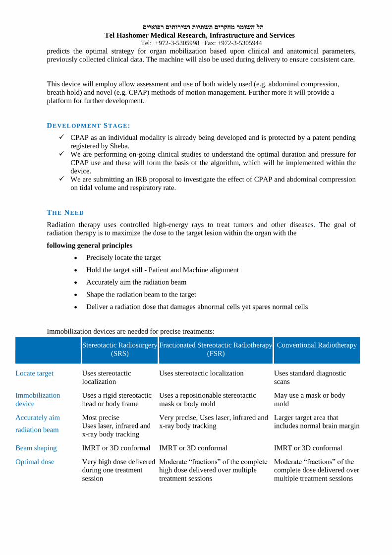
תל השומר מחקרים תשתיות ושירותים רפואיים
Tel Hashomer Medical Research, Infrastructure and Services Tel: +972-3-5305998 Fax: +972-3-5305944
predicts the optimal strategy for organ mobilization based upon clinical and anatomical parameters,
previously collected clinical data. The machine will also be used during delivery to ensure consistent care.
This device will employ allow assessment and use of both widely used (e.g. abdominal compression,
breath hold) and novel (e.g. CPAP) methods of motion management. Further more it will provide a
platform for further development.
DEVELOPMENT STAGE:
CPAP as an individual modality is already being developed and is protected by a patent pending
registered by Sheba.
We are performing on-going clinical studies to understand the optimal duration and pressure for
CPAP use and these will form the basis of the algorithm, which will be implemented within the
device.
We are submitting an IRB proposal to investigate the effect of CPAP and abdominal compression
on tidal volume and respiratory rate.
THE NEED
Radiation therapy uses controlled high-energy rays to treat tumors and other diseases. The goal of
radiation therapy is to maximize the dose to the target lesion within the organ with the
following general principles
Precisely locate the target
Hold the target still - Patient and Machine alignment
Accurately aim the radiation beam
Shape the radiation beam to the target
Deliver a radiation dose that damages abnormal cells yet spares normal cells
Immobilization devices are needed for precise treatments:
Stereotactic Radiosurgery
(SRS)
Fractionated Stereotactic Radiotherapy
(FSR)
Conventional Radiotherapy
Locate target Uses stereotactic
localization
Uses stereotactic localization Uses standard diagnostic
scans
Immobilization
device
Uses a rigid stereotactic
head or body frame
Uses a repositionable stereotactic
mask or body mold
May use a mask or body
mold
Accurately aim
radiation beam
Most precise
Uses laser, infrared and
x-ray body tracking
Very precise, Uses laser, infrared and
x-ray body tracking
Larger target area that
includes normal brain margin
Beam shaping IMRT or 3D conformal IMRT or 3D conformal IMRT or 3D conformal
Optimal dose Very high dose delivered
during one treatment
session
Moderate “fractions” of the complete
high dose delivered over multiple
treatment sessions
Moderate “fractions” of the
complete dose delivered over
multiple treatment sessions
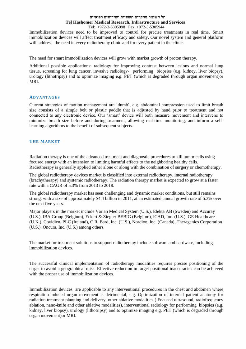
תל השומר מחקרים תשתיות ושירותים רפואיים
Tel Hashomer Medical Research, Infrastructure and Services Tel: +972-3-5305998 Fax: +972-3-5305944
Immobilization devices need to be improved to control for precise treatments in real time. Smart
immobilization devices will affect treatment efficacy and safety. Our novel system and general platform
will address the need in every radiotherapy clinic and for every patient in the clinic.
The need for smart immobilization devices will grow with market growth of proton therapy.
Additional possible applications: radiology for improving contrast between lesions and normal lung
tissue, screening for lung cancer, invasive radiology- performing biopsies (e.g. kidney, liver biopsy),
urology (lithotripsy) and to optimize imaging e.g. PET (which is degraded through organ movement)or
MRI.
ADVANTAGES
Current strategies of motion management are ‘dumb’, e.g. abdominal compression used to limit breath
size consists of a simple belt or plastic paddle that is adjusted by hand prior to treatment and not
connected to any electronic device. Our ‘smart’ device will both measure movement and intervene to
minimize breath size before and during treatment, allowing real-time monitoring, and inform a self-
learning algorithms to the benefit of subsequent subjects.
THE MARK ET
Radiation therapy is one of the advanced treatment and diagnostic procedures to kill tumor cells using
focused energy with an intension to limiting harmful effects to the neighboring healthy cells.
Radiotherapy is generally applied either alone or along with the combination of surgery or chemotherapy.
The global radiotherapy devices market is classified into external radiotherapy, internal radiotherapy
(brachytherapy) and systemic radiotherapy. The radiation therapy market is expected to grow at a faster
rate with a CAGR of 5.3% from 2013 to 2018.
The global radiotherapy market has seen challenging and dynamic market conditions, but still remains
strong, with a size of approximately $4.4 billion in 2011, at an estimated annual growth rate of 5.3% over
the next five years.
Major players in the market include Varian Medical System (U.S.), Elekta AB (Sweden) and Accuray
(U.S.), IBA Group (Belgium), Eckert & Ziegler BEBIG (Belgium), iCAD, Inc. (U.S.), GE Healthcare
(U.K.), Covidien, PLC (Ireland), C.R. Bard, Inc. (U.S.), Nordion, Inc. (Canada), Theragenics Corporation
(U.S.), Oncura, Inc. (U.S.) among others.
The market for treatment solutions to support radiotherapy include software and hardware, including
immobilization devices.
The successful clinical implementation of radiotherapy modalities requires precise positioning of the
target to avoid a geographical miss. Effective reduction in target positional inaccuracies can be achieved
with the proper use of immobilization devices.
Immobilization devices are applicable to any interventional procedures in the chest and abdomen where
respiration-induced organ movement is detrimental, e.g. Optimization of internal patient anatomy for
radiation treatment planning and delivery, other ablative modalities ( Focused ultrasound, radiofrequency
ablation, nano-knife and other ablative modalities), interventional radiology for performing biopsies (e.g.
kidney, liver biopsy), urology (lithotripsy) and to optimize imaging e.g. PET (which is degraded through
organ movement)or MRI.

תל השומר מחקרים תשתיות ושירותים רפואיים
Tel Hashomer Medical Research, Infrastructure and Services Tel: +972-3-5305998 Fax: +972-3-5305944
The immobilization device market includes stereotactic frame, Talon system, thermoplastic molds, Alpha
Cradles, and Vac-Lok system. Main players are VARIAN, Elekta, Orfit Industries, Bionix Radiation
Therapy , Kobold , Qfix and others. The overall market reaches over 430 M USD during 2013. The
driving force of this market will be precise treatment and proton therapy using immobilization with real
time controlled operation systems.
FUTURE OUTLOOK
New therapeutic approaches to disease are increasingly minimally invasive and non-operative. All require
precision, and if located within the chest or abdomen, they are negatively affected by respiratory motion.
Hence there is a growing need for “organ stabilization” techniques.
IP STATUS- NEW APPLICATION , PENDING
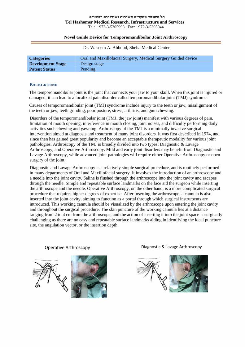
תל השומר מחקרים תשתיות ושירותים רפואיים
Tel Hashomer Medical Research, Infrastructure and Services Tel: +972-3-5305998 Fax: +972-3-5305944
Novel Guide Device for Temporomandibular Joint Arthroscopy
Dr. Waseem A. Abboud, Sheba Medical Center
Categories Oral and Maxillofacial Surgery, Medical Surgery Guided device
Development Stage Design stage
Patent Status Pending
BACKGROUND
The temporomandibular joint is the joint that connects your jaw to your skull. When this joint is injured or
damaged, it can lead to a localized pain disorder called temporomandibular joint (TMJ) syndrome.
Causes of temporomandibular joint (TMJ) syndrome include injury to the teeth or jaw, misalignment of
the teeth or jaw, teeth grinding, poor posture, stress, arthritis, and gum chewing.
Disorders of the temporomandibular joint (TMJ, the jaw joint) manifest with various degrees of pain,
limitation of mouth opening, interference in mouth closing, joint noises, and difficulty performing daily
activities such chewing and yawning. Arthroscopy of the TMJ is a minimally invasive surgical
intervention aimed at diagnosis and treatment of many joint disorders. It was first described in 1974, and
since then has gained great popularity and become an acceptable therapeutic modality for various joint
pathologies. Arthroscopy of the TMJ is broadly divided into two types; Diagnostic & Lavage
Arthroscopy, and Operative Arthroscopy. Mild and early joint disorders may benefit from Diagnostic and
Lavage Arthroscopy, while advanced joint pathologies will require either Operative Arthroscopy or open
surgery of the joint.
Diagnostic and Lavage Arthroscopy is a relatively simple surgical procedure, and is routinely performed
in many departments of Oral and Maxillofacial surgery. It involves the introduction of an arthroscope and
a needle into the joint cavity. Saline is flushed through the arthroscope into the joint cavity and escapes
through the needle. Simple and repeatable surface landmarks on the face aid the surgeon while inserting
the arthroscope and the needle. Operative Arthroscopy, on the other hand, is a more complicated surgical
procedure that requires higher degrees of expertise. After inserting the arthroscope, a cannula is also
inserted into the joint cavity, aiming to function as a portal through which surgical instruments are
introduced. This working cannula should be visualized by the arthroscope upon entering the joint cavity
and throughout the surgical procedure. The skin puncture of the working cannula lies at a distance
ranging from 2 to 4 cm from the arthroscope, and the action of inserting it into the joint space is surgically
challenging as there are no easy and repeatable surface landmarks aiding in identifying the ideal puncture
site, the angulation vector, or the insertion depth.
Diagnostic & Lavage Arthroscopy Operative Arthroscopy
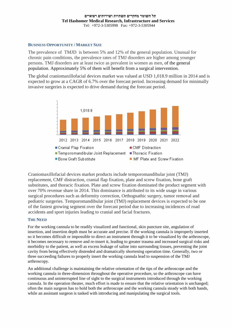
תל השומר מחקרים תשתיות ושירותים רפואיים
Tel Hashomer Medical Research, Infrastructure and Services Tel: +972-3-5305998 Fax: +972-3-5305944
BUSINESS OPPORTUNITY / MARKET SIZE
The prevalence of TMJD is between 5% and 12% of the general population. Unusual for
chronic pain conditions, the prevalence rates of TMJ disorders are higher among younger
persons. TMJ disorders are at least twice as prevalent in women as men, of the general
population. Approximately 5% of them will benefit from a surgical intervention.
The global craniomaxillofacial devices market was valued at USD 1,018.9 million in 2014 and is
expected to grow at a CAGR of 6.7% over the forecast period. Increasing demand for minimally
invasive surgeries is expected to drive demand during the forecast period.
Craniomaxillofacial devices market products include temporomandibular joint (TMJ)
replacement, CMF distraction, cranial flap fixation, plate and screw fixation, bone graft
substitutes, and thoracic fixation. Plate and screw fixation dominated the product segment with
over 70% revenue share in 2014. This dominance is attributed to its wide usage in various
surgical procedures such as deformity correction, Orthognathic surgery, tumor removal and
pediatric surgeries. Temporomandibular joint (TMJ) replacement devices is expected to be one
of the fastest growing segment over the forecast period due to increasing incidences of road
accidents and sport injuries leading to cranial and facial fractures.
THE NEED
For the working cannula to be readily visualized and functional, skin puncture site, angulation of
insertion, and insertion depth must be accurate and precise. If the working cannula is improperly inserted
so it becomes difficult or impossible to direct an instrument through it to be visualized by the arthroscope,
it becomes necessary to remove and re-insert it, leading to greater trauma and increased surgical risks and
morbidity to the patient, as well as excess leakage of saline into surrounding tissues, preventing the joint
cavity from being effectively distended and dramatically shortening operation time. Generally, two or
three succeeding failures to properly insert the working cannula lead to suspension of the TMJ
arthroscopy.
An additional challenge is maintaining the relative orientation of the tips of the arthroscope and the
working cannula in three-dimension throughout the operative procedure, so the arthroscope can have
continuous and uninterrupted line of sight to the surgical instruments introduced through the working
cannula. In the operation theater, much effort is made to ensure that the relative orientation is unchanged;
often the main surgeon has to hold both the arthroscope and the working cannula steady with both hands,
while an assistant surgeon is tasked with introducing and manipulating the surgical tools.
תל השומר מחקרים תשתיות ושירותים רפואיים
Tel Hashomer Medical Research, Infrastructure and Services Tel: +972-3-5305998 Fax: +972-3-5305944
TECHNOLOGY AND INNOVATION
A guide device that enables accurate insertion of the working cannula relative to the arthroscope, and
maintains this relation throughout the procedure. The device in essence is a rod that has the same length
and is parallel to the arthroscope, and mounted on it. After proper insertion of the working cannula by the
guide and visualizing it by the arthroscope, the working cannula is locked in this fixed orientation relative
to the arthroscope, allowing the surgeon to hold the arthroscope and working cannula with one hand using
the guide device, and performing the surgical instrumentation with the other hand. The guide device has
multiple joints and can be configured to various positions, all of which maintain the relative orientation
between the arthroscope and the working cannula.
ADVANTAGES
Facilitating proper insertion of the working cannula into the joint cavity with one single attempt, and
fixating the relation of the arthroscope and the working cannula steady throughout the procedure
achieves the following goals:
Minimal violation of joint capsule → improved hydro-distention of joint cavity → improved vision
and easier surgical instrumentation.
Minimal puncturing of skin → decreased surgical risks associated with facial nerve injury, ear injury,
and scarring of skin.
Minimal sweeping in joint cavity looking for the working cannula → prevents scuffing of joint lining
and iatrogenic damage to intra-articular tissues.
More simple, predictable, and easy operation → enables the novice arthroscopist of performing
Operative Arthroscopy.
More simple, predictable, and easy operation → allows patients with various TMJ disorders to undergo
minimally invasive arthroscopic intervention instead of open surgery of the joint.
Fixating the arthroscope and working cannula in 3D → the main surgeon can perform the surgical
instrumentation instead of merely stabilizing the relation between the cannula and arthroscope
throughout the operation.
Decreased morbidity for the patient in terms of pain, swelling, and hospitalization time.
Less operative time in the surgical theatre.
More predictable procedure.
IP - Pending


