Modeling and Simulation of Soft Tissue Deformation
Yuping Duan1, Weimin Huang1, Huibin Chang2, Wenyu Chen1, Kyaw Kyar Toe1,Jiayin Zhou1, Tao Yang1, Jiang Liu1, Soo Kng Teo3, Chi Wan Lim3, Yi Su3, Chee
Kong Chui4, and Stephen Chang5
1 Institute for Infocomm Research, A*STAR, 1 Fusionopolis Way, Singapore, 1386322 Department of Mathematical Sciences, Tianjin Normal University, 241 Weijin Rd, Tianjin,
China, 3003873 Institute of High Performance Computing, A*STAR, 1 Fusionopolis Way, Singapore, 138632
4 Department of Mechanical Engineering, National University of Singapore, 21 Lower KentRidge Rd, Singapore, 119077
5 National University Hospital, 5 Lower Kent Ridge Rd, Singapore, 119074
Abstract. A stable and accurate deformable model to simulate the deformationof soft tissues is a challenging area of research. This paper describes a soft tissuesimulation method that can deform multiple organs synchronously and interactwith virtual surgical instruments accurately. The model we used in our methodis a multi-organ system by point masses and springs. The organs that anatomi-cally connect to each other are jointed together by high stiffness springs. Herewe propose a volume preserved mass-spring model for simulation of soft organdeformation. It does not rely on any direct constraint on the volume of tetrahe-drons, but rather two constraints on the length of springs and the third constrainton the direction of springs. To provide reliable interaction between the soft tis-sues and kinematic instruments we incorporate the position-based attachment toaccurately move the soft tissue with the tools. Experiments have been designedfor evaluation of our method on porcine organs. Using a pair of freshly harvestedporcine liver and gallbladder, the real organ deformation is CT scanned as groundtruth for evaluation. Compared to the porcine model, our model achieves a meanabsolute error 1.5024 mm on landmarks with a overall surface error 1.2905 mmfor a small deformation (the deformation of the hanging point is 49.1091 mm)and a mean absolute error 2.9317 mm on landmarks with a overall surface er-ror 2.6400 mm for a large deformation (the deformation of the hanging pointis 83.1376 mm). The change of volume for the two deformations are limited to0.22% and 0.59%, respectively. Finally, we show that the proposed model is ableto simulate the large deformation of the liver and gallbladder system in real-timecalculations.
Keywords: Physically based modeling, mass-spring, time integration, surgerysimulation, volume preservation
1 IntroductionWith the development of laparoscopic techniques, surgery simulation becomes an in-creasingly relevant alternative to traditional training methods. Elastic deformation mod-els have been greatly studied in the last two decades for the surgery simulation. Nealenet al. [10] gave a good overview of the deformable models used in computer graph-ics. Generally speaking, the vast number of techniques used for soft tissue deformation
modeling can be classified into two different categories: the heuristic models such asMass-Spring Model (MSM) and continuum-mechanical approach such as the Finite El-ement Method (FEM). The FEMs are based on a physical model of deformation andable to accurately compute complex deformations of soft tissues. Compared to a linearFEM, which is numerically fast but not well suited to moderate deformations and rota-tions, a nonlinear FEM has advantage that it is more reliable to rotations and large de-formations. However, it lead to a requirement of both high computational cost and largememory usage in simulation. To achieve real-time deformation, non-linear FEMs relyon either the pre-computation or GPU-based accelerations. On the other hand, MSMs,which are robust to topology changes and large deformations, are also widely used tomodel deformable objets [11] due to the ability in generating dynamic behaviors in realtime.
A MSM is a discrete model, which consists of a set of point masses connectedby ideal weightless elastic springs. To model a solid 3D object, a tetrahedral meshmodeling both the surface and internal structure of the object can be constructed usingthe MSM. Suppose a MSM is composed of n point masses xi ∈ R3, i = 1, . . . , n withmass mi ∈ R and the forces acting on the point is fi ∈ R3. The geometric state ofall points is simply x ∈ R3n, f ∈ R3n and m ∈ R3n×3n, respectively. The relationbetween the acceleration and force can be described by Newton’s Law of motion asfollows:
mx = f , (1)
where x is the second derivative of the position with respect to time.
In this work, we present a soft tissue simulator that uses a fast tetrahedral massspring model to calculate soft tissue deformation, where the model parameters are se-lected according to soft tissue properties. The position based interaction with kinematicvirtual surgical tools is applied to achieve accurate attachment. The deformation of ourmulti-organ system is realized according to (1) by accumulating external and internalforces on point masses. We model the connecting tissues between soft organs as springswith high stiffness, the so-called repulsive springs. With the repulsive springs, no col-lision detection is required for the synchronous deformation illustrated by the liver andgallbladder. Volume preservation is important for realistic modeling of soft yet solid tis-sues. Lasseter [5] states, “The most important rule to squash and stretch is that, no mat-ter how squashed or stretched out a particular object gets, its volume remains constant.”To reach this point, two constraints on the length of springs and the third constrainton the direction of springs are constructed to serve as post processing to the MSM.The constraints introduce extra non-linearity to the conventional MSM. Unlike priorworks [1, 3], in which the volume of tetrahedrons is investigated in computation, ourconstraints act on mass points instead of tetrahedrons. The proposed volume preservedMSM is validated on the real deformation of a porcine liver with gallbladder. The CTscanned deformation is compared with the computational deformation of MSM. Thecomparison results demonstrate the accurate performance of our method. Finally, weachieve a real-time dynamic system with reasonable accuracy of organ deformationand interaction with a kinematic virtual tool for a simplified mesh model.
2 Volume Preserved MSMThe deformation is estimated based on Verlet integration according to total forces. Wediscuss different forces in our system, which contain both the external forces (pullingattachment and gravitation) and the internal forces (spring forces, damping forces andcontact forces). Novel constraints on point masses are incorporated into the system torealize the volume conservation for soft tissues.
2.1 Deformation EstimationFor the numerical simulation, we first separate Equation (1) into two coupled first orderequations by introducing the velocity v ∈ R3 as follows{
v = f(t)/m,x = v.
(2)
We use Verlet integration, which is among the simplest and most popular explicitschemes in real-time applications to solve (2). The basic idea is to keep the position atprevious time t−∆t and use this information to obtain a more accurate prediction fort + ∆t. Taylor expansion of the position in the two time directions yields
x(t + ∆t) = x(t) + x(t)∆t +12x(t)∆t2 +
16
...x(t)∆t3 + O(∆t4),
x(t−∆t) = x(t)− x(t)∆t +12x(t)∆t2 − 1
6...x(t)∆t3 + O(∆t4).
By adding the above two equations and bringing in (1) and ignoring the high orderterms, we have the so-called Verlet integration scheme for the MSM as follows{
x(t + ∆t) = x(t) + v(t)∆t + f(t)∆t2/m,
v(t + ∆t) = (x(t + ∆t)− x(t))/∆t.(3)
2.2 Forces Modeling
Attachment In order to accurately move vertices of soft tissues along with the inter-acted kinematic instruments, we use the position-based attachment [9]. The positionof selected vertices are updated at every time step to coincide with the motion of thekinematic instrument. Suppose the initial and objective position of mass point xi areP0(xi) and P (xi) and the attachment is done in n iteration, the movement of the pointxi in each iteration is (P (xi)− P0(xi))/n.
Gravitation The force of gravity is acting on every point mass in the system and writtenas fg(xi) = mig, where g is the gravitational acceleration.
Spring Forces Springs are modeled with linear elasticity. The force acting on mass igenerated by the spring connecting i and j is in direct proportion with the extension ofthe spring. Therefore, according to Hooke’s Law, the spring force is defined as follows
fsi = k(i,j)(‖xj − xi‖ − l0) ·
xj − xi
‖xj − xi‖, (4)
where k(i,j) is the spring stiffness and l0 is the rest length of spring (i, j).
Damping Forces Due to imperfect elasticity of physical bodies, energy dissipationoccurs during the deformation. We use spring damping to represent the viscous force.These damping forces are defined as
fdi = d(i,j)
(vj − vi) · (xj − xi)‖xj − xi‖
· xj − xi
‖xj − xi‖, (5)
where d(i,j) is the spring’s damping constant of spring (i, j).
Contact Forces The gallbladder is connected to the lower surface of the liver at thegallbladder fossa by connecting tissues. Therefore, when external forces are applied tothe liver/gallbladder, there exist contact forces in the contact surface of the liver andgallbladder. We generate repulsive springs in the contact surface of the liver and gall-bladder to model contact forces. The contact forces are the forces combined of springforces and damping forces from repulsive springs.
2.3 Deformable ConstraintsWe obtain both the position x and velocity v after Verlet integration. Next, we introduceconstraints on the position as the post-processing process to regularize the simulationresults. These constraints introduce extra non-linearity to the conventional MSM.
Spring Length Correction It is well-known when a concentration of large forces oc-curs in a small region of soft tissues, the simulation result of a MSM falls into the prob-lem of local deformation (“super-elastic” effects) [13]. We design a pair of constraintson the spring length based on the deformation rate, which is defined as τ = (l− l0)/l0.To identify the influence of the stretch and compression in the deformation, differentdeformation rates are used for the over-stretching compensation and over-compressioncompensation.
Over-stretching Compensation. We set a critical stretching rate τs to the springs toprotect the spring from being stretched too much. More specifically, when the length ofthe spring exceeds (1 + τs)× l0, the constraint is applied to try to push the spring backto (1 + τs) × l0. Therefore, we define the over-stretching correction as the followinginequality constraint:
Cstretch(xi,xj) = (1 + τs)× l0 − ‖xi − xj‖ ≥ 0, (6)
If the above inequality constraint is not satisfied, we compute the corrections on thepoint xi and xj along the gradient of Cstretch. The formulae of the correction term∆xi and ∆xj are given as follows:
∆xi =12× Cstretch(xi,xj) ·
xi − xj
‖xi − xj‖,
∆xj = −12× Cstretch(xi,xj) ·
xi − xj
‖xi − xj‖.
After the implementation of over-stretching constraint, ∀xi, i = 1, . . . , n, we sum upall the corrections ∆xi contributed by the edges containing the point xi, namely
xi = xi +1m
∑Ei
∆xi, (7)
where Ei denotes the set of edges containing the point xi and m is the total number ofedges in Ei.
Over-compressing Compensation. On the other hand, as long as the length of thespring is less than (1 − τc) × l0, τc is the critical compressing rate, we use anotherconstraint to push the spring back to (1− τc)× l0. The constraint for over-compressingsprings is defined as
Ccompress(xi,xj) = ‖xi − xj‖ − (1− τc)× l0 ≥ 0, (8)
The method to compute the update from (8) and the following direction constraint issimilar to over-stretching constraint by computing the gradient of the constraint.
Spring Direction Correction During the simulation, sudden change of the spring di-rection may cause serious problem, such as instability and self-collision. Therefore, wedefine another constraint on the spring direction to guarantee that the direction of thespring is within a critical rotation angle θ in certain iterations. More specifically, wedefine the constraint on the spring direction as follows
Cdirection(xi,xj) = θ − arccos( (xj − xi) · (xp
j − xpi )
‖xj − xi‖‖xpj − xp
i ‖
)≥ 0, (9)
where xpi denote the previous state of the system, whose initial value is the initial posi-
tion of meshes and is updated every certain number of iterations to the current position.
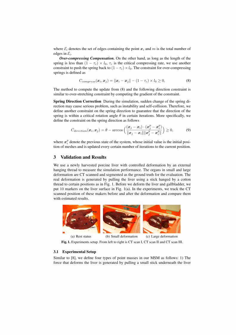
3 Validation and ResultsWe use a newly harvested porcine liver with controlled deformation by an externalhanging thread to measure the simulation performance. The organs in small and largedeformation are CT scanned and segmented as the ground truth for the evaluation. Thereal deformation is generated by pulling the liver using a stick hanged by a cottonthread to certain positions as in Fig. 1. Before we deform the liver and gallbladder, weput 10 markers on the liver surface in Fig. 1(a). In the experiments, we track the CTscanned position of these makers before and after the deformation and compare themwith estimated results.
(a) Rest status (b) Small deformation (c) Large deformation
Fig. 1. Experiments setup. From left to right is CT scan I, CT scan II and CT scan III.
3.1 Experimental SetupSimilar to [8], we define four types of point masses in our MSM as follows: 1) Theforce that deforms the liver is generated by pulling a small stick underneath the liver
Lobe 1 through a cotton thread (Fig. 1(b)). To mimic the pulling force in the simulation,attachment points along the stick and the thread are manually selected and the positionof these points are described based on the tool position in the experiment. 2) We observethat there is nearly no deformation of Marker 8-10 and the other lobe (Lobe 2) for bothdeformations. For simplicity, all point masses in the posterior and Lobe 2 of the liverthat touch to the ground surface are regarded as fixed points during the simulation. 3)To generate the repulsive springs between the liver and gallbladder, we select the pointson the gallbladder that are in contact with the liver and then find the joint points on theliver by the smallest distance. 4) All other liver and gallbladder points are free points,the movement of which are completely determined by the resulting forces acting onthem.
Point Masses Assume the mass of a tetrahedron is equally divided among its vertices.The mass mi of mass point i is estimated as:
mi =∑∀j∈Ti
14ρVj , (10)
where Ti is the union of all tetrahedrons that contain point i, Vj is the volume of tetra-hedron j and ρ is the tissue density. In our experiments, the mass density of the liver is1060 kg/m3 [2] and the mass density of the bile (gallbladder) is 1000 kg/m3 [6].
Spring Stiffness The parametrization of spring stiffness provides certain stress-stainrelationship of soft tissues. In [7], a formula was established to compute spring stiffnessfor a regular tetrahedron with unique spring length based on an isotropic elastic materialwith Young’s modulus E. For our irregular tetrahedral formulation, we calculate anequivalent edge length le from the volume for each tetrahedron element e, i.e., le =(Ve
12√2)
13 . According to [7], we compute spring stiffness for body springs from:
k(i,j) =∑
e∈T(i,j)
2√
225
leE, (11)
where T(i,j) is the set of tetrahedrons that contain the edge (i, j). For our experiments,the Young’s modulus for the liver and gallbladder are E = 3.5 kPa [12] and E =1.5 kPa [6], respectively. On the other hand, we use very stiff spring parameters forrepulsive springs due to their function in preventing collision.
Spring Damping In [11], the authors have given the formula to calculate the dampingconstant to ensure the best behavior consistency for different and combined resolution.For the spring connecting point mass mi and mj with initial length l0, we use thefollowing formula to compute the damping constant:
d(i,j) =2√
k(i,j)(mi + mj)l0
. (12)
3.2 Evaluation Criteria
To compare the simulation results with the porcine model (PM), the absolute error ofthe markers in estimated meshes by the MSM is computed as follows:
εi = ‖xPMi − xMSM
i ‖. (13)
In (13), xi denotes the position of the ith node.
3.3 Model Evaluation
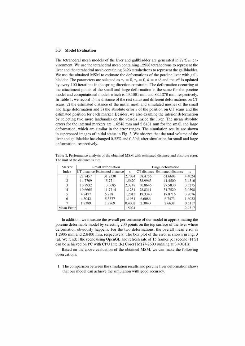
The tetrahedral mesh models of the liver and gallbladder are generated in TetGen en-vironment. We use the tetrahedral mesh containing 12958 tetrahedrons to represent theliver and the tetrahedral mesh containing 5423 tetrahedrons to represent the gallbladder.We use the obtained MSM to estimate the deformations of the porcine liver with gall-bladder. The parameters are selected as τs = 0, τc = 0, θ = π/3 and the xp is updatedby every 100 iterations in the spring direction constraint. The deformation occurring atthe attachment points of the small and large deformation is the same for the porcinemodel and computational model, which is 49.1091 mm and 83.1376 mm, respectively.In Table 1, we record 1) the distance of the rest status and different deformations on CTscans, 2) the estimated distance of the initial mesh and simulated meshes of the smalland large deformation and 3) the absolute error ε of the position on CT scans and theestimated position for each marker. Besides, we also examine the interior deformationby selecting two more landmarks on the vessels inside the liver. The mean absoluteerrors for the internal markers are 1.6245 mm and 2.6431 mm for the small and largedeformation, which are similar in the error ranges. The simulation results are shownin superposed images of initial status in Fig. 2. We observe that the total volume of theliver and gallbladder has changed 0.22% and 0.59% after simulation for small and largedeformation, respectively.
Table 1. Performance analysis of the obtained MSM with estimated distance and absolute error.The unit of the distance is mm.
Marker Small deformation Large deformationIndex CT distance Estimated distance εi CT distance Estimated distance εi
1 28.7457 31.2330 2.7084 58.4756 61.6608 4.40242 14.7709 15.7711 1.5620 38.9963 41.4500 3.43103 10.7932 13.0685 2.3248 30.8646 27.5830 3.52754 10.6665 11.7714 1.1251 28.8311 31.7520 3.03985 4.9477 5.7381 1.2013 19.3340 17.8716 3.90766 4.3042 5.3377 1.1951 6.6086 6.7473 1.60227 1.8389 1.8769 0.4002 2.3040 2.6638 0.6117
Mean Error – – 1.5024 – – 2.9317
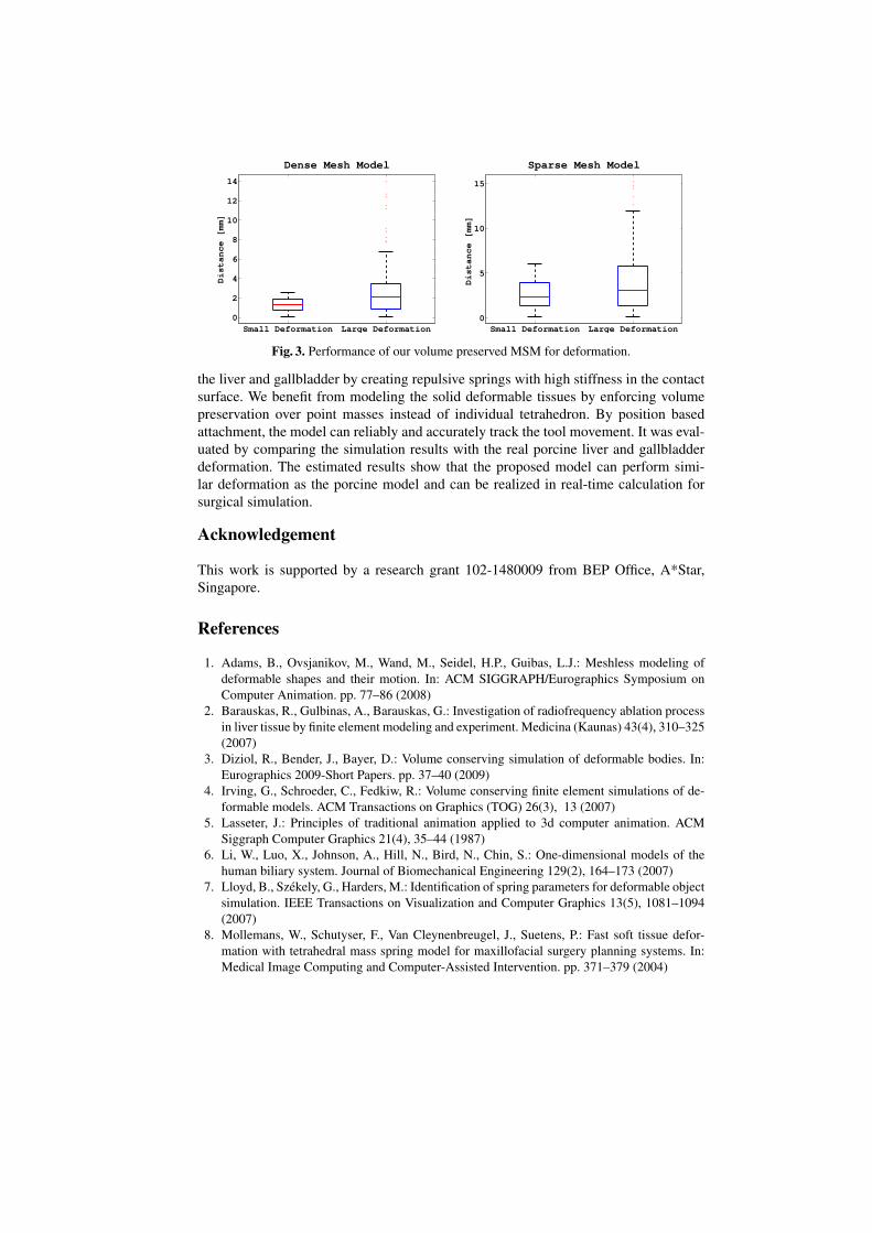
In addition, we measure the overall performance of our model in approximating theporcine deformable model by selecting 200 points on the top surface of the liver wheredeformation obviously happens. For the two deformations, the overall mean error is1.2905 mm and 2.6400 mm, respectively. The box plot of the error is shown in Fig. 3(a). We render the scene using OpenGL and refresh rate of 15 frames per second (FPS)can be achieved on PC with CPU Intel(R) Core(TM) i7-2600 running at 3.40GHz.
Based on the above evaluation of the obtained MSM, we can make the followingobservations:
1. The comparison between the simulation results and porcine liver deformation showsthat our model can achieve the simulation with good accuracy.
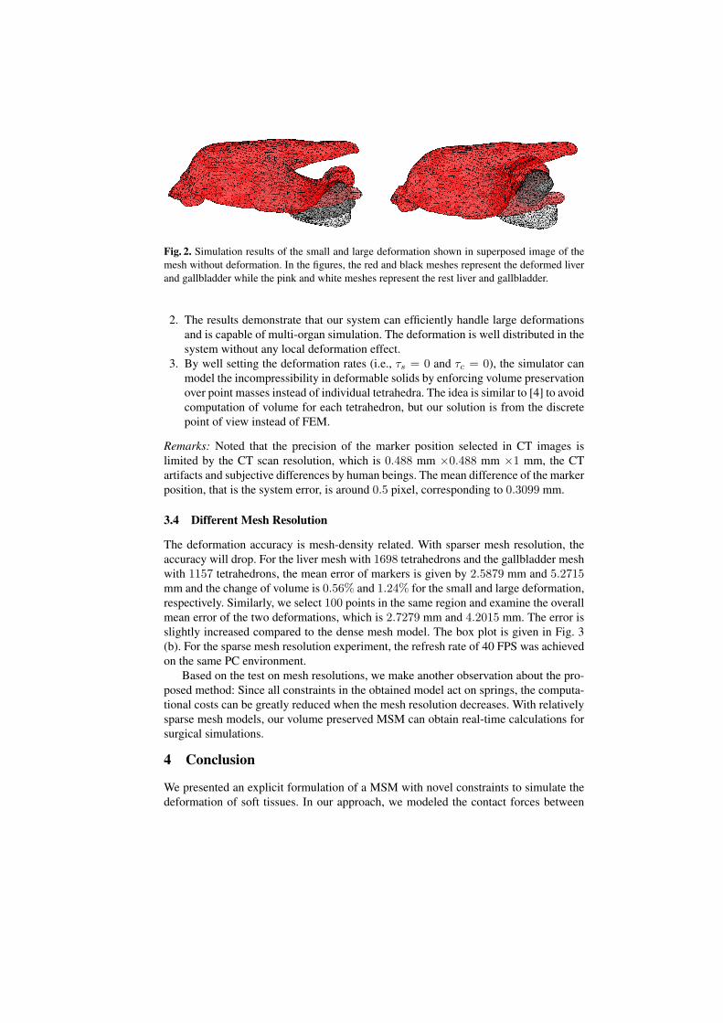
Fig. 2. Simulation results of the small and large deformation shown in superposed image of themesh without deformation. In the figures, the red and black meshes represent the deformed liverand gallbladder while the pink and white meshes represent the rest liver and gallbladder.
2. The results demonstrate that our system can efficiently handle large deformationsand is capable of multi-organ simulation. The deformation is well distributed in thesystem without any local deformation effect.
3. By well setting the deformation rates (i.e., τs = 0 and τc = 0), the simulator canmodel the incompressibility in deformable solids by enforcing volume preservationover point masses instead of individual tetrahedra. The idea is similar to [4] to avoidcomputation of volume for each tetrahedron, but our solution is from the discretepoint of view instead of FEM.
Remarks: Noted that the precision of the marker position selected in CT images islimited by the CT scan resolution, which is 0.488 mm ×0.488 mm ×1 mm, the CTartifacts and subjective differences by human beings. The mean difference of the markerposition, that is the system error, is around 0.5 pixel, corresponding to 0.3099 mm.
3.4 Different Mesh Resolution
The deformation accuracy is mesh-density related. With sparser mesh resolution, theaccuracy will drop. For the liver mesh with 1698 tetrahedrons and the gallbladder meshwith 1157 tetrahedrons, the mean error of markers is given by 2.5879 mm and 5.2715mm and the change of volume is 0.56% and 1.24% for the small and large deformation,respectively. Similarly, we select 100 points in the same region and examine the overallmean error of the two deformations, which is 2.7279 mm and 4.2015 mm. The error isslightly increased compared to the dense mesh model. The box plot is given in Fig. 3(b). For the sparse mesh resolution experiment, the refresh rate of 40 FPS was achievedon the same PC environment.
Based on the test on mesh resolutions, we make another observation about the pro-posed method: Since all constraints in the obtained model act on springs, the computa-tional costs can be greatly reduced when the mesh resolution decreases. With relativelysparse mesh models, our volume preserved MSM can obtain real-time calculations forsurgical simulations.
4 Conclusion
We presented an explicit formulation of a MSM with novel constraints to simulate thedeformation of soft tissues. In our approach, we modeled the contact forces between
Small Deformation Large Deformation0
2
4
6
8
10
12
14
Dense Mesh Model
Distance [mm]
Small Deformation Large Deformation0
5
10
15
Sparse Mesh Model
Distance [mm]
Fig. 3. Performance of our volume preserved MSM for deformation.
the liver and gallbladder by creating repulsive springs with high stiffness in the contactsurface. We benefit from modeling the solid deformable tissues by enforcing volumepreservation over point masses instead of individual tetrahedron. By position basedattachment, the model can reliably and accurately track the tool movement. It was eval-uated by comparing the simulation results with the real porcine liver and gallbladderdeformation. The estimated results show that the proposed model can perform simi-lar deformation as the porcine model and can be realized in real-time calculation forsurgical simulation.
Acknowledgement
This work is supported by a research grant 102-1480009 from BEP Office, A*Star,Singapore.
References
1. Adams, B., Ovsjanikov, M., Wand, M., Seidel, H.P., Guibas, L.J.: Meshless modeling ofdeformable shapes and their motion. In: ACM SIGGRAPH/Eurographics Symposium onComputer Animation. pp. 77–86 (2008)
2. Barauskas, R., Gulbinas, A., Barauskas, G.: Investigation of radiofrequency ablation processin liver tissue by finite element modeling and experiment. Medicina (Kaunas) 43(4), 310–325(2007)
3. Diziol, R., Bender, J., Bayer, D.: Volume conserving simulation of deformable bodies. In:Eurographics 2009-Short Papers. pp. 37–40 (2009)
4. Irving, G., Schroeder, C., Fedkiw, R.: Volume conserving finite element simulations of de-formable models. ACM Transactions on Graphics (TOG) 26(3), 13 (2007)
5. Lasseter, J.: Principles of traditional animation applied to 3d computer animation. ACMSiggraph Computer Graphics 21(4), 35–44 (1987)
6. Li, W., Luo, X., Johnson, A., Hill, N., Bird, N., Chin, S.: One-dimensional models of thehuman biliary system. Journal of Biomechanical Engineering 129(2), 164–173 (2007)
7. Lloyd, B., Szekely, G., Harders, M.: Identification of spring parameters for deformable objectsimulation. IEEE Transactions on Visualization and Computer Graphics 13(5), 1081–1094(2007)
8. Mollemans, W., Schutyser, F., Van Cleynenbreugel, J., Suetens, P.: Fast soft tissue defor-mation with tetrahedral mass spring model for maxillofacial surgery planning systems. In:Medical Image Computing and Computer-Assisted Intervention. pp. 371–379 (2004)

9. Muller, M., Heidelberger, B., Hennix, M., Ratcliff, J.: Position based dynamics. Journal ofVisual Communication and Image Representation 18(2), 109–118 (2007)
10. Nealen, A., Muller, M., Keiser, R., Boxerman, E., Carlson, M.: Physically based deformablemodels in computer graphics. In: Computer Graphics Forum. vol. 25, pp. 809–836 (2006)
11. Paloc, C., Bello, F., Kitney, R., Darzi, A.: Online multiresolution volumetric mass springmodel for real time soft tissue deformation. In: Medical Image Computing and Computer-Assisted Intervention. pp. 219–226 (2002)
12. Peterlık, I., Duriez, C., Cotin, S.: Modeling and real-time simulation of a vascularized livertissue. In: Medical Image Computing and Computer-Assisted Intervention. pp. 50–57 (2012)
13. Provot, X.: Deformation constraints in a mass-spring model to describe rigid cloth behaviour.In: Graphics interface. pp. 147–147 (1995)


