© IJCIRAS | ISSN (O) - 2581-5334
October 2018 | Vol. 1 Issue. 5
IJCIRAS1522 WWW.IJCIRAS.COM 5
MORPHO-ANATOMICAL STUDIES OF INDIGOFERA
LINNAEI
Dr. P. N. Pawade1, Komal P. Chinchamalatpure2
1Associate Professor, Department of Botany, Arts, Commerce & Science College, Kiran Nagar, Amravati, Maharashtra, India 2Research Scholar, Department of Botany, Arts, Commerce & Science College, Kiran Nagar, Amravati, Maharashtra, India
Abstract
Indigoferalinnaei known as Birdsville indigo and
nine-leaved indigo. It is a low ground herb and is an
important species of herbaceous cover.It forms
extensive mats of populations carpeting the soil.The
plant provides food for certain local insects and
protects the soil cover with its clustered root system
and spreading form of multi-stemmed branching
pattern. Therefore Indigofera linnaei has the
potential for use in the restoration of destroyed
degraded and damaged habitats. Thepresent
investigation has been carried out to study the
morphological and anatomical features of whole
plant of Indigofera linnaei . Anatomical studies of
plant parts show various significant characters.
Epidermal cells of stem are radially elongated,
collenchymatous cortex contain starch sheath and
resin duct. Pericycle is homogenous formed of
several layer of parenchymatous region. Vessels are
in radial multiplies some are solitary performing
various shape. Two different types of stomata are
present on adaxial and abaxial surface. Paracytic
type of stomata is present in adaxial surface and
anisocytic stomata present in abaxial surface, also
variation in both epidermal cells. Cortical region of
root contain crystals of calcium oxalate, xylem
vessels are radial, predominantly paired. Rachis
shows 5 vascular bundles, one is large, lateral two
are equal in size and remaining two are smaller.
Keyword: Morphology and anatomy of Indigofera
linnaei, Root, stem,leaf and petiole
1.INTRODUCTION
Indigofera linnaei is a species of leguminous shrub in
the genus Indigofera of the subfamily Papilionaceae. The
genus name Indigofera is derived from Latin and means
containing Indigo-a purple dye originally obtained from
some Indigofera species while linnaeiderived from
Linnaeous .Wilson (1987)and Wilson and Ross (2004)
provided a general account on Indigofera species.
Indigofera is a large genus of over 750 species of
flowering plants of the subfamily papilionaceae
belonging to the pea family Fabaceae.It is widely
distributed in tropical and subtropical regions .The
genus is one of the nine genera which are members of
the tribe Galegeae. (Nwachukwu and Mbagwu, 2007).
Indigofera in Greek means Indigo dye which is famous
for the natural blue colour obtained from the leaflets
and branches of this herb.The fruits are oval shaped and
elongated,4-angled or flattened and often curved with
many seeds.The dye which is among the most widely
used natural dye in the world is obtained mainly from
the leaves through a process of fermentation. Indigofera
genus possess wide range of uses ranging from several
economical and ecological purposes, feed for livestock,
ornamental, medicinal ,plant recipes as well as dye for
commercial purposes. Indigoferaspecies are used as
food plants by the larvae of some Lepidoptera species
including TurnipMoth.
The leafy twigs are the main source of indigo dye used
since very ancient times for dyeing textile blue.The
leaves and twigs do not contain indigo but colourless
precursors that extracted and processed to produce the
indigo dye.
The herb layer has structural and functional significance
in both forest and non-forest ecosystems.Certain low
ground herbs have the ability in retarding soil,water and
nutrient erosion and these abilities vary with different
weed species.(Kumar,et.al. 1997).
© IJCIRAS | ISSN (O) - 2581-5334
October 2018 | Vol. 1 Issue. 5
IJCIRAS1522 WWW.IJCIRAS.COM 6
Indigofera linnaei is a perennial plant with prostrate
sometime, ascending, much-branched stem that can
become more or less woody ,especially near the base
and persist. The plant is harvested from the wild for local
use as a medicine, in times of need the plant is harvested
from the wild for its seed which are an emergency
source of food. It causes however a disease of horses but
is obviously suitable for feeding sheep and cattle. The
juice of the plant is used as an antiscorbutic and diuretic.
It is considered to be alternative in the treatment of old
venereal affections.
From the available literature no anatomical studies
werecarried out on this species. The present
investigation has been carried out the details
morphological and anatomical study of the complete
plant of Indigofera linnaei.The study was aimed to
provide valuable and reliable illustrated anatomical
descriptions of the species.
2.MATERIAL AND METHOD
Mature and fresh samples of root, stem and leaves were
collected from grassland area in Amravati region. Fresh
materials were fixed in Formalin Acetic Acid (FAA) for 48
hrs. , washed in several changes of distilled water,
dehydrated through alcohol series (30%, 50%, 70%, 90%
and 100%), 2hrs. in each solutionand embedded in wax.
Sections in each case were cut on a Leica 2125 rotary
microtome at thickness 5µ. The sections were de-waxed
with pure xylene and rehydrated in alcohol series
following Cutler (1978) with modifications. Staining was
achieved by dipping the slides in 1% alcian blue (light
green) for about 5 min. washed with distilled water and
counter stained with 1% safranin for 2 min. The stained
sections were dehydrated through alcohol series and
mounted permanently in DPX. Photomicrographs of
anatomical sections were taken with a Coslab camera
fitted with 4X, 10X, 40X microscopic objective lens.
To study leaf architecture, the mature leaves from fresh
materials were cleared by treating them with 5%
aqueous sodium hydroxide which was repeatedly
replaced by fresh solution until leaf material got cleared,
followed by treatment with 2% acetic acid for 1-2 hours
to neutralize residual sodium hydroxide. The cleared
leaves after washing with distilled water stained with
aqueous safranin and mounted in glycerin or
dehydrated. Major and minor venation patterns and
details of leaf architecture, were studied under
compound microscope. Terminology of Hickey (1971,
1973) is followed for describing leaf architecture. Whole
lamina photographs were taken directly using coslab
camera fitted with 4X, 10X, 40X microscopic objective
lens.
3.RESULT & DISCUSSION
3.1. Morphological features
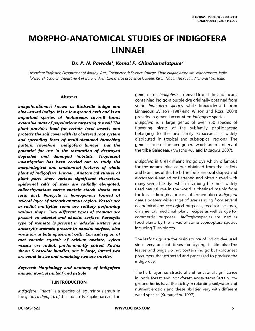
Diffuse, Prostrate, annual or perennial herb with woody
root stock, branches trailing 30-40cm long, grey-
pubscent. Stem is branched, herbaceous, cylindrical, and
velvet-hairy. Leaves are pinnately compound 1-2cm
long cuspidate, imparipinnate with 5-9 alternately
arranged leaflets, 7-12mm long, 2-5mm broad, sessile,
oblanceolate or obovate. Inflorescence is a
spicataracemes, peduncle 0.2cm long. Flowers red, in
many flowered axillary crowded, pedicel absent, bract
scarious, 2mm long, ovate, acuminate, persistant,
sepals-5, polysepalous, 3-4mm long, hairy outside, teeth
long. Petals-5, gamopetalous, papilionaceous
corolla,bright red, slightly exerted 4mm long. Fruit3-
6mm long, 2-3mm broad, pods very small, cylindric, 3-
4mm long, sparsely clothed with white appressed hairs,
apiculate, pubscent,1-2 seeded, globose. Flowering June
to December.
Fig. No. 1: Indigofera linnaei Habit(Axillary crowded
head)
Located along grassland area in Amravati region,
common on rocky soil or old walls etc. very common
throughout in open grasslands, fields etc.
© IJCIRAS | ISSN (O) - 2581-5334
October 2018 | Vol. 1 Issue. 5
IJCIRAS1522 WWW.IJCIRAS.COM 7
3.2. Anatomical features
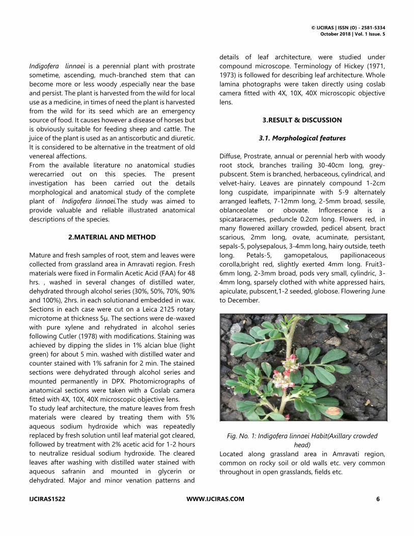
3.2.1. Primary structure (Young Stem)
Young stem roughly triangular in shape, Epidermis is
single layered, cells are angular small in size covered
with thick cuticle. Following epidermis there is single
layer of hypodermal stone cells interrupted by
chollenchymatous cells, cells are loosely placed, Cortex
narrow, parenchymatous, cells thin walled, enclosing
small intercellular spaces, endodermis distinct, 16
vascular bundles are present, cambium in the form of
complete ring from the beginning. Pith large, cells
parenchymatous, rounded or oval in shape, loosely
arranged with small intercellular space with styloids
scattered present in small amount.
Fig.No.02: Primary structure (Entire View) Fig.No.03: Primary structure (Cross View)
Fig.No.04: Primary structure (Pith)
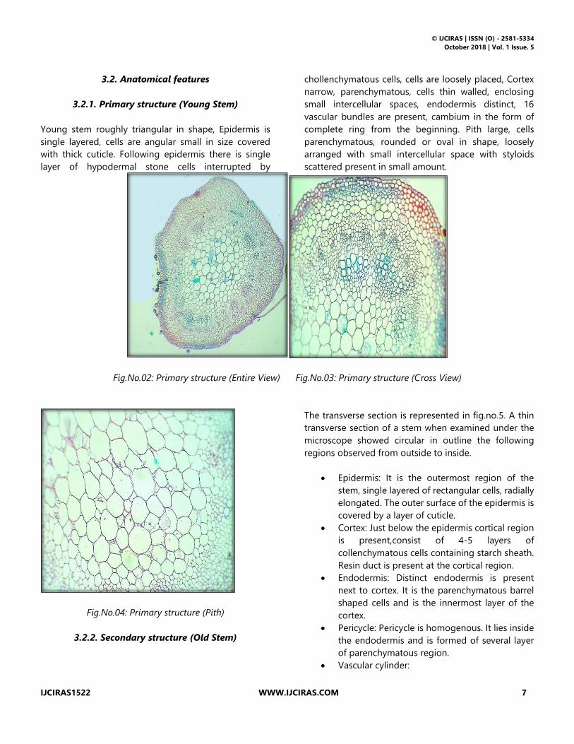
3.2.2. Secondary structure (Old Stem)
The transverse section is represented in fig.no.5. A thin
transverse section of a stem when examined under the
microscope showed circular in outline the following
regions observed from outside to inside.
• Epidermis: It is the outermost region of the
stem, single layered of rectangular cells, radially
elongated. The outer surface of the epidermis is
covered by a layer of cuticle.
• Cortex: Just below the epidermis cortical region
is present,consist of 4-5 layers of
collenchymatous cells containing starch sheath.
Resin duct is present at the cortical region.
• Endodermis: Distinct endodermis is present
next to cortex. It is the parenchymatous barrel
shaped cells and is the innermost layer of the
cortex.
• Pericycle: Pericycle is homogenous. It lies inside
the endodermis and is formed of several layer
of parenchymatous region.
• Vascular cylinder:
© IJCIRAS | ISSN (O) - 2581-5334
October 2018 | Vol. 1 Issue. 5
IJCIRAS1522 WWW.IJCIRAS.COM 8
a) Secondary phloem: Secondary phloem
is found next to pericycle region, 3-4
layered parenchymatous cells.
b) Cambium: The cambium has been
found to initiate in the vascular bundle
between xylem and phloem. Gradually
it extends towards the upper part.
c) Secondary xylem: Xylem inner to
cambium in close cylinder transversed
by narrow rays. Vessels in radial
multiples of 6-7, some are solitary.
d) circular, angular, triangle in outline,
secondary xylem with vessels
predominantly in radial multiples.
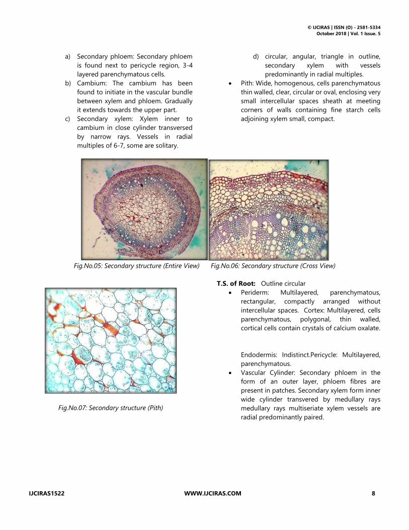
• Pith: Wide, homogenous, cells parenchymatous
thin walled, clear, circular or oval, enclosing very
small intercellular spaces sheath at meeting
corners of walls containing fine starch cells
adjoining xylem small, compact.
Fig.No.05: Secondary structure (Entire View) Fig.No.06: Secondary structure (Cross View)
Fig.No.07: Secondary structure (Pith)
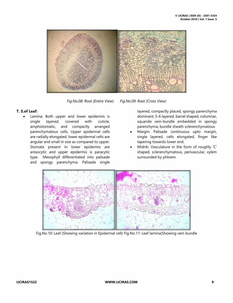
T.S. of Root: Outline circular
• Periderm: Multilayered, parenchymatous,
rectangular, compactly arranged without
intercellular spaces. Cortex: Multilayered, cells
parenchymatous, polygonal, thin walled,
cortical cells contain crystals of calcium oxalate.
Endodermis: Indistinct.Pericycle: Multilayered,
parenchymatous.
• Vascular Cylinder: Secondary phloem in the
form of an outer layer, phloem fibres are
present in patches. Secondary xylem form inner
wide cylinder transvered by medullary rays
medullary rays multiseriate xylem vessels are
radial predominantly paired.
© IJCIRAS | ISSN (O) - 2581-5334
October 2018 | Vol. 1 Issue. 5
IJCIRAS1522 WWW.IJCIRAS.COM 9
Fig.No.08: Root (Entire View) Fig.No.09: Root (Cross View)
T. S.of Leaf:
• Lamina: Both upper and lower epidermis is
single layered, covered with cuticle,
amphistomatic, and compactly arranged
parenchymatous cells. Upper epidermal cells
are radially elongated; lower epidermal cells are
angular and small in size as compared to upper.
Stomata present in lower epidermis are
anisocytic and upper epidermis is paracytic
type. Mesophyll differentiated into palisade
and spongy parenchyma. Palisade single
layered, compactly placed, spongy parenchyma
dominant, 5-6 layered, barrel shaped, columnar,
squaride vein-bundle embedded in spongy
parenchyma, bundle sheath sclerenchymatous.
• Margin: Palisade continuous upto margin,
single layered, cells elongated, finger like
tapering towards lower end.
• Midrib: Vasculature in the form of roughly ‘C’
shaped, sclerenchymatous, perivascular, xylem
surrounded by phloem.
Fig.No.10: Leaf (Showing variation in Epidermal cell) Fig.No.11: Leaf lamina(Showing vein bundle

© IJCIRAS | ISSN (O) - 2581-5334
October 2018 | Vol. 1 Issue. 5
IJCIRAS1522 WWW.IJCIRAS.COM 10
Fig.No.12: Upper epidermis (Paracytic)Fig.No.13: Lower epidermis (Anisocytic)
3.2.3. Trichome (Unicellular macroform two armed
hair):
• Foot: Simple with the base circular or oval in
outline, cells broader than long, contents scanty
on upper and absent in lower. Lateral walls
moderately thickened, smooth.
• Body: Unicellular, two armed; arms equal or
nearly so, T-shaped, gradually tapering pointed
at both ends; wall moderately thickened, surface
smooth or punctuate.
Length of trichome onadaxial surface is 75µm and width
in middle is 5µm.
Length of trichome on abaxial surface is 125µm and
width in middle is 10µm.
Fig.No.14: Trichome filled with scanty contents (adaxial)Fig.No.15: Trichomewithout contents (abaxial)
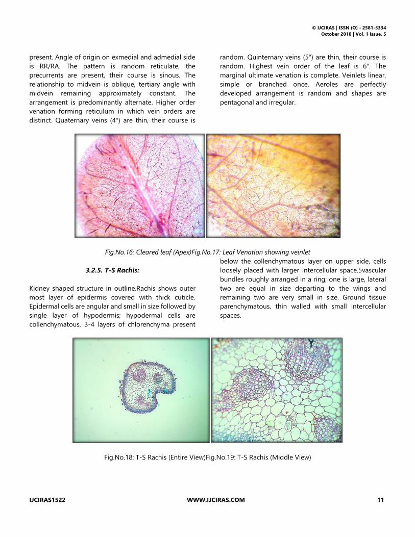
3.2.4. Leaf architecture
The basic axis of orientation in the leaflet is apical. The
leaf organization is compound with respect to leaf shape
and size, the length of leaflet is 7-12mm long and width
is 2-5mm broad. The lamina is asymmetrical, base is
slight asymmetrical, form is narrow obovate, apex is
obtuse and base is acute. The margin is entire, leaf
texture is membranaceous. The glands are absent
on the lamina and the petiole is normal. Type of
venation is pinnate. Semicraspedodromous, primary
vein (1°) is moderate, its course is straight and
unbranched secondary vein (2°) is present. The angle of
divergen is acute upper secondary veins more obtuse
than lower and nearly uniform. Secondary veins are
moderate, the course is curved, abruptly; loop forming
branches are joining supra adjacent secondary at an
acute angle. Intersecondary veins are simple,
intermarginal vein is absent. Tertiary veins (3°) are
© IJCIRAS | ISSN (O) - 2581-5334
October 2018 | Vol. 1 Issue. 5
IJCIRAS1522 WWW.IJCIRAS.COM 11
present. Angle of origin on exmedial and admedial side
is RR/RA. The pattern is random reticulate, the
precurrents are present, their course is sinous. The
relationship to midvein is oblique, tertiary angle with
midvein remaining approximately constant. The
arrangement is predominantly alternate. Higher order
venation forming reticulum in which vein orders are
distinct. Quaternary veins (4°) are thin, their course is
random. Quinternary veins (5°) are thin, their course is
random. Highest vein order of the leaf is 6°. The
marginal ultimate venation is complete. Veinlets linear,
simple or branched once. Aeroles are perfectly
developed arrangement is random and shapes are
pentagonal and irregular.
Fig.No.16: Cleared leaf (Apex)Fig.No.17: Leaf Venation showing veinlet
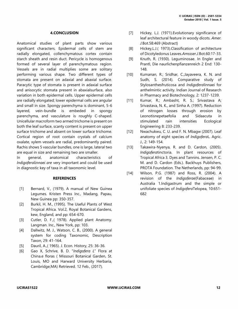
3.2.5. T-S Rachis:
Kidney shaped structure in outline.Rachis shows outer
most layer of epidermis covered with thick cuticle.
Epidermal cells are angular and small in size followed by
single layer of hypodermis; hypodermal cells are
collenchymatous, 3-4 layers of chlorenchyma present
below the collenchymatous layer on upper side, cells
loosely placed with larger intercellular space.5vascular
bundles roughly arranged in a ring; one is large, lateral
two are equal in size departing to the wings and
remaining two are very small in size. Ground tissue
parenchymatous, thin walled with small intercellular
spaces.
Fig.No.18: T-S Rachis (Entire View)Fig.No.19: T-S Rachis (Middle View)
© IJCIRAS | ISSN (O) - 2581-5334
October 2018 | Vol. 1 Issue. 5
IJCIRAS1522 WWW.IJCIRAS.COM 12
4.CONCLUSION
Anatomical studies of plant parts show various
significant characters. Epidermal cells of stem are
radially elongated, collenchymatous cortex contain
starch sheath and resin duct. Pericycle is homogenous
formed of several layer of parenchymatous region.
Vessels are in radial multiplies some are solitary
performing various shape. Two different types of
stomata are present on adaxial and abaxial surface.
Paracytic type of stomata is present in adaxial surface
and anisocytic stomata present in abaxialsurface, also
variation in both epidermal cells. Upper epidermal cells
are radially elongated; lower epidermal cells are angular
and small in size. Spongy parenchyma is dominant, 5-6
layered, vein-bundle is embeded in spongy
parenchyma, and vasculature is roughly C-shaped.
Unicellular macroform two armed trichome is present on
both the leaf surface, scanty content is present on upper
surface trichome and absent on lower surface trichome.
Cortical region of root contain crystals of calcium
oxalate, xylem vessels are radial, predominantly paired.
Rachis shows 5 vascular bundles, one is large, lateral two
are equal in size and remaining two are smaller.
In general, anatomical characteristics of
Indigoferalinnaei are very important and could be used
in diagnostic key of taxa in all taxonomic level.
REFERENCES
[1] Bernard, V., (1979). A manual of New Guinea
Legumes. Kristen Press Inc., Madang, Papau,
New Guinea pp: 350-357.
[2] Burkil, H. M., (1995). The Useful Plants of West
Tropical Africa. Vol.2, Royal Botanical Gardens,
kew, England, and pp: 654-670.
[3] Cutler, D. F.,( 1978). Applied plant Anatomy.
Langman. Inc., New York, pp: 103.
[4] Dallwitz, M. J., Watson, C. B., (2000). A general
system for coding Taxonomic, Description
Taxon, 29: 41-164.
[5] Dauril, A.,( 1965). J. Econ. History, 25: 36-36.
[6] Gao X, Schrive, B. D. “Indigofera L” Flora at
China.e floras ( Missouri Botanical Garden, St.
Louis, MO and Harward University Herbaria,
Cambridge,MA) Retrieved. 12 Feb., (2017).
[7] Hickey, L.J. (1971).Evolutionary significance of
leaf architectural feature in woody dicots. Amer.
J.Bot.58:469 (Abstract)
[8] Hickey,L.J.( 1973).Classification of architecture
of Dicotyledonus Leaves.Amssser.J.Bot.60:17-33.
[9] Knuth, R. (1930). Leguminosae. In Engler and
Prantl, Die naurlichenpfianzenreich 2 End: 130-
148.
[10] Kumanan, R.; Sridhar, C.;Jayaveera, K. N. and
Sudh, S. (2014). Comparative study of
Stylosanthesfruticosa and Indigoferalinnaei for
anthelmintic activity. Indian Journal of Research
in Pharmacy and Biotechnology, 2: 1237-1239.
[11] Kumar, R.; Ambasht, R. S.; Srivastava A;
Srivastava, N. K., and Sinha A. (1997). Reduction
of nitrogen losses through erosion by
Leonotisnepetaefolia and Sidaacuta in
stimulated rain intensities Ecological
Engineering 8: 233-239.
[12] Nwachukwu, C. U. and F. N. Mbagw (2007). Leaf
anatomy of eight species of IndigoferaL. Agric.
J., 2: 149-154.
[13] Takawira-Nyenya, R. and D. Cardon, (2005).
Indigoferatinctoria. In plant resources of
Tropical Africa 3. Dyes and Tannins. Jensen, P. C.
M. and D. Cardon (Eds.). Backhuys Publishers,
PROTA Foundation. The Netherlands, pp: 94-99.
[14] Wilson, P.G. (1987) and Ross, R. (2004). A
revision of the Indigoferae(Fabaceae) in
Australia 1.Indigastrum and the simple or
unifoliate species of IndigoferaTelopea, 10:651-
682


